RESPIRATORY SYSTEM. The respiratory system is formed of 2 functional components: –Conducting...
15
RESPIRATORY RESPIRATORY SYSTEM SYSTEM
-
Upload
shanon-bryan -
Category
Documents
-
view
223 -
download
0
Transcript of RESPIRATORY SYSTEM. The respiratory system is formed of 2 functional components: –Conducting...

RESPIRATORY RESPIRATORY SYSTEMSYSTEM

The respiratory system is formed of The respiratory system is formed of 2 functional 2 functional componentscomponents: :
– Conducting portion:Conducting portion: for the transport of inspired and for the transport of inspired and expired air between the atmosphere and the circulatory expired air between the atmosphere and the circulatory system.system.
– Respiratory portion:Respiratory portion: for the exchange of gases between for the exchange of gases between the atmospheric air and blood.the atmospheric air and blood.
Conducting partsConducting parts include: include:– Nasal cavityNasal cavity– PharynxPharynx– LarynxLarynx– TracheaTrachea– Bronchi (1ry & 2ry)Bronchi (1ry & 2ry)– Bronchioles (terminal bronchioles)Bronchioles (terminal bronchioles)
Respiratory parts:Respiratory parts: include: include:– Respiratory bronchiolesRespiratory bronchioles– Alveolar ductsAlveolar ducts– Alveolar sacs (alveoli)Alveolar sacs (alveoli)

Extrapulmonary conducting portionExtrapulmonary conducting portion– This extends from nasal cavity to primary bronchi. It is This extends from nasal cavity to primary bronchi. It is
characterized by:characterized by:Ciliated pseudostratified columnar epithelium rich in goblet cells.Ciliated pseudostratified columnar epithelium rich in goblet cells.Submucous loose C.T. rich in mixed seromucous glands.Submucous loose C.T. rich in mixed seromucous glands.
Function of the conducting passages:Function of the conducting passages:– Trapping the inhaled particles and debris by the sero-Trapping the inhaled particles and debris by the sero-
mucous secretion which have suitable consistency to be mucous secretion which have suitable consistency to be expelled out towards the nose. expelled out towards the nose.
Individuals who suffer from immobile cilia have chronic lung Individuals who suffer from immobile cilia have chronic lung infections. infections. The function of cilia is also lost in smokers thus inviting infections.The function of cilia is also lost in smokers thus inviting infections.
– The seromucous secretion has detoxifying action on the The seromucous secretion has detoxifying action on the soluble gases.soluble gases.
– The subepithelial C.T. (lamina propria) contains diffuse The subepithelial C.T. (lamina propria) contains diffuse lymphocytes, which produce secretory immunoglobulin A, lymphocytes, which produce secretory immunoglobulin A, which kills the bacteria and viruses and prevents them from which kills the bacteria and viruses and prevents them from penetrating the epithelium.penetrating the epithelium.

Anatomically the respiratory system is divided Anatomically the respiratory system is divided into 2 parts:into 2 parts:
– Upper respiratory tract. Upper respiratory tract. – Lower respiratory tract.Lower respiratory tract.
TheThe upper respiratory tractupper respiratory tract includesincludes: : – Nasal cavity. Nasal cavity. – Paranasal sinuses. Paranasal sinuses. – Nasopharynx. Nasopharynx.
Its basic function is filtration, humidification and Its basic function is filtration, humidification and adjusting the temperature of the inspired air.adjusting the temperature of the inspired air.

TheThe lower respiratory tractlower respiratory tract includesincludes: : – Larynx which continues as the trachea, which divides and re-Larynx which continues as the trachea, which divides and re-
divides almost about 20 times. divides almost about 20 times. – At first, the trachea divides into At first, the trachea divides into 2 2 primary or main primary or main bronchibronchi. . – Each Each primary bronchusprimary bronchus gives rise to gives rise to secondary or lobar secondary or lobar
bronchibronchi supplying the lobes of the lungs. supplying the lobes of the lungs. – These again divide into These again divide into tertiary or segmental bronchitertiary or segmental bronchi, ,
which supply segments of each lobe. which supply segments of each lobe. – The The tertiary bronchitertiary bronchi divide into smaller airways called divide into smaller airways called
bronchiolesbronchioles. . The smallest of such division is called The smallest of such division is called terminal bronchiolesterminal bronchioles. .
– The terminal bronchioles then divide into The terminal bronchioles then divide into respiratory respiratory bronchiolesbronchioles and and alveolar ductsalveolar ducts.. These passages finally These passages finally terminate in dilated spaces called terminate in dilated spaces called alveolar sacsalveolar sacs,, which open which open into into alveolialveoli..
Each type of airway has its own features but there is a Each type of airway has its own features but there is a gradual change in structure.gradual change in structure.

Respiratory mucosaRespiratory mucosa– Epithelium:Epithelium: pseudostratified columnar ciliated with pseudostratified columnar ciliated with
goblet cells.goblet cells.– Lamina propria:Lamina propria: loose C.T. containing: loose C.T. containing:
Rich blood vessels for warming of air.Rich blood vessels for warming of air.
Lymphocytes for immune response.Lymphocytes for immune response.
Seromucous (Seromucous (branched tubuloalveolar glands of branched tubuloalveolar glands of BowmanBowman) glands for secretion of mucous and absorption ) glands for secretion of mucous and absorption and detoxification of gases.and detoxification of gases.
Note:Note: In some regions of nasal mucosa, there are veins In some regions of nasal mucosa, there are veins resembling erectile tissue termed resembling erectile tissue termed swell bodiesswell bodies.. These These are specialized veins which engorge periodically and are specialized veins which engorge periodically and alternatively to close one side of the nasal cavity, thus alternatively to close one side of the nasal cavity, thus giving it time to recover from drying. This cyclic process giving it time to recover from drying. This cyclic process is controlled by autonomic nerves.is controlled by autonomic nerves.

Olfactory mucosa:Olfactory mucosa:– It is thicker than that of the respiratory mucosa and lacks the It is thicker than that of the respiratory mucosa and lacks the
Goblet cells. Goblet cells. – The glands in lamina propria are purely serous because the The glands in lamina propria are purely serous because the
fluid secretion dissolves the odoriferous substances and also fluid secretion dissolves the odoriferous substances and also rapidly washed away to clear the receptors for new stimuli.rapidly washed away to clear the receptors for new stimuli.
– The lamina propria is rich in vascular plexusesThe lamina propria is rich in vascular plexuses..– The olfactory epithelium consists of 3 types of cells:The olfactory epithelium consists of 3 types of cells:
Supporting cells:Supporting cells: have narrow bases and broad apices that carry have narrow bases and broad apices that carry microvilli. The nucleus is just above the center of the cell. The microvilli. The nucleus is just above the center of the cell. The cytoplasm contains cytoplasm contains lipofuscin pigmentslipofuscin pigments that give the area its yellow that give the area its yellow coloration.coloration.Bipolar olfactory cells:Bipolar olfactory cells: which carry the receptors of smell which carry the receptors of smell (bipolar (bipolar neurons).neurons). The peripheral part is modified dendrite, which ends in a The peripheral part is modified dendrite, which ends in a bulb called bulb called olfactory knobolfactory knob, which gives long non-motile cilia lying flat , which gives long non-motile cilia lying flat on the surface, and they carry the receptors for smell. The basal part on the surface, and they carry the receptors for smell. The basal part is the axon, which passes through the basement membrane, and is the axon, which passes through the basement membrane, and joins other axons forming the joins other axons forming the olfactory nerveolfactory nerve that pass through the that pass through the cribriform plate to reach the brain.cribriform plate to reach the brain.Basal cells:Basal cells: short pyramidal with basal nuclei, they are short pyramidal with basal nuclei, they are undifferentiated and can give other types of the above cells.undifferentiated and can give other types of the above cells.
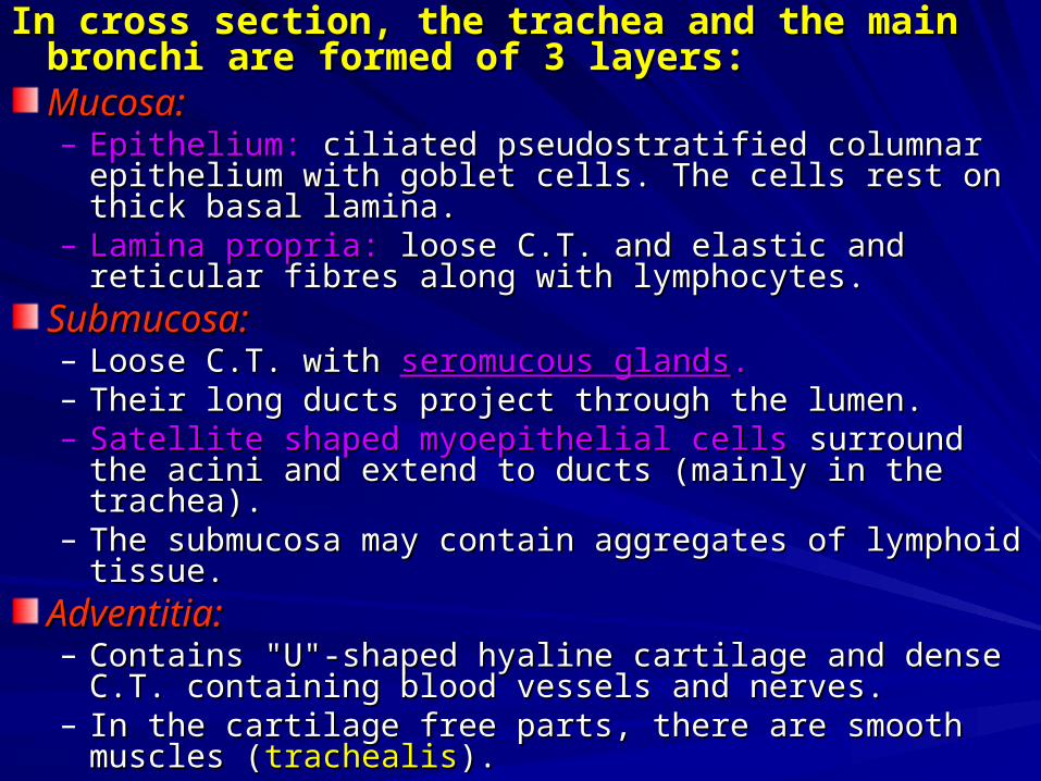
In cross section, the trachea and the main bronchi In cross section, the trachea and the main bronchi are formed of 3 layers:are formed of 3 layers:Mucosa:Mucosa:– Epithelium:Epithelium: ciliated pseudostratified columnar epithelium with ciliated pseudostratified columnar epithelium with
goblet cells. The cells rest on thick basal lamina.goblet cells. The cells rest on thick basal lamina.– Lamina propria:Lamina propria: loose C.T. and elastic and reticular fibres loose C.T. and elastic and reticular fibres
along with lymphocytes.along with lymphocytes.
Submucosa:Submucosa:– Loose C.T. with Loose C.T. with seromucous glandsseromucous glands.. – Their long ducts project through the lumen. Their long ducts project through the lumen. – Satellite shaped myoepithelial cellsSatellite shaped myoepithelial cells surround the acini and surround the acini and
extend to ducts (mainly in the trachea). extend to ducts (mainly in the trachea). – The submucosa may contain aggregates of lymphoid tissue.The submucosa may contain aggregates of lymphoid tissue.
Adventitia:Adventitia:– Contains "U"-shaped hyaline cartilage and dense C.T. Contains "U"-shaped hyaline cartilage and dense C.T.
containing blood vessels and nerves. containing blood vessels and nerves. – In the cartilage free parts, there are smooth muscles In the cartilage free parts, there are smooth muscles
((trachealistrachealis).).

Types of cells in lining of trachea and bronchi:Types of cells in lining of trachea and bronchi:
3 types of cells are seen by light microscope:3 types of cells are seen by light microscope:– Ciliated columnar cellsCiliated columnar cells..– Goblet cellsGoblet cells which produce mucoid secretion. which produce mucoid secretion.– Basal cells:Basal cells: these are undifferentiated cells which can divide these are undifferentiated cells which can divide
by mitosis and can give other types of cells.by mitosis and can give other types of cells.
2 2 types seen by electron microscope:types seen by electron microscope:– Brush cellsBrush cells which contain glycogen granules and carry which contain glycogen granules and carry
microvilli. They may be either immature columnar cells or microvilli. They may be either immature columnar cells or degranuled goblet cells or sensory cells because their bases degranuled goblet cells or sensory cells because their bases make contact with nerve processes.make contact with nerve processes.
– Granular cellsGranular cells which are of 2 types according to function of which are of 2 types according to function of granules:granules:
Catecholamine secreting cellsCatecholamine secreting cells (neurosecretory cells). (neurosecretory cells).
Protein hormone secreting cellsProtein hormone secreting cells (enteroendocrine like cells). They (enteroendocrine like cells). They secrete amines and amine precursors.secrete amines and amine precursors.

Secondary bronchiSecondary bronchiThe The intrapulmonary secondary bronchiintrapulmonary secondary bronchi differs from differs from the the extrapulmonary primary bronchiextrapulmonary primary bronchi by the following, by the following, otherwise the structure is the same:otherwise the structure is the same:– The mucosa is The mucosa is foldedfolded..– The presence of The presence of muscularis mucosamuscularis mucosa which is composed of which is composed of
smooth muscle sheet beneath the lamina propria, so the smooth muscle sheet beneath the lamina propria, so the mucosa is folded after fixation.mucosa is folded after fixation.
– Adventitia contains Adventitia contains discontinuous cartilage platesdiscontinuous cartilage plates and dense and dense C.T. The cartilage plates surround the bronchi leading to a C.T. The cartilage plates surround the bronchi leading to a circular appearance and not flattened at one side like the circular appearance and not flattened at one side like the trachea and main bronchi.trachea and main bronchi.
Note:Note: Smaller generations of secondary bronchi are Smaller generations of secondary bronchi are lined by ciliated simple columnar epithelium and their lined by ciliated simple columnar epithelium and their adventitia contains isolated smaller cartilages.adventitia contains isolated smaller cartilages.

BronchiolesBronchiolesThese are smaller in diameter. Its structure is as follows:-These are smaller in diameter. Its structure is as follows:-– Mucosa is folded and contains: Mucosa is folded and contains: ciliated simple columnarciliated simple columnar cells with goblet cells with goblet
cells.cells.– Muscularis mucosa: relatively thick smooth muscle layer.Muscularis mucosa: relatively thick smooth muscle layer.– Submucosa: no glands or lymphoid tissue.Submucosa: no glands or lymphoid tissue.– Adventitia: loose connective tissue, Adventitia: loose connective tissue, no cartilageno cartilage..– The bronchioles give rise to terminal bronchioles, which give respiratory The bronchioles give rise to terminal bronchioles, which give respiratory
bronchioles, that receives the alveoli.bronchioles, that receives the alveoli.
Terminal bronchiolesTerminal bronchiolesThese are the smallest branches of the conducting system. These are the smallest branches of the conducting system. Their mucosa is characterized by:Their mucosa is characterized by:– The epithelium is The epithelium is cuboidal ciliatedcuboidal ciliated..– Absent goblet cells.Absent goblet cells.– Presences of bronchiolar secretary cells called Presences of bronchiolar secretary cells called Clara cellsClara cells. They are . They are
non-ciliated with projecting apex-carrying microvilli. They secrete non-ciliated with projecting apex-carrying microvilli. They secrete surfactant, a phospholipid that alters the surface tension of the fluid layer surfactant, a phospholipid that alters the surface tension of the fluid layer covering the surface.covering the surface.
The Respiratory bronchiolesThe Respiratory bronchioles are lined with simple cuboidal are lined with simple cuboidal ciliated epithelium, some of them are ciliated, and others are ciliated epithelium, some of them are ciliated, and others are Clara cellsClara cells..

Alveolar DuctsAlveolar Ducts– The wall is deficient except in small areas lined by cuboidal cells in The wall is deficient except in small areas lined by cuboidal cells in
between the between the alveolar sacsalveolar sacs. . It looks like a long corridor along which open It looks like a long corridor along which open many lobbies (the alveolar sacs or antrum). The lobbies lead to the many lobbies (the alveolar sacs or antrum). The lobbies lead to the rooms (the alveoli).rooms (the alveoli).
The AlveoliThe Alveoli– The alveoli are lined by 2 types of cells:The alveoli are lined by 2 types of cells:
Flattened simple squamous cells (Flattened simple squamous cells (type I pneumocytestype I pneumocytes).).Cuboid cells Cuboid cells ((type II pneumocytestype II pneumocytes)) called called secretory cellssecretory cells or or septal cellsseptal cells that that bulge into the alveolar lumen and contain multi-lamellar bodies. Their bulge into the alveolar lumen and contain multi-lamellar bodies. Their secretion is rich in phospholipids “secretion is rich in phospholipids “surfactantsurfactant” because it reduces the surface ” because it reduces the surface tension and prevents collapse of alveoli during expiration. Absence of the tension and prevents collapse of alveoli during expiration. Absence of the surfactant cells leads to respiratory distress syndrome (RDS).surfactant cells leads to respiratory distress syndrome (RDS).
The spaces between the alveoli are called inter-alveolar septa, The spaces between the alveoli are called inter-alveolar septa, which consists of C.T. containing:which consists of C.T. containing:– Fibres:Fibres: reticular and elastic. reticular and elastic.– Septal cellsSeptal cells (type II pneumocytes). (type II pneumocytes).– Other Cells:Other Cells: Fibroblasts, mast cells, leucocytes, macrophages. Other Fibroblasts, mast cells, leucocytes, macrophages. Other
septal cells contain bundles of actin and myosin filaments which contract septal cells contain bundles of actin and myosin filaments which contract in response to hypoxia (their role is unknown).in response to hypoxia (their role is unknown).
– Blood capillariesBlood capillaries (continuous non-fenestrated); (continuous non-fenestrated);
N.B.:N.B.: the capillary endothelial cells and type I flat cells share the capillary endothelial cells and type I flat cells share fused basement membranes.fused basement membranes.

SUMMARY OF STRUCTURES OF THE RESPIRATORY TREESUMMARY OF STRUCTURES OF THE RESPIRATORY TREEThe structure of respiratory changes according to the functional The structure of respiratory changes according to the functional need:need:– The air is inhaled through mouth or nose where it is humidified, warmed The air is inhaled through mouth or nose where it is humidified, warmed
and the suspended particles trapped by the hair of nasal cavity and the and the suspended particles trapped by the hair of nasal cavity and the mucous present on the mucosa. These particles are ultimately expelled mucous present on the mucosa. These particles are ultimately expelled out by the cilia.out by the cilia.
– As we go down the respiratory tree the cartilage decreases till it is As we go down the respiratory tree the cartilage decreases till it is completely absent in the bronchioles. In trachea it is present as a single completely absent in the bronchioles. In trachea it is present as a single semilunar plate while in secondary and tertiary bronchi, it is present in semilunar plate while in secondary and tertiary bronchi, it is present in few discontinuous patches.few discontinuous patches.
– The height of the epithelium decreases down the tree till it becomes The height of the epithelium decreases down the tree till it becomes simple cuboidal in the terminal bronchioles.simple cuboidal in the terminal bronchioles.
– The cilia decrease in number as we go down till they are absent in the The cilia decrease in number as we go down till they are absent in the terminal bronchioles. terminal bronchioles.
– The goblet cells disappear with the absence of cilia.The goblet cells disappear with the absence of cilia.– The smooth muscle increase in quantity till it is maximum in the The smooth muscle increase in quantity till it is maximum in the
bronchioles where it serves to act as a sphincter or control valve for bronchioles where it serves to act as a sphincter or control valve for regulation of air.regulation of air.

LOCAL DEFENSE MEHCHANISMLOCAL DEFENSE MEHCHANISM– Alveolar MacrophagesAlveolar Macrophages
Present in the interalveolar septum and on alveoli, help in Present in the interalveolar septum and on alveoli, help in phagocytosis and disposal of antigens reaching the alveoli.phagocytosis and disposal of antigens reaching the alveoli.
Bacteria and viruses are easily phagocytosed and degraded Bacteria and viruses are easily phagocytosed and degraded Indigested inert particles as carbon remain in macrophages for a long Indigested inert particles as carbon remain in macrophages for a long time and are deposited in islands of collagen (scar tissue). time and are deposited in islands of collagen (scar tissue). Heavy Heavy exposure to asbestos, coal and other industrial particles, toxic gases exposure to asbestos, coal and other industrial particles, toxic gases also lead to their accumulation in scar tissues.also lead to their accumulation in scar tissues.
Alveolar FluidAlveolar Fluid– This neutralizes bacteria and viruses by carrying the This neutralizes bacteria and viruses by carrying the
secretory IgA produced by B-lymphocytes (in local lymphoid secretory IgA produced by B-lymphocytes (in local lymphoid tissue). There are also lymph nodes present in hila of the tissue). There are also lymph nodes present in hila of the lungs, so both humoral and cell-mediated immune lungs, so both humoral and cell-mediated immune responses play a role in lung defense against infection.responses play a role in lung defense against infection.
– Cigarette smokeCigarette smoke interferes with the normal macrophage interferes with the normal macrophage and immune function of the lung and increase susceptibility and immune function of the lung and increase susceptibility to infection.to infection.

CLINICAL NOTESCLINICAL NOTES– Absence of cartilageAbsence of cartilage from the wall of bronchioles is from the wall of bronchioles is
a potential hazard, since these airways can a potential hazard, since these airways can constrict to a point of closing if the tone of their constrict to a point of closing if the tone of their muscles is increased. This is the problem of asthma muscles is increased. This is the problem of asthma which is an allergic condition to non-specific lung which is an allergic condition to non-specific lung irritant. irritant. Wheezing noisesWheezing noises and and difficulty in breathingdifficulty in breathing occurs during occurs during expirationexpiration rather than inspiration. rather than inspiration.
– Pulmonary Surfactant:Pulmonary Surfactant: Type II pneumocytes Type II pneumocytes secrete phospholipid surfactant that decreases the secrete phospholipid surfactant that decreases the alveolar surface tension forces to a minimal level alveolar surface tension forces to a minimal level thus preventing the alveoli from collapse. The thus preventing the alveoli from collapse. The presence of this secretion is important for the presence of this secretion is important for the newbornnewborn to obtain their first breath of air. In to obtain their first breath of air. In premature delivery type II cells are immature. This premature delivery type II cells are immature. This leads to fatal respiratory difficulty in new born leads to fatal respiratory difficulty in new born ((respiratory distress syndrome).respiratory distress syndrome).


![Anatomy and Physiology Respiratory System [Tab 2] Respiratory System.](https://static.fdocuments.net/doc/165x107/56649ebd5503460f94bc631f/anatomy-and-physiology-respiratory-system-tab-2-respiratory-system.jpg)