Respiratory System Overview-The Respiratory System The respiratory system includes the lungs and a...
38
Respiratory System Overview-The Respiratory System • The respiratory system includes the lungs and a series of airways that connect the lungs to the external environment. • The respiratory system can be functionally divided into two major divisions: – a conducting portion, consisting of airways that deliver air to the lungs – and a respiratory portion, consisting of structures within the lungs in which oxygen in the inspired air is exchanged for carbon dioxide in the blood. • The components of the respiratory system possess characteristic lining epithelia, supporting structures, glands, and other features
-
date post
19-Dec-2015 -
Category
Documents
-
view
215 -
download
0
Transcript of Respiratory System Overview-The Respiratory System The respiratory system includes the lungs and a...

Respiratory System Overview-The Respiratory System • The respiratory system includes the lungs and a series of
airways that connect the lungs to the external environment.
• The respiratory system can be functionally divided into two major divisions: – a conducting portion, consisting of airways that deliver air to
the lungs– and a respiratory portion, consisting of structures within the
lungs in which oxygen in the inspired air is exchanged for carbon dioxide in the blood.
• The components of the respiratory system possess characteristic lining epithelia, supporting structures, glands, and other features
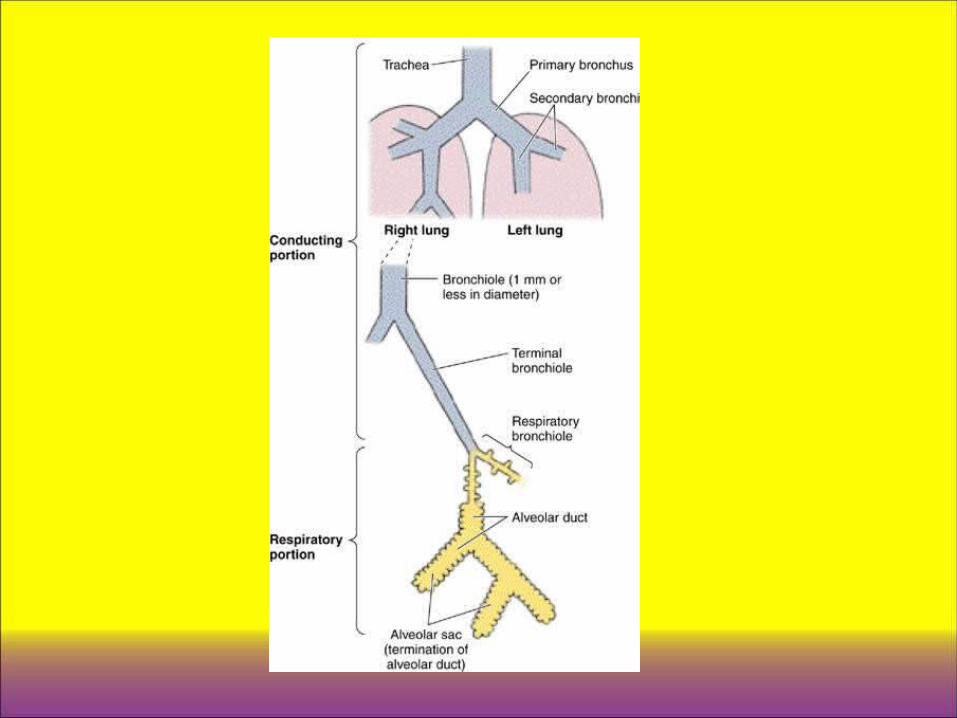

Conducting Portion of the Respiratory System
• This portion of the respiratory system includes the nose, nasopharynx, larynx, trachea, bronchi, and bronchioles down to and including the terminal bronchioles.
• These structures warm, moisten, and filter the air before it reaches the sites where gas exchange occurs.

A. Nasal cavity • 1. The nares are the nostrils; their outer portions
are lined by thin skin. They open into the vestibule.
• 2. The vestibule is the first portion of the nasal cavity, where the epithelial lining becomes nonkeratinized. Posteriorly, the lining changes to respiratory epithelium (pseudostratified ciliated columnar epithelium with goblet cells). – a. The vestibule contains vibrissae (thick, short hairs),
which filter large particles from the inspired air. – b. It has a lamina propria that is vascular (many
venous plexuses) and contains seromucous glands.


• 3. Olfactory epithelium – a. Overview
• 1) The olfactory epithelium is located in the roof of the nasal cavity, on either side of the nasal septum and on the superior nasal conchae
• 2) It is a tall pseudostratified columnar epithelium consisting of olfactory cells, supporting (sustentacular) cells, and basal cells.
• 3) It has a lamina propria that contains many veins and unmyelinated nerves and houses Bowman glands
– b. Olfactory cells are bipolar nerve cells characterized by a bulbous apical projection (olfactory vesicle) from which several modified cilia extend. • 1) Olfactory cilia (olfactory hairs)• 2) Supporting (sustentacular) cells • 3) Basal cells• 4) Bowman glands


• B. Nasopharynx – 1. The nasopharynx is the posterior continuation
of the nasal cavities and becomes continuous with the oropharynx at the level of the soft palate.
– 2. It is lined by respiratory epithelium; the oropharynx and laryngopharynx are lined by stratified squamous nonkeratinized epithelium.
– 3. The lamina propria of the nasopharynx, located beneath the respiratory epithelium, contains mucous and serous glands as well as an abundance of lymphoid tissue, including the pharyngeal tonsil. When the pharyngeal tonsil is inflamed, it is called an adenoid.

• C. Larynx – 1. Overview • a. connects the pharynx with the trachea. • b. The wall of the larynx is supported by hyaline
cartilages (thyroid, cricoid, and lower part of arytenoids) and elastic cartilages (epiglottis, corniculate, and tips of arytenoids). • c. The wall also possesses striated muscle, connective
tissue, and glands.

– 2. The vocal cords consist of skeletal muscle (the vocalis muscle), the vocal ligament (formed by a band of elastic fibers), and a covering of stratified squamous nonkeratinized epithelium. • a. Contraction of the laryngeal muscles changes the size of
the opening between the vocal cords, which affects the pitch of the sounds caused by air passing through the larynx.
• b. Inferior to the vocal cords, the lining epithelium changes to respiratory epithelium, which lines air passages down through the trachea and intrapulmonary bronchi.
– 3. Vestibular folds (false vocal cords) lie superior to the vocal cords. • a. These folds of loose connective tissue contain glands,
lymphoid aggregations, and fat cells. • b. They are covered by stratified squamous nonkeratinized
epithelium.

– 4. The epiglottis is a big piece of elastic cartilage, covered by stratified squamous epithelium where it should contact food and by respiratory epithelium below. A mix of serous, mucinous, and mixed glands are found over the epiglottis.


• D. Trachea and extrapulmonary (primary) bronchi – 1. Overview • a. The walls of these structures are supported by C-
shaped hyaline cartilages (C-rings), whose open ends face posteriorly. Smooth muscle (trachealis muscle in the trachea) extends between the open ends ofthese cartilages. • b. Dense fibroelastic connective tissue is located
between adjacent C-rings, which permits the elongation ofthe trachea during inhalation.

– 2. Mucosa • a. The respiratory epithelium in the trachea possesses the
following cell types. – 1) Ciliated cells – 2) Goblet cells are goblet-shaped and are filled with large mucinogen
droplets, which are secreted to trap inhaled particles. – 3) “Brush" cells – 4) Small granule cells or Diffuse neuroendocrine cells (DNES cells): – 5) Short (basal) cells
• b. The basement membrane is a very thick layer underlying the epithelium.
• c. The lamina propria is a thin layer of connective tissue that lies beneath the basement membrane. It contains longitudinally oriented elastic fibers separating the lamina propria from the submucosa.
– 3. The submucosa is a connective tissue layer containing many seromucous glands.
– 4. The adventitia contains C-shaped hyaline cartilages and forms the outermost layer of the trachea


• E. Intrapulmonary bronchi (secondary bronchi) – 1. Intrapulmonary bronchi arise from subdivisions
of the primary bronchi. – 2. They divide many times and give rise to lobar
and segmental bronchi. – 3. Their walls contain irregular cartilage plates. – 4. They are lined by respiratory epithelium. – 5. Spiraling smooth muscle bundles separate the
lamina propria from the submucosa, which contains seromucous glands.

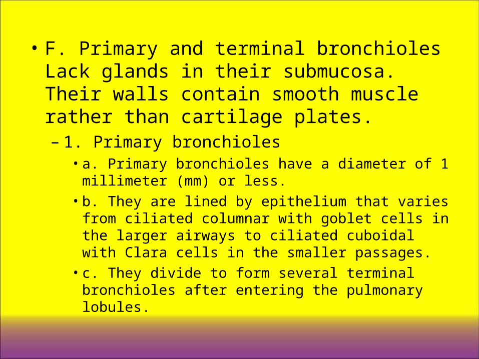
• F. Primary and terminal bronchioles Lack glands in their submucosa. Their walls contain smooth muscle rather than cartilage plates. – 1. Primary bronchioles • a. Primary bronchioles have a diameter of 1 millimeter
(mm) or less. • b. They are lined by epithelium that varies from ciliated
columnar with goblet cells in the larger airways to ciliated cuboidal with Clara cells in the smaller passages. • c. They divide to form several terminal bronchioles
after entering the pulmonary lobules.

– 2. Terminal bronchioles• a. Terminal bronchioles are the most distal part of the
conducting portion of the respiratory system. • b. They have a diameter of less than 0.5 mm. • c. They are lined by a simple cuboidal epithelium that
contains mostly Clara cells, some ciliated cells, and no goblet cells. • d. Function. Clara cells have the following functions:
– 1) Clara cells divide, and some of them differentiate to form ciliated cells.
– 2) They secrete glycosaminoglycans. – 3) They metabolize airborne toxins, a process that is carried
out by cytochrome P-450 enzymes present in their abundant smooth endoplasmic reticulum (SER).


Respiratory Portion of the Respiratory System.
• A. Respiratory bronchioles – 1. The respiratory bronchioles mark the transition
from the conducting to the respiratory portion of the respiratory system.
– 2. They are lined by a simple cuboidal epithelium consisting of Clara cells and some ciliated cells, except where their walls are interrupted by alveoli, the sites where gas exchange occurs.

• B. Alveolar ducts – 1. Alveolar ducts are linear passageways
continuous with the respiratory bronchioles. – 2. Their walls consist of adjacent alveoli, which are
separated from one another only by an interalveolar septum. smooth muscle, which is present in their walls at the openings of adjacent alveoli.
– 4. They are lined by type II pneumocytes and the highly attenuated simple squamous epithelium of type I pneumocytes.


• D. Alveoli – 1. Overview
• a. Alveoli are pouch-like evaginations [about 200 um in diameter] present in the walls of respiratory bronchioles, in alveolar ducts, and in alveolar sacs.
• b. They have thin walls, across which oxygen and carbon dioxide can diffuse between the air and the blood.
• c. They are separated from each other by interalveolar septae, which may contain one or more alveolar pores (pores of Kohn). These pores permit equalization of pressure between alveoli.
• d. They are rimmed by elastic fibers at their openings and are supported by many reticular fibers in their walls.
• e. They are lined by a highly attenuated simple squamous epithelium composed of type I and type II pneumocytes.

– 2. Alveolar cells • a. Type I pneumocytes (type I alveolar cells):
– 1) Cover about 95% ofthe alveolar surface and form part ofthe blood-gas barrier where exchange of 02 and CO2 occurs
– 2) Have an extremely thin cytoplasm that may be less than 80 nanometers (nm) in thickness
– 3) Form tight junctions with adjacent cells – 4) May have phagocytic capabilities – 5) Are not able to divide

– b. Type II pneumocytes (type II alveolar cells; great alveolar cells; granular pneumocytes; septal cells): • 1) Are cuboidal in shape and are most often located
near septal intersections • 2) Bulge into the alveolus and have a free surface that
contains short microvilli around their peripheral borders • 3) Are able to divide and regenerate both types of
alveolar pneumocytes • 4) Form tight junctions with adjacent cells • 5) Synthesize pulmonary surfactant, which is stored in
cytoplasmic lamellar bodies

– c. Alveolar macrophages (alveolar phagocytes; dust cells): • 1) Are the principal mononuclear phagocytes of the
alveolar surface • 2) Remove inhaled dust, bacteria, and other particulate
matter trapped in the pulmonary surfactant, thus providing a vital line of defense in the lungs • 3) Migrate to the bronchioles after becoming filled with
debris. From there, they are carried via ciliary action to the upper airways, eventually reaching the oropharynx, where they are swallowed or expectorated • 4) May also exit by migrating into the interstitium and
leaving via lymphatic vessels


• E. Interalveolar septum – 1. The interalveolar septum is the wall, or
partition, between two adjacent alveoli. – 2. It is bounded on its outer surfaces by the
extremely thin simple squamous epithelium lining the alveoli.
– 3. It contains many elastic and reticular fibers in its thicker regions.
– 4. It houses continuous capillaries in its central (interior) region.
– 5. It accommodates the blood-gas barrier, which separates the alveolar airspace from the capillary lumen.

• a. Structure of blood-gas barrier– 1) The thinnest regions of the barrier are 0.2 um or less in
thickness and consist of the following layers: » (a) Type I pneumocytes and layer of surfactant lining the
alveolar airspace » (b) Fused basal laminae of type I pneumocytes and
capillary endothelial cells » (c) Endothelium of the continuous capillaries within the
interalveolar septum – 2) Thicker regions of the barrier measure about 0.5 um across
and have an interstitial area interposed between the two basal laminae, which are not fused.
• b. Function of blood-gas barrier. The blood-gas barrier permits the diffusion of gases between the alveolar airspace and the blood. Oxygen passes from the alveolus into the capillary, and carbon dioxide passes from the capillary blood into the alveolus.


Pulmonary Vascular Supply • A. Pulmonary artery
– 1. The pulmonary artery carries blood to the lungs to be oxygenated.
– 2. It enters the root of each lung and extends branches along the divisions of the bronchial tree.
– 3. It enters lung lobules, where its branches follow the bronchioles.
• B. Pulmonary veins – 1. In lung lobules, pulmonary veins run in the intersegmental
connective tissue, separated from the arteries. – 2. After leaving the lobules, the pulmonary veins come close to
divisions of the bronchial tree and run parallel to branches of the pulmonary artery as they accompany bronchi to the root of the lung.


• C. Bronchial arteries and veins – 1. Bronchial arteries and veins provide nutrients
to and remove wastes from the nonrespiratory portions of the lung (bronchi, bronchioles, interstitium, and pleura).
– 2. They follow the branching pattern of the bronchial tree and form anastomoses with the pulmonary vessels near capillary beds.

Pulmonary Nerve Supply
• A. Parasympathetic stimulation causes contraction of pulmonary smooth muscle.
• B. Sympathetic stimulation causes relaxation of pulmonary smooth muscle and can be mimicked by certain drugs that cause dilation of bronchi and bronchioles.