RESONANT RADIOFREQUENCY FIELDS DAMAGING SACCHAROMYCES ...
3
RESONANT RADIOFREQUENCY FIELDS DAMAGING SACCHAROMYCES CEREVISIAE 1 W S Dias, 1 E H M Liquer, 2 L C Gontijo, 2 T A Oakes, 2 G S Dias, 2 C Marques, and 3 H S Chavez 1 WS-Tecnologia, Serra-ES, Brazil 2 Department of Physics - IFES - Instituto Federal do Espírito Santo (IFES), Av. Vitória 1729, Jucutuquara, Vitória, ES, Brazil, CEP 29040-780 and 3 UCL-Faculdade do Centro Leste, Rodovia ES 010, Km 6 BR 101, Serra-ES, Brazil * (Dated: July 7, 2021) This work describes an experiment and some results from instability involving the proliferation achieved by cells over some radio frequency electromagnetic field. Saccharomyces cerevisiae cells were cultured, and separate samples were examined within 6 hours. The frequency, the pulse width and peak-to-peak voltages were fixed. Producing at least 1 kV across the cell membrane in milk suspension. It was observed that in the presence of the electromagnetic field the cells had their time life drastically reduced when compared to the control sample. I. INTRODUCTION The use of electric fields to control the proliferation of microorganisms were first described and patented by [1] in the early 1960s. After, reference [2] analyzed the ef- fects of DC pulses on microorganisms in a systematically way. Following these works, radio frequency technology has proven useful for a wide range of applications over the decades since the initial research. The extensive use of electromagnetic field has been applied in food preser- vation as discussed in [3–7]. The authors describe indus- trial processes with the application of radio frequencies for the elimination of microorganisms in food production, in order to increase shelf life and final product quality. Other applications are: the interruption of the forma- tion processes or even the synchrony of the cellular repro- duction process, as it is well known. According to them, only by varying the potential of the field applied, one can stimulate and inhibit cellular processes. The main goal of this work is to study the effects of the exposure of high frequency electromagnetic waves in a solution of milk and Saccharomyces cerevisiae and to evaluate the effects of fields. II. PRESENTATION OF THE EXPERIMENT AND MAIN RESULTS Saccharomyces Cerevisiae S288C is an unicellular eu- karyotic organism that belongs to the kingdom of fungi. It is the yeast used in the production of bread and also of beer, besides being used for the production of ethanol. A milk solution containing 20 g of dry biological yeast (Sac- charomyces cerevisiae), 20 g of sucrose and 200 ml of milk was prepared at 28 Celsius degrees. The solution was di- vided into four equal samples, two for control and two for the application of the electromagnetic field. We use * [email protected]; [email protected] the Radio Frequency Generator Kenwood Transceiver, Model TS-2000, operating in a constant frequency mode of 28 MHz frequency carrier modulation. This frequency was chosen because it was part of the used range in the cited studies involving radio frequency. The antenna that was developed for our purposes is called magnetic loop and is shown at figure 1. It constitutes of an RLC circuit with a metal circle in a torus geometry, which resonates at the frequency of the experiment. Measurements were calculated in the MMANA antenna simulator software, obtained at [8], following the field setup in order to ex- pose the samples. See figure below. Figure 1. Field geometric setup of an antenna. Based on these materials, the antenna was placed to- gether with the radio frequency generating equipment on a wooden table covered by glass. Inside the loop of the antenna - area of the greatest electromagnetic field - two beakers were arranged with 50 ml of the solution in each one. Two additional 50 ml beakers were placed outside and away from the antenna loop, spaced at a distance of at least one meter from the radiating assembly and adopted as a control sample. A power of 25 watts was applied to the antenna, bring- ing the peak-to-peak voltage at the coil to 2.8 v. In the figure below it can be verified that a fluorescent lamp remains lit without any electrical contact inside arXiv:1807.11721v1 [physics.bio-ph] 31 Jul 2018
Transcript of RESONANT RADIOFREQUENCY FIELDS DAMAGING SACCHAROMYCES ...

RESONANT RADIOFREQUENCY FIELDS DAMAGING SACCHAROMYCESCEREVISIAE
1W S Dias, 1E H M Liquer, 2L C Gontijo, 2T A Oakes, 2G S Dias, 2C Marques, and 3H S Chavez1WS-Tecnologia, Serra-ES, Brazil
2Department of Physics - IFES - Instituto Federal do Espírito Santo (IFES),Av. Vitória 1729, Jucutuquara, Vitória, ES, Brazil, CEP 29040-780 and
3UCL-Faculdade do Centro Leste, Rodovia ES 010, Km 6BR 101, Serra-ES, Brazil*
(Dated: July 7, 2021)
This work describes an experiment and some results from instability involving the proliferationachieved by cells over some radio frequency electromagnetic field. Saccharomyces cerevisiae cellswere cultured, and separate samples were examined within 6 hours. The frequency, the pulse widthand peak-to-peak voltages were fixed. Producing at least 1 kV across the cell membrane in milksuspension. It was observed that in the presence of the electromagnetic field the cells had their timelife drastically reduced when compared to the control sample.
I. INTRODUCTION
The use of electric fields to control the proliferation ofmicroorganisms were first described and patented by [1]in the early 1960s. After, reference [2] analyzed the ef-fects of DC pulses on microorganisms in a systematicallyway. Following these works, radio frequency technologyhas proven useful for a wide range of applications overthe decades since the initial research. The extensive useof electromagnetic field has been applied in food preser-vation as discussed in [3–7]. The authors describe indus-trial processes with the application of radio frequenciesfor the elimination of microorganisms in food production,in order to increase shelf life and final product quality.
Other applications are: the interruption of the forma-tion processes or even the synchrony of the cellular repro-duction process, as it is well known. According to them,only by varying the potential of the field applied, one canstimulate and inhibit cellular processes.
The main goal of this work is to study the effects ofthe exposure of high frequency electromagnetic waves ina solution of milk and Saccharomyces cerevisiae and toevaluate the effects of fields.
II. PRESENTATION OF THE EXPERIMENTAND MAIN RESULTS
Saccharomyces Cerevisiae S288C is an unicellular eu-karyotic organism that belongs to the kingdom of fungi.It is the yeast used in the production of bread and also ofbeer, besides being used for the production of ethanol. Amilk solution containing 20 g of dry biological yeast (Sac-charomyces cerevisiae), 20 g of sucrose and 200 ml of milkwas prepared at 28 Celsius degrees. The solution was di-vided into four equal samples, two for control and twofor the application of the electromagnetic field. We use
* [email protected]; [email protected]
the Radio Frequency Generator Kenwood Transceiver,Model TS-2000, operating in a constant frequency modeof 28 MHz frequency carrier modulation. This frequencywas chosen because it was part of the used range in thecited studies involving radio frequency. The antenna thatwas developed for our purposes is called magnetic loopand is shown at figure 1. It constitutes of an RLC circuitwith a metal circle in a torus geometry, which resonatesat the frequency of the experiment. Measurements werecalculated in the MMANA antenna simulator software,obtained at [8], following the field setup in order to ex-pose the samples. See figure below.
Figure 1. Field geometric setup of an antenna.
Based on these materials, the antenna was placed to-gether with the radio frequency generating equipment ona wooden table covered by glass. Inside the loop of theantenna - area of the greatest electromagnetic field - twobeakers were arranged with 50 ml of the solution in eachone. Two additional 50 ml beakers were placed outsideand away from the antenna loop, spaced at a distanceof at least one meter from the radiating assembly andadopted as a control sample.
A power of 25 watts was applied to the antenna, bring-ing the peak-to-peak voltage at the coil to 2.8 v. Inthe figure below it can be verified that a fluorescentlamp remains lit without any electrical contact inside
arX
iv:1
807.
1172
1v1
[ph
ysic
s.bi
o-ph
] 3
1 Ju
l 201
8
2
the loop, demonstrating the high intensity of the linesof force that concentrate there. The experiment envi-ronment was closely supervised to verify that the tem-perature remained the same as the initial temperature.In the temperature measurements performed every hour,during the 6 hours of the experiment, no difference intemperature between the samples was noticed. This pre-caution was taken so that the temperature variation didnot interfere in the development of the cells in the anal-ysis of the result. After 6 hours of exposure, individualsamples of each beaker were collected and survivor cellswere counted throughout photos captured from a micro-scope. The general state of the cells of each beaker couldbe visualized as shown at the figures2 and 3 in the nextsection.
Figure 2. Experiment setup showing the control and treatedsamples.
Principal results are shown at the figures below, wherewe see a stringent correlation between electromagneticfield application and mortality of the micro organisms.
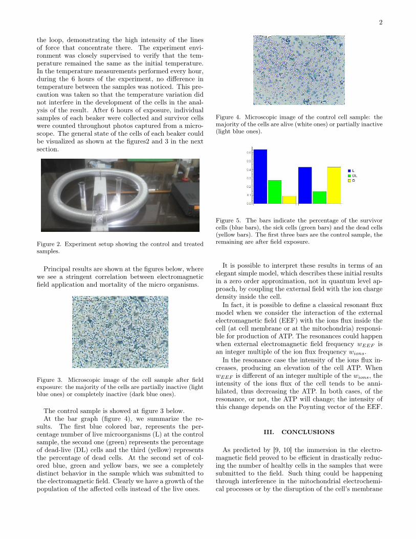
Figure 3. Microscopic image of the cell sample after fieldexposure: the majority of the cells are partially inactive (lightblue ones) or completely inactive (dark blue ones).
The control sample is showed at figure 3 below.At the bar graph (figure 4), we summarize the re-
sults. The first blue colored bar, represents the per-centage number of live microorganisms (L) at the controlsample, the second one (green) represents the percentageof dead-live (DL) cells and the third (yellow) representsthe percentage of dead cells. At the second set of col-ored blue, green and yellow bars, we see a completelydistinct behavior in the sample which was submitted tothe electromagnetic field. Clearly we have a growth of thepopulation of the affected cells instead of the live ones.
Figure 4. Microscopic image of the control cell sample: themajority of the cells are alive (white ones) or partially inactive(light blue ones).
Figure 5. The bars indicate the percentage of the survivorcells (blue bars), the sick cells (green bars) and the dead cells(yellow bars). The first three bars are the control sample, theremaining are after field exposure.
It is possible to interpret these results in terms of anelegant simple model, which describes these initial resultsin a zero order approximation, not in quantum level ap-proach, by coupling the external field with the ion chargedensity inside the cell.
In fact, it is possible to define a classical resonant fluxmodel when we consider the interaction of the externalelectromagnetic field (EEF) with the ions flux inside thecell (at cell membrane or at the mitochondria) responsi-ble for production of ATP. The resonances could happenwhen external electromagnetic field frequency 𝑤𝐸𝐸𝐹 isan integer multiple of the ion flux frequency 𝑤𝑖𝑜𝑛𝑠.
In the resonance case the intensity of the ions flux in-creases, producing an elevation of the cell ATP. When𝑤𝐸𝐸𝐹 is different of an integer multiple of the 𝑤𝑖𝑜𝑛𝑠, theintensity of the ions flux of the cell tends to be anni-hilated, thus decreasing the ATP. In both cases, of theresonance, or not, the ATP will change; the intensity ofthis change depends on the Poynting vector of the EEF.
III. CONCLUSIONS
As predicted by [9, 10] the immersion in the electro-magnetic field proved to be efficient in drastically reduc-ing the number of healthy cells in the samples that weresubmitted to the field. Such thing could be happeningthrough interference in the mitochondrial electrochemi-cal processes or by the disruption of the cell’s membrane

3
integrity.We devise, following [9] that the long exposition of
electromagnetic field may cause a variation of the ATPdemand for the basic process inside the cells, stressingthem and killing cells. The basic physics behind thisprocess can be related to a dynamically redefinition ofion concentrations or ion gradients inside the cells. Thishypothesis needs deeper analyses and future experiments.Which are being considered for future developments.
As we see, a wide range of applications not only in thearea of food preservation but also in the medical fieldcan be glimpsed from this technique, enabling the de-velopment of industrial and medical equipment aimed atmicrobiological control. Other possible application is totry to reduce the tumoral cell growth by applying spe-cific fields setups that, in some way, could fully break
mitochondrial metabolism.In particular, we are developing the study of the ef-
fect of radiation at the proliferation of more complexorganisms, whose large reproduction causes diseases atagricultural plants and forests like the Atlantic Forest inEspírito Santo, Brazil.
ACKNOWLEDGMENTS
We thank Dr. Adriana Korres from Federal Institute,IFES, for the support in the experiment at the Microbi-ology Lab. Also we thank Isabelle Cohen Dias for thesuggestions in the writting of this paper.
[1] Doevenspeck, Verfahren und Vorrichtung zur Gewin-nung der einzelnen Phasen aus dispersen Systemen. DE1, 1960, 𝑝𝑝.237− 541.
[2] A. J. H. Sale e W. A. Hamilton, Effects of high electricfields on micro-organisms III. Lysis of erythrocytes andprotoplasts, Biochimica e Biophysica Acta, vol. 163, pp.37− 43, 1968.
[3] Awuah, George B., J. Tang, and Hosahalli S. Ra-maswamy. "Radio-Frequency heating in food processing:Principles and applications". CRC Press, 2014.
[4] Ofelia Q. F. et al . Electrical stimulation of saccha-romyces cerevisiae cultures. Braz. J. Microbiol, SãoPaulo, v. 35, n. 1− 2, p. 97− 103, junho 2004.
[5] Doevenspeck, Verfahren und Vorrichtung zur Gewin-nung der einzelnen Phasen aus dispersen Systemen. DE1, 1960, 𝑝𝑝.237− 541.
[6] Doevenspeck, Influencing cells and cell walls by electro-static impulses, Fleischwirtschaft, vol. 13, 1961.
[7] A. Hamilton and A. J. H. Sale, Effect of high electricfields on microorganisms. II. Mechanisms of action ofthe lethal effect, Biochim Biophys Acta, vol.148, 𝑝𝑝.789−800, 1967.
[8] http://hamsoft.ca/pages/mmana-gal.php and adjustedat the experiment. Acessado em 30/07/2018.
[9] Steven L. Harrison, Gustavo V. Barbosa-Canovas, andBarry G. Swanson. Saccharomyces cerevisiaeStructuralChanges Induced by Pulsed Electric Field Treatment.LWT-Food Science and Technology 30.3(1997) : 236 −240.
[10] Olga N. Pakhomova, et al. "Electroporation-induced elec-trosensitization." PloS one 6.2(2011) : 𝑒17100.