ResolutionofDi useIntrahepaticBiliaryStricturesafter...
3
CASE REPORT | BILIARY Resolution of Di ffuse Intrahepatic Biliary Strictures after Chemotherapy for Metastatic Ovarian Cancer Daniel Lew, MD 1 , Vinay Sundaram, MD 2 , Brad D. Barrows, DO 3 , Simon K. Lo, MD, FACP 2 , and Srinivas Gaddam, MD, MPH 2 1 Department of Medicine, Cedars-Sinai Medical Center, Los Angeles, CA 2 Division of Gastroenterology, Cedars-Sinai Medical Center, Los Angeles, CA 3 Department of Pathology, Cedars-Sinai Medical Center, Los Angeles, CA ABSTRACT Sclerosing cholangitis and cholestatic jaundice secondary to metastatic disease is a rare complication. We report a rare case of secondary sclerosing cholangitis (SSC) due to lymphatic spread from ovarian cancer with complete resolution after chemotherapy. The diagnosis of SSC from metastatic ovarian cancer was clinically challenging, as endoscopic retrograde cholangiopancreatography revealed irregular hepatic ducts consistent with sclerosing cholangitis, but it did not identify any malignant cells. The final diagnosis was made with liver biopsy revealing high-grade metastatic Mullerian carcinoma. The patient responded well to chemotherapy and is in remission. A timely diagnosis is important and can lead to complete resolution of the disease. INTRODUCTION Sclerosing cholangitis is a chronic and progressive cholestatic disease characterized by inflammation, fibrosis, and stricturing of the bile ducts. The majority of cases are idiopathic, known as primary sclerosing cholangitis (PSC). 1 A small number of patients are diagnosed with secondary sclerosing cholangitis (SSC) caused by metastatic disease, infection, ischemia, autoimmune dysfunction, obstruction, and toxins. 1,2 The prognosis of SSC depends on the tim- ing of diagnosis and the underlying etiology. The definitive treatment for advanced cases is liver transplantation. 3 CASE REPORT A 53-year-old woman with a history of BRCA2 stage III recurrent ovarian cancer presented with complaints of ab- dominal pain for the past 3 weeks. She characterized the pain as burning and mid-epigastric, radiating to the back and right shoulder. Pain progressed from intermittent during the initial few days to constant in the past week. Eating did not exacerbate or alleviate the pain. Associated symptoms included yellowing of the eyes and skin, nau- sea with non-bilious and non-bloody vomiting, and generalized fatigue for the past 3 days. She denied pruritis, bowel habit changes, fevers, chills, night sweats, or recent travel. There was no personal or family history of hepa- tobiliary disease. Eight weeks prior, the patient was hospitalized for similar symptoms. Liver serologies showed aspartate transami- nase 2,585 U/L, alanine transaminase 3,545 U/L, alkaline phosphatase 162 U/L, and total bilirubin 7.3 mg/dL. Workup for viral, autoimmune, and ischemic hepatitides was non-revealing. Two months prior, the patient was diagnosed with her second recurrence of ovarian cancer to the lymph nodes in the porta hepatis, retroperitoneum, and mediastinum, and she was started on olaparib (Lynparza, AstraZeneca Pharmaceuticals LP, Wilmington, DE), a poly-ADP ribose polymerase inhibitor approved by the U.S. Food and Drug Administration (FDA). Drug-induced liver injury was considered, but discontinuation of olaparib resulted only in transient improvement in symptoms ACG Case Rep J 2017;4:e77. doi:10.14309/crj.2017.77. Published online: June 7, 2017. Correspondence: Daniel Lew, Cedars-Sinai Medical Center, Department of Medicine, 8700 Beverly Blvd, Los Angeles, CA 90048 ([email protected]). Copyright: © 2017 Lew et al. This work is licensed under a Creative Commons Attribution-NonCommercial-NoDerivatives 4.0 International License. To view a copy of this license, visit http://creativecommons.org/licenses/by-nc-nd/4.0. ACG Case Reports Journal / Volume 4 acgcasereports.gi.org 1 ACG CASE REPORTS JOURNAL
-
Upload
nguyentuyen -
Category
Documents
-
view
214 -
download
0
Transcript of ResolutionofDi useIntrahepaticBiliaryStricturesafter...
CASE REPORT | BILIARY
Resolution of Diffuse Intrahepatic Biliary Strictures afterChemotherapy for Metastatic Ovarian CancerDaniel Lew, MD1, Vinay Sundaram, MD2, Brad D. Barrows, DO3, Simon K. Lo, MD, FACP2, andSrinivas Gaddam, MD, MPH2
1Department of Medicine, Cedars-Sinai Medical Center, Los Angeles, CA2Division of Gastroenterology, Cedars-Sinai Medical Center, Los Angeles, CA3Department of Pathology, Cedars-Sinai Medical Center, Los Angeles, CA
ABSTRACTSclerosing cholangitis and cholestatic jaundice secondary to metastatic disease is a rare complication. We reporta rare case of secondary sclerosing cholangitis (SSC) due to lymphatic spread from ovarian cancer with completeresolution after chemotherapy. The diagnosis of SSC from metastatic ovarian cancer was clinically challenging,as endoscopic retrograde cholangiopancreatography revealed irregular hepatic ducts consistent with sclerosingcholangitis, but it did not identify any malignant cells. The final diagnosis was made with liver biopsy revealinghigh-grade metastatic Mullerian carcinoma. The patient responded well to chemotherapy and is in remission. Atimely diagnosis is important and can lead to complete resolution of the disease.
INTRODUCTIONSclerosing cholangitis is a chronic and progressive cholestatic disease characterized by inflammation, fibrosis, andstricturing of the bile ducts. The majority of cases are idiopathic, known as primary sclerosing cholangitis (PSC).1 Asmall number of patients are diagnosed with secondary sclerosing cholangitis (SSC) caused by metastatic disease,infection, ischemia, autoimmune dysfunction, obstruction, and toxins.1,2 The prognosis of SSC depends on the tim-ing of diagnosis and the underlying etiology. The definitive treatment for advanced cases is liver transplantation.3
CASE REPORTA 53-year-old woman with a history of BRCA2 stage III recurrent ovarian cancer presented with complaints of ab-dominal pain for the past 3 weeks. She characterized the pain as burning and mid-epigastric, radiating to the backand right shoulder. Pain progressed from intermittent during the initial few days to constant in the past week.Eating did not exacerbate or alleviate the pain. Associated symptoms included yellowing of the eyes and skin, nau-sea with non-bilious and non-bloody vomiting, and generalized fatigue for the past 3 days. She denied pruritis,bowel habit changes, fevers, chills, night sweats, or recent travel. There was no personal or family history of hepa-tobiliary disease.
Eight weeks prior, the patient was hospitalized for similar symptoms. Liver serologies showed aspartate transami-nase 2,585 U/L, alanine transaminase 3,545 U/L, alkaline phosphatase 162 U/L, and total bilirubin 7.3 mg/dL.Workup for viral, autoimmune, and ischemic hepatitides was non-revealing. Two months prior, the patient wasdiagnosed with her second recurrence of ovarian cancer to the lymph nodes in the porta hepatis, retroperitoneum,and mediastinum, and she was started on olaparib (Lynparza, AstraZeneca Pharmaceuticals LP, Wilmington, DE), apoly-ADP ribose polymerase inhibitor approved by the U.S. Food and Drug Administration (FDA). Drug-inducedliver injury was considered, but discontinuation of olaparib resulted only in transient improvement in symptoms
ACG Case Rep J 2017;4:e77. doi:10.14309/crj.2017.77. Published online: June 7, 2017.
Correspondence: Daniel Lew, Cedars-Sinai Medical Center, Department of Medicine, 8700 Beverly Blvd, Los Angeles, CA 90048 ([email protected]).
Copyright: © 2017 Lew et al. This work is licensed under a Creative Commons Attribution-NonCommercial-NoDerivatives 4.0 InternationalLicense. To view a copy of this license, visit http://creativecommons.org/licenses/by-nc-nd/4.0.
ACG Case Reports Journal / Volume 4 acgcasereports.gi.org 1
ACGCASE REPORTS JOURNAL
and serologies. Imaging did not reveal any new signs offocal metastasis or liver abnormality. The patient improvedwith conservative management and did not resume olaparib.Liver serologies at discharge showed aspartate transaminase2,145 U/L, alanine transaminase 1,299 U/L, alkaline phospha-tase 120 U/L, and total bilirubin 5.3 mg/dL.
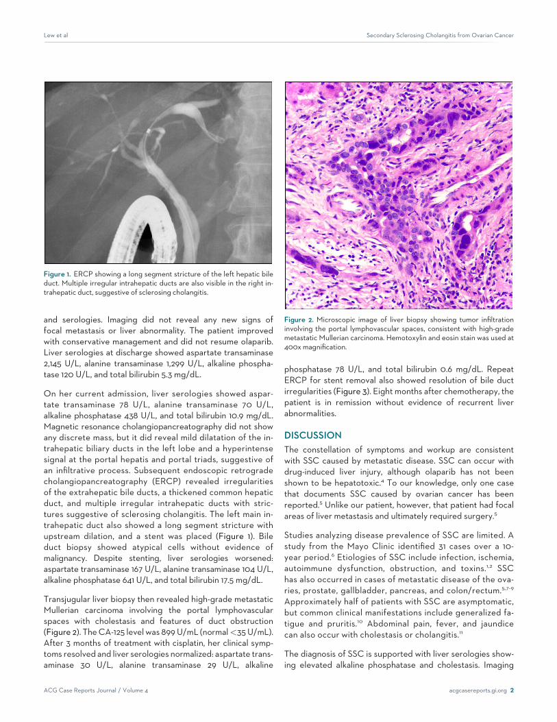
On her current admission, liver serologies showed aspar-tate transaminase 78 U/L, alanine transaminase 70 U/L,alkaline phosphatase 438 U/L, and total bilirubin 10.9 mg/dL.Magnetic resonance cholangiopancreatography did not showany discrete mass, but it did reveal mild dilatation of the in-trahepatic biliary ducts in the left lobe and a hyperintensesignal at the portal hepatis and portal triads, suggestive ofan infiltrative process. Subsequent endoscopic retrogradecholangiopancreatography (ERCP) revealed irregularitiesof the extrahepatic bile ducts, a thickened common hepaticduct, and multiple irregular intrahepatic ducts with stric-tures suggestive of sclerosing cholangitis. The left main in-trahepatic duct also showed a long segment stricture withupstream dilation, and a stent was placed (Figure 1). Bileduct biopsy showed atypical cells without evidence ofmalignancy. Despite stenting, liver serologies worsened:aspartate transaminase 167 U/L, alanine transaminase 104 U/L,alkaline phosphatase 641 U/L, and total bilirubin 17.5 mg/dL.
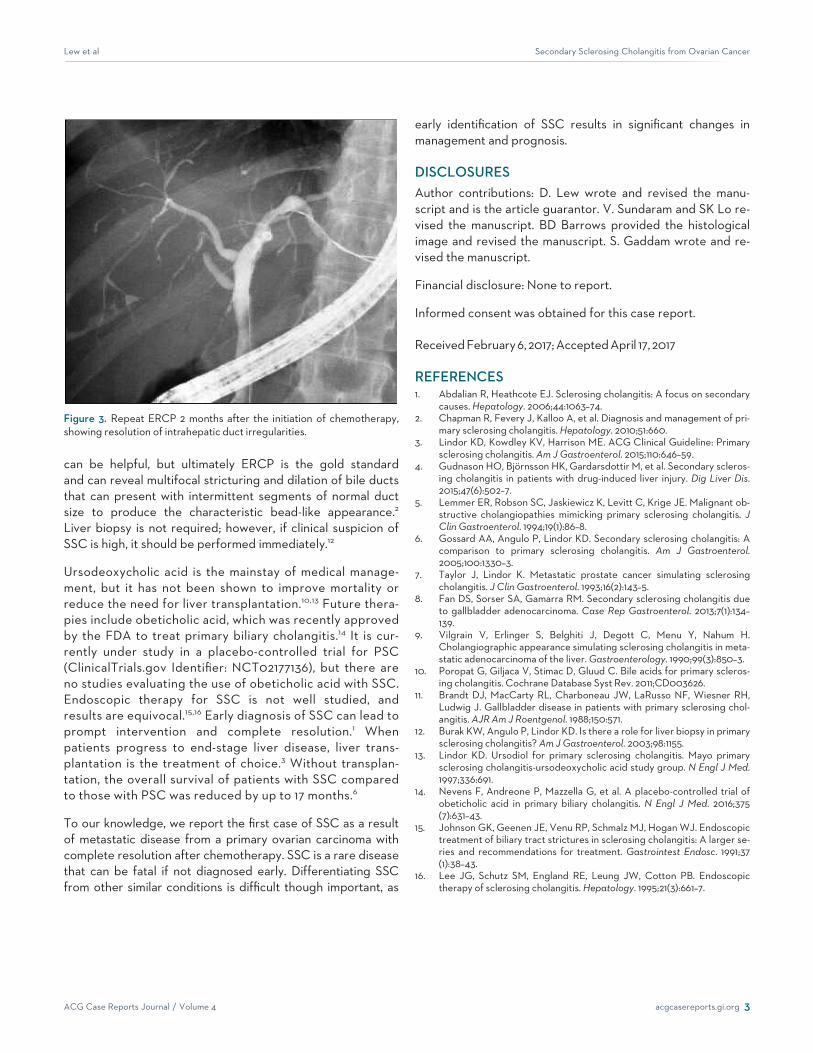
Transjugular liver biopsy then revealed high-grade metastaticMullerian carcinoma involving the portal lymphovascularspaces with cholestasis and features of duct obstruction(Figure 2). TheCA-125 level was 899U/mL (normal<35U/mL).After 3 months of treatment with cisplatin, her clinical symp-toms resolved and liver serologies normalized: aspartate trans-aminase 30 U/L, alanine transaminase 29 U/L, alkaline
phosphatase 78 U/L, and total bilirubin 0.6 mg/dL. RepeatERCP for stent removal also showed resolution of bile ductirregularities (Figure 3). Eight months after chemotherapy, thepatient is in remission without evidence of recurrent liverabnormalities.
DISCUSSIONThe constellation of symptoms and workup are consistentwith SSC caused by metastatic disease. SSC can occur withdrug-induced liver injury, although olaparib has not beenshown to be hepatotoxic.4 To our knowledge, only one casethat documents SSC caused by ovarian cancer has beenreported.5 Unlike our patient, however, that patient had focalareas of liver metastasis and ultimately required surgery.5
Studies analyzing disease prevalence of SSC are limited. Astudy from the Mayo Clinic identified 31 cases over a 10-year period.6 Etiologies of SSC include infection, ischemia,autoimmune dysfunction, obstruction, and toxins.1,2 SSChas also occurred in cases of metastatic disease of the ova-ries, prostate, gallbladder, pancreas, and colon/rectum.5,7-9
Approximately half of patients with SSC are asymptomatic,but common clinical manifestations include generalized fa-tigue and pruritis.10 Abdominal pain, fever, and jaundicecan also occur with cholestasis or cholangitis.11
The diagnosis of SSC is supported with liver serologies show-ing elevated alkaline phosphatase and cholestasis. Imaging
Figure 1. ERCP showing a long segment stricture of the left hepatic bileduct. Multiple irregular intrahepatic ducts are also visible in the right in-trahepatic duct, suggestive of sclerosing cholangitis.
Figure 2. Microscopic image of liver biopsy showing tumor infiltrationinvolving the portal lymphovascular spaces, consistent with high-grademetastatic Mullerian carcinoma. Hemotoxylin and eosin stain was used at400xmagnification.
Lew et al Secondary Sclerosing Cholangitis from Ovarian Cancer
ACG Case Reports Journal / Volume 4 acgcasereports.gi.org 2
can be helpful, but ultimately ERCP is the gold standardand can reveal multifocal stricturing and dilation of bile ductsthat can present with intermittent segments of normal ductsize to produce the characteristic bead-like appearance.2
Liver biopsy is not required; however, if clinical suspicion ofSSC is high, it should be performed immediately.12
Ursodeoxycholic acid is the mainstay of medical manage-ment, but it has not been shown to improve mortality orreduce the need for liver transplantation.10,13 Future thera-pies include obeticholic acid, which was recently approvedby the FDA to treat primary biliary cholangitis.14 It is cur-rently under study in a placebo-controlled trial for PSC(ClinicalTrials.gov Identifier: NCT02177136), but there areno studies evaluating the use of obeticholic acid with SSC.Endoscopic therapy for SSC is not well studied, andresults are equivocal.15,16 Early diagnosis of SSC can lead toprompt intervention and complete resolution.1 Whenpatients progress to end-stage liver disease, liver trans-plantation is the treatment of choice.3 Without transplan-tation, the overall survival of patients with SSC comparedto those with PSC was reduced by up to 17 months.6
To our knowledge, we report the first case of SSC as a resultof metastatic disease from a primary ovarian carcinoma withcomplete resolution after chemotherapy. SSC is a rare diseasethat can be fatal if not diagnosed early. Differentiating SSCfrom other similar conditions is difficult though important, as
early identification of SSC results in significant changes inmanagement and prognosis.
DISCLOSURESAuthor contributions: D. Lew wrote and revised the manu-script and is the article guarantor. V. Sundaram and SK Lo re-vised the manuscript. BD Barrows provided the histologicalimage and revised the manuscript. S. Gaddam wrote and re-vised the manuscript.
Financial disclosure: None to report.
Informed consent was obtained for this case report.
ReceivedFebruary6,2017;AcceptedApril 17,2017
REFERENCES1. Abdalian R, Heathcote EJ. Sclerosing cholangitis: A focus on secondary
causes.Hepatology. 2006;44:1063–74.2. Chapman R, Fevery J, Kalloo A, et al. Diagnosis and management of pri-
mary sclerosing cholangitis.Hepatology. 2010;51:660.3. Lindor KD, Kowdley KV, Harrison ME. ACG Clinical Guideline: Primary
sclerosing cholangitis. Am J Gastroenterol. 2015;110:646–59.4. Gudnason HO, Björnsson HK, Gardarsdottir M, et al. Secondary scleros-
ing cholangitis in patients with drug-induced liver injury. Dig Liver Dis.2015;47(6):502–7.
5. Lemmer ER, Robson SC, Jaskiewicz K, Levitt C, Krige JE. Malignant ob-structive cholangiopathies mimicking primary sclerosing cholangitis. JClin Gastroenterol. 1994;19(1):86–8.
6. Gossard AA, Angulo P, Lindor KD. Secondary sclerosing cholangitis: Acomparison to primary sclerosing cholangitis. Am J Gastroenterol.2005;100:1330–3.
7. Taylor J, Lindor K. Metastatic prostate cancer simulating sclerosingcholangitis. J Clin Gastroenterol. 1993;16(2):143–5.
8. Fan DS, Sorser SA, Gamarra RM. Secondary sclerosing cholangitis dueto gallbladder adenocarcinoma. Case Rep Gastroenterol. 2013;7(1):134–139.
9. Vilgrain V, Erlinger S, Belghiti J, Degott C, Menu Y, Nahum H.Cholangiographic appearance simulating sclerosing cholangitis in meta-static adenocarcinoma of the liver.Gastroenterology. 1990;99(3):850–3.
10. Poropat G, Giljaca V, Stimac D, Gluud C. Bile acids for primary scleros-ing cholangitis. Cochrane Database Syst Rev. 2011;CD003626.
11. Brandt DJ, MacCarty RL, Charboneau JW, LaRusso NF, Wiesner RH,Ludwig J. Gallbladder disease in patients with primary sclerosing chol-angitis.AJRAm J Roentgenol. 1988;150:571.
12. Burak KW, Angulo P, Lindor KD. Is there a role for liver biopsy in primarysclerosing cholangitis? Am JGastroenterol. 2003;98:1155.
13. Lindor KD. Ursodiol for primary sclerosing cholangitis. Mayo primarysclerosing cholangitis-ursodeoxycholic acid study group. N Engl J Med.1997;336:691.
14. Nevens F, Andreone P, Mazzella G, et al. A placebo-controlled trial ofobeticholic acid in primary biliary cholangitis. N Engl J Med. 2016;375(7):631–43.
15. Johnson GK, Geenen JE, Venu RP, Schmalz MJ, HoganWJ. Endoscopictreatment of biliary tract strictures in sclerosing cholangitis: A larger se-ries and recommendations for treatment. Gastrointest Endosc. 1991;37(1):38–43.
16. Lee JG, Schutz SM, England RE, Leung JW, Cotton PB. Endoscopictherapy of sclerosing cholangitis.Hepatology. 1995;21(3):661–7.
Figure 3. Repeat ERCP 2 months after the initiation of chemotherapy,showing resolution of intrahepatic duct irregularities.
Lew et al Secondary Sclerosing Cholangitis from Ovarian Cancer
ACG Case Reports Journal / Volume 4 acgcasereports.gi.org 3