RESEARCH Open Access Tissue print of prostate biopsy: a ...
7
RESEARCH Open Access Tissue print of prostate biopsy: a novel tool in the diagnostic procedure of prostate cancer Adriano Angelucci 1* , Gianna Pace 2 , Patrizia Sanità 1 , Carlo Vicentini 2 and Mauro Bologna 1 Abstract Background: Nowadays, the histological examination of prostate core needle biopsies is still regarded as the gold standard in the diagnosis of prostate cancer (PCa). We investigated if the tissue print of core needle biopsy (biopsy print) could be used as adjunctive molecular investigative procedures in conjunction with routine histological examination of biopsy to improve PCa diagnosis. Methods: The direct contact of PCa core biopsy to nitrocellulose membrane resulted in the release of a cellular micropeel that was used for downstream analytical procedures. Results: By zymogram print-phoresis we demonstrated that matrix metalloproteases MMP-2 and MMP-9 could be visualized in biopsy prints and that the gelatinolytic activity was positively correlated with immunohistochemistry analysis of the same markers in matched bioptic specimens. Moreover, we compared the ability to detect the PCa- associated hypermethylation of GSTP1 promoter in DNA extracted from biopsy prints with those of the corresponding core needle biopsies. Biopsy prints demonstrated the same specificity of biopsies in detecting PCa (50%) while the sensitivity and the positive predictive value were lower than biopsies (56% vs 78% and 63% vs 70%, respectively). Conclusions: Biopsy print, combining a molecular point of view to the routinely hystopathological analysis of prostate biopsies, should be a useful tool to improve the diagnosis of PCa. Introduction To date, the diagnosis of prostate cancer (PCa) is based upon the histological examination of prostate core nee- dle biopsies. Other diagnostic procedures, although use- ful in improving the detection of PCa, such as measure of serum prostate specific antigen (PSA), still lack in adequate specificity to be used alone as diagnostic tech- nique. The importance of prostate needle biopsies is supported by the continuous progress in biopsy scheme in the attempt to improve the detection rate of PCa. In fact, in the last few years the standard sextant biopsy has been replaced by 12 cores and even more extensive biopsy schemes [1,2]. Despite the current debate on the optimal biopsy scheme, the core needle biopsy is still regarded by urologists as the gold standard in the diag- nosis of PCa. Moreover, in the attempt of better detect- ing PCa, several immunohistochemistry innovations have been developed, and some markers, as metallopro- teases, has been associated to the progression of PCa [3]. The Gleason score offers a good description of the aggressiveness of the tumor, but it is insufficient in esti- mating the prognosis. In order to achieve an optimal therapeutic approach a molecular analysis of needle biopsy should be useful. However, a single core biopsy, due to its small size, cannot be reserved for other analy- tical procedures than pathologic examination. Although the idea of reproducing an exact anatomical copy of a tissue on a solid support is not a novelty, it has been applied mainly in botanic studies [4]. Tissue print is based on the transfer, by direct contact, of the superfi- cial cellular contents of a fresh tissue to an adhesive or adsorptive surface. Tissue print of vegetal tissues on nylon or nitrocellulose membrane has demonstrated to be an effective tool to spatially detect the presence of specific proteins and mRNA [5]. Tissue copies on adhe- sive surface from rat and mouse tissues have been uti- lized in DNA-DNA and RNA-RNA hybridization assays * Correspondence: [email protected] 1 Department of Experimental Medicine, University of L’Aquila, L’Aquila, 67100, Italy Full list of author information is available at the end of the article Angelucci et al. Diagnostic Pathology 2011, 6:34 http://www.diagnosticpathology.org/content/6/1/34 © 2011 Angelucci et al; licensee BioMed Central Ltd. This is an Open Access article distributed under the terms of the Creative Commons Attribution License (http://creativecommons.org/licenses/by/2.0), which permits unrestricted use, distribution, and reproduction in any medium, provided the original work is properly cited.
Transcript of RESEARCH Open Access Tissue print of prostate biopsy: a ...
RESEARCH Open Access
Tissue print of prostate biopsy: a novel tool inthe diagnostic procedure of prostate cancerAdriano Angelucci1*, Gianna Pace2, Patrizia Sanità1, Carlo Vicentini2 and Mauro Bologna1
Abstract
Background: Nowadays, the histological examination of prostate core needle biopsies is still regarded as the goldstandard in the diagnosis of prostate cancer (PCa). We investigated if the tissue print of core needle biopsy (biopsyprint) could be used as adjunctive molecular investigative procedures in conjunction with routine histologicalexamination of biopsy to improve PCa diagnosis.
Methods: The direct contact of PCa core biopsy to nitrocellulose membrane resulted in the release of a cellularmicropeel that was used for downstream analytical procedures.
Results: By zymogram print-phoresis we demonstrated that matrix metalloproteases MMP-2 and MMP-9 could bevisualized in biopsy prints and that the gelatinolytic activity was positively correlated with immunohistochemistryanalysis of the same markers in matched bioptic specimens. Moreover, we compared the ability to detect the PCa-associated hypermethylation of GSTP1 promoter in DNA extracted from biopsy prints with those of thecorresponding core needle biopsies. Biopsy prints demonstrated the same specificity of biopsies in detecting PCa(50%) while the sensitivity and the positive predictive value were lower than biopsies (56% vs 78% and 63% vs70%, respectively).
Conclusions: Biopsy print, combining a molecular point of view to the routinely hystopathological analysis ofprostate biopsies, should be a useful tool to improve the diagnosis of PCa.
IntroductionTo date, the diagnosis of prostate cancer (PCa) is basedupon the histological examination of prostate core nee-dle biopsies. Other diagnostic procedures, although use-ful in improving the detection of PCa, such as measureof serum prostate specific antigen (PSA), still lack inadequate specificity to be used alone as diagnostic tech-nique. The importance of prostate needle biopsies issupported by the continuous progress in biopsy schemein the attempt to improve the detection rate of PCa. Infact, in the last few years the standard sextant biopsyhas been replaced by 12 cores and even more extensivebiopsy schemes [1,2]. Despite the current debate on theoptimal biopsy scheme, the core needle biopsy is stillregarded by urologists as the gold standard in the diag-nosis of PCa. Moreover, in the attempt of better detect-ing PCa, several immunohistochemistry innovations
have been developed, and some markers, as metallopro-teases, has been associated to the progression of PCa[3].The Gleason score offers a good description of the
aggressiveness of the tumor, but it is insufficient in esti-mating the prognosis. In order to achieve an optimaltherapeutic approach a molecular analysis of needlebiopsy should be useful. However, a single core biopsy,due to its small size, cannot be reserved for other analy-tical procedures than pathologic examination. Althoughthe idea of reproducing an exact anatomical copy of atissue on a solid support is not a novelty, it has beenapplied mainly in botanic studies [4]. Tissue print isbased on the transfer, by direct contact, of the superfi-cial cellular contents of a fresh tissue to an adhesive oradsorptive surface. Tissue print of vegetal tissues onnylon or nitrocellulose membrane has demonstrated tobe an effective tool to spatially detect the presence ofspecific proteins and mRNA [5]. Tissue copies on adhe-sive surface from rat and mouse tissues have been uti-lized in DNA-DNA and RNA-RNA hybridization assays
* Correspondence: [email protected] of Experimental Medicine, University of L’Aquila, L’Aquila,67100, ItalyFull list of author information is available at the end of the article
Angelucci et al. Diagnostic Pathology 2011, 6:34http://www.diagnosticpathology.org/content/6/1/34
© 2011 Angelucci et al; licensee BioMed Central Ltd. This is an Open Access article distributed under the terms of the CreativeCommons Attribution License (http://creativecommons.org/licenses/by/2.0), which permits unrestricted use, distribution, andreproduction in any medium, provided the original work is properly cited.
and in immunochemistry [6-8]. In more recent studies,Gaston and colleagues have successfully applied the tis-sue print technique to the molecular investigation ofPCa biopsy and of the entire prostate surface. Theydemonstrated that the cellular micropeel retained on themembrane is enough to detect proteins and mRNA in awide range of application, from print-phoresis to RT-PCR [9].Our aim has been to verify whether the biopsy print
should be an adequate copy of the prostate biopsy andif it may be useful in the routinely clinical diagnosticinvestigation as based on an easy procedure and on awide range of methods available for its analysis.
Materials and methodsPatientsWe selected 18 consecutive patients with a histologicaldiagnosis of PCa, who underwent radical retropubicprostatectomy (RRP) in our Urology Department of theG. Mazzini Hospital, Teramo, Italy. Patients were eligi-ble if they did not undergo previous anti-hormonal,radiation or chemotherapies. We considered the serumPSA value at the diagnosis and Gleason score obtainedafter RRP. As controls we enrolled 6 patients affected byBenign Prostatic Hyperplasia (BPH) and diagnosed bythe histopathological analysis of the tissue obtained aftera transvescical retropubic adenomectomy (TV adeno-mectomy) or a transurethral resection prostatectomy(TURP). Our institutional review board approved theprotocol. All patients signed the informed consent.
Reagents and plasticwareReagents, if not differently indicated, were purchasedfrom Sigma-Aldrich (St. Luois, MI, USA). Plasticwarewas purchased by BD Biosciences (Franklin Lakes, NJUSA). Human prostate cancer cell line, PC3 was origin-ally obtained by ATCC (Rockville, MD, USA) and main-tained in DMEM supplemented with 10% fetal bovineserum (FBS), glutamine, and penicillin-streptomycin.Conditioned medium was obtained culturing PC3 cellsfor 48 h in FBS-free medium.
Biopsy and tissue printThree needle biopsies were performed immediately afterthe surgical treatment in the area where the tumor waslocalized. All the procedures were performed in asepticatmosphere. Immediately, all three bioptic tissues werecompletely rinsed in phosphate buffer without Ca2+ andMg2+ and then the excess of liquid was dried off by anabsorbent paper. Each of the three biopsies were spreadon a dry nitrocellulose membrane (0.2 μm Protran,Whatman plc, Kent, UK) and left to adhere for about 30seconds. Then were delicately recovered with tweezerspulling the tissue from one end. The exceeding parts,
without the print, were removed from nitrocellulosemembranes. The resulting biopsy prints were conservedin a dry box at -20°C until processed.
Zymogram print-phoresisGelatin zymogram was performed according to standardprocedure with few modifications. A 10% SDS-polyacry-lamide gel copolymerized with 0.1 mg/ml gelatine wasutilized placing tissue print on the top of the resolvinggel and then pouring the 6% SDS-polyacrylamide stack-ing gel. At the end of the run gel was washed threetimes with 50 mM Tris-HCl (pH 7.4) containing 2% Tri-ton X-100 for 15 minutes under agitation to removeSDS, and was incubated with Tris-HCl (pH 7.4) plus10 mM CaCl2 and 200 mM NaCl for 24 hours at 37°C.Gel was then fixed and stained with 0.1% CoomassieBlue solution. Enzyme digested regions were identifiedas white bands against a blue background.
ImmunohistochemistryThe three core needle biopsies were fixed in 4% formal-dehyde in 0.1 M phosphate buffer, pH 7.2, andembedded in paraffin. Slide-mounted tissue sectionswere incubated with the anti-MMP2 or anti-MMP9 pri-mary antibodies (Santa Cruz, CA, USA) for 1 h at roomtemperature. Antibody binding was revealed using theUltra-Vision detection system anti-Polyvalent HRP/dia-minobenzidine kit according to the manufacturer’sinstructions. Each case was evaluated blindly by twoindependent readers (A. A. and P. G.). The number ofcells expressing the marker was assessed using a semi-quantitative three-grade scale (score 0 = 0%, score 1 =0-50%, or score 2 >50%). Cases in which the two obser-vers had obtained different results were collectivelyreviewed, and a consensus was obtained.
Methylation-specific PCRThe three needle biopsies obtained by focusing on thearea of the tumor and the related tissue prints wereincubated with lysis buffer (10 mmol/L Tris-HCl, pH8.0, 0.1 mmol/L EDTA, 1% v/v SDS) containing 2 mg/ml proteinase K at 55°C for 4 h. DNA was thenextracted with 1:1 v/v phenol/chloroform and precipi-tated with 50 mmol/L sodium acetate in ethanol. Theconcentration of extracted DNA was measured withspectrometry, and a range of 50-150 ng DNA was usedin the following steps. Using the EpiTec Bisulfite Kit(Qiagen, Venlo, The Netherlands) DNA was modifiedby bisulfite treatment for the detection of methylatedCpG residues, and was purified prior of the PCR ampli-fication. Bisulfite-converted DNA was subjected tomethylation-specific PCR (MSP) using primers specificfor the hypermethylated form of glutathione-S-trans-pherase 1 (GSTP1) promoter (forward: 5’-
Angelucci et al. Diagnostic Pathology 2011, 6:34http://www.diagnosticpathology.org/content/6/1/34
Page 2 of 7
GTTGCGCGGCGATTTC- 3’; reverse: 5’-GCCCCAA-TACTAAATCACGACG- 3’ ). The PCR reaction wasperformed with 2.5 μl of bisulfite-modified DNA tem-plate in 25 μl of reaction mixture containing 2.5 μl 10 ×PCR buffer, 200 μmol/L of each dNTP, 3.0 mmol/LMgCl2, 0.25 μmol/L each primer, 1.25 U of Ampli TaqGold. The PCR reaction was subjected to hot start at95°C for 10 minutes followed by 30 cycles of denatura-tion at 95°C for 30 seconds, annealing at 60°C for 1minute, and extension at 72°C for 1 minute. A secondset of primers detecting an unrelated not methylatedgene, MYOD1 was used as control for the efficacy ofbisulfite conversion (forward: 5’-CCAACTC-CAAATCCCCTCTCTAT-3’; reverse: 5’-TGATTAATT-TAGATTGGGTTTAGAGAAGGA-3’). Ten microL ofeach PCR reaction were loaded onto a 1.8% agarose gelcontaining ethidium bromide and PCR amplified DNAwas visualized by UV transilluminator.
Statistical analysisSPSS for Windows (version 10.0.7) computer packagewas used for statistical analysis of the data. The studyvariables were normally distributed (Shapiro-Wilk test; P< 0.05). The Pearson’s correlation test was used. Wealso evaluated the positive predictive value (PPV), thenegative predictive value (NPV), the sensitivity and spe-cificity of the performed method (biopsy print) with95% Coefficient of Confidence (CI). Values < 0.01 wereconsidered significant.
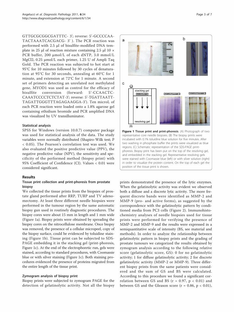
ResultsTissue print collection and print-phoresis from prostatebiopsyWe collected the tissue prints from the biopsies of pros-tate gland performed after RRP, TURP and TV adeno-mectomy. At least three different needle biopsies wereperformed in the tumour region by the same automaticbiopsy gun used in routinely diagnostic procedures. Thebiopsy cores were about 15 mm in length and 1 mm wide(Figure 1a). Biopsy prints were obtained by spreading thebiopsy cores on the nitrocellulose membrane. Once biopsywas removed, the presence of a cellular micropeel, copy ofthe biopsy surface, could be evidenced by toluidine stain-ing (Figure 1b). Tissue print can be subjected to SDS-PAGE embedding it in the stacking gel (print-phoresis,Figure 1c). At the end of the electrophoretic run, gels werestained, according to standard procedures, with Coomassieblue or with silver staining (Figure 1c). Both staining pro-cedures evidenced the presence of proteins migrated fromthe entire length of the tissue print.
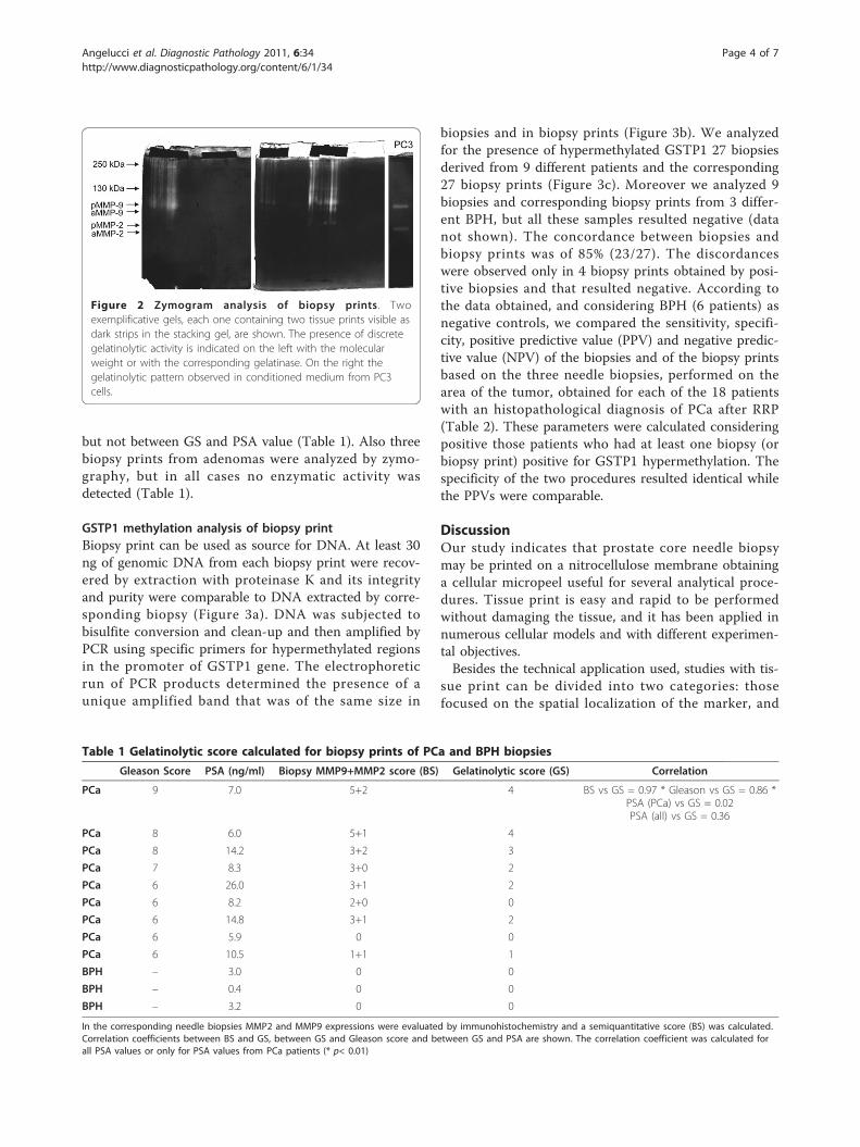
Zymogram analysis of biopsy printBiopsy prints were subjected to zymogram PAGE for thedetection of gelatinolytic activity. Not all the biopsy
prints demonstrated the presence of the lytic enzymes.When the gelatinolytic activity was evident we observedboth a diffuse and a discrete lytic activity. The more fre-quent discrete bands were identified as MMP-2 andMMP-9 (pro- and active forms), as suggested by thecorrespondence with the gelatinolytic pattern by condi-tioned media from PC3 cells (Figure 2). Immunohisto-chemistry analyses of needle biopsies used for tissueprints were performed for verifying the presence ofMMP-2 and MMP-9 and the results were expressed in asemiquantitative scale of intensity (BS, see material andmethods). In order to analyze the relationship betweengelatinolytic pattern in biopsy prints and the grading ofprostate tumours we categorized the results obtained byzymogram analysis according to the following relativescore (gelatinolytic score, GS): 0 for no gelatinolyticactivity; 1 for diffuse gelatinolytic activity; 2 for discretegelatinolytic activity (MMP-2 or MMP-9). Three differ-ent biopsy prints from the same patients were consid-ered and the sum of GS and BS were calculated.According to this procedure we found a significant cor-relation between GS and BS (r = 0.97, p < 0.01) andbetween GS and the Gleason score (r = 0.86, p < 0.01),
Figure 1 Tissue print and print-phoresis. (A) Photograph of tworepresentative core needle biopsies. (B) The biopsy prints wereincubated with 0.1% toluidine blue solution for five minutes. Aftertwo washing in phosphate buffer the prints were visualized as blueregions. (C) Schematic representation of the SDS-PAGE print-phoresis. Biopsy print has been put on the top of the resolving gel,and embedded in the stacking gel. Representative resolving gelswere stained with Coomassie blue (left) or with silver solution (right)in order to visualize the protein content. On the top of each gel theposition of the tissue print is shown.
Angelucci et al. Diagnostic Pathology 2011, 6:34http://www.diagnosticpathology.org/content/6/1/34
Page 3 of 7
but not between GS and PSA value (Table 1). Also threebiopsy prints from adenomas were analyzed by zymo-graphy, but in all cases no enzymatic activity wasdetected (Table 1).
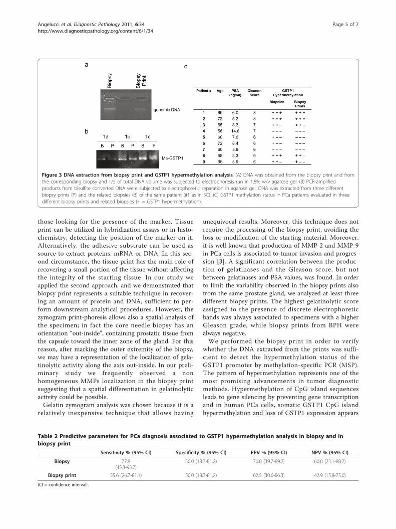
GSTP1 methylation analysis of biopsy printBiopsy print can be used as source for DNA. At least 30ng of genomic DNA from each biopsy print were recov-ered by extraction with proteinase K and its integrityand purity were comparable to DNA extracted by corre-sponding biopsy (Figure 3a). DNA was subjected tobisulfite conversion and clean-up and then amplified byPCR using specific primers for hypermethylated regionsin the promoter of GSTP1 gene. The electrophoreticrun of PCR products determined the presence of aunique amplified band that was of the same size in
biopsies and in biopsy prints (Figure 3b). We analyzedfor the presence of hypermethylated GSTP1 27 biopsiesderived from 9 different patients and the corresponding27 biopsy prints (Figure 3c). Moreover we analyzed 9biopsies and corresponding biopsy prints from 3 differ-ent BPH, but all these samples resulted negative (datanot shown). The concordance between biopsies andbiopsy prints was of 85% (23/27). The discordanceswere observed only in 4 biopsy prints obtained by posi-tive biopsies and that resulted negative. According tothe data obtained, and considering BPH (6 patients) asnegative controls, we compared the sensitivity, specifi-city, positive predictive value (PPV) and negative predic-tive value (NPV) of the biopsies and of the biopsy printsbased on the three needle biopsies, performed on thearea of the tumor, obtained for each of the 18 patientswith an histopathological diagnosis of PCa after RRP(Table 2). These parameters were calculated consideringpositive those patients who had at least one biopsy (orbiopsy print) positive for GSTP1 hypermethylation. Thespecificity of the two procedures resulted identical whilethe PPVs were comparable.
DiscussionOur study indicates that prostate core needle biopsymay be printed on a nitrocellulose membrane obtaininga cellular micropeel useful for several analytical proce-dures. Tissue print is easy and rapid to be performedwithout damaging the tissue, and it has been applied innumerous cellular models and with different experimen-tal objectives.Besides the technical application used, studies with tis-
sue print can be divided into two categories: thosefocused on the spatial localization of the marker, and
Figure 2 Zymogram analysis of biopsy prints . Twoexemplificative gels, each one containing two tissue prints visible asdark strips in the stacking gel, are shown. The presence of discretegelatinolytic activity is indicated on the left with the molecularweight or with the corresponding gelatinase. On the right thegelatinolytic pattern observed in conditioned medium from PC3cells.
Table 1 Gelatinolytic score calculated for biopsy prints of PCa and BPH biopsies
Gleason Score PSA (ng/ml) Biopsy MMP9+MMP2 score (BS) Gelatinolytic score (GS) Correlation
PCa 9 7.0 5+2 4 BS vs GS = 0.97 * Gleason vs GS = 0.86 *PSA (PCa) vs GS = 0.02PSA (all) vs GS = 0.36
PCa 8 6.0 5+1 4
PCa 8 14.2 3+2 3
PCa 7 8.3 3+0 2
PCa 6 26.0 3+1 2
PCa 6 8.2 2+0 0
PCa 6 14.8 3+1 2
PCa 6 5.9 0 0
PCa 6 10.5 1+1 1
BPH – 3.0 0 0
BPH – 0.4 0 0
BPH – 3.2 0 0
In the corresponding needle biopsies MMP2 and MMP9 expressions were evaluated by immunohistochemistry and a semiquantitative score (BS) was calculated.Correlation coefficients between BS and GS, between GS and Gleason score and between GS and PSA are shown. The correlation coefficient was calculated forall PSA values or only for PSA values from PCa patients (* p< 0.01)
Angelucci et al. Diagnostic Pathology 2011, 6:34http://www.diagnosticpathology.org/content/6/1/34
Page 4 of 7
those looking for the presence of the marker. Tissueprint can be utilized in hybridization assays or in histo-chemistry, detecting the position of the marker on it.Alternatively, the adhesive substrate can be used assource to extract proteins, mRNA or DNA. In this sec-ond circumstance, the tissue print has the main role ofrecovering a small portion of the tissue without affectingthe integrity of the starting tissue. In our study weapplied the second approach, and we demonstrated thatbiopsy print represents a suitable technique in recover-ing an amount of protein and DNA, sufficient to per-form downstream analytical procedures. However, thezymogram print-phoresis allows also a spatial analysis ofthe specimen; in fact the core needle biopsy has anorientation “out-inside”, containing prostatic tissue fromthe capsule toward the inner zone of the gland. For thisreason, after marking the outer extremity of the biopsy,we may have a representation of the localization of gela-tinolytic activity along the axis out-inside. In our preli-minary study we frequently observed a nonhomogeneous MMPs localization in the biopsy printsuggesting that a spatial differentiation in gelatinolyticactivity could be possible.Gelatin zymogram analysis was chosen because it is a
relatively inexpensive technique that allows having
unequivocal results. Moreover, this technique does notrequire the processing of the biopsy print, avoiding theloss or modification of the starting material. Moreover,it is well known that production of MMP-2 and MMP-9in PCa cells is associated to tumor invasion and progres-sion [3]. A significant correlation between the produc-tion of gelatinases and the Gleason score, but notbetween gelatinases and PSA values, was found. In orderto limit the variability observed in the biopsy prints alsofrom the same prostate gland, we analyzed at least threedifferent biopsy prints. The highest gelatinolytic scoreassigned to the presence of discrete electrophoreticbands was always associated to specimens with a higherGleason grade, while biopsy prints from BPH werealways negative.We performed the biopsy print in order to verify
whether the DNA extracted from the prints was suffi-cient to detect the hypermethylation status of theGSTP1 promoter by methylation-specific PCR (MSP).The pattern of hypermethylation represents one of themost promising advancements in tumor diagnosticmethods. Hypermethylation of CpG island sequencesleads to gene silencing by preventing gene transcriptionand in human PCa cells, somatic GSTP1 CpG islandhypermethylation and loss of GSTP1 expression appears
Table 2 Predictive parameters for PCa diagnosis associated to GSTP1 hypermethylation analysis in biopsy and inbiopsy print
Sensitivity % (95% CI) Specificity % (95% CI) PPV % (95% CI) NPV % (95% CI)
Biopsy 77.8(45.3-93.7)
50.0 (18.7-81.2) 70.0 (39.7-89.2) 60.0 (23.1-88.2)
Biopsy print 55.6 (26.7-81.1) 50.0 (18.7-81.2) 62.5 (30.6-86.3) 42.9 (15.8-75.0)
(CI = confidence interval).
Figure 3 DNA extraction from biopsy print and GSTP1 hypermethylation analysis. (A) DNA was obtained from the biopsy print and fromthe corresponding biopsy and 1/5 of total DNA volume was subjected to electrophoresis run in 1.8% w/v agarose gel. (B) PCR-amplifiedproducts from bisulfite converted DNA were subjected to electrophoretic separation in agarose gel. DNA was extracted from three differentbiopsy prints (P) and the related biopsies (B) of the same patient (#1 as in 3C). (C) GSTP1 methylation status in PCa patients evaluated in threedifferent biopsy prints and related biopsies (+ = GSTP1 hypermethylation).
Angelucci et al. Diagnostic Pathology 2011, 6:34http://www.diagnosticpathology.org/content/6/1/34
Page 5 of 7
to be the most common and consistent genomeabnormality [10,11]. In our study, we obtained DNAamounts sufficient for MSP in all 27 PCa biopsy printsexamined. We detected hypermethylation in GSTP1promoter in 78% of patients (7/9), a value that is similarto those observed by other authors in PCa tissue [12].The sensitivity of biopsy print-MSP resulted lower thanthose of biopsy-MSP (56% vs 79%). The decrease in sen-sitivity is due to a lower number of hypermethylation-positive biopsy prints respect to positive biopsies. Thisresult should be probably accounted to the fact that thetissue print is a partial representation of the biopsy, andit collects DNA just from a limited portion of the tissuebiopsy. For this reason if the printed tissue surface doesnot contain tumor cells or it contains tumor cells in avery low percentage, the tissue print may result negativealso in presence of a positive biopsy. The decrease insensitivity should be minimized by analyzing multiplebiopsy prints from the same patient. On the contrary,we did not observe false positive in biopsy print sam-ples, confirming the specificity of the biopsy analysis.Both urologists and histopathologist point of view,
who routinely work with us as a well integratedresearch group, underline that the future handling ofpatients with localized prostate cancer will undoubt-edly depend upon a more sophisticated prognosticationthan that available today. The basis will continue to bethe histopathological evaluation of tumor size, grade,localization and distribution within the gland unlessthe future need for objective techniques is well recog-nized. The option of combining an histopathologicalanalysis of prostate biopsies to molecular informationshould be of importance in improving the currentdiagnostic procedures available, particularly in uncer-tain cases. Focusing on tissue print of prostate biop-sies, the results of the current study are undoubtedlyinteresting unless need to be confirmed by the use inthe clinical practise to verify whether it is really usefulfor the diagnosis of PCa. Because of widely adoptedscreening programs for the early detection of prostatecancer, several patients who undergo RP are diagnosedwith tumors of small volume, and their extent and dis-tribution in the gland can only be determined by amicroscopic examination of the surgical specimen. His-torically, one of the most important predictor of can-cer control following RP is the absence of cancer atthe surgical margins. Tissue print may be able inincreasing specificity of the conventional histopathol-ogy mainly into the assessment of RP margins as it is amethod for obtaining molecular information about thecancer that can add to the macroscopic and micro-scopic anatomical findings.An important limitation of the present study is the
low number of cases examined. However, this study has
to be considered preliminary and performed in theattempt to check the methodology. In this regard we tryto point out the promising aspects of the proposedmethodology: first, biopsy print is a very simple andquick procedure that can be performed soon after thebiopsy. The resulting print can be stored at -20°C untilthe execution of the analytic procedure, while the coreneedle biopsy can be processed for the histological ana-lysis. Second, biopsy print resulted in an accurate copyof the needle biopsy offering reliable molecular informa-tion about the prostatic tissue. In conclusion, the appli-cation of biopsy print to a larger number of cases willclarify whether this promising technique should be use-ful in supporting the current procedures used for thediagnosis of PCa.
AcknowledgementsThe authors would like to thank dr Pomante R. for critical discussion aboutthe clinical interpretation of data. The authors declare that there is noconflict of interest that would prejudice the impartiality of the reportedresearch.
Author details1Department of Experimental Medicine, University of L’Aquila, L’Aquila,67100, Italy. 2Department of Health Sciences, University of L’Aquila, L’Aquila,67100, Italy.
Authors’ contributionsAA carried out the methylation studies and drafted the manuscript. GPcollected human specimens, performed the statistical analysis and helped todraft the manuscript. PS carried out the zymographies and prepared thefigures. CV was the coordinator of clinical management of patients andparticipated in the design of the study. MB was the scientific coordinatorand supervisor of the study and participated in its design. All authors readand approved the final manuscript.
Competing interestsThe authors declare that they have no competing interests.
Received: 12 January 2011 Accepted: 13 April 2011Published: 13 April 2011
References1. Scattoni V, Zlotta A, Montironi R, Schulman C, Rigatti P, Montorsi F:
Extended and saturation prostatic biopsy in the diagnosis andcharacterisation of prostate cancer: a critical analysis of the literature.Eur Urol 2007, 52:1309-1322.
2. de la Taille A, Jones JS, Klein EA: Open to debate. The motion: at least 18cores are necessary to make a prostatic biopsy useful. Eur Urol 2008,53:659-662, discussion 662.
3. Zucker S, Hymowitz M, Conner C, Zarrabi HM, Hurewitz AN, Matrisian L,Boyd D, Nicolson G, Montana S: Measurement of matrixmetalloproteinases and tissue inhibitors of metalloproteinases in bloodand tissues. Clinical and experimental applications. Ann N Y Acad Sci1999, 878:212-227.
4. Daoust R: Localization of deoxyribonuclease in tissue sections; a newapproach to the histochemistry of enzymes. Exp Cell Res 1957, 12:203-211.
5. Varner JE, Ye Z: Tissue printing. Faseb J 1994, 8:378-384.6. Hernandez Bronchud M, Webb S, Esiri MM: Brain blotting: a method to
detect multiple DNA copies in specific brain regions. J HistochemCytochem 1988, 36:1191-1195.
7. Lipkin WI, Villarreal LP, Oldstone MB: Whole animal section in situhybridization and protein blotting: new tools in molecular analysis ofanimal models for human disease. Curr Top Microbiol Immunol 1989,143:33-54.
Angelucci et al. Diagnostic Pathology 2011, 6:34http://www.diagnosticpathology.org/content/6/1/34
Page 6 of 7
8. Barres BA, Koroshetz WJ, Chun LL, Corey DP: Ion channel expression bywhite matter glia: the type-1 astrocyte. Neuron 1990, 5:527-544.
9. Gaston SM, Soares MA, Siddiqui MM, Vu D, Lee JM, Goldner DL, Brice MJ,Shih JC, Upton MP, Perides G, Baptista J, Lavin PT, Bloch BN, Genega EM,Rubin MA, Lenkinski RE: Tissue-print and print-phoresis as platformtechnologies for the molecular analysis of human surgical specimens:mapping tumor invasion of the prostate capsule. Nat Med 2005,11:95-101.
10. Lee WH, Morton RA, Epstein JI, Brooks JD, Campbell PA, Bova GS, Hsieh WS,Isaacs WB, Nelson WG: Cytidine methylation of regulatory sequences nearthe pi-class glutathione S-transferase gene accompanies humanprostatic carcinogenesis. Proc Natl Acad Sci USA 1994, 91:11733-11737.
11. Lin X, Tascilar M, Lee WH, Vles WJ, Lee BH, Veeraswamy R, Asgari K, Freije D,van Rees B, Gage WR, Bova GS, Isaacs WB, Brooks JD, DeWeese TL, DeMarzo AM, Nelson WG: GSTP1 CpG island hypermethylation isresponsible for the absence of GSTP1 expression in human prostatecancer cells. Am J Pathol 2001, 159:1815-1826.
12. Nakayama M, Gonzalgo ML, Yegnasubramanian S, Lin X, De Marzo AM,Nelson WG: GSTP1 CpG island hypermethylation as a molecularbiomarker for prostate cancer. J Cell Biochem 2004, 91:540-552.
doi:10.1186/1746-1596-6-34Cite this article as: Angelucci et al.: Tissue print of prostate biopsy: anovel tool in the diagnostic procedure of prostate cancer. DiagnosticPathology 2011 6:34.
Submit your next manuscript to BioMed Centraland take full advantage of:
• Convenient online submission
• Thorough peer review
• No space constraints or color figure charges
• Immediate publication on acceptance
• Inclusion in PubMed, CAS, Scopus and Google Scholar
• Research which is freely available for redistribution
Submit your manuscript at www.biomedcentral.com/submit
Angelucci et al. Diagnostic Pathology 2011, 6:34http://www.diagnosticpathology.org/content/6/1/34
Page 7 of 7