RESEARCH Open Access Low doses of esmolol and ... · Low doses of esmolol and phenylephrine act as...
8
RESEARCH Open Access Low doses of esmolol and phenylephrine act as diuretics during intravenous anesthesia Yu Hong Li 1 , Hai Bin Zhu 2 , Xiaozhu Zheng 3 , Han Jian Chen 1 , Liang Shao 4 and Robert G Hahn 5* Abstract Introduction: The renal clearance of infused crystalloid fluid is very low during anaesthesia and surgery, but experiments in conscious sheep indicate that the renal fluid clearance might approach a normal rate when the adrenergic balance is modified. Methods: Sixty females (mean age, 32 years) undergoing laparoscopic gynecological surgery were randomized to control group and received only the conventional anesthetic drugs and 20 ml/kg of lactated Ringer’s over 30 mins. The others were also given an infusion of 50 μg/kg/min of esmolol (beta 1 -receptor blocker) or 0.01 μg/kg/min of phenylephrine (alpha 1 -adrenergic agonist) over 3 hours. The distribution and elimination of infused fluid were studied by volume kinetic analysis based on urinary excretion and blood hemoglobin level. Results: Both drugs significantly increased urinary excretion while heart rate and arterial pressure remained largely unaffected. The urine flows during non-surgery were 43, 147, and 176 ml in the control, esmolol, and phenylephrine groups, respectively (medians, P < 0.03). When surgery had started the corresponding values were 34, 65 and 61 ml (P < 0.04). At 3 hours, averages of 9%, 20%, and 25% of the infused volume had been excreted in the three groups (P < 0.01). The kinetic analyses indicated that both treatments slowed down the distribution of fluid from the plasma to the interstitial fluid space, thereby preventing hypovolemia. Conclusions: Esmolol doubled and phenylephrine almost tripled urinary excretion during anesthesia-induced depression of renal fluid clearance. Introduction During general anesthesia and surgery, renal clearance of crystalloid fluid is only 15% to 20% of that found in conscious volunteers [1-3]. If not carefully balanced, fluid therapy entails a risk of inducing edema. Slow elimination might be counteracted by manipulation of the adrenergic balance. In awake sheep, b 1 -receptor sti- mulation by isoprenaline caused fluid retention and excessive plasma volume expansion that closely resem- bles the kinetic situation during anesthesia and surgery. In contrast, a 1 -receptor stimulation by phenylephrine increased urinary excretion [4,5]. The aim of the present study was to examine to what degree the slow turnover of lactated Ringer’ s solution during anesthesia and surgery can be rectified by infus- ing esmolol (a b 1 -receptor blocker) or phenylephrine in patients undergoing laparoscopic gynecological surgery during intravenous anesthesia. Based on the previous animal studies [4,5], our hypothesis was that urinary excretion would increase in response to esmolol and phenylephrine. Kinetic analysis was also performed to elucidate whether the infusions cause an aberrant distri- bution of fluid. Safety reasons stipulated that the amounts of drug provided be low. The rate of infusion of phenylephrine represented only 1% of that used for hemodynamic sup- port. Esmolol was given in the starting dose recom- mended for the management of supraventricular tachyarrhythmias. Materials and methods Patients Between November 2008 and October 2010, 60 Ameri- can Society of Anesthesiologists (ASA) I patients between 21 and 49 (mean of 32) years of age were stu- died during elective laparoscopic removal of ovarian cysts or laparoscopic supracervical hysterectomy (uterus * Correspondence: [email protected] 5 Section for Anesthesia, Linköping University, 581 85 Linköping, Sweden Full list of author information is available at the end of the article Li et al. Critical Care 2012, 16:R18 http://ccforum.com/content/16/1/R18 © 2011 Li et al.; licensee BioMed Central Ltd. This is an open access article distributed under the terms of the Creative Commons Attribution License (http://creativecommons.org/licenses/by/2.0), which permits unrestricted use, distribution, and reproduction in any medium, provided the original work is properly cited.
Transcript of RESEARCH Open Access Low doses of esmolol and ... · Low doses of esmolol and phenylephrine act as...

RESEARCH Open Access
Low doses of esmolol and phenylephrine act asdiuretics during intravenous anesthesiaYu Hong Li1, Hai Bin Zhu2, Xiaozhu Zheng3, Han Jian Chen1, Liang Shao4 and Robert G Hahn5*
Abstract
Introduction: The renal clearance of infused crystalloid fluid is very low during anaesthesia and surgery, butexperiments in conscious sheep indicate that the renal fluid clearance might approach a normal rate when theadrenergic balance is modified.
Methods: Sixty females (mean age, 32 years) undergoing laparoscopic gynecological surgery were randomized tocontrol group and received only the conventional anesthetic drugs and 20 ml/kg of lactated Ringer’s over 30 mins.The others were also given an infusion of 50 μg/kg/min of esmolol (beta1-receptor blocker) or 0.01 μg/kg/min ofphenylephrine (alpha1-adrenergic agonist) over 3 hours. The distribution and elimination of infused fluid werestudied by volume kinetic analysis based on urinary excretion and blood hemoglobin level.
Results: Both drugs significantly increased urinary excretion while heart rate and arterial pressure remained largelyunaffected. The urine flows during non-surgery were 43, 147, and 176 ml in the control, esmolol, andphenylephrine groups, respectively (medians, P < 0.03). When surgery had started the corresponding values were34, 65 and 61 ml (P < 0.04). At 3 hours, averages of 9%, 20%, and 25% of the infused volume had been excreted inthe three groups (P < 0.01). The kinetic analyses indicated that both treatments slowed down the distribution offluid from the plasma to the interstitial fluid space, thereby preventing hypovolemia.
Conclusions: Esmolol doubled and phenylephrine almost tripled urinary excretion during anesthesia-induceddepression of renal fluid clearance.
IntroductionDuring general anesthesia and surgery, renal clearanceof crystalloid fluid is only 15% to 20% of that found inconscious volunteers [1-3]. If not carefully balanced,fluid therapy entails a risk of inducing edema. Slowelimination might be counteracted by manipulation ofthe adrenergic balance. In awake sheep, b1-receptor sti-mulation by isoprenaline caused fluid retention andexcessive plasma volume expansion that closely resem-bles the kinetic situation during anesthesia and surgery.In contrast, a1-receptor stimulation by phenylephrineincreased urinary excretion [4,5].The aim of the present study was to examine to what
degree the slow turnover of lactated Ringer’s solutionduring anesthesia and surgery can be rectified by infus-ing esmolol (a b1-receptor blocker) or phenylephrine inpatients undergoing laparoscopic gynecological surgery
during intravenous anesthesia. Based on the previousanimal studies [4,5], our hypothesis was that urinaryexcretion would increase in response to esmolol andphenylephrine. Kinetic analysis was also performed toelucidate whether the infusions cause an aberrant distri-bution of fluid.Safety reasons stipulated that the amounts of drug
provided be low. The rate of infusion of phenylephrinerepresented only 1% of that used for hemodynamic sup-port. Esmolol was given in the starting dose recom-mended for the management of supraventriculartachyarrhythmias.
Materials and methodsPatientsBetween November 2008 and October 2010, 60 Ameri-can Society of Anesthesiologists (ASA) I patientsbetween 21 and 49 (mean of 32) years of age were stu-died during elective laparoscopic removal of ovariancysts or laparoscopic supracervical hysterectomy (uterus
* Correspondence: [email protected] for Anesthesia, Linköping University, 581 85 Linköping, SwedenFull list of author information is available at the end of the article
Li et al. Critical Care 2012, 16:R18http://ccforum.com/content/16/1/R18
© 2011 Li et al.; licensee BioMed Central Ltd. This is an open access article distributed under the terms of the Creative CommonsAttribution License (http://creativecommons.org/licenses/by/2.0), which permits unrestricted use, distribution, and reproduction inany medium, provided the original work is properly cited.
less than 1 kg) performed under intravenous generalanesthesia. The protocol (reference number 080186),including the sampled blood volume of approximately75 mL, was approved by the ethics committee of Zhe-jiang University (Hangzhou, China). Each patient gaveher informed consent to participate.
ProcedurePatients fasted overnight. No premedication was given.A radial artery catheter was inserted for sampling andfor monitoring arterial blood pressure. Anesthesia wasinduced with midazolam 50 μg/kg, propofol 1.5 mg/kg,cis-atracurium 0.15 mg/kg, and fentanyl 3 μg/kg andthis was followed by endotracheal intubation andmechanical ventilation. The anesthesia was maintainedwith propofol 6 mg/kg per hour and cis-atracurium 0.15mg/kg per hour; fentanyl was infused at a rate of 2 μg/kg per hour during the first 30 minutes and then at 1.5μg/kg per minute; these rates could be slightly adjusteddepending on the depth of anesthesia.An indwelling catheter was placed into the bladder.
The patients were then randomly assigned by the Excelrandom number generator (Microsoft Corporation,Redmond, WA, USA) to receive a 3-hour continuousinfusion of 10 mL/hour lactated Ringer’s solution thatprovided no drug (controls); 50 μg/kg per minute of theb1-receptor blocker esmolol (Qilu Pharmaceutical Co.,Shandong, China); or 0.01 μg/kg per minute of the alpha1adrenergic receptor agonist phenylephrine (Hefeng Phar-maceutical Co., Shanghai, China). The line was primed toprovide the drug from the first minute. The anesthesiaand the research were managed by two anesthesiologistsblinded to the study drug.After 10 minutes for adjustment to the effects of the
drug, plasma volume expansion was induced by infusing20 mL/kg of lactated Ringer’s solution (Pharmacia-Baxter, Shanghai, China) over the course of 30 minutesvia an infusion pump. No other fluid (except drug vehi-cles) was given. No study data were collected aftertermination of the anesthesia.
MeasurementsMonitoring included pulse oximetry, electrocardiogra-phy, heart rate, and invasive mean arterial pressure(MAP). Data were displayed on a multifunction monitor(Datex-Ohmeda, Hoevelaken, The Netherlands) andsaved digitally. The depth of anesthesia was monitoredby a bispectral index (BIS) sensor applied to the fore-head. The signal was recorded on a BIS monitor ModelA-2000TM (Aspect Medical System, Natick, MA, USA).Urine output was measured every 20 minutes during thestudy. Arterial blood samples (2 mL each) were col-lected every 5 minutes during the first 60 minutes andevery 10 minutes during the following 120 minutes. The
blood hemoglobin (Hb) concentration was measured ona GEM Premier 3000 (Instrumentation Laboratory, Lex-ington, IL, USA). Duplicate samples collected at baselineensured a coefficient of variation of 1.5%. Blood loss wasassessed from the content of blood in suction bottlesand on sponges.
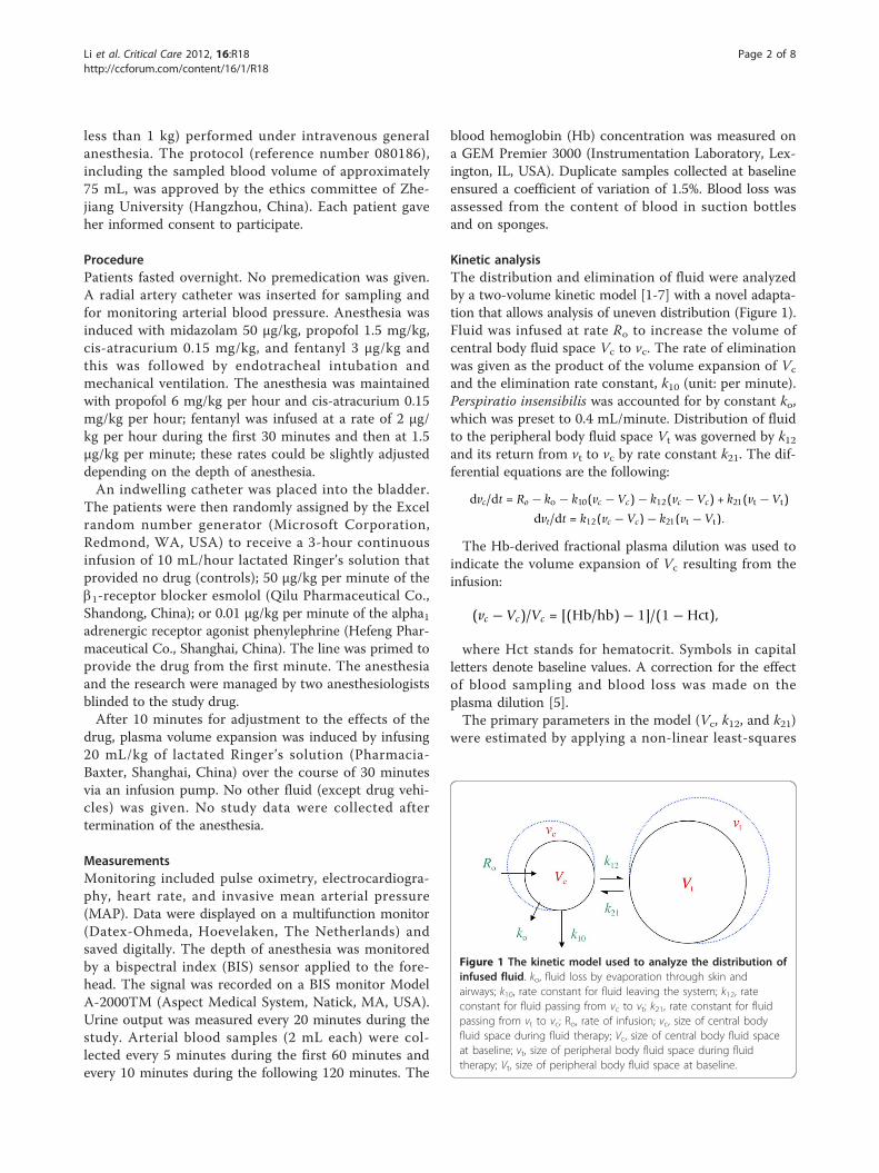
Kinetic analysisThe distribution and elimination of fluid were analyzedby a two-volume kinetic model [1-7] with a novel adapta-tion that allows analysis of uneven distribution (Figure 1).Fluid was infused at rate Ro to increase the volume ofcentral body fluid space Vc to vc. The rate of eliminationwas given as the product of the volume expansion of Vc
and the elimination rate constant, k10 (unit: per minute).Perspiratio insensibilis was accounted for by constant ko,which was preset to 0.4 mL/minute. Distribution of fluidto the peripheral body fluid space Vt was governed by k12and its return from vt to vc by rate constant k21. The dif-ferential equations are the following:
dvc/dt = Ro − ko − k10(vc − Vc)− k12(vc − Vc) + k21(vt − Vt)
dvt/dt = k12(vc − Vc)− k21(vt − Vt).
The Hb-derived fractional plasma dilution was used toindicate the volume expansion of Vc resulting from theinfusion:
(vc − Vc)/Vc = [(Hb/hb)− 1]/(1−Hct),
where Hct stands for hematocrit. Symbols in capitalletters denote baseline values. A correction for the effectof blood sampling and blood loss was made on theplasma dilution [5].The primary parameters in the model (Vc, k12, and k21)
were estimated by applying a non-linear least-squares
Figure 1 The kinetic model used to analyze the distribution ofinfused fluid. ko, fluid loss by evaporation through skin andairways; k10, rate constant for fluid leaving the system; k12, rateconstant for fluid passing from vc to vt; k21, rate constant for fluidpassing from vt to vc; Ro, rate of infusion; vc, size of central bodyfluid space during fluid therapy; Vc, size of central body fluid spaceat baseline; vt, size of peripheral body fluid space during fluidtherapy; Vt, size of peripheral body fluid space at baseline.
Li et al. Critical Care 2012, 16:R18http://ccforum.com/content/16/1/R18
Page 2 of 8

regression routine (fminsearch) by using Matlab R2010asoftware (The MathWorks, Inc., Natick, MA, USA),whereas k10 was calculated as a secondary parameter asfollows:
k10 =∑
urine volume/AUC for (vc - Vc),
where AUC is the area under the curve. The renalclearance is the product of Vc and k10.
Statistical analysisThe study was powered (80%) to detect a doubling ofurinary excretion in any of the study groups as com-pared with the controls on the basis of the ratiobetween the excreted urine and infused fluid previouslyfound during surgery [2,3]. An interim analysis wasmade after 42 patients.Data are presented as the median and 25th to 75th
percentile range. Comparisons between the three groupswere made by the Kruskal-Wallis test, which, if signifi-cant, this is so. was followed by the Mann-Whitney Utest for post hoc analysis. Changes were assessed by theWilcoxon matched-pair test. Correlations between vari-ables were studied by simple and multiple regressionanalysis by using square root transformation if distribu-tion was skewed. A P value of less than 0.05 was consid-ered statistically significant.
ResultsDemographics, hemodynamics, and anesthesiaThere were no differences among the three study groupswith respect to baseline parameters, operating time,blood loss (Table 1), and heart rate (Figure 2, left).Esmolol infusion was followed by a modest decrease inMAP during surgery in comparison with baseline (P <0.01), whereas no change versus baseline occurred inthe other two groups (Figure 2, middle). During surgery,MAP was 5 mm Hg higher in the phenylephrine groupthan in the esmolol group (P < 0.01). Phenylephrine wasalso associated with a slightly higher overall MAP thanthe other groups (Table 1). The administered amountsof anesthetic drugs were similar between the groups, butthe esmolol patients received less fentanyl than theothers between 60 and 120 minutes (Table 2). The BISrecordings were virtually identical (Figure 2, right).
Plasma dilutionThe decrease in Hb concentration during the infusionsaveraged 25 g/L. There were no differences in Hb-derived plasma dilution at 30, 60, or 120 minutes, butboth treatment drugs were associated with a slightlyless pronounced plasma dilution at 180 minutes incomparison with the controls (both differences P <0.01) (Table 3).
Urinary excretionThe cumulative urinary excretion was significantly largerin the two treatment groups than in the control groupfrom 60 minutes onward. At 180 minutes, both treat-ments had more than doubled urinary excretion and theratio between excreted and infused fluid volumes (Krus-kal-Wallis test, P < 0.01) (Figure 3 and Table 4). Study-ing only the periods of non-surgery and surgery alsorevealed that the urinary excretion was larger and theurine flow higher in the treatment groups comparedwith the control group (Table 4).
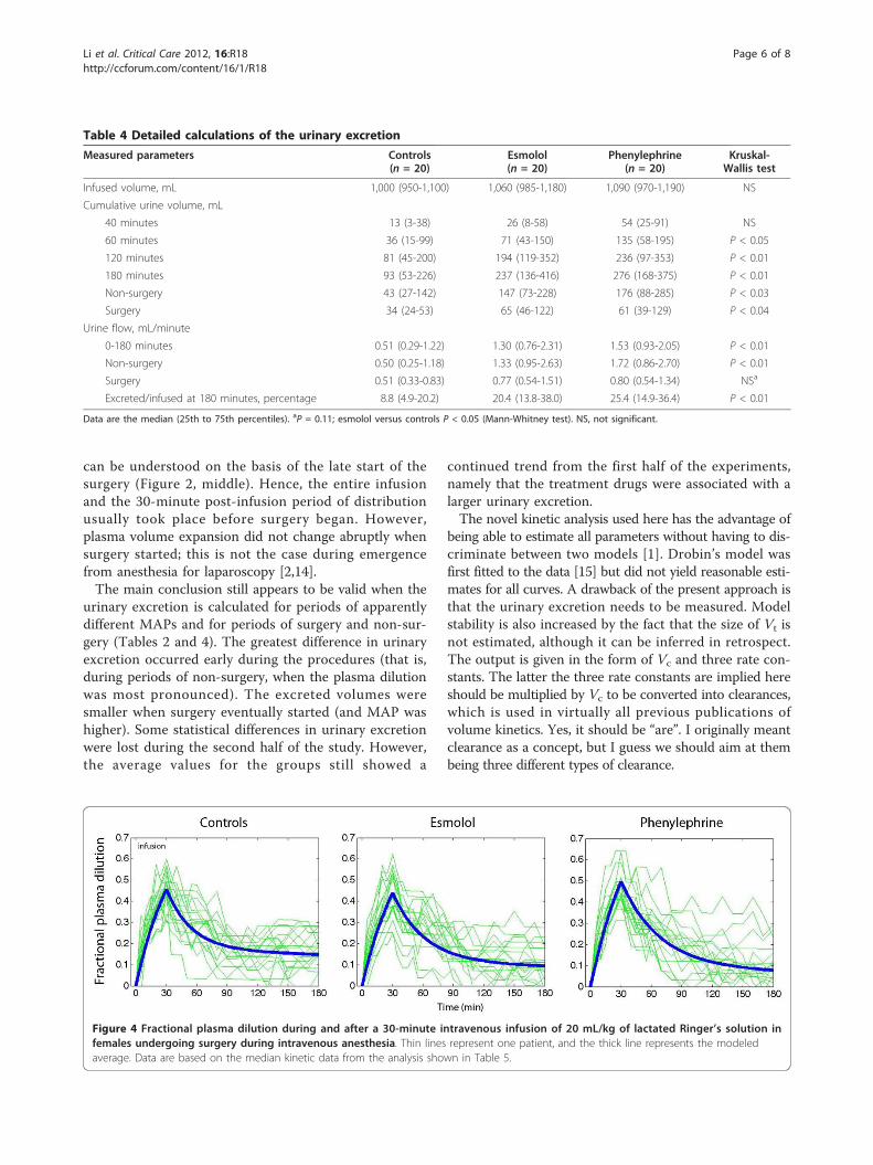
Kinetic analysisFigure 4 shows all individual plasma dilution-time pro-files together with the optimal curve fit for each group.The three model parameters - Vc, k12, and k21 - couldbe estimated in all experiments (Table 5). The elimina-tion rate parameter, k10, was higher in the two treatmentgroups (P < 0.03 and P < 0.01) than in the controlgroup. Moreover, k21 was lower in the phenylephrinegroup than in the controls (P < 0.02).Simulations of the fluid distribution showed that the
volume expansion of vc during the infusion was similaramong the groups (Figure 5, top row) and virtually iden-tical on correction for body weight. However, at 180minutes, less fluid resided in vc in the phenylephrinegroup (90 mL) than in the control and esmolol groups(P < 0.03).Fluid was distributed more slowly to vt in the two
treatment groups. At 60 minutes, 59% of the fluidresided in vt in the control group, 48% in the esmolol
Table 1 Baseline characteristics of patients andoperations
Parameter Controls(n = 20)
Esmolol(n = 20)
Phenylephrine(n = 20)
Kruskal-Wallistest
Age, years 32 (27-38) 32 (28-39) 33 (27-41) NS
Body weight, kg 50 (48-55) 53 (49-59) 55 (48-60) NS
Start of surgery,minutes
70 (65-80) 70 (70-80) 80 (60-90) NS
End of surgery,minutes
130 (115-155)
150 (130-180)
150 (130-180) NS
Operating time,minutes
60 (40-80) 75 (50-100)
70 (50-100) NS
Blood loss, mLa 30 (30-55) 35 (25-70) 40 (30-70) NS
MAP, mm Hg
Baseline 88 (81-94) 92 (87-102)
92 (88-98) NS
Non-surgery 77 (68-80) 78 (73-85) 83 (75-93) NS
Surgery 92 (87-96) 90 (84-92) 95 (85-94) P < 0.03
5 to 180minutes
82 (59-86) 82 (80-90) 92 (85-94) P < 0.02
Data are the median (25th to 75th percentile) for the group. aCollected bysuction and on sponges, not including blood in removed tissue. MAP, meanarterial pressure; NS, not significant.
Li et al. Critical Care 2012, 16:R18http://ccforum.com/content/16/1/R18
Page 3 of 8

group, and 43% in the phenylephrine group (P < 0.01;controls versus phenylephrine P < 0.004) (Figure 5, mid-dle row). At 180 minutes, the volume in vt did not differsignificantly. The kinetic model slightly overestimatedurinary excretion early in the operations (Figure 5, bot-tom row).
Linear regression analysisSimple regression showed that MAP explained 11% ofthe variability in the urinary excretion (P < 0.02). How-ever, multiple regression of the effects of MAP and thetwo treatments in turn showed that only the treatmentdrugs served as statistically significant predictors of theurinary excretion and the ratio of excreted/infused fluid.
DiscussionShifting the catecholamine balance by small amounts ofdrug promoted urinary excretion. In the present study,esmolol doubled and phenylephrine almost tripled theexcretion of infused lactated Ringer’s during laparo-scopic surgery. Despite the diuretic effect, the modifica-tion of the adrenergic stimulus pattern was not strongenough to clearly affect MAP or heart rate.
The average ratio of excreted/infused fluid after 3hours in the control group (9%) was similar to thatfound for acetated Ringer’s during laparoscopic chole-cystectomy [2] and thyroid surgery [3]. However, evenwith the two treatment drugs, urinary excretion was stillfar below the 50% to 70% of the infused volume seenwithin 3 hours in unstressed volunteers [6] and patientsawaiting laparoscopic surgery [7]. Hence, the adminis-tered esmolol and phenylephrine were not sufficient toreverse the strong antidiuretic effect of general anesthe-sia and surgery. On the other hand, the present resultsopen up for the use of these drugs when both adrener-gic and diuretic effects are desired. How long the differ-ence in urinary excretion during surgery prevails isunknown. The rate is likely to increase slightly whenanesthesia is terminated [2], but much of the fluidretained during surgery remains in the body for severaldays [8]. In contrast, infusions of crystalloid salt [7] andglucose [9] solution initiated several hours later areexcreted effectively.Why the diuretic response to plasma volume expansion
is blunted during anesthesia and surgery is poorly under-stood but probably can be attributed to vasodilatation,
Figure 2 Heart rate, mean arterial pressure, and bispectral index in patients receiving esmolol, phenylephrine, or sham (controls). Dataare the median values.
Table 2 Doses of anesthetic drugs for each hour of the experiments
Measured parameters Controls(n = 20)
Esmolol(n = 20)
Phenylephrine(n = 20)
Kruskal-Wallis test
Propofol, mg
0-60 minutes 350 (279-350) 300 (300-337) 290 (250-300) NS
60-120 minutes 316 (291-358) 324 (290-384) 320 (254-400) NS
120-180 minutes 150 (125-225) 210 (168-249) 209 (169-271) NS
Fentanyl, μg
0-60 minutes 250 (250-300) 250 (213-250) 300 (250-300) NS
60-120 minutes 200 (200-250) 150 (150-200) 216 (150-268) P < 0.05
120-180 minutes 100 (100-125) 125 (100-150) 112 (100-150) NS
Cis-atracurium, mg
0-60 minutes 18 (15-20) 18 (15-20) 19 (15-20) NS
60-120 minutes 9 (8-10) 10 (8-10) 9 (7-10) NS
120-180 minutes 0 (0-0) 3 (0-5) 5 (3-5) P < 0.01
Data are the median (25th to 75th percentiles). aP = 0.11; phenylephrine versus controls P < 0.05 (Mann-Whitney test). NS, not significant.
Li et al. Critical Care 2012, 16:R18http://ccforum.com/content/16/1/R18
Page 4 of 8

reduced arterial pressure, and activation of the renin-aldosterone hormonal axis [1]. Beta-receptor activity mayalso play a part. The renal clearance of crystalloid fluid isslightly higher during open laparotomy [10] than duringlaparoscopy [2]; therefore, body position and pneumoper-itoneum might also depress the renal excretion of fluid.
The physiological effects of intravenous anesthesia alsopromote a pronounced plasma volume expansion inresponse to crystalloid fluid. When the infusions ended,60% to 70% of the infused fluid remained in the func-tional plasma volume, vc. Similarly high percentageshave been reported during isoflurane anesthesia [11]and during general anesthesia and surgery [2,3,10]. Pro-nounced plasma volume expansion during infusionimplies that Vc is low, and this is typical of anesthesia-induced vasodilatation [1]. Here, the small Vc can alsobe explained by the relatively low body weight (50 to 55kg) of the Chinese women studied. The study drugsmight have altered Vc, although the average volume inthe esmolol and phenylephrine groups differed very littlefrom that of the controls.A key finding of the kinetic analysis is that both study
drugs slowed down the distribution of fluid from vc tovt and this probably reflects minimally decreased filtra-tion due to vasoconstriction. This counteracted anyhypovolemic effect of the increased diuresis during thefirst half of the experiments. The fluid volumes residingin vc became reduced at the end of the study, but, atthat time, differences were hardly worth noting. Thefluid volumes in vt did not differ significantly at 180minutes (Figure 5).Variable results for the diuretic effect of esmolol in
previous work suggest that the present study may berelevant only to anesthetized subjects, possibly becausethe renin-aldosterone axis is then stimulated. Completeb1-receptor blockade in conscious humans lowers theresting plasma levels of renin, angiotensin, and aldoster-one but without affecting the diuretic response tovolume loading with saline [12]. In awake sheep, esmo-lol reduced the diuretic response to infused fluid [13].Moreover, more fluid tended to accumulate in the extra-vascular space [13] and this was not encountered duringintravenous anesthesia in humans (Figure 5).Phenylephrine increases the urinary excretion by pres-
sure diuresis if the increase in MAP is profound, butalso by increasing the release of atrial natriuretic pep-tides [4]. The small doses of drugs used in the presentstudy make the role of these mechanisms speculative.These doses are, in fact, also a limitation since we can-not outline the diuretic effects of larger amounts. Insheep, larger doses of phenylephrine markedly lower theplasma volume [4,5] but this was not an issue in thepresent study (Figures 4 and 5).Another limitation was that the operations were per-
formed on ASA I patients only. Therefore, the effects ofthe study drugs on patients with cardiovascular diseaseare unclear. Operations were also performed in a teach-ing hospital and this explains why more than 1 hourhad passed before the surgery started. The restoration ofbaseline MAP between 70 and 90 minutes in all groups
Table 3 Blood hemoglobin concentration and thehematocrit
Measured parameters Controls(n = 20)
Esmolol(n = 20)
Phenylephrine(n = 20)
Hematocrit at baseline,ratio
0.36 (0.35-0.39)
0.37 (0.34-0.39)
0.39 (0.37-0.31)
Hemoglobinconcentration, g/L
Baseline 112 (110-121)
113 (104-120)
121 (114-27)
30 minutes 87 (84-96) 87 (84-96) 95 (89-98)
60 minutes 96 (90-104) 95 (90-102) 102 (96-107)
120 minutes 102 (99-105) 105 (98-114) 109 (105-117)
180 minutes 102 (97-105) 105 (99-114) 112 (105-118)
Mean 5-180 minutes 98 (94-103) 99 (93-107) 105 (99-111)
Plasma dilution, no units
30 minutes 0.45 (0.43-0.51)
0.42 (0.38-0.54)
0.45 (0.42-0.49)
60 minutes 0.30 (0.25-0.35)
0.31 (0.25-0.37)
0.30 (0.25-0.34)
120 minutes 0.18 (0.13-0.20)
0.10 (0.07-0.17)
0.12 (0.08-0.22)
180 minutesa 0.19 (0.15-0.25)
0.09 (0.04-0.17)
0.11 (0.03-0.17)
Mean 5-180 minutes 0.28 (0.24-0.30)
0.24 (0.20-0.29)
0.22 (0.20-0.26)
Data are the median (25th to 75th percentiles). aP < 0.01 by the Kruskal-Wallistest.
Figure 3 Urinary excretion during 3 hours of anesthesia andsurgery. Twenty patients were in each group.
Li et al. Critical Care 2012, 16:R18http://ccforum.com/content/16/1/R18
Page 5 of 8
can be understood on the basis of the late start of thesurgery (Figure 2, middle). Hence, the entire infusionand the 30-minute post-infusion period of distributionusually took place before surgery began. However,plasma volume expansion did not change abruptly whensurgery started; this is not the case during emergencefrom anesthesia for laparoscopy [2,14].The main conclusion still appears to be valid when the
urinary excretion is calculated for periods of apparentlydifferent MAPs and for periods of surgery and non-sur-gery (Tables 2 and 4). The greatest difference in urinaryexcretion occurred early during the procedures (that is,during periods of non-surgery, when the plasma dilutionwas most pronounced). The excreted volumes weresmaller when surgery eventually started (and MAP washigher). Some statistical differences in urinary excretionwere lost during the second half of the study. However,the average values for the groups still showed a
continued trend from the first half of the experiments,namely that the treatment drugs were associated with alarger urinary excretion.The novel kinetic analysis used here has the advantage of
being able to estimate all parameters without having to dis-criminate between two models [1]. Drobin’s model wasfirst fitted to the data [15] but did not yield reasonable esti-mates for all curves. A drawback of the present approach isthat the urinary excretion needs to be measured. Modelstability is also increased by the fact that the size of Vt isnot estimated, although it can be inferred in retrospect.The output is given in the form of Vc and three rate con-stants. The latter the three rate constants are implied hereshould be multiplied by Vc to be converted into clearances,which is used in virtually all previous publications ofvolume kinetics. Yes, it should be “are”. I originally meantclearance as a concept, but I guess we should aim at thembeing three different types of clearance.
Table 4 Detailed calculations of the urinary excretion
Measured parameters Controls(n = 20)
Esmolol(n = 20)
Phenylephrine(n = 20)
Kruskal-Wallis test
Infused volume, mL 1,000 (950-1,100) 1,060 (985-1,180) 1,090 (970-1,190) NS
Cumulative urine volume, mL
40 minutes 13 (3-38) 26 (8-58) 54 (25-91) NS
60 minutes 36 (15-99) 71 (43-150) 135 (58-195) P < 0.05
120 minutes 81 (45-200) 194 (119-352) 236 (97-353) P < 0.01
180 minutes 93 (53-226) 237 (136-416) 276 (168-375) P < 0.01
Non-surgery 43 (27-142) 147 (73-228) 176 (88-285) P < 0.03
Surgery 34 (24-53) 65 (46-122) 61 (39-129) P < 0.04
Urine flow, mL/minute
0-180 minutes 0.51 (0.29-1.22) 1.30 (0.76-2.31) 1.53 (0.93-2.05) P < 0.01
Non-surgery 0.50 (0.25-1.18) 1.33 (0.95-2.63) 1.72 (0.86-2.70) P < 0.01
Surgery 0.51 (0.33-0.83) 0.77 (0.54-1.51) 0.80 (0.54-1.34) NSa
Excreted/infused at 180 minutes, percentage 8.8 (4.9-20.2) 20.4 (13.8-38.0) 25.4 (14.9-36.4) P < 0.01
Data are the median (25th to 75th percentiles). aP = 0.11; esmolol versus controls P < 0.05 (Mann-Whitney test). NS, not significant.
Figure 4 Fractional plasma dilution during and after a 30-minute intravenous infusion of 20 mL/kg of lactated Ringer’s solution infemales undergoing surgery during intravenous anesthesia. Thin lines represent one patient, and the thick line represents the modeledaverage. Data are based on the median kinetic data from the analysis shown in Table 5.
Li et al. Critical Care 2012, 16:R18http://ccforum.com/content/16/1/R18
Page 6 of 8

ConclusionsLow doses of esmolol doubled and phenylephrine almosttripled urinary excretion. These drugs may potentially beused as diuretics when renal fluid clearance is reduced byanesthesia and surgery-associated stress. Further work isneeded to elucidate how the diuretic effects of thesedrugs are best employed. Phenylephrine might be analternative to a conventional diuretic such as furosemideif the hemodynamics is unstable. Esmolol could be usedas a mild diuretic in patients with supraventriculararrhythmias.
Key messages• The clearance of crystalloid fluid is very low duringintravenous anesthesia.
Table 5 Kinetic parameters
Estimatedparameters
Controls(n = 20)
Esmolol(n = 20)
Phenylephrine(n = 20)
Kruskal-Wallistest
Vc, L 1.5 (1.1-1.8)
1.7 (1.2-2.1)
1.6 (1.2-1.8) NS
k12, 10-3/minute 28 (21-
48)22 (18-34)
18 (14-31) NS
k21, 10-3/minute 11 (4-23) 7 (2-17) 4 (0-11) P < 0.05
k10, 10-3/minute 2.1 (0.9-
4.2)4.2 (2.7-8.3)
4.7 (3.3-10.0) P < 0.02
Data are the median (25th to 75th percentile) for the group during the entireexperiment (0 to 180 minutes). k10, rate constant for fluid leaving the system;k12, rate constant for fluid passing from vc to vt; k21, rate constant for fluidpassing from vt to vc; NS, not significant; vc, size of central body fluid spaceduring fluid therapy; Vc, size of central body fluid space at baseline; vt, size ofperipheral body fluid space during fluid therapy.
Figure 5 Simulated volume-time curves for the central (Vc) and peripheral (Vt) fluid spaces and urinary excretion. Thin lines aresimulations based on k12, k21, and k10 from each operation, and thick lines are the median for each group. The points in the lower panel are themeasured urinary excretion (median values). k10, rate constant for fluid leaving the system; k12, rate constant for fluid passing from vc to vt; k21,rate constant for fluid passing from vt to vc; vc, size of central body fluid space during fluid therapy; Vc, size of central body fluid space atbaseline; vt, size of peripheral body fluid space during fluid therapy; Vt, size of peripheral body fluid space at baseline.
Li et al. Critical Care 2012, 16:R18http://ccforum.com/content/16/1/R18
Page 7 of 8

• Small doses of esmolol and phenylephrine increasethe clearance but not up to the normal values forconscious volunteers.• These drugs at least doubled the urinary excretionin comparison with controls.• The increased diuresis did not cause hypovolemiaas both esmolol and phenylephrine slowed down therate of distribution of lactated Ringer’s from plasmato interstitium.
AbbreviationsHb: hemoglobin; MAP: mean arterial pressure; ko: fluid loss by evaporationthrough skin and airways; k10: rate constant for fluid leaving the system; k12:rate constant for fluid passing from vc to vt; k21: rate constant for fluidpassing from vt to vc; Ro: rate of infusion; vc: size of central body fluid spaceduring fluid therapy; Vc: size of central body fluid space at baseline; vt: sizeof peripheral body fluid space during fluid therapy; Vt: size of peripheralbody fluid space at baseline.
AcknowledgementsTobias Gebäck, of the Chalmers School of Technology, Gothenburg, Sweden,created the computer programs.
Author details1Department of Anesthesiology, First Affiliated Hospital, College of Medicine,Zhejiang University, 79 Qingchun Road, 310003 Hangzhou, China.2Department of Obstetrics and Gynecology, First Affiliated Hospital, Collegeof Medicine, Zhejiang University, 79 Qingchun Road, 310003 Hangzhou,China. 3Zhejiang Hospital, 12 Linying Road, 310013 Hangzhou, China.4Yuhuan County People’s Hospital, Changle Road, Yuhan County, 317600Taizhou City, Zhejiang, China. 5Section for Anesthesia, Linköping University,581 85 Linköping, Sweden.
Authors’ contributionsYHL wrote the ethics application, organized the study, and collected data.HBZ, XZ, HJC, and LS collected data and performed the operations. RGHdesigned the trial, performed the calculations, and wrote the manuscript. Allauthors read and approved the final manuscript.
Competing interestsThe authors declare that they have no competing interests.
Received: 27 July 2011 Revised: 1 January 2012Accepted: 30 January 2012 Published: 30 January 2012
References1. Hahn RG: Volume kinetics of infusion fluids. Anesthesiology 2010,
113:470-481.2. Olsson J, Svensén CH, Hahn RG: The volume kinetics of acetated Ringer’s
solution during laparoscopic cholecystectomy. Anesth Analg 2004,99:1854-1860.
3. Ewaldsson C-A, Hahn RG: Kinetics and extravascular retention of acetatedRinger’s solution during isoflurane and propofol anesthesia for thyroidsurgery. Anesthesiology 2005, 103:460-469.
4. Vane LA, Prough DS, Kinsky MA, Willimas CA, Grady JJ, Kramer GC: Effectsof different catecholamines on the dynamics of volume expansion ofcrystalloid infusion. Anesthesiology 2004, 101:1136-1144.
5. Ewaldsson C-A, Vane LA, Kramer GC, Hahn RG: Catecholamines alter boththe fluid kinetics and the hemodynamic responses to volume expansionin sheep. J Surg Res 2006, 131:7-14.
6. Drobin D, Hahn RG: Kinetics of isotonic and hypertonic plasma volumeexpanders. Anesthesiology 2002, 96:1371-1380.
7. Borup T, Hahn RG, Holte K, Ravn L, Kehlet H: Intraoperative colloidadministration increases the clearance of a postoperative fluid load. ActaAnesthesiol Scand 2009, 53:311-317.
8. Brandstrup B, Tonnesen H, Beier-Holgersen R Hjortsø E, Ørding H, Lindorff-Larsen K, Rasmussen MS, Lanng C, Wallin L, Iversen LH, Gramkow CS,
Okholm M, Blemmer T, Svendsen PE, Rottensten HH, Thage B, Riis J,Jeppesen IS, Teilum D, Christensen AM, Graungaard B, Pott F, Danish StudyGroup on Perioperative Fluid Therapy: Effects of intravenous fluidrestriction on postoperative complications: comparison of twoperioperative fluid regimens. A randomized assessor-blinded multicentertrial. Ann Surg 2003, 238:641-648.
9. Strandberg P, Hahn RG: Volume kinetics of glucose 2.5% given byintravenous infusion after hysterectomy. Br J Anaesth 2005, 94:30-38.
10. Svensén CH, Olsson J, Hahn RG: Intravascular fluid administration andhemodynamic performance during open abdominal surgery. AnesthAnalg 2006, 103:671-676.
11. Norberg Å, Hahn RG, Li H, Olsson J, Prough DS, Börsheim E, Wolf S,Minton R, Svensén CH: Population volume kinetics predicts retention of0.9% saline infused in awake and isoflurane-anesthetized volunteers.Anesthesiology 2007, 107:24-32.
12. Molstrom S, Larsen NH, Simonsen JA, Washington R, Bie P: Normotensivesodium loading in normal man: regulation of renin secretion during β-receptor blockade. Am J Physiol Regul Integr Comp Physiol 2009, 206:R436-R445.
13. Kinsky MP, Vaid SU, Vane LA, Prough DS, Kramer GC: Effect of esmolol onfluid therapy in normovolemia and hypovolemia. Shock 2008, 30:55-63.
14. Sjöstrand F, Hahn RG: Volume kinetics of 2.5% glucose solution duringlaparoscopic cholecystectomy. Br J Anesth 2004, 92:485-492.
15. Drobin D: A single-model solution for volume kinetic analysis of isotonicfluid infusions. Acta Anaesthesiol Scand 2006, 50:1074-1080.
doi:10.1186/cc11175Cite this article as: Li et al.: Low doses of esmolol and phenylephrineact as diuretics during intravenous anesthesia. Critical Care 2012 16:R18.
Submit your next manuscript to BioMed Centraland take full advantage of:
• Convenient online submission
• Thorough peer review
• No space constraints or color figure charges
• Immediate publication on acceptance
• Inclusion in PubMed, CAS, Scopus and Google Scholar
• Research which is freely available for redistribution
Submit your manuscript at www.biomedcentral.com/submit
Li et al. Critical Care 2012, 16:R18http://ccforum.com/content/16/1/R18
Page 8 of 8