RESEARCH Open Access Comparison of ... - Head & Face Medicine
7
RESEARCH Open Access Comparison of pre-bent titanium mesh versus polyethylene implants in patient specific orbital reconstructions Marcin Kozakiewicz and Piotr Szymor * Abstract Introduction: Computerized tomography DICOM file can be relatively easily transformed to a virtual 3D model. With the help of additional software we are able to create the mirrored model of an undamaged orbit and on this basis produce an individual implant for the patient Authors decided to apply implants with any thickness, which are authors own invention to obtain volumetric support and more stable orbital wall reconstruction outcome. Material of choice was ultra-high molecular weight polyethylene (UHMWPE). Objective: The aim of this study was to present and compare functional results of individual reconstructions of orbital wall using either titanium mesh or ultra-high molecular weight polyethylene. Materials and methods: 57 consecutive patients affected by orbital wall fracture (46 males, 11 females, mean age 34±14 year) were treated in Department of Maxillofacial Surgery from 2010 to 2012. In the first group we used patient specific treatment by titanium mesh shaped on a 3D printed model of a mirrored intact orbit (37 orbits) or by individually manufactured UHMW-PE implantby CAM milling in second group (20 orbits). All of these patients were subjected to preoperative helical computerized tomography and consultation of an ophthalmologist (including binocular single vision loss test - BSVL). Further on, patients were operated under general anaesthesia using transconjuctival approach. BSVL was again evaluated post-operationally in 1 month and 6 months later. Results: Functional treatment results (BSVL) for both groups were similar in 1 month as well as 6 months post operational time. There was no statistically significant difference between these two groups. Conclusions: This study of 6 months functional result assessment of pre-bent individual implants and CNC milled ultra-high molecular weight polyethylene of the orbital wall has shown it to be a predictable reconstruction method. Individually shaped UHMWPE seems to be as good as pre-bent titanium mesh. Keywords: Facial skeleton injuries, Individual implants, Titanium mesh, Ultrahigh molecular weight polyethylene, Diplopia Introduction Orbital wall fractures are quite common consequence of maxillofacial trauma. 3 [1]-32% [2] of all maxillofacial fractures are present within the orbit. Although aetiology of facial trauma varies in different countries the most common causes worldwide are assault, traffic accident, sports and fall. Common complications of such fractures are diplopia and enophtalmos [3,4]. In 2000s individual reconstructions in maxillofacial surgery begun to be more popular [5-12]. Technical equipment and software is more available nowadays. Computerized tomography DICOM file can be rela- tively easily transformed to a virtual model [8,13]. With the help of additional software the mirrored model of an undamaged orbit can be created and exported as a . stl file for external 3D printer. On such printed model it is possible to bend titanium mesh preoperatively. This allows to reduce operating time, improve safety [13,14] and achieve much better accuracy of orbital reconstruc- tion [9,10,15]. However, sometimes it is observed that * Correspondence: [email protected] Department of Maxillofacial Surgery, Medical University of Lodz, ul. Zeromskiego 113, 90-549 Lodz, Poland HEAD & FACE MEDICINE © 2013 Kozakiewicz and Szymor; licensee BioMed Central Ltd. This is an open access article distributed under the terms of the Creative Commons Attribution License (http://creativecommons.org/licenses/by/2.0), which permits unrestricted use, distribution, and reproduction in any medium, provided the original work is properly cited. Kozakiewicz and Szymor Head & Face Medicine 2013, 9:32 http://www.head-face-med.com/content/9/1/32
Transcript of RESEARCH Open Access Comparison of ... - Head & Face Medicine
HEAD & FACE MEDICINE
Kozakiewicz and Szymor Head & Face Medicine 2013, 9:32http://www.head-face-med.com/content/9/1/32
RESEARCH Open Access
Comparison of pre-bent titanium mesh versuspolyethylene implants in patient specific orbitalreconstructionsMarcin Kozakiewicz and Piotr Szymor*
Abstract
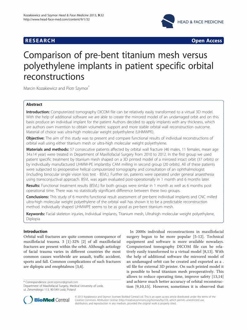
Introduction: Computerized tomography DICOM file can be relatively easily transformed to a virtual 3D model.With the help of additional software we are able to create the mirrored model of an undamaged orbit and on thisbasis produce an individual implant for the patient Authors decided to apply implants with any thickness, whichare authors own invention to obtain volumetric support and more stable orbital wall reconstruction outcome.Material of choice was ultra-high molecular weight polyethylene (UHMWPE).
Objective: The aim of this study was to present and compare functional results of individual reconstructions oforbital wall using either titanium mesh or ultra-high molecular weight polyethylene.
Materials and methods: 57 consecutive patients affected by orbital wall fracture (46 males, 11 females, mean age34±14 year) were treated in Department of Maxillofacial Surgery from 2010 to 2012. In the first group we usedpatient specific treatment by titanium mesh shaped on a 3D printed model of a mirrored intact orbit (37 orbits) orby individually manufactured UHMW-PE implantby CAM milling in second group (20 orbits). All of these patientswere subjected to preoperative helical computerized tomography and consultation of an ophthalmologist(including binocular single vision loss test - BSVL). Further on, patients were operated under general anaesthesiausing transconjuctival approach. BSVL was again evaluated post-operationally in 1 month and 6 months later.
Results: Functional treatment results (BSVL) for both groups were similar in 1 month as well as 6 months postoperational time. There was no statistically significant difference between these two groups.
Conclusions: This study of 6 months functional result assessment of pre-bent individual implants and CNC milledultra-high molecular weight polyethylene of the orbital wall has shown it to be a predictable reconstructionmethod. Individually shaped UHMWPE seems to be as good as pre-bent titanium mesh.
Keywords: Facial skeleton injuries, Individual implants, Titanium mesh, Ultrahigh molecular weight polyethylene,Diplopia
IntroductionOrbital wall fractures are quite common consequence ofmaxillofacial trauma. 3 [1]-32% [2] of all maxillofacialfractures are present within the orbit. Although aetiologyof facial trauma varies in different countries the mostcommon causes worldwide are assault, traffic accident,sports and fall. Common complications of such fracturesare diplopia and enophtalmos [3,4].
* Correspondence: [email protected] of Maxillofacial Surgery, Medical University of Lodz,ul. Zeromskiego 113, 90-549 Lodz, Poland
© 2013 Kozakiewicz and Szymor; licensee BioMCreative Commons Attribution License (http:/distribution, and reproduction in any medium
In 2000s individual reconstructions in maxillofacialsurgery begun to be more popular [5-12]. Technicalequipment and software is more available nowadays.Computerized tomography DICOM file can be rela-tively easily transformed to a virtual model [8,13]. Withthe help of additional software the mirrored model ofan undamaged orbit can be created and exported as a .stl file for external 3D printer. On such printed model itis possible to bend titanium mesh preoperatively. Thisallows to reduce operating time, improve safety [13,14]and achieve much better accuracy of orbital reconstruc-tion [9,10,15]. However, sometimes it is observed that
ed Central Ltd. This is an open access article distributed under the terms of the/creativecommons.org/licenses/by/2.0), which permits unrestricted use,, provided the original work is properly cited.
Kozakiewicz and Szymor Head & Face Medicine 2013, 9:32 Page 2 of 7http://www.head-face-med.com/content/9/1/32
implants may become misplaced or deformed, especiallyin cases of severe damaged orbits and old cases wherethin titanium mesh cannot be efficiently supported.Therefore authors decided to use implants of authorsown invention with any desired thickness to obtain volu-metric support and more stable orbital wall reconstructionoutcome [16]. Material of choice was ultra-high molecularweight polyethylene (UHMW-PE) commonly used inmedicine [17-19] especially as acetabulum replacement intotal hip prosthesis. An initial interest in UHMWE-PE touse it in maxillofacial surgery is dated at 1999 [20]. Theproblem of shrinkage of the polymer during its processingled to rejection of this material from maxillofacial recon-structions at that time. To eliminate this problem comput-erized numerical control milling from earlier preparedsolid bar of UHMW-PE was suggested. This led to the firstsuccessful human application of solid patient specificorbital wall implant made from UHMWE-PE at 2012 [16].
ObjectiveThe objective of this study was to present and comparefunctional results of individual reconstructions of orbitalwall using either titanium mesh or ultra-high molecularweight polyethylene.
Material and methodsThe Medical University of Lodz Ethic Committee ap-proval was obtained for this study [RNN/266/11/KB,RNN/141/12/KB, RNN/267/11/KB]. Participants pro-vided their written informed consent in a form acceptedby Medical University of Lodz Ethic Committee to partici-pate in this study. 57 consecutive patients affected by or-bital wall fracture (46 males, 11 females, mean age 34±14year) were treated in Department of Maxillofacial Surgeryfrom 2010 to 2012. Inclusion criteria was unilateral sideinjury i.e. 57 reconstructions were performed. In thefirst group pre-bent titanium mesh (37 orbits) was usedand in the second group computerized numerical con-trol milled ultrahigh molecular weight polyethylene [16]implants (20 orbits) were used. Patients to both groupswere assigned randomly.
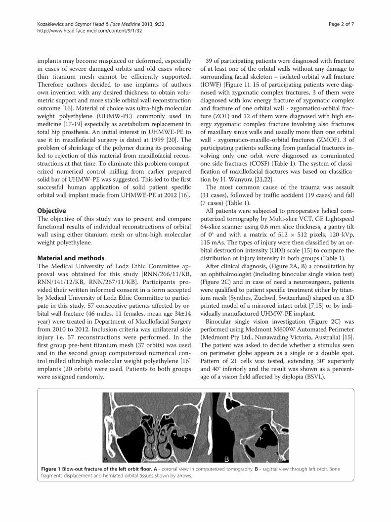
Figure 1 Blow-out fracture of the left orbit floor. A - coronal view in cofragments displacement and herniated orbital tissues shown by arrows.
39 of participating patients were diagnosed with fractureof at least one of the orbital walls without any damage tosurrounding facial skeleton – isolated orbital wall fracture(IOWF) (Figure 1). 15 of participating patients were diag-nosed with zygomatic complex fractures, 3 of them werediagnosed with low energy fracture of zygomatic complexand fracture of one orbital wall - zygomatico-orbital frac-ture (ZOF) and 12 of them were diagnosed with high en-ergy zygomatic complex fracture involving also fracturesof maxillary sinus walls and usually more than one orbitalwall - zygomatico-maxillo-orbital fractures (ZMOF). 3 ofparticipating patients suffering from panfacial fractures in-volving only one orbit were diagnosed as comminutedone-side fractures (COSF) (Table 1). The system of classi-fication of maxillofacial fractures was based on classifica-tion by H. Wanyura [21,22].The most common cause of the trauma was assault
(31 cases), followed by traffic accident (19 cases) and fall(7 cases) (Table 1).All patients were subjected to preoperative helical com-
puterized tomography by Multi-slice VCT, GE Lightspeed64-slice scanner using 0.6 mm slice thickness, a gantry tiltof 0° and with a matrix of 512 × 512 pixels, 120 kVp,115 mAs. The types of injury were then classified by an or-bital destruction intensity (ODI) scale [15] to compare thedistribution of injury intensity in both groups (Table 1).After clinical diagnosis, (Figure 2A, B) a consultation by
an ophthalmologist (including binocular single vision test)(Figure 2C) and in case of need a neurosurgeon, patientswere qualified to patient specific treatment either by titan-ium mesh (Synthes, Zuchwil, Switzerland) shaped on a 3Dprinted model of a mirrored intact orbit [7,15] or by indi-vidually manufactured UHMW-PE implant.Binocular single vision investigation (Figure 2C) was
performed using Medmont M600W Automated Perimeter(Medmont Pty Ltd., Nunawading Victoria, Australia) [15].The patient was asked to decide whether a stimulus seenon perimeter globe appears as a single or a double spot.Pattern of 21 cells was tested, extending 30° superiorlyand 40° inferiorly and the result was shown as a percent-age of a vision field affected by diplopia (BSVL).
mputerized tomography. B - sagittal view through left orbit. Bone

Table 1 Specific to implant type patient data
UHMW-PE Titanium mesh Total value
Total 20 37 57
Males 14 32 46
Females 6 5 11
Mean age 31.35 35.00
Mean ODI 3.75 2.27
Trauma cause
Assault 12 19 31
Traffic accident 6 13 19
Fall 2 5 7
Diagnosis
ZMOF 6 6 12
IOMF 12 27 39
ZOF 2 1 3
COSF 0 3 3
Mean BSVL_PRE 27.08% 20.22% n.s.
Mean BSVL_01 29.20% 20.32% n.s
Mean BSVL_06 15.95% 15.49% n.s
Abbreviations:n.s. no significant statistical difference.UHMW-PE ultra high molecular weight polyethylene.ODI orbital destruction intensity scale.ZMOF zygomatico-maxillo-orbital fractures.IOMF isolated orbital wall fracture.ZOF zygomatico-orbital bone fracture.COSF comminuted one-side fracture of the orbit.BSVL_PRE preoperative binocular single vision loss test [0% intact vision, 100%double in whole field of view].BSVL_01 1 month postoperative binocular single vision loss test.BSVL_06 6 months postoperative binocular single vision loss test.
Kozakiewicz and Szymor Head & Face Medicine 2013, 9:32 Page 3 of 7http://www.head-face-med.com/content/9/1/32
UHMW-PE implants were designed and produced inaccordance with the method described previously by theauthors [16]. The chosen substrate material was medicalUHMW-PE for surgical implants produced in accord-ance with ISO 5834–1 2007 type 1, ISO 5834–2 2006type 1 and ASTM F 648–07 type 1 standards (TiconaEngineering Polymers, Florence, USA; www.ticona.com).After compression moulding and ram extrusion, materialwas formed into stock shapes or solid blocks, as necessaryfor milling. Designing implants began with segmentingacquired DICOM data using Amira 5.4 (Visage ImagingGmbH, Germany) and creating 3D model of the patient’sfacial skeleton. In the next stage with use of GeomagicStudio 11 (Geomagic Corp., Morrisville, USA), a mirroredmodel of the unaffected side was superimposed on modelof fractured side. To ensure proper alignment analysisof symmetry was performed. The reference areas wereundamaged upper rim and upper wall. Proper superim-position allowed creating superior (from undamagedmirrored orbit) and inferior (from model of fracturedorbit) surfaces of implant. Subsequently, the 3D modelwas transferred to a CAD program SolidWorks (Dassault
Systèmes SolidWorks Corp., Waltham, USA) and pre-pared for CNC milling. Each virtual implant was inspectedand approved after necessary corrections by a maxillofacialsurgeon (MK) before manufacturing. All UHMW-PE im-plants were produced on computer numerical controlled,5-axis milling machine Speed Hawk 650 (OPS-IngersollFunkenerosion GmbH, Burbach, Germany) with accuracyof 0.05 mm.Further on, patients were operated under general anaes-
thesia by the same surgeon (author MK) (Figure 3). Trans-conjunctival approach was used in all cases (Figure 3A). Inthe first group flat titanium 0.4 mm thick mesh wasshaped preoperatively by operating surgeon(MK) on asolid individual model [Ti-Mesh] [7,15]. In the next grouppreviously prepared ultrahigh molecular weight polyethyl-ene [UHMW-PE] [16] implants were used to reconstructaffected lower or lower and medial wall of the orbit. Thecorrectness of position of implant during operation wascontrolled by checking implant alignment in previouslydesigned reference areas (usually lower orbital rim anteri-orly and orbital process of palatal bone posteriorly). Inboth groups the anatomical orbital wall reconstructionwere obtained. Computerized tomography was performedin the first week after surgery (Figure 2D) to evaluate thequality of the reconstruction and the condition of sur-rounding tissues to exclude any complications. Binocularsingle vision was again evaluated post-operationally in 1month and 6 months later (Figure 2F).Collected data were statistically analysed in Statgraphics
Centurion XVI (STATPOINT TECHNOLOGIES, INC.,Warrenton, Virginia, USA) (summary statistics, ANOVA,analysis of linear regression, t-test). Statistical significancewas determined as p< 0.05.
ResultsAlthough patients were assigned to both groups atrandom there was a statistically significant differencebetween mean ODI value in polyethylene and titan-ium mesh group (p<0.01). There was a higher orbitalwall destruction in a group treated with UHMW-PE(Figure 4). Despite that both groups were similar asfar as pre-treatment BSVL is considered. There wasa statically significant difference between mean ODIvalue between fractures of left (ODI=2) and right(ODI=4) side of the face (ANOVA p<0.00001).Despite the differences treatment results (BSVL) for
both groups were similar in 1 month as well as 6 monthspost operational time. For pre-bent titanium mesh meanBSVL preoperational was 20.22%, 1 month after operationit was 20.32% and 6 months after the operation lowered to15.48%. For UHMW-PE these values were 27.07% preop-erational, 29.2% 1 month post operational and 15.95% 6months post operational. There was no statistically signifi-cant difference between these two groups (Figure 5).

Figure 2 Results of treatment. A – computerized tomography in sagittal view: titanium mesh deformated during intra-orbital maneuvres inprimary surgery (asterisk); its deepest located part hurts the inferior rectus muscle. B – downgaze significantly limited in the left eye; C – result ofbinocular single vision loss test: diplopia caused by titanium mesh is mainly up- and downgaze. D - computerized tomography in sagittal viewafter corrective surgery and exchange of the implant to stiff patient specific ultra-high molecular weight polyethylene implant: orbital floor isreconstructed (arrow) and inferior rectus muscle is free. E – normal eye globe motility: full downgaze 1 month post-operationally. F - result ofbinocular single vision loss test: residual diplopia in upgaze/left.
Kozakiewicz and Szymor Head & Face Medicine 2013, 9:32 Page 4 of 7http://www.head-face-med.com/content/9/1/32
There was no statistically significant difference betweengroups divided by the cause of trauma neither in pre-operative nor in late postoperative results.In this study 80.7% of patients with orbital wall frac-
ture were male what coincides with results of otherstudies [3,23,24]. Gender had no statistically significantinfluence on BSVL preoperatively or on treatment re-sults (Figure 6).
Figure 3 Surgical steps in exchange titanium mesh to individual ultra-happroach exposes the titanium mesh immerse in scar tissue in orbital floor. Oeylid in the lower part of the picture. B – mesh impressions in orbital floor scatransconjunctival approach. D – implant position in the orbit, implant alignmeimplant to lower orbital rim. Implant position fixed with single 6mm long selfZuchwil, Switzerland).
Clinical classification of a fracture has a statisticallysignificant impact on pre-operational BSVL results. Pa-tients who suffered from isolated orbital wall fracture,or zygomato-orbital bone fracture, had a mean BSVLmuch lower (18.09%; 12.83%) than those who sufferedfrom zygomatico-maxillo-orbital fractures or commi-nuted one-side fractures (36.96%; 34.0%). These statisti-cally important differences between groups disappear in
igh molecular weight polyethylene implant. A – transconjunctivalrbital spatula holding the globe in the upper section, hooks holding ther tissue. C – individual polyethylene implant insertion through thent checked in previously designed reference areas, here visible fit of the-tapping screw from MatrixMIDFACE system by SYNTHES (Synthes,
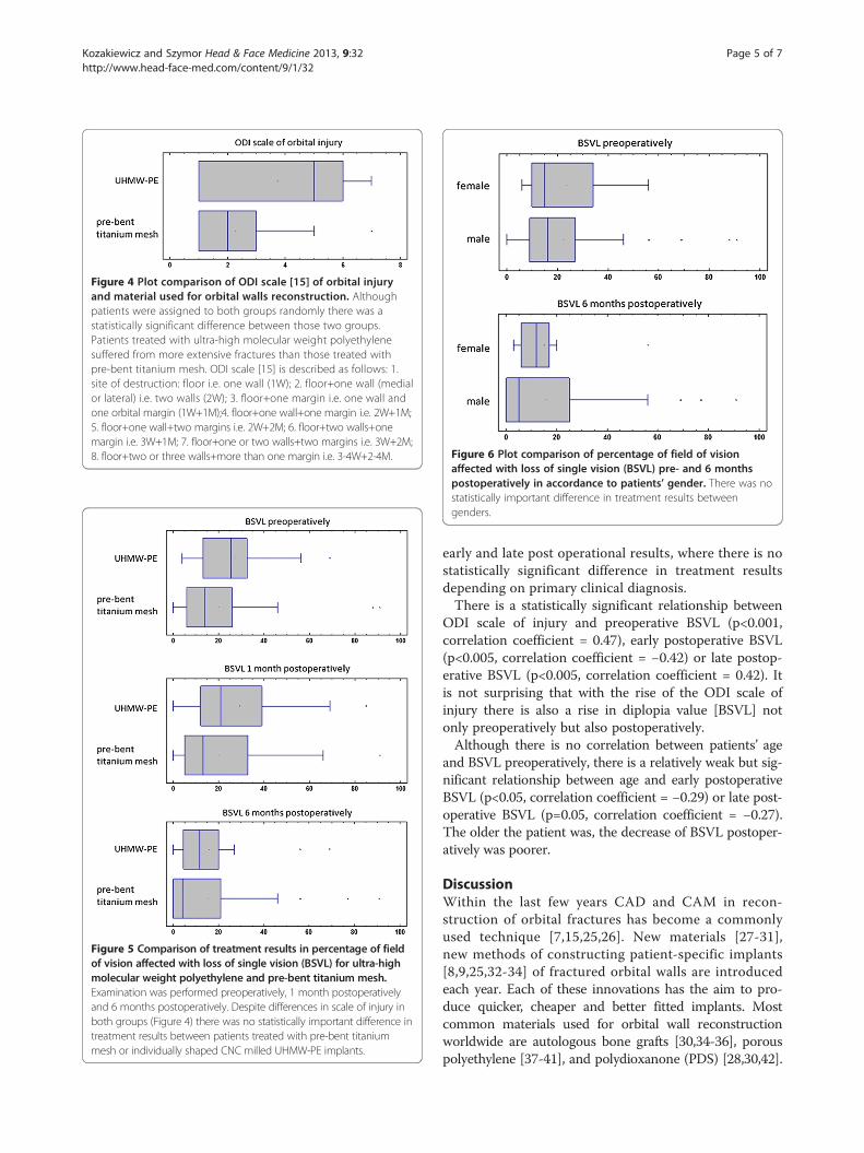
Figure 4 Plot comparison of ODI scale [15] of orbital injuryand material used for orbital walls reconstruction. Althoughpatients were assigned to both groups randomly there was astatistically significant difference between those two groups.Patients treated with ultra-high molecular weight polyethylenesuffered from more extensive fractures than those treated withpre-bent titanium mesh. ODI scale [15] is described as follows: 1.site of destruction: floor i.e. one wall (1W); 2. floor+one wall (medialor lateral) i.e. two walls (2W); 3. floor+one margin i.e. one wall andone orbital margin (1W+1M);4. floor+one wall+one margin i.e. 2W+1M;5. floor+one wall+two margins i.e. 2W+2M; 6. floor+two walls+onemargin i.e. 3W+1M; 7. floor+one or two walls+two margins i.e. 3W+2M;8. floor+two or three walls+more than one margin i.e. 3-4W+2-4M.
Figure 5 Comparison of treatment results in percentage of fieldof vision affected with loss of single vision (BSVL) for ultra-highmolecular weight polyethylene and pre-bent titanium mesh.Examination was performed preoperatively, 1 month postoperativelyand 6 months postoperatively. Despite differences in scale of injury inboth groups (Figure 4) there was no statistically important difference intreatment results between patients treated with pre-bent titaniummesh or individually shaped CNC milled UHMW-PE implants.
Figure 6 Plot comparison of percentage of field of visionaffected with loss of single vision (BSVL) pre- and 6 monthspostoperatively in accordance to patients’ gender. There was nostatistically important difference in treatment results betweengenders.
Kozakiewicz and Szymor Head & Face Medicine 2013, 9:32 Page 5 of 7http://www.head-face-med.com/content/9/1/32
early and late post operational results, where there is nostatistically significant difference in treatment resultsdepending on primary clinical diagnosis.There is a statistically significant relationship between
ODI scale of injury and preoperative BSVL (p<0.001,correlation coefficient = 0.47), early postoperative BSVL(p<0.005, correlation coefficient = −0.42) or late postop-erative BSVL (p<0.005, correlation coefficient = 0.42). Itis not surprising that with the rise of the ODI scale ofinjury there is also a rise in diplopia value [BSVL] notonly preoperatively but also postoperatively.Although there is no correlation between patients’ age
and BSVL preoperatively, there is a relatively weak but sig-nificant relationship between age and early postoperativeBSVL (p<0.05, correlation coefficient = −0.29) or late post-operative BSVL (p=0.05, correlation coefficient = −0.27).The older the patient was, the decrease of BSVL postoper-atively was poorer.
DiscussionWithin the last few years CAD and CAM in recon-struction of orbital fractures has become a commonlyused technique [7,15,25,26]. New materials [27-31],new methods of constructing patient-specific implants[8,9,25,32-34] of fractured orbital walls are introducedeach year. Each of these innovations has the aim to pro-duce quicker, cheaper and better fitted implants. Mostcommon materials used for orbital wall reconstructionworldwide are autologous bone grafts [30,34-36], porouspolyethylene [37-41], and polydioxanone (PDS) [28,30,42].

Kozakiewicz and Szymor Head & Face Medicine 2013, 9:32 Page 6 of 7http://www.head-face-med.com/content/9/1/32
Titanium mesh has already proven its usefulness in recon-structing orbital walls [35,43]. Alternative materials for or-bital walls reconstruction such as hydroxyapatite, porouspolyethylene [37] or polylactide provide as good treatmentresults as titanium mesh [30]. Ultra high molecular weightpolyethylene used in this study has proven to be as usefulin reconstructing orbital walls as pre-bent titanium mesh.No statistical differences in post operational results de-pending gender, age or primary clinical diagnosis showthat polymers may be broadly used instead of titaniummesh. An ability to create implants with nearly any thick-ness due to CNC milling seems to be helpful in recon-structing heavily destroyed orbits and especially in delayedsurgery cases. It is possible to adjust implant thickness tofully recreate orbital walls. Costs of producing ultra-highmolecular weight polyethylene implants are similar tousing titanium mesh, decreased of a cost of 3D printedacrylic model of an orbit. Compared to porous polyethyl-ene [30,41] UHMW-PE implants should probably havelower a risk of infection due to their solid structure.Therefore an elevated risk of deep implant infection beforecompleting vascularization should not occur as there is novascular ingrowth into implant. In our study there was nocase of postoperational implant infection but further stud-ies concerning this issue are required.A major drawback of polyethylene implants is their
radiolucency. It is required to use radio-opaque agentcombined with the polyethylene to make implants visibleon computed tomography for post-operational controlof implant position.
ConclusionsThis study of 6 months functional result assessment ofpre-bent individual implants and CNC milled ultra-highmolecular weight polyethylene of the orbital wall hasshown it to be a predictable reconstruction method.UHMW-PE implant seems to be as good as pre-benttitanium mesh.
Competing interestThe authors declare that they have no competing interests.
Authors’ contributionsMK designed the study, preformed all of the operations and statisticallyanalysed the results, also helped to draft the manuscript. PS made literaturesearch, gathered the results and drafted the manuscript. Both authors readand approved the final manuscript.
AcknowledgementsStudy was supported by grant: 502-03/5-138-03/502-54-084, 503/5-061-02/503-01.
Received: 8 July 2013 Accepted: 24 October 2013Published: 29 October 2013
References1. Roden KS, Tong W, Surrusco M, Shockley WW, Van Aalst JA, Hultman CS:
Changing characteristics of facial fractures treated at a regional, level 1trauma center, from 2005 to 2010: an assessment of patient
demographics, referral patterns, etiology of injury, anatomic location,and clinical outcomes. Ann Plas Surg 2012, 68:461–466.
2. Smith H, Peek-Asa C, Nesheim D, Nish A, Normandin P, Sahr S: Etiology,diagnosis, and characteristics of facial fracture at a midwestern level Itrauma center. J Traum Nursing: the official journal of the Society of TraumaNurses 2012, 19:57–65.
3. Chi MJ, Ku M, Shin KH, Baek S: An analysis of 733 surgically treatedblowout fractures. Ophthalmologica 2010, 224:167–175.
4. Loba P, Kozakiewicz M, Nowakowska O, Omulecki W, Broniarczyk-Loba A:Management of persistent diplopia after surgical repair of orbitalfractures. J AAPOS: the official publication of the American Association forPediatric Ophthalmology and Strabismus / American Association for PediatricOphthalmology and Strabismus 2012, 16:548–553.
5. Schön R, Metzger MC, Zizelmann C, Weyer N, Schmelzeisen R: Individuallypreformed titanium mesh implants for a true-to-original repair of orbitalfractures. Int J Oral Max Surg 2006, 35:990–995.
6. An J-G, Zhang Y, Zhang Z-Y: Computer-assisted fabricated individualtitanium mesh for reconstruction of orbital wall. Beijing da xue xue bao Yixue ban = Journal of Peking University Health sciences 2008, 40:88–91.
7. Kozakiewicz M, Elgalal M, Loba P, Komuński P, Arkuszewski P, Broniarczyk-LobaA, Stefańczyk L: Clinical application of 3D pre-bent titanium implants fororbital floor fractures. J Cranio Maxill Surg: official publication of the EuropeanAssociation for Cranio-Maxillo-Facial Surgery 2009, 37:229–234.
8. Elgalal M, Kozakiewicz M, Loba P, Walkowiak B, Olszycki M, Stefańczyk L:Patient specific implants, designed using Rapid Prototyping anddiagnostic imaging, for the repair of orbital fractures. Med Sci Monit 2010,16:75–79.
9. Essig H, Dressel L, Rana M, Rana M, Kokemueller H, Ruecker M, Gellrich N-C:Precision of posttraumatic primary orbital reconstruction usingindividually bent titanium mesh with and without navigation: aretrospective study. Head Face Med 2013, 9:18.
10. Metzger MC, Schön R, Weyer N, Rafii A, Gellrich N-C, Schmelzeisen R, StrongBE: Anatomical 3-dimensional pre-bent titanium implant for orbital floorfractures. Ophthalmology 2006, 113:1863–1868.
11. Stoetzer M, Rana M, von See C, Eckardt AM, Gellrich N-C: Reconstruction ofdefects of maxillary sinus wall after removal of a huge odontogeniclesion using prebended 3D titanium-mesh and CAD/CAM technique.Head Face Med 2011, 7:21.
12. Essig H, Rana M, Kokemueller H, von See C, Ruecker M, Tavassol F, GellrichN-C: Pre-operative planning for mandibular reconstruction - a full digitalplanning workflow resulting in a patient specific reconstruction. HeadNeck Oncol 2011, 3:45.
13. Olszewski R, Reychler H: Three-dimensional surgical guide for frontal-nasal-ethmoid-vomer disjunction in Le Fort III osteotomy. J CraniofacSurg 2011, 22:1791–1792.
14. Reychler H, Olszewski R: Intracerebral penetration of a zygomatic dentalimplant and consequent therapeutic dilemmas: case report. Int J OralMax Impl 2010, 25:416–418.
15. Kozakiewicz M, Elgalal M, Piotr L, Broniarczyk-Loba A, Stefanczyk L:Treatment with individual orbital wall implants in humans - 1-Yearophthalmologic evaluation. J Cranio Maxill Surg: official publication of theEuropean Association for Cranio-Maxillo-Facial Surgery 2011, 39:30–36.
16. Kozakiewicz M, Elgalal M, Walkowiak B, Stefanczyk L: Technical concept ofpatient-specific, ultrahigh molecular weight polyethylene orbital wallimplant. J Cranio Maxill Surg: official publication of the European Associationfor Cranio-Maxillo-Facial Surgery 2013, 41:282–290.
17. Hinrichs F, Boudriot U, Held T, Griss P: 10-Jahres-Ergebnisse einerMonobloc-Hüftendoprothesenpfanne mit mehrlagigerReintitangitterschale zur zementfreien Implantation. Z Orthop Grenzgeb2001, 139:212–216.
18. Jasty M, Rubash HE, Muratoglu O: Highly cross-linked polyethylene: thedebate is over–in the affirmative. J Arthroplasty 2005, 20(4 Suppl 2):55–58.
19. Reynolds SE, Malkani AL, Ramakrishnan R, Yakkanti MR: Wear analysis of first-generation highly cross-linked polyethylene in primary total hiparthroplasty: an average 9-year follow-up. J Arthroplasty 2012, 27:1064–1068.
20. Rimell JT, Marquis PM: Selective laser sintering of ultra high molecularweight polyethylene for clinical applications. J Biomed Mater Res 2000,53:414–420.
21. Wanyura H: Clinical and anatomopathologic classification of fractures ofthe orbit. Revue de stomatologie et de chirurgie maxillo-faciale 1998,99:80–87.

Kozakiewicz and Szymor Head & Face Medicine 2013, 9:32 Page 7 of 7http://www.head-face-med.com/content/9/1/32
22. Wanyura H, Kowalczyk P, Bossak M, Samolczyk-wanyura D, Stopa Z: Finiteelement analysis of external loads resulting in orbito-nasal dislocations.J Stoma 2011, 64:411–424.
23. Shere JL, Boole JR, Holtel MR, Amoroso PJ: An analysis of 3599 midfacialand 1141 orbital blowout fractures among 4426 United States ArmySoldiers, 1980–2000. Otolaryngology–head and neck surgery: official journalof American Academy of Otolaryngology-Head and Neck Surgery 2004,130:164–170.
24. Josuel Raimundo C, Karis Barbosa G, Egito Vasconcelos BCd, de HollandaVasconcellos RJ: Epidemiological study of patients with facial traumatreated at the Antônio Targino Hospital - Campina Grande/Paraíba.Braz J Otorhinolaryngology 2009, 75:628–633.
25. Metzger MC, Schön R, Schmelzeisen R: Preformed titanium meshes: a newstandard? Skull base: official journal of North American Skull Base Society.[et al.] 2007, 17:269–272.
26. Hohlweg-Majert B, Schön R, Schmelzeisen R, Gellrich N-C, Schramm A:Navigational maxillofacial surgery using virtual models. World J Surg2005, 29:1530–1538.
27. Ciprandi MTO, Primo BT, Gassen HT, Closs LQ, Hernandez PAG, Silva AN:Calcium phosphate cement in orbital reconstructions. J Craniofac Surg2012, 23:145–148.
28. Gierloff M, Seeck NGK, Springer I, Becker S, Kandzia C, Wiltfang J: Orbitalfloor reconstruction with resorbable polydioxanone implants. J CraniofacSurg 2012, 23:161–164.
29. Loba P, Kozakiewicz M, Elgalal M, Stefańczyk L, Broniarczyk-Loba A,Omulecki W: The use of modern imaging techniques in the diagnosisand treatment planning of patients with orbital floor fractures. Med SciMonitor: international medical journal of experimental and clinical research2011, 17:CS94–CS98.
30. Avashia YJ, Sastry A, Fan KL, Mir HS, Thaller SR: Materials used forreconstruction after orbital floor fracture. J Craniofac Surg 2012,23(7 Suppl 1):1991–1997.
31. Schumann P, Lindhorst D, Wagner MEH, Schramm A, Gellrich N-C, Rücker M:Perspectives on resorbable osteosynthesis materials incraniomaxillofacial surgery. Pathobiology: journal of immunopathology,molecular and cellular biology 2013, 80:211–217.
32. Mustafa SF, Evans PL, Bocca a, Patton DW, Sugar a W, Baxter PW:Customized titanium reconstruction of post-traumatic orbital walldefects: a review of 22 cases. Int J Oral Max Surg 2011, 40:1357–1362.
33. Zizelmann C, Gellrich NC, Metzger MC, Schoen R, Schmelzeisen R, SchrammA: Computer-assisted reconstruction of orbital floor based on conebeam tomography. Brit J Oral Max Surg 2007, 45:79–80.
34. Schmelzeisen R, Gellrich NC, Schoen R, Gutwald R, Zizelmann C, SchrammA: Navigation-aided reconstruction of medial orbital wall and floorcontour in cranio-maxillofacial reconstruction. Injury 2004, 35:955–962.
35. Ellis E, Tan Y: Assessment of internal orbital reconstructions for pureblowout fractures: cranial bone grafts versus titanium mesh. J Oral MaxSurg: official journal of the American Association of Oral and MaxillofacialSurgeons 2003, 61:442–453.
36. Gellrich N-C, Schramm A, Hammer B, Rojas S, Cufi D, Lagrèze W,Schmelzeisen R: Computer-assisted secondary reconstruction of unilateralposttraumatic orbital deformity. Plast Reconstr Surg 2002, 110:1417–1429.
37. Sadiq SA, Mengher LS, Lowry J, Downes R: Integrated orbital implants–acomparison of hydroxyapatite and porous polyethylene implants. Orbit(Amsterdam, Netherlands) 2008, 27:37–40.
38. Kim CY, Jeong BJ, Lee SY, Yoon JS: Comparison of surgical outcomes oflarge orbital fractures reconstructed with porous polyethylene channeland porous polyethylene titan barrier implants. Ophthal Plast Recons2012, 28:176–180.
39. Saiepour D, Messo E, Hedlund AJO, Nowinski DJ: Radiologic and long-termclinical outcome from treatment of isolated medial orbital wall blowoutfractures. J Craniofac Surg 2012, 23:1252–1255.
40. Eski M, Sahin I, Deveci M, Turegun M, Isik S, Sengezer M: A retrospectiveanalysis of 101 zygomatico-orbital fractures. J Craniofac Surg 2006,17:1059–1064.
41. Lee S, Maronian N, Most SP, Whipple ME, McCulloch TM, Stanley RB, FarwellDG: Porous high-density polyethylene for orbital reconstruction. ArchOtolaryngol 2005, 131:446–450.
42. Gerressen M, Gillessen S, Riediger D, Hölzle F, Modabber A, Ghassemi A:Radiologic and facial morphologic long-term results in treatment oforbital floor fracture with flexible absorbable alloplastic material. J OralMaxil Surg: official journal of the American Association of Oral andMaxillofacial Surgeons 2012, 70:2375–2385.
43. Gear AJL, Lokeh A, Aldridge JH, Migliori MR, Benjamin CI, Schubert W:Safety of titanium mesh for orbital reconstruction. Ann Plas Surg 2002,48:1–7. discussion 7–9.
doi:10.1186/1746-160X-9-32Cite this article as: Kozakiewicz and Szymor: Comparison of pre-benttitanium mesh versus polyethylene implants in patient specific orbitalreconstructions. Head & Face Medicine 2013 9:32.
Submit your next manuscript to BioMed Centraland take full advantage of:
• Convenient online submission
• Thorough peer review
• No space constraints or color figure charges
• Immediate publication on acceptance
• Inclusion in PubMed, CAS, Scopus and Google Scholar
• Research which is freely available for redistribution
Submit your manuscript at www.biomedcentral.com/submit