Research Article Phyllanthus emblica L. Enhances Human...
10
Hindawi Publishing Corporation Evidence-Based Complementary and Alternative Medicine Volume 2013, Article ID 720728, 9 pages http://dx.doi.org/10.1155/2013/720728 Research Article Phyllanthus emblica L. Enhances Human Umbilical Vein Endothelial Wound Healing and Sprouting Linda Chularojmontri, 1 Maneewan Suwatronnakorn, 2 and Suvara K. Wattanapitayakul 2 1 Department of Preclinical Sciences, Faculty of Medicine, ammasat University, Pathum ani 12121, ailand 2 Department of Pharmacology, Faculty of Medicine, Srinakharinwirot University, Bangkok 10110, ailand Correspondence should be addressed to Suvara K. Wattanapitayakul; [email protected] Received 19 January 2013; Accepted 5 March 2013 Academic Editor: Ludger Beerhues Copyright © 2013 Linda Chularojmontri et al. is is an open access article distributed under the Creative Commons Attribution License, which permits unrestricted use, distribution, and reproduction in any medium, provided the original work is properly cited. Endothelial dysfunction is the hallmark of impaired wound healing and increased risk of cardiovascular disease. Antioxidants from natural sources decrease oxidative stress and protect against cellular damage caused by reactive oxygen species (ROS). In this study, we examined the antioxidant constituents and capacity of Phyllanthus emblica L. (PE) fruit in freeze-dried power form. e pharmacological properties of PE were investigated using human umbilical vein endothelial cells (HUVECs) in the aspects of endothelial cell proliferation, nitric oxide (NO) production, wound healing, cell migration, in vitro angiogenesis, and VEGF gene expression. e ASC content of PE was 1.574% + 0.046% (w/w) as determined by HPLC and the total phenolic content was 36.1% ± 0.7% gallic acid equivalent when measured by Folin-Ciocalteu assay. e FRAP assay revealed a relatively high antioxidant capacity at 3,643 + 192.5 mole/mg. PE at 0.1 to 10 g/mL did not significantly influence endothelial cell proliferation, but at higher concentrations PE decreased cell survival to 62%. PE significantly promoted NO production, endothelial wound closure, endothelial sprouting, and VEGF mRNA expression. erefore, PE is a candidate for antioxidant supplement that promotes endothelial function and restores wound healing competency. 1. Introduction Endothelial dysfunction is the foremost step in the develop- ment of atherosclerosis and causes impairment in cell pro- liferation, migration, wound healing, and angiogenic func- tion [1]. Pathologic oxidative stress conditions, such as dia- betes and cardiovascular disease, potentiate endothelial dys- function eventually leading to ischemic vascular damage and become impediment to wound healing. Current therapeutic strategies focusing on protection of endothelial oxidative damage, accelerating endothelial wound healing, or promot- ing angiogenesis may have a role in diabetic and oxidative related vascular disease [2]. Impaired endothelial function is mainly characterized by reduced nitric oxide (NO) production, decreased cell migra- tion to repair the endothelial damage, and being deficient in the capacity to form new collateral vessels. Substances that enhance NO bioavailability and promote vascular endothelial growth factor (VEGF) synthesis are shown to improve wound healing and/or angiogenesis [3, 4]. Several oxidative stress markers such as homocysteine and long-standing hypoxia are known to impair endothelial wound healing while using antioxidants have been proven to improve endothelial func- tion and wound repair [5, 6]. Of these, natural antioxidants are the current main focus in complementary and alterna- tive medicine research. In traditional medicine, Phyllanthus emblica L. (PE), an indigenous plant grown in ailand and some parts of Asia, has long been used as a wound healing agent either in single formulation or combined components in traditional preparations. e effect of PE on wound repair has been demonstrated in rats and the mechanisms involve modulation of collagen synthesis, extracellular matrix (ECM) protein synthesis, and antioxidant status [7, 8]. However, the pharmacological action of PE exclusively on endothelial function, wound repair, and tube formation has not been reported. In this study, we investigated the properties of PE fruits in the aspects of antioxidant capacity, ascorbic acid content, total phenolics level, and vasculogenic property
Transcript of Research Article Phyllanthus emblica L. Enhances Human...

Hindawi Publishing CorporationEvidence-Based Complementary and Alternative MedicineVolume 2013 Article ID 720728 9 pageshttpdxdoiorg1011552013720728
Research ArticlePhyllanthus emblica L Enhances Human Umbilical VeinEndothelial Wound Healing and Sprouting
Linda Chularojmontri1 Maneewan Suwatronnakorn2 and Suvara K Wattanapitayakul2
1 Department of Preclinical Sciences Faculty of Medicine Thammasat University PathumThani 12121 Thailand2Department of Pharmacology Faculty of Medicine Srinakharinwirot University Bangkok 10110 Thailand
Correspondence should be addressed to Suvara K Wattanapitayakul suvaragmailcom
Received 19 January 2013 Accepted 5 March 2013
Academic Editor Ludger Beerhues
Copyright copy 2013 Linda Chularojmontri et al This is an open access article distributed under the Creative Commons AttributionLicense which permits unrestricted use distribution and reproduction in any medium provided the original work is properlycited
Endothelial dysfunction is the hallmark of impaired wound healing and increased risk of cardiovascular disease Antioxidantsfrom natural sources decrease oxidative stress and protect against cellular damage caused by reactive oxygen species (ROS) Inthis study we examined the antioxidant constituents and capacity of Phyllanthus emblica L (PE) fruit in freeze-dried power formThe pharmacological properties of PE were investigated using human umbilical vein endothelial cells (HUVECs) in the aspectsof endothelial cell proliferation nitric oxide (NO) production wound healing cell migration in vitro angiogenesis and VEGFgene expression The ASC content of PE was 1574 + 0046 (ww) as determined by HPLC and the total phenolic content was361plusmn 07 gallic acid equivalent when measured by Folin-Ciocalteu assayThe FRAP assay revealed a relatively high antioxidantcapacity at 3643 + 1925120583molemg PE at 01 to 10 120583gmL did not significantly influence endothelial cell proliferation but at higherconcentrations PEdecreased cell survival to 62 PE significantly promotedNOproduction endothelial wound closure endothelialsprouting and VEGF mRNA expression Therefore PE is a candidate for antioxidant supplement that promotes endothelialfunction and restores wound healing competency
1 Introduction
Endothelial dysfunction is the foremost step in the develop-ment of atherosclerosis and causes impairment in cell pro-liferation migration wound healing and angiogenic func-tion [1] Pathologic oxidative stress conditions such as dia-betes and cardiovascular disease potentiate endothelial dys-function eventually leading to ischemic vascular damage andbecome impediment to wound healing Current therapeuticstrategies focusing on protection of endothelial oxidativedamage accelerating endothelial wound healing or promot-ing angiogenesis may have a role in diabetic and oxidativerelated vascular disease [2]
Impaired endothelial function is mainly characterized byreduced nitric oxide (NO) production decreased cell migra-tion to repair the endothelial damage and being deficient inthe capacity to form new collateral vessels Substances thatenhanceNObioavailability and promote vascular endothelialgrowth factor (VEGF) synthesis are shown to improvewound
healing andor angiogenesis [3 4] Several oxidative stressmarkers such as homocysteine and long-standing hypoxiaare known to impair endothelial wound healing while usingantioxidants have been proven to improve endothelial func-tion and wound repair [5 6] Of these natural antioxidantsare the current main focus in complementary and alterna-tive medicine research In traditional medicine Phyllanthusemblica L (PE) an indigenous plant grown in Thailand andsome parts of Asia has long been used as a wound healingagent either in single formulation or combined componentsin traditional preparations The effect of PE on wound repairhas been demonstrated in rats and the mechanisms involvemodulation of collagen synthesis extracellularmatrix (ECM)protein synthesis and antioxidant status [7 8] Howeverthe pharmacological action of PE exclusively on endothelialfunction wound repair and tube formation has not beenreported In this study we investigated the properties of PEfruits in the aspects of antioxidant capacity ascorbic acidcontent total phenolics level and vasculogenic property
2 Evidence-Based Complementary and Alternative Medicine
that promoted endothelial NO production wound healingand in vitro angiogenesis using human umbilical cord veinendothelial cells (HUVECs)
2 Materials and Methods
21 Chemicals and Phyllanthus emblica L Fruit Extract (PE)All chemicals were purchased from Sigma-Aldrich (St LouisMO USA) or otherwise indicated PE fruits were obtainedfrom Nakhon Ratchasima province Northeast of ThailandPE fruit juicewas extracted from303 g (57 fresh fruits) using afruit juice extractor and yielded approximately 254mLfruitPE juice was then filtered through 02 120583m membrane filterand underwent freeze-drying process giving 161 yield(wv) The yellowish dry power of PE was kept at 4∘C untiluse The aqueous stock solutions at 10mgmL were preparedfreshly at the time of use in each experiment
22 Ascorbic Acid Content Antioxidant Activity and TotalPhenolic Compounds of PE It is well recognized that PEexerts various biological activities partly due to its antioxidantactivity such as ascorbic acid (ASC) polyphenolic com-pounds and flavonoids This study evaluated ASC contentwhich is the major antioxidant component of the extractusing HPLC method (Thermo Scientific) Standard curveof ASC was established by measuring the areas under thepeak after injecting a series of ASC stock solutions intoreversed phase HPLC column (Luna C18 5 120583m dimension150 times 460mm Phenomenex Thailand) with mobile phase(100mM phosphate buffer 95 methanol 5) at the flowrate of 04mLmin isocratic elution and detected by UVabsorption at 243 nm [9]
Total antioxidant capacity of PEwas determined by Ferricreducing antioxidant power (FRAP) assay The FRAP assaydetermined the ability of PE to deliver one electron to FeIII-TPTZ (246-Tri(2-pyridyl)-s-triazine) complex to form acolor ferrous ion Briefly tenmicroliters of standard FeSO
4or
sample solutions were added to a 96-well microplate followedby adding 200120583L of FRAP reagent (acetate buffer 300mM(pH 36) TPTZ 10mM in HCl 40mM and FeCl
3sdot6H2O
20mM) and the development of blue color was monitoredat 595 nm (Synergy BioTek USA) [10] The stability of anti-oxidant capacity was determined from the dry power of PEsamples stored at 4∘C for 12 months
Total phenolic content was evaluated by Folin-Ciocalteuassay using gallic acid (GA) as an assay standard [11] Brieflya series of standard GA concentrations (0 3125 625 125250 500 and 1000 120583M) or samples at the volume of 16mLwere added to a reaction test tubes followed by 100 120583L ofFolin-Ciocalteu reagent and 300 120583L of Na
2CO3solution The
mixture was incubated at 40∘C for 30min and then cooleddown at room temperature for 5minThe products of pheno-lic compounds reaction were measured at 756 nm (ShimadzuUV-1601 Japan) The amount of total phenolic compoundswas represented as GA equivalence (GAE)
23 Human Umbilical Vein Endothelial Cell (HUVEC) Cul-ture Human umbilical cords were collected from the laborroom of the university hospital and HUVECs were isolated
within 48 h as described previously [12] Cells were culturedinM199medium supplementedwith 20 fetal bovine serum(FBS) with antibiotic and antimycotic agents (InvitrogenUSA) in a humidified atmosphere of 95 air and 5 CO
2
at 37∘C Cells between passages 3 and 5 were used in theexperiments and were cultured in low serum medium (1FBS) during PE incubation or other treatments
24 Cytotoxicity of PE and Ascorbic Acid (ASC) There havebeen reported that PE induced apoptosis in many cell typesincluding at least 7 cancer cell lines and primary osteoclasts[13ndash15] Therefore this experiment was aimed to deter-mine nontoxic doses of PE and its major antioxidant ASCfor further experiments HUVECs were treated with var-ious concentrations of PE or ASC for 48 h Cell survivalwas evaluated using MTT (3-(45-dimethylthiazol-2-yl)-25-diphenyltetrazolium bromide) assay and sulforhodamine B(SRB) cytotoxicity assay For MTT assay ten microliters ofMTT stock solution (5mgmL) were added to the culturemedium and incubated for 4 h or until dark blue-purplecrystalline precipitates were visualized under an invertedmicroscope Then one hundred microliters of DMSO wereadded to dissolve the formazan products The plate was thenshacked at 200 rpm for 20min and the relative cell viabilitywas detected absorbance at 550 nm by Synergy plate reader(BioTek USA)
SRB assay was performed in 96-well plate after removalof culture medium and washed once with PBS Two hundredmicroliters of cold 10 trichloroacetic acid (TCA) wereadded to the wells to fix cells Following 30min incubation at4∘CTCAwas aspirated and thewells were rinsedwithwater 5times Plates were air-dried and 100 120583L of SRB solution (04in 1 acetic acid) was added to each well and allowed cells tobe stained for 30min Cells were then washed with 1 aceticacid until unincorporated dye was removed (approximately5 times) The plates were air-dried at room temperature for30min The bound SRB dye was solubilized with 200120583L of10mMTris (pH 105) for 5 minutes at room temperatureTherelative cell viability was determined by absorbance at 510 nm(BioTek USA)
25 Endothelial Wound Healing Assay Scratch wound assaywas used to evaluate the ability of PE in promoting endothe-lial wound in vitro HUVECs were seeded in 6-well plate(Nunc Thermo Fisher Scientific USA) at 5 times 105 cellswellin culture medium and allowed the cells to grow to 90 con-fluence The culture medium was then replaced with 1 FBSM199 overnight before the scratch wounds were initiatedusing a sterile 200120583L pipette tip Photos of wounds werecaptured by a digital camera (Olympus DP20 Japan) at thesame positions at 0 24 and 48 h The length of woundconfluence was measured by CellandB program (OlympusJapan)
26 Cell Migration Assay Migration of HUVECs toward achemoattractant VEGF and testing substances (PE and ASC)in the cellrsquos surrounding environment was determined byBoyden chamber in 24-well plate (Corning USA) HUVECswere cultured and 200120583L of cells suspension (2times105 cellsmL
Evidence-Based Complementary and Alternative Medicine 3
in 1 FBS)was seeded into the chamber with translucent PETmembranes 8120583m pore size Seven hundred and fifty micro-liters of media containing vehicle (CTRL group) 100 ngmLVEGF PE or ASC were added to the lower chamber Theplate was incubated at 37∘C in a CO
2incubator for 16 hours
The inserts were then transferred to 025 trypsin-EDTAsolution and 300 120583L of 5 120583M calcein-acetoxymethyl ester(CAL-AM Sigma) was added to the lower chamber andincubated at 37∘C in a CO
2incubator for 45 minutes Cells
migrated through the micropores used enzyme esterases tohydrolyze the nonfluorescent CAL-AM to highly fluorescentproduct which was monitored at wavelengths 485528 nm forexcitationemission respectively
27 Nitric Oxide (NO) Production Endothelial NO is impor-tant to maintain endothelial healthcell survival and activa-tion cell migration In this study the stable product of NOnitrite was evaluated by 23-diaminonaphthalene (DAN)assay which is far more sensitive than Griess reaction [16]This fluorometric method uses the substrate DAN (nonfluo-rescent) to react with nitrite under acidic conditions to gen-erate 23-diaminonaphthotriazole or 1-(H)-naphthotriazole(NATH) the fluorescent product NATH fluorescent signalis further enhanced by alkalinization of medium to yield 23-naphthotriazole anion (NAT) NaNO
2standard solution was
freshly prepared ranging from 013 to 1333 120583M in DMEMbefore experiment Seventy-five microliters of standard solu-tions or the supernatant media from PE or ASC treatmentswere transferred into 96-well microplate then 10 120583L of DANsolution (50 120583gmL in 062NHCl) was added to each well for10min at room temperature in the dark Finally 5 120583L of 28NNaOHsolutionwas subsequently added to eachwell andfluo-rescence was measured using a fluorescent microplate reader(Synergy HT BioTek) with excitationemission wavelengthsof 360460 nm Nitrite levels of sample were calculated fromNaNO
2standard curve
28 In Vitro Angiogenesis Assay The 3D spheroid-based an-giogenesis assay was performed as described by Korff [17]EachHUVEC spheroidwas composed of 500 cells distributedin 14 methyl cellulose (Fisher Scientific USA) Forty-eightspheroids were embedded in 1mL rat tail collagen (2mgmL)and performed in 24-well tissue culture plates For angiogenicstimulation test the collagen gels were overlaid with 100120583Lmedium containing 50 ngmL vascular endothelial growthfactor (VEGF PeproTech USA) or PE at various concentra-tions and the gels were allowed to be stored at 37∘C and5 CO
2for 24 h Pictures of each spheroid were captured at
0 and 24 h with a digital camera and the cumulative sproutlength was measured for at least 10 individual spheroids pertreatment (Olympus Japan)
29 VEGF mRNA Expression VEGF gene expression wasdetermined based on SYBR-Green fluorescence RT-PCRusing commercial kits (BioRad USA) Total RNA wasextracted from cultured HUVECs using Trizol reagent (Invi-trogen USA) Amplification of target genes was initiatedby denaturation at 70∘C and cooling to 37∘C prior to
01 1 10 100 10000
50
100
150
400
ASC-SRBPE-SRBPE-MTT ASC-MTT
Cel
l via
bilit
y (c
ontro
l )
Concentration (120583gmL)
Figure 1 Cytotoxic doses of PE and ASC in HUVECs comparingtwo methods of cell viability assays HUVECs were incubated withPhyllanthus emblica (PE) or ascorbic acid (ASC) at designatedconcentrations for 48 h SRB and MTT assays were performed asdescribed in Section 2
performing reverse transcription at 37∘C for 1 h as describedin the manufacturerrsquos manual PCR amplification for 120573-actinand VEGF was carried out in parallel using primer pairsas follows 120573-actin forward 51015840-GGACTTCGAGCAAGAGATGG-31015840 reverse 51015840-AGCACTGTGTTGGCGTACAG-31015840VEGF forward 51015840-TATTTTTCTTGCTGCTAAAT-31015840 re-verse 51015840-AATGTTATTGGTGTCTTCAC-31015840 (Gen BankNM 001025366) Reaction cycles were performed for 40cycles with two denaturation steps (94∘C 4min and 94∘C4 s) followed by annealing at 58∘C 30 s and extension at 72∘Cfor 10 s Amplification was terminated with 10min extensionat 72∘C (Roter-Gene 6000 Corbett Life Science) RelativemRNA amounts of samples were calculated using the 2minusΔΔCTmethod
3 Results
31 Ascorbic Acid (ASC) Content Antioxidants Capacity andPhenolic Content of PE PE possessed antioxidant capacity of36433plusmn1925 120583molemgwhichwas not significantly changedwhen kept at 4∘C for 12 months (36945 plusmn 1058 120583molemg)The amount of total phenolic content which is well corre-sponding to antioxidant capacity was calculated as 0361 plusmn0005mg GAEmg PE powder HPLC analysis revealed thatPE dry powder consisted of 157 ASC (ww) or equivalentto 253mgmL fresh juice or approximately 642mgfruit
32 Cytotoxic Effects of PE and ASC MTT showed thatonly PE at 1000 120583gmL caused cytotoxicity while SRB assaydetected decreases in cell survival in a concentration-dependentmanner beginning fromPE at 10120583gmL (Figure 1)Interestingly MTT assay showed that ASC at 100 and1000 120583gmL dramatically increased cell survival by 15315and 39988 respectively But SRB assay revealed that ASC1000 120583gmL decreased cell survival by 42 Cell survival
4 Evidence-Based Complementary and Alternative Medicine
0 24 480
20
40
60
80
100
Time (hour)
Wou
nd co
nflue
nce (
)
VEGF 50ngmLCTRL
ASC 001 120583gmL
PE 01 120583gmLPE 1120583gmLPE 10120583gmL
ASC 01 120583gmLASC 1120583gmLASC 10120583gmL
lowast
lowast
lowast
lowast
(a)
0h 24h 48h
CTRL
VEGF50ngmL
PE01 120583gmL
ASC001 120583gmL
(b)
Figure 2 Scratch wound assay HUVEC wounds were created using pipette tips and the widths of wound areas were measured at the samepositions at time 0 24 and 48 h after wounds were initiated (a) PE (01ndash100120583gmL) or ascorbic acid (ASC 001ndash10 120583gmL) was incubatedwith endothelial wounds at time 0 (b) Representative photographs of wound closure at different time points Dashed line and the woundareas were accentuated for visual purpose The wound confluence was calculated compared to vehicle treated group (CTRL) lowast119875 lt 005versus CTRL
Evidence-Based Complementary and Alternative Medicine 5
0
50
100
150
200
250
CTRL VEGF
Cel
l mig
ratio
n(
CTR
L)
lowast
lowastlowastlowast
01 1 10 001 01 1
PE (120583gmL) ASC (120583gmL)
Figure 3 Effect of PE and ASC on cell migration Comparison ofHUVECmigration under influence of the chemotactic factor VEGF(100 ngmL) and PE (01 1 10120583gmL) or ASC (001 01 1120583gmL)wasdetermined using Boyden chamber assay as described in Section 2lowast
119875 lt 005 versus control
was then confirmed by cell morphology observed underinverted microscope (data not shown) The cell integrity andmorphology of live cells were in accordance with SRB assaythus noncytotoxic doses of PE and ASC were determinedbased on SRB assay
33 PE and ASC Enhanced Endothelial Wound HealingEffects of PE and ASC on endothelial wound healing wereinfluenced by the concentrations applied to the scratchwound At relatively lower concentrations (01 120583gmL for PEand 001120583gmL for ASC) these compounds significantly pro-moted wound confluence only at 48 h while VEGF 50 ngmLshowed significant enhance in wound healing rate since24 h PE 01 120583gmL completely healed endothelial scratchwound at 48 h which was comparable to wound treated withVEGF No change was observed in the healing of HUVECwound when treated with PE at 1 10 120583gmL and ASC at01 1120583gmL (Figure 2) Interestingly high dose of ASC at10 120583gmL marked impaired endothelial wound confluence at48 h
34 PE andASCPromotedCellMigration Migrated endothe-lial cells through modified Boyden chambers were measuredby reading the fluorescent product of CAL-AM resulted fromthe metabolism of live cells migrated through the microporesof the upper chamber Figure 3 demonstrated that PE (011 and 10 120583gmL) and ASC (001 01 and 1120583gmL) inverselyenhanced cell migration with respect to increasing dosesThe lowest two concentrations of PE and ASC used in thisexperiment significantly promoted endothelial cell migrationwhile the maximum responses were obtained at the degreecomparable to the action of the chemoattractant VEGF(100 ngmL)
35 Effects of PE and ASC on NO Production It is wellestablished that NO promotes HUVEC migration Here we
0
50
100
150
CTRL
Nitr
ite (
CTR
L)
lowast
01 1 10 001 01 1
PE (120583gmL) ASC (120583gmL)
Figure 4 Effects of PE and ASC on nitric oxide levels The stableproduct of nitric oxide nitrite was determined using DAN assayChanges in the levels of nitrite in culturemediawere analyzed at 48 hfollowing PE or ASC treatment as described in Section 2 lowast119875 lt 005versus CTRL
evaluated the stable product of NO nitrite in the culturemedia of cells treated with PE or ASC corresponding tothe doses that shown to promote endothelial migration Itappeared that significant increase in NO production wasobserved only in HUVEC treated with PE at 10120583gmLwhereas PE at lower doses (01 and 1 120583gmL) or all dosesof ASC (001 01 and 1 120583gmL) did not change nitrite levels(Figure 4)
36 Low Doses of PE Promoted Endothelial Sprouting PEenhanced endothelial sprouting from spheroidswas observedat 01 and 1120583gmL which was similar to its effect on endothe-lial wound closure Low PE doses promoted endothelialsprouting but no significant difference fromCTRL group wasobserved at the higher concentration 10120583gmL (Figure 5)Effects of ASC on endothelial sprout length were inverselyrelated to stepwise increases in concentrations used in theexperiment (001 01 and 1 120583gmL) while ASC at 10 120583gmLsignificantly suppressed in vitro angiogenesis
37 VEGF mRNA Expression The expression of VEGFmRNA was determined at 24 and 48 h following PE or ASCtreatment (as shown in Figure 6) At 24 h after treatmentPE (1120583gmL) and ASC (01 120583gmL) significantly enhancedVEGF mRNA expression by approximately 35- and 3-foldrespectively while other concentrations did not significantlyalter VEGF expression No change was observed in VEGFgene expression when detected at 48 h
4 Discussion
Antioxidant diets and supplements have increasingly becomean important strategy to prevent or slow deterioration of vas-cular endothelium in well-recognized high oxidative stressconditions such as diabetes and cardiovascular disease PEis one of the most studied natural antioxidants beneficial
6 Evidence-Based Complementary and Alternative Medicine
CTRL 0h
(a)
CTRL 24h
(b)
VEGF
(c)
PE 01
(d)
PE 1
(e)
PE 10
(f)
ASC 001
(g)
ASC 01
(h)
ASC 1
(i)
0
20
40
60
80
CTRL VEGF 01 1 10 10001 01 1
PE (120583gmL) ASC (120583gmL)
lowast
lowast
lowastlowast
Cum
ulat
ive s
prou
t len
gth
(120583m
)
(j)
Figure 5 Effects of PE and ASC on endothelial sprouting Evaluation of in vitro model of spheroid angiogenesis was performed 24 h afterspheroids were embedded in the collagenmatrix containing vehicle (CTRL) VEGF 100 ngmL (VEGF) PE (01 1 and 10 120583gmL) and ascorbicacid (ASC 001 01 and 1 120583gmL) Cumulative sprout length per spheroid was determined using an image program CellandB as described inSection 2 (a) to (i) are representative photographs of spheroids with different treatments indicated in the figure (j) bar graph demonstratescumulative sprout length of all the treatments lowast119875 lt 005 versus control
to endothelial health in several models of oxidative damageboth in vitro and in vivo [18 19] Here we found thatPE promoted endothelial wound healing cell migrationnitric oxide production and endothelial sprouting which
are crucial in endothelial cell function and restoration ofendothelial integrity following oxidative damage
PE is known to contain high antioxidant capacity amongmedicinal plants and most antioxidant properties found
Evidence-Based Complementary and Alternative Medicine 7
0
1
2
3
4
VEG
F ex
pres
sion
(fold
chan
ge)
CTRL 01 1 10 001 01 1
PE (120583gmL) ASC (120583gmL)
lowastlowast
24h48h
Figure 6 Effects of PE and ASC on VEGF mRNA expressionChanges in VEGF mRNA expression at 24 and 48 h following PEor ASC treatment were determined by real-time PCR as describedin Section 2 lowast119875 lt 005 versus CTRL
are commonly contributed by ASC and polyphenolic con-stituents including polyphenols which are best defined in thehuman diet [20] The ASC content of PE sample falls into therange of 11ndash17 (ww) reported by others [21] Consumingnine PE fruits or 25mL of fruit juice could receive sufficientASC at recommended daily intake (RDI) for Thai people(60mg)This study also revealed that PE fruit juice when keptin dry powder at 4∘C retained antioxidant activity for at least1 year as evaluated by FRAP assay and total phenolic contentThis data suggest that preparing PE fruit juice in powderform may be suitable to maintain antioxidant property ofthe product if further used for food processing and nutritionindustry
Prior to further study for other pharmacological activ-ities endothelial cell survival was evaluated after beingexposed to varied concentrations of PE Several methodsare available to evaluate cell survival or cell death The twocolorimetric MTT and LDH assays and the two fluorometricassays resazurin and CFDA-AM are among the most assaysthat used cytotoxicity assays in the field of pharmacologyand ecology [22] Selecting appropriate assay is based ondetection mechanism specificity and sensitivity This studyfirst usedMTTassay to evaluate the toxicity of PE inHUVECIt appeared that there might be an interaction between MTTreagent and chemical components in PE At high con-centrations of PE and ASC the absorbance readings didnot correlate with cell viability as evaluated under invertedmicroscope As a result SRB assay was introduced to evaluatecell survival which has been proven to be more sensitiveto cell death than MTT assay [23] PE induced cytotoxicityin endothelial cell culture at concentrations above 10120583gmLtherefore PE concentrations used in this studywere chosen tobe lower than the toxic level It is critical to choose appropriatecell viability assay for medicinal plant research since this mayinfluence study design and hence interpretations of resultsfrom the study especially when cell death accounts for theinhibitory effect such in wound healing or angiogenesis assay
PE accelerated endothelial wound closure only at lowconcentration (01120583gmL) while higher concentrations didnot have any significant effect PE inclined to causecytotoxicity at higher concentrations observed in this studyand others For example the plant extract induced cell deathor apoptosis in many human carcinoma cell lines at the rangeof 50ndash200120583gmL [14 24] Nonetheless it also depends on themethod of plant extraction and preparation The mechanismof wound healing is complex but when exclusively consideredat endothelial layer there are two main factors involvedthat is cell proliferation and cell migration Increased cellproliferation may not contribute to the healing effect sinceno significant change in cell number was observed at this PEconcentration On the other hand increased cell migrationcould be the mechanism underlining accelerated woundclosure induced by PE where the migration effect correlatedwell with wound closure phenomenon Likewise ascorbicacid at concentrations closely related to its composition ratioin PE demonstrated similar effect on endothelial woundhealing and cell migration However high dose of ASC(10 120583gmL) inhibited scratch wound closure and angiogenesiswhich is similar to a report byMikirova et al [25] in that highamount of ASC (3mgmL) inhibited in vitro tube formationand at higher concentrations (10ndash20mgmL) ASC inhibitedcell migration 50ndash60 at 8 h Thus it is possible that PEpromoted endothelial wound healing and sprouting at leastin part through the action of ASC at low concentrations
The main characteristic of endothelial dysfunction is thereduction of nitric oxide (NO) bioavailability Although thedose of PE that increased NO production did not relate to itseffect on endothelial wound healing or sprouting this mayhave significant impact on maintaining vascular homeostasisand providing microenvironment that can partly restorewoundhealing deficitMoreover nitrate and nitrite have beenshown to undergo reduction to NO in the vasculature [26] Itis possible that PE may promote endothelial function at thedoses different from other effects and this effect is unlikelycaused by ASC constituent but rather induced by other activeconstituents in PE such as gallic acid [27] Nonethelessdeterminations of changes in eNOS or iNOS expression areneeded to confirm the influence of PE on NO production
VEGF is an important growth factor for angiogenesisin promoting cell proliferations and cell migration throughthe signaling of VEGF receptors (VEGFR) Upon bindingof VEGF to VEGFR the protein-tyrosine kinase receptorundergoes autophosphorylation and sends signals to PI3KAKT resulted in increased NO production (through eNOS)and thereby enhancing cell permeability vasodilation andcellmigration and survivalTheVEGF activation cascade alsoincludes Cd42p38MAPKAP-K2 pathway where it activatesactin polymerization stress fiber formation that rendersthe cell to migrate [28] Signaling of 120572V1205733 integrin alsoparticipates in cell migration through the activation of FAK(phosphorylation at Ser732) that leads to focal adhesionturnover and hence cell migration Our results did not showcorrelation between the concentrations of PE induced NOproduction and the doses that promoted in vitro angiogen-esis Thus it is possible that PE NO alone may not be thesole mechanism that induces cell migration and endothelial
8 Evidence-Based Complementary and Alternative Medicine
sprouting or that the kinetics of NO in the mode of paracrineeffect escape sensitivity of assay Similarly changes in VEGFmRNA expression did not correlate well in the direction thatfavors the explanation of NO action The interpretation ofVEGF mRNA expression is complicated due to the intrinsicactivity of test substances experiment conditions and timesof detection For instance phorbol-12-myristate-13-acetate(PMA) maximally stimulated VEGF gene expression inHUVEC at 3 h and returned to baseline level within 12 hwhile hypoxia showed peak VEGF mRNA levels at 48 h [29]Nonetheless this study is the first to report the effect of PEon VEGF expression and this is substantiated by its effecton accelerated wound healing effect [7] Interestingly theinhibitory effect onVEGF expression is observedwhen eitherhigh dose of ASC was applied to endothelial cells or malig-nant cells and appeared to correspond to antiangiogenesiseffect [30 31] This may be due to intracellular redox balanceand the crosstalk between oxidative stress signaling andangiogenesis activation cascade [32] Given that PE possesseshigh antioxidant capacity PE might influence cellular redoxstatus which may relate to angiogenesis in the condition thatlacks influence from additional growth factors in the culturesystem
One advantage usingmedicinal plant in crude form is thesynergistic effect among different chemical constituents Inmany cases purified fractions or single chemicals demon-strate less effective than the crude extract when used atequivalent doses For example there are pharmacodynamicsynergies among Cinchona alkaloids and pharmacokineticsinteractions between Artemisia annua tea that the crudeextracts decrease IC50 by many folds for antimalarial effectwhen compared to using single compounds alone [33] Themechanism of synergy is not fully understood but it involvesthe action at different pharmacological targets in the waythat the operations are in parallel In the case of PE theconcentrations that demonstrated significant changes fromeach experiment are incongruent in some parts A certainset of components in PE may be responsible for the acti-vation of NO production while another separate group ofcompoundswork in the samepharmacological targets towardcell migration and endothelial tube formation Accordinglyfurther studies to identify active ingredient(s) and the precisemechanism of PE induced endothelial migration and differ-entiation are warranted
5 Conclusion
Endothelial dysfunction and endothelial damage are impor-tant risk factors associated with pathogenesis of impairedwound healing and cardiovascular disease Consuming anti-oxidants and compounds that activate endothelial woundhealing during oxidative stress may promote endothelialhealth and favor cardiovascular risk reduction This studydemonstrated that PE possessed high antioxidant capacityand enhanced endothelial wound healing and sprouting atlow concentrations but the opposite effects were observedwhen investigated at high concentration These beneficialeffects on endothelial cells are partly due to its antioxi-dant constituent ascorbic acid Therefore it is important to
consider this dose-related biphasic effects when designingexperiments and applying information to clinical studies inorder to obtain desirable pharmacological activities
Acknowledgments
This work was supported by Thailand Research Fund(DBG5180023) Grants from Faculty of Medicine (1092554)and Graduate School (2032552) Srinakharinwirot Univer-sity We appreciate the help from Labor Room Departmentof Gynecology Princess Maha Chakri Sirindhorn MedicalCenter Nakhon Nayok for umbilical cord collection
References
[1] G K Kolluru S C Bir and C G Kevil ldquoEndothelial dysfunc-tion and diabetes effects on angiogenesis vascular remodelingand wound healingrdquo International Journal of VascularMedicinevol 2012 Article ID 918267 30 pages 2012
[2] T Laing R Hanson F Chan and D Bouchier-Hayes ldquoTherole of endothelial dysfunction in the pathogenesis of impaireddiabetic wound healing a novel therapeutic targetrdquo MedicalHypotheses vol 69 no 5 pp 1029ndash1031 2007
[3] T Laing R Hanson F Chan and D Bouchier-Hayes ldquoEffect ofpravastatin on experimental diabetic wound healingrdquo Journal ofSurgical Research vol 161 no 2 pp 336ndash340 2010
[4] H W Lam H C Lin S C Lao et al ldquoThe angiogenic effectsof Angelica sinensis extract on HUVEC in vitro and zebrafish invivordquo Journal of Cellular Biochemistry vol 103 no 1 pp 195ndash2112008
[5] C H Chen R S Beard and S E Bearden ldquoHomocysteineimpairs endothelial wound healing by activating metabotropicglutamate receptor 5rdquo Microcirculation vol 19 no 4 pp 285ndash295 2012
[6] A Shukla A M Rasik and B N Dhawan ldquoAsiaticoside-induced elevation of antioxidant levels in healing woundsrdquoPhytotherapy Research vol 13 no 1 pp 50ndash54 1999
[7] M Sumitra P Manikandan V S Gayathri P Mahendran andL Suguna ldquoEmblica officinalis exerts wound healing actionthrough up-regulation of collagen and extracellular signal-regulated kinases (ERK12)rdquo Wound Repair and Regenerationvol 17 no 1 pp 99ndash107 2009
[8] M S Kumar S Kirubanandan R Sripriya and P K SehgalldquoTriphala promotes healing of infected full-thickness dermalwoundrdquo Journal of Surgical Research vol 144 no 1 pp 94ndash1012008
[9] P Scartezzini F Antognoni M A Raggi F Poli and CSabbioni ldquoVitamin C content and antioxidant activity of thefruit and of the Ayurvedic preparation of Emblica officinalisGaertnrdquo Journal of Ethnopharmacology vol 104 no 1-2 pp 113ndash118 2006
[10] I F F Benzie and J J Strain ldquoFerric reducingantioxidant powerassay direct measure of total antioxidant activity of biologicalfluids and modified version for simultaneous measurementof total antioxidant power and ascorbic acid concentrationrdquoMethods in Enzymology vol 299 pp 15ndash27 1998
[11] V L Singleton R Orthofer and R M Lamuela-RaventosldquoAnalysis of total phenols and other oxidation substrates andantioxidants by means of folin-ciocalteu reagentrdquo Methods inEnzymology vol 299 pp 152ndash178 1998
Evidence-Based Complementary and Alternative Medicine 9
[12] S KWattanapitayakul M Suwatronnakorn L Chularojmontriet al ldquoKaempferia parviflora ethanolic extract promoted nitricoxide production in human umbilical vein endothelial cellsrdquoJournal of Ethnopharmacology vol 110 no 3 pp 559ndash562 2007
[13] Z G Zhong D P Wu J L Huang et al ldquoProgallin A isolatedfrom the acetic ether part of the leaves of Phyllanthus emblicaL induces apoptosis of human hepatocellular carcinoma BEL-7404 cells by up-regulation of Bax expression and down-regulation of Bcl-2 expressionrdquo Journal of Ethnopharmacologyvol 133 no 2 pp 765ndash772 2011
[14] C Ngamkitidechakul K Jaijoy P Hansakul N Soonthorn-chareonnon and S Sireeratawong ldquoAntitumour effects of Phyl-lanthus emblica L induction of cancer cell apoptosis andinhibition of in vivo tumour promotion and in vitro invasionof human cancer cellsrdquo Phytotherapy Research vol 24 no 9 pp1405ndash1413 2010
[15] R Piva L Penolazzi M Borgatti et al ldquoApoptosis of humanprimary osteoclasts treated with molecules targeting nuclearfactor-120581BrdquoAnnals of the NewYork Academy of Sciences vol 1171pp 448ndash456 2009
[16] D J Kleinhenz X Fan J Rubin and C M Hart ldquoDetection ofendothelial nitric oxide release with the 23-diaminonapthaleneassayrdquo Free Radical Biology andMedicine vol 34 no 7 pp 856ndash861 2003
[17] T Korff ldquoThree-dimensional in vitro angiogenesis assayrdquo inMethods in Endothelial Cell Biology pp 115ndash123 SpringerBerlin Germany 2004
[18] P Nain V Saini S Sharma and J Nain ldquoAntidiabetic and anti-oxidant potential of Emblica officinalis Gaertn leaves extract instreptozotocin-induced type-2 diabetes mellitus (T2DM) ratsrdquoJournal of Ethnopharmacology vol 142 no 1 pp 65ndash71 2012
[19] S V Nampoothiri A Prathapan O L Cherian K G RaghuV V Venugopalan and A Sundaresan ldquoIn vitro antioxidantand inhibitory potential of Terminalia bellerica and Emblicaofficinalis fruits against LDL oxidation and key enzymes linkedto type 2 diabetesrdquo Food and Chemical Toxicology vol 49 no 1pp 125ndash131 2011
[20] R Kamal S Yadav M Mathur and P Katariya ldquoAntiradicalefficiency of 20 selected medicinal plantsrdquo Natural ProductResearch vol 26 no 11 pp 1054ndash1062 2012
[21] T S Chen S Y Liou and Y L Chang ldquoSupplementation ofEmblica officinalis (Amla) extract reduces oxidative stress inuremic patientsrdquo American Journal of Chinese Medicine vol 37no 1 pp 19ndash25 2009
[22] C P Hsu S J Lin M Y Chung and T M Lu ldquoAsymmet-ric dimethylarginine predicts clinical outcomes in ischemicchronic heart failurerdquo Atherosclerosis vol 225 no 2 pp 504ndash510 2012
[23] L Badimon J C Romero J Cubedo and M Borrell-PagesldquoCirculating biomarkersrdquoThrombosis Research vol 130 supple-ment 1 pp S12ndashS15 2012
[24] N V Rajeshkumar M Radahakrishna Pillai and R KuttanldquoInduction of apoptosis in mouse and human carcinoma celllines by Emblica officinalis polyphenols and its effect on chemi-cal carcinogenesisrdquo Journal of Experimental and Clinical CancerResearch vol 22 no 2 pp 201ndash212 2003
[25] N A Mikirova T E Ichim and N H Riordan ldquoAnti-angiogenic effect of high doses of ascorbic acidrdquo Journal ofTranslational Medicine vol 6 article 50 2008
[26] L Xi S G Zhu A Das et al ldquoDietary inorganic nitrate allevi-ates doxorubicin cardiotoxicity mechanisms and implicationsrdquoNitric Oxide vol 26 no 4 pp 274ndash284 2012
[27] A Chatterjee S Chatterjee A Biswas S Bhattacharya S Chat-topadhyay and S K Bandyopadhyay ldquoGallic acid enriched frac-tion of Phyllanthus emblica potentiates indomethacin-inducedgastric ulcer healing via e-NOS-dependent pathwayrdquo Evidence-Based Complementary and Alternative Medicine vol 2012Article ID 487380 13 pages 2012
[28] L Lamalice F Le Boeuf and JHuot ldquoEndothelial cellmigrationduring angiogenesisrdquo Circulation Research vol 100 no 6 pp782ndash794 2007
[29] A Namiki E Brogi M Kearney et al ldquoHypoxia induces vas-cular endothelial growth factor in cultured human endothelialcellsrdquo Journal of Biological Chemistry vol 270 no 52 pp 31189ndash31195 1995
[30] C H Yeom G Lee J H Park et al ldquoHigh dose concentra-tion administration of ascorbic acid inhibits tumor growth inBALBC mice implanted with sarcoma 180 cancer cells via therestriction of angiogenesisrdquo Journal of Translational Medicinevol 7 article 70 2009
[31] HNKimHKim JMKong et al ldquoVitaminCdown-regulatesVEGF production in B16F10 murine melanoma cells via thesuppression of p4244 MAPK activationrdquo Journal of CellularBiochemistry vol 112 no 3 pp 894ndash901 2011
[32] E Daghini X Y Zhu D Versari et al ldquoAntioxidant vitaminsinduce angiogenesis in the normal pig kidneyrdquo AmericanJournal of Physiology vol 293 no 1 pp F371ndashF381 2007
[33] P Rasoanaivo C W Wright M L Willcox and B GilbertldquoWhole plant extracts versus single compounds for the treat-ment of malaria synergy and positive interactionsrdquo MalariaJournal vol 10 no 1 article S4 2011
Submit your manuscripts athttpwwwhindawicom
Stem CellsInternational
Hindawi Publishing Corporationhttpwwwhindawicom Volume 2014
Hindawi Publishing Corporationhttpwwwhindawicom Volume 2014
MEDIATORSINFLAMMATION
of
Hindawi Publishing Corporationhttpwwwhindawicom Volume 2014
Behavioural Neurology
EndocrinologyInternational Journal of
Hindawi Publishing Corporationhttpwwwhindawicom Volume 2014
Hindawi Publishing Corporationhttpwwwhindawicom Volume 2014
Disease Markers
Hindawi Publishing Corporationhttpwwwhindawicom Volume 2014
BioMed Research International
OncologyJournal of
Hindawi Publishing Corporationhttpwwwhindawicom Volume 2014
Hindawi Publishing Corporationhttpwwwhindawicom Volume 2014
Oxidative Medicine and Cellular Longevity
Hindawi Publishing Corporationhttpwwwhindawicom Volume 2014
PPAR Research
The Scientific World JournalHindawi Publishing Corporation httpwwwhindawicom Volume 2014
Immunology ResearchHindawi Publishing Corporationhttpwwwhindawicom Volume 2014
Journal of
ObesityJournal of
Hindawi Publishing Corporationhttpwwwhindawicom Volume 2014
Hindawi Publishing Corporationhttpwwwhindawicom Volume 2014
Computational and Mathematical Methods in Medicine
OphthalmologyJournal of
Hindawi Publishing Corporationhttpwwwhindawicom Volume 2014
Diabetes ResearchJournal of
Hindawi Publishing Corporationhttpwwwhindawicom Volume 2014
Hindawi Publishing Corporationhttpwwwhindawicom Volume 2014
Research and TreatmentAIDS
Hindawi Publishing Corporationhttpwwwhindawicom Volume 2014
Gastroenterology Research and Practice
Hindawi Publishing Corporationhttpwwwhindawicom Volume 2014
Parkinsonrsquos Disease
Evidence-Based Complementary and Alternative Medicine
Volume 2014Hindawi Publishing Corporationhttpwwwhindawicom
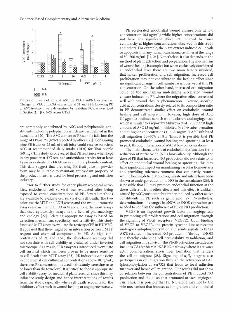


![Renal Protective Efficacy of Terminalia chebula ... · treatment of Terminalia chebula, Terminalia bellirica, Phyllanthus emblica and formulated drug Triphala [21]. The findings are](https://static.fdocuments.net/doc/165x107/5f1c729bd8c97163d7231976/renal-protective-efficacy-of-terminalia-chebula-treatment-of-terminalia-chebula.jpg)