RESEARCH ARTICLE Open Access Mast cell activation and ... · Mast cell activation and clinical...
8
RESEARCH ARTICLE Open Access Mast cell activation and clinical outcome in pediatric cholelithiasis and biliary dyskinesia Craig A Friesen 1* , Nancy Neilan 1 , James F Daniel 1 , Kim Radford 1 , Jennifer V Schurman 1 , Ding-You Li 1 , Linda Andre 1 , Shawn D St Peter 2 and George W Holcomb III 2 Abstract Background: The current study was undertaken to determine the degree of activation of gallbladder mucosal mast cells, whether mast cell (MC) density or activation differ between patients with and without a positive clinical response to cholecystectomy, and whether either density or activation correlate with gallbladder emptying. Results: Fifteen biliary dyskinesia (BD) and 13 symptomatic cholelithiasis (CL) patients undergoing cholecystectomy were prospectively enrolled. Gallbladder wall MC density (by immunohistochemistry) and activation (by electron microscopy) were determined. Clinical response was evaluated 30 days post-cholecystectomy on a 5-point Likert- type scale. A complete or nearly complete clinical response was seen in 100% of CL and in 87% of BD patients. The overall degranulation indices were 49.4 ± 18.7% for CL patients and 44.2 ± 16.8% for BD patients. Neither MC density nor activation correlated with the gallbladder ejection fraction. A complete clinical response was associated with lower epithelial MC density. Conclusion: Cholecystectomy is efficacious in relieving pain in both CL and BD patients. BD and CL are associated not only with increased MC density but a moderate to high degree of MC activation. A possible relationship between MC density and outcome for BD warrants further investigation. Keywords: mast cell, biliary dyskinesia, cholelithiasis, cholecystectomy Background The Rome III working group defined a number of func- tional gastrointestinal disorders in adults and children [1,2]. Functional gallbladder disorder (FGD), previously known as biliary dysfunction) was included in the adult but not the pediatric diagnoses. FGD is defined by recurrent symptoms of pain in the epigastrium or right upper quadrant lasting 30 minutes or longer and occur- ring at different intervals, among other criteria [3]. Sup- port for this diagnosis is provided by an abnormal gallbladder ejection fraction with a normal hepatobiliary ultrasound and laboratories [3]. For children, the entity which would most closely resemble FGD has been termed biliary dyskinesia. Biliary dyskinesia (BD) and cholelithiasis (CL) are the most common indications for cholecystectomy in children and adolescents [4-6]. Although there has been extensive evaluation of the pathophysiology of cholelithiasis, the pathophysiology of FGD or BD has not been well described. Previously we evaluated gallbladder wall inflammatory cells in children with symptomatic CL and BD [7]. These two conditions were associated with a 9 to 12-fold increase in mucosal MC density. This parallels other functional gastrointest- inal disorders, specifically functional dyspepsia and irri- table bowel syndrome, which have been associated with increased MCs in the stomach, small bowel, and/or large bowel [8-11]. While we have previously demonstrated increased MC density in BD and symptomatic CL, this is not sufficient alone to implicate MCs in the pathogenesis or genera- tion of symptoms in these patients. A large part of the biologic activity of MCs is attributable to released med- iators which are active in a concentration-dependent fashion [12]. Thus, the biologic activity of MCs is defined not only by density but by the degree and loca- tion of activation. This distinction can be demonstrated * Correspondence: [email protected] 1 Department of Pediatrics, The Children’s Mercy Hospital, 2401 Gillham Rd., Kansas City, Missouri, USA Full list of author information is available at the end of the article Friesen et al. BMC Research Notes 2011, 4:322 http://www.biomedcentral.com/1756-0500/4/322 © 2010 Friesen et al; licensee BioMed Central Ltd. This is an open access article distributed under the terms of the Creative Commons Attribution License (http://creativecommons.org/licenses/by/2.0), which permits unrestricted use, distribution, and reproduction in any medium, provided the original work is properly cited.
Transcript of RESEARCH ARTICLE Open Access Mast cell activation and ... · Mast cell activation and clinical...
RESEARCH ARTICLE Open Access
Mast cell activation and clinical outcome inpediatric cholelithiasis and biliary dyskinesiaCraig A Friesen1*, Nancy Neilan1, James F Daniel1, Kim Radford1, Jennifer V Schurman1, Ding-You Li1, Linda Andre1
, Shawn D St Peter2 and George W Holcomb III2
Abstract
Background: The current study was undertaken to determine the degree of activation of gallbladder mucosalmast cells, whether mast cell (MC) density or activation differ between patients with and without a positive clinicalresponse to cholecystectomy, and whether either density or activation correlate with gallbladder emptying.
Results: Fifteen biliary dyskinesia (BD) and 13 symptomatic cholelithiasis (CL) patients undergoing cholecystectomywere prospectively enrolled. Gallbladder wall MC density (by immunohistochemistry) and activation (by electronmicroscopy) were determined. Clinical response was evaluated 30 days post-cholecystectomy on a 5-point Likert-type scale. A complete or nearly complete clinical response was seen in 100% of CL and in 87% of BD patients.The overall degranulation indices were 49.4 ± 18.7% for CL patients and 44.2 ± 16.8% for BD patients. Neither MCdensity nor activation correlated with the gallbladder ejection fraction. A complete clinical response was associatedwith lower epithelial MC density.
Conclusion: Cholecystectomy is efficacious in relieving pain in both CL and BD patients. BD and CL are associatednot only with increased MC density but a moderate to high degree of MC activation. A possible relationshipbetween MC density and outcome for BD warrants further investigation.
Keywords: mast cell, biliary dyskinesia, cholelithiasis, cholecystectomy
BackgroundThe Rome III working group defined a number of func-tional gastrointestinal disorders in adults and children[1,2]. Functional gallbladder disorder (FGD), previouslyknown as biliary dysfunction) was included in the adultbut not the pediatric diagnoses. FGD is defined byrecurrent symptoms of pain in the epigastrium or rightupper quadrant lasting 30 minutes or longer and occur-ring at different intervals, among other criteria [3]. Sup-port for this diagnosis is provided by an abnormalgallbladder ejection fraction with a normal hepatobiliaryultrasound and laboratories [3]. For children, the entitywhich would most closely resemble FGD has beentermed biliary dyskinesia. Biliary dyskinesia (BD) andcholelithiasis (CL) are the most common indications forcholecystectomy in children and adolescents [4-6].
Although there has been extensive evaluation of thepathophysiology of cholelithiasis, the pathophysiology ofFGD or BD has not been well described. Previously weevaluated gallbladder wall inflammatory cells in childrenwith symptomatic CL and BD [7]. These two conditionswere associated with a 9 to 12-fold increase in mucosalMC density. This parallels other functional gastrointest-inal disorders, specifically functional dyspepsia and irri-table bowel syndrome, which have been associated withincreased MCs in the stomach, small bowel, and/orlarge bowel [8-11].While we have previously demonstrated increased MC
density in BD and symptomatic CL, this is not sufficientalone to implicate MCs in the pathogenesis or genera-tion of symptoms in these patients. A large part of thebiologic activity of MCs is attributable to released med-iators which are active in a concentration-dependentfashion [12]. Thus, the biologic activity of MCs isdefined not only by density but by the degree and loca-tion of activation. This distinction can be demonstrated
* Correspondence: [email protected] of Pediatrics, The Children’s Mercy Hospital, 2401 Gillham Rd.,Kansas City, Missouri, USAFull list of author information is available at the end of the article
Friesen et al. BMC Research Notes 2011, 4:322http://www.biomedcentral.com/1756-0500/4/322
© 2010 Friesen et al; licensee BioMed Central Ltd. This is an open access article distributed under the terms of the Creative CommonsAttribution License (http://creativecommons.org/licenses/by/2.0), which permits unrestricted use, distribution, and reproduction inany medium, provided the original work is properly cited.
by previous reports implicating MCs in the pathogenesisof IBS in adults. IBS has been associated with increasedMCs in the terminal ileum, cecum, proximal descendingcolon, and rectum [9-11,13]. IBS patients also demon-strate increased density of activated or degranulatingMCs as compared to controls and in IBS patients, MCsin close proximity to nerves are more likely to be acti-vated [11,13]. Only the MC density in close proximityto nerves is correlated with the severity and frequencyof pain in these patients [11].If MCs are to be implicated in the pathogenesis of BD
or symptomatic CL, not only do MCs need to beincreased in density but it needs to be demonstratedthat MCs are actively releasing mediators. The currentstudy was undertaken to assess the degree of gallbladdermucosal MC activation. In addition, this study evaluatedwhether MC density or activation differ betweenpatients with and without a positive clinical response tocholecystectomy.
MethodsThis was a prospective study evaluating gallbladder his-tology (specifically MC density and activation) and post-operative clinical response in patients undergoinglaparoscopic cholecystectomy between February, 2006and September, 2008 for symptomatic CL and BD. Thestudy was approved by the Institutional Review Board atChildren’s Mercy Hospital in Kansas City and writtenconsent/assent was obtained for all participants prior toparticipation in the study.
Patient SelectionPatients were eligible for enrollment if they had rightupper quadrant (RUQ) or epigastric pain of at least 8weeks duration from the onset of the first bout andwere scheduled for cholecystectomy. BD patients wererequired to demonstrate delayed gallbladder emptying(EF < 35%) following CCK stimulation on a single cho-lescintigraphy. Reproduction of pain during cholescinti-graphy was not considered. CL patients were requiredto have the presence of a gallstone within the gallblad-der on ultrasound.Patients were not excluded for pain in other regions in
addition to the RUQ or epigastric pain. Patients wereexcluded from either group for evidence of biliaryobstruction defined as direct bilirubin ≥ 1.0 mg/dL onany previous laboratory evaluations or common bileduct diameter ≥ 7 mm on any previous ultrasound.Patients were excluded if they had received anti-inflam-matory medications or had a history of inflammatorybowel disease, rheumatologic disease, or cancer. In addi-tion, BD patients were required to exhibit normal filling(< 1 hour)on cholescintigraphy.
HistologySections from the body of the resected gallbladders wereembedded and processed in the usual fashion includingstaining with hematoxylin and eosin for the usual evalua-tion by a board certified pediatric pathologist. Additionalspecimens were obtained for immunohistochemical (IHC)staining for MCs. For IHC evaluation, serial 3- μm paraf-fin sections were air dried and fixed on slides. The sec-tions were deparraffinized using a xylene substitute andthen rehydrated in alcohol to tris-buffered saline. Endo-genous peroxidase activity was blocked using 3% hydro-gen peroxide followed by a protein block with 5% goatserum. Residual biotin and avidin activity were quenchedusing avidin and biotin block, respectively. Monoclonalmouse anti-human mast cell tryptase, Clone AA1 wasused as the primary antibody and applied as a 1:1000dilution for 1 hour at room temperature (18°C - 25°C).Labeled streptavidin-biotin (LSAB) was used for thedetection system with diaminobenzidine tetrahydrochlor-ide (DAB) as the chromogen. Sections were counter-stained with hematoxylin. MCs were enumerated for 10high power fields per layer (epithelium, lamina propria,and muscularis mucosae) by a single investigator (NN).The largest MC count in any 40X high power field (hpf)was reported as the peak and the average number/hpfwas reported as the mean.
Electron MicroscopySpecimens for electron microscopic (EM) evaluationwere obtained in the operating room immediately fol-lowing cholecystectomy and were placed in 2% glutaral-dehyde in 0.1 M sodium cacodylate, pH 7.4. Allspecimens were taken from the body of the gallbladderand were approximately 5 mm × 5 mm. Specimens werepost fixed in osmium tetroxide. The tissue was thendehydrated in graded alcohols and embedded in epoxyresin. Sections were cut at 80 nm thickness and con-trasted with uranyl citrate and lead citrate. TransmittingEM was performed and MCs were identified and photo-graphed at 4800X. The photographs were then evaluatedand the MC degranulation index was calculated by pre-viously described methodology [14]. The number ofgranules exhibiting loss of density or vacuolization wasdivided by the total number of granules. Degranulationindices were not evaluated by layer as the layers couldnot be precisely distinguished on EM.
Clinical OutcomeA standardized history regarding the patient’s symp-toms was obtained pre-operatively and 30 days follow-ing cholecystectomy. The follow-up evaluation alsoincluded an assessment of the patient’s global clinicalresponse utilizing a Likert-type scale adapted to assess
Friesen et al. BMC Research Notes 2011, 4:322http://www.biomedcentral.com/1756-0500/4/322
Page 2 of 8
the change in pain by patient report. The five painrelief grades were:
Grade 1 Worse- clinical deterioration with increas-ing pain intensity and/or frequency.Grade 2 No change- no increase or decrease in painintensity or frequencyGrade 3 Moderate improvement- partial clinicalresponse with definite improvement in pain, but notmeeting the criteria for a Grade 4 responseGrade 4 Good- nearly complete relief of symptomswith minimal residual pain and pain not interferingwith daily activitiesGrade 5 Excellent- complete relief of pain
StatisticsThe frequencies for individual symptoms were com-pared between the pre- and post-cholecystectomy timepoints by chi square analysis or the Fisher’s exact test.The percentage of patients with elevated peak and meancell densities, respectively, was determined. The upperlimit of normal was determined as the mean + 2 SDfrom previous control data with upper limits defined asmean > 3.5/hpf in the LP, peak > 5.78/hpf in the LP,mean > 1.66/hpf in the MM, and peak > 4.19 in theMM [7]. MC densities were compared between BD andCL patients by the student’s t test. MC densities werecompared between patients with and without chroniccholecystitis for all patients and for BD patients only bythe student’s t test. Pearson correlation coefficients weredetermined for the gallbladder ejection fractions withthe MC densities and MC degranulation indices, respec-tively. Pearson correlation coefficients were determinedfor length of symptoms with the MC densities and MCdegranulation indices, respectively. Mucosal MC densi-ties were compared by layer between patients with acomplete clinical response (grade 5) and lesser respon-ders by ANOVA. A p value < .05 was consideredsignificant.
ResultsFifteen patients with BD and 13 symptomatic patientswith CL who were scheduled for cholecystectomy wereprospectively enrolled in this study. BD patients rangedin age from 8 to 17 years (mean 13.3 ± 2.7 years). Mostwere female (87%) and Caucasian (93%). CL patientsranged in age from 6 to 18 years (mean 12.2 ± 3.0years). Again, most (85%) were female, 77% were Cauca-sian, 15% were African-American, and 8% were Hispa-nic. The presenting symptoms are shown in Table 1.For patients with BD, 66.7% had pain only in the rightupper quadrant (RUQ), 6.7% only in the epigastrium,and 6.7% in both the RUQ and epigastrium. For CL
patients, 69.2% had pain only in the RUQ, 15.4% only inthe epigastrium, and 7.7% in both the RUQ and epigas-trium. The remaining patients also had pain in otherareas of the abdomen.The percentage of patients exhibiting a particular
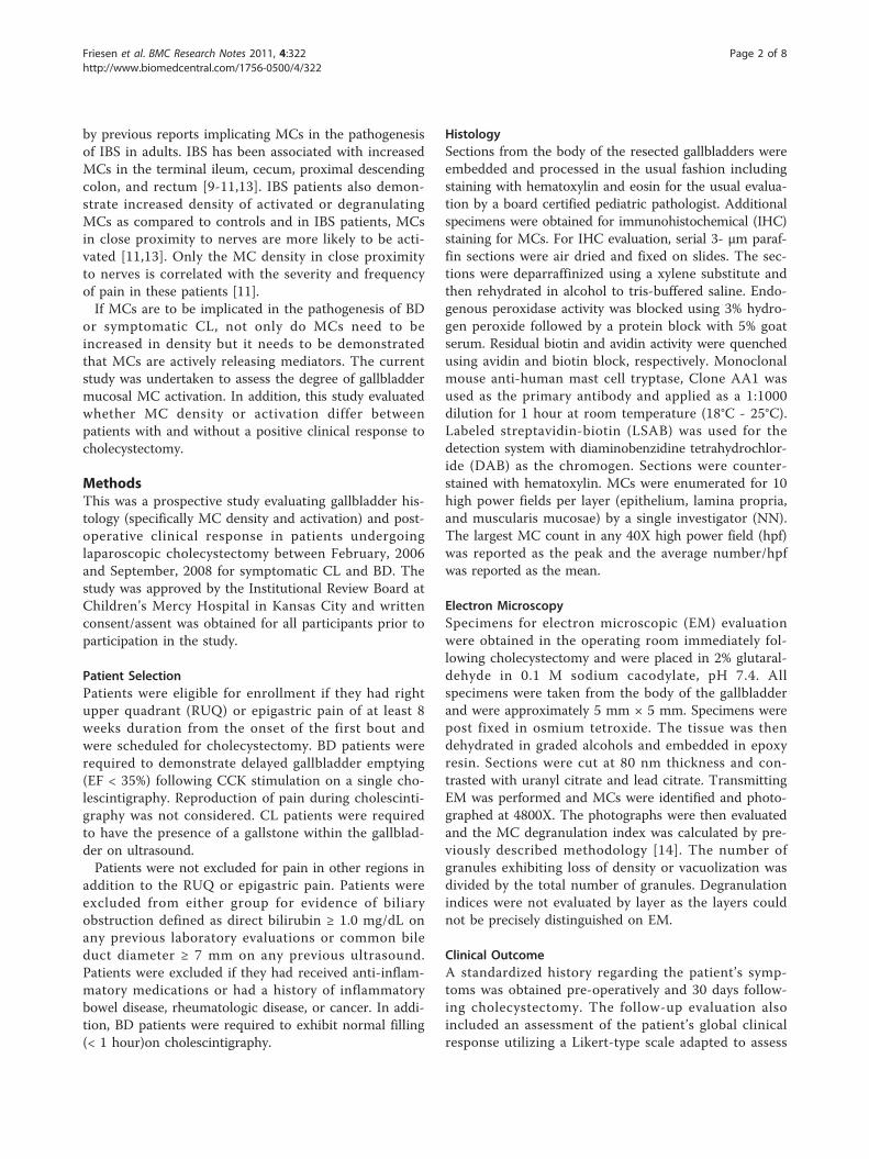
symptom at 30 days post-cholecystectomy is also shownin Table 1. One CL patient was lost to follow-up. Therewas a significant decrease in the frequency of pain, painwith eating, night time waking with pain, and nausea,respectively, in both patient groups. There was a signifi-cant decrease in vomiting frequency in the CL patientsonly. The global response grades are shown in Figure 1.Ninety- two percent of CL patients achieved a grade 4or 5 response. Ninety-three percent of BD patients hada positive clinical response with 87% achieving a grade 4or 5 response.
Table 1 The percentage of patients exhibiting particularsymptoms preoperatively and 30 days post-cholecystectomy
Cholelithiasis Biliary Dyskinesia
Symptom Pre Post pvalue
Pre Post pvalue
Pain 100% 15.4% < .01 100% 20% < .01
Pain with eating 61.5% 15.4% < .01 80% 6.7% .04
Night waking with pain 84.6% 7.7% .02 60% 6.7% < .01
Nausea 100% 15.4% < .01 80% 20% < .01
Vomiting 53.8% 7.7% < .01 40% 6.7% .03
Diarrhea 23.1% 7.7% .17 33.3% 6.7% .17
Constipation 30.8% 46.2% .40 20% 20% 1.00
Pain decreased withstool
7.7% 7.7% 1.00 26.7% 20% 1.00
Figure 1 Global clinical response grades for biliary dyskinesia(BD) and cholelithiasis (CL) patients 30 days post-cholecystectomy.
Friesen et al. BMC Research Notes 2011, 4:322http://www.biomedcentral.com/1756-0500/4/322
Page 3 of 8
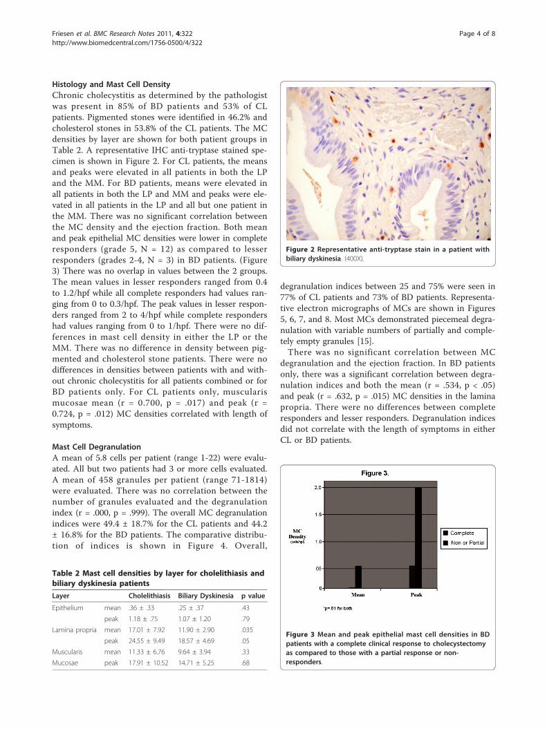
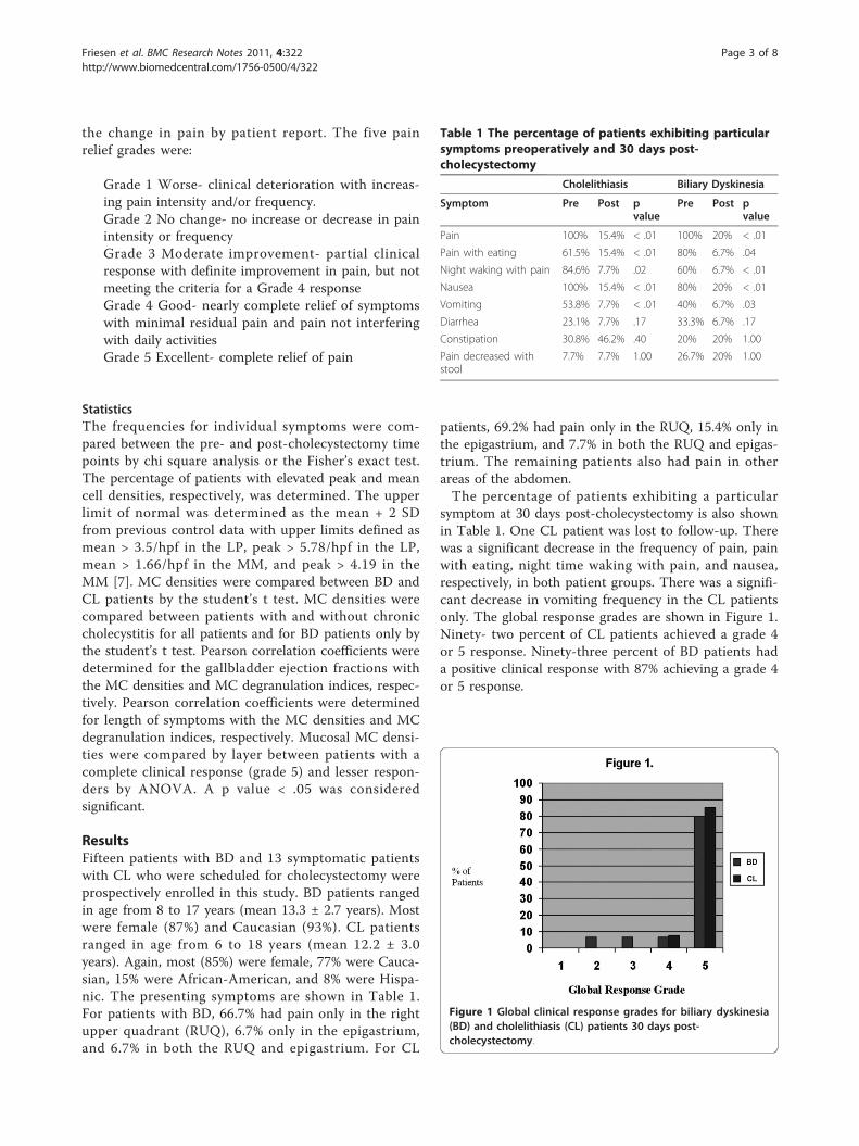
Histology and Mast Cell DensityChronic cholecystitis as determined by the pathologistwas present in 85% of BD patients and 53% of CLpatients. Pigmented stones were identified in 46.2% andcholesterol stones in 53.8% of the CL patients. The MCdensities by layer are shown for both patient groups inTable 2. A representative IHC anti-tryptase stained spe-cimen is shown in Figure 2. For CL patients, the meansand peaks were elevated in all patients in both the LPand the MM. For BD patients, means were elevated inall patients in both the LP and MM and peaks were ele-vated in all patients in the LP and all but one patient inthe MM. There was no significant correlation betweenthe MC density and the ejection fraction. Both meanand peak epithelial MC densities were lower in completeresponders (grade 5, N = 12) as compared to lesserresponders (grades 2-4, N = 3) in BD patients. (Figure3) There was no overlap in values between the 2 groups.The mean values in lesser responders ranged from 0.4to 1.2/hpf while all complete responders had values ran-ging from 0 to 0.3/hpf. The peak values in lesser respon-ders ranged from 2 to 4/hpf while complete respondershad values ranging from 0 to 1/hpf. There were no dif-ferences in mast cell density in either the LP or theMM. There was no difference in density between pig-mented and cholesterol stone patients. There were nodifferences in densities between patients with and with-out chronic cholecystitis for all patients combined or forBD patients only. For CL patients only, muscularismucosae mean (r = 0.700, p = .017) and peak (r =0.724, p = .012) MC densities correlated with length ofsymptoms.
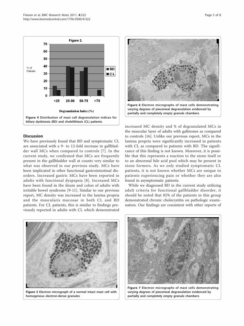
Mast Cell DegranulationA mean of 5.8 cells per patient (range 1-22) were evalu-ated. All but two patients had 3 or more cells evaluated.A mean of 458 granules per patient (range 71-1814)were evaluated. There was no correlation between thenumber of granules evaluated and the degranulationindex (r = .000, p = .999). The overall MC degranulationindices were 49.4 ± 18.7% for the CL patients and 44.2± 16.8% for the BD patients. The comparative distribu-tion of indices is shown in Figure 4. Overall,
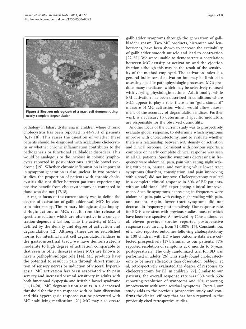
degranulation indices between 25 and 75% were seen in77% of CL patients and 73% of BD patients. Representa-tive electron micrographs of MCs are shown in Figures5, 6, 7, and 8. Most MCs demonstrated piecemeal degra-nulation with variable numbers of partially and comple-tely empty granules [15].There was no significant correlation between MC
degranulation and the ejection fraction. In BD patientsonly, there was a significant correlation between degra-nulation indices and both the mean (r = .534, p < .05)and peak (r = .632, p = .015) MC densities in the laminapropria. There were no differences between completeresponders and lesser responders. Degranulation indicesdid not correlate with the length of symptoms in eitherCL or BD patients.
Table 2 Mast cell densities by layer for cholelithiasis andbiliary dyskinesia patients
Layer Cholelithiasis Biliary Dyskinesia p value
Epithelium mean .36 ± .33 .25 ± .37 .43
peak 1.18 ± .75 1.07 ± 1.20 .79
Lamina propria mean 17.01 ± 7.92 11.90 ± 2.90 .035
peak 24.55 ± 9.49 18.57 ± 4.69 .05
Muscularis mean 11.33 ± 6.76 9.64 ± 3.94 .33
Mucosae peak 17.91 ± 10.52 14.71 ± 5.25 .68
Figure 2 Representative anti-tryptase stain in a patient withbiliary dyskinesia. (400X).
Figure 3 Mean and peak epithelial mast cell densities in BDpatients with a complete clinical response to cholecystectomyas compared to those with a partial response or non-responders.
Friesen et al. BMC Research Notes 2011, 4:322http://www.biomedcentral.com/1756-0500/4/322
Page 4 of 8
DiscussionWe have previously found that BD and symptomatic CLare associated with a 9- to 12-fold increase in gallblad-der wall MCs when compared to controls [7]. In thecurrent study, we confirmed that MCs are frequentlypresent in the gallbladder wall at counts very similar towhat was observed in our previous study. MCs havebeen implicated in other functional gastrointestinal dis-orders. Increased gastric MCs have been reported inadults with functional dyspepsia [8]. Increased MCshave been found in the ileum and colon of adults withirritable bowel syndrome [9-11]. Similar to our previousreport, MC density was increased in the lamina propriaand the muscularis mucosae in both CL and BDpatients. For CL patients, this is similar to findings pre-viously reported in adults with CL which demonstrated
increased MC density and % of degranulated MCs inthe muscular layer of adults with gallstones as comparedto controls [16]. Unlike our previous report, MCs in thelamina propria were significantly increased in patientswith CL as compared to patients with BD. The signifi-cance of this finding is not known. Moreover, it is possi-ble that this represents a reaction to the stone itself orto an abnormal bile acid pool which may be present instone formers. As we only studied symptomatic CLpatients, it is not known whether MCs are unique topatients experiencing pain or whether they are alsofound in asymptomatic patients.While we diagnosed BD in the current study utilizing
adult criteria for functional gallbladder disorder, itshould be noted that 85% of the patients in this groupdemonstrated chronic cholecystitis on pathologic exami-nation. Our findings are consistent with other reports of
Figure 4 Distribution of mast cell degranulation indices forbiliary dyskinesia (BD) and cholelithiasis (CL) patients.
Figure 5 Electron micrograph of a normal intact mast cell withhomogenous electron-dense granules.
Figure 6 Electron micrographs of mast cells demonstratingvarying degrees of piecemeal degranulation evidenced bypartially and completely empty granule chambers.
Figure 7 Electron micrographs of mast cells demonstratingvarying degrees of piecemeal degranulation evidenced bypartially and completely empty granule chambers.
Friesen et al. BMC Research Notes 2011, 4:322http://www.biomedcentral.com/1756-0500/4/322
Page 5 of 8
pathology in biliary dyskinesia in children where chroniccholecystitis has been reported in 44-93% of patients[6,17,18]. This raises the question of whether thesepatients should be diagnosed with acalculous cholecysti-tis or whether chronic inflammation contributes to thepathogenesis or functional gallbladder disorders. Thiswould be analogous to the increase in colonic lympho-cytes reported in post-infectious irritable bowel syn-drome [19]. Whether chronic inflammation is importantin symptom generation is also unclear. In two previousstudies, the proportion of patients with chronic chole-cystitis did not differ between patients experiencingpositive benefit from cholecystectomy as compared tothose who did not [17,18].A major focus of the current study was to define the
degree of activation of gallbladder wall MCs by elec-tron microscopy. The primary biologic and pathophy-siologic actions of MCs result from the release ofspecific mediators which are often active in a concen-tration-dependent fashion. Thus the activity of MCs isdefined by the density and degree of activation anddegranulation [12]. Although there are no establishednorms for intestinal mast cell degranulation indices inthe gastrointestinal tract, we have demonstrated amoderate to high degree of activation comparable tothat seen in other diseases where MCs are known tohave a pathophysiologic role [14]. MC products havethe potential to result in pain through direct stimula-tion of sensory nerves or induction of visceral hyperal-gesia. MC activation has been associated with painseverity and increased visceral sensitivity in adults withboth functional dyspepsia and irritable bowel syndrome[11,14,20]. MC degranulation results in a decreasedthreshold for the pain response with balloon distensionand this hyperalgesic response can be prevented withMC-stabilizing medication [21] MC may also create
gallbladder symptoms through the generation of gall-bladder spasm. Two MC products, histamine and leu-kotrienes, have been shown to increase the excitabilityof gallbladder smooth muscle and lead to contraction[22-25]. We were unable to demonstrate a correlationbetween MC density or activation and the ejectionfraction although this may be the result of the sensitiv-ity of the method employed. The activation index is ageneral indicator of activation but may be limited inassessing specific pathophysiologic processes. MCs pro-duce many mediators which may be selectively releasedwith varying physiologic actions. Additionally, whileEM activation has been described in conditions whereMCs appear to play a role, there is no “gold standard”measure of MC activation which would allow assess-ment of the accuracy of degranulation indices. Furtherwork is necessary to determine if specific mediatorsare responsible for the observed dysmotility.Another focus of the current study was to prospectively
evaluate global response, to determine which symptomsimprove with cholecystectomy, and to evaluate whetherthere is a relationship between MC density or activationand clinical response. Consistent with previous reports, acomplete or nearly complete clinical response was seenin all CL patients. Specific symptoms decreasing in fre-quency were abdominal pain, pain with eating, night wak-ing with pain, nausea, and vomiting while lower tractsymptoms (diarrhea, constipation, and pain improvingwith a stool) did not improve. Cholecystectomy resultedin a complete clinical response in 80% of BD patients,with an additional 15% experiencing clinical improve-ment. Specific symptoms decreasing in frequency wereabdominal pain, pain with eating, night waking with pain,and nausea. Again, lower tract symptoms did notdecrease in frequency postoperatively. Our response ratefor BD is consistent with previous studies, most of whichhave been retrospective. As reviewed by Constantinou, etal, eleven previous studies reported postoperativeresponse rates varying from 71-100% [17]. Constantinou,et al, also reported outcomes following cholecystectomyin 100 children with BD where outcome data were col-lected prospectively [17]. Similar to our patients, 77%reported resolution of symptoms at 6 months to 5 yearspostoperatively. The only randomized trial for BD wasperformed in adults [26] This study found cholecystect-omy to be more efficacious than observation. Siddiqui, etal, retrospectively evaluated the degree of response tocholecystectomy for BD in children [27]. Similar to ourpatients, the overall response rate was 93% with 65%reporting resolution of symptoms and 28% reportingimprovement with some residual symptoms. Overall, ourstudy adds to the previous prospective study and con-firms the clinical efficacy that has been reported in thepreviously cited retrospective studies.
Figure 8 Electron micrograph of a mast cell demonstratingnearly complete degranulation.
Friesen et al. BMC Research Notes 2011, 4:322http://www.biomedcentral.com/1756-0500/4/322
Page 6 of 8
The only relationship between MC activation or densityand the clinical response grade for either patient groupwas demonstrated in the BD patients. A complete clinicalresponse was associated with lower epithelial MC densitywhen compared to a partial or non-response. The reasonfor this is not clear. It is possible that higher MC densitymay be associated with MCs in the gastrointestinal tractresulting in dyspepsia or irritable bowel syndrome. Thiscould not be evaluated in the current study as only a fewpatients underwent endoscopic evaluation. There areinsufficient data to determine whether there is clinicalvalue to obtaining gastrointestinal biopsies prior to chole-cystectomy. These data (both the positive findings in theepithelium and the negative findings in other layers) needto be interpreted with caution as there were so few par-tial or non-responders for comparison. The positive find-ings may simply represent a type II statistical error.Larger studies are necessary to evaluate the relationshipbetween MCs and outcome.
ConclusionsIn the current study, we have demonstrated a moderateto high degree of mast cell activation in BD and CLwhich along with the previously demonstrated high MCdensity (confirmed in the current study) suggests a pos-sible role for MCs in the pathophysiology of these con-ditions. Future work evaluating concentrations ofgallbladder wall mediators such as proteases or cyto-kines is necessary to further delineate possible patho-physiologic mechanisms. We have demonstrated arelationship between MC density and outcome for bili-ary dyskinesia which also warrants further investigation.A better understanding of the pathophysiology may leadto better identification of patients who will have clinicalbenefit from cholecystectomy or who might respond tomedications such as mast cell stabilizers.
List of AbbreviationsMC: mast cell; BD: biliary dyskinesia; CL: cholelithiasis; FGD: functionalgallbladder disorder; IBS: irritable bowel syndrome; RUQ: right upperquadrant; CCK: cholecystokinin; IHC: immunohistochemical; EM: electronmicroscopic; LP: lamina propria; MM: muscularis mucosae.
AcknowledgementsThis work was supported by the Kathryn B. Richardson Foundation. Thefunders had no role in the study design, data collection and analysis,decision to publish, or preparation of the manuscript.
Author details1Department of Pediatrics, The Children’s Mercy Hospital, 2401 Gillham Rd.,Kansas City, Missouri, USA. 2Department of Surgery, The Children’s MercyHospital, 2401 Gillham Rd., Kansas City, Missouri, USA.
Authors’ contributionsCF conceived of the study, analyzed the data, and wrote the first draft ofthe manuscript. All authors participated in the design of the study andcritically revised the manuscript. NN performed all of the lab work. Authorsread and approved the final manuscript.
Competing interestsThe authors declare that they have no competing interests.
Received: 25 May 2011 Accepted: 6 September 2011Published: 6 September 2011
References1. Drossman DA: The functional gastrointestinal disorders and the Rome III
process. Gastroenterology 2006, 130:1377-1390.2. Rasquin A, Di Lorenzo C, Forbes D, Guiraldes E, Hyams JS, Staiano A,
Walker LS: Childhood functional gastrointestinal disorders: child/adolescent. Gastroenterology 2006, 130:1527-1537.
3. Behar J, Corazziari E, Guelrud M, Hogan W, Sherman S, Toouli J: Functionalgallbladder and sphincter of Oddi disorders. Gastroenterology 2006,130:1498-1509.
4. St Peter SD, Keckler SJ, Nair A, Andrews WS, Sharp RJ, Snyder CL, Ostlie DJ,Holcomb GW III: Laparoscopic cholecystectomy in the pediatricpopulation. J Laparoendosc Adv Surg Tech 2008, 18:127-130.
5. Vegunta RK, Raso M, Pollock J, Misra S, Wallace LJ, Torres A Jr, Pearl RH:Biliary dyskinesia: the most common indication for cholecystectomy inchildren. Surgery 2005, 138:726-733.
6. Hofeldt M, Richmond B, Huffman K, Nestor J, Maxwell D: Laparoscopicchole-cystectomy for treatment of biliary dyskinesia is safe and effectivein the pediatric population. Amer Surg 2008, 74:1069-1072.
7. Rau B, Friesen CA, Daniel JF, Qadeer A, Li D-Y, Roberts CC, Holcomb GW III:Gallbladder wall inflammatory cells in pediatric patients with biliarydyskinesia and cholelithiasis: a pilot study. J Pediatr Surg 2006,41:1545-1548.
8. Hall W, Buckley M, Crotty P, O’Morain CA: Gastric mucosal mast cells areincreased in Helicobacter pylori-negative functional dyspepsia. ClinGastroenterol Hepatol 2003, 1:363-369.
9. Weston AP, Biddle WL, Bhatia PS, Miner PB Jr: Terminal ileal mucosal mastcells in irritable bowel syndrome. Dig Dis Sci 1993, 38:1590-1595.
10. O’Sullivan M, Clayton N, Breslin NP, Harman I, Bountra C, McLaren A,O’Morain CA: Increased mast cells in the irritable bowel syndrome.Neurogastroenterol Motil 2000, 12:449-457.
11. Barbara G, Stranghellini V, De Giorgio R, Cremon C, Cottrell GS, Santini D,Pasquinelli G, Morselli-Labate AM, Grady EF, Bunnett NW, Collins SM,Corinaldesi R: Activated mast cells in proximity to colonic nervescorrelate with abdominal pain in irritable bowel syndrome.Gastroenterology 2004, 126:693-702.
12. Stone KD, Prussin C, Metcalfe DD: IgE, mast cells, basophils, andeosinophils. J Allergy Clin Immunol 2010, 125:S73-S80.
13. Park CH, Joo YE, Choi SK, Rew JS, Kim SJ, Lee MC: Activated mast cellsinfiltrate in close proximity to enteric nerves in diarrhea-predominantirritable bowel syndrome. J Korean Med Soc 2003, 18:204-210.
14. Drake-Lee AB, Price J: Ultrastructure of nasal mast cells in normalsubjects and patients with perennial allergic rhinitis. J Laryngol Otol 1991,105:1006-1013.
15. Dvorak AM: Ultrastructural studies of human basophils and mast cells. JHistochem Cytochem 2005, 53:1043-1070.
16. Carotti S, Guarino MPL, Cicala M, Perrone G, Alloni R, Segreto F, Rabitti C,Morini S: Effect of ursodeoxycholic acid on inflammatory infiltrate ingallbladder muscleof cholesterol gallstone patients. NeurogastroenterolMotil 2010, 22:866-e232.
17. Constantinou C, Sucandy I, Ramenofsky M: Laparoscopic cholecystectomyfor biliary dyskinesia in children: report of 100 cases from a singleinstitution. Amer Surg 2008, 74:587-592.
18. Kaye AJ, Jatla M, Mattei P, Kelly J, Nance ML: Use of laparoscopic chole-cystectomy for biliary dyskinesia in the child. J Pediatr Surg 2008,43:1057-1059.
19. Spiller RC: Postinfectious irritable bowel syndrome. Gastroenterology 2003,124:1662-1671.
20. Hou X-H, Zhu L-R, Li Q-X, Chen JDZ: Alterations in mast cells and 5-HT-positive cells in gastric mucosa in functional dyspepsia patients withhypersensitivity. Neurogastroenterol Motil 2001, 13:398-399.
21. Wood JD: Neuropathophysiology of irritable bowel syndrome. J ClinGastroenterol 2002, 35(1 Suppl):s11-s22.
22. Jennings LJ, Salido GM, Pozo MJ, Davison JS, Sharkey KA, Lea RW, Singh J:The source and action of histamine in the isolated guinea-piggallbladder. Inflamm Res 1995, 44:447-453.
Friesen et al. BMC Research Notes 2011, 4:322http://www.biomedcentral.com/1756-0500/4/322
Page 7 of 8
23. Woods CM, Mawe GM, Shaffer EA, Tooulli J, T P Saccone G: Effects ofbioactive agents on biliary motor function. Curr Gastroenterol Rep 2003,5:154-159.
24. Freedman SM, Wallace JL, Shaffer EA: Characterization of leukotriene-induced contraction of the guinea-pig gallbladder in vitro. Can J PhysiolPharmacol 1993, 71:145-150.
25. Yusko P, Hall RA, Ford-Hutchinson AW: Contraction of guinea piggallbladder strips by leukotrienes and other agonists. Prostaglandins1983, 25:397-403.
26. Yap L, Wycherley AG, Morphett AD, Toouli J: Acalculous biliary pain:cholecystectomy alleviates symptoms in patients with abnormalcholescintigraphy. Gastroenterology 1991, 101:786-793.
27. Siddiqui S, Newbrough S, Alterman D, Anderson A, Kennedy A Jr: Efficacyof laparoscopic cholecystectomy in the pediatric population. J PediatrSurg 2008, 43:109-113.
doi:10.1186/1756-0500-4-322Cite this article as: Friesen et al.: Mast cell activation and clinicaloutcome in pediatric cholelithiasis and biliary dyskinesia. BMC ResearchNotes 2011 4:322.
Submit your next manuscript to BioMed Centraland take full advantage of:
• Convenient online submission
• Thorough peer review
• No space constraints or color figure charges
• Immediate publication on acceptance
• Inclusion in PubMed, CAS, Scopus and Google Scholar
• Research which is freely available for redistribution
Submit your manuscript at www.biomedcentral.com/submit
Friesen et al. BMC Research Notes 2011, 4:322http://www.biomedcentral.com/1756-0500/4/322
Page 8 of 8