Research Article New Bioactive Fungal Molecules with...
12
Hindawi Publishing Corporation BioMed Research International Volume 2013, Article ID 497492, 11 pages http://dx.doi.org/10.1155/2013/497492 Research Article New Bioactive Fungal Molecules with High Antioxidant and Antimicrobial Capacity Isolated from Cerrena unicolor Idiophasic Cultures Magdalena Jaszek, Monika OsiNska-Jaroszuk, Grzegorz Janusz, Anna Matuszewska, Dawid Stefaniuk, Justyna Sulej, Jolanta Polak, Marta Ruminowicz, Krzysztof Grzywnowicz, and Anna Jarosz-WilkoBazka Department of Biochemistry, Maria Curie-Skłodowska University, Akademicka 19, 20-033 Lublin, Poland Correspondence should be addressed to Magdalena Jaszek; [email protected] Received 29 March 2013; Revised 7 June 2013; Accepted 23 June 2013 Academic Editor: Elvira Gonzalez De Mejia Copyright © 2013 Magdalena Jaszek et al. is is an open access article distributed under the Creative Commons Attribution License, which permits unrestricted use, distribution, and reproduction in any medium, provided the original work is properly cited. ree bioactive fractions, extracellular laccase (ex-LAC), crude endopolysaccharides (c-EPL), and a low molecular subfraction of secondary metabolites (ex-LMS), were isolated from the idiophasic cultures of the white rot fungus Cerrena unicolor. For the first time, we determined the antioxidant properties of these samples by chemiluminometric measurement (a) and assessment of the scavenging effect on ABTS (b) and the DPPH reduction rate (c). e highest reducing capability was found for the ex-LMS fraction: 39–90% for (a), 20–90% for (b), and 10–59% for (c) at the concentration of 6.25–800 g/mL. e scavenging abilities of the C. unicolor c-EPL were between 36 and 70% for (a), 2 and 60% for (b), and 28 and 32% for (c) at the concentration of 6.25– 800 g/mL. A very high prooxidative potential was observed for the ex-LAC probes. e preliminary toxicity tests were done using the Microtox system and revealed the following percentage of the toxic effect against Vibrio fischeri: 85.37% for c-EPL, 50.67% for ex-LAC, and 99.8% for ex-LMS, respectively. e ex-LAC sample showed the antibacterial activity against Escherichia coli, c-EPL against Staphylococcus aureus, and ex-LMS against both bacterial strains, respectively, but the stronger inhibitory effect was exerted on S. aureus. 1. Introduction A large number of fungal bioactive compounds, both cellular components and secondary metabolites, have been shown to affect the human immune system and could be used to treat a variety of diseases [1]. Recent intensification in the application of bioactive compounds produced by white rot fungi in the food processing or pharmaceutical industry [2] is stimulating a worldwide search for new natural bioactive compounds of fungal origin. Moreover, their production has become an important field of contemporary biotechnology. A number of these substances including intra- and extracellular low molecular weight compounds, proteins, polysaccharides, or polysaccharide-protein complexes have been isolated from wood-degrading fungal strains [3, 4]. Given the worldwide research, some authors have proposed dividing the bioactive compounds isolated from higher mushrooms into the follow- ing groups: (a) secondary metabolites (terpenoids, polyphe- nols, alkaloids, lactones, sterols, etc.), (b) (glyco)proteins, and (c) high molecular weight polysaccharides [5]. Numerous publications indicate that the most important problem for living organisms is posed by uncontrolled pro- duction of reactive oxygen species (ROS), very active by- products having one or more unpaired electrons [6]. Reactive oxygen derivatives are generated during the normal cellular metabolism or as a consequence of exposition to some stress factors, such as changes in temperature, and the presence of metal ions or redox-cycling xenobiotics [6, 7]. As a protection
Transcript of Research Article New Bioactive Fungal Molecules with...

Hindawi Publishing CorporationBioMed Research InternationalVolume 2013, Article ID 497492, 11 pageshttp://dx.doi.org/10.1155/2013/497492
Research ArticleNew Bioactive Fungal Molecules with HighAntioxidant and Antimicrobial Capacity Isolated fromCerrena unicolor Idiophasic Cultures
Magdalena Jaszek, Monika OsiNska-Jaroszuk, Grzegorz Janusz, Anna Matuszewska,Dawid Stefaniuk, Justyna Sulej, Jolanta Polak, Marta Ruminowicz, Krzysztof Grzywnowicz,and Anna Jarosz-WilkoBazka
Department of Biochemistry, Maria Curie-Skłodowska University, Akademicka 19, 20-033 Lublin, Poland
Correspondence should be addressed to Magdalena Jaszek; [email protected]
Received 29 March 2013; Revised 7 June 2013; Accepted 23 June 2013
Academic Editor: Elvira Gonzalez De Mejia
Copyright © 2013 Magdalena Jaszek et al. This is an open access article distributed under the Creative Commons AttributionLicense, which permits unrestricted use, distribution, and reproduction in any medium, provided the original work is properlycited.
Three bioactive fractions, extracellular laccase (ex-LAC), crude endopolysaccharides (c-EPL), and a low molecular subfractionof secondary metabolites (ex-LMS), were isolated from the idiophasic cultures of the white rot fungus Cerrena unicolor. For thefirst time, we determined the antioxidant properties of these samples by chemiluminometric measurement (a) and assessment ofthe scavenging effect on ABTS (b) and the DPPH reduction rate (c). The highest reducing capability was found for the ex-LMSfraction: 39–90% for (a), 20–90% for (b), and 10–59% for (c) at the concentration of 6.25–800 𝜇g/mL. The scavenging abilities ofthe C. unicolor c-EPL were between 36 and 70% for (a), 2 and 60% for (b), and 28 and 32% for (c) at the concentration of 6.25–800𝜇g/mL. A very high prooxidative potential was observed for the ex-LAC probes.The preliminary toxicity tests were done usingthe Microtox system and revealed the following percentage of the toxic effect against Vibrio fischeri: 85.37% for c-EPL, 50.67% forex-LAC, and 99.8% for ex-LMS, respectively. The ex-LAC sample showed the antibacterial activity against Escherichia coli, c-EPLagainst Staphylococcus aureus, and ex-LMS against both bacterial strains, respectively, but the stronger inhibitory effect was exertedon S. aureus.
1. Introduction
A large number of fungal bioactive compounds, both cellularcomponents and secondary metabolites, have been shownto affect the human immune system and could be used totreat a variety of diseases [1]. Recent intensification in theapplication of bioactive compounds produced by white rotfungi in the food processing or pharmaceutical industry [2]is stimulating a worldwide search for new natural bioactivecompounds of fungal origin. Moreover, their production hasbecome an important field of contemporary biotechnology. Anumber of these substances including intra- and extracellularlowmolecular weight compounds, proteins, polysaccharides,or polysaccharide-protein complexes have been isolated from
wood-degrading fungal strains [3, 4]. Given the worldwideresearch, some authors have proposed dividing the bioactivecompounds isolated from highermushrooms into the follow-ing groups: (a) secondary metabolites (terpenoids, polyphe-nols, alkaloids, lactones, sterols, etc.), (b) (glyco)proteins, and(c) high molecular weight polysaccharides [5].
Numerous publications indicate that the most importantproblem for living organisms is posed by uncontrolled pro-duction of reactive oxygen species (ROS), very active by-products having one ormore unpaired electrons [6]. Reactiveoxygen derivatives are generated during the normal cellularmetabolism or as a consequence of exposition to some stressfactors, such as changes in temperature, and the presence ofmetal ions or redox-cycling xenobiotics [6, 7]. As a protection

2 BioMed Research International
system for the balance between the production and inactiva-tion of ROS, living organisms have evolved specific defensemechanisms for detoxification, consisting of enzymatic andnonenzymatic antioxidants [8]. An uncontrolled increase inthe level of free radicals can cause damage to numerous cellu-lar compounds includingDNA, proteins, ormembrane lipids,in consequence leading to many serious human diseases anddisorders, such as atherosclerosis, coronary heart disease,cancer, impaired immune function, or aging processes [6,9, 10]. This outcome suggests that, with their capacity toscavenge free radicals, antioxidants may protect organismsfrom oxidative stress-caused damage. Many polysaccharidesextracted from Lentinus polychrous, Ganoderma atrum, Gri-fola frondosa, and Lentinus edodeshave been reported to havenot only antioxidant activity but also they are believed tobe bioactive ingredients involved in both the antitumor andanti-inflammatory processes [6, 10]. Furthermore, differentother fractions isolated from Ganoderma lucidum, Phellinuslinteus, Agaricus campestris, Lentinus edodes, Agaricus blazei,or Pleurotus ostreatus were reported to possess the sametherapeutic effects [10–12].
At the same time, investigation and isolation of newnatural bioactive substances are also important for foodindustry due to the growing importance of their antioxidativeactivity, which is crucial in food preservation processes.Unfortunately, the commonly used synthetic substances suchas hydroxyanisole (BHA) and hydroxytoluene (BHT) arelikely to be toxic for living organisms [13].
Interestingly, the physiological life cycle of the white rotBasidiomycota is associated with a relatively high concen-tration of ROS, which might initiate the secondary woodcell wall decay processes [14]. Therefore, these organismsalso possess a very efficient antioxidative system consistingof enzymatic (peroxidases, laccase, catalase, and superoxidedismutase) and nonenzymatic elements (phenolic derivativesor polysaccharides) [15, 16]. Given the information relatedto the antioxidant potential of wood degrading, medicinalmushrooms [10, 13, 17], in this study we described bioac-tive compounds derived from the fungus Cerrena unicolorbelonging to the Polyporaceae family [18], which till now hasbeen poorly examined in this respect. In the present report,the antioxidative properties of crude endopolysaccharidesfromC. unicolor submerged cultures were determined for thefirst time. It is known that, beside the polysaccharides, fungiare able to produce many secondary metabolites with antiox-idative activities including a number of phenolic compounds(e.g., hispidin and its dimmers or fungal pigments usuallyisolated from fruiting bodies) [13, 19]. Because the white rotfungi are capable of producing large amounts of metabolitesto the culture fluid during their growth, we developed amethod of isolation of the extracellular lowmolecular weightsubfraction (ex-LMS). This ex-LMS is a side product inthe production of biotechnologically important enzymes, forexample, laccase. In our work, the antioxidative properties ofex-LMS were tested.
For many years, C. unicolor laccase has been studiedextensively as a very efficient biocatalyst [20–22]. This mul-tifunction enzyme belongs to the “blue-copper” family ofoxidases. A majority of laccases characterized so far have
been derived from fungi, especially from white rot Basid-iomycotawhich are very efficient lignin degraders [21, 23].Laccases have also been reported to synthesize products ofpharmaceutical importance. Some authors carried out anassay for HIV reverse-transcriptase inhibitory activity usingTricholoma giganteum laccase purified from its fruiting body[24]. The laccase inhibited HCV replication and prolifera-tion of hepatoma Hep G2 and breast cancer MCF-7 cells[25]. Although laccase is known to exhibit a significantpharmacological activity, the mechanisms responsible forthese properties remain unknown. It is known that thequinone cycle catalyzed by laccase causes oxygen activation,production of superoxide anion radicals, and subsequentlyproduction of hydrogen peroxide [16, 26]. It can be assumedthat this prooxidative potential of the enzyme is likely to be atool in their action towards pathogenic cells [25].This was thereason why the prooxidative properties of C. unicolor laccasehave been determined in the recent report.
There are no available reports describing toxicity of extra-cellular laccase, the crude extract of endopolysaccharides,and the extracellular subfraction of low molecular weightmetabolites isolated from the fungus C. unicolor. Therefore,the aim of our study, besides the assessment of their anti-or prooxidative potential, was preliminary characterizationof these biological samples in terms of their effects on Vibriofischeri, E. coli, and S. aureus cells.
2. Materials and Methods
2.1. Strain, Medium, Growth Processing, and PreliminarySeparation of Fungal Samples. C. unicolor (Bull. ex Fr.) Murr.was obtained from the culture collection of the RegensburgUniversity and deposited in the fungal collection of theDepartment of Biochemistry (Maria Curie-Sklodowska Uni-versity, Poland) under the strain number 139 (ITS sequencedeposited in GenBank under accession number DQ056858)[27]. The fermentor scale cultivation was performed at28∘C in a 2.5 L Bioflo III (New Brunswick Scientific, NewBrunswick, NJ, USA) fermentor containing 2 L of a sterilizedLindenberg and Holm medium optimized as in [21]. Thefermentor was inoculated with crumbled fungal mats (10%of total volume), aerated at 1 L air per minute, and stirred at100 rpm. Antifoam B emulsion (Sigma, St. Louis, MO, USA)was occasionally added to the fermentor cultures for breakingthe foam. The beginning of the idiophase was determinedaccording to Jennings and Lysek’s recommendation [28]. 10-day-old idiophasic cultures were filtered through Miracloth(Calbiochem) and used for further assays. After washingwith MQ water, the separated fungal biomass was used forisolation of crude endopolysaccharides (c-EPL). The cultureliquid obtained after mycelium separation was centrifuged at10.000 × g for 15min. The supernatant was subdivided intotwo fractions on the ultrafiltration system Pellicon 2 Miniholder (Millipore, Bedford, MA, USA) with an Ultracel minicartridge (10 kD cut-off). The first fraction containing sub-stances above 10 kDa was used as the source of crude laccase,which was purified according to the modified method ofPozdnyakova [29].The second fraction containing substances

BioMed Research International 3
below 10 kDa was used as a source of low molecular weightmetabolites (extracellular low molecular weight subfraction,ex-LMS).
2.2. Preparation of Fungal Bioactive Fractions
2.2.1. Laccase (ex-LAC) Isolation and Detection. The super-natant of the culture fluid was concentrated on the ultrafiltra-tion systemPellicon 2Mini holder.The concentrated proteinswere separated by anion exchange chromatography an aDEAE Sepharose column (2.5 × 15 cm) working under theFPLC system (Bio-Rad, Richmond, VA, USA) equilibratedwith 20mM Tris-HCl buffer (pH 6.5). Proteins were elutedwith a linear gradient of NaCl (0.1–0.5M) at a flow rate of1mL/min for 360min and detected at 280 nm; the fractionscontaining LAC activity were collected and desalted onthe Sephadex G-50 column (5.0 × 20 cm). The purificationprocesses were performed in 4∘C. The solution of laccaseisoforms mixture was lyophilized using the Freezone 12Freeze Dry System (Labconco, Kansas City, MO, USA).
2.2.2. Crude Polysaccharide (c-EPL) Extraction. Mycelia werecollected from the cultures by filtration though Miracloth(Calbiochem), washed three times with distilled water,oven-dried at 60∘C during 12 hours, and weighed for dryweight.The polysaccharide fractions were extracted from thedried mycelia with hot water (90∘C, 4 h) in a 1 : 100 (w/w)ratio, cooled, and then centrifuged at 9.000 × g for 20minaccording to modified Freimund’s method [30]. The crudepolysaccharide was precipitated from the supernatant by fourvolumes of cold ethanol followed by an overnight incubationat 4∘C. The precipitated polysaccharides were collected bycentrifugation at 9.000 × g for 20min and washed threetimes with ethanol. The samples of the crude polysaccharidewere redissolved in distilled water (1mg/mL) and used forfurther testing. In the c-EPLobtained, the amount of total andreducing carbohydrates, proteins, and phenolic compoundswas examined.
2.2.3. Preparation of the Extracellular Low Molecular WeightSubfraction (ex-LMS). The subfraction of the culture fluidbelow 10 kDa, separated by ultrafiltration with a Biomax10 membrane (10 kDa cut-off), was concentrated using areverse osmosis column, lyophilized, and used as a source ofnatural low molecular weight metabolites (extracellular lowmolecular weight subfraction, ex-LMS). The samples of ex-LMS were dissolved in distilled water (1mg/mL) and used forfurther tests. Each sample of ex-LMS was analyzed to assessthe amount of total and reducing carbohydrates, proteins, andphenolic compounds.
2.3. Analytical Methods
2.3.1. Determination of Carbohydrates, Proteins, and PhenolicCompounds. The total carbohydrate content of c-EPL andex-LMS was determined by the phenol-sulfuric acid assaywith D-glucose as a standard [31]. The concentration ofreducing sugars was measured using the Somogyi-Nelson
method [32]. The concentration of total polysaccharides wascalculated by subtraction of the concentration of reducingsugars separated from the total carbohydrate concentration.Protein concentrations were determined using the Bradfordreagent and bovine serum albumin as a standard [33]. Thetotal content of the phenolic compounds was determinedwith diazosulfanilamide by the DASA test [34], where theabsorbance was measured at 500 nm and vanillic acid wasused as a standard.
2.3.2. Ex-LACActivity Assay. LAC activity wasmeasured fol-lowing oxidation of 0.025mM of syringaldazine (4-hydroxy3,5-dimetoxybenzaldehyde) in 50mM buffer at pH 5.3 [35].Theoxidation of the substratewas recorded at 525 nmat 20∘C.One unit of ex-LAC activity was defined as the amount of theenzyme required to oxidize 1mol of syringaldazine per sec.(the extinction coefficient for production of 65000M−1 cm−1)and expressed in nanokatals per mg of protein (nkat/mg).
2.4. Antioxidant Activity Assays
2.4.1. Chemiluminescence (CL) Assay of Antioxidant Activity.Antioxidant properties of the c-EPL, ex-LMS, and ex-LACfractions were measured using the transient property of theFenton reaction and scavenging reaction between luminoland hydroxyl radicals described by Cheng et al. [36]. Thereaction mixture contained 1.5mM Fe2+-EDTA solution,4.4mM H
2O2and 2mM luminol in 95% ethanol, 50mM
phosphate buffer (pH 7.4), and the tested samples. Thechemiluminescent (CL) reaction was conducted at roomtemperature. The samples of the tested compounds (100 𝜇L)at concentrations ranging from6.25 to 800𝜇g/mLwere addedto 600 𝜇L buffer and 100 𝜇L of luminol andmixed. In the nextstage, 100 𝜇L of Fe (II) and 100𝜇L of H
2O2were added to the
reaction mixture. The Fe (II)-H2O2-luminol reaction signal
profiles were detected by a Lumat LB 9506 luminometer(Berthold, Germany) with two dispensers. The CL peakvalues were recorded in the absence (𝐼
0) or presence (𝐼
1) of
the tested compounds.The inhibitory rate (𝐼𝑅) was calculated
according to the following equation:
𝐼𝑅= (1 −𝐼1
𝐼0
) × 100%. (1)
The trolox calibration curve was prepared in the rangeof the concentrations from 6.25 to 800 𝜇g/mL and EC
50
values were obtained. EC50value is described as the effective
concentration atwhich the radicals present in the investigatedsamples were scavenged by 50%; the antioxidant activity was50%.
2.4.2. ABTS Radical-Scavenging Test. The ABTS radical-scavenging activities of the c-EPL, ex-LMS, and ex-LACfractions were conducted according to the method of vanden Berg et al. [37], Duo-Chuan [38], and Re et al. [39] withmodification. The stock solution was prepared by dissolving7.4mMABTS and 2.6mMpotassium persulfate inMQwater,and this solution was stored in the dark for 16 h at room
4 BioMed Research International
temperature before use. In the next stage, the concentratedABTS stock solution was diluted with phosphate bufferedsaline (PBS) pH 7.4 to absorbance 0.700 ± 0.02 recordedat 734 nm. For measuring the antioxidant capacity, 10 𝜇L ofthe tested compound at concentrations ranging from 6.25to 800𝜇g/mL was mixed with 990 𝜇L of the ABTS radicalsolution.The percentage of inhibition of ABTS oxidation wascalculated by the following formula:
ABTS∙+ scavenging effect (%) = [(𝐴0− 𝐴1)
𝐴0
] × 100, (2)
where 𝐴0means the absorbance of the control and 𝐴
1the
absorbance at 734 nm of the tested compound/standard. Thetrolox calibration curve was prepared for a concentrationrange from6.25 to 800 𝜇g/mL and EC
50values were obtained.
2.4.3. DPPH Free Radical-Scavenging Test. The DPPH freeradical-scavenging activity of the c-EPL, ex-LMS, and ex-LAC fractions was estimated according to the proceduredescribed by Paduch et al. [40]. This method is based onthe ability of 1,1-diphenyl-2-picrylhydrazyl (DPPH), a stablefree radical, to decolorize in the presence of antioxidants.Thetested compound (0.1mL) at concentrations ranging from6.25 to 800𝜇g/mL was added to 0.1mL of a DPPH∙ solution(0.2mg/mL in ethanol). Trolox and ascorbic acid (Vit. C),the well-known standards with strong antioxidant activities,were used as positive controls. Absorbance at 515 nm wasdetermined after 2, 5, 10, 15, 20, and 30min of incubationat room temperature. The percentage of inhibition of DPPHoxidation was calculated according to the following formula:
DPPH∙ scavenging effect (%) = [(𝐴0− 𝐴1)
𝐴0
] × 100, (3)
where𝐴0means the absorbance of the control sample and𝐴
1
means the absorbance of the standard or tested compound.The inhibition curves were prepared and EC
50values were
obtained.
2.5. FT-IR Spectroscopy Analysis of c-EPL and ex-LMS Sam-ples. To determine the composition of the endopolysaccha-rides (c-EPL), their completed acid hydrolysis was carriedout with 4.95N trifluoroacetic acid (TFA) at 80∘C in aheating block for 4 h. The mixture was cooled to the roomtemperature, evaporated, and then analyzed by infraredspectroscopy. The analyses of ex-LMS were performed usingthe crude lyophilized filtrate obtained from the concentratedculture fluid separated with a Biomax 10 kDa membrane. FT-IR spectroscopy was recorded with a spectrometer (ThermoScientific Nicolet 8700A with FT Ramana Nicolet NXRmodule) in the wavelength range 4000–400 cm−1.
2.6. Visualization of Endopolysaccharides Using ConfocalLaser Scanning Microscopy. Fluorescence Brightener 28 wasused in order to detect 𝛽-linked polysaccharides. Thelyophilized samples of c-EPL (1mg) were washed one timewithMQwater and, after centrifugation at 10000×g for 5min,
stained for 30min with 200𝜇L of 25𝜇g/mL FluorescenceBrightener 28. In the next step, the dye solution was removedand the precipitate was washed two times with MQ water,placed on a glass slide, and observed under a microscope.For visualization of the endopolysaccharides, an invertedmicroscope Axiovert 200M equipped with an LSM 5 Pascalhead (with magnification 200x) was used.
2.7. Estimation of the Toxicity Effect of the c-EPL, ex-LMS,and ex-LACFractions. The toxic effects of the tested fractionsfrom C. unicolor cultures against marine bacterium Vibriofischeri were estimated using the Microtox Model 500 Ana-lyzer detection system.The test is based on the luminescenceability of the bacteria. Any inhibition of cell respiration iscorrelated with the cellular activity and results in a reductionof luminescence.The light output ofV. fischeriwas measuredat 0, 5, and 15min after the treatment with c-EPL, ex-LMS,and ex-LAC.The analytical procedure was applied accordingto the Screening Test Protocol of the Microtox assay.
2.8. Analysis of the Antibacterial Activity of the ex-LAC, c-EPL, and ex-LMS Fractions. The antibacterial activity of thesamples was studied by the method of well diffusion assaywith slight modifications. Escherichia coli (ATCC 25992) andStaphylococcus aureus (ATCC 25923) were used as indicatorbacteria and inoculated into commercially available Muller-Hinton Agar II medium (LabM (TM), IDG plc, UK), 38 g/L,with the inoculum solution (100 𝜇L) about 1 × 10⁵ CFU/mL ofeach kind of microorganism smeared on the standard assaymedium. Volume of 100 𝜇L of the specimen (concentration1mg/mL) was added into the agar well in the center ofthe Petri dishes and left to incubate for 2 hours at roomtemperature; afterwards the plates were transferred to the37∘C for 18 h. Sterilized physiological saline was used as acontrol. After the medium was incubated, the inhibitionzones were measured.
2.9. Statistical Analysis. All the results are expressed asmean ± SD from three experiments (𝑛 = 3). Values of P ≤0.05 were only reported as a statistically significant.Themeanvalues and standard deviation were calculated using the Excelprogram fromMicrosoft Office 2010 package.
3. Results and Discussion
For the past two decades, C. unicolor regarded as a nonediblefungus has been intensively studied as a very efficient sourceof extracellular laccase produced in noninduced conditionsof growth [20, 21, 41]. C. unicolor belongs to the phylumBasidiomycota consisting of a number of species that arethe most versatile among other wood-decaying species andare very useful biotechnological tools in various industrialprocesses [13]. Much attention has been directed especiallyto the typical edible mushrooms belonging to Basidiomycota,which are being evaluated for their nutritional and phar-macological properties. On the other hand, many untypicaledible species of this phylum are completely unknown as asource of pharmaceutical and nutritional substances.
BioMed Research International 5
In the present work, it was detected that the 10-day-oldidiophasic cultures of C. unicolor could be used as a sourceof three fractions of potentially bioactive fungal metabolites:extracellular laccase (ex-LAC), endopolysaccharides (c-EPL),and the extracellular subfraction of low molecular weightcompounds (ex-LMS). According to the available knowledge,this is the first attempt at testing this type of preparationsfrom C. unicolor in terms of their antioxidant properties andtoxicity.
3.1. Determination of Extracellular Laccase (ex-LAC)Properties
3.1.1. General. The extracellular laccase was isolated andpartially purified from the idiophasic cultures of C. unicoloraccording to Pozdnyakova et al. [29]. 1mg of lyophilizedlaccase isoforms mixture dissolved in 1mL of MQ water pos-sessed the activity of 1 150 110 nkat and protein concentrationc = 329 𝜇g/mL.The specific activity of the laccase preparationwas 3495.4 nkat/mg of protein. It has been reported that C.unicolor is an excellent source of very active ex-LAC [20,21, 41]. In contrast to the many laccase producers like T.versicolor, this fungus is able to produce high amounts ofthis enzyme without any extra supplementation, for example,some aromatic compounds [41, 42]. This outcome may beexploited to produce high amounts of this enzyme as abiologically active substance with pharmacological potential.
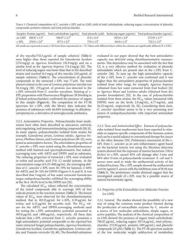
3.1.2. Prooxidative, Toxic, and Antibacterial Effects. Someavailable reports proposed using LAC from other fungi(Clitocybe maxima, P. ostreatus, or P. eryngii) as cytotoxic andantiviral agents [24, 42–45], but themechanisms of this actionare still unknown [25].Theprooxidative properties of ex-LAChave been described in relation to quinone cycles catalyzedby an enzyme causing oxygen activation, production ofsuperoxide anion radicals, and the subsequent productionof hydrogen peroxide [16, 26]. Based on these data, we canspeculate that one possibility of damagemechanisms towardspathogenic cells can be probably based on the prooxidativeaction of this enzyme, especially in the presence of redoxcycling compounds. There are no data describing this whiterot fungus enzyme from this point of view. Having measuredthe potential of the investigated ex-LAC to produce reactiveoxygen species (ROS) using chemiluminometric detection, avery strong prooxidative action of the investigated enzymewas found (Figure 1). 200% higher levels of ROS productionwere observed in the tested samples for amounts of proteinscorresponding to the 800𝜇g/mL of trolox and ascorbic acidused as controls. Luminol is widely used for studying radicalreactions and is accepted when a single oxidant, likewise thepurified ex-LAC used in the present paper, is measured [46].The obtained results showed linearity dependence betweenthe chemiluminescence and enzyme concentration. Becausethe compounds of the reaction mixture used in ABTS andDPPH free radical scavenging test inhibited the ex-LACactivity, they are not proper for this kind of estimation(data not shown). The estimation of ex-LAC toxicity using aMicrotox detection system showed that the exposure of the
0
50
100
1506.25 12.5 25 50 100 200 400 800
Inhi
bito
ry ra
te o
f che
milu
min
esce
nce (
%)
TroloxVitamin Cex-LAC
−200
−150
−100
−50
Concentration (𝜇g/mL)
Figure 1: Inhibitory rate of laccase (ex-LAC) from C. unicolorassessed with the chemiluminescence method. Data are mean ± SDfor three measurements (𝑛 = 3).
marine bacterium Vibrio fischeri to ex-LAC caused 38% cellsdamage after 5min and 51% after 15min treatment with thisenzyme.The ex-LAC sample was found to be effective againstE. coli, with the inhibition zone of 13.66mm (Table 3). Thepreliminary results obtained are in agreement with the ideaof the antimicrobial, antiviral, and antiproliferative actions ofselected fungal proteins, including laccase [1].
3.2. Properties of the Crude Extract of Endopolysaccharides(c-EPL)
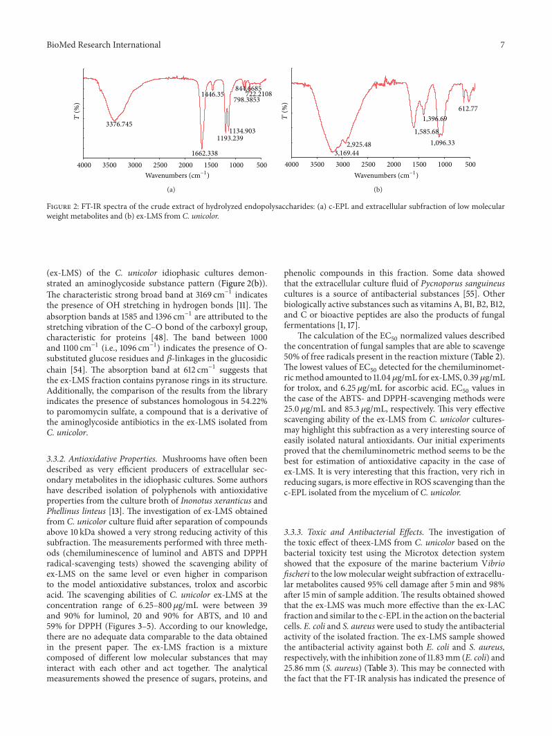
3.2.1. General. Recently, fungal polysaccharides have beenproposed as a very promising factor for various industrialapplications including biopharmacy and cosmetology. Themost abundant fungal mushroom polysaccharides are chitin,𝛼- and 𝛽-glucans, xylans, mannans, and galactans [47]. In thepresent report, the FT-IR spectra of the crude endopolysac-charides (c-EPL) isolated from C. unicolor showed a typicalcarbohydrate pattern. As shown in Figure 2(a), the FT-IRspectrum of c-EPL displays a broad stretching intense char-acteristic peak at 3376 cm−1 characteristic for the hydroxylgroup [11]. The absorption bands at 1662 and 1446 cm−1suggest the presence of the deprotonated carboxylic group(COO−) and proteins which were detected in crude polysac-charide extract [48, 49]. The sharp bands at 1193 and1134 cm−1 in the FT-IR spectra indicate the presence of C–O bonds. Characteristic 𝛼-linked glycosyl residues in thepolysaccharides at 844 cm−1 were also present [50]. Thedetermination of the chemical properties of c-EPL showedthe total carbohydrate content reaching 8.77% of the extract(Table 1). The available data of Lentinus edodes show thatcrude endopolysaccharides isolated from mycelia contained22.8% of total carbohydrates. The values obtained for thesamples extracted from the fresh fruiting bodies of Lentinuspolychrous were significantly higher (45.9%) in comparisonto the mycelia obtained from submerged cultures [10]. Thetotal polysaccharides of the presented c-EPL (73.2mg/g
6 BioMed Research International
Table 1: Chemical composition of C. unicolor c-EPL and ex-LMS: yield of total carbohydrate, reducing sugars, concentration of phenoliccompounds, proteins contents, and total polysaccharides.
Samples Protein (𝜇g/mL) Total carbohydrate (𝜇g/mL) Total phenolic (𝜇M) Reducing sugars (𝜇g/mL) Total polysaccharides (𝜇g/mL)ex-LMS 188.97 ± 1.3a 780.07 ± 2.7a 15.0 ± 0.4a 507.14 ± 2.8a 272.93 ± 2.7a
c-EPL 270.0 ± 2.6b 877.3 ± 2.1b 77.0 ± 1.3b 144.8 ± 1.5b 732.5 ± 2.5b
All results are expressed as mean ± SD from three experiments (𝑛 = 3). Values with different letters within the columns are significantly different (P ≤ 0.05).
of dry mycelia/732.5 𝜇g/mL of sample solution) (Table 1)were higher than those reported for Ganoderma lucidum(27.6mg/g) or Agaricus brasiliensis (45.9mg/g) and on asimilar level as for Agaricus bisporus (74.4mg/g) [11]. Thelevel of reducing sugars was lower than that in the describedstrains and reached 14.5mg/g of dry mycelia (145 𝜇g/mL ofsample solution) (Table 1). The concentration of phenoliccompounds in the extracted c-EPL was 77 𝜇M. The totalphenol content in the case of Lentinus polychrousmycelia was58.4mg/g [10]. 270𝜇g/mL of proteins was detected in thec-EPL extracted from C. unicolor mycelium. Staining of c-EPL preparation with Fluorescence Brightener 28 (binding to𝛽-linked polysaccharides) confirmed the presence of sugarsin this sample (Figure 6). The comparison of the FT-IRspectrum for c-EPL with the library data indicates thepresence of substances with 46.68% homology to dihydrox-ystreptomycin, a derivative of aminoglycoside antibiotics.
3.2.2. Antioxidative Properties. Polysaccharides from mush-rooms have often been described as anticancer, antiviral,anticoagulating, and immunostimulating compounds [10, 11].In many papers, polysaccharides isolated from strains forexample, Ganoderma atrum, Lentinus edodes, Agaricus bis-porus, Phellinus linteus, and Lentinus polychrous [10–12], weretested as antioxidative factors.The antioxidative properties ofC. unicolor c-EPL were tested using the chemiluminescencemethod with luminol and spectrophotometric free radical-scavenging tests with ABTS and DPPH used as substrates.The reducing properties of extracted c-EPL were evaluatedin trolox and ascorbic acid (Vit. C) model systems. At theconcentration range of 6.25–800 𝜇g/mL, the scavenging abil-ities of C. unicolor c-EPL were 36–70% for luminol, 2–60%for ABTS, and 28–32% for DPPH (Figures 3, 4, and 5). It wasdescribed that 5mg/mL of hot water extracted Ganodermatsugae endopolysaccharides exhibited the scavenging abilityat the 36.4–58.4% level in the DPPH test [51].
The calculated EC50
values reflected the concentrationof the tested compounds able to scavenge 50% of freeradicals present in the reaction mixture (Table 2). The lowestvalues of EC
50were observed for the chemiluminometric
method, that is, 183.15 𝜇g/mL for c-EPL, 0.39 𝜇g/mL fortrolox, and 6.25𝜇g/mL for ascorbic acid. The EC
50val-
ues for the ABTS- and DPPH-scavenging methods usedfor testing the c-EPL antioxidative potential amounted to493.8 𝜇g/mL and >800𝜇g/mL, respectively. All these dataindicate that c-EPL extracted from C. unicolor possesses ahigh antioxidative potential comparable to the reported hotwater extracted fungal polysaccharides, Agaricus brasiliensis,Ganoderma lucidum, Ganoderma applanatum, Lentinus edo-des, and Trametes versicolor [11, 49]. The threefold estimation
evaluated in our paper showed that the best antioxidativecapacity was detected using chemiluminometric measure-ments. This dependence may be associated with the fact thatCL is a very effective method for evaluation of phenolicantioxidants which are present also in the c-EPL from C.unicolor [36]. To sum up, the high antioxidative capacityof the c–EPL from C. unicolor was confirmed and it washigher than the antioxidative properties of polysaccharidesisolated from other fungi, for example, Agaricus bisporus(obtained from hot water extracted fresh fruit bodies) [11]or Agaricus blazei and Lentinus edodes (obtained from drypowder formulation) [52]. In the case of polysaccharidesisolated from these strains, the EC
50values (measured with
DPPH) were on the levels 2.0mg/mL, 6.77mg/mL, and26.32mg/mL, respectively [11, 52]. Considering these facts,C. unicolor mycelium seems to be a very promising newsource of endopolysaccharides with important antioxidantproperties.
3.2.3. Toxic and Antimicrobial Effect. Extracts of polysaccha-rides isolated from mushrooms have been reported to stim-ulate or suppress specific components of the immune systemand can be a useful adjunct to conventional therapy for canceror the other diseases [53]. Preliminary assessment of the c-EPL from C. unicolor as an anti-inflammatory agent basedon the bacterial toxicity test using the Microtox detectionsystem showed that the exposure of marine bacterium Vibriofischeri to c-EPL caused 85% cell damage after 5min and88% after 15min of polysaccharide treatment. E. coli and S.aureus were used to study the antibacterial activity of theisolated fractions.The c-EPL sample showed the antibacterialactivity against S. aureuswith the inhibition zone of 18.96mm(Table 3). The preliminary results obtained suggest that theinvestigated sample of c-EPL may be a possible source ofnatural bacteriostatic agents.
3.3. Properties of the Extracellular Low Molecular Fraction(ex-LMS)
3.3.1. General. The studies showed the possibility of a newway of using the common waste product formed duringthe production of extracellular enzymes by C. unicolor asa source of low molecular antioxidants, simple sugars, oractive peptides. The analysis of the chemical composition ofex-LMS showed the presence of sugars (total carbohydrate:780.07𝜇g/mL, reducing sugars: 507.14𝜇g/mL, total polysac-charides: 272.93𝜇g/mL, proteins (189 𝜇g/mL), and phenoliccompounds (15 𝜇M) (Table 1)). The FT-IR spectrum analysisof the low molecular weight subfraction of metabolites
BioMed Research International 7
722.2108798.3853844.6685
1134.9031193.239
1446.35
1662.338
3376.745
5001000150020002500300035004000Wavenumbers (cm−1)
T(%
)
(a)
612.77
1,096.33
1,396.69
1,585.68
2,925.483,169.44
5001000150020002500300035004000Wavenumbers (cm−1)
T(%
)
(b)
Figure 2: FT-IR spectra of the crude extract of hydrolyzed endopolysaccharides: (a) c-EPL and extracellular subfraction of low molecularweight metabolites and (b) ex-LMS from C. unicolor.
(ex-LMS) of the C. unicolor idiophasic cultures demon-strated an aminoglycoside substance pattern (Figure 2(b)).The characteristic strong broad band at 3169 cm−1 indicatesthe presence of OH stretching in hydrogen bonds [11]. Theabsorption bands at 1585 and 1396 cm−1 are attributed to thestretching vibration of the C–O bond of the carboxyl group,characteristic for proteins [48]. The band between 1000and 1100 cm−1 (i.e., 1096 cm−1) indicates the presence of O-substituted glucose residues and 𝛽-linkages in the glucosidicchain [54]. The absorption band at 612 cm−1 suggests thatthe ex-LMS fraction contains pyranose rings in its structure.Additionally, the comparison of the results from the libraryindicates the presence of substances homologous in 54.22%to paromomycin sulfate, a compound that is a derivative ofthe aminoglycoside antibiotics in the ex-LMS isolated fromC. unicolor.
3.3.2. Antioxidative Properties. Mushrooms have often beendescribed as very efficient producers of extracellular sec-ondary metabolites in the idiophasic cultures. Some authorshave described isolation of polyphenols with antioxidativeproperties from the culture broth of Inonotus xeranticus andPhellinus linteus [13]. The investigation of ex-LMS obtainedfrom C. unicolor culture fluid after separation of compoundsabove 10 kDa showed a very strong reducing activity of thissubfraction. The measurements performed with three meth-ods (chemiluminescence of luminol and ABTS and DPPHradical-scavenging tests) showed the scavenging ability ofex-LMS on the same level or even higher in comparisonto the model antioxidative substances, trolox and ascorbicacid. The scavenging abilities of C. unicolor ex-LMS at theconcentration range of 6.25–800 𝜇g/mL were between 39and 90% for luminol, 20 and 90% for ABTS, and 10 and59% for DPPH (Figures 3–5). According to our knowledge,there are no adequate data comparable to the data obtainedin the present paper. The ex-LMS fraction is a mixturecomposed of different low molecular substances that mayinteract with each other and act together. The analyticalmeasurements showed the presence of sugars, proteins, and
phenolic compounds in this fraction. Some data showedthat the extracellular culture fluid of Pycnoporus sanguineuscultures is a source of antibacterial substances [55]. Otherbiologically active substances such as vitamins A, B1, B2, B12,and C or bioactive peptides are also the products of fungalfermentations [1, 17].
The calculation of the EC50normalized values described
the concentration of fungal samples that are able to scavenge50% of free radicals present in the reactionmixture (Table 2).The lowest values of EC
50detected for the chemiluminomet-
ricmethod amounted to 11.04 𝜇g/mL for ex-LMS, 0.39𝜇g/mLfor trolox, and 6.25𝜇g/mL for ascorbic acid. EC
50values in
the case of the ABTS- and DPPH-scavenging methods were25.0𝜇g/mL and 85.3𝜇g/mL, respectively. This very effectivescavenging ability of the ex-LMS from C. unicolor cultures-may highlight this subfraction as a very interesting source ofeasily isolated natural antioxidants. Our initial experimentsproved that the chemiluminometric method seems to be thebest for estimation of antioxidative capacity in the case ofex-LMS. It is very interesting that this fraction, very rich inreducing sugars, is more effective in ROS scavenging than thec-EPL isolated from the mycelium of C. unicolor.
3.3.3. Toxic and Antibacterial Effects. The investigation ofthe toxic effect of theex-LMS from C. unicolor based on thebacterial toxicity test using the Microtox detection systemshowed that the exposure of the marine bacterium Vibriofischeri to the lowmolecular weight subfraction of extracellu-lar metabolites caused 95% cell damage after 5min and 98%after 15min of sample addition. The results obtained showedthat the ex-LMS was much more effective than the ex-LACfraction and similar to the c-EPL in the action on the bacterialcells. E. coli and S. aureuswere used to study the antibacterialactivity of the isolated fraction. The ex-LMS sample showedthe antibacterial activity against both E. coli and S. aureus,respectively, with the inhibition zone of 11.83mm (E. coli) and25.86mm (S. aureus) (Table 3). This may be connected withthe fact that the FT-IR analysis has indicated the presence of

8 BioMed Research International
0
20
40
60
80
100
0 100 200 300 400 500 600 700 800
Scav
engi
ng eff
ect (
%)
TroloxVitamin C
ex-LMSc-EPL
Concentration (𝜇g/mL)
Figure 3: Inhibitory rate of the crude extract of endopolysaccha-rides (c-EPL) and extracellular sub-fraction of lowmolecular weightmetabolites (ex-LMS) from C. unicolor assessed with the chemilu-minescence method. Data are mean ± SD for three measurements(𝑛 = 3).
0
20
40
60
80
100
Scav
engi
ng eff
ect (
%)
0 100 200 300 400 500 600 700 800
TroloxVitamin C
ex-LMSc-EPL
Concentration (𝜇g/mL)
Figure 4: Scavenging effects of the crude extract of endopolysac-charides (c-EPL) and extracellular sub-fraction of low molecularweight metabolites (ex-LMS) from C. unicolor assessed with theABTS radical-scavenging method. Data are mean ± SD for threemeasurements (𝑛 = 3).
derivatives, similar to known aminoglycoside antibiotics, inthe investigated samples (ex-LMS, c-EPL).
4. Conclusion
Our paper proposes a new insight into the possibility ofapplication of common wood-destroying fungus C. unicoloras a source of three fractions of potentially bioactive metabo-lites: extracellular laccase (ex-LAC), intracellular nonpurifiedpolysaccharides (c-EPL), and a lowmolecularweight subfrac-tion of extracellular metabolites (ex-LMS). Each of them canbe investigated differentially.
To the best of our knowledge, this is the first reportdescribing the very high ROS-scavenging potential of fungal
0
20
40
60
80
100
Scav
engi
ng eff
ect (
%)
0 100 200 300 400 500 600 700 800
TroloxVitamin C
ex-LMSc-EPL
Concentration (𝜇g/mL)
Figure 5: Scavenging effects of the crude extract of endopolysac-charides (c-EPL) and extracellular sub-fraction of low molecularweight metabolites (ex-LMS) from C. unicolor assessed with theDPPH radical-scavenging method. Data are mean ± SD for threemeasurements (𝑛 = 3).
Figure 6: Visualization of endopolysaccharides using confocal laserscanning microscopy. The lyophilized samples of c-EPL, washedwith MQ water, were stained for 30min with 200𝜇L of 25𝜇g/mLFluorescence Brightener 28 commonly used in order to detect 𝛽-linked polysaccharides. For visualization of the endopolysaccha-rides from C. unicolor, the inverted microscope Axiovert 200Mequipped with an LSM 5 Pascal head (with magnification 200x) wasused. The letter (A) indicates the luminous areas exhibiting visible𝛽-linked polysaccharide fragments.
preparations such as endopolysaccharides and extracellularlow molecular weight compounds measured by three dif-ferent methods. These substances may potentially be usedas a new source of effective antioxidants that can be easilyproduced in controlled laboratory conditions. Therefore, theresults obtained introduce a new, nonedible fungus to themedicinal mushroom family, comprising species like Tram-etes versicolor (crestin source) and Schizophyllum commune

BioMed Research International 9
Table 2: EC50 values (effective concentration at which the radicals present in the investigated samples were scavenged by 50%; the antioxidantactivity was 50%) of c-EPL and ex-LMS isolated from C. unicolor submerged cultures in comparison to trolox and Vit C.
EC50 (𝜇g/mL)c-EPL ex-LMS trolox Vit C
chemiluminescence method 183.15 ± 1.2 11.04 ± 0.2 0.39 ± 0.1 6.25 ± 0.2
ABTS radical scavenging 493.8 ± 2.2 25.0 ± 0.1 315.9 ± 2.1 268.4 ± 2.2
DPPH radical scavenging >800 85.3 ± 0.7 59.52 ± 0.7 82.56 ± 1.1
All results are expressed asmean± SD from three experiments (𝑛 = 3). Values within the column and the row for investigated samples are significantly different(P ≤ 0.05). EC50 > 800𝜇g/mL cannot be calculated from the graphs.
Table 3: The antibacterial activities of ex-LAC, c-EPL, and ex-LMS(1mg/mL) isolated from C. unicolor submerged cultures.
Diameters of inhibition zone (mm)E. coli S. aureus
ex-LAC 13.66 ± 0.4 —a
c-EPL — 18.96 ± 0.4
ex-LMS 11.83 ± 0.2 25.86 ± 0.2
Physiological saline — —All results are expressed as mean ± SD from three experiments (𝑛 =3). Values within the columns are significantly different (P ≤ 0.05). aNotdetected.
(schizophyllan source), which are regarded as nonediblestrains but as producers of bioactive substances.
The prooxidative potential of laccase and the toxic effecton bacterial cells of all the three fractions suggest continu-ation of the presented studies in terms of pharmacologicaleffects. However, it is worth noting that further studiesare needed comprising isolation and characterization of theabove-mentioned bioactive substances, and their possible useas crucial factors in new therapy and as a natural source ofantioxidative molecules.
Conflict of Interests
The authors declare that there is no conflict of interests.
References
[1] X. Xu, H. Yan, J. Chen, and X. Zhang, “Bioactive proteins frommushrooms,” Biotechnology Advances, vol. 29, no. 6, pp. 667–674, 2011.
[2] J. H. Wong, T. B. Ng, R. C. F. Cheung et al., “Proteins with anti-fungal properties and other medicinal applications from plantsand mushrooms,” Applied Microbiology and Biotechnology, vol.87, no. 4, pp. 1221–1235, 2010.
[3] I. C. F. R. Ferreira, J. A. Vaz, M. H. Vasconcelos, and A.Martins,“Compounds from wild mushrooms with antitumor potential,”Anti-Cancer Agents in Medicinal Chemistry, vol. 10, no. 5, pp.424–436, 2010.
[4] S. P. Wasser, “Medicinal mushroom science: history, currentstatus, future trends and unsolved problems,” InternationalJournal of Medicinal Mushrooms, vol. 12, no. 1, pp. 1–16, 2010.
[5] J. Erjavec, J. Kos, M. Ravnikar, T. Dreo, and J. Sabotic, “Proteinsof higher fungi-from forest to application,” Trends in Biotech-nology, vol. 30, no. 5, pp. 259–273, 2012.
[6] X. J. Wu and C. Hansen, “Antioxidant capacity, phenoliccontent, and polysaccharide content of Lentinus edodes grownin whey permeate-based submerged culture,” Journal of FoodScience, vol. 73, no. 1, pp. M1–M8, 2008.
[7] L. Fan, J. Li, K. Deng, and L. Ai, “Effects of drying methodson the antioxidant activities of polysaccharides extracted fromGanoderma lucidum,” Carbohydrate Polymers, vol. 87, no. 2, pp.1849–1854, 2012.
[8] D. J. Jamieson, “The effect of oxidative stress on Saccharomycescerevisiae,” Redox Report, vol. 1, pp. 89–95, 1995.
[9] B. Halliwell, “Antioxidants and human disease: a general intro-duction,” Nutrition Reviews, vol. 55, no. 1, part 2, pp. S44–S52,1997.
[10] C.Thetsrimuang, S. Khammuang, K. Chiablaem, C. Srisomsap,and R. Sarnthima, “Antioxidant properties and cytotoxicityof crude polysaccharides from Lentinus polychrous Lev,” FoodChemistry, vol. 128, no. 3, pp. 634–639, 2011.
[11] M. Kozarski, A. Klaus, M. Niksic, D. Jakovljevic, J. P. F. G.Helsper, and L. J. L. D. van Griensven, “Antioxidative andimmunomodulating activities of polysaccharide extracts of themedicinal mushrooms Agaricus bisporus, Agaricus brasiliensis,Ganoderma lucidum and Phellinus linteus,” Food Chemistry, vol.129, no. 4, pp. 1667–1675, 2011.
[12] Y. Chen, M.-Y. Xie, S.-P. Nie, C. Li, and Y.-X. Wang, “Purifi-cation, composition analysis and antioxidant activity of apolysaccharide from the fruiting bodies of Ganoderma atrum,”Food Chemistry, vol. 107, no. 1, pp. 231–241, 2008.
[13] J.-Y. Jung, I.-K. Lee, S.-J. Seok, H.-J. Lee, Y.-H. Kim, and B.-S. Yun, “Antioxidant polyphenols from the mycelial culture ofthe medicinal fungi Inonotus xeranticus and Phellinus linteus,”Journal of Applied Microbiology, vol. 104, no. 6, pp. 1824–1832,2008.
[14] K. E. Hammel, A. N. Kapich, K. A. Jensen Jr., and Z. C. Ryan,“Reactive oxygen species as agents of wood decay by fungi,”Enzyme and Microbial Technology, vol. 30, no. 4, pp. 445–453,2002.
[15] W. Chen, Z. Zhao, S.-F. Chen, and Y.-Q. Li, “Optimization forthe production of exopolysaccharide from Fomes fomentarius insubmerged culture and its antitumor effect in vitro,” BioresourceTechnology, vol. 99, no. 8, pp. 3187–3194, 2008.
[16] M. Jaszek, J. Zuchowski, E. Dajczak, K. Cimek, M. Graz,and K. Grzywnowicz, “Ligninolytic enzymes can participatein a multiple response system to oxidative stress in white-rotbasidiomycetes: Fomes fomentarius and Tyromyces pubescens,”International Biodeterioration and Biodegradation, vol. 58, no.3-4, pp. 168–175, 2006.
[17] S. Ghorai, S. P. Banik, D. Verma, S. Chowdhury, S. Mukherjee,and S. Khowala, “Fungal biotechnology in food and feedprocessing,” Food Research International, vol. 42, no. 5-6, pp.577–587, 2009.
10 BioMed Research International
[18] W. C. Roody, Mushrooms of West Virginia and the CentralAppalachians, University Press of Kentucky, 2003.
[19] C. Gonindard, C. Bergonzi, C. Denier et al., “Synthetic hispidin,a PKC inhibitor, is more cytotoxic toward cancer cells thannormal cells in vitro,” Cell Biology and Toxicology, vol. 13, no.3, pp. 141–153, 1997.
[20] J. Rogalski, A. Dawidowicz, E. Jozwik, and A. Leonowicz,“Immobilization of laccase fromCerrena unicolor on controlledporosity glass,” Journal of Molecular Catalysis B, vol. 6, no. 1-2,pp. 29–39, 1999.
[21] G. Janusz, J. Rogalski, and J. Szczodrak, “Increased productionof laccase by Cerrena unicolor in submerged liquid cultures,”World Journal of Microbiology and Biotechnology, vol. 23, no. 10,pp. 1459–1464, 2007.
[22] J. Polak and A. Jarosz-Wilkolazka, “Fungal laccases as greencatalysts for dye synthesis,” Process Biochemistry, vol. 47, no. 9,pp. 1295–1307, 2012.
[23] G. Janusz, K. H. Kucharzyk, A. Pawlik, M. Staszczak, andA. J. Paszczynski, “Fungal laccase, manganese peroxidase andlignin peroxidase: gene expression and regulation,” Enzyme andMicrobial Technology, vol. 52, no. 1, pp. 1–12, 2012.
[24] H. X. Wang and T. B. Ng, “Purification of a novel low-molecular-mass laccase with HIV-1 reverse transcriptaseinhibitory activity from the mushroom Tricholoma giganteum,”Biochemical and Biophysical Research Communications, vol.315, no. 2, pp. 450–454, 2004.
[25] M. Li, G. Zhang, H. Wang, and T. Ng, “Purification andcharacterization of a laccase from the edible wild mushroomTricholoma mongolicum,” Journal of Microbiology and Biotech-nology, vol. 20, no. 7, pp. 1069–1076, 2010.
[26] A. T. Martınez, M. Speranza, F. J. Ruiz-Duenas et al., “Biodegra-dation of lignocellulosics: microbial, chemical, and enzymaticaspects of the fungal attack of lignin,” International Microbiol-ogy, vol. 8, no. 3, pp. 195–204, 2005.
[27] G. Janusz, A. Mazur, A. Checinsks, W. Malek, J. Rogalski, andS. Ohga, “Cloning and characterization of a lacease gene frombiotechnologically important basidiomyceteCerrerna unicolor,”Journal of the Faculty of Agriculture, Kyushu University, vol. 57,no. 1, pp. 41–49, 2012.
[28] D. Jennings and G. Lysek, Fungal Biology: Understanding theFungal Lifestyle, BIOS Scientific Publishers Ltd., Oxford, UK,1999.
[29] N. N. Pozdnyakova, J. Rodakiewicz-Nowak, O. V. Turkovskaya,and J. Haber, “Oxidative degradation of polyaromatic hydro-carbons catalyzed by blue laccase from Pleurotus ostreatus D1in the presence of synthetic mediators,” Enzyme and MicrobialTechnology, vol. 39, no. 6, pp. 1242–1249, 2006.
[30] S. Freimund, M. Sauter, O. Kappeli, and H. Dutler, “A new non-degrading isolation process for 1,3-𝛽-D-glucan of high purityfrombaker’s yeast Saccharomyces cerevisiae,”Carbohydrate Poly-mers, vol. 54, no. 2, pp. 159–171, 2003.
[31] M. DuBois, K. A. Gilles, J. K. Hamilton, P. A. Rebers, and F.Smith, “Colorimetric method for determination of sugars andrelated substances,”Analytical Chemistry, vol. 28, no. 3, pp. 350–356, 1956.
[32] C. F. A. Hope and R. G. Burns, “Activity, origins and location ofcellulases in a silt loam soil,” Biology and Fertility of Soils, vol. 5,no. 2, pp. 164–170, 1987.
[33] M. M. Bradford, “A rapid and sensitive method for the quanti-tation of microgram quantities of protein utilizing the principleof protein dye binding,”Analytical Biochemistry, vol. 72, no. 1-2,pp. 248–254, 1976.
[34] E. Malarczyk, “Transformation of phenolic acids by Nocardia,”Acta Microbiologica Polonica, vol. 38, no. 1, pp. 45–53, 1998.
[35] A. Leonowicz andK. Grzywnowicz, “Quantitative estimation oflaccase forms in some white-rot fungi using syringaldazine as asubstrate,” Enzyme and Microbial Technology, vol. 3, no. 1, pp.55–58, 1981.
[36] Z. Cheng, G. Yan, Y. Li, and W. Chang, “Determination ofantioxidant activity of phenolic antioxidants in a Fenton-typereaction system by chemiluminescence assay,” Analytical andBioanalytical Chemistry, vol. 375, no. 3, pp. 376–380, 2003.
[37] R. van den Berg, G. R. M. M. Haenen, H. van den Berg,and A. Bast, “Applicability of an improved Trolox equivalentantioxidant capacity (TEAC) assay for evaluation of antioxidantcapacity measurements of mixtures,” Food Chemistry, vol. 66,no. 4, pp. 511–517, 1999.
[38] L. Duo-Chuan, “Review of fungal chitinases,” Mycopathologia,vol. 161, no. 6, pp. 345–360, 2006.
[39] R. Re, N. Pellegrini, A. Proteggente, A. Pannala,M. Yang, andC.Rice-Evans, “Antioxidant activity applying an improved ABTSradical cation decolorization assay,” Free Radical Biology andMedicine, vol. 26, no. 9-10, pp. 1231–1237, 1999.
[40] R. Paduch, G. Matysik, M. Wojciak-Kosior et al., “Lamiumalbum extracts express free radical scavenging and cytotoxicactivities,” Polish Journal of Environmental Studies, vol. 17, no.4, pp. 569–580, 2008.
[41] A. Leonowicz, L. Gianfreda, J. Rogalski et al., “Appearance oflaccase in wood-rotting fungi and its inducibility,” Journal of theKorean Wood Science and Technology, vol. 25, pp. 29–36, 1997.
[42] J.-A.Majeau, S. K. Brar, andR.D. Tyagi, “Laccases for removal ofrecalcitrant and emerging pollutants,” Bioresource Technology,vol. 101, no. 7, pp. 2331–2350, 2010.
[43] H. X. Wang and T. B. Ng, “Purification of a laccase fromfruiting bodies of the mushroom Pleurotus eryngii,” AppliedMicrobiology and Biotechnology, vol. 69, no. 5, pp. 521–525, 2006.
[44] E. M. EL-Fakharany, B. M. Haroun, T. B. Ng, and E.-R. M.Redwan, “Oyster mushroom laccase inhibits hepatitis C virusentry into peripheral blood cells and hepatoma cells,” Proteinand Peptide Letters, vol. 17, no. 8, pp. 1031–1039, 2010.
[45] G.-Q. Zhang, Y.-F. Wang, X.-Q. Zhang, T. B. Ng, and H.-X.Wang, “Purification and characterization of a novel laccasefrom the ediblemushroomClitocybemaxima,”Process Biochem-istry, vol. 45, no. 5, pp. 627–633, 2010.
[46] R. L. Prior, X. Wu, and K. Schaich, “Standardized methodsfor the determination of antioxidant capacity and phenolicsin foods and dietary supplements,” Journal of Agricultural andFood Chemistry, vol. 53, no. 10, pp. 4290–4302, 2005.
[47] M. Zhang, S.W. Cui, P. C. K. Cheung, andQ.Wang, “Antitumorpolysaccharides from mushrooms: a review on their isola-tion process, structural characteristics and antitumor activity,”Trends in Food Science and Technology, vol. 18, no. 1, pp. 4–19,2007.
[48] G. D. Manrique and F. M. Lajolo, “FT-IR spectroscopy as a toolfor measuring degree of methyl esterification in pectins isolatedfrom ripening papaya fruit,”Postharvest Biology andTechnology,vol. 25, no. 1, pp. 99–107, 2002.
[49] M. Kozarski, A. Klaus, M. Niksic et al., “Antioxidative activ-ities and chemical characterization of polysaccharide extractsfrom the widely used mushrooms Ganoderma applanatum,Ganoderma lucidum, Lentinus edodes and Trametes versicolor,”Journal of Food Composition and Analysis, vol. 26, no. 1-2, pp.144–153, 2012.

BioMed Research International 11
[50] J. Jelsma and D. R. Kreger, “Polymorphism in crystalline (1→3)-𝛼-d-glucan from fungal cell-walls,” Carbohydrate Research,vol. 71, no. 1, pp. 51–64, 1979.
[51] Y.-H. Tseng, J.-H. Yang, and J.-L. Mau, “Antioxidant propertiesof polysaccharides from Ganoderma tsugae,” Food Chemistry,vol. 107, no. 2, pp. 732–738, 2008.
[52] A. A. J. Carneiro, I. C. F. R. Ferreira,M. Duenas et al., “Chemicalcomposition and antioxidant activity of dried powder formula-tions of Agaricus blazei and Lentinus edodes,” Food Chemistry,vol. 138, no. 4, pp. 2168–2173, 2013.
[53] S. Wasser, “Medicinal mushrooms as a source of antitumorand immunomodulating polysaccharides,” Applied Microbiol-ogy and Biotechnology, vol. 60, no. 3, pp. 258–274, 2002.
[54] B. Stone and A. Clarke, Chemistry and Biology of (1, 3)-Β-Glucans, La Trobe University Press, 1992.
[55] A. Smania, F. D. Monache, E. F. A. Smania, M. L. Gil, L. C.Benchetrit, and F. S. Cruz, “Antibacterial activity of a substanceproduced by the fungus Pycnoporus sanguineus (Fr.) Murr,”Journal of Ethnopharmacology, vol. 45, no. 3, pp. 177–181, 1995.

Submit your manuscripts athttp://www.hindawi.com
PainResearch and TreatmentHindawi Publishing Corporationhttp://www.hindawi.com Volume 2014
The Scientific World JournalHindawi Publishing Corporation http://www.hindawi.com Volume 2014
Hindawi Publishing Corporationhttp://www.hindawi.com
Volume 2014
ToxinsJournal of
VaccinesJournal of
Hindawi Publishing Corporation http://www.hindawi.com Volume 2014
Hindawi Publishing Corporationhttp://www.hindawi.com Volume 2014
AntibioticsInternational Journal of
ToxicologyJournal of
Hindawi Publishing Corporationhttp://www.hindawi.com Volume 2014
StrokeResearch and TreatmentHindawi Publishing Corporationhttp://www.hindawi.com Volume 2014
Drug DeliveryJournal of
Hindawi Publishing Corporationhttp://www.hindawi.com Volume 2014
Hindawi Publishing Corporationhttp://www.hindawi.com Volume 2014
Advances in Pharmacological Sciences
Tropical MedicineJournal of
Hindawi Publishing Corporationhttp://www.hindawi.com Volume 2014
Medicinal ChemistryInternational Journal of
Hindawi Publishing Corporationhttp://www.hindawi.com Volume 2014
AddictionJournal of
Hindawi Publishing Corporationhttp://www.hindawi.com Volume 2014
Hindawi Publishing Corporationhttp://www.hindawi.com Volume 2014
BioMed Research International
Emergency Medicine InternationalHindawi Publishing Corporationhttp://www.hindawi.com Volume 2014
Hindawi Publishing Corporationhttp://www.hindawi.com Volume 2014
Autoimmune Diseases
Hindawi Publishing Corporationhttp://www.hindawi.com Volume 2014
Anesthesiology Research and Practice
ScientificaHindawi Publishing Corporationhttp://www.hindawi.com Volume 2014
Journal of
Hindawi Publishing Corporationhttp://www.hindawi.com Volume 2014
Pharmaceutics
Hindawi Publishing Corporationhttp://www.hindawi.com Volume 2014
MEDIATORSINFLAMMATION
of