Research Article Myriocin-Ceramide Inhibitor Modulates...
13
D. Lizarazo et al. / Journal of Biochemical and Pharmacological Research, Vol. 1 (3): 148-160, September 2013 ISSN 2168-8761 print/ISSN 2168-877X online ~ 148 ~ http://www.researchpub.org/journal/jbpr/jbpr.html Abstract: Background: Alcohol-related liver disease (ALD) is mediated in part by insulin resistance, which dysregulates lipid metabolism, increasing hepatic ceramides, lipotoxicity and endoplasmic reticulum (ER) stress. However, toxic ceramides increase inflammation and worsen insulin resistance. Aim : We tested the hypothesis that ceramide inhibitors could reduce ER stress and restore insulin signaling, including expression of insulin responsive genes such as aspartyl- asparaginyl-β-hydroxylase (AAH), which mediates structural remodeling of the liver. Design: Using a liver slice culture (LSC) model generated from control and ethanol-exposed rats, we examined effects of Myriocin (serine palmitoyl transferase inhibitor) on insulin/IGF signaling, and expression of AAH and ER stress-related proteins in liver. Results: Ethanol-induced hepatic steatosis resulted in increased cytotoxicity and triglyceride and ceramide accumulations. Ethanol also inhibited signaling through the insulin and IGF-1 receptors, IRS-1, Akt, and p70S6k, increased activation of GSK-3β, reduced AAH, Humbug, and β-actin, and altered ER stress pathway protein expression. Myriocin reduced hepatic steatosis and ceramide levels, normalized components of insulin/IGF-1 signaling, and enhanced expression of AAH, but did not reduce cytotoxicity, triglycerides, or ER stress proteins in ethanol exposed LSCs. Conclusions: Ceramide accumulation partly mediates liver insulin resistance in ALD. Ceramide inhibitors may provide supplementary treatment for restoring liver function in ALD. __________________________________________ a Liver Research Center, Divisions of Gastroenterology and Neuropathology, and Departments of Medicine, Pathology, Neurology, and Neurosurgery, Rhode Island Hospital and the Warren Alpert Medical School of Brown University, Providence, RI *Corresponding author. Tel: +1 401-444-7364, Fax: +1 401-444-2939 Email: [email protected] (Received April 4, 2013; Revised April 4, 2013; Accepted April 5, 2013; Published online April 12, 2013) Keywords: alcoholic liver disease; ceramides; steatohepatitis; insulin resistance; Notch; aspartyl-asparaginyl-β-hydroxylase; myriocin 1. Introduction lcohol abuse is a leading cause of liver-related morbidity and mortality [1-3]. Excessive chronic or binge alcohol consumption causes steatohepatitis, which progresses to chronic alcoholic liver disease (ALD) [4] due to sustained insulin and insulin-like growth factor (IGF) resistance [5-7], inflammation, oxidative and endoplasmic reticulum (ER) stress, lipotoxicity with ceramide accumulation, and mitochondrial dysfunction [5-9]. Ethanol-impaired insulin and IGF type 1 (IGF-1) signaling [5, 6, 10-15] inhibits downstream activation of ERK mitogen-activated protein kinase (MAPK), which supports liver growth and repair after injury, and phosphatidyl-inositol-3-kinase (PI3K)-Akt [5, 16, 17], which promotes hepatocellular survival, motility (for remodeling), and metabolism [18]. In addition, ethanol activates glycogen synthase kinase 3β (GSK-3β) [11], and phosphatases that negatively regulate receptor tyrosine kinases and PI3K [11, 19]. Therefore, inhibition of insulin/IGF signaling accounts for many of the toxic and degenerative effects of ethanol on the liver. Aspartyl-asparaginyl-β-hydroxylase (AAH) is an important downstream target of insulin signaling because of its role in regulating cell motility, which is needed for liver remodeling, repair, and regeneration [6, 20-22]. AAH is an ~86 kD type 2 transmembrane protein that undergoes physiological cleavage, releasing a ~52-54 kD C-terminal fragment that has catalytic (hydroxylase) activity, and a smaller ~32-34 kD N-terminal fragment that is highly homologous to another protein called Humbug [23-25]. AAH’s C-terminal catalytic fragment confers cell motility, invasiveness, and adhesion [20, 22], whereas both Humbug and the N-terminus of AAH may mediate calcium flux between the endoplasmic reticulum (ER) and cytosol via ryanodine receptors [26], and thereby modulate cell adhesion [20]. Insulin and IGFs regulate AAH by increasing its mRNA, Research Article Myriocin-Ceramide Inhibitor Modulates Insulin/IGF and ER Stress Pathways in Experimental Alcohol-Related Steatohepatitis Diana Lizarazo a , Valerie Zabala a , Ming Tong a , Suzanne M. de la Monte a,* A
-
Upload
nguyentram -
Category
Documents
-
view
213 -
download
0
Transcript of Research Article Myriocin-Ceramide Inhibitor Modulates...
D. Lizarazo et al. / Journal of Biochemical and Pharmacological Research, Vol. 1 (3): 148-160, September 2013
ISSN 2168-8761 print/ISSN 2168-877X online ~ 148 ~ http://www.researchpub.org/journal/jbpr/jbpr.html
Abstract: Background: Alcohol-related liver disease (ALD) is mediated in part by insulin resistance, which dysregulates lipid metabolism, increasing hepatic ceramides, lipotoxicity and endoplasmic reticulum (ER) stress. However, toxic ceramides increase inflammation and worsen insulin resistance. Aim: We tested the hypothesis that ceramide inhibitors could reduce ER stress and restore insulin signaling, including expression of insulin responsive genes such as aspartyl- asparaginyl-β-hydroxylase (AAH), which mediates structural remodeling of the liver. Design: Using a liver slice culture (LSC) model generated from control and ethanol-exposed rats, we examined effects of Myriocin (serine palmitoyl transferase inhibitor) on insulin/IGF signaling, and expression of AAH and ER stress-related proteins in liver. Results: Ethanol-induced hepatic steatosis resulted in increased cytotoxicity and triglyceride and ceramide accumulations. Ethanol also inhibited signaling through the insulin and IGF-1 receptors, IRS-1, Akt, and p70S6k, increased activation of GSK-3β, reduced AAH, Humbug, and β-actin, and altered ER stress pathway protein expression. Myriocin reduced hepatic steatosis and ceramide levels, normalized components of insulin/IGF-1 signaling, and enhanced expression of AAH, but did not reduce cytotoxicity, triglycerides, or ER stress proteins in ethanol exposed LSCs. Conclusions: Ceramide accumulation partly mediates liver insulin resistance in ALD. Ceramide inhibitors may provide supplementary treatment for restoring liver function in ALD. __________________________________________ a Liver Research Center, Divisions of Gastroenterology and Neuropathology, and Departments of Medicine, Pathology, Neurology, and Neurosurgery, Rhode Island Hospital and the Warren Alpert Medical School of Brown University, Providence, RI *Corresponding author. Tel: +1 401-444-7364, Fax: +1 401-444-2939 Email: [email protected] (Received April 4, 2013; Revised April 4, 2013; Accepted April 5, 2013; Published online April 12, 2013)
Keywords: alcoholic liver disease; ceramides; steatohepatitis; insulin resistance; Notch; aspartyl-asparaginyl-β-hydroxylase; myriocin 1. Introduction
lcohol abuse is a leading cause of liver-related morbidity and mortality [1-3]. Excessive chronic or binge alcohol
consumption causes steatohepatitis, which progresses to chronic alcoholic liver disease (ALD) [4] due to sustained insulin and insulin-like growth factor (IGF) resistance [5-7], inflammation, oxidative and endoplasmic reticulum (ER) stress, lipotoxicity with ceramide accumulation, and mitochondrial dysfunction [5-9]. Ethanol-impaired insulin and IGF type 1 (IGF-1) signaling [5, 6, 10-15] inhibits downstream activation of ERK mitogen-activated protein kinase (MAPK), which supports liver growth and repair after injury, and phosphatidyl-inositol-3-kinase (PI3K)-Akt [5, 16, 17], which promotes hepatocellular survival, motility (for remodeling), and metabolism [18]. In addition, ethanol activates glycogen synthase kinase 3β (GSK-3β) [11], and phosphatases that negatively regulate receptor tyrosine kinases and PI3K [11, 19]. Therefore, inhibition of insulin/IGF signaling accounts for many of the toxic and degenerative effects of ethanol on the liver. Aspartyl-asparaginyl-β-hydroxylase (AAH) is an important downstream target of insulin signaling because of its role in regulating cell motility, which is needed for liver remodeling, repair, and regeneration [6, 20-22]. AAH is an ~86 kD type 2 transmembrane protein that undergoes physiological cleavage, releasing a ~52-54 kD C-terminal fragment that has catalytic (hydroxylase) activity, and a smaller ~32-34 kD N-terminal fragment that is highly homologous to another protein called Humbug [23-25]. AAH’s C-terminal catalytic fragment confers cell motility, invasiveness, and adhesion [20, 22], whereas both Humbug and the N-terminus of AAH may mediate calcium flux between the endoplasmic reticulum (ER) and cytosol via ryanodine receptors [26], and thereby modulate cell adhesion [20]. Insulin and IGFs regulate AAH by increasing its mRNA,
Research Article
Myriocin-Ceramide Inhibitor Modulates Insulin/IGF and ER Stress Pathways in Experimental Alcohol-Related
Steatohepatitis
Diana Lizarazoa, Valerie Zabalaa, Ming Tonga, Suzanne M. de la Montea,*
A
D. Lizarazo et al. / Journal of Biochemical and Pharmacological Research, Vol. 1 (3): 148-160, September 2013
ISSN 2168-8761 print/ISSN 2168-877X online ~ 149 ~ http://www.researchpub.org/journal/jbpr/jbpr.html
catalytic activity [20, 27-29], and protein [21, 27, 28]. Insulin/IGF stimulation causes AAH protein to accumulate by inhibiting GSK-3β phosphorylation, which leads to AAH’s proteolytic degradation [28-30]. Ethanol can inhibit AAH’s expression and function by impairing insulin/IGF signaling, and increasing GSK-3β activity [5, 29, 31, 32]. Growing evidence suggests that besides insulin/IGF resistance, ethanol mediates its adverse effects on liver by causing lipid dyshomeostasis [33]. Lipid dyshomeostasis results in increased lipolysis and ceramide accumulation. Attendant lipotoxicity further impairs insulin signaling [34-36], activates pro-inflammatory cytokines, inhibits PI3K-Akt [37-40], and increases ER and oxidative stress [9, 41, 42]. The contributions of ceramide accumulation to the pathogenesis of ALD are further suggested by the findings that: 1) mice deficient in acidic sphingomyelinase are resistant to ethanol-induced hepatic steatosis [43]; 2) ceramides inhibit 5' adenosine monophosphate-activated protein kinase (AMPK) [44] and promote local hepatocellular injury [45], possibly because AMPK reduces ER stress and apoptosis [46]; and 3) hepatic ceramide levels are elevated and their profiles substantially altered in advanced human ALD [7, 47]. Previous studies showed that treatment with peroxisome proliferator activated receptor (PPAR) agonists, which are insulin sensitizers that have anti-inflammatory actions, could prevent structural, biochemical, and molecular pathologic changes in experimental chronic ALD [6, 42]. However, not all aspects of ALD were resolved with PPAR agonist treaments. In particular, ceramide load remained elevated [48], setting the stage for ALD to eventually progress due to persistent insulin/IGF resistance and inhibition of target genes and signaling molecules that regulate liver remodeling. Therefore, additional approaches are needed to effectively treat chronic ALD. Herein, we evaluate the therapeutic effects of a ceramide inhibitor with respect to resolution of hepatic steatosis, insulin resistance, impaired AAH expression, and ER stress. 2. Materials and methods 2.1. Materials The Taqman Gene expression master mix, Amplex UltraRed soluble fluorophore, Amplex Red Triglyceride Assay Kit, Akt Pathway Total and Phospho 7-Plex panels, and the 4-Methylumbelliferyl phosphate (4-MUP) fluorophore were obtained from Invitrogen (Carlsbad, CA). QIAzol Lysis RNA extraction Reagent was bought from Qiagen, Inc (Valencia, CA). MaxiSorp plates, OptiPlates (96-well) needed for enzyme-linked immunosorbent assays (ELISAs), BD Falcon culture inserts, and Nunc culture supplies were obtained from Thermo Scientific (Rochester, NY). SuperBlock TBS, bicinchoninic assay, and enhanced chemiluminescence reagents were purchased from Pierce (Rockford, IL). Mouse monoclonal anti-ceramide and Myriocin were from Sigma-Aldrich Co (St. Louis, MO). Rabbit polyclonal antibody to large acidic ribosomal protein (RPLPO) was from Proteintech, Inc (Chicago, IL). The A85E6, A85G6, and FB50 mouse monoclonal antibodies to AAH were characterized as
described [29, 31]. All other antibodies and reagents for ELISAs were purchased from Abcam (Cambridge, MA), Upstate (Billerica, MA), Vector Laboratories (Burlingame, CA), Invitrogen (Carlsbad, CA), or Chemicon (Temecula, CA). Other fine chemicals were purchased from CalBiochem (Carlsbad, CA) or Sigma-Aldrich (St. Louis, MO). 2.2. Organotypic slice culture Liver slice cultures (LSCs) were generated with livers from adult Long Evans rats maintained on Lieber-deCarli isocaloric liquid diets containing 0% (control) or 37% ethanol by caloric content. At harvest, livers were placed into ice-cold Hanks Balanced Salt Solution (HBSS). Eight mm diameter cylindrical cores were sliced at a thickness of 150 µm using a McIlwain tissue chopper (Mickle Laboratory Engineering Co. Ltd, UK). After gentle separation under a dissecting microscope, 2 or 3 slices were placed (non-overlapping) on the bottoms of BD Falcon 8-μm pore culture inserts and seated into 12-well Nunc plates. Each well contained 1 ml of pre-warmed (37°C) Dulbecco’s Modified Eagle’s medium supplemented with 10% fetal bovine serum, 4 mM L-glutamine, 10 mM non-essential amino acids, 120 U/mL Penicillin, 120 μg/ml Streptomycin, and either 0 mM or 50 mM ethanol (corresponding with in vivo blood alcohol levels). After 24 hours incubation, cultures were treated with 0.1% DMSO (vehicle) or 10 µM Myriocin, a serine palmitoyl transferase (de novo pathway) inhibitor [49]. LSCs were maintained at 37°C in a standard humidified CO2 incubator and on a rotating platform. LSC medium was changed daily with fresh additions of chemical inhibitors. We used the LDH Cytotoxicity Assay Kit to assess effects of ceramide inhibitor treatments on culture viability. In brief, duplicate 50 µl aliquots of cell culture medium were transferred to individual wells of a 96-well white OptiPlate. After combining with 50 µl of reaction mixture containing 4 mM resazurin, the samples were incubated at 37°C for 30 minutes. Fluorescence was measured (Ex 530 nm/Em 590 nm) in a SpectraMax M5 microplate reader (Molecular Devices Corp., Sunnyvale, CA) and results were normalized to protein content. LSCs were harvested after 72 h and tissues were snap-frozen in a dry ice/methanol bath and stored at -80°C for protein studies. 2.3. LSC studies LSC homogenates were prepared in lysis buffer containing 50 mM Tris (pH 7.5), 150 mM NaCl, 5 mM EDTA (pH 8.0), 50 mM NaF, 0.1% Triton X-100, and protease and phosphatase inhibitors [7]. Protein concentration was measured with BCA reagents. Immunoreactivity was measured by direct binding duplex ELISAs and normalized to large acidic ribosomal protein (RPLPO) measured in the same wells [7]. Protein homogenates (100 ng/50 µl) were adsorbed to the well bottoms of MaxiSorp plates by overnight incubation at 4°C, and then blocked for 3 hours with 1% bovine serum albumin (BSA) in TRIS buffered saline (TBS). After washing, the samples were incubated with primary antibody (0.1-0.4 µg/ml) overnight at 4°C. Immunoreactivity was detected with horseradish peroxidase-conjugated secondary antibody and Amplex
D. Lizarazo et al. / Journal of Biochemical and Pharmacological Research, Vol. 1 (3): 148-160, September 2013
ISSN 2168-8761 print/ISSN 2168-877X online ~ 150 ~ http://www.researchpub.org/journal/jbpr/jbpr.html
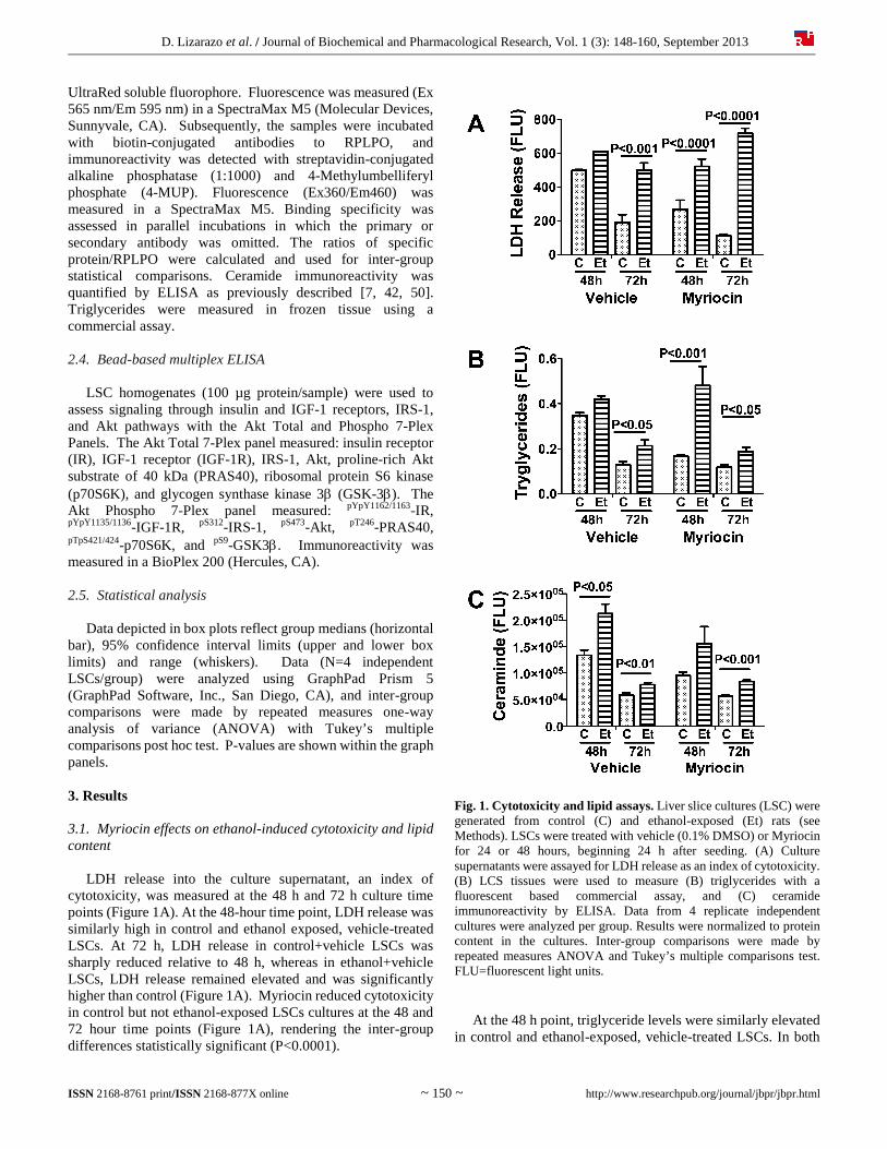
UltraRed soluble fluorophore. Fluorescence was measured (Ex 565 nm/Em 595 nm) in a SpectraMax M5 (Molecular Devices, Sunnyvale, CA). Subsequently, the samples were incubated with biotin-conjugated antibodies to RPLPO, and immunoreactivity was detected with streptavidin-conjugated alkaline phosphatase (1:1000) and 4-Methylumbelliferyl phosphate (4-MUP). Fluorescence (Ex360/Em460) was measured in a SpectraMax M5. Binding specificity was assessed in parallel incubations in which the primary or secondary antibody was omitted. The ratios of specific protein/RPLPO were calculated and used for inter-group statistical comparisons. Ceramide immunoreactivity was quantified by ELISA as previously described [7, 42, 50]. Triglycerides were measured in frozen tissue using a commercial assay. 2.4. Bead-based multiplex ELISA LSC homogenates (100 µg protein/sample) were used to assess signaling through insulin and IGF-1 receptors, IRS-1, and Akt pathways with the Akt Total and Phospho 7-Plex Panels. The Akt Total 7-Plex panel measured: insulin receptor (IR), IGF-1 receptor (IGF-1R), IRS-1, Akt, proline-rich Akt substrate of 40 kDa (PRAS40), ribosomal protein S6 kinase (p70S6K), and glycogen synthase kinase 3 (GSK-3). The Akt Phospho 7-Plex panel measured: pYpY1162/1163-IR, pYpY1135/1136-IGF-1R, pS312-IRS-1, pS473-Akt, pT246-PRAS40, pTpS421/424-p70S6K, and pS9-GSK3. Immunoreactivity was measured in a BioPlex 200 (Hercules, CA). 2.5. Statistical analysis Data depicted in box plots reflect group medians (horizontal bar), 95% confidence interval limits (upper and lower box limits) and range (whiskers). Data (N=4 independent LSCs/group) were analyzed using GraphPad Prism 5 (GraphPad Software, Inc., San Diego, CA), and inter-group comparisons were made by repeated measures one-way analysis of variance (ANOVA) with Tukey’s multiple comparisons post hoc test. P-values are shown within the graph panels. 3. Results 3.1. Myriocin effects on ethanol-induced cytotoxicity and lipid content LDH release into the culture supernatant, an index of cytotoxicity, was measured at the 48 h and 72 h culture time points (Figure 1A). At the 48-hour time point, LDH release was similarly high in control and ethanol exposed, vehicle-treated LSCs. At 72 h, LDH release in control+vehicle LSCs was sharply reduced relative to 48 h, whereas in ethanol+vehicle LSCs, LDH release remained elevated and was significantly higher than control (Figure 1A). Myriocin reduced cytotoxicity in control but not ethanol-exposed LSCs cultures at the 48 and 72 hour time points (Figure 1A), rendering the inter-group differences statistically significant (P<0.0001).
Fig. 1. Cytotoxicity and lipid assays. Liver slice cultures (LSC) were generated from control (C) and ethanol-exposed (Et) rats (see Methods). LSCs were treated with vehicle (0.1% DMSO) or Myriocin for 24 or 48 hours, beginning 24 h after seeding. (A) Culture supernatants were assayed for LDH release as an index of cytotoxicity. (B) LCS tissues were used to measure (B) triglycerides with a fluorescent based commercial assay, and (C) ceramide immunoreactivity by ELISA. Data from 4 replicate independent cultures were analyzed per group. Results were normalized to protein content in the cultures. Inter-group comparisons were made by repeated measures ANOVA and Tukey’s multiple comparisons test. FLU=fluorescent light units. At the 48 h point, triglyceride levels were similarly elevated in control and ethanol-exposed, vehicle-treated LSCs. In both
D. Lizarazo et al. / Journal of Biochemical and Pharmacological Research, Vol. 1 (3): 148-160, September 2013
ISSN 2168-8761 print/ISSN 2168-877X online ~ 151 ~ http://www.researchpub.org/journal/jbpr/jbpr.html
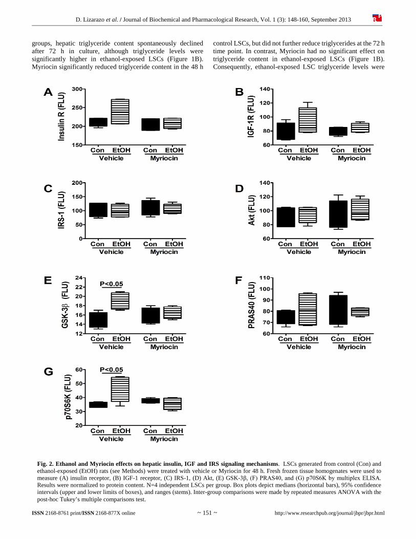
Fig. 2. Ethanol and Myriocin effects on hepatic insulin, IGF and IRS signaling mechanisms. LSCs generated from control (Con) and ethanol-exposed (EtOH) rats (see Methods) were treated with vehicle or Myriocin for 48 h. Fresh frozen tissue homogenates were used to measure (A) insulin receptor, (B) IGF-1 receptor, (C) IRS-1, (D) Akt, (E) GSK-3β, (F) PRAS40, and (G) p70S6K by multiplex ELISA. Results were normalized to protein content. N=4 independent LSCs per group. Box plots depict medians (horizontal bars), 95% confidence intervals (upper and lower limits of boxes), and ranges (stems). Inter-group comparisons were made by repeated measures ANOVA with the post-hoc Tukey’s multiple comparisons test.
groups, hepatic triglyceride content spontaneously declined after 72 h in culture, although triglyceride levels were significantly higher in ethanol-exposed LSCs (Figure 1B). Myriocin significantly reduced triglyceride content in the 48 h
control LSCs, but did not further reduce triglycerides at the 72 h time point. In contrast, Myriocin had no significant effect on triglyceride content in ethanol-exposed LSCs (Figure 1B). Consequently, ethanol-exposed LSC triglyceride levels were
D. Lizarazo et al. / Journal of Biochemical and Pharmacological Research, Vol. 1 (3): 148-160, September 2013
ISSN 2168-8761 print/ISSN 2168-877X online ~ 152 ~ http://www.researchpub.org/journal/jbpr/jbpr.html
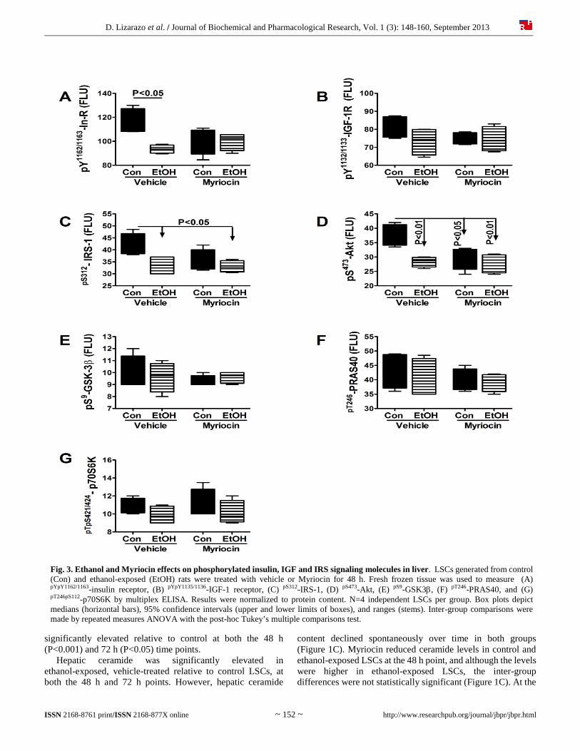
Fig. 3. Ethanol and Myriocin effects on phosphorylated insulin, IGF and IRS signaling molecules in liver. LSCs generated from control (Con) and ethanol-exposed (EtOH) rats were treated with vehicle or Myriocin for 48 h. Fresh frozen tissue was used to measure (A) pYpY1162/1163-insulin receptor, (B) pYpY1135/1136-IGF-1 receptor, (C) pS312-IRS-1, (D) pS473-Akt, (E) pS9-GSK3, (F) pT246-PRAS40, and (G) pT246pS112-p70S6K by multiplex ELISA. Results were normalized to protein content. N=4 independent LSCs per group. Box plots depict medians (horizontal bars), 95% confidence intervals (upper and lower limits of boxes), and ranges (stems). Inter-group comparisons were made by repeated measures ANOVA with the post-hoc Tukey’s multiple comparisons test.
significantly elevated relative to control at both the 48 h (P<0.001) and 72 h (P<0.05) time points. Hepatic ceramide was significantly elevated in ethanol-exposed, vehicle-treated relative to control LSCs, at both the 48 h and 72 h points. However, hepatic ceramide
content declined spontaneously over time in both groups (Figure 1C). Myriocin reduced ceramide levels in control and ethanol-exposed LSCs at the 48 h point, and although the levels were higher in ethanol-exposed LSCs, the inter-group differences were not statistically significant (Figure 1C). At the
D. Lizarazo et al. / Journal of Biochemical and Pharmacological Research, Vol. 1 (3): 148-160, September 2013
ISSN 2168-8761 print/ISSN 2168-877X online ~ 153 ~ http://www.researchpub.org/journal/jbpr/jbpr.html
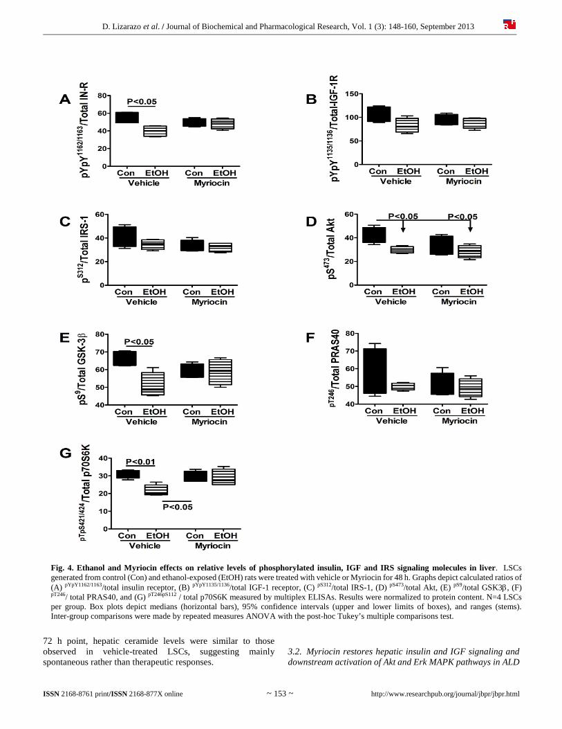
Fig. 4. Ethanol and Myriocin effects on relative levels of phosphorylated insulin, IGF and IRS signaling molecules in liver. LSCs generated from control (Con) and ethanol-exposed (EtOH) rats were treated with vehicle or Myriocin for 48 h. Graphs depict calculated ratios of (A) pYpY1162/1163/total insulin receptor, (B) pYpY1135/1136/total IGF-1 receptor, (C) pS312/total IRS-1, (D) pS473/total Akt, (E) pS9/total GSK3, (F) pT246/ total PRAS40, and (G) pT246pS112 / total p70S6K measured by multiplex ELISAs. Results were normalized to protein content. N=4 LSCs per group. Box plots depict medians (horizontal bars), 95% confidence intervals (upper and lower limits of boxes), and ranges (stems). Inter-group comparisons were made by repeated measures ANOVA with the post-hoc Tukey’s multiple comparisons test.
72 h point, hepatic ceramide levels were similar to those observed in vehicle-treated LSCs, suggesting mainly spontaneous rather than therapeutic responses.
3.2. Myriocin restores hepatic insulin and IGF signaling and downstream activation of Akt and Erk MAPK pathways in ALD
D. Lizarazo et al. / Journal of Biochemical and Pharmacological Research, Vol. 1 (3): 148-160, September 2013
ISSN 2168-8761 print/ISSN 2168-877X online ~ 154 ~ http://www.researchpub.org/journal/jbpr/jbpr.html
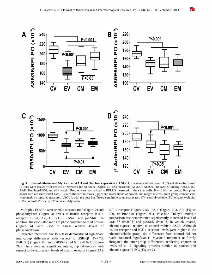
Fig. 5. Effects of ethanol and Myriocin on AAH and Humbug expression in LSCs. LSCs generated from control (C) and ethanol-exposed (E) rats were treated with vehicle or Myriocin for 48 hours. Duplex ELISAs measured (A) AAH-A85G6, (B) AAH+Humbug-A85E6, (C) AAH+Humbug-FB50, and (D) β-actin. Results were normalized to RPLPO measured in the same wells. N=4 LSCs per group. Box plots depict medians (horizontal bars), 95% confidence intervals (upper and lower limits of boxes), and ranges (stems). Inter-group comparisons were made by repeated measures ANOVA with the post-hoc Tukey’s multiple comparisons test. CV=control+vehicle; EV=ethanol+vehicle; CM= control+Myriocin; EM=ethanol+Myriocin.
Multiplex ELISAs were used to measure total (Figure 2) and phosphorylated (Figure 3) levels of insulin receptor, IGF-1 receptor, IRS-1, Akt, GSK-3, PRAS40, and p70S6K. In addition, the calculated ratios of phosphorylated to total protein (Figure 4) were used to assess relative levels of phosphorylation. Repeated measures ANOVA tests demonstrated significant inter-group differences with respect to GSK-3 (F=4.73; P=0.021) (Figure 2E) and p70S6K (F=4.63; P=0.022) (Figure 2G). There were no significant inter-group differences with respect to the expression levels of insulin receptor (Figure 2A),
IGF-1 receptor (Figure 2B), IRS-1 (Figure 2C), Akt (Figure 2D), or PRAS40 (Figure 2G). Post-hoc Tukey’s multiple comparison test demonstrated significantly increased levels of GSK-3β (P<0.05) and p70S6K (P<0.05) in vehicle-treated, ethanol-exposed relative to control+vehicle LSCs. Although insulin receptor and IGF-1 receptor levels were higher in the ethanol+vehicle group, the differences from control did not reach statistical significance. Myriocin treatment uniformly abrogated the inter-group differences, rendering expression levels of all 7 signaling proteins similar in control and ethanol-exposed LSCs (Figure 2).
D. Lizarazo et al. / Journal of Biochemical and Pharmacological Research, Vol. 1 (3): 148-160, September 2013
ISSN 2168-8761 print/ISSN 2168-877X online ~ 155 ~ http://www.researchpub.org/journal/jbpr/jbpr.html
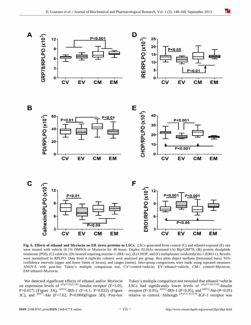
Fig. 6. Effects of ethanol and Myriocin on ER stress proteins in LSCs. LSCs generated from control (C) and ethanol-exposed (E) rats were treated with vehicle (0.1% DMSO) or Myriocin for 48 hours. Duplex ELISAs measured (A) Bip/GRP78, (B) protein disulphide isomerase (PDI), (C) calnexin, (D) inositol requiring enzyme-1 (IRE-1), (E) CHOP, and (F) endoplasmic oxidoreductin-1 (ERO-1). Results were normalized to RPLPO. Data from 4 replicate cultures were analyzed per group. Box plots depict medians (horizontal bars), 95% confidence intervals (upper and lower limits of boxes), and ranges (stems). Inter-group comparisons were made using repeated measures ANOVA with post-hoc Tukey’s multiple comparison test. CV=control+vehicle; EV=ethanol+vehicle; CM= control+Myriocin; EM=ethanol+Myriocin.
We detected significant effects of ethanol and/or Myriocin on expression levels of pYpY1162/1163-Insulin receptor (F=5.05; P=0.017) (Figure 3A), pS312-IRS-1 (F=4.1; P=0.032) (Figure 3C), and pS473-Akt (F=7.62; P=0.004)(Figure 3D). Post-hoc
Tukey’s multiple comparison test revealed that ethanol+vehicle LSCs had significantly lower levels of pYpY1162/1163-Insulin receptor (P<0.05), pS312-IRS-1 (P<0.05), and pS473-Akt (P<0.01) relative to control. Although pYpY1135/1136-IGF-1 receptor was
D. Lizarazo et al. / Journal of Biochemical and Pharmacological Research, Vol. 1 (3): 148-160, September 2013
ISSN 2168-8761 print/ISSN 2168-877X online ~ 156 ~ http://www.researchpub.org/journal/jbpr/jbpr.html
also reduced in ethanol+vehicle LSCs (Figure 3B), the difference did not reach statistical significance. Myriocin rendered the mean levels of pYpY1162/1163-Insulin receptor, pYpY1135/1136-IGF-1 receptor, pS312-IRS-1, and pS473-Akt similar in control and ethanol-exposed LSCs by reducing the levels in control cultures and/or increasing the levels in ethanol-exposed LSCs. In contrast, ethanol and Myriocin had little or no effects on the expression of pS9-GSK-3 (Figure 3E), pT246-PRAS40 (Figure 3F), and pTpS421/424-p70S6K (Figure 3G). Significant inter-group differences were also observed for the calculated ratios of pYpY1162/1163/total insulin receptor (F=4.47; P=0.025) (Figure 4A), pS473/total Akt (F=4.37; P=0.027) (Figure 4D), pS9/total GSK-3 (F=4.60; P=0.023) (Figure 4E), and pTpS421/424/total p70S6K (F=5.87; P=0.01) (Figure 4G), but not for pYpY1135/1136/total IGF-1 receptor (Figure 4B), pS312/total IRS-1 (Figure 4C), or pT246/total PRAS40 (Figure 4F). Myriocin completely abrogated the effects of ethanol, resolving all significant inter-group differences with respect to the relative levels of phospho/total signaling proteins (Figure 4). 3.3. Myriocin restores ethanol-impaired AAH expression Duplex ELISAs measured effects of ethanol and Myriocin on AAH, Humbug, and β-actin expression in LSCs (Figure 5). AAH was detected with the A85G6 monoclonal antibody, which binds to an epitope in the C-terminus, in the vicinity of the catalytic domain [31]. The A85E6 and FB50 monoclonal antibodies bind to different epitopes in the N-terminus of AAH [31]. However, because the N-terminus of AAH is virtually identical to Humbug, FB50 and A85E6 detect AAH and Humbug together [31, 51-53]. We observed significant inter-group differences in expression of AAH-A85G6 (F=65.56; P<0.0001) (Figure 5A), AAH+Humbug-A85E6 (F=24.03; P<0.0001) (Figure 5B), AAH+Humbug-FB50 (F=63.73; P<0.0001) (Figure 5C), and β-Actin (F=31.98; P<0.0001) (Figure 5D). Post hoc tests demonstrated significantly reduced levels of A85G6 (P<0.001), A85E6 (P<0.001), FB50 (P<0.001), and β-actin (P<0.001) immunoreactivity in ethanol+vehicle relative to control+vehicle LSCs. Myriocin reduced A85G6 (P<0.001), A85E6 (P<0.001), FB50 (P<0.001), and β-actin (P<0.001) in control LSCs, and increased/normalized A85G6 (P<0.001) in ethanol-exposed LSCs. In contrast, Myriocin had no significant effect on A85E6, FB50 or β-actin expression in ethanol-exposed LSCs. 3.4. Myriocin effects on hepatic ER stress in LSCs We measured immunoreactivity to proteins that mediate ER stress at various levels in the cascade [54]. ANOVA tests demonstrated significant inter-group differences with respect to GRP78 (F=14.93; P<0.0001) (Figure 6A), PDI (F=7.32; P=0.0002) (Figure 6B), Calnexin (F=5.31; P=0.002) (Figure 6C), IRE-1 (F=7.038; P=0.0003) (Figure 6D), CHOP (F=30.11; P<0.0001) (Figure 6E), ERO1 (F=0.34; P<0.0001) (Figure 6F). Ethanol significantly reduced hepatic expression of calnexin (P<0.01), IRE-1 (P<0.05), and CHOP (P<0.001), and increased ERO1 (P<0.001) relative to vehicle-treated controls.
In contrast, GRP78 and PDI were expressed at similar levels in control and ethanol exposed LSCs. Myriocin significantly increased expression of GRP78 (P<0.001) and PDI (P<0.01) in control LSCs (relative to vehicle), and GRP78 (P<0.001), calnexin (P<0.05), and IRE-1 (P<0.01) in ethanol-fed rats (relative to vehicle). In contrast, Myriocin had no significant effects on PDI, CHOP, or ERO1 expression in ethanol-exposed LSCs, or Calnexin, IRE-1, CHOP, or ERO1 in control LSCs. Consequently, the major effects of Myriocin were to increase and normalize expression of Calnexin and IRE-1 in ethanol-exposed LSCs, similarly increase GRP78 in control and ethanol-exposed LSCs, and selectively increase PDI in control LSCs. 4. Discussion 4.1. Rationale for using the ceramide-inhibitor approach for ALD The present study examines the potential therapeutic effects of Myriocin on liver function after chronic in vivo ethanol exposure. Myriocin inhibits serine palmitoyl transferase, which mediates de novo ceramide synthesis. The studies utilized a liver slice culture model to evaluate early responses to Myriocin. We focused on insulin/IGF-1 signaling networks and expression of AAH, a downstream target that regulates cell motility and adhesion [6, 20-22]. 4.2. Ceramide inhibitor effects on cytotoxicity and lipid storage in ethanol-exposed LSCs Therapeutic effectiveness of the Myriocin treatments was demonstrated by the lowering of ceramide levels in both control and ethanol exposed LSCs at the 48 h time point. The subsequent spontaneous declines in ceramide levels, irrespective of treatment could reflect general adaptive responses to the LSC environment. Despite early reductions of ceramide levels, the failure of Myriocin to reduce cytotoxicity in ethanol-exposed LSCs indicates that the hepatotoxic effects of ethanol and/or acetaldehyde are not ameliorated by this approach. Similarly, hepatic triglyceride storage is not measurably decreased following short-term inhibition of ceramide synthesis. 4.3. Short-term effects of myriocin on insulin/IGF-1/IRS-1 signaling in ALD Ethanol exposure broadly impaired insulin and IGF-1 signaling by inhibiting the absolute and relative levels of phosphorylated receptors and IRS-1. Correspondingly, insulin/IGF-1 signaling through Akt and p70S6K was reduced, whereas PRAS40 was relatively unaffected. In addition, the total levels of GSK-3β were increased, and relative levels of GSK-3β phosphorylation were decreased in ethanol-exposed LSCs. These results indicate that ethanol exposure led to GSK-3β activation since Ser9-phosphorylation of GSK-3β inhibits the kinase. These broad impairments in insulin/IGF signaling correspond with the decreased levels of AAH and
D. Lizarazo et al. / Journal of Biochemical and Pharmacological Research, Vol. 1 (3): 148-160, September 2013
ISSN 2168-8761 print/ISSN 2168-877X online ~ 157 ~ http://www.researchpub.org/journal/jbpr/jbpr.html
AAH+Humbug expression, which are stimulated by insulin and IGF-1, and decreased by GSK-3β [30, 31]. Myriocin effectively restored insulin and IGF-1 signaling networks, rendering the total and relative phosphorylated levels of most proteins similar in control and ethanol-exposed LSCs. Therefore, reducing hepatic ceramide load may be beneficial for restoring hepatic insulin responsiveness needed to maintain liver function in ALD. 4.4. Myriocin effects on AAH expression in ethanol-exposed livers The finding that ethanol inhibits AAH and AAH+Humbug expression corroborates previous in vivo studies of ALD [6, 31]. The significant increases in AAH protein expression (A85G6 immunoreactivity) in ethanol-exposed livers treated with Myriocin correlate with the enhancements in insulin/IGF-1 signaling. On the other hand, the modest effects of Myriocin on A85E6 and FB50 immunoreactivity suggest that other therapeutic approaches are needed to restore Humbug expression in ALD. The same conclusion could be drawn with respect to β-actin, in which the inhibitory effects of ethanol were not reversed by Myriocin. 4.5. Myriocin effects on ER stress signaling proteins Insulin resistance and ceramide accumulation promote ER stress, and ER stress exacerbates insulin resistance, inflammation, oxidative stress, and ceramide accumulation [55-62]. The ER mediates protein synthesis, folding, maturation, and trafficking [63], and is critical for Ca2+ homeostasis and triglyceride synthesis. ER stress occurs when homeostatic mechanisms get disrupted and unfolded proteins accumulate, leading to increased formation of reactive oxygen species. Prolonged activation of the unfolded protein response (UPR) leads to inflammation, injury, steatosis, and apoptosis [8, 54, 63, 64]. The ER adapts to stress by activating the UPR [60, 65], which quickly increases levels of ER stress sensor proteins including: inositol-requiring enzyme 1 (IRE-1), PKR-like ER-localized eIF2 kinase (PERK), and the activating transcription factor 6α (ATF-6α; ER membrane-anchored transcription factor). PERK and IRE1 transmit stress signals in response to protein misfolding or unfolding, and thereby activate ER stress networks [63]. PERK drives global arrest of protein synthesis and up-regulates the transcription factor C/EBP homologous protein CHOP, which promotes apoptosis. IRE1 increases transcription of chaperones and ER associated protein degradation (ERAD) machinery. Insulin resistance drives ER stress because vital ER functions such as protein synthesis, modification, and folding, calcium signaling, and lipid biosynthesis utilize glucose as the main source of energy; insulin resistance impairs glucose uptake and metabolism. Moreover, pathogenic lipolysis leading to ceramide accumulation is initiated by critical levels of ER stress and mitochondrial dysfunction [45, 66-68]. Ethanol- activates all major ER stress sensor cascades [66-69], and the resulting increased levels of ER stress exacerbate insulin
resistance and lipolysis. End results include, increased ceramide production [70, 71], worsening of inflammation and insulin resistance, and lipotoxicity [8, 72, 73]. Herein we observed relative inhibition of calnexin, IRE-1, and CHOP/GADD153, and increased levels of ERO-1 in ethanol+vehicle relative to control+vehicle LSCs. ERO-1, complexed with the PDI molecular chaperone, mediates oxidation of disulfide bonds in folding proteins. Therefore the ethanol associated increases in ERO-1 could reflect increased disposal of mal-folded proteins in liver. At the same time, reduced expression of calnexin and IRE-1 could indicate impaired folding and assembly of newly synthesized proteins in the ER (calnexin) and reduced transcription of chaperones and ER associated protein degradation machinery (ERAD). Consequences would include that in these short term experiments, the ethanol-exposed hepatocytes respond to increased formation of mal-folded proteins and can utilize homeostatic mechanisms to prevent their accumulation. Myriocin-mediated restoration of hepatic insulin/IGF signaling mechanisms in ethanol-exposed LSCs was associated with slight but significant increases in GRP78, calnexin, and IRE-1. This suggests that Myriocin-induced reduction in hepatic ceramide levels in ethanol-exposed LSCs was correlated with increased activation of ER stress responses. While these effects may seem paradoxical, the outcomes suggest they are protective and needed to restore homeostasis vis-à-vis persistent hepatic injury from continuous ethanol exposure. On the other hand, the persistently lower levels of PDI, CHOP, and ERO-1α in ethanol exposed LSCs suggests that, in these short-term experiments, the mechanisms designed to destroy cells with activated UPRs and accumulated mal-folded proteins were not mobilized by the Myriocin treatments. This phenomenon could partly account for the persistent cytotoxicity (high-level LDH release) in ethanol-exposed LSCs, irrespective of Myriocin treatment. Acknowledgements This work was supported by AA-11431, AA-12908, and 5T32DK060415 from the National Institutes of Health. Conflict of interest: None declared. References [1] Miller AM, Horiguchi N, Jeong WI, Radaeva S, Gao B.
Molecular mechanisms of alcoholic liver disease: innate immunity and cytokines. Alcohol Clin Exp Res 2011; 35:787-793.
[2] McCullough AJ, O'Shea RS, Dasarathy S. Diagnosis and management of alcoholic liver disease. J Dig Dis 2011; 12:257-262.
[3] Paula H, Asrani SK, Boetticher NC, Pedersen R, Shah VH, Kim WR. Alcoholic liver disease-related mortality in the United States: 1980-2003. Am J Gastroenterol 2010; 105:1782-1787.
D. Lizarazo et al. / Journal of Biochemical and Pharmacological Research, Vol. 1 (3): 148-160, September 2013
ISSN 2168-8761 print/ISSN 2168-877X online ~ 158 ~ http://www.researchpub.org/journal/jbpr/jbpr.html
[4] O'Shea RS, Dasarathy S, McCullough AJ. Alcoholic liver disease. Hepatology 2010; 51:307-328.
[5] de la Monte SM, Yeon JE, Tong M, Longato L, Chaudhry R, Pang MY, et al. Insulin resistance in experimental alcohol-induced liver disease. J Gastroenterol Hepatol 2008; 23:e477-486.
[6] Pang M, de la Monte SM, Longato L, Tong M, He J, Chaudhry R, et al. PPARdelta agonist attenuates alcohol-induced hepatic insulin resistance and improves liver injury and repair. J Hepatol 2009; 50:1192-1201.
[7] Longato L, Ripp K, Setshedi M, Dostalek M, Akhlaghi F, Branda M, et al. Insulin resistance, ceramide accumulation, and endoplasmic reticulum stress in human chronic alcohol-related liver disease. Oxid Med Cell Longev 2012; 2012:479348.
[8] Kaplowitz N, Ji C. Unfolding new mechanisms of alcoholic liver disease in the endoplasmic reticulum. J Gastroenterol Hepatol 2006; 21 Suppl 3:S7-9.
[9] Lieber CS. Alcoholic fatty liver: its pathogenesis and mechanism of progression to inflammation and fibrosis. Alcohol 2004; 34:9-19.
[10] Denucci SM, Tong M, Longato L, Lawton M, Setshedi M, Carlson RI, et al. Rat strain differences in susceptibility to alcohol-induced chronic liver injury and hepatic insulin resistance. Gastroenterology research and practice 2010; 2010:312790.
[11] He J, de la Monte S, Wands JR. Acute ethanol exposure inhibits insulin signaling in the liver. Hepatology 2007; 46:1791-1800.
[12] Onishi Y, Honda M, Ogihara T, Sakoda H, Anai M, Fujishiro M, et al. Ethanol feeding induces insulin resistance with enhanced PI 3-kinase activation. Biochem Biophys Res Commun 2003; 303:788-794.
[13] Ronis MJ, Wands JR, Badger TM, de la Monte SM, Lang CH, Calissendorff J. Alcohol-induced disruption of endocrine signaling. Alcohol Clin Exp Res 2007; 31:1269-1285.
[14] Sasaki Y, Wands JR. Ethanol impairs insulin receptor substrate-1 mediated signal transduction during rat liver regeneration. Biochem Biophys Res Commun 1994; 199:403-409.
[15] Setshedi M, Longato L, Petersen DR, Ronis M, Chen WC, Wands JR, et al. Limited Therapeutic Effect of N-Acetylcysteine on Hepatic Insulin Resistance in an Experimental Model of Alcohol-Induced Steatohepatitis. Alcoholism, clinical and experimental research 2011; 35:2139-2151.
[16] Mohr L, Tanaka S, Wands JR. Ethanol inhibits hepatocyte proliferation in insulin receptor substrate 1 transgenic mice. Gastroenterology 1998; 115:1558-1565.
[17] Sasaki Y, Hayashi N, Ito T, Fusamoto H, Kamada T, Wands JR. Influence of ethanol on insulin receptor substrate-1-mediated signal transduction during rat liver regeneration. Alcohol Alcohol 1994; 1:99-106.
[18] Roberts RA, James NH, Cosulich SC. The role of protein kinase B and mitogen-activated protein kinase in epidermal growth factor and tumor necrosis factor alpha-mediated rat hepatocyte survival and apoptosis. Hepatology 2000; 31:420-427.
[19] Yeon JE, Califano S, Xu J, Wands JR, De La Monte SM. Potential role of PTEN phosphatase in ethanol-impaired survival signaling in the liver. Hepatology 2003; 38:703-714.
[20] Cantarini MC, de la Monte SM, Pang M, Tong M, D'Errico A, Trevisani F, et al. Aspartyl-asparagyl beta hydroxylase over-expression in human hepatoma is linked to activation of
insulin-like growth factor and notch signaling mechanisms. Hepatology 2006; 44:446-457.
[21] de la Monte SM, Tamaki S, Cantarini MC, Ince N, Wiedmann M, Carter JJ, et al. Aspartyl-(asparaginyl)-beta-hydroxylase regulates hepatocellular carcinoma invasiveness. Journal of hepatology 2006; 44:971-983.
[22] Ince N, de la Monte SM, Wands JR. Overexpression of human aspartyl (asparaginyl) beta-hydroxylase is associated with malignant transformation. Cancer research 2000; 60:1261-1266.
[23] Lavaissiere L, Jia S, Nishiyama M, de la Monte S, Stern AM, Wands JR, et al. Overexpression of human aspartyl(asparaginyl)beta-hydroxylase in hepatocellular carcinoma and cholangiocarcinoma. The Journal of clinical investigation 1996; 98:1313-1323.
[24] Jia S, McGinnis K, VanDusen WJ, Burke CJ, Kuo A, Griffin PR, et al. A fully active catalytic domain of bovine aspartyl (asparaginyl) beta-hydroxylase expressed in Escherichia coli: characterization and evidence for the identification of an active-site region in vertebrate alpha-ketoglutarate-dependent dioxygenases. Proc Natl Acad Sci U S A 1994; 91:7227-7231.
[25] Jia S, VanDusen WJ, Diehl RE, Kohl NE, Dixon RA, Elliston KO, et al. cDNA cloning and expression of bovine aspartyl (asparaginyl) beta-hydroxylase. J Biol Chem 1992; 267:14322-14327.
[26] Feriotto G, Finotti A, Breveglieri G, Treves S, Zorzato F, Gambari R. Transcriptional activity and Sp 1/3 transcription factor binding to the P1 promoter sequences of the human AbetaH-J-J locus. The FEBS journal 2007; 274:4476-4490.
[27] Lahousse SA, Carter JJ, Xu XJ, Wands JR, de la Monte SM. Differential growth factor regulation of aspartyl-(asparaginyl)-beta-hydroxylase family genes in SH-Sy5y human neuroblastoma cells. BMC cell biology 2006; 7:41.
[28] Lawton M, Tong M, Gundogan F, Wands JR, de la Monte SM. Aspartyl-(asparaginyl) beta-hydroxylase, hypoxia-inducible factor-alpha and Notch cross-talk in regulating neuronal motility. Oxidative medicine and cellular longevity 2010; 3:347-356.
[29] Carter JJ, Tong M, Silbermann E, Lahousse SA, Ding FF, Longato L, et al. Ethanol impaired neuronal migration is associated with reduced aspartyl-asparaginyl-beta-hydroxylase expression. Acta neuropathologica 2008; 116:303-315.
[30] Silbermann E, Moskal P, Bowling N, Tong M, de la Monte SM. Role of aspartyl-(asparaginyl)-beta-hydroxylase mediated notch signaling in cerebellar development and function. Behavioral and brain functions : BBF 2010; 6:68.
[31] de la Monte SM, Tong M, Carlson RI, Carter JJ, Longato L, Silbermann E, et al. Ethanol inhibition of aspartyl-asparaginyl-beta-hydroxylase in fetal alcohol spectrum disorder: Potential link to the impairments in central nervous system neuronal migration. Alcohol 2009; 43:225-240.
[32] Gundogan F, Elwood G, Longato L, Tong M, Feijoo A, Carlson RI, et al. Impaired placentation in fetal alcohol syndrome. Placenta 2008; 29:148-157.
[33] Kao Y, Youson JH, Holmes JA, Al-Mahrouki A, Sheridan MA. Effects of insulin on lipid metabolism of larvae and metamorphosing landlocked sea lamprey, Petromyzon marinus. Gen Comp Endocrinol 1999; 114:405-414.
[34] Holland WL, Summers SA. Sphingolipids, insulin resistance, and metabolic disease: new insights from in vivo manipulation of sphingolipid metabolism. Endocr Rev 2008; 29:381-402.
D. Lizarazo et al. / Journal of Biochemical and Pharmacological Research, Vol. 1 (3): 148-160, September 2013
ISSN 2168-8761 print/ISSN 2168-877X online ~ 159 ~ http://www.researchpub.org/journal/jbpr/jbpr.html
[35] Langeveld M, Aerts JM. Glycosphingolipids and insulin resistance. Prog Lipid Res 2009; 48:196-205.
[36] Kraegen EW, Cooney GJ. Free fatty acids and skeletal muscle insulin resistance. Curr Opin Lipidol 2008; 19:235-241.
[37] Bourbon NA, Sandirasegarane L, Kester M. Ceramide-induced inhibition of Akt is mediated through protein kinase Czeta: implications for growth arrest. J Biol Chem 2002; 277:3286-3292.
[38] Hajduch E, Balendran A, Batty IH, Litherland GJ, Blair AS, Downes CP, et al. Ceramide impairs the insulin-dependent membrane recruitment of protein kinase B leading to a loss in downstream signalling in L6 skeletal muscle cells. Diabetologia 2001; 44:173-183.
[39] Nogueira TC, Anhe GF, Carvalho CR, Curi R, Bordin S, Carpinelli AR. Involvement of phosphatidylinositol-3 kinase/AKT/PKCzeta/lambda pathway in the effect of palmitate on glucose-induced insulin secretion. Pancreas 2008; 37:309-315.
[40] Powell DJ, Hajduch E, Kular G, Hundal HS. Ceramide disables 3-phosphoinositide binding to the pleckstrin homology domain of protein kinase B (PKB)/Akt by a PKCzeta-dependent mechanism. Mol Cell Biol 2003; 23:7794-7808.
[41] de la Monte SM, Longato L, Tong M, DeNucci S, Wands JR. The liver-brain axis of alcohol-mediated neurodegeneration: role of toxic lipids. Int J Environ Res Public Health 2009; 6:2055-2075.
[42] Ramirez T, Tong M, Chen WC, Nguyen QG, Wands JR, de la Monte SM. Chronic alcohol-induced hepatic insulin resistance and endoplasmic reticulum stress ameliorated by peroxisome-proliferator activated receptor-delta agonist treatment. J Gastroenterol Hepatol 2013; 28:179-187.
[43] Garcia-Ruiz C, Colell A, Mari M, Morales A, Calvo M, Enrich C, et al. Defective TNF-alpha-mediated hepatocellular apoptosis and liver damage in acidic sphingomyelinase knockout mice. J Clin Invest 2003; 111:197-208.
[44] Liangpunsakul S, Sozio MS, Shin E, Zhao Z, Xu Y, Ross RA, et al. Inhibitory effect of ethanol on AMPK phosphorylation is mediated in part through elevated ceramide levels. Am J Physiol Gastrointest Liver Physiol 2010; 298:G1004-1012.
[45] Anderson N, Borlak J. Molecular mechanisms and therapeutic targets in steatosis and steatohepatitis. Pharmacol Rev 2008; 60:311-357.
[46] Kuznetsov JN, Leclerc GJ, Leclerc GM, Barredo JC. AMPK and Akt determine apoptotic cell death following perturbations of one-carbon metabolism by regulating ER stress in acute lymphoblastic leukemia. Mol Cancer Ther 2011; 10:437-447.
[47] de la Monte SM. Alcohol-induced liver and brain degeneration: roles of insulin resistance, toxic ceramides, and endoplasmic reticulum stress. In: Watson RR, Preedy VR, Zipadi S, editors. Alcohol, Nutrition, and Health Consequences. New York: Humana Press, 2012. p. 507-522.
[48] Ramirez T, Longato L, Dostalek M, Tong M, Wands JR, de la Monte SM. Insulin resistance, ceramide accumulation and endoplasmic reticulum stress in experimental chronic alcohol-induced steatohepatitis. Alcohol Alcohol 2013; 48:39-52.
[49] Glaros EN, Kim WS, Wu BJ, Suarna C, Quinn CM, Rye KA, et al. Inhibition of atherosclerosis by the serine palmitoyl transferase inhibitor myriocin is associated with reduced plasma glycosphingolipid concentration. Biochem Pharmacol 2007; 73:1340-1346.
[50] Lyn-Cook LE, Jr., Lawton M, Tong M, Silbermann E, Longato L, Jiao P, et al. Hepatic Ceramide May Mediate Brain Insulin
Resistance and Neurodegeneration in Type 2 Diabetes and Non-alcoholic Steatohepatitis. J Alzheimers Dis 2009; 16:715-729.
[51] Luu M, Sabo E, de la Monte SM, Greaves W, Wang J, Tavares R, et al. Prognostic value of aspartyl (asparaginyl)-beta-hydroxylase/humbug expression in non-small cell lung carcinoma. Human pathology 2009; 40:639-644.
[52] Palumbo KS, Wands JR, Safran H, King T, Carlson RI, de la Monte SM. Human aspartyl (asparaginyl) beta-hydroxylase monoclonal antibodies: potential biomarkers for pancreatic carcinoma. Pancreas 2002; 25:39-44.
[53] Wang J, de la Monte SM, Sabo E, Kethu S, Tavares R, Branda M, et al. Prognostic value of humbug gene overexpression in stage II colon cancer. Human pathology 2007; 38:17-25.
[54] Jager R, Bertrand MJ, Gorman AM, Vandenabeele P, Samali A. The unfolded protein response at the crossroads of cellular life and death during ER stress. Biol Cell 2012; 104:259-270.
[55] Senkal CE, Ponnusamy S, Bielawski J, Hannun YA, Ogretmen B. Antiapoptotic roles of ceramide-synthase-6-generated C16-ceramide via selective regulation of the ATF6/CHOP arm of ER-stress-response pathways. FASEB journal : official publication of the Federation of American Societies for Experimental Biology 2010; 24:296-308.
[56] Boslem E, MacIntosh G, Preston AM, Bartley C, Busch AK, Fuller M, et al. A lipidomic screen of palmitate-treated MIN6 beta-cells links sphingolipid metabolites with endoplasmic reticulum (ER) stress and impaired protein trafficking. The Biochemical journal 2011; 435:267-276.
[57] Carracedo A, Lorente M, Egia A, Blazquez C, Garcia S, Giroux V, et al. The stress-regulated protein p8 mediates cannabinoid-induced apoptosis of tumor cells. Cancer Cell 2006; 9:301-312.
[58] Hu P, Han Z, Couvillon AD, Kaufman RJ, Exton JH. Autocrine tumor necrosis factor alpha links endoplasmic reticulum stress to the membrane death receptor pathway through IRE1alpha-mediated NF-kappaB activation and down-regulation of TRAF2 expression. Mol Cell Biol 2006; 26:3071-3084.
[59] McCullough KD, Martindale JL, Klotz LO, Aw TY, Holbrook NJ. Gadd153 sensitizes cells to endoplasmic reticulum stress by down-regulating Bcl2 and perturbing the cellular redox state. Mol Cell Biol 2001; 21:1249-1259.
[60] Ozcan U, Cao Q, Yilmaz E, Lee AH, Iwakoshi NN, Ozdelen E, et al. Endoplasmic reticulum stress links obesity, insulin action, and type 2 diabetes. Science 2004; 306:457-461.
[61] Urano F, Bertolotti A, Ron D. IRE1 and efferent signaling from the endoplasmic reticulum. Journal of cell science 2000; 113 Pt 21:3697-3702.
[62] Sauane M, Su ZZ, Dash R, Liu X, Norris JS, Sarkar D, et al. Ceramide plays a prominent role in MDA-7/IL-24-induced cancer-specific apoptosis. J Cell Physiol 2010; 222:546-555.
[63] Hotamisligil GS. Endoplasmic reticulum stress and the inflammatory basis of metabolic disease. Cell 2010; 140:900-917.
[64] Pandol SJ, Gorelick FS, Gerloff A, Lugea A. Alcohol abuse, endoplasmic reticulum stress and pancreatitis. Dig Dis 2010; 28:776-782.
[65] Malhi H, Kaufman RJ. Endoplasmic reticulum stress in liver disease. Journal of hepatology 2011; 54:795-809.
[66] Kaplowitz N, Than TA, Shinohara M, Ji C. Endoplasmic reticulum stress and liver injury. Semin Liver Dis 2007; 27:367-377.
D. Lizarazo et al. / Journal of Biochemical and Pharmacological Research, Vol. 1 (3): 148-160, September 2013
ISSN 2168-8761 print/ISSN 2168-877X online ~ 160 ~ http://www.researchpub.org/journal/jbpr/jbpr.html
[67] Malhi H, Gores GJ. Molecular mechanisms of lipotoxicity in nonalcoholic fatty liver disease. Semin Liver Dis 2008; 28:360-369.
[68] Sundar-Rajan S, Srinivasan V, Balasubramanyam M, Tatu U. Endoplasmic reticulum (ER) stress & diabetes. Indian J Med Res 2007; 125:411-424.
[69] Sharma NK, Das SK, Mondal AK, Hackney OG, Chu WS, Kern PA, et al. Endoplasmic reticulum stress markers are associated with obesity in nondiabetic subjects. The Journal of clinical endocrinology and metabolism 2008; 93:4532-4541.
[70] de la Monte SM. Triangulated mal-signaling in Alzheimer's Disease: Roles of neurotoxic ceramides, ER stress, and insulin resistance reviewed. J Alzheimers Dis 2012; 30:S231-S249.
[71] Szulc ZM, Bielawski J, Gracz H, Gustilo M, Mayroo N,
Hannun YA, et al. Tailoring structure-function and targeting properties of ceramides by site-specific cationization. Bioorg Med Chem 2006; 14:7083-7104.
[72] Ronis MJ, Butura A, Korourian S, Shankar K, Simpson P, Badeaux J, et al. Cytokine and chemokine expression associated with steatohepatitis and hepatocyte proliferation in rats fed ethanol via total enteral nutrition. Exp Biol Med (Maywood) 2008; 233:344-355.
[73] Banerjee A, Russell WK, Jayaraman A, Ramaiah SK. Identification of proteins to predict the molecular basis for the observed gender susceptibility in a rat model of alcoholic steatohepatitis by 2-D gel proteomics. Proteomics 2008; 8:4327-4337.