
Research Article Immunomodulatory Effects of...
9
Research Article Immunomodulatory Effects of Lactobacillus plantarum Lp62 on Intestinal Epithelial and Mononuclear Cells Thalis Ferreira dos Santos, 1 Tauá Alves Melo, 2 Milena Evangelista Almeida, 2 Rachel Passos Rezende, 2 and Carla Cristina Romano 2 1 State University of Feira de Santana, Transnordestina Avenue S/N, 44030-900 Feira de Santana, BA, Brazil 2 State University of Santa Cruz, Highway Ilh´ eus-Itabuna, km 16 S/N, 45662-900 Ilh´ eus, BA, Brazil Correspondence should be addressed to Carla Cristina Romano; [email protected] Received 7 March 2016; Revised 3 June 2016; Accepted 14 June 2016 Academic Editor: David Bernardo Copyright © 2016 alis Ferreira dos Santos et al. is is an open access article distributed under the Creative Commons Attribution License, which permits unrestricted use, distribution, and reproduction in any medium, provided the original work is properly cited. Probiotic lactic acid bacteria are known for their ability to modulate the immune system. ey have been shown to inhibit inflammation in experiments with animal models, cell culture, and clinical trials. e objective of this study was to elucidate the anti- inflammatory potential of Lactobacillus plantarum Lp62, isolated from cocoa fermentation, in a cell culture model. Lp62 inhibited IL-8 production by Salmonella Typhi-stimulated HT-29 cells and prevented the adhesion of pathogens to these epithelial cells. e probiotic strain was able to modulate TNF-, IL1-, and IL-17 secretion by J774 macrophages. J774 activation was reduced by coincubation with Lp62. PBMC culture showed significantly higher levels of CD4 + CD25 + T lymphocytes following treatment with Lp62. Probiotics also induced increased IL-10 secretion by mononuclear cells. L. plantarum Lp62 was able to inhibit inflammatory stimulation in epithelial cells and macrophages and activated a tolerogenic profile in mononuclear cells of healthy donors. ese results indicate this strain for a possible application in the treatment or prevention of inflammatory diseases. 1. Background Probiotics are defined as live microorganisms which, when administered in adequate amounts, promote beneficial effects on the host’s health. Microbial genera commonly associated with probiotic effects usually have the ability to restore the balance of microbiota, regulate intestinal traffic, produce short-chain fatty acids, and compete with pathogens for adhesion sites. Other properties, such as immune modulation and production of specific bioactive substances, are restricted to some strains. Traditionally, probiotics are used to treat or prevent the imbalance of the intestinal microbiota caused by pathogens and/or resulting from antibiotic therapy. However, new approaches have demonstrated the potential of these microorganisms as adjuncts to the treatment or prevention of intestinal and extraintestinal chronic diseases [1–3]. Inflammatory bowel diseases (IBD) have increased espe- cially in western countries. Despite being considered to be caused by multifactorial conditions, the gut microbial population plays a central role in the development of IBD in genetically susceptible individuals [4]; therefore, therapeutic approaches that modify the local microbiota are very attrac- tive. In this context, probiotics can stimulate the immune system, resulting in modulation of inflammatory mediators that are responsible for the maintenance of the pathological process or directing the innate and adaptive responses in a regulatory sense [5]. L. plantarum is a Gram-positive rod-shaped bacterium found in a wide variety of niches such as vegetables, meat, fish, and the gastrointestinal tract. Due to its ubiquity and importance in various fermentation processes, it was the first species of the genus Lactobacillus to have its genome sequenced. Further sequencing revealed considerable genetic diversity among strains isolated from different environments, which explains the high adaptability of these lactic acid bacteria [6]. A number of studies prove the applicability of various strains of L. plantarum as probiotic. e 299v strain, used in an already marketed probiotic, reduced in vitro expression of proinflammatory genes in a culture model of colonic mucosa [7]. In addition to anti-Helicobacter pylori Hindawi Publishing Corporation BioMed Research International Volume 2016, Article ID 8404156, 8 pages http://dx.doi.org/10.1155/2016/8404156
Transcript of Research Article Immunomodulatory Effects of...
Research ArticleImmunomodulatory Effects of Lactobacillus plantarum Lp62 onIntestinal Epithelial and Mononuclear Cells
Thalis Ferreira dos Santos,1 Tauá Alves Melo,2 Milena Evangelista Almeida,2
Rachel Passos Rezende,2 and Carla Cristina Romano2
1State University of Feira de Santana, Transnordestina Avenue S/N, 44030-900 Feira de Santana, BA, Brazil2State University of Santa Cruz, Highway Ilheus-Itabuna, km 16 S/N, 45662-900 Ilheus, BA, Brazil
Correspondence should be addressed to Carla Cristina Romano; [email protected]
Received 7 March 2016; Revised 3 June 2016; Accepted 14 June 2016
Academic Editor: David Bernardo
Copyright © 2016 Thalis Ferreira dos Santos et al. This is an open access article distributed under the Creative CommonsAttribution License, which permits unrestricted use, distribution, and reproduction in any medium, provided the original work isproperly cited.
Probiotic lactic acid bacteria are known for their ability to modulate the immune system. They have been shown to inhibitinflammation in experimentswith animalmodels, cell culture, and clinical trials.Theobjective of this studywas to elucidate the anti-inflammatory potential of Lactobacillus plantarum Lp62, isolated from cocoa fermentation, in a cell culture model. Lp62 inhibitedIL-8 production by Salmonella Typhi-stimulated HT-29 cells and prevented the adhesion of pathogens to these epithelial cells.The probiotic strain was able to modulate TNF-𝛼, IL1-𝛽, and IL-17 secretion by J774 macrophages. J774 activation was reduced bycoincubation with Lp62. PBMC culture showed significantly higher levels of CD4+CD25+ T lymphocytes following treatment withLp62. Probiotics also induced increased IL-10 secretion by mononuclear cells. L. plantarum Lp62 was able to inhibit inflammatorystimulation in epithelial cells and macrophages and activated a tolerogenic profile in mononuclear cells of healthy donors. Theseresults indicate this strain for a possible application in the treatment or prevention of inflammatory diseases.
1. Background
Probiotics are defined as live microorganisms which, whenadministered in adequate amounts, promote beneficial effectson the host’s health. Microbial genera commonly associatedwith probiotic effects usually have the ability to restore thebalance of microbiota, regulate intestinal traffic, produceshort-chain fatty acids, and compete with pathogens foradhesion sites. Other properties, such as immunemodulationand production of specific bioactive substances, are restrictedto some strains. Traditionally, probiotics are used to treat orprevent the imbalance of the intestinal microbiota caused bypathogens and/or resulting from antibiotic therapy. However,new approaches have demonstrated the potential of thesemicroorganisms as adjuncts to the treatment or preventionof intestinal and extraintestinal chronic diseases [1–3].
Inflammatory bowel diseases (IBD) have increased espe-cially in western countries. Despite being considered tobe caused by multifactorial conditions, the gut microbialpopulation plays a central role in the development of IBD in
genetically susceptible individuals [4]; therefore, therapeuticapproaches that modify the local microbiota are very attrac-tive. In this context, probiotics can stimulate the immunesystem, resulting in modulation of inflammatory mediatorsthat are responsible for the maintenance of the pathologicalprocess or directing the innate and adaptive responses in aregulatory sense [5].
L. plantarum is a Gram-positive rod-shaped bacteriumfound in a wide variety of niches such as vegetables, meat,fish, and the gastrointestinal tract. Due to its ubiquity andimportance in various fermentation processes, it was thefirst species of the genus Lactobacillus to have its genomesequenced. Further sequencing revealed considerable geneticdiversity among strains isolated from different environments,which explains the high adaptability of these lactic acidbacteria [6]. A number of studies prove the applicabilityof various strains of L. plantarum as probiotic. The 299vstrain, used in an alreadymarketed probiotic, reduced in vitroexpression of proinflammatory genes in a culture model ofcolonic mucosa [7]. In addition to anti-Helicobacter pylori
Hindawi Publishing CorporationBioMed Research InternationalVolume 2016, Article ID 8404156, 8 pageshttp://dx.doi.org/10.1155/2016/8404156
2 BioMed Research International
activity [8], it was also able to improve the symptoms ofirritable bowel syndrome in a clinical study using 200 patients[9]. L. plantarum Lp91 showed strong immunoregulatorycapacity in a murine colitis model induced by TNBS [10],and the WCFS1 strain was effective in generating regulatoryT cells in healthy individuals [11].
The probiotic characteristics of each isolated strain arespecific. Different species or variants within the same speciescan interact with the local microbiota and the host immunesystem in particular ways. Consequently, the use of Lacto-bacillus species as a probiotic needs careful selection to clarifytheir potential, mechanisms, and technological properties.L. plantarum Lp62 was isolated from a batch of fermentingcocoa beans and identified by 16S rDNA gene sequencing(GenBank access number KU291427). Its probiotic potentialwas attested previously in a study that evaluated its anti-inflammatory capacity in a colitis model induced by aceticacid in mice [12]. However, strain Lp62 was administered in apool of other strains, making it difficult to establish the role ofeach microorganism in the observed effect. In this study, wesought to refine this research, by endeavoring to propose apossible in vitro anti-inflammatory mechanism. Strain Lp62modulated the inflammatory response in epithelial cells bypreventing S. Typhi adhesion, inhibited macrophage activa-tion and thereby decreased the levels of cytokines involvedin IBD pathogenesis, and, finally, increased IL-10 levels inmononuclear cells of healthy donors.
2. Materials and Methods
2.1. Cell Strains. HT-29 cells, a cell line derived from humancolon adenocarcinoma, were cultured in 24-well plates, inDMEM (Gibco�) supplemented with 10% fetal bovine serum(Gibco) and 100U⋅mL−1 penicillin and streptomycin, at aninitial concentration of 106 cells⋅mL−1, at 37∘C and 5% CO
2.
The cultures were maintained for 15 d until the experimentday, and, during that period, the medium was replaced everytwo days.
The macrophage cell line J774A.1 (ATCC� TIB-67) wascultured at a concentration of 5 × 10⁵ cells⋅mL−1 in RPMI(Gibco) medium supplemented with 10% fetal bovine serumand 100U⋅mL−1 streptomycin and penicillin, for 7 d in 5%CO2and 37∘C atmosphere, and the medium was replaced
every two days until the experiment day.Before inoculating microorganisms in the cell cultures,
the medium was replaced with no added antibiotic.
2.2.Microorganisms. L. plantarum Lp62was cultured inMRSmedium (HiMedia) for 18 h at 37∘C. The culture was thenwashed twice in 0.9% NaCl solution and used at a titer of1 × 10
9 CFU⋅mL−1.Salmonella enterica serovar Typhi ATCC 6539 was cul-
tured in Tryptic Soy Broth medium (HiMedia) for 18 h at37∘C, while stirring at 180 rpm. The culture was washed with0.9% NaCl solution and diluted to reach 𝐴
600= 0.1, which
corresponds to 108 CFU⋅mL−1.
2.3. Separation of Peripheral Blood Mononuclear Cells(PBMCs). Ten healthy donors were selected for blood
collection. The group was composed of six men and fourwomen, average age 26 years. Each individual took part inthe study by signing the free informed consent term. Thecollection of blood from healthy donors was approved by thelocal ethics committee on human research (access number106909), in accordance with guidelines established by theNational Health Council. Blood was collected from donorsin heparinized tubes and peripheral blood mononuclear cellswere separated using Hystopaque� Sigma. 5mL Hystopaqueand 5mL blood were added to a conical tube. Aftercentrifuging at 400×g for 30min, the mononuclear cellswere collected and washed with RPMI. The concentrationwas adjusted to 5 × 10⁵ cells⋅mL−1 and the cells were thengrown in RPMI supplemented with 10% fetal bovine serumat 37∘C and 5% CO
2.
2.4. Cytometry. For cytometric analysis, the cells werewashed with PBS (2000 rpm, 10min). To detect internalantigens, the cells were permeabilized using formaldehyde/saponin-based permeabilization IntraPrep� Kit (Beckman-Coulter). The macrophage lineage J774A.1 was externallylabeled with anti-CD86-APC and anti-CD14-FITC. The HT-29 line was externally and internally labeled with anti-TLR-4-PE and anti-TLR2-FITC. Mouse IgG conjugated to FITC,PE, or APC was used as isotype control. PBMCs wereexternally labeled with anti-CD4-FITC, anti-CD25-PE, andintracellular anti-Foxp3-PE staining. Analyses were madein FC500 Beckman-Coulter cytometer. Data were processedusing the Kaluza� flow analysis software.
2.5. ELISA. After bacterial cell coculture assays, the super-natants were collected for cytokine quantification by ELISA.The sandwich-ELISA procedures were performed accordingto the manufacturer’s instructions. Kits for measurementof IL-8, IL-10, IL-1𝛽, IL-12, IFN-𝛾, TNF-𝛼, and IL-17 wereobtained from PeproTech, Brazil.
2.6. Coculture Assays. An HT-29 cell culture was inoculatedwith L. plantarum Lp62 (109 CFU⋅mL−1) and incubated for2 h at 37∘C and 5% CO
2. Then, the wells were washed with
PBS, inoculated with S. Typhi 6539 at a concentration of108 CFU⋅mL−1, and incubated for 2 h. In parallel, S. Typhi6539 and Lp62 were added to HT-29 culture for 2 h, simulta-neously. After cell-bacteria interaction, the supernatants werecollected for cytokine assay. HT-29 cells were treated withtrypsin-EDTA solution 0.25%, for cell detachment.The plateswere incubated for 10 minutes at 37∘C and then the trypsinwas inactivated with fetal bovine serum. The cells werewashed with RPMI and sent to flow cytometry. The effect ofnonviable Lactobacillus plantarum Lp62 cells was also tested.Accordingly, a bacterial cell suspension was inactivated byheating at 80∘C for 10 minutes. Cell viability was testedby plating on MRS medium. In addition, the proportionof adhering S. Typhi related to the initial inoculum wasassessed by serial dilution and plating on MacConkey agarand the adherence percentage was calculated by the formula% adherence = CFUfinal/CFUinitial ∗ 100. J774 cells werestimulated with 50 𝜇L L. plantarum Lp62 (09 CFU⋅mL−1) andLPS (200 ng⋅mL−1) and incubated for 2 h at 5%CO
2and 37∘C.
BioMed Research International 3
PBMC cultures were similarly challenged, but the sampleswere incubated for 24 h. Supernatants were collected andJ774 cells were detached by using cold RPMI. The cells wereprocessed and analyzed by flow cytometry.
2.7. Data Analysis. The data shown represent the mean ±SD of the triplicate from three independent experiments.The statistical difference between the media (ANOVA) wasassessed using GraphPad Prism 5.0 software.
3. Results and Discussion
The gastrointestinal tract mucosa is home to a diverse andlarge population of microorganisms. The epithelial layer andmucosa-associated immune system should be regulated inorder to tolerate the resident microbiota and food antigensand simultaneously remain ready to respond to invasion ofenteric pathogens. Accordingly, imbalance in the axis toler-ance versus response leads to the development of a state ofchronic intestinal inflammation, including ulcerative colitis(UC) and Crohn’s disease (CD). Despite their peculiarities,inflammatory bowel diseases (IBD) are characterized by lossof epithelial barrier integrity, changes in expression level andspatial location of innate receptors, and increased productionof proinflammatory cytokines [13]. In view of their effects onthe immune response, probiotics have been used effectivelyin the treatment of gastrointestinal tract disorders. As theimmune system is complex and compartmentalized, eachprobiotic strain interacts in a particular way, resulting in aspecific response. In this study, we aimed at determiningthe anti-inflammatory effect of the L. plantarum Lp62 strain,testing its activity in vitro in a cell culture model.
Lp62 was isolated from cocoa pulp during seed fermen-tation. This strain was originally tested in a fermented milkdrink containing other isolates from the same environmentand was able to reverse chemically induced colitis in anonisogenic animal model. However, the process of stan-dardization, quality control, and industrial-scale productionof multistrain probiotic formulations is quite laborious, sowe prefer to focus studies on the strain with the mostpromising results. Initially, the Lp62 anti-inflammatory effectwas tested on the HT-29 intestinal epithelial cell line andthe pathogenic bacterium S. Typhi 6539 was used as aninflammatory stimulus. For this approach, the probioticbacteria were added before adding the pathogen, or bothwereadded simultaneously to the cell culture. After incubation, IL-8 production and the expression of Toll-like receptors 2 and 4were evaluated. We also quantified pathogen adhesion to theepithelial cell in all treatments.
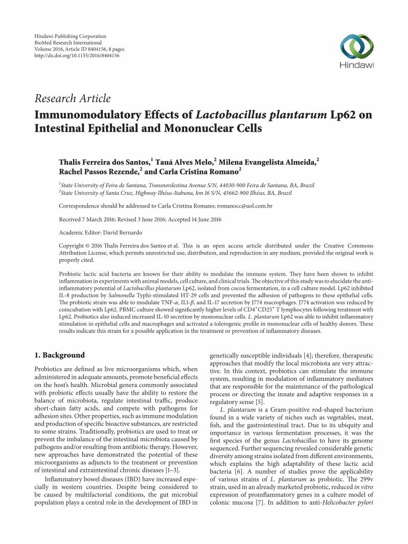
L. plantarum Lp62 significantly reduced IL-8 productionbyHT-29 cells. In comparisonwith the control (0.8 ng⋅mL−1),which was only S. Typhi-stimulated, there was an approx-imately 80-fold reduction in both groups, treated with theprobiotic prior to addition or simultaneously to the pathogenchallenge (0.01 ng⋅mL−1). When the epithelial cell culturewas stimulated with the probiotic alone, there was no sig-nificant cytokine production. Additionally, heat-inactivatedLp62 anti-inflammatory activity was investigated and itwas observed that this group showed no decrease in IL-8
1.0
0.9
0.8
0.7
0.6
0.5
0.020
0.015
0.010
0.005
0.000
a
bb
c
Med
ium
S. T
yphi
Lp62
Lp62
/S. T
yphi
Lp62
/S. T
yphi
(S)
Lp62
(HI)
Lp62
(HI)
/S. T
yphi
[IL-
8] (
ng· m
L−1)
Figure 1: Quantification of IL-8 secreted by HT-29 in culturesupernatant. HT-29 cells were treated with Lp62 and S. Typhi.Levels of IL-8 secreted into the culture medium were measured.Unstimulated cultures or cultures stimulated only with Lp62 or S.Typhi 𝑖 were used as controls. S: inoculated simultaneously; HI:heat-inactivated. aSignificant difference from the medium (withoutany stimulation). bSignificant difference from S. Typhi-stimulatedgroup. cSignificant difference from Lp62/S. Typhi; 𝑃 < 0.05.
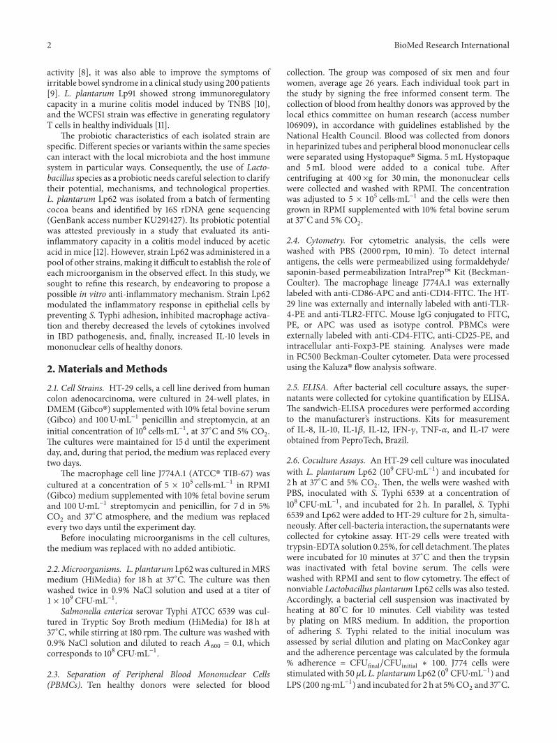
(±0.87 ng⋅mL−1), detected by ELISA (Figure 1). In accordancewith these data, adherence of S. Typhi 6539 toHT-29 cells wasstatistically reduced in the groups treated with the probioticLp62 (Figure 2), showing that its anti-inflammatory action, inthis model, may be related to the probiotic ability to preventcontact of the epithelial cell with the pathogen or competitionfor adhesion sites. Interestingly, the group treated with heat-inactivated probiotics had a higher percentage of pathogensattached to epithelial cells compared to other groups treatedwith probiotics, although it was significantly lower whencompared to the control treated only with S. Typhi. This isprobably the reason why this treatment has been unable toreduce IL-8 levels.
IL-8 is a chemokine that has chemoattractant activity,leading neutrophils to the site of the inflammatory stimulus.Like TNF-𝛼 and IL-1, it is expressed at high levels in thecolonic mucosa of IBD patients [14]. The ability of probioticsto reduce in vitro IL-8 levels is well documented and servesas one of the basic parameters in the selection of probioticbacteria with this potential. Ren et al. [15] observed adecrease in IL-8 produced by Caco-2 cells prestimulated by L.plantarum and challenged with Salmonella Typhimurium. Inline with our findings, the probiotic caused strong inhibitionof pathogen adhesion. The heat inactivation also led toloss of the anti-inflammatory effect. Carey and Kostrzynska[16] reported that preincubation with Lactobacillus and Bifi-dobacterium supernatant was able to inhibit IL-8 secretionby epithelial cells challenged with S. Typhimurium. Theeffect was lost when probiotics were inactivated by heat. Asin the present study, these observations suggest that some
4 BioMed Research International
40
30
20
10
0.8
0.6
0.4
0.2
0.0
%S.
Typ
hi ad
here
nce t
o H
T-29
cells
S. T
yphi
Lp62
/S. T
yphi
Lp62
/S. T
yphi
(S)
Lp62
(HI)
/S. T
yphi
∗
∗
∗
Figure 2: Percentage of S. Typhi adherence to HT-29 cells. HT-29cells were treated with Lp62, and then S. Typhi was added to theculture. After incubation, the probiotic ability to inhibit pathogenbinding to the epithelial cell was measured. The percentage of S.Typhi adherencewas calculated in relation to the initial inoculum 1×10⁸. S: simultaneously inoculated; HI: heat-inactivated. ∗Significant
difference in relation to S. Typhi-stimulated group (𝑃 < 0.05).
factor released by metabolically active probiotic bacteria isresponsible for the observed effect. We may thus emphasizethat the inhibition of pathogen adhesion can contribute to theanti-inflammatory action.
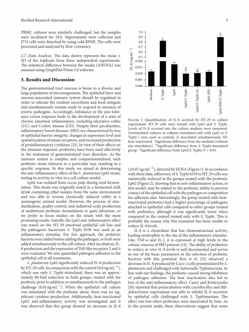
No changes were detected in TLR2 expression inany experimental group. Interestingly, TLR-4 intracellularexpression was found to be increased in Lp62-treated groupsbut did not differ significantly from the S. Typhi-stimulatedcontrol. When probiotic and pathogen were given simultane-ously, there was a significant increase in receptor expression(Figure 3). LPS is a TLR-4 agonist. Under stimulation, thereceptor triggers transcription of proinflammatory genes. Inthe intestinal mucosa, the receptors that recognize microbe-associated molecular patterns are expressed at low levelsto avoid overstimulation and thus chronic inflammation.Alternatively, these receptors are expressed in a compart-mentalized way, like TLR-5, which recognizes flagellin andis expressed basolaterally and is activated only if the colonicmucosa is invaded [17]. Despite its anti-inflammatory profile,Lp62 was able to raise TLR-4 expression; however, it wasdetectable only internally. According to Karlsson et al. [18],L. plantarum can be recognized by TLR-4, but, in ourexperiments, we believe that it was not able to activate thedownstream route that leads to the production of proin-flammatory cytokines such as IL-8. However, we did notinvestigate other products of TLR-4 activation in this cellmodel.
Macrophages located in the intestinal lamina itself rep-resent the major reservoir of these cells in the human body.They are adapted to efficiently remove any pathogen thattries to cross the mucosa, while maintaining homeostasis of
60
40
20
0
TLR-
4-P
E H
T-29
mea
n flu
ores
cenc
e int
ensit
y
a
aa
a, b
Med
ium
S. T
yphi
Lp62
Lp62
/S. T
yphi
Lp62
/S. T
yphi
(S)
Figure 3: TLR-4 expression in HT-29 cells. HT-29 cells werestimulated with Lp62 and then challenged with S. Typhi. In parallel,the effect of simultaneous (S) addition of the two microorganismswas tested. HT-29 cells were labeled internally with anti-TLR-4and analyzed by flow cytometry. aStatistically different from themedium (unstimulated cell). bStatistically different from the S.Typhi-stimulated group; 𝑃 < 0.05.
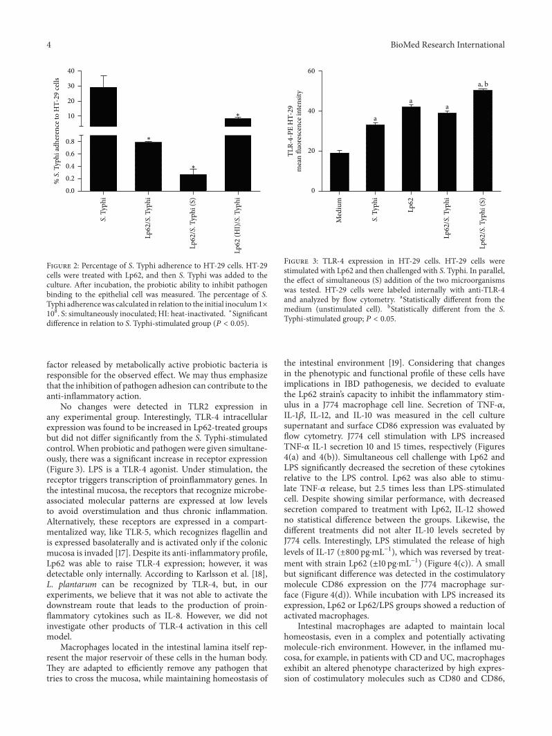
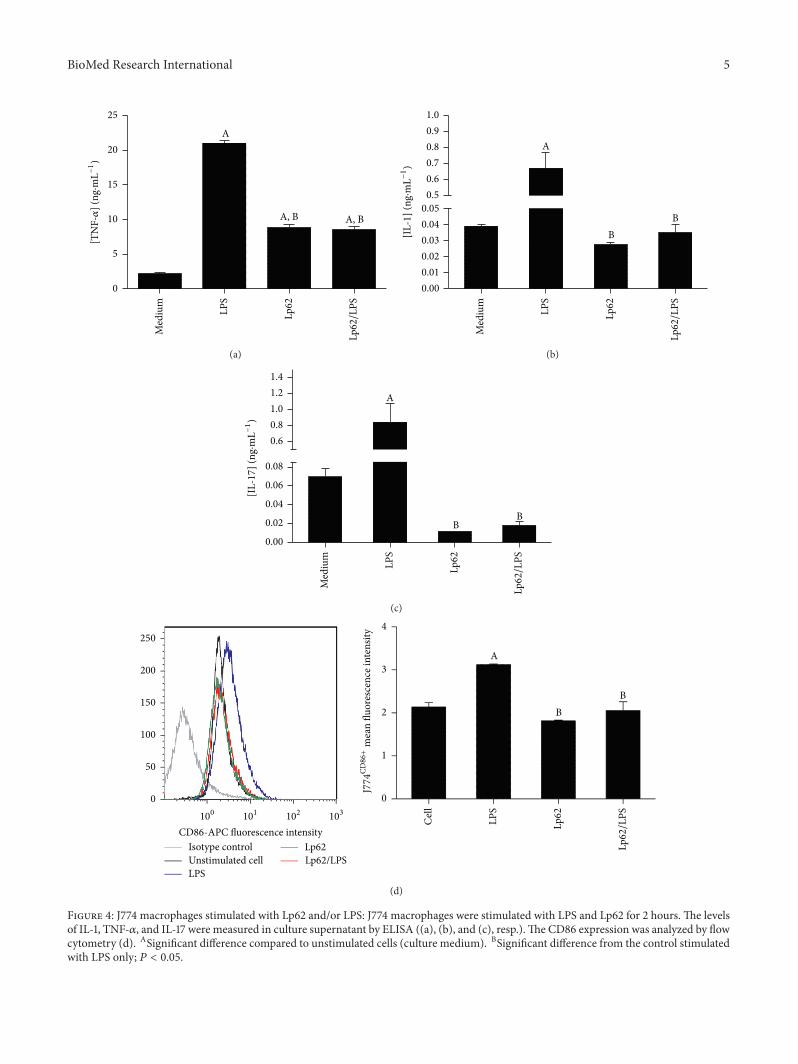
the intestinal environment [19]. Considering that changesin the phenotypic and functional profile of these cells haveimplications in IBD pathogenesis, we decided to evaluatethe Lp62 strain’s capacity to inhibit the inflammatory stim-ulus in a J774 macrophage cell line. Secretion of TNF-𝛼,IL-1𝛽, IL-12, and IL-10 was measured in the cell culturesupernatant and surface CD86 expression was evaluated byflow cytometry. J774 cell stimulation with LPS increasedTNF-𝛼 IL-1 secretion 10 and 15 times, respectively (Figures4(a) and 4(b)). Simultaneous cell challenge with Lp62 andLPS significantly decreased the secretion of these cytokinesrelative to the LPS control. Lp62 was also able to stimu-late TNF-𝛼 release, but 2.5 times less than LPS-stimulatedcell. Despite showing similar performance, with decreasedsecretion compared to treatment with Lp62, IL-12 showedno statistical difference between the groups. Likewise, thedifferent treatments did not alter IL-10 levels secreted byJ774 cells. Interestingly, LPS stimulated the release of highlevels of IL-17 (±800 pg⋅mL−1), which was reversed by treat-ment with strain Lp62 (±10 pg⋅mL−1) (Figure 4(c)). A smallbut significant difference was detected in the costimulatorymolecule CD86 expression on the J774 macrophage sur-face (Figure 4(d)). While incubation with LPS increased itsexpression, Lp62 or Lp62/LPS groups showed a reduction ofactivated macrophages.
Intestinal macrophages are adapted to maintain localhomeostasis, even in a complex and potentially activatingmolecule-rich environment. However, in the inflamed mu-cosa, for example, in patients with CD and UC, macrophagesexhibit an altered phenotype characterized by high expres-sion of costimulatory molecules such as CD80 and CD86,
BioMed Research International 5
25
20
15
10
5
0
[TN
F-𝛼
] (ng
·mL−
1)
A
A, B A, B
Med
ium
LPS
Lp62
Lp62/L
PS(a)
1.0
0.9
0.8
0.7
0.6
0.5
0.05
0.04
0.03
0.02
0.01
0.00
[IL-
1] (
ng·m
L−1)
A
BB
Med
ium
LPS
Lp62
Lp62/L
PS
(b)
1.4
1.2
1.0
0.8
0.6
0.08
0.06
0.04
0.02
0.00
[IL-
17
] (ng
·mL−
1)
Med
ium
LPS
Lp62
A
BB
Lp62/L
PS
(c)
4
3
2
1
0
Cel
l
LPS
Lp62
Lp62/L
PS
A
BB
250
200
150
100
50
0
100
101
102
103
CD86-APC fluorescence intensityIsotype controlUnstimulated cellLPS
Lp62Lp62/LPS
J774
CD86+
mea
n flu
ores
cenc
e int
ensit
y
(d)
Figure 4: J774 macrophages stimulated with Lp62 and/or LPS: J774 macrophages were stimulated with LPS and Lp62 for 2 hours. The levelsof IL-1, TNF-𝛼, and IL-17 were measured in culture supernatant by ELISA ((a), (b), and (c), resp.).The CD86 expression was analyzed by flowcytometry (d). ASignificant difference compared to unstimulated cells (culture medium). BSignificant difference from the control stimulatedwith LPS only; 𝑃 < 0.05.
6 BioMed Research International
8
6
4
2
0
% P
BMC
expr
essin
g CD
4/C
D25
Med
ium
LPS
Lp62
Lp62
/LPS
BA, B
(a)
[IL-
10
] (ng
·mL−
1)
2.5
2.0
1.5
1.0
0.5
0.0
Cel
l
LPS
Lp62
Lp62
/LPS
A
B
B
(b)
Figure 5: CD4+CD25+ T lymphocytes and IL-10 secretion in PBMC treated with Lp62. Cultures of peripheral blood mononuclear cells werechallenged with LPS and Lp62. The proportion of CD4+CD25+ cells was determined by flow cytometry (a). IL-10 production was examinedin the culture supernatant by ELISA (b). AStatistical difference compared to the control without stimulation. BStatistical difference from thecontrol only stimulated with LPS; 𝑃 < 0.05.
as well as the innate receptors TLR-2 and TLR-4, specializedin detecting bacterial antigens [20, 21]. In this context, thesecells become potent producers of proinflammatory cytokinessuch as IL-1𝛽, TNF-𝛼, IL-6, and MCP-1. Trials with murineand human cells have shown that probiotics can prevent orreverse the functional change of macrophages, characteristicof chronic inflammatory diseases. According to Pathmakan-than et al. [22], L. plantarum 299v reduced the secretionof TNF-𝛼 and IL-1𝛽 in mucosal mononuclear cells fromIBD patients stimulated with E. coli or Salmonella Dublinand increased the IL-10 levels. TNF-𝛼 production is alsoaffected by the LPS-stimulated macrophage RAW 2647 andtreated with Lactobacillus rhamnosus GG [23]. Matsumotoand Benno [24] found that metabolites released in thestools of patients fedwith yoghurt containingBifidobacteriumanimalis LKM512 were able to reverse the inflammationcaused by LPS in J774 cells.The effect of probiotic bacteria onantigens presenting cells such as macrophages and dendriticcells is strain-dependent, since they also may be able toupregulate the production of costimulatory molecules andproinflammatory cytokines [25]. IL17 induces neutrophilrecruitment to the inflamed site and triggers the release ofinflammatory cytokines in macrophages. However, its rolein inducing colitis remains uncertain, as it even presentsa protective activity in the gut, depending on the modelstudied. The main source of this cytokine is Th17 cells;however, the innate immunity cells, including macrophages,can produce it [26]. Here, we observe that Lp62 modulatedIL-17 secretion in J774 macrophages. Further studies areneeded to determine the impact of this probiotic on IL-17 production in the in vivo colitis model. In the presentstudy, we speculate that L. plantarum Lp62 was capableof limiting J774 macrophage activation and consequentlypreventing proinflammatory cytokine secretion, contributingto the maintenance of local homeostasis.
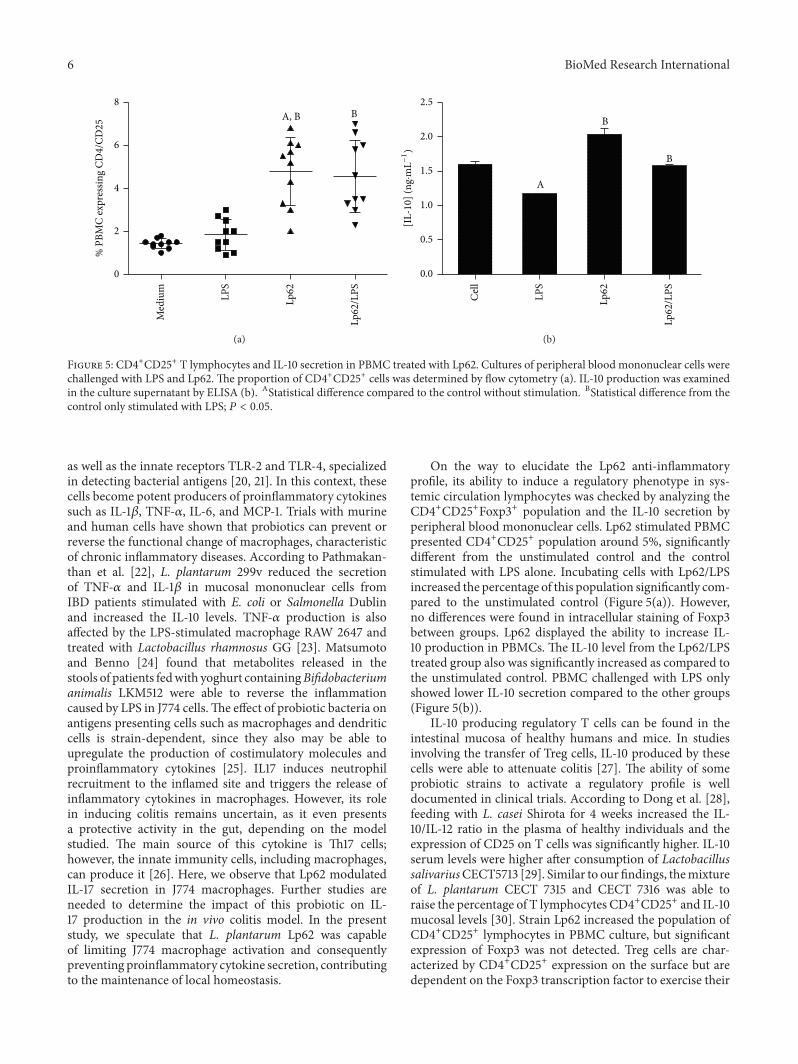
On the way to elucidate the Lp62 anti-inflammatoryprofile, its ability to induce a regulatory phenotype in sys-temic circulation lymphocytes was checked by analyzing theCD4+CD25+Foxp3+ population and the IL-10 secretion byperipheral blood mononuclear cells. Lp62 stimulated PBMCpresented CD4+CD25+ population around 5%, significantlydifferent from the unstimulated control and the controlstimulated with LPS alone. Incubating cells with Lp62/LPSincreased the percentage of this population significantly com-pared to the unstimulated control (Figure 5(a)). However,no differences were found in intracellular staining of Foxp3between groups. Lp62 displayed the ability to increase IL-10 production in PBMCs. The IL-10 level from the Lp62/LPStreated group also was significantly increased as compared tothe unstimulated control. PBMC challenged with LPS onlyshowed lower IL-10 secretion compared to the other groups(Figure 5(b)).
IL-10 producing regulatory T cells can be found in theintestinal mucosa of healthy humans and mice. In studiesinvolving the transfer of Treg cells, IL-10 produced by thesecells were able to attenuate colitis [27]. The ability of someprobiotic strains to activate a regulatory profile is welldocumented in clinical trials. According to Dong et al. [28],feeding with L. casei Shirota for 4 weeks increased the IL-10/IL-12 ratio in the plasma of healthy individuals and theexpression of CD25 on T cells was significantly higher. IL-10serum levels were higher after consumption of LactobacillussalivariusCECT5713 [29]. Similar to our findings, themixtureof L. plantarum CECT 7315 and CECT 7316 was able toraise the percentage of T lymphocytes CD4+CD25+ and IL-10mucosal levels [30]. Strain Lp62 increased the population ofCD4+CD25+ lymphocytes in PBMC culture, but significantexpression of Foxp3 was not detected. Treg cells are char-acterized by CD4+CD25+ expression on the surface but aredependent on the Foxp3 transcription factor to exercise their
BioMed Research International 7
function on colonic lamina propria. Increased IL-10 levelsafter treatment with Lp62 point to a regulatory T cell profile,but cytokine production by other cells present in the cultureshould be considered.
In the intestinal environment, epithelial cells, microor-ganisms, and immune cell aggregates contribute to maintain-ing homeostasis. According to the widely accepted model,epithelial cells are responsible for releasing factors that willdirect the antigen presenting cells to a nonresponsive profileor activating a regulatory response. The T cells generatedin this environment would be responsible for maintaininghomeostasis by releasing considerable amounts of IL-10 andTGF-𝛽. Evidence suggests that the composition of the localflora is directly correlated to the balance between responseand tolerance. In this sense, probiotics have been effectivein restoring the tolerogenic profile of the intestinal mucosa,by modulating the activity of the cells that participate inthis process [17, 19, 27]. In this paper, the marked anti-inflammatory effect related to the lactic acid bacteria L.plantarum Lp62 was observed on intestinal epithelial cells,macrophage, and lymphocyte. In a cell culture model, thisstrain was able to prevent S. Typhi adhesion to epithelialcells and hence inhibit IL-8 secretion. A slight decrease inmacrophage activation was also observed which may havecontributed to reducing proinflammatory cytokine produc-tion. Finally, the Lp62 strain was able to enhance IL-10secretion and increase the CD4+CD25+ cell population. Sinceit showed immunomodulatory capacity on the main cellsinvolved in the intestinal mucosal immunity, Lp62 is a strongcandidate to assist in therapy for inflammatory diseases.
4. Conclusions
The results presented in this paper should serve as a basisfor further studies that can investigate the pathways involvedin the Lp62 anti-inflammatory effect. Equally important areapproaches in search of safe use of all the newly discoveredstrains, mainly because probiotics are used in the context ofa previously damagedmucosa. Furthermore, in vivo trials areessential in the study of probiotic action due to particularitiesand the high complexity of the intestinal environment.
Competing Interests
The authors declare no competing interests between theauthors and the sponsoring institutions of this research.
References
[1] L. M. T. Dicks and M. Botes, “Probiotic lactic acid bacteria inthe gastro-intestinal tract: health benefits, safety and mode ofaction,” Beneficial Microbes, vol. 1, no. 1, pp. 11–29, 2010.
[2] S. Fijan, “Microorganisms with claimed probiotic properties: anoverview of recent literature,” International Journal of Environ-mental Research and Public Health, vol. 11, no. 5, pp. 4745–4767,2014.
[3] C. Hill, F. Guarner, G. Reid et al., “Expert consensus docu-ment: the international scientific association for probiotics andprebiotics consensus statement on the scope and appropriate
use of the term probiotic,”Nature Reviews Gastroenterology andHepatology, vol. 11, no. 8, pp. 506–514, 2014.
[4] R. Bringiotti, R. Lerardi, R. Lovero, G. Losurdo, A. Di Leo,and M. Principi, “Intestinal microbiota: the explosive mixtureat the origin of inflammatory bowel disease?”World Journal ofGastrointestinal Pathophysiology, vol. 5, no. 4, pp. 550–559, 2014.
[5] J. Plaza-Diaz, C. Gomez-Llorente, L. Fontana, and A. Gil,“Modulation of immunity and inflammatory gene expressionin the gut, in inflammatory diseases of the gut and in the liverby probiotics,”World Journal of Gastroenterology, vol. 20, no. 42,pp. 15632–15649, 2014.
[6] R. J. Siezen and J. E. T. Vlieg, “Genomic diversity and versa-tility of Lactobacillus plantarum, a natural metabolic engineer,”Microbial Cell Factories, vol. 10, no. 1, article S3, 2011.
[7] C. Bauerl, M. Llopis, M. Antolın et al., “Lactobacillus paracaseiand Lactobacillus plantarum strains downregulate proinflam-matory genes in an ex vivo system of cultured human colonicmucosa,” Genes & Nutrition, vol. 8, no. 2, pp. 165–180, 2013.
[8] S. Rokka, A. Pihlanto, H. Korhonen, and V. Joutsjoki, “Invitro growth inhibition of Helicobacter pylori by lactobacillibelonging to the Lactobacillus plantarum group,” Letters inApplied Microbiology, vol. 43, no. 5, pp. 508–513, 2006.
[9] P. Ducrotte, P. Sawant, and V. Jayanthi, “Clinical trial: Lacto-bacillus plantarum 299v (DSM 9843) improves symptoms ofirritable bowel syndrome,” World Journal of Gastoenterology,vol. 18, no. 30, pp. 4012–4018, 2012.
[10] R. K.Duary,M.A. Bhausaheb, V. K. Batish, and S. Grover, “Anti-inflammatory and immunomodulatory efficacy of indigenousprobiotic Lactobacillus plantarum Lp91 in colitis mouse model,”Molecular Biology Reports, vol. 39, no. 4, pp. 4765–4775, 2012.
[11] M. J. Smelt, B. J. de Haan, P. A. Bron et al., “The Impact ofLactobacillus plantarum WCFS1 teichoic acid D-alanylationon the generation of effector and regulatory T-cells in healthymice,” PLoS ONE, vol. 8, no. 4, article e63099, 2013.
[12] T. F.Dos Santos,Probiotic potential of lactic acid bacteria isolatedfrom cocoa beans fermentation in southern Bahia, Brazil [M.S.thesis], Santa Cruz State University, Biology Department, 2010,http://www.dominiopublico.gov.br/pesquisa/DetalheObra-Form.do?select action=&co obra=187827.
[13] M. Z. Cader and A. Kaser, “Recent advances in inflammatorybowel disease: mucosal immune cells in intestinal inflamma-tion,” Gut, vol. 62, no. 11, pp. 1653–1664, 2013.
[14] C. Banks, A. Bateman, R. Payne, P. Johnson, and N. Sheron,“Chemokine expression in IBD.Mucosal chemokine expressionis unselectively increased in both ulcerative colitis and Crohn’sdisease,” Journal of Pathology, vol. 199, no. 1, pp. 28–35, 2003.
[15] D.-Y. Ren, C. Li, Y.-Q.Qin et al., “Lactobacilli reduce chemokineIL-8 production in response to TNF-𝛼 and Salmonella challengeof caco-2 cells,”BioMed Research International, vol. 2013, ArticleID 925219, 9 pages, 2013.
[16] C. M. Carey andM. Kostrzynska, “Lactic acid bacteria and bifi-dobacteria attenuate the proinflammatory response in intesti-nal epithelial cells induced by salmonella enterica serovartyphimurium,” Canadian Journal of Microbiology, vol. 59, no. 1,pp. 9–17, 2013.
[17] J. G. Magalhaes, I. Tattoli, and S. E. Girardin, “The intestinalepithelial barrier: how to distinguish between the microbialflora and pathogens,” Seminars in Immunology, vol. 19, no. 2, pp.106–115, 2007.
[18] H. Karlsson, C. Hessle, and A. Rudin, “Innate immune re-sponses of human neonatal cells to bacteria from the normal
8 BioMed Research International
gastrointestinal flora,” Infection and Immunity, vol. 70, no. 12,pp. 6688–6696, 2002.
[19] M. Schenk andC.Mueller, “Adaptations of intestinalmacropha-ges to an antigen-rich environment,” Seminars in Immunology,vol. 19, no. 2, pp. 84–93, 2007.
[20] J. Rugtveit, A. Bakka, and P. Brandtzaeg, “Differential distribu-tion of B7.1 (CD80) and B7.2 (CD86) costimulatory moleculesonmucosal macrophage subsets in human inflammatory boweldisease (IBD),” Clinical and Experimental Immunology, vol. 110,no. 1, pp. 104–113, 1997.
[21] M. Hausmann, S. Kiessling, S. Mestermann et al., “Toll-likereceptors 2 and 4 are up-regulated during intestinal inflamma-tion,” Gastroenterology, vol. 122, no. 7, pp. 1987–2000, 2002.
[22] S. Pathmakanthan, C. K. F. Li, J. Cowie, and C. J. Hawkey, “Lac-tobacillus plantarum 299v: beneficial in vitro immunomodu-lation in cells extracted from inflamed human colon,” Journalof Gastroenterology and Hepatology, vol. 19, no. 2, pp. 166–173,2004.
[23] J. A. Pena and J. Versalovic, “Lactobacillus rhamnosus GGdecreases TNF-𝛼 production in lipopolysaccharide-activatedmurine macrophages by a contact-independent mechanism,”Cellular Microbiology, vol. 5, no. 4, pp. 277–285, 2003.
[24] M. Matsumoto and Y. Benno, “Anti-inflammatory metaboliteproduction in the gut from the consumption of probiotic yogurtcontaining Bifidobacterium animalis subsp. lactis LKM512,”Bioscience, Biotechnology and Biochemistry, vol. 70, no. 6, pp.1287–1292, 2006.
[25] S. Latvala, T. E. Pietila, V. Veckman et al., “Potentially probioticbacteria induce efficient maturation but differential cytokineproduction in humanmonocyte-derived dendritic cells,”WorldJournal of Gastroenterology, vol. 14, no. 36, pp. 5570–5583, 2008.
[26] A. Geremia, P. Biancheri, P. Allan, G. R. Corazza, and A. DiSabatino, “Innate and adaptive immunity in inflammatorybowel disease,” Autoimmunity Reviews, vol. 13, no. 1, pp. 3–10,2014.
[27] J. L. Coombes and K. J. Maloy, “Control of intestinal home-ostasis by regulatory T cells and dendritic cells,” Seminars inImmunology, vol. 19, no. 2, pp. 116–126, 2007.
[28] H. Dong, I. Rowland, L. V. Thomas, and P. Yaqoob, “Immuno-modulatory effects of a probiotic drink containing Lactobacilluscasei Shirota in healthy older volunteers,” European Journal ofNutrition, vol. 52, no. 8, pp. 1853–1863, 2013.
[29] S. Sierra, F. Lara-Villoslada, L. Sempere, M. Olivares, J. Boza,and J. Xaus, “Intestinal and immunological effects of daily oraladministration of Lactobacillus salivarius CECT5713 to healthyadults,” Anaerobe, vol. 16, no. 3, pp. 195–200, 2010.
[30] J. Mane, E. Pedrosa, V. Loren et al., “A mixture of Lactobacillusplantarum CECT 7315 and CECT 7316 enhances systemicimmunity in elderly subjects. A dose-response, double-blind,placebo-controlled, randomized pilot trial,” Nutricion Hospita-laria, vol. 26, no. 1, pp. 228–235, 2011.

Submit your manuscripts athttp://www.hindawi.com
Hindawi Publishing Corporationhttp://www.hindawi.com Volume 2014
Anatomy Research International
PeptidesInternational Journal of
Hindawi Publishing Corporationhttp://www.hindawi.com Volume 2014
Hindawi Publishing Corporation http://www.hindawi.com
International Journal of
Volume 2014
Zoology
Hindawi Publishing Corporationhttp://www.hindawi.com Volume 2014
Molecular Biology International
GenomicsInternational Journal of
Hindawi Publishing Corporationhttp://www.hindawi.com Volume 2014
The Scientific World JournalHindawi Publishing Corporation http://www.hindawi.com Volume 2014
Hindawi Publishing Corporationhttp://www.hindawi.com Volume 2014
BioinformaticsAdvances in
Marine BiologyJournal of
Hindawi Publishing Corporationhttp://www.hindawi.com Volume 2014
Hindawi Publishing Corporationhttp://www.hindawi.com Volume 2014
Signal TransductionJournal of
Hindawi Publishing Corporationhttp://www.hindawi.com Volume 2014
BioMed Research International
Evolutionary BiologyInternational Journal of
Hindawi Publishing Corporationhttp://www.hindawi.com Volume 2014
Hindawi Publishing Corporationhttp://www.hindawi.com Volume 2014
Biochemistry Research International
ArchaeaHindawi Publishing Corporationhttp://www.hindawi.com Volume 2014
Hindawi Publishing Corporationhttp://www.hindawi.com Volume 2014
Genetics Research International
Hindawi Publishing Corporationhttp://www.hindawi.com Volume 2014
Advances in
Virolog y
Hindawi Publishing Corporationhttp://www.hindawi.com
Nucleic AcidsJournal of
Volume 2014
Stem CellsInternational
Hindawi Publishing Corporationhttp://www.hindawi.com Volume 2014
Hindawi Publishing Corporationhttp://www.hindawi.com Volume 2014
Enzyme Research
Hindawi Publishing Corporationhttp://www.hindawi.com Volume 2014
International Journal of
Microbiology


