Research Article Effect ofBetula pendula Leaf Extract on ...
9
Research Article Effect of Betula pendula Leaf Extract on -Glucosidase and Glutathione Level in Glucose-Induced Oxidative Stress Kristina BljajiT, 1 Nina ŠoštariT, 1 Roberta Petlevski, 1 Lovorka VujiT, 1 Andrea BrajkoviT, 1,2 Barbara FumiT, 1 Isabel Saraiva de Carvalho, 2 and Marijana Zovko KonIiT 1 1 Faculty of Pharmacy and Biochemistry, University of Zagreb, A. Kovaˇ ci´ ca 1, 10 000 Zagreb, Croatia 2 Faculty of Sciences and Technology, University of Algarve, Campus de Gambelas, Bd. 8, 8005-139 Faro, Portugal Correspondence should be addressed to Marijana Zovko Konˇ ci´ c; [email protected] Received 20 May 2016; Accepted 18 August 2016 Academic Editor: Hilal Zaid Copyright © 2016 Kristina Bljaji´ c et al. is is an open access article distributed under the Creative Commons Attribution License, which permits unrestricted use, distribution, and reproduction in any medium, provided the original work is properly cited. B. pendula leaf is a common ingredient in traditional herbal combinations for treatment of diabetes in southeastern Europe. Present study investigated B. pendula ethanolic and aqueous extract as inhibitors of carbohydrate hydrolyzing enzymes, as well as their ability to restore glutathione concentration in Hep G2 cells subjected to glucose-induced oxidative stress. Phytochemical analysis revealed presence of rutin and other quercetin derivatives, as well as chlorogenic acid. In general, ethanolic extract was richer in phenolic substances than the aqueous extract. Furthermore, a comprehensive analysis of antioxidant activity of two extracts (deter- mined by DPPH and ABTS radical scavenging activity, total antioxidant activity, and chelating activity as well as ferric-reducing antioxidant power) has shown that ethanolic extract was better radical scavenger and metal ion reductant. In addition, ethanolic extract effectively increased cellular glutathione levels caused by hyperglycemia and inhibited -glucosidase with the activity com- parable to that of acarbose. erefore, in vitro research using B. pendula plant extracts has confirmed their antidiabetic properties. 1. Introduction Chronic hyperglycemia, which may arise as a consequence of diabetes or metabolic syndrome, induces oxidative stress in sensitive tissues because glucose in high concentrations forms reactive oxygen species (ROS). Elevated glucose concentra- tions, and thus induced oxidative damage, may adversely affect pancreatic islet cells, leading to disturbances in insulin production and further aggravating hyperglycemic status. e harm caused by ROS causes damage and impair- ment of function of the classical secondary targets of dia- betes, such as blood vessels, kidneys, nerves, and eyes [1], leading to cardiovascular diseases, as well as microvascular diabetic complications, including nephropathy, retinopathy, and neuropathy [2]. In addition to that, recent research has provided evidence that insulin resistance and impaired insulin signaling, typical for type 2 diabetes, may be a contributory factor to the progression of dementia and other neurological disorders [3]. Endo- or exogenous antioxidants play an important role in alleviating oxidative stress and its consequences. One of the most important cellular nonprotein antioxidants is glu- tathione (GSH). GSH protects cells by scavenging oxygen and nitrogen radicals and by reducing H 2 O 2 . GSH is also impor- tant in maintaining the concentration of other nonenzymatic antioxidants. For example, the oxidized vitamin C can be restored to the reduced form by enzymatic reaction which uses GSH as substrate [4]. Besides contributing to protection of cells against oxidative damage, GSH detoxifies xenobiotics and regulates the functions of redox-sensitive proteins [5]. However, in states of increased oxidants, production levels of glutathione in cells can deplete. erefore, in such conditions, constant and rapid replenishment of GSH is required, which is accomplished through both the reduction of oxidized GSH and its de novo synthesis. However, high concentration of glucose leads to glycation of glutamate-cysteine ligase, the first enzyme of the glutathione biosynthetic pathway, thus leading to further decrease of GSH levels [6]. ere is in vitro and clinical evidence that abnormally low levels of glutathione in cells may lead to -cell dysfunction and the pathogenesis of long-term complications of diabetes. As a Hindawi Publishing Corporation Evidence-Based Complementary and Alternative Medicine Volume 2016, Article ID 8429398, 8 pages http://dx.doi.org/10.1155/2016/8429398 brought to you by CORE View metadata, citation and similar papers at core.ac.uk provided by Crossref
Transcript of Research Article Effect ofBetula pendula Leaf Extract on ...
Research ArticleEffect of Betula pendula Leaf Extract on 𝛼-Glucosidase andGlutathione Level in Glucose-Induced Oxidative Stress
Kristina BljajiT,1 Nina ŠoštariT,1 Roberta Petlevski,1 Lovorka VujiT,1 Andrea BrajkoviT,1,2
Barbara FumiT,1 Isabel Saraiva de Carvalho,2 and Marijana Zovko KonIiT1
1Faculty of Pharmacy and Biochemistry, University of Zagreb, A. Kovacica 1, 10 000 Zagreb, Croatia2Faculty of Sciences and Technology, University of Algarve, Campus de Gambelas, Bd. 8, 8005-139 Faro, Portugal
Correspondence should be addressed to Marijana Zovko Koncic; [email protected]
Received 20 May 2016; Accepted 18 August 2016
Academic Editor: Hilal Zaid
Copyright © 2016 Kristina Bljajic et al. This is an open access article distributed under the Creative Commons Attribution License,which permits unrestricted use, distribution, and reproduction in any medium, provided the original work is properly cited.
B. pendula leaf is a common ingredient in traditional herbal combinations for treatment of diabetes in southeastern Europe. Presentstudy investigated B. pendula ethanolic and aqueous extract as inhibitors of carbohydrate hydrolyzing enzymes, as well as theirability to restore glutathione concentration in Hep G2 cells subjected to glucose-induced oxidative stress. Phytochemical analysisrevealed presence of rutin and other quercetin derivatives, as well as chlorogenic acid. In general, ethanolic extract was richer inphenolic substances than the aqueous extract. Furthermore, a comprehensive analysis of antioxidant activity of two extracts (deter-mined by DPPH and ABTS radical scavenging activity, total antioxidant activity, and chelating activity as well as ferric-reducingantioxidant power) has shown that ethanolic extract was better radical scavenger and metal ion reductant. In addition, ethanolicextract effectively increased cellular glutathione levels caused by hyperglycemia and inhibited 𝛼-glucosidase with the activity com-parable to that of acarbose. Therefore, in vitro research using B. pendula plant extracts has confirmed their antidiabetic properties.
1. Introduction
Chronic hyperglycemia, which may arise as a consequence ofdiabetes or metabolic syndrome, induces oxidative stress insensitive tissues because glucose in high concentrations formsreactive oxygen species (ROS). Elevated glucose concentra-tions, and thus induced oxidative damage, may adverselyaffect pancreatic islet 𝛽 cells, leading to disturbances ininsulin production and further aggravating hyperglycemicstatus. The harm caused by ROS causes damage and impair-ment of function of the classical secondary targets of dia-betes, such as blood vessels, kidneys, nerves, and eyes [1],leading to cardiovascular diseases, as well as microvasculardiabetic complications, including nephropathy, retinopathy,and neuropathy [2]. In addition to that, recent researchhas provided evidence that insulin resistance and impairedinsulin signaling, typical for type 2 diabetes, may be acontributory factor to the progression of dementia and otherneurological disorders [3].
Endo- or exogenous antioxidants play an important rolein alleviating oxidative stress and its consequences. One of
the most important cellular nonprotein antioxidants is glu-tathione (GSH). GSHprotects cells by scavenging oxygen andnitrogen radicals and by reducing H
2O2. GSH is also impor-
tant in maintaining the concentration of other nonenzymaticantioxidants. For example, the oxidized vitamin C can berestored to the reduced form by enzymatic reaction whichuses GSH as substrate [4]. Besides contributing to protectionof cells against oxidative damage, GSH detoxifies xenobioticsand regulates the functions of redox-sensitive proteins [5].However, in states of increased oxidants, production levels ofglutathione in cells can deplete.Therefore, in such conditions,constant and rapid replenishment of GSH is required, whichis accomplished through both the reduction of oxidized GSHand its de novo synthesis. However, high concentration ofglucose leads to glycation of glutamate-cysteine ligase, thefirst enzyme of the glutathione biosynthetic pathway, thusleading to further decrease of GSH levels [6]. There is invitro and clinical evidence that abnormally low levels ofglutathione in cells may lead to 𝛽-cell dysfunction and thepathogenesis of long-term complications of diabetes. As a
Hindawi Publishing CorporationEvidence-Based Complementary and Alternative MedicineVolume 2016, Article ID 8429398, 8 pageshttp://dx.doi.org/10.1155/2016/8429398
brought to you by COREView metadata, citation and similar papers at core.ac.uk
provided by Crossref
2 Evidence-Based Complementary and Alternative Medicine
consequence, interest has been developed in the potentialfor therapeutic modification of glutathione status in thetreatment of diabetes. For example, changing the GSH statuscan be achieved by using natural antioxidants such as lipoicacid [7], curcumin, or sulforaphane [4]. Such approach couldbe used for development of nutraceuticals with potential inthe treatment of metabolic disorder and diabetes [6].
Besides their influences on GSH content, natural sub-stances can exert other biological activities which can bebeneficial in treatment of diabetes and its complications. Forexample, due to their antioxidant activity, directly or viatheir influence on endogenous antioxidants, they can protectcellular targets and consequently the tissues which are mostsusceptible to diabetic complications [8]. Besides that, theycan influence the enzymes which participate in carbohydratemetabolism, such as 𝛼-amylase and 𝛼-glucosidase, thusretarding the postprandial increase of glucose concentration.Alpha-amylase is an enzyme secreted in saliva and pancreaticjuice which catalyzes the hydrolysis of starch to a mixtureof smaller oligosaccharides, which are then degraded toglucose by 𝛼-glucosidase, enzyme located in the mucosalbrush border of the small intestine. Alpha-amylase and 𝛼-glucosidase inhibitors can thus be helpful in the developmentof compounds for the treatment of diabetes, obesity, andhyperlipemia. Medicinal plants may constitute a good sourceof 𝛼-amylase and 𝛼-glucosidase inhibitors [9, 10].
Betula pendula, Roth (Betulaceae) commonly known assilver birch, is a tree native to Europe and Asia. In traditionalmedicine of Eastern Europe, it is used as diuretic, especiallyin cases of cystitis, as well as treatment for rheumatism andarthritic diseases. Recent studies have shown that its diureticpotential may stem from its endopeptidases-inhibiting prop-erties, while xanthine oxidase inhibitory properties may beresponsible for its use in gout treatment [11, 12]. Furthermore,B. pendula leaf extract can inhibit growth and cell division ofinflammatory lymphocytes [12], as well as inhibiting tyrosi-nase, the enzyme that catalyzes the first stages of melaninbiosynthesis. Birch leaf extract also displayed antioxidant andmetal chelating properties [13].
Besides its well known use as diuretic and anti-inflammatory drug, B. pendula leaf is often used as partof herbal mixtures for treatment of diabetes in traditionalmedicine of southeastern Europe. The aim of this work wasto evaluate suitability of such use by studying the inhibitoryeffects of B. pendula extracts against 𝛼-amylase and 𝛼-glucosidase, as well as to evaluate their antioxidant and GSHprotecting activities in hyperglycemic conditions. An addi-tional aim was to determine the best solvent for extraction ofactive principles of B. pendula leaf. To the best of our knowl-edge, this is the first time that potential use of birch extract assupplementary treatment for diabetes has been investigated.
2. Materials and Methods
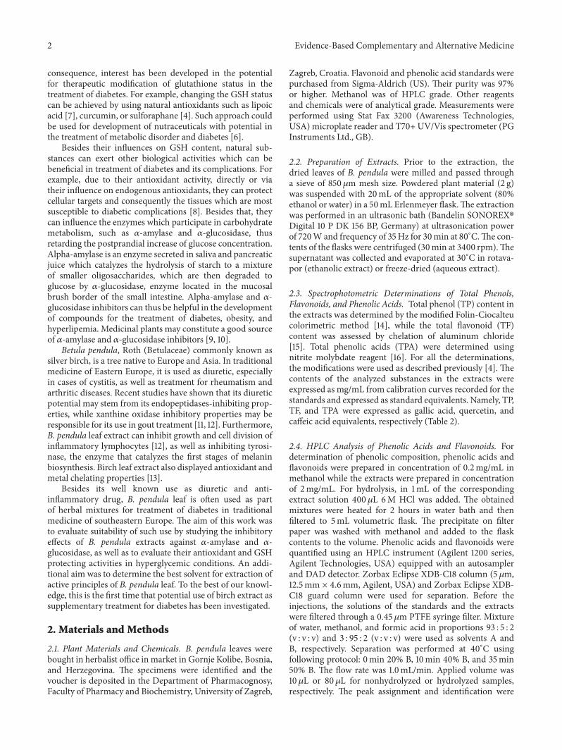
2.1. Plant Materials and Chemicals. B. pendula leaves werebought in herbalist office in market in Gornje Kolibe, Bosnia,and Herzegovina. The specimens were identified and thevoucher is deposited in the Department of Pharmacognosy,Faculty of Pharmacy and Biochemistry, University of Zagreb,
Zagreb, Croatia. Flavonoid and phenolic acid standards werepurchased from Sigma-Aldrich (US). Their purity was 97%or higher. Methanol was of HPLC grade. Other reagentsand chemicals were of analytical grade. Measurements wereperformed using Stat Fax 3200 (Awareness Technologies,USA) microplate reader and T70+ UV/Vis spectrometer (PGInstruments Ltd., GB).
2.2. Preparation of Extracts. Prior to the extraction, thedried leaves of B. pendula were milled and passed througha sieve of 850𝜇m mesh size. Powdered plant material (2 g)was suspended with 20mL of the appropriate solvent (80%ethanol or water) in a 50mL Erlenmeyer flask.The extractionwas performed in an ultrasonic bath (Bandelin SONOREX�Digital 10 P DK 156 BP, Germany) at ultrasonication powerof 720W and frequency of 35Hz for 30min at 80∘C.The con-tents of the flasks were centrifuged (30min at 3400 rpm).Thesupernatant was collected and evaporated at 30∘C in rotava-por (ethanolic extract) or freeze-dried (aqueous extract).
2.3. Spectrophotometric Determinations of Total Phenols,Flavonoids, and Phenolic Acids. Total phenol (TP) content inthe extracts was determined by the modified Folin-Ciocalteucolorimetric method [14], while the total flavonoid (TF)content was assessed by chelation of aluminum chloride[15]. Total phenolic acids (TPA) were determined usingnitrite molybdate reagent [16]. For all the determinations,the modifications were used as described previously [4]. Thecontents of the analyzed substances in the extracts wereexpressed as mg/mL from calibration curves recorded for thestandards and expressed as standard equivalents. Namely, TP,TF, and TPA were expressed as gallic acid, quercetin, andcaffeic acid equivalents, respectively (Table 2).
2.4. HPLC Analysis of Phenolic Acids and Flavonoids. Fordetermination of phenolic composition, phenolic acids andflavonoids were prepared in concentration of 0.2mg/mL inmethanol while the extracts were prepared in concentrationof 2mg/mL. For hydrolysis, in 1mL of the correspondingextract solution 400 𝜇L 6M HCl was added. The obtainedmixtures were heated for 2 hours in water bath and thenfiltered to 5mL volumetric flask. The precipitate on filterpaper was washed with methanol and added to the flaskcontents to the volume. Phenolic acids and flavonoids werequantified using an HPLC instrument (Agilent 1200 series,Agilent Technologies, USA) equipped with an autosamplerand DAD detector. Zorbax Eclipse XDB-C18 column (5 𝜇m,12.5mm × 4.6mm, Agilent, USA) and Zorbax Eclipse XDB-C18 guard column were used for separation. Before theinjections, the solutions of the standards and the extractswere filtered through a 0.45 𝜇m PTFE syringe filter. Mixtureof water, methanol, and formic acid in proportions 93 : 5 : 2(v : v : v) and 3 : 95 : 2 (v : v : v) were used as solvents A andB, respectively. Separation was performed at 40∘C usingfollowing protocol: 0min 20% B, 10min 40% B, and 35min50% B. The flow rate was 1.0mL/min. Applied volume was10 𝜇L or 80 𝜇L for nonhydrolyzed or hydrolyzed samples,respectively. The peak assignment and identification were
Evidence-Based Complementary and Alternative Medicine 3
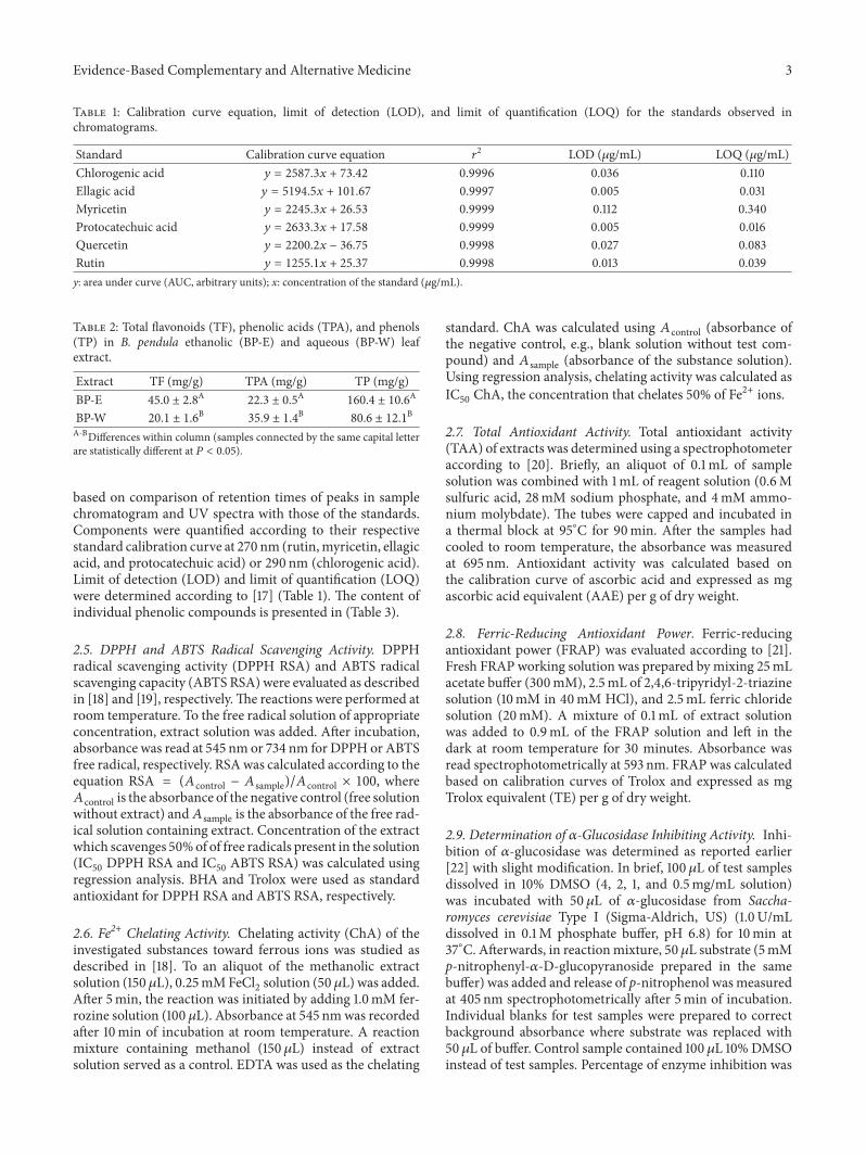
Table 1: Calibration curve equation, limit of detection (LOD), and limit of quantification (LOQ) for the standards observed inchromatograms.
Standard Calibration curve equation 𝑟2 LOD (𝜇g/mL) LOQ (𝜇g/mL)
Chlorogenic acid 𝑦 = 2587.3𝑥 + 73.42 0.9996 0.036 0.110Ellagic acid 𝑦 = 5194.5𝑥 + 101.67 0.9997 0.005 0.031Myricetin 𝑦 = 2245.3𝑥 + 26.53 0.9999 0.112 0.340Protocatechuic acid 𝑦 = 2633.3𝑥 + 17.58 0.9999 0.005 0.016Quercetin 𝑦 = 2200.2𝑥 − 36.75 0.9998 0.027 0.083Rutin 𝑦 = 1255.1𝑥 + 25.37 0.9998 0.013 0.039y: area under curve (AUC, arbitrary units); x: concentration of the standard (𝜇g/mL).
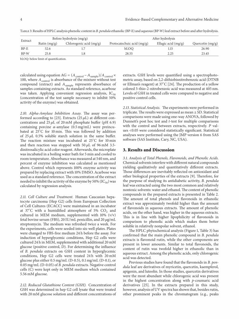
Table 2: Total flavonoids (TF), phenolic acids (TPA), and phenols(TP) in B. pendula ethanolic (BP-E) and aqueous (BP-W) leafextract.
Extract TF (mg/g) TPA (mg/g) TP (mg/g)BP-E 45.0 ± 2.8
A22.3 ± 0.5
A160.4 ± 10.6
A
BP-W 20.1 ± 1.6B
35.9 ± 1.4B
80.6 ± 12.1B
A-BDifferences within column (samples connected by the same capital letterare statistically different at 𝑃 < 0.05).
based on comparison of retention times of peaks in samplechromatogram and UV spectra with those of the standards.Components were quantified according to their respectivestandard calibration curve at 270 nm (rutin,myricetin, ellagicacid, and protocatechuic acid) or 290 nm (chlorogenic acid).Limit of detection (LOD) and limit of quantification (LOQ)were determined according to [17] (Table 1). The content ofindividual phenolic compounds is presented in (Table 3).
2.5. DPPH and ABTS Radical Scavenging Activity. DPPHradical scavenging activity (DPPH RSA) and ABTS radicalscavenging capacity (ABTS RSA) were evaluated as describedin [18] and [19], respectively.The reactions were performed atroom temperature. To the free radical solution of appropriateconcentration, extract solution was added. After incubation,absorbance was read at 545 nm or 734 nm for DPPH or ABTSfree radical, respectively. RSAwas calculated according to theequation RSA = (𝐴control − 𝐴 sample)/𝐴control × 100, where𝐴control is the absorbance of the negative control (free solutionwithout extract) and𝐴 sample is the absorbance of the free rad-ical solution containing extract. Concentration of the extractwhich scavenges 50%of of free radicals present in the solution(IC50DPPH RSA and IC
50ABTS RSA) was calculated using
regression analysis. BHA and Trolox were used as standardantioxidant for DPPH RSA and ABTS RSA, respectively.
2.6. Fe2+ Chelating Activity. Chelating activity (ChA) of theinvestigated substances toward ferrous ions was studied asdescribed in [18]. To an aliquot of the methanolic extractsolution (150 𝜇L), 0.25mMFeCl
2solution (50 𝜇L) was added.
After 5min, the reaction was initiated by adding 1.0mM fer-rozine solution (100 𝜇L). Absorbance at 545 nmwas recordedafter 10min of incubation at room temperature. A reactionmixture containing methanol (150 𝜇L) instead of extractsolution served as a control. EDTA was used as the chelating
standard. ChA was calculated using 𝐴control (absorbance ofthe negative control, e.g., blank solution without test com-pound) and 𝐴 sample (absorbance of the substance solution).Using regression analysis, chelating activity was calculated asIC50ChA, the concentration that chelates 50% of Fe2+ ions.
2.7. Total Antioxidant Activity. Total antioxidant activity(TAA) of extracts was determined using a spectrophotometeraccording to [20]. Briefly, an aliquot of 0.1mL of samplesolution was combined with 1mL of reagent solution (0.6Msulfuric acid, 28mM sodium phosphate, and 4mM ammo-nium molybdate). The tubes were capped and incubated ina thermal block at 95∘C for 90min. After the samples hadcooled to room temperature, the absorbance was measuredat 695 nm. Antioxidant activity was calculated based onthe calibration curve of ascorbic acid and expressed as mgascorbic acid equivalent (AAE) per g of dry weight.
2.8. Ferric-Reducing Antioxidant Power. Ferric-reducingantioxidant power (FRAP) was evaluated according to [21].Fresh FRAP working solution was prepared by mixing 25mLacetate buffer (300mM), 2.5mL of 2,4,6-tripyridyl-2-triazinesolution (10mM in 40mM HCl), and 2.5mL ferric chloridesolution (20mM). A mixture of 0.1mL of extract solutionwas added to 0.9mL of the FRAP solution and left in thedark at room temperature for 30 minutes. Absorbance wasread spectrophotometrically at 593 nm. FRAP was calculatedbased on calibration curves of Trolox and expressed as mgTrolox equivalent (TE) per g of dry weight.
2.9. Determination of 𝛼-Glucosidase Inhibiting Activity. Inhi-bition of 𝛼-glucosidase was determined as reported earlier[22] with slight modification. In brief, 100 𝜇L of test samplesdissolved in 10% DMSO (4, 2, 1, and 0.5mg/mL solution)was incubated with 50 𝜇L of 𝛼-glucosidase from Saccha-romyces cerevisiae Type I (Sigma-Aldrich, US) (1.0U/mLdissolved in 0.1M phosphate buffer, pH 6.8) for 10min at37∘C. Afterwards, in reactionmixture, 50 𝜇L substrate (5mMp-nitrophenyl-𝛼-D-glucopyranoside prepared in the samebuffer) was added and release of p-nitrophenol wasmeasuredat 405 nm spectrophotometrically after 5min of incubation.Individual blanks for test samples were prepared to correctbackground absorbance where substrate was replaced with50 𝜇L of buffer. Control sample contained 100𝜇L 10% DMSOinstead of test samples. Percentage of enzyme inhibition was
4 Evidence-Based Complementary and Alternative Medicine
Table 3: Results ofHPLC analysis phenolic content inB. pendula ethanolic (BP-E) and aqueous (BP-W) leaf extract before and after hydrolysis.
Extract Before hydrolysis (mg/g) After hydrolysisRutin (mg/g) Chlorogenic acid (mg/g) Protocatechuic acid (mg/g) Ellagic acid (mg/g) Quercetin (mg/g)
BP-E 52.6 1.7 bLOQ 1.15 26.90BP-W 25.8 1.8 28.55 2.25 23.45bLOQ: below limit of quantification.
calculated using equation AG = (𝐴control −𝐴 sample)/𝐴control ×100, where 𝐴control is absorbance of the mixture without testcompound (extract) and 𝐴 sample represents absorbance ofsamples containing extracts. As standard reference, acarbosewas taken. Applying convenient regression analysis, IC
50
(concentration of the test sample necessary to inhibit 50%activity of the enzyme) was obtained.
2.10. Alpha-Amylase Inhibition Assay. The assay was per-formed according to [23]. Extracts (25𝜇L) at different con-centrations and 25 𝜇L of 20mM phosphate buffer (pH 6.9)containing porcine 𝛼-amylase (0.5mg/mL) were preincu-bated at 25∘C for 10min. This was followed by additionof 25 𝜇L 0.5% soluble starch solution in the same buffer.The reaction mixture was incubated at 25∘C for 10minand then reaction was stopped with 50 𝜇L of 96mM 3.5-dinitrosalicylic acid color reagent. Afterwards, themicroplatewas incubated in a boiling water bath for 5min and cooled toroom temperature. Absorbance wasmeasured at 540 nm, andpercent of enzyme inhibition was calculated as mentionedabove. Control which represents 100% enzyme activity wasprepared by replacing extract with 10%DMSO. Acarbose wasused as a standard reference.The concentration of the extractneeded to inhibit the activity of the enzyme by 50% (IC
50) was
calculated by regression analysis.
2.11. Cell Culture and Treatment. Human Caucasian hepa-tocyte carcinoma (Hep G2) cells from European Collectionof Cell Cultures (ECACC) were maintained in an incubatorat 37∘C with a humidified atmosphere of 5% CO
2and
cultured in MEM medium, supplemented with 10% (v/v)fetal bovine serum (FBS), 20 IU/mL penicillin, and 20 𝜇g/mLstreptomycin. The medium was refreshed twice a week. Forthe experiments, cells were seeded into six-well plates. Plateswere changed to FBS-free medium 24 h before the assay. Forinduction of hyperglycemic conditions, Hep G2 cells werecultured 24 h inMEM, supplemented with additional 20mMglucose (positive control, D). For determining the influenceof B. pendula extracts on GSH content in hyperglycemicconditions, Hep G2 cells were treated 24 h with 20mMglucose plus either 0.5mg/mL (D-0.5), 0.1mg/mL (D-0.1), or0.05mg/mL (D-0.05) of B. pendula extracts. Negative controlcells (C) were kept only in MEM medium which contained5.56mM glucose.
2.12. Reduced Glutathione Content (GSH). Concentration ofGSH was determined in hep G2 cell lysate that were treatedwith 20mM glucose solution and different concentrations of
extracts. GSH levels were quantified using a spectrophoto-metric assay, based on 2,2-dithiobisnitrobenzoic acid (DTNBor Ellman’s reagent) at 37∘C [24]. The production of a yellowcolored 5-thio-2-nitrobenzoic acid was measured at 405 nm.Levels of GSH in treated cells were compared to negative andpositive control cells.
2.13. Statistical Analysis. The experiments were performed intriplicate.The results were expressed asmean ± SD. Statisticalcomparisons were made using one-way ANOVA, followed byDunnett’s post hoc test and t-test for multiple comparisonswith the control and between extracts, respectively. 𝑃 val-ues <0.05 were considered statistically significant. Statisticalanalyses were performed using the JMP version 6 from SASsoftware (SAS Institute, Cary, NC, USA).
3. Results and Discussion
3.1. Analysis of Total Phenols, Flavonoids, and Phenolic Acids.Chemical solvents interferewith different natural compoundsyielding qualitatively and quantitatively different extracts.Those differences are inevitably reflected on antioxidant andother biological properties of the extracts [9]. Therefore, forthe purpose of studying its antidiabetic activity, B. pendulaleaf was extracted using the two most common and relativelynontoxic solvents: water and ethanol.The content of phenoliccompounds in the prepared extracts is presented in Table 2.The amount of total phenols and flavonoids in ethanolicextract was approximately twofold higher than the amountof flavonoids in aqueous extracts. The amount of phenolicacids, on the other hand, was higher in the aqueous extracts.This is in line with higher lipophilicity of flavonoids incomparison to phenolic acids which makes them bettersoluble in relatively nonpolar solvent, ethanol.
The HPLC phytochemical analysis (Figure 1, Table 3) hasconfirmed that the main phenolic compound in B. pendulaextracts is flavonoid rutin, while the other components arepresent in lower amounts. Similar to total flavonoids, thecontent of rutin was twofold higher in ethanolic than inaqueous extract. Among the phenolic acids, only chlorogenicacid was detected.
Previous studies have found that the flavonoids in B. pen-dula leaf are derivatives of myricetin, quercetin, kaempferol,apigenin, and luteolin. In those studies, quercetin derivativeswere the most abundant while chlorogenic acid was presentin the highest concentration along with p-coumaric acidderivatives [25]. In the extracts prepared in this study,however, analysis ofUV spectra has shown that, besides rutin,other prominent peaks in the chromatogram (e.g., peaks

Evidence-Based Complementary and Alternative Medicine 5
Chlorogenic acidRutin
2.52
42.
925
3.84
44.
698 4.
916
5.21
4
6.94
67.
237
7.61
2
9.45
2
11.2
5111
.898
13.0
8313
.482
14.3
35
15.8
70
17.3
06
44.3
05
46.0
26
20 30 40 5010(min)
0
10
20
30
40
50
(mAU
)
(a)
Chlorogenic acid Rutin
2.58
4 3.85
64.
174 4.59
04.
944
5.23
9
6.37
37.
172
7.63
6
8.75
09.
490
11.2
7511
.944
13.1
13
14.3
76
17.3
51
44.3
22
20 30 40 5010(min)
05
101520253035
(mAU
)
(b)
Figure 1: Chromatogram of ethanolic (a) and aqueous (b) B. pendula extract recorded at 320 nm.
at 9.49min, 13.11, and 14.38min) also belong to quercetinderivatives.This was confirmed by the analysis of the extractssubjected to acid hydrolysis. Both hydrolyzed extracts con-tained quercetin while a very low amount of myricetin waspresent only in hydrolyzed ethanolic extracts (the amountof myricetin was too low for quantification). In additionto flavonoids, protocatechuic acid, product of the flavonoldegradation [26], was also detected, as well as low amountof ellagic acid.The presence of other used flavonoid aglyconeand phenolic acid standards (baicalein, chrysin, hesperetin,luteolin, kaempferol, cinnamic, caffeic, chlorogenic, ferulic,rosmarinic, syringic, vanillic, and sinapic acid) was notdetected in the extracts.
The three main proposed mechanisms through whichantioxidantsmay play their protective role are hydrogen atomtransfer, single electron transfer, andmetal chelation [27].Theproportion of each of those mechanisms in total antioxidantactivity of a herbal extract depends on various influences.Therefore, use of more than one method is recommendedto give a comprehensive analysis of antioxidant efficiency ofcomplex mixtures such as natural extracts. In the presentedstudy, the following five assays were conducted: total antiox-idant activity, DPPH and ABTS radical scavenging assay,and chelating and ferric-reducing antioxidant power assay.BHA, ascorbic acid, EDTA, and Trolox, antioxidants and ionchelators often employed in the food and pharmaceuticalindustry, were used as positive controls [28].
Comparison of antioxidant activities of the preparedextracts is presented in Table 4. Radical scavenging abilityfor DPPH free radical did not differ statistically between thetwo extracts, but ethanolic extract was better ABTS radicalscavenger. However, as shown bymarkedly lower IC
50values,
aqueous extracts were shown to be better Fe3+ ion chelator ofmetal ions than ethanolic extract. On the other hand, TAAand FRAP, methods that are based on reducing propertiesof the chemical species, were higher in case of ethanolicextract. Since phenolic compounds are considered to be themajor compounds that contribute to the antioxidant activitiesof herbal extracts [29, 30], better antioxidant activity ofethanolic extract is not surprising.
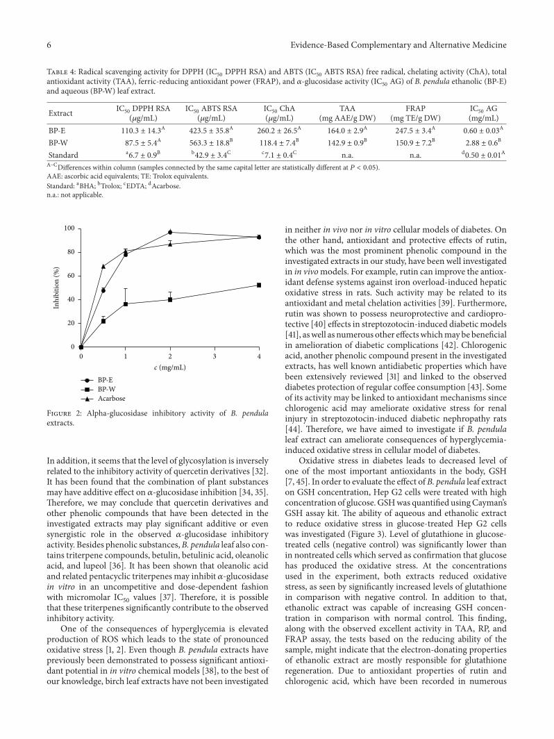
The prepared B. pendula leaf extracts were tested for their𝛼-glucosidase and 𝛼-amylase inhibitory properties. Whilethe extracts did not show any inhibitory activity toward𝛼-amylase, their 𝛼-glucosidase activity was excellent andcomparable to the activity of standard, antidiabetic drugacarbose (Figure 2). Ethanolic extract whose IC
50value did
not statistically differ from acarbose was especially active(Table 4). In an attempt to determine the phytochemicalsresponsible for the observed𝛼-glucosidase inhibitory activity,the activity of rutin and chlorogenic acid have also beentested. It was previously reported that chlorogenic acid maysuppress postprandial hyperglycemia in rats by inhibiting𝛼-glucosidase [31]. However, in the concentrations presentin the active amounts of extracts in this study, rutin andchlorogenic acid did not present observable 𝛼-glucosidaseinhibitory activity. If we compare the results obtained in thisstudy with the IC
50values of rutin and caffeic acid needed
for inhibition of 𝛼-glucosidase in previously published works[32, 33], we may observe that the concentration of thosephenols in the present study may not be sufficient for dis-playing significant inhibitory potential. However, it has beenfound that rutin and chlorogenic acid display significantlylower anti-𝛼-glucosidase activity then their nonconjugatedcounterparts, quercetin and caffeic acid, respectively [32, 33].
6 Evidence-Based Complementary and Alternative Medicine
Table 4: Radical scavenging activity for DPPH (IC50DPPH RSA) and ABTS (IC
50ABTS RSA) free radical, chelating activity (ChA), total
antioxidant activity (TAA), ferric-reducing antioxidant power (FRAP), and 𝛼-glucosidase activity (IC50AG) of B. pendula ethanolic (BP-E)
and aqueous (BP-W) leaf extract.
Extract IC50DPPH RSA(𝜇g/mL)
IC50ABTS RSA(𝜇g/mL)
IC50ChA
(𝜇g/mL)TAA
(mg AAE/g DW)FRAP
(mg TE/g DW)IC50AG
(mg/mL)BP-E 110.3 ± 14.3
A423.5 ± 35.8
A260.2 ± 26.5
A164.0 ± 2.9
A247.5 ± 3.4
A0.60 ± 0.03
A
BP-W 87.5 ± 5.4A
563.3 ± 18.8B
118.4 ± 7.4B
142.9 ± 0.9B
150.9 ± 7.2B
2.88 ± 0.6B
Standard a6.7 ± 0.9
B b42.9 ± 3.4
C c7.1 ± 0.4
C n.a. n.a. d0.50 ± 0.01
A
A–CDifferences within column (samples connected by the same capital letter are statistically different at 𝑃 < 0.05).AAE: ascorbic acid equivalents; TE: Trolox equivalents.Standard: aBHA; bTrolox; cEDTA; dAcarbose.n.a.: not applicable.
BP-EBP-WAcarbose
1 2 3 40c (mg/mL)
0
20
40
60
80
100
Inhi
bitio
n (%
)
Figure 2: Alpha-glucosidase inhibitory activity of B. pendulaextracts.
In addition, it seems that the level of glycosylation is inverselyrelated to the inhibitory activity of quercetin derivatives [32].It has been found that the combination of plant substancesmay have additive effect on 𝛼-glucosidase inhibition [34, 35].Therefore, we may conclude that quercetin derivatives andother phenolic compounds that have been detected in theinvestigated extracts may play significant additive or evensynergistic role in the observed 𝛼-glucosidase inhibitoryactivity. Besides phenolic substances,B. pendula leaf also con-tains triterpene compounds, betulin, betulinic acid, oleanolicacid, and lupeol [36]. It has been shown that oleanolic acidand related pentacyclic triterpenes may inhibit 𝛼-glucosidasein vitro in an uncompetitive and dose-dependent fashionwith micromolar IC
50values [37]. Therefore, it is possible
that these triterpenes significantly contribute to the observedinhibitory activity.
One of the consequences of hyperglycemia is elevatedproduction of ROS which leads to the state of pronouncedoxidative stress [1, 2]. Even though B. pendula extracts havepreviously been demonstrated to possess significant antioxi-dant potential in in vitro chemical models [38], to the best ofour knowledge, birch leaf extracts have not been investigated
in neither in vivo nor in vitro cellular models of diabetes. Onthe other hand, antioxidant and protective effects of rutin,which was the most prominent phenolic compound in theinvestigated extracts in our study, have been well investigatedin in vivomodels. For example, rutin can improve the antiox-idant defense systems against iron overload-induced hepaticoxidative stress in rats. Such activity may be related to itsantioxidant and metal chelation activities [39]. Furthermore,rutin was shown to possess neuroprotective and cardiopro-tective [40] effects in streptozotocin-induced diabeticmodels[41], aswell as numerous other effectswhichmay be beneficialin amelioration of diabetic complications [42]. Chlorogenicacid, another phenolic compound present in the investigatedextracts, has well known antidiabetic properties which havebeen extensively reviewed [31] and linked to the observeddiabetes protection of regular coffee consumption [43]. Someof its activity may be linked to antioxidant mechanisms sincechlorogenic acid may ameliorate oxidative stress for renalinjury in streptozotocin-induced diabetic nephropathy rats[44]. Therefore, we have aimed to investigate if B. pendulaleaf extract can ameliorate consequences of hyperglycemia-induced oxidative stress in cellular model of diabetes.
Oxidative stress in diabetes leads to decreased level ofone of the most important antioxidants in the body, GSH[7, 45]. In order to evaluate the effect of B. pendula leaf extracton GSH concentration, Hep G2 cells were treated with highconcentration of glucose. GSHwas quantified usingCayman’sGSH assay kit. The ability of aqueous and ethanolic extractto reduce oxidative stress in glucose-treated Hep G2 cellswas investigated (Figure 3). Level of glutathione in glucose-treated cells (negative control) was significantly lower thanin nontreated cells which served as confirmation that glucosehas produced the oxidative stress. At the concentrationsused in the experiment, both extracts reduced oxidativestress, as seen by significantly increased levels of glutathionein comparison with negative control. In addition to that,ethanolic extract was capable of increasing GSH concen-tration in comparison with normal control. This finding,along with the observed excellent activity in TAA, RP, andFRAP assay, the tests based on the reducing ability of thesample, might indicate that the electron-donating propertiesof ethanolic extract are mostly responsible for glutathioneregeneration. Due to antioxidant properties of rutin andchlorogenic acid, which have been recorded in numerous

Evidence-Based Complementary and Alternative Medicine 7
∗†
∗†
∗†
∗ ∗
0
20
40
60
80
GSH
(mM
)
BP-W
(0.1
)
BP-E
(0.0
5)
BP-W
(0.5
)C
BP-W
(0.0
5)
BP-E
(0.1
)D
BP-E
(0.5
)
†
Figure 3: Glutathione (GSH) concentration in Hep G2 cells. C: cellsin MEM; D: cells in MEM supplemented with 20mM glucose; BP-E and BP-W: cells in MEM supplemented with 20mM glucose andthe corresponding B. pendula extract (numbers in bracket representextract concentration inmg/mL);∗ and †: value statistically differentfrom C and D, respectively (𝑃 < 0.05, Dunnett’s test). Values areaverage of 3 replications ± SD.
studies [39, 44, 46], it may be concluded that a significant partof the observed activity could be attributed to the presence ofthose antioxidants.
4. Conclusions
B. pendula extracts posses significant antioxidant and antidi-abetic properties as demonstrated by several antioxidantassays, ability to increase intracellular GSH concentration,and inhibition of 𝛼-glucosidase. Solvent choice can signifi-cantly affect biological properties of herbal extracts. In thisstudy, ethanol was able to efficiently extract more or B. pen-dula leaf bioactive principles yielding the extract with highercontent of phenolic antioxidants and better 𝛼-glucosidaseinhibiting and GSH regenerating properties. Some of theobserved biological properties could be attributed to rutin,natural flavonoid which was themain phenolic component ofthe investigated ethanolic extract. Future in vitro and in vivostudies are needed to further investigate antidiabetic potentialof B. pendula ethanolic extract and its mechanism of action.
Competing Interests
The authors declare that there is no conflict of interestsregarding the publication of this paper.
Acknowledgments
Financial support of University of Zagreb is kindly acknowl-edged.
References
[1] R. P. Robertson and J. S. Harmon, “Diabetes, glucose toxicity,and oxidative stress: a case of double jeopardy for the pancreaticislet 𝛽 cell,” Free Radical Biology and Medicine, vol. 41, no. 2, pp.177–184, 2006.
[2] A. Giaccari, G. Sorice, andG.Muscogiuri, “Glucose toxicity: theleading actor in the pathogenesis and clinical history of type 2diabetes—mechanisms and potentials for treatment,”Nutrition,Metabolism and Cardiovascular Diseases, vol. 19, no. 5, pp. 365–377, 2009.
[3] G. Verdile, K. N. Keane, V. F. Cruzat et al., “Inflamma-tion and oxidative stress: the molecular connectivity betweeninsulin resistance, obesity, and Alzheimer’s disease,” Mediatorsof Inflammation, vol. 2015, Article ID 105828, 17 pages, 2015.
[4] H. J. Forman, H. Zhang, and A. Rinna, “Glutathione: overviewof its protective roles, measurement, and biosynthesis,”Molecu-lar Aspects of Medicine, vol. 30, no. 1-2, pp. 1–12, 2009.
[5] L. Yuan and N. Kaplowitz, “Glutathione in liver diseases andhepatotoxicity,” Molecular Aspects of Medicine, vol. 30, no. 1-2,pp. 29–41, 2009.
[6] D. A. Dickinson and H. J. Forman, “Cellular glutathione andthiols metabolism,” Biochemical Pharmacology, vol. 64, no. 5-6,pp. 1019–1026, 2002.
[7] C. Livingstone and J. Davis, “Targeting therapeutics againstglutathione depletion in diabetes and its complications,” BritishJournal of Diabetes and Vascular Disease, vol. 7, no. 6, pp. 258–265, 2007.
[8] N. Orsolic, D. Sirovina, M. Z. Koncic, G. Lackovic, and G. Gre-gorovic, “Effect of Croatian propolis on diabetic nephropathyand liver toxicity inmice,”BMCComplementary andAlternativeMedicine, vol. 12, no. 1, article 117, 2012.
[9] A. D. Tchamgoue, L. R. Y. Tchokouaha, P. A. Tarkang, J.-R.Kuiate, and G. A. Agbor, “Costus afer possesses carbohydratehydrolyzing enzymes inhibitory activity and antioxidant capac-ity in vitro,” Evidence-Based Complementary and AlternativeMedicine, vol. 2015, Article ID 987984, 10 pages, 2015.
[10] G. Mahendran, G. Thamotharan, S. Sengottuvelu, and V.Narmatha Bai, “Anti-diabetic activity of Swertia corymbosa(Griseb.) Wight ex C.B. Clarke aerial parts extract in strepto-zotocin induced diabetic rats,” Journal of Ethnopharmacology,vol. 151, no. 3, pp. 1175–1183, 2014.
[11] J. Havlik, R. G. de la Huebra, K. Hejtmankova et al., “Xanthineoxidase inhibitory properties of Czech medicinal plants,” Jour-nal of Ethnopharmacology, vol. 132, no. 2, pp. 461–465, 2010.
[12] C. Grundemann, C. W. Gruber, A. Hertrampf, M. Zehl, B.Kopp, and R. Huber, “An aqueous birch leaf extract of Betulapendula inhibits the growth and cell division of inflammatorylymphocytes,” Journal of Ethnopharmacology, vol. 136, no. 3, pp.444–451, 2011.
[13] M. P. Germano, F. Cacciola, P. Donato et al., “Betula pendulaleaves: polyphenolic characterization and potential innovativeuse in skin whitening products,” Fitoterapia, vol. 83, no. 5, pp.877–882, 2012.
[14] V. L. Singleton, R. Orthofer, and R. M. Lamuela-Raventos,“Analysis of total phenols and other oxidation substrates andantioxidants by means of folin-ciocalteu reagent,” Methods inEnzymology, vol. 299, pp. 152–178, 1999.
[15] S. Kumazawa, T. Hamasaka, and T. Nakayama, “Antioxidantactivity of propolis of various geographic origins,” Food Chem-istry, vol. 84, no. 3, pp. 329–339, 2004.

8 Evidence-Based Complementary and Alternative Medicine
[16] C. Nicolle, A. Carnat, D. Fraisse et al., “Characterisation andvariation of antioxidant micronutrients in lettuce (Lactucasativa folium),” Journal of the Science of Food and Agriculture,vol. 84, no. 15, pp. 2061–2069, 2004.
[17] J. Ermer, Ed.,Method Validation in Pharmaceutical Analysis: AGuide to Best Practice, Wiley-VCH, Weinheim, Germany, 2005.
[18] M. Z. Koncic,M. Barbaric, I. Perkovic, andB. Zorc, “Antiradical,chelating and antioxidant activities of hydroxamic acids andhydroxyureas,”Molecules, vol. 16, no. 8, pp. 6232–6242, 2011.
[19] R. Re, N. Pellegrini, A. Proteggente, A. Pannala,M. Yang, andC.Rice-Evans, “Antioxidant activity applying an improved ABTSradical cation decolorization assay,” Free Radical Biology andMedicine, vol. 26, no. 9-10, pp. 1231–1237, 1999.
[20] P. Prieto, M. Pineda, and M. Aguilar, “Spectrophotometricquantitation of antioxidant capacity through the formation ofa phosphomolybdenum complex: specific application to thedetermination of vitamin E,” Analytical Biochemistry, vol. 269,no. 2, pp. 337–341, 1999.
[21] I. F. F. Benzie and J. J. Strain, “The ferric reducing ability ofplasma (FRAP) as a measure of ‘antioxidant power’: the FRAPassay,” Analytical Biochemistry, vol. 239, no. 1, pp. 70–76, 1996.
[22] A. K. Tiwari, M. Swapna, S. B. Ayesha, A. Zehra, S. B. Agawane,and K. Madhusudana, “Identification of proglycemic and anti-hyperglycemic activity in antioxidant rich fraction of somecommon food grains,” International Food Research Journal, vol.18, no. 3, pp. 915–923, 2011.
[23] E. Apostolidis, Y.-I. Kwon, and K. Shetty, “Inhibitory potentialof herb, fruit, and fungal-enriched cheese against key enzymeslinked to type 2 diabetes and hypertension,” Innovative FoodScience and Emerging Technologies, vol. 8, no. 1, pp. 46–54, 2007.
[24] K. J. Lee, E.-R. Woo, C. Y. Choi et al., “Protective effect ofacteoside on carbon tetrachloride-induced hepatotoxicity,” LifeSciences, vol. 74, no. 8, pp. 1051–1064, 2004.
[25] M. Keinanen and R. Julkunen-Tiitto, “High-performance liquidchromatographic determination of flavonoids inBetula pendulaand Betula pubescens leaves,” Journal of Chromatography A, vol.793, no. 2, pp. 370–377, 1998.
[26] T. E. Moussa-Ayoub, S. K. El-Samahy, L. W. Kroh, and S. Rohn,“Identification and quantification of flavonol aglycons in cactuspear (Opuntia ficus indica) fruit using a commercial pectinaseand cellulase preparation,” Food Chemistry, vol. 124, no. 3, pp.1177–1184, 2011.
[27] M. Leopoldini, N. Russo, andM. Toscano, “Themolecular basisof working mechanism of natural polyphenolic antioxidants,”Food Chemistry, vol. 125, no. 2, pp. 288–306, 2011.
[28] “Antioxidants in Food: Practical Applications,” https://www.crcpress.com/Antioxidants-in-Food-Practical-Applications/Pokorny-Yanishlieva-Gordon/p/book/9780849312229.
[29] M. Plaza, A. G. Batista, C. B. B. Cazarin et al., “Characterizationof antioxidant polyphenols fromMyrciaria jaboticaba peel andtheir effects on glucose metabolism and antioxidant status: apilot clinical study,” Food Chemistry, vol. 211, pp. 185–197, 2016.
[30] J. Giacometti, D. Muhvic, A. Pavletic, and L. Đudaric, “Cocoapolyphenols exhibit antioxidant, anti-inflammatory, antican-cerogenic, and anti-necrotic activity in carbon tetrachloride-intoxicated mice,” Journal of Functional Foods, vol. 23, pp. 177–187, 2016.
[31] S. Meng, J. Cao, Q. Feng, J. Peng, and Y. Hu, “Roles ofchlorogenic acid on regulating glucose and lipids metabolism:a review,” Evidence-Based Complementary and AlternativeMedicine, vol. 2013, Article ID 801457, 11 pages, 2013.
[32] Y. Q. Li, F. C. Zhou, F. Gao, J. S. Bian, and F. Shan, “Comparativeevaluation of quercetin, isoquercetin and rutin as inhibitors of𝛼-glucosidase,” Journal of Agricultural and Food Chemistry, vol.57, no. 24, pp. 11463–11468, 2009.
[33] G. Oboh, O. M. Agunloye, S. A. Adefegha, A. J. Akinyemi,and A. O. Ademiluyi, “Caffeic and chlorogenic acids inhibitkey enzymes linked to type 2 diabetes (in vitro): a comparativestudy,” Journal of Basic and Clinical Physiology and Pharmacol-ogy, vol. 26, no. 2, pp. 165–170, 2016.
[34] S. Adisakwattana, T. Ruengsamran, P. Kampa, andW. Sompong,“In vitro inhibitory effects of plant-based foods and theircombinations on intestinal 𝛼-glucosidase and pancreatic 𝛼-amylase,” BMC Complementary and Alternative Medicine, vol.12, article 110, pp. 1–8, 2012.
[35] S. Adisakwattana, O. Lerdsuwankij, U. Poputtachai, A.Minipun, and C. Suparpprom, “Inhibitory activity of cinnamonbark species and their combination effect with acarbose againstintestinal 𝛼-glucosidase and pancreatic 𝛼-amylase,” Plant Foodsfor Human Nutrition, vol. 66, no. 2, pp. 143–148, 2011.
[36] K. Duric, E. Kovac-Besovic, H. Niksic, and E. Sofic, “Antibac-terial activity of methanolic extracts, decoction and isolatedtriterpene products from different parts of birch, betula pen-dula, roth,” Journal of Plant Studies, vol. 2, no. 2, 2013.
[37] J. M. Castellano, A. Guinda, T. Delgado, M. Rada, and J.A. Cayuela, “Biochemical basis of the antidiabetic activity ofoleanolic acid and related pentacyclic triterpenes,”Diabetes, vol.62, no. 6, pp. 1791–1799, 2013.
[38] L. Raudone, R. Raudonis, V. Janulis, and P. Viskelis, “Qualityevaluation of different preparations of dry extracts of birch(Betula pendulaRoth) leaves,”Natural Product Research, vol. 28,no. 19, pp. 1645–1648, 2014.
[39] S. A. H. Aziza, M. E.-S. Azab, and S. K. El-Shall, “Amelioratingrole of rutin on oxidative stress induced by iron overload inhepatic tissue of rats,”Pakistan Journal of Biological Sciences, vol.17, no. 8, pp. 964–977, 2014.
[40] J. F. C. Guimaraes, B. P. Muzio, C. M. Rosa et al., “Rutinadministration attenuates myocardial dysfunction in diabeticrats,” Cardiovascular Diabetology, vol. 14, no. 1, article 90, 2015.
[41] M. S. Ola, M. M. Ahmed, R. Ahmad, H. M. Abuohashish, S.S. Al-Rejaie, and A. S. Alhomida, “Neuroprotective effects ofrutin in streptozotocin-induced diabetic rat retina,” Journal ofMolecular Neuroscience, vol. 56, no. 2, pp. 440–448, 2015.
[42] S. Habtemariam and G. Lentini, “The therapeutic potentialof rutin for diabetes: an update,” Mini-Reviews in MedicinalChemistry, vol. 15, no. 7, pp. 524–528, 2015.
[43] R. M. M. Santos and D. R. A. Lima, “Coffee consumption,obesity and type 2 diabetes: a mini-review,” European Journalof Nutrition, vol. 55, no. 4, pp. 1345–1358, 2016.
[44] H.-Y. Ye, Z.-Y. Li, Y. Zheng, Y. Chen, Z.-H. Zhou, and J. Jin,“The attenuation of chlorogenic acid on oxidative stress forrenal injury in streptozotocin-induced diabetic nephropathyrats,”Archives of Pharmacal Research, vol. 39, no. 7, pp. 989–997,2016.
[45] L. Lash, “Mitochondrial glutathione in diabetic nephropathy,”Journal of Clinical Medicine, vol. 4, no. 7, pp. 1428–1447, 2015.
[46] B.-M. Lue, N. S. Nielsen, C. Jacobsen, L. Hellgren, Z. Guo,and X. Xu, “Antioxidant properties of modified rutin esters byDPPH, reducing power, iron chelation and human low densitylipoprotein assays,” Food Chemistry, vol. 123, no. 2, pp. 221–230,2010.
Submit your manuscripts athttp://www.hindawi.com
Stem CellsInternational
Hindawi Publishing Corporationhttp://www.hindawi.com Volume 2014
Hindawi Publishing Corporationhttp://www.hindawi.com Volume 2014
MEDIATORSINFLAMMATION
of
Hindawi Publishing Corporationhttp://www.hindawi.com Volume 2014
Behavioural Neurology
EndocrinologyInternational Journal of
Hindawi Publishing Corporationhttp://www.hindawi.com Volume 2014
Hindawi Publishing Corporationhttp://www.hindawi.com Volume 2014
Disease Markers
Hindawi Publishing Corporationhttp://www.hindawi.com Volume 2014
BioMed Research International
OncologyJournal of
Hindawi Publishing Corporationhttp://www.hindawi.com Volume 2014
Hindawi Publishing Corporationhttp://www.hindawi.com Volume 2014
Oxidative Medicine and Cellular Longevity
Hindawi Publishing Corporationhttp://www.hindawi.com Volume 2014
PPAR Research
The Scientific World JournalHindawi Publishing Corporation http://www.hindawi.com Volume 2014
Immunology ResearchHindawi Publishing Corporationhttp://www.hindawi.com Volume 2014
Journal of
ObesityJournal of
Hindawi Publishing Corporationhttp://www.hindawi.com Volume 2014
Hindawi Publishing Corporationhttp://www.hindawi.com Volume 2014
Computational and Mathematical Methods in Medicine
OphthalmologyJournal of
Hindawi Publishing Corporationhttp://www.hindawi.com Volume 2014
Diabetes ResearchJournal of
Hindawi Publishing Corporationhttp://www.hindawi.com Volume 2014
Hindawi Publishing Corporationhttp://www.hindawi.com Volume 2014
Research and TreatmentAIDS
Hindawi Publishing Corporationhttp://www.hindawi.com Volume 2014
Gastroenterology Research and Practice
Hindawi Publishing Corporationhttp://www.hindawi.com Volume 2014
Parkinson’s Disease
Evidence-Based Complementary and Alternative Medicine
Volume 2014Hindawi Publishing Corporationhttp://www.hindawi.com