Research Article Dose Dependence of …downloads.hindawi.com/journals/jchem/2013/930818.pdf ·...
5
Hindawi Publishing Corporation Journal of Chemistry Volume 2013, Article ID 930818, 4 pages http://dx.doi.org/10.1155/2013/930818 Research Article Dose Dependence of Mechanoluminescence Properties in MgAl 2 O 4 : Dy Phosphor Kabita K. Satapathy 1 and G. C. Mishra 2 1 Department of Applied Chemistry, O.P. Jindal Institute of Technology, Raigarh 496001, India 2 Department of Applied Physics, O.P. Jindal Institute of Technology, Raigarh 496001, India Correspondence should be addressed to Kabita K. Satapathy; [email protected] Received 24 May 2013; Accepted 8 October 2013 Academic Editor: Ewa Schab-Balcerzak Copyright © 2013 K. K. Satapathy and G. C. Mishra. is is an open access article distributed under the Creative Commons Attribution License, which permits unrestricted use, distribution, and reproduction in any medium, provided the original work is properly cited. A reliable dosimetry is fundamental for quality assurance of the processes and irradiation products. All dosimetric systems for high doses have some limitation with regard to their use. Dosimetric system should be easy to use, fast to measure, and of low cost. Good phosphor which shows high luminescence properties may fulfil the above criteria in some way. MgAl 2 O 4 : Dy phosphor has been prepared by solution combustion technique and confirmed with the help of XRD. ML has been excited impulsively by dropping a load of mass 0.7 kg onto the phosphors from various heights; two distinct ML peaks are observed for all the samples. It is observed that MgAl 2 O 4 : Dy phosphor shows linear response to gamma-ray dose and low fading which can be used for dosimetric purpose. 1. Introduction Dosimeters have to measure accurately radiation intensities. eir applications include personnel monitoring, environ- mental monitoring, radiation therapy, diagnostic radiology, and other radiation measurements. Ionization chambers measure directly intensities of ionizing radiation. For many applications, such as personnel and environmental monitor- ing, the absorbed energy has to be stored for long periods. is is done by suitable storing element fading of lumines- cence. e longer a phosphor stands aſter the excitation, the more trapped carriers will leave the traps, and the subse- quently measured luminescence peaks will be weaker. Ioniz- ing radiation is a technique widely employed in last decades. It was used for a long time for many applications such as sterilization of medical devices. Rare earth doped phosphors have a vital role as radiation detectors in many fields of fundamental and applied research, such as clinical, personal, and environmental monitoring of ionizing radiation [1, 2]. Various methods of preparation have also been developed for easy synthesis of these materials to make them available easily. While irradiation usually leads to the creation of structural defects, the healing effect of irradiation is also known [3]. Metals made of powders preliminary irradiated by electrons or gamma irradiations are characterized by the absence of big pores and fine homogeneous grain structure. e average sizes of grains are 4-5 times lower than that in conventional technology [4]. Ionising radiation has been found to be widely applicable in modifying the structure and properties of polymers and can be used to tailor the performance of either bulk materials or surfaces. Improved luminescent char- acteristics of some of aluminates have also found their place in optoelectronics. e thermoluminescence (TL) materials have been widely applied to defect studying and dosimetry, such as the detection of ionizing radiation and dating in archaeology [5–8]. e ability to detect and perform energy- dispersive spectroscopy of high-energy radiation such as X- rays, -rays, and other uncharged and charged particles has improved dramatically in recent years [9]. is is of great importance in a wide range of applications including medical imaging, industrial process monitoring, national security and treaty verification, environmental safety and remediation, and basic science. A reliable dosimetry is fundamental for quality assurance of the processes and irradiation products. Almost all dosimetric systems for high doses present some limitable disadvantages with regard to their use. A dosimeter
Transcript of Research Article Dose Dependence of …downloads.hindawi.com/journals/jchem/2013/930818.pdf ·...

Hindawi Publishing CorporationJournal of ChemistryVolume 2013, Article ID 930818, 4 pageshttp://dx.doi.org/10.1155/2013/930818
Research ArticleDose Dependence of Mechanoluminescence Propertiesin MgAl2O4: Dy Phosphor
Kabita K. Satapathy1 and G. C. Mishra2
1 Department of Applied Chemistry, O.P. Jindal Institute of Technology, Raigarh 496001, India2Department of Applied Physics, O.P. Jindal Institute of Technology, Raigarh 496001, India
Correspondence should be addressed to Kabita K. Satapathy; [email protected]
Received 24 May 2013; Accepted 8 October 2013
Academic Editor: Ewa Schab-Balcerzak
Copyright © 2013 K. K. Satapathy and G. C. Mishra. This is an open access article distributed under the Creative CommonsAttribution License, which permits unrestricted use, distribution, and reproduction in any medium, provided the original work isproperly cited.
A reliable dosimetry is fundamental for quality assurance of the processes and irradiation products. All dosimetric systems for highdoses have some limitation with regard to their use. Dosimetric system should be easy to use, fast tomeasure, and of low cost. Goodphosphor which shows high luminescence properties may fulfil the above criteria in some way. MgAl
2O4: Dy phosphor has been
prepared by solution combustion technique and confirmed with the help of XRD. ML has been excited impulsively by dropping aload of mass 0.7 kg onto the phosphors from various heights; two distinct ML peaks are observed for all the samples. It is observedthat MgAl
2O4: Dy phosphor shows linear response to gamma-ray dose and low fading which can be used for dosimetric purpose.
1. Introduction
Dosimeters have to measure accurately radiation intensities.Their applications include personnel monitoring, environ-mental monitoring, radiation therapy, diagnostic radiology,and other radiation measurements. Ionization chambersmeasure directly intensities of ionizing radiation. For manyapplications, such as personnel and environmental monitor-ing, the absorbed energy has to be stored for long periods.This is done by suitable storing element fading of lumines-cence. The longer a phosphor stands after the excitation, themore trapped carriers will leave the traps, and the subse-quently measured luminescence peaks will be weaker. Ioniz-ing radiation is a technique widely employed in last decades.It was used for a long time for many applications such assterilization of medical devices. Rare earth doped phosphorshave a vital role as radiation detectors in many fields offundamental and applied research, such as clinical, personal,and environmental monitoring of ionizing radiation [1, 2].Various methods of preparation have also been developed foreasy synthesis of thesematerials tomake themavailable easily.While irradiation usually leads to the creation of structuraldefects, the healing effect of irradiation is also known [3].
Metals made of powders preliminary irradiated by electronsor gamma irradiations are characterized by the absence ofbig pores and fine homogeneous grain structure.The averagesizes of grains are 4-5 times lower than that in conventionaltechnology [4]. Ionising radiation has been found to bewidely applicable in modifying the structure and propertiesof polymers and can be used to tailor the performance ofeither bulkmaterials or surfaces. Improved luminescent char-acteristics of some of aluminates have also found their placein optoelectronics. The thermoluminescence (TL) materialshave been widely applied to defect studying and dosimetry,such as the detection of ionizing radiation and dating inarchaeology [5–8]. The ability to detect and perform energy-dispersive spectroscopy of high-energy radiation such as X-rays, 𝛾-rays, and other uncharged and charged particles hasimproved dramatically in recent years [9]. This is of greatimportance in a wide range of applications includingmedicalimaging, industrial processmonitoring, national security andtreaty verification, environmental safety and remediation,and basic science. A reliable dosimetry is fundamental forquality assurance of the processes and irradiation products.Almost all dosimetric systems for high doses present somelimitable disadvantages with regard to their use. A dosimeter
2 Journal of Chemistry
0100020003000400050006000
20 30 40 50 60 70 80
Inte
nsity
(a.u
.)
2𝜃 (deg)
Figure 1: XRD pattern of MgAl2O4phosphor.
that fulfils the traditional requirements of precision, dose-rate independence, and postirradiation stability is desirable[10]. For industrial needs a dosimetric system must fulfilsome criteria: being easy to use, fast to measure, and of lowcost. The dosimeter cost should be negligible compared tothe radiation processing final price. Sugar has showed goodresults as dosimeter using EPR technique [11–13] but thehigh cost of the equipment is a serious handicap for large-scale routine application. The other techniques that havebeen used to reduce the cost are lyoluminescence [14, 15].Mechanoluminescence can also be used to reduce the cost.Mechanoluminescence is an interesting phenomenon whichis a light emission caused by mechanical stimuli such asgrinding, cutting, collision, striking, and friction [16, 17].Alkaline earth aluminate ceramics are important hostmateri-als that have been prepared and studied by several researchersfor luminescence applications [18]. Alkaline earth aluminatebelongs to the spinel group of minerals (MAl
2O4) with
general chemical composition, AB2O4, where A is a divalent
atom [19]. The magnesium aluminate spinel (MgAl2O4) has
good thermal and mechanical properties, high hardnessand low electrical loss, and chemical properties. As such,it is currently utilized as a refractory for furnace wallsand firebricks and also has the potential for application asenvironment humidity sensors, laser materials, and substratein integrated electronics [20–27]. The objective of this studyis to find out the effect of dose onML properties of MgAl
2O4:
Dy phosphor and to know how it is efficient for dosimeter.
2. Materials and Methods
The samples were prepared by solution combustion synthesistechnique. The ingredients used were Mg (NO
3)2⋅6H2O, Al
(NO3)3⋅9H2O, urea, and dysprosium in the form of nitrate.
Magnesium nitrate, aluminium nitrate, urea, and the desiredamount of dopant were taken in a glass beaker and dissolvedin distilled water. The beaker was kept in a furnace set ataround 300∘C. The reaction is self-propagating and is ableto sustain this high temperature long enough. The entirecombustion process was over in about 5min. Formations ofthe samples were confirmed by XRD pattern recorded by X-ray defractometer (PW-1710).The gamma-ray irradiationwascarried out using 60Co source. ML was excited impulsively bydropping a load on the sample placed on a Lucite plate withdifferent impact velocities. The luminescence was monitoredby a 931A photomultiplier tube positioned below the Lucite
012345678
0 0.5 1 1.5 2 2.5 3 3.5Time (ms)
ML
inte
nsity
(a.u
.)
2.200 kGy1.100 kGy
0.550 kGy0.275 kGy
Figure 2: Dependence of ML intensity of MgAl2O4: Dy (0.1mol%)
phosphor on gamma dose.
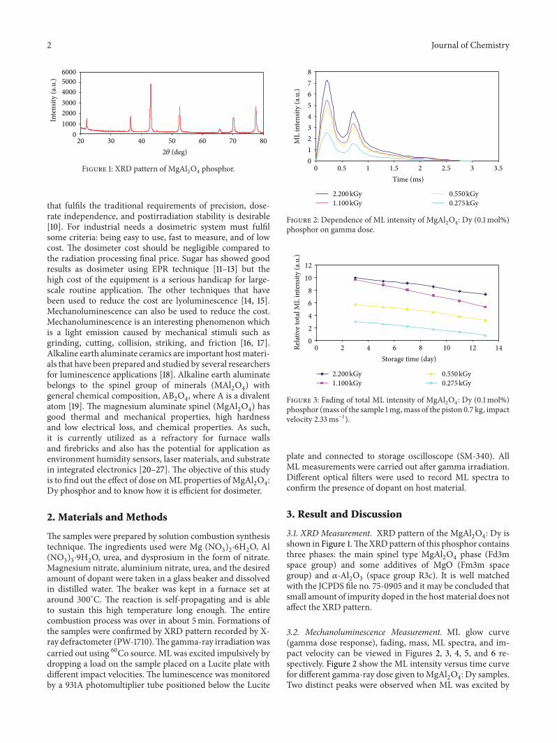
0
2
4
6
8
10
12
0 2 4 6 8 10 12 14Storage time (day)
Relat
ive t
otal
ML
inte
nsity
(a.u
.)
2.200 kGy1.100 kGy
0.550 kGy0.275 kGy
Figure 3: Fading of total ML intensity of MgAl2O4: Dy (0.1mol%)
phosphor (mass of the sample 1mg,mass of the piston 0.7 kg, impactvelocity 2.33ms−1).
plate and connected to storage oscilloscope (SM-340). AllML measurements were carried out after gamma irradiation.Different optical filters were used to record ML spectra toconfirm the presence of dopant on host material.
3. Result and Discussion
3.1. XRD Measurement. XRD pattern of the MgAl2O4: Dy is
shown in Figure 1.TheXRDpattern of this phosphor containsthree phases: the main spinel type MgAl
2O4phase (Fd3m
space group) and some additives of MgO (Fm3m spacegroup) and 𝛼-Al
2O3(space group R3c). It is well matched
with the JCPDS file no. 75-0905 and it may be concluded thatsmall amount of impurity doped in the hostmaterial does notaffect the XRD pattern.
3.2. Mechanoluminescence Measurement. ML glow curve(gamma dose response), fading, mass, ML spectra, and im-pact velocity can be viewed in Figures 2, 3, 4, 5, and 6 re-spectively. Figure 2 show the ML intensity versus time curvefor different gamma-ray dose given toMgAl
2O4: Dy samples.
Two distinct peaks were observed when ML was excited by

Journal of Chemistry 3
0
2
4
6
8
10
12
0 0.5 1 1.5 2 2.5 3Mass of the sample (mg)
Tota
l ML
inte
nsity
(a.u
.)
2.200 kGy1.100 kGy
0.550 kGy0.275 kGy
Figure 4: Dependence of ML intensity of MgAl2O4: Dy (0.1mol%)
phosphor on mass of the phosphor for different gamma dose.
0
1
2
3
4
5
6
7
400 450 500 550 600 650 700Wavelength (nm)
Tota
l ML
inte
nsity
(a.u
.)
2.200 kGy1.100 kGy
0.550 kGy0.275 kGy
Figure 5: ML emission spectra of MgAl2O4: Dy (0.1mol%) phos-
phor for different gamma-ray dose.
dropping a load of mass 0.7 kg onto it for the entire sample.ML intensity initially increased with time attained an opti-mum value for a particular time and then decreased againincreases and finally disappeared for all the samples. MLintensity is observed maximum for higher gamma-ray dose.
Figure 3 shows the effect of storage (at room temperature)on ML of 𝛾-irradiated MgAl
2O4: Dy (0.1mol%). It is clear
that fading in ML intensity of the entire sample is very low.Themaximum fading observed is around 5 to 6%when it wasrecorded after 15 days of irradiation.
Figure 4 shows that ML intensity increased almost lin-early with increasing the mass (0.5 to 2.5mg) of the sampledeformed for recording ML for different gamma-ray dose inthe rage 0.275–2.200 kGy investigated. When we increase themass of the sample, the number of crystallites in the sampleincreases and thereby the ML intensity and the total MLintensity increase and are optimum for higher dose.
Figure 5 shows ML emission spectra of MgAl2O4: Dy
(0.1mol%) phosphors. In order to find the luminescence cen-tres responsible forML emission, we have recordedML spec-trum. Two distinct peaks, one around 482 nm and another
0
1
2
3
4
5
6
0 0.5 1 1.5 2 2.5Impact velocity (m/s)
Tota
l ML
inte
nsity
(a.u
.)
2.200 kGy1.100 kGy
0.550 kGy0.275 kGy
Figure 6: Dependence of ML intensity of MgAl2O4: Dy (0.1mol%)
phosphor on impact velocity for different gamma dose.
around 585 nm, were observed due to 4F9/2→6H13/2
and4F9/2→6H15/2
transition of Dy3+ ions, respectively, which isthe characteristic emission of Dy3+.
Figure 6 shows the dependence of total ML intensity of𝛾-irradiatedMgAl
2O4: Dy (0.1mol%) phosphors on different
impact velocities. In present investigation impact velocitywasvaried from 0.5 to 2.33m/s. It is observed that ML intensityincreases almost linearly with increasing impact velocity.
The origin of light emission is not piezoelectricity asMgAl
2O4has a centrosymmetric structure (Fd3m).Therefore
it is suggested that ML of MgAl2O4: Dy3+ is related to the
movement of dislocations and the recombination of activatedelectrons and holes. The movement of dislocations excitescarriers from the filled traps and the subsequent recombina-tion of the electrons andholes in defect centres.On increasingthe 𝛾-dose, the density of defect centres increases and when asample of a givenmass is deformed at a given impact velocity,ML intensity should increase with the density of defect cen-tres. InMgAl
2O4: RE, themost probable centres which can be
observed are theV centres (a hole trapped at a cation vacancy)and F centres (an electron trapped at an anion vacancy) [28].It is known that the cation disorder and nonstoichiometry ofaluminates like MgAl
2O4provide a large number of lattice
defects, which may serve as trapping centres. It seems thatduring the preparation of MgAl
2O4: RE phosphor, two ions
of RE3+ replace threeMg2+ ions, creatingMg2+ ion vacancies.The Re3+ ion can easily enter the lattice, in place of Mg2+ ion,as the ionic radius of RE3+ is close to the ionic radius of Mg2+ion. SinceML glow curve shows the characteristic emission ofRE3+, this energy may be transferred nonradiatively to RE3+ions causing their excitation and subsequent deexcitation ofexcited RE3+ ions. It is observed that ML of MgAl
2O4: Dy3+
phosphor is optimum for higher gamma-ray dose and fadingof the sample is very low for all doses given to the sample.
4. Conclusions
MgAl2O4: Dy3+ was synthesized via a solution combus-
tion process from metal nitrates and organic fuel. Well-crystallized powders were obtained at 477∘C within 5min.
4 Journal of Chemistry
ML emission spectrum shows the characteristic emissionof Dy3+ ions. ML intensity increases linearly with mass ofthe sample and gamma-ray doses given to the sample. Twodistinct peaks were observed in ML curve of MgAl
2O4: Dy3+
phosphors. It is observed that MgAl2O4: Dy3+ phosphor
possesses high sensitivity to enable measurements of verylow radiation doses, linear response to increasing gamma-raydose, and very low fading. SinceML emission in this system isinduced by the gamma ray andML increases with gamma-raydose, it may be use in ML dosimetry. This fundamental workmight be important in developing new luminescent devicesapplicable for ML sensors and dosimeter.
References
[1] S. J. Dhoble, “Preparation of (K:Eu) NaSO4 phosphor for ly-oluminescence dosimetry of ionising radiation,” Radiation Pro-tection Dosimetry, vol. 100, no. 1–4, pp. 285–287, 2002.
[2] N. D. Yordanov and Y. Karakirova, “EPR of gamma irradiatedsolid sucrose and UV spectra of its solution. An attempt for cal-ibration of solid state/EPRdosimetry,”RadiationMeasurements,vol. 42, no. 3, pp. 347–351, 2007.
[3] R. L. Clough, “High-energy radiation and polymers: a reviewof commercial processes and emerging applications,” NuclearInstruments and Methods in Physics Research B, vol. 185, pp. 8–33, 2001.
[4] Y. A. Zaykin and B. A. Aliyev, “Radiation effects in high-disperse metal media and their application in powder metal-lurgy,” Radiation Physics and Chemistry, vol. 63, no. 3–6, pp.227–230, 2002.
[5] F. Daniels, C. A. Boyd, and D. F. Saunders, “Thermolumines-cence as a research tool,” Science, vol. 117, no. 3040, pp. 343–349,1953.
[6] A. J. J. Bos, “High sensitivity thermoluminescence dosimetry,”Nuclear Instruments andMethods in Physics Research B, vol. 184,pp. 3–28, 2001.
[7] R. P. Yavetskiy, E. F. Dolzhenkova, A. V. Tolmachev, S. V.Parkhomenko, V. N. Baumer, and A. L. Prosvirnin, “Radiationdefects in SrB
4O7:Eu2+ crystals,” Journal of Alloys and Com-
pounds, vol. 441, no. 1-2, pp. 202–205, 2007.[8] T. E. Schlesinger, J. E. Toney, H. Yoon et al., “Cadmium zinc
telluride and its use as a nuclear radiation detector material,”Materials Science and Engineering R, vol. 32, no. 4-5, pp. 103–189, 2001.
[9] W. L. McLaughlin, A. W. Boyed, K. C. Chadwich, J. C. Mcdon-ald, and A. Miller, Dosimetry for Radiation Processing, Taylor &Francis, London, UK, 1989.
[10] T. Nakajima and T. Otsuki, “Dosimetry for radiation emer-gencies: radiation-induced free radicals in sugar of variouscountries and the effect of pulverizing on the ESR signal,”Applied Radiation and Isotopes, vol. 41, no. 4, pp. 359–365, 1990.
[11] T. Nakajima, “ESR of sugar as a personnel monitor for radiationemergencies,” Applied Radiation and Isotopes, vol. 46, no. 8, pp.819–825, 1995.
[12] A. Tchen, C. L. Greenstock, and A. Trivedi, “The use of sugarpellets in ESR dosimetry,” Radiation Protection Dosimetry, vol.46, no. 2, pp. 119–121, 1993.
[13] N. A. Atari and K. V. Ettinger, “Lyoluminescence of irradiatedsaccharides,” Radiation Effects, vol. 20, no. 1-2, pp. 135–139, 1973.
[14] T.Nickel, E. Pitt, andA. Scharmann, “Lyoluminescence dosime-trywith a sugar after accidental gamma ray exposure,”RadiationProtection Dosimetry, vol. 35, no. 3, pp. 173–177, 1991.
[15] A. J. Walton, “Triboluminescence,” Advances in Physics, vol. 26,no. 6, pp. 887–948, 1977.
[16] G. Alzetta, I. Chudasek, and R. Scarmozzino, “Excitation oftriboluminescence by deformation of single crystals,” PhysicaStatus Solidi A, vol. 1, no. 4, pp. 775–785, 1970.
[17] Y. Enomoto and H. Hashimoto, “Emission of charged particlesfrom indentation fracture of rocks,” Nature, vol. 346, no. 6285,pp. 641–643, 1990.
[18] T. Matsuzawa, Y. Aokiy, N. Takeuchi, and Y. Murayama, “Anew long phosphorescent phosphor with high brightness, SrAl
2
O4: Eu2+, Dy3+,” Journal of the Electrochemical Society, vol. 143,
pp. 2670–2673, 1996.[19] R. Dekkers and C. F. Woensdregt, “Crystal structural control
on surface topology and crystal morphology of normal spinel(MgAl
2O4),” Journal of Crystal Growth, vol. 236, no. 1–3, pp.
441–454, 2002.[20] C. Baudı́n, R. Mart́ınez, and P. Pena, “High-temperature me-
chanical behavior of stoichiometric magnesium spinel,” Journalof the American Ceramic Society, vol. 78, pp. 1857–1862, 1995.
[21] M. Sindel, N. A. Travitzky, and N. Claussen, “Influence of mag-nesium-aluminum spinel on the directed oxidation of moltenaluminum alloys,” Journal of the American Ceramic Society, vol.73, pp. 2615–2618, 1990.
[22] J. T. Baitley and R. Russell, “Sintered spinel ceramics,” Journal ofthe American Ceramic Society, vol. 47, pp. 1025–1029, 1968.
[23] C. C. Wang, “Growth and characterization of spinel singlecrystals for substrate use in integrated electronics,” Journal ofApplied Physics, vol. 40, no. 9, pp. 3433–3444, 1969.
[24] K. Petermann, R. Clausen, E. Heumann, and M. Ledig, “Timeresolved excited state absorption of Mn2+,” Optics Communica-tions, vol. 70, no. 6, pp. 483–486, 1989.
[25] R. Clausen and K. Petermann, “Mn/sup 2+/ as a potential solid-state laser ion,” IEEE Journal of QuantumElectronics, vol. 24, no.6, pp. 1114–1117, 1988.
[26] C. Wyon, J. J. Aubert, and F. Auzel, “Czochralski growth andoptical properties of magnesium-aluminium spinel doped withnickel,” Journal of Crystal Growth, vol. 79, no. 1–3, pp. 710–713,1986.
[27] A. Jouini, A. Yoshikawa, A. Brenier, T. Fukuda, and G. Boulon,“Optical properties of transition metal ion-doped MgAl
2O4
spinel for laser application,” Physica Status Solidi C, vol. 4, no.3, pp. 1380–1383, 2007.
[28] E. A. Raja, B. Dhabekar, S. Menon, S. P. More, T. K. G. Rao,and R. K. Kher, “Role of defect centres in thermoluminescencemechanism of Tb 3+doped MgAl
2O4,” Indian Journal of Pure
and Applied Physics, vol. 47, no. 6, pp. 420–425, 2009.

Submit your manuscripts athttp://www.hindawi.com
Hindawi Publishing Corporationhttp://www.hindawi.com Volume 2014
Inorganic ChemistryInternational Journal of
Hindawi Publishing Corporation http://www.hindawi.com Volume 2014
International Journal ofPhotoenergy
Hindawi Publishing Corporationhttp://www.hindawi.com Volume 2014
Carbohydrate Chemistry
International Journal of
Hindawi Publishing Corporationhttp://www.hindawi.com Volume 2014
Journal of
Chemistry
Hindawi Publishing Corporationhttp://www.hindawi.com Volume 2014
Advances in
Physical Chemistry
Hindawi Publishing Corporationhttp://www.hindawi.com
Analytical Methods in Chemistry
Journal of
Volume 2014
Bioinorganic Chemistry and ApplicationsHindawi Publishing Corporationhttp://www.hindawi.com Volume 2014
SpectroscopyInternational Journal of
Hindawi Publishing Corporationhttp://www.hindawi.com Volume 2014
The Scientific World JournalHindawi Publishing Corporation http://www.hindawi.com Volume 2014
Medicinal ChemistryInternational Journal of
Hindawi Publishing Corporationhttp://www.hindawi.com Volume 2014
Chromatography Research International
Hindawi Publishing Corporationhttp://www.hindawi.com Volume 2014
Applied ChemistryJournal of
Hindawi Publishing Corporationhttp://www.hindawi.com Volume 2014
Hindawi Publishing Corporationhttp://www.hindawi.com Volume 2014
Theoretical ChemistryJournal of
Hindawi Publishing Corporationhttp://www.hindawi.com Volume 2014
Journal of
Spectroscopy
Analytical ChemistryInternational Journal of
Hindawi Publishing Corporationhttp://www.hindawi.com Volume 2014
Journal of
Hindawi Publishing Corporationhttp://www.hindawi.com Volume 2014
Quantum Chemistry
Hindawi Publishing Corporationhttp://www.hindawi.com Volume 2014
Organic Chemistry International
ElectrochemistryInternational Journal of
Hindawi Publishing Corporation http://www.hindawi.com Volume 2014
Hindawi Publishing Corporationhttp://www.hindawi.com Volume 2014
CatalystsJournal of