
Research Article Apoptosis-Inducing Effect of Three...
9
Research Article Apoptosis-Inducing Effect of Three Medicinal Plants on Oral Cancer Cells KB and ORL-48 Mohd Zabidi Majid, 1 Zuraiza Mohamad Zaini, 2 and Fathilah Abdul Razak 1 1 Department of Oral Biology and Biomedical Sciences, Faculty of Dentistry, University of Malaya, 50603 Kuala Lumpur, Malaysia 2 Department of Oro-Maxillofacial Surgical and Medical Sciences, Faculty of Dentistry, University of Malaya, 50603 Kuala Lumpur, Malaysia Correspondence should be addressed to Fathilah Abdul Razak; [email protected] Received 11 May 2014; Accepted 2 July 2014; Published 24 July 2014 Academic Editor: Chen Yao Copyright © 2014 Mohd Zabidi Majid et al. is is an open access article distributed under the Creative Commons Attribution License, which permits unrestricted use, distribution, and reproduction in any medium, provided the original work is properly cited. Brucea javanica, Azadirachta indica, and Typhonium flagelliforme are medicinal plants commonly used to treat conditions associated with tumour formation. is study aimed to determine the antiproliferative activity of these plants extracts on KB and ORL-48 oral cancer cell lines and to suggest their mode of cell death. e concentration producing 50% cell inhibition (IC 50 ) was determined and the activity was examined under an inverted microscope. Immunohistochemistry fluorescent staining method (TUNEL) was performed to indicate the mechanism of cell death and the fragmented DNA band pattern produced was obtained for verification. Compared to Azadirachta sp. and Typhonium sp., the antiproliferative activity of Brucea sp. extract was the most potent on both KB and ORL-48 cells with IC 50 of 24.37 ± 1.75 and 6.67 ± 1.15 g/mL, respectively. Signs of cell attrition were observed 24 hr aſter treatment. Green fluorescent spots indicating cell death by apoptosis were observed in images of both cells following treatment with all the three extracts. DNA fragments harvested from Brucea-treated cells produced bands in a ladder pattern suggesting the apoptotic effect of the extract. It is thus concluded that Brucea sp. extract exhibited cytotoxic activity on ORL-48 cells and their action mechanism is via apoptosis. 1. Introduction All mammalian cells carry a similar molecular machinery to regulate cell proliferation, differentiation, and death. Cancer cells have defects in these regulatory circuits that govern normal cell proliferation and homeostasis [1]. e ability of tumour cell populations to expand in number is determined by the rate of cell proliferation/cell death. A major source of cell death is apoptosis or programmed cell death. A cancerous cell acquires resistance against apoptosis. Uncontrolled proliferation of cells in the oral mucosa may start as an abnormal cell overgrowth in the oral cavity. Oſten referred to as leukoplakia, a disorder normally presents itself as patches of white areas on the inner surface of the mouth which have been known to potentially become cancer [2]. ere are no specific causes to explain the onset of such cancerous activities but one of the main characteristics of a cancer cell is the loss of controlled proliferative activity. Among the approaches to the treatment of cancer is the use of therapeutic agents and alternative medicine from plants in combination with surgery and/or radiotherapy. Intensive efforts to discover natural agents for the treatment of cancer had resulted from complaints on the many side effects experienced by cancer patients receiving radiotherapy and chemotherapy treatments. Traditional Chinese medicine (TCM) has shown success in treating various types of cancer. Among the many ingredients of TCM are the water extracts of Lobelia chinensis, Rheum officinale Baill, Agrimoniapilosa Ledeb., Sanguisorba officinalis Linn., and Paris polyphylla Smith that are known to be effective in inhibiting the growth of human lung adenocarcinoma A549 and human breast cancer MCF-7 cells [3]. In the rain forest of Malaysia, many plants have been used in traditional preparations to prevent and treat various types of cancers. Brucea sp., Azadirachta sp., and Typhonium sp. are three plants commonly used in folklore medicines in the Hindawi Publishing Corporation e Scientific World Journal Volume 2014, Article ID 125353, 8 pages http://dx.doi.org/10.1155/2014/125353
Transcript of Research Article Apoptosis-Inducing Effect of Three...
Research ArticleApoptosis-Inducing Effect of Three Medicinal Plants on OralCancer Cells KB and ORL-48
Mohd Zabidi Majid,1 Zuraiza Mohamad Zaini,2 and Fathilah Abdul Razak1
1 Department of Oral Biology and Biomedical Sciences, Faculty of Dentistry, University of Malaya, 50603 Kuala Lumpur, Malaysia2 Department of Oro-Maxillofacial Surgical and Medical Sciences, Faculty of Dentistry, University of Malaya,50603 Kuala Lumpur, Malaysia
Correspondence should be addressed to Fathilah Abdul Razak; [email protected]
Received 11 May 2014; Accepted 2 July 2014; Published 24 July 2014
Academic Editor: Chen Yao
Copyright © 2014 Mohd Zabidi Majid et al. This is an open access article distributed under the Creative Commons AttributionLicense, which permits unrestricted use, distribution, and reproduction in any medium, provided the original work is properlycited.
Brucea javanica, Azadirachta indica, andTyphoniumflagelliforme aremedicinal plants commonly used to treat conditions associatedwith tumour formation.This study aimed to determine the antiproliferative activity of these plants extracts on KB and ORL-48 oralcancer cell lines and to suggest their mode of cell death. The concentration producing 50% cell inhibition (IC
50) was determined
and the activity was examined under an inverted microscope. Immunohistochemistry fluorescent staining method (TUNEL) wasperformed to indicate the mechanism of cell death and the fragmented DNA band pattern produced was obtained for verification.Compared to Azadirachta sp. and Typhonium sp., the antiproliferative activity of Brucea sp. extract was the most potent on bothKB and ORL-48 cells with IC
50of 24.37 ± 1.75 and 6.67 ± 1.15𝜇g/mL, respectively. Signs of cell attrition were observed 24 hr after
treatment. Green fluorescent spots indicating cell death by apoptosis were observed in images of both cells following treatmentwith all the three extracts. DNA fragments harvested from Brucea-treated cells produced bands in a ladder pattern suggesting theapoptotic effect of the extract. It is thus concluded that Brucea sp. extract exhibited cytotoxic activity on ORL-48 cells and theiraction mechanism is via apoptosis.
1. Introduction
All mammalian cells carry a similar molecular machinery toregulate cell proliferation, differentiation, and death. Cancercells have defects in these regulatory circuits that governnormal cell proliferation and homeostasis [1]. The ability oftumour cell populations to expand in number is determinedby the rate of cell proliferation/cell death. A major source ofcell death is apoptosis or programmed cell death. A cancerouscell acquires resistance against apoptosis.
Uncontrolled proliferation of cells in the oralmucosamaystart as an abnormal cell overgrowth in the oral cavity. Oftenreferred to as leukoplakia, a disorder normally presents itselfas patches of white areas on the inner surface of the mouthwhich have been known to potentially become cancer [2].There are no specific causes to explain the onset of suchcancerous activities but one of the main characteristics ofa cancer cell is the loss of controlled proliferative activity.
Among the approaches to the treatment of cancer is theuse of therapeutic agents and alternative medicine fromplants in combination with surgery and/or radiotherapy.Intensive efforts to discover natural agents for the treatmentof cancer had resulted from complaints on the many sideeffects experienced by cancer patients receiving radiotherapyand chemotherapy treatments. Traditional Chinese medicine(TCM) has shown success in treating various types of cancer.Among the many ingredients of TCM are the water extractsof Lobelia chinensis, Rheum officinale Baill, AgrimoniapilosaLedeb., Sanguisorba officinalis Linn., and Paris polyphyllaSmith that are known to be effective in inhibiting the growthof human lung adenocarcinoma A549 and human breastcancer MCF-7 cells [3].
In the rain forest ofMalaysia, many plants have been usedin traditional preparations to prevent and treat various typesof cancers. Brucea sp., Azadirachta sp., and Typhonium sp.are three plants commonly used in folklore medicines in the
Hindawi Publishing Corporatione Scientific World JournalVolume 2014, Article ID 125353, 8 pageshttp://dx.doi.org/10.1155/2014/125353
2 The Scientific World Journal
Southeast Asia region. The active polar compounds of theseplants are traditionally consumed in the form of a decoction[4]. Brucea sp. is a plant under the family Simaroubaceae.The seeds of this plant have been reported to exhibit severalbiological activities including anticancer ones [5]. Due toits many bioactivities, Brucea sp. is used as a commoningredient in traditional Indonesian and Chinese medicines.According to Polonsky et al. [6], the active component ofplants in the Simaroubaceae family is a group of alkaloidsknown as quassinoids that gives out its distinct bitter taste.Azadirachta sp. (Neem) on the other hand is intensivelyemployed as a folklore remedy for awide spectrumof diseasesin India including in the treatment of cancer [7]. This plantalso possesses insecticidal [8] and antibacterial activities [9].Typhonium sp. is a herbal plant that is locally known as keladitikus or rodent tuber by the English.The tuber of this plant isused in combating a range of cancerous cell activities [10].
The aim of this study was to investigate the effect ofBrucea sp., Azadirachta sp., and Typhonium sp. extracts onthe proliferation of cancerous oral cell lines KB and ORL-48.Responses of the cancer cells to the extracts were monitoredand periodically captured for examination under an invertedmicroscope. Immunohistochemistry (IHC) fluorescent stain-ing of extract-treated cells was performed as an indication ofthe mode of cell death, and the pattern of their fragmentedDNA was produced for verification of the activity.
2. Methods
2.1. Preparation of Plant Materials. Fresh seeds of Bruceajavanica, tubers of Typhonium flagelliforme, and leaves ofAzadirachta indica were scientifically identified by a botanistand the voucher specimens were deposited at the Herbariumof Rimba Ilmu Botanical Garden, University of Malaya. Thevoucher specimen number of Azadirachta indica is KLU47778. The references for the other two plants are howeverstill under preparation.
100 g of each specimen was cleaned in running water andoven-dried at 37∘C for 24 hours. The dried specimens werehomogenized in distilled water at a ratio of sample to water of1 : 10. The homogenate was boiled to one-third of the originalvolume and filtered through a filter paper (Whatman no. 1) toremove debris before it was further boiled to a final volume of100mL. The decoction was freeze-dried (EYELA FDU-1200,Tokyo) overnight and the powder obtained was stored in adry cabinet for use in the study. Stock solution of the extractat the concentration of 100mg/mL was prepared and dilutedto the concentrations required for the respective experiments.The extract was sterilised by filtration using 0.2𝜇mnylonsyringe filter (Milipore, USA).
2.2. Preparation of Cell Lines. Two cancerous oral mucosalcell lines were used in the cytotoxic assay. KB cells werepurchased from the American Type Cell Culture (ATCC,USA) while ORL-48 cells were obtained from the CancerResearch Initiatives Foundation (CARIF, Malaysia). ORL-48 cell line was developed from a female patient with gumtumour [11]. Fibroblast cell line was developed from an
explant of gingival tissue scraped from an extracted tooth atthe faculty’s polyclinic. The cell line was used as a control torepresent normal oralmucosa cells for comparative purposes.
2.3. Assessment of Antiproliferative Activity. The antiprolifer-ative activity of the respective plants extracts was assessedbased on a colorimetric assay using neutral red dye [12]. KBandORL-48 cells at the concentration of 3× 10⁴ cells/mLweredispensed into a 96-well culture plate (Nunc, Germany). A20mg/mL stock extract of each plant was diluted to varyingconcentrations of 0.1, 1, 10, 50, and 100𝜇g/mL in separatewellsusing DMEM medium containing 10% fetal bovine serum,1% penicillin-streptomycin, and amphotericin B.The cultureswere incubated in a humidified incubator over a period of72 hr at 37∘C and 5% CO
2(Thermo Forma, USA). Wells
containing cells in the absence of the extracts represented thenegative control for the test. Following incubation, the culturemedium was discarded and replaced with 100𝜇L neutralred (1% v/v). The culture plates were further incubated for2 hours after which the cells were washed with 1mL ofsolution containing 1% sodium dodecyl sulfate. The cultureplates were placed on a rocker (Nunc, Germany) for 30minand the density of the detached viable cells that absorbedthe red dye was assessed based on the optical absorbanceread using an ELISA microplate reader (Bio Tek, USA) at awavelength of 540 nm. The concentration of extract causing50% of cell death known as the inhibition concentration(IC50) was determined by a graph of percentage of cell death
versus concentrations of the plant extracts. The experimentwas repeated three times in triplicate (𝑛 = 9) [13]. Similarprocedure was carried out on normal fibroblast cells forcomparative purposes.
2.4. Assessment of Cell Morphology in Response to the Extracts.Changes to the morphology of the cells in response totreatment by the respective extracts were monitored andperiodically captured and analysed. A workstation for theanalysis was set up encompassing a direct heat CO
2incubator
(Thermo Forma), an inverted microscope (Olympus CK40),and a live resolution digital microscope (Moticam 2300).A concentration of 3 × 10⁵ cells was dispensed into 6-well titre plates, and 200𝜇L of the respective plant extractsat concentrations within the range of 0.1, 1, 10, 50, and100 𝜇g/mL was added. The proliferation of cells under theinfluence of the respective extracts was closely monitoredat specific incubation period of 0, 3, 6, 9, 12, 24, 48, and72 hr following the addition of the extract. Responses ofthe proliferating cells upon exposure to the extracts wereviewed and captured under an inverted microscope. Imagesof cells captured at the 0 hr represented the control for theexperiment.
2.5. Immunohistochemical (IHC) Staining: TUNEL Assay.The terminal deoxynucleotidyltransferase-mediated dUTPnick end labelling (TUNEL) technique was used to detectthe presence of apoptotic cells. KB and ORL-48 cells (1.0 ×10⁵ cells) were seeded into 60 mm culture dishes with coverslips. Brucea sp., Typhonium sp., and Azadirachta sp. at a
The Scientific World Journal 3
concentration of 100𝜇g/mL were added and the cells wereincubated in a CO
2incubator for 72 hr. Following incubation,
the cells were centrifuged at 4∘C and the media removed.Thecells were resuspended in 4% formaldehyde/PBS to a densityof 1× 10⁶ cell/mL, left to stand for 10min at room temperature,pelleted down by centrifugation, and then resuspended andfixed in 80% ethanol. 1mL of the suspension was transferredinto a fresh microfuge tube and centrifuged. Following theremoval of ethanol, 200𝜇L of TBS (Tris buffered saline) wasadded and left to stand for 10min before they were pelleteddown and the TBS removed. The cells were resuspended in100 𝜇L of 20𝜇g/mL proteinase K and left to stand at roomtemperature for 5min before they were pelleted down bycentrifugation.
The cells were then resuspended in 100𝜇L of TdT equili-bration buffer, incubated at room temperature for 10–30min,centrifuged, and resuspended in 60 𝜇L of TdT labellingreaction mixture. Following gentle mixing, the mixture wasincubated in the dark at 37∘C for 1–1.5 hr. Following cen-trifugation, the reaction mixture was discarded and the cellswere resuspended in 200 𝜇L of TBS. The cells were washed,centrifuged, and examined under a fluorescence microscope.
2.6. DNA Fragmentation Analysis. Gel electrophoresis wascarried out to determine the band pattern of DNA fragmentsfromKB andORL-48 extract-treated cells.The Suicide-TrackDNA Ladder Isolation kit that uses a nonisotopic methodfor the detection of DNA laddering in monolayered cells wasused.
2.6.1. Collection of DNA. Cells cultured in 6-well titre platesfor 48 hours were detached by the addition of accutase(PAA, Austria). Following centrifugation at 9,100×g, the cellpellet was resuspended to a concentration of 5 × 10⁵ to 1 ×10⁶ cells/mL using a haemocytometer. Prior to electrophore-sis, the cells were centrifuged and resuspended in 55 𝜇L ofsolution #1 (Kit component no. JA1825-1.38ML) followed bythe addition of 20𝜇L of solution #2 (Kit component no.JA1826-.5ML). Following an hour incubation at 37∘C, 25 𝜇L ofsolution #3 (Kit component no. JA1827-.625ML) was added,gently mixed, and reincubated overnight at 50∘C. 2𝜇L of Pel-let Paint Co-Precipitant (Kit component no. JA1836-0.05ML),60 𝜇L of 3M sodium acetate, and 662𝜇L of 2-propanol werethen added. Following gentle mixing, the microvial was leftto stand at room temperature for 2min or until a pinkpellet was clearly visible at its bottom. The microvial wascentrifuged at 15,000–16,000×g for 5min, and the cell pelletwas rinsed twice with 500𝜇L of 70% ethanol followed by500𝜇L of 100% ethanol. Following centrifugation, the finalcell pellet was collected, air dried to remove excess ethanol,and resuspended in 50 𝜇L of resuspension buffer.
2.6.2. Gel Preparation. Agarose gel (1.5%) of 0.75 cm thickwas prepared in TBE (Tris/borate/EDTA) with the additionof 0.5mg/mL of ethidium bromide. The agarose mixture waspoured into an electrophoresis chamber, and a gel comb wasinserted to create wells for samples. Once solidified, the gelwas transferred into a gel buffer tank. 5𝜇L of DNA ladder
Table 1: The IC50 of Brucea sp., Azadirachta sp., and Typhonium sp.extracts on KB and ORL-48 cancer cells. Fibroblasts represented asnormal cells in the study.
Extracts Antiproliferative activity, IC50 (𝜇g/mL)Fibroblasts KB cells ORL-48 cells
Brucea sp. ND 24.37 ± 1.75 6.67 ± 1.15
Azadirachta sp. ND 95.67 ± 1.15 >100Typhonium sp. ND 80.75 ± 6.01 >100ND: not determinable.
samples in gel loading buffer was carefully loaded into thewells, and 5 𝜇Lof 100 bp labDNA ladderwas used as amarker.The electrophoresis was run at a constant ∼50 volts until thedye front has reached 1-2 cm from the bottom of the gel.The gel was then examined through UV illumination for thedetection of DNA products of the cancer cells.
2.7. Statistical Analysis. Statistical analyses were performedusing the SPSS version 11.5 software.The distributions of datawere evaluated using nonparametric tests, and the resultswere considered statistically significant if the 𝑃 value < 0.05from two-sided tests.
3. Results
3.1. Antiproliferative Activity. Based on the IC50
value, theextract of Brucea sp. exhibited antiproliferative activity onKB cells at 24.37 ± 1.75 𝜇g/mL while some inhibition byTyphonium sp. and Azadirachta sp. was observed at 95.67 ±1.15 𝜇g/mL and 80.75 ± 6.01 𝜇g/mL, respectively (Table 1).Brucea sp. also exhibited strong potency against ORL-48 cellsat 6.67±1.15 𝜇g/mL.The other two extracts showed no effecton the proliferation of ORL-48 cells.
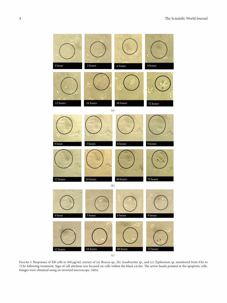
3.2. Morphological Changes of Cancer Cells in Response toExtracts. Obvious changes to KB and ORL-48 cells wereobserved at 24 hr of incubation following treatment with100 𝜇g/mL of Brucea sp. extracts. The black circles markedthe area in which the cells were observed throughout a 72 hrincubation period. Morphological changes include reductionin the size of the cells. The cells gradually become flatand shrunken with the appearance of small vesicle bodies(apoptotic bodies). Similar observations were also madewhen the cells were treated with 100 𝜇g/mL of Azadirachtasp. and Typhonium sp. However, the first sign of apoptoticactivity was observed much later following treatment at 72 hrof incubation (Figures 1 and 2).
3.3. TUNEL Assay. Green fluorescent spots were presentin all the images of KB and ORL-48 extract-treated cells(Figure 3). These observations were indicative of the apop-totic activity of Brucea sp., Azadirachta sp., and Typho-nium sp. extracts on both cancer cells. This IHC assaywas employed to confirm the apoptotic activity induced bythe respective extracts on KB and ORL-48 cells. ApoptoticKB or ORL-48 cells appeared as fluorescence bright green
4 The Scientific World Journal
0 hour 3 hours 6 hours 9 hours
12 hours 24 hours 48 hours 72 hours
(a)
0 hour 6 hours 9 hours
12 hours 24 hours 48 hours 72 hours
3 hours
(b)
0 hour 3 hours 6 hours 9 hours
12 hours 24 hours 48 hours 72 hours
(c)
Figure 1: Responses of KB cells to 100 𝜇g/mL extract of (a) Brucea sp., (b) Azadirachta sp., and (c) Typhonium sp. monitored from 0 hr to72 hr following treatment. Sign of cell attrition was focused on cells within the black circles. The arrow heads pointed at the apoptotic cells.Images were obtained using an inverted microscope. (40x).
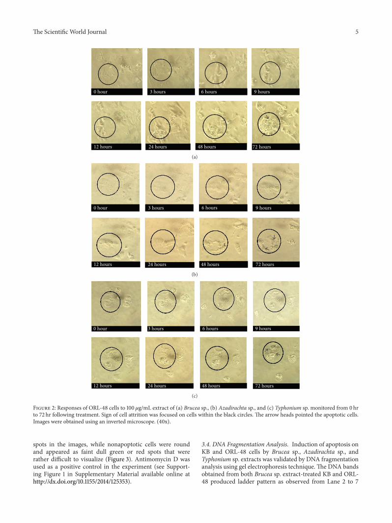
The Scientific World Journal 5
0 hour 3 hours 6 hours 9 hours
12 hours 24 hours 48 hours 72 hours
(a)
0 hour 3 hours 6 hours 9 hours
12 hours 24 hours 48 hours 72 hours
(b)
0 hour 3 hours 6 hours 9 hours
12 hours 24 hours 48 hours 72 hours
(c)
Figure 2: Responses of ORL-48 cells to 100 𝜇g/mL extract of (a) Brucea sp., (b) Azadirachta sp., and (c) Typhonium sp. monitored from 0 hrto 72 hr following treatment. Sign of cell attrition was focused on cells within the black circles. The arrow heads pointed the apoptotic cells.Images were obtained using an inverted microscope. (40x).
spots in the images, while nonapoptotic cells were roundand appeared as faint dull green or red spots that wererather difficult to visualize (Figure 3). Antimomycin D wasused as a positive control in the experiment (see Support-ing Figure 1 in Supplementary Material available online athttp://dx.doi.org/10.1155/2014/125353).
3.4. DNA Fragmentation Analysis. Induction of apoptosis onKB and ORL-48 cells by Brucea sp., Azadirachta sp., andTyphonium sp. extracts was validated by DNA fragmentationanalysis using gel electrophoresis technique.The DNA bandsobtained from both Brucea sp. extract-treated KB and ORL-48 produced ladder pattern as observed from Lane 2 to 7

6 The Scientific World Journal
Negative control KB
(a)
Azadirachta sp. (100𝜇g/mL)
(b)
Brucea sp. (100𝜇g/mL)
(c)
Typhonium sp. (100𝜇g/mL)
(d)
Negative control ORL-48
(e)
Azadirachta sp. (100𝜇g/mL)
(f)
Brucea sp. (100𝜇g/mL)
(g)
Typhonium sp. (100𝜇g/mL)
(h)
Figure 3: Images of fluorescent-stained KB and ORL-48 cells following treatments with 100 𝜇g/mL of Azadirachta sp. ((b), (f)), Brucea sp.((c), (g)), and Typhonium sp. ((d), (h)) at 10x magnification. (a) and (b) were the negative controls for KB and ORL-48 cells, respectively.
1000 900800700600500400300200
100
M 1 2 3 4 5 6 7(bp)
(a)
1000 900800700600500400300200
100
M 1 2 3 4 5 6 7(bp)
(b)
Figure 4: DNA band patterns of (a) KB and (b) ORL-48 cells treated with various concentrations of Brucea sp. Lane 1: negative control; Lanes2 to 7 were bands of cancer cells treated with 1.0, 10.0, 25.0, 50.0, 75.0, and 100.0𝜇g/mL extract of Brucea sp.
(Figure 4). A ladder formation was used to indicate thatthe DNA has undergone fragmentation, and each fragmentcorresponded to a band in the ladder. A similarDNAbandingpattern but of lower intensity was also observed on KBand ORL-48 cells following treatment with the extracts ofTyphonium sp. andAzadirachta sp. (Supporting Figures 2 and3).
4. Discussion
Theability of cancerous cell populations to expand in numberis determined by their ability to proliferate and, inmany cases,to form tumour.Three plants species with reputed anticancer
potentials among the local practitioners were selected for thisresearch. Other than topical application of paste preparedfrom these plants to the tumour area, consumption ofdecoctions prepared from the plants is also being practiced.Despite their popular usage as local medicines, informationon the exact formulation and amount of plant specimenused in the treatments is not standardised. Results obtainedfrom this study revealed information with regards to theability of aqueous extracts of Brucea sp., Typhonium sp., andAzadirachta sp. to inhibit the proliferation of cancer cell linesoriginating from the oral mucosa.
Antiproliferative assessment showed that the prolifera-tion of both KB and ORL-48 cells was effectively inhibited
The Scientific World Journal 7
by the extract of Brucea sp. with activity on the lattercells being more prominent (Table 1). Brucea sp. inducedmorphological effects on both KB and ORL-48 at 24 hr oftreatment compared to at 72 hr forAzadirachta sp. andTypho-nium sp. Among the signs of cells going through attritionprocess is the appearance of smaller, flattened, and shrunkencells (Figures 1 and 2). These are among the characteristicsof cells undergoing apoptosis. Cell death can be achievedvia apoptosis as well as necrosis. In the search for activecompoundswith anticancer activity, an agent that induces celldeath via apoptosis is preferred [2].
Apoptosis is a programmed cell death that removesor eliminates targeted unwanted or dead cells. Other thanshrunken cells, characteristics of apoptotic cells includecondensation of the cytoplasm and nucleus, aggregationof chromatin, and formation of membrane-bound vesiclesknown as apoptotic bodies [14]. Necrosis on the other handrefers to a pathological activity. Necrosis is known to beproinflammatory and is marked by swelling of the cell thatis often accompanied by chromatin condensation. Necroticcells eventually experienced cellular and nuclear lysis alongwith subsequent inflammation [15], which would be unfavor-able for an anticancer agent. In this study, results obtainedfrom the IHC staining procedure suggested the apoptoticactivity of Brucea sp., Typhonium sp., and Azadirachta sp.(Figure 3). Green fluorescent spots indicating the presence ofapoptotic cells were observed when the extract-treated cellswere examined under a fluorescentmicroscope (Figure 3). Asanticipated with the strong antiproliferative activity of Bruceasp., the population of green spots in images of Brucea-treatedcells was higher compared to cells treated with the other twoextracts.
Verification of the apoptotic activity of Brucea sp. wascarried out based on the pattern of DNA bands producedfrom a gel electrophoresis. In apoptosis, cells are lysedgradually and systematically to produce membrane-boundapoptotic bodies, which was suggested to play a major role insuppressing inflammatory responses to other neighbouringcells. Apoptotic bodies or cells which underwent apoptosisproduce a specific pattern of DNA fragments with the mul-tiples of 200 bp due to specific action of activated nucleases[16]. These isolated fragments produced bands in a ladderpattern, in contrast with the smeared pattern produced fromnecrosis activity (Figure 4). Results obtained in this studyin a way supported the various claims made by researcheson the anticancer properties of this plant [17–19]. Furtherstudies are being carried out to identify the active principle ofBrucea sp. extract. Thus, it can be concluded that the strongantiproliferative activity of Brucea sp. extract on oral cancercells suggests its possible development as an anticancer agent.The mode of action of Brucea sp. was by the induction ofapoptotic activity on cancer cells.
Conflict of Interests
The authors declare that there is no conflict of interestsregarding the publication of this paper.
Acknowledgments
This study was financially supported by the High ImpactResearch MoE Grant UM.C/625/1/HIR/MoE/15, Universityof Malaya Research Grant (UMRG020/09HTM), and Post-graduate Research Fund (PS164/2011B).
References
[1] D.Hanahan andR.A.Weinberg, “Thehallmarks of cancer,”Cell,vol. 100, no. 1, pp. 57–70, 2000.
[2] V. Kumar, R. S. Cotran, and S. L. Robbins, Basic Pathology, WBSaunders, Philadelphia, Pa, USA, 6th edition, 2006.
[3] P. Li, Y. Yin, D. Li, W. S. Kim, and G. Wu, “Amino acids andimmune function,”The British Journal of Nutrition, vol. 98, no.2, pp. 237–252, 2007.
[4] J. Samy, M. Sugumaran, and L. W. Kate Lee, Herbs of Malaysia,Marshall Cavendish Sdn Bhd, Malaysia, 2009.
[5] L. A. Anderson, A. Harris, and J. D. Phillipson, “Production ofcytotoxic canthin-6-one alkaloids by Ailanthus altissima plantcell cultures,” Journal of Natural Products, vol. 46, no. 3, pp. 374–378, 1983.
[6] J. Polonsky, J. Varenne, T. Prange, and C. Pascard, “Antileukae-mic quassinoids: structure (X-ray analysis) of bruceine C andrevised structure of bruceantinol,” Tetrahedron Letters, vol. 21,no. 19, pp. 1853–1856, 1980.
[7] M. S. Mulla and T. Su, “Activity and biological effects of neemproducts against arthropods of medical and veterinary impor-tance,” Journal of the American Mosquito Control Association,vol. 15, no. 2, pp. 133–152, 1999.
[8] M. M. el-Shazly and E. D. el-Sharnoubi, “Toxicity of a Neem(Azadirachta indica) insecticide to certain aquatic organisms.,”Journal of the Egyptian Society of Parasitology, vol. 30, no. 1, pp.221–231, 2000.
[9] B. K. Das, S. C. Mukherjee, B. B. Sahu, and G. Murjani,“Neem (Azadirachta indica) extract as an antibacterial agentagainst fish pathogenic bacteria,” Indian Journal of ExperimentalBiology, vol. 37, no. 11, pp. 1097–1100, 1999.
[10] K. H. Teo and B. I. Ch’ng Beng,Cancer—YetThey Live, CACare,Penang, Malaysia, 2nd edition, 1999.
[11] S. Hamid, K. P. Lim, R. B. Zain et al., “Establishment andcharacterization of Asian oral cancer cell lines as in vitromodelsto study a disease prevalent in Asia,” International Journal ofMolecular Medicine, vol. 19, no. 3, pp. 453–460, 2007.
[12] E. Borenfreund and J. A. Puerner, “A simple quantitativeprocedure using monolayer cultures for cytotoxicity assays(HTD/NR-90),” Journal of Tissue Culture Methods, vol. 9, no. 1,pp. 7–9, 1985.
[13] A. R. Fathilah, R. Sujata, A. W. Norhanom, and M. I. Adenan,“Antiproliferative activity of aqueous extract of Piper betle L.and Psidium guajava L. on KB and HeLa cell lines,” Journal ofMedicinal Plants Research, vol. 4, no. 11, pp. 987–990, 2010.
[14] J. J. Cohen, “Apoptosis,” Immunology Today, vol. 14, no. 3, pp.126–130, 1993.
[15] A. H. Wyllie, J. F. R. Kerr, and A. R. Currie, “Cell death: thesignificance of apoptosis,” International Review of Cytology, vol.68, pp. 251–306, 1980.
[16] C. D. Bortner, N. B. E. Oldenburg, and J. A. Cidlowski, “The roleof DNA fragmentation in apoptosis,” Trends in Cell Biology, vol.5, no. 1, pp. 21–26, 1995.

8 The Scientific World Journal
[17] H. M. Chang and P. P. But, “Yadanzi,” in Pharmacology andApplications of Chinese MateriaMedica. Vol II, World Scientific,Singapore, 1987.
[18] F. Y. Lau, C. H. Chui, R. Gambari et al., “Antiproliferativeand apoptosis-inducing activity of Brucea javanica extract onhuman carcinoma cells,” International Journal of MolecularMedicine, vol. 16, no. 6, pp. 1157–1162, 2005.
[19] J. Kim, E. K. Lau, L. Pan, and E. J. Carcache De Blanco, “NF-𝜅Binhibitors from Brucea javanica exhibiting intracellular effectson reactive oxygen species,” Anticancer Research, vol. 30, no. 9,pp. 3295–3300, 2010.

Submit your manuscripts athttp://www.hindawi.com
Stem CellsInternational
Hindawi Publishing Corporationhttp://www.hindawi.com Volume 2014
Hindawi Publishing Corporationhttp://www.hindawi.com Volume 2014
MEDIATORSINFLAMMATION
of
Hindawi Publishing Corporationhttp://www.hindawi.com Volume 2014
Behavioural Neurology
EndocrinologyInternational Journal of
Hindawi Publishing Corporationhttp://www.hindawi.com Volume 2014
Hindawi Publishing Corporationhttp://www.hindawi.com Volume 2014
Disease Markers
Hindawi Publishing Corporationhttp://www.hindawi.com Volume 2014
BioMed Research International
OncologyJournal of
Hindawi Publishing Corporationhttp://www.hindawi.com Volume 2014
Hindawi Publishing Corporationhttp://www.hindawi.com Volume 2014
Oxidative Medicine and Cellular Longevity
Hindawi Publishing Corporationhttp://www.hindawi.com Volume 2014
PPAR Research
The Scientific World JournalHindawi Publishing Corporation http://www.hindawi.com Volume 2014
Immunology ResearchHindawi Publishing Corporationhttp://www.hindawi.com Volume 2014
Journal of
ObesityJournal of
Hindawi Publishing Corporationhttp://www.hindawi.com Volume 2014
Hindawi Publishing Corporationhttp://www.hindawi.com Volume 2014
Computational and Mathematical Methods in Medicine
OphthalmologyJournal of
Hindawi Publishing Corporationhttp://www.hindawi.com Volume 2014
Diabetes ResearchJournal of
Hindawi Publishing Corporationhttp://www.hindawi.com Volume 2014
Hindawi Publishing Corporationhttp://www.hindawi.com Volume 2014
Research and TreatmentAIDS
Hindawi Publishing Corporationhttp://www.hindawi.com Volume 2014
Gastroenterology Research and Practice
Hindawi Publishing Corporationhttp://www.hindawi.com Volume 2014
Parkinson’s Disease
Evidence-Based Complementary and Alternative Medicine
Volume 2014Hindawi Publishing Corporationhttp://www.hindawi.com


![Antiproliferative and Apoptosis-inducing Effects of Abrus … · 2019-01-22 · of apoptosis in the development of therapeutic agents for treating cancer[8]. The homeostasis in eukaryotic](https://static.fdocuments.net/doc/165x107/5e7531a7e0b7db69cc34d0b0/antiproliferative-and-apoptosis-inducing-effects-of-abrus-2019-01-22-of-apoptosis.jpg)

