Research Article Antioxidant Activity of Extract and Its Major Constituents from Okra...
10
Research Article Antioxidant Activity of Extract and Its Major Constituents from Okra Seed on Rat Hepatocytes Injured by Carbon Tetrachloride Lianmei Hu, 1 Wenlan Yu, 1 Ying Li, 1 Nagendra Prasad, 2 and Zhaoxin Tang 1 1 College of Veterinary Medicine, South China Agricultural University, Guangzhou 510642, China 2 Chemical Engineering Discipline, School of Engineering, Monash University, 46150 Bandar Sunway, Malaysia Correspondence should be addressed to Nagendra Prasad; [email protected] and Zhaoxin Tang; [email protected] Received 2 January 2014; Accepted 16 January 2014; Published 27 February 2014 Academic Editor: Jos´ e Carlos Tavares Carvalho Copyright © 2014 Lianmei Hu et al. is is an open access article distributed under the Creative Commons Attribution License, which permits unrestricted use, distribution, and reproduction in any medium, provided the original work is properly cited. e antioxidant activities and protective effects of total phenolic extracts (TPE) and their major components from okra seeds on oxidative stress induced by carbon tetrachloride (CCl 4 ) in rat hepatocyte cell line were investigated. e major phenolic compounds were identified as quercetin 3-O-glucosyl (1→6) glucoside (QDG) and quercetin 3-O-glucoside (QG). TPE, QG, and QDG from okra seeds exhibited excellent reducing power and free radical scavenging capabilities including , -diphenyl--picrylhydrazyl (DPPH), superoxide anions, and hydroxyl radical. Overall, DPPH radical scavenging activity and reducing power of QG and QDG were higher than those of TPE while superoxide and hydroxyl radical scavenging activities of QG and TPE were higher than those of QDG. Furthermore, TPE, QG, and QDG pretreatments significantly alleviated the cytotoxicity of CCl 4 on rat hepatocytes, with attenuated lipid peroxidation, increased SOD and CAT activities, and decreased GPT and GOT activities. e protective effects of TPE and QG on rat hepatocytes were stronger than those of QDG. However, the cytotoxicity of CCl 4 on rat hepatocytes was not affected by TPE, QG, and QDG posttreatments. It was suggested that the protective effects of TPE, QG, and QDG on rat hepatocyte against oxidative stress were related to the direct antioxidant capabilities and the induced antioxidant enzymes activities. 1. Introduction Oxidation is essential for living organisms. Reactive oxygen species (ROS) also are produced during oxidation [1]. Organ- isms can maintain a dynamic equilibrium between produc- tion and elimination of ROS in normal conditions. However, when organisms are subjected to stress conditions, this equi- librium is disrupted. Excessive accumulation of ROS will result in cellular injuries, including lipid peroxidation, pro- tein oxidation, and DNA damage, which are involved in development of a variety of diseases including cellular aging, mutagenesis, carcinogenesis, hepatopathies, diabetes, and neurodegeneration [2]. erefore, cellular antioxidant defense systems play important roles in counteracting these deleterious effects of ROS. Almost all organisms possess antioxidant defense systems including antioxidant enzymes and nonenzymatic antioxi- dants. However, these systems are insufficient to prevent the damage entirely in some cases [3]. Plants are the most impor- tant source of natural antioxidants [4]. Phenolic compounds or polyphenols, which consist of secondary metabolites, constitute a wide and complex array of phytochemicals that exhibit antioxidant actions. Epidemiological studies have indicated that regular consumption of foods rich in phenolic compounds is associated with reduced risk of cardiovascular diseases, neurodegenerative diseases, and certain cancers [5, 6]. ese phenolic compounds hold promising potentials in the development of health foods, nutritional supplements, and herbal medicines for the application as antioxidants and ROS-related disease chemopreventive agents. Okra (Hibiscus esculentus L.), also known as lady’s fin- ger and gumbo, belongs to Malvaceae family, which is distributed widely in Africa, Asia, Southern Europe, and America [7]. e plant is a common vegetable in most regions for its nutrition value. Okra pod contains thick slimy polysaccharides, which are used to thicken soups and stews, Hindawi Publishing Corporation BioMed Research International Volume 2014, Article ID 341291, 9 pages http://dx.doi.org/10.1155/2014/341291
Transcript of Research Article Antioxidant Activity of Extract and Its Major Constituents from Okra...

Research ArticleAntioxidant Activity of Extract and Its Major Constituents fromOkra Seed on Rat Hepatocytes Injured by Carbon Tetrachloride
Lianmei Hu,1 Wenlan Yu,1 Ying Li,1 Nagendra Prasad,2 and Zhaoxin Tang1
1 College of Veterinary Medicine, South China Agricultural University, Guangzhou 510642, China2 Chemical Engineering Discipline, School of Engineering, Monash University, 46150 Bandar Sunway, Malaysia
Correspondence should be addressed to Nagendra Prasad; [email protected] Zhaoxin Tang; [email protected]
Received 2 January 2014; Accepted 16 January 2014; Published 27 February 2014
Academic Editor: Jose Carlos Tavares Carvalho
Copyright © 2014 Lianmei Hu et al. This is an open access article distributed under the Creative Commons Attribution License,which permits unrestricted use, distribution, and reproduction in any medium, provided the original work is properly cited.
The antioxidant activities and protective effects of total phenolic extracts (TPE) and their major components from okra seeds onoxidative stress induced by carbon tetrachloride (CCl
4) in rat hepatocyte cell line were investigated.Themajor phenolic compounds
were identified as quercetin 3-O-glucosyl (1 → 6) glucoside (QDG) and quercetin 3-O-glucoside (QG). TPE, QG, and QDG fromokra seeds exhibited excellent reducing power and free radical scavenging capabilities including 𝛼, 𝛼-diphenyl-𝛽-picrylhydrazyl(DPPH), superoxide anions, and hydroxyl radical. Overall, DPPH radical scavenging activity and reducing power of QG and QDGwere higher than those of TPE while superoxide and hydroxyl radical scavenging activities of QG and TPE were higher than thoseof QDG. Furthermore, TPE, QG, and QDG pretreatments significantly alleviated the cytotoxicity of CCl
4on rat hepatocytes, with
attenuated lipid peroxidation, increased SOD and CAT activities, and decreased GPT and GOT activities. The protective effects ofTPE and QG on rat hepatocytes were stronger than those of QDG. However, the cytotoxicity of CCl
4on rat hepatocytes was not
affected by TPE, QG, and QDG posttreatments. It was suggested that the protective effects of TPE, QG, and QDG on rat hepatocyteagainst oxidative stress were related to the direct antioxidant capabilities and the induced antioxidant enzymes activities.
1. Introduction
Oxidation is essential for living organisms. Reactive oxygenspecies (ROS) also are produced during oxidation [1]. Organ-isms can maintain a dynamic equilibrium between produc-tion and elimination of ROS in normal conditions. However,when organisms are subjected to stress conditions, this equi-librium is disrupted. Excessive accumulation of ROS willresult in cellular injuries, including lipid peroxidation, pro-tein oxidation, and DNA damage, which are involved indevelopment of a variety of diseases including cellularaging, mutagenesis, carcinogenesis, hepatopathies, diabetes,and neurodegeneration [2]. Therefore, cellular antioxidantdefense systems play important roles in counteracting thesedeleterious effects of ROS.
Almost all organisms possess antioxidant defense systemsincluding antioxidant enzymes and nonenzymatic antioxi-dants. However, these systems are insufficient to prevent the
damage entirely in some cases [3]. Plants are the most impor-tant source of natural antioxidants [4]. Phenolic compoundsor polyphenols, which consist of secondary metabolites,constitute a wide and complex array of phytochemicals thatexhibit antioxidant actions. Epidemiological studies haveindicated that regular consumption of foods rich in phenoliccompounds is associated with reduced risk of cardiovasculardiseases, neurodegenerative diseases, and certain cancers [5,6]. These phenolic compounds hold promising potentials inthe development of health foods, nutritional supplements,and herbal medicines for the application as antioxidants andROS-related disease chemopreventive agents.
Okra (Hibiscus esculentus L.), also known as lady’s fin-ger and gumbo, belongs to Malvaceae family, which isdistributed widely in Africa, Asia, Southern Europe, andAmerica [7]. The plant is a common vegetable in mostregions for its nutrition value. Okra pod contains thick slimypolysaccharides, which are used to thicken soups and stews,
Hindawi Publishing CorporationBioMed Research InternationalVolume 2014, Article ID 341291, 9 pageshttp://dx.doi.org/10.1155/2014/341291

2 BioMed Research International
as an egg white substitute, and as a fat substitute in chocolatebar cookies and in chocolate frozen dairy dessert [7]. Okraseed is rich in protein and unsaturated fatty acids such aslinoleic acid [8]. In some countries, okra also is used infolk medicine as antiulcerogenic, gastroprotective, diureticagents [9]. In addition, Arapitsas [10] reported that okraseed was rich in phenolic compounds, mainly composed offlavonol derivatives and oligomeric catechins, suggesting thatit might possess some antioxidant properties. However, littleinformation on antioxidant capabilities of major phenoliccompounds from okra seed is available.
Carbon tetrachloride (CCl4), a well-known environmen-
tal biohazard, can be particularly toxic to liver. CCl4-
induced hepatic injury, a classic experimental model, hasbeen extensively used to evaluate the potential of drugs anddietary antioxidants against the oxidative damage [11, 12].The objectives of the study were to evaluate the antioxidantactivity of major phenolic compounds in vitro and theireffects on oxidative stress induced by carbon tetrachloride(CCl4) in rat hepatocyte cell line.
2. Materials and Methods
2.1. Plant Materials. Okra pods (Hibiscus esculentus L.) wereharvested from a commercial orchard in Guangzhou, Guang-dong, China. The fruit were manually separated, and theseeds were collected, sun-dried, and pulverized to a powder.Thematerials were stored at room temperature in a desiccatoruntil use.
2.2. Extraction, Isolation, and Purification. Dried seed pow-der ofH. esculentuswas exhaustively extractedwithmethanolat temperature (25–32∘C) for 3 days. The extracts were con-centrated with a rotary evaporator (RE52AA, Yarong Equip-ment Co., Shanghai, China) under reduced pressure at 55∘Cand then fractionated sequentially by petroleum ether andEtOAc.The extraction with petroleum ether was to eliminatethe pigments. EtOAc extract was obtained by evaporationunder reduced pressure and then subjected to purification bysilica gel column using CHCl
3-MeOH solvent system with
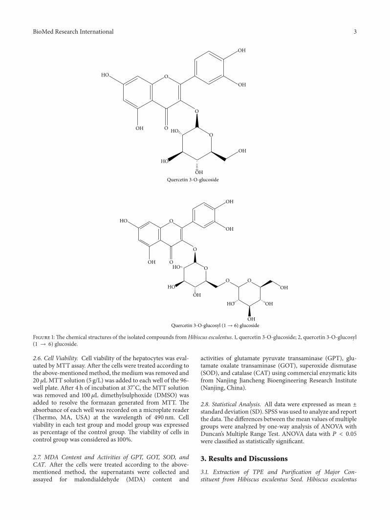
increased polarity (0 : 100–60 : 40) to yield eight fractions.We were only interested in the major phenolic compounds.Therefore, the largest fractions were further purified by silicagel column and Sephadex LH-20 to yield compound 1 andcompound 2, respectively. Compound 1 and compound 2were identified as quercetin 3-O-glucosyl (1 → 6) glucosideand quercetin 3-O-glucoside (Figure 1), by comparison ofexperimental and literature NMR data [13].
2.3. Determination of Total Phenol Content. Total phenolcontent of TPE inH. esculentuswas determined by the Folin-Ciocalteu method [14]. Chlorogenic acid was used as a stan-dard. The total phenol content was determined in triplicateand expressed as chlorogenic acid equivalents in mg/g ofplant material.
2.4. Evaluation of Antioxidant Activities. Antioxidant capa-bilities of total phenolic extracts and their major component
fromokrawere evaluated according to the describedmethodsby Duan et al. [15] with minor modifications. To evaluateDPPH scavenging, 0.1mL various concentrations of sampleswere mixed with 2.9mL 0.1mM DPPH-methanol solution.After 30min of incubation at 25∘C in the dark, the absorbanceat 517 nm was measured. DPPH radical scavenging activityof the samples was calculated using the following formula:DPPHscavenging activity (%) = [1−(absorbance of sample−absorbance of blank)/absorbance of control] × 100.
Superoxide radicals were generated by illuminating asolution containing riboflavin. The photoinduced reactionswere performed at about 4000 lux. 25 𝜇L various concentra-tions of sample were mixed with 3mL reaction buffer [1.3𝜇Mriboflavin, 13mM methionine, 63 𝜇M nitroblue tetrazolium(NBT), and 100 𝜇M EDTA, pH 7.8]. The reaction solutionwas illuminated at 25∘C for 15min. The reaction mixturewithout sample was used as a control.The scavenging activitywas calculated as follows: scavenging activity (%) = (1 −absorbance of sample/absorbance of control) × 100.
Hydroxyl radical scavenging activity was determinedby evaluating inhibitory effect of samples on deoxyribosedegradation. 0.2mL different concentrations of samples wereincubated with 1mL reaction buffer (100𝜇M FeCl
3, 104 𝜇M
EDTA, 1.5mMH2O2, 2.5mM deoxyribose, and 100 𝜇M L-
ascorbic acid, pH 7.4) for 1 h at 37∘C. After adding 1mL0.5% 2-thiobarbituric acid and 1mL 2.8% trichloroacetic acid,the mixture was heated for 30min at 80∘C and then cooledon ice. The absorbance at 532 nm was measured. Percentinhibition of deoxyribose degradation was calculated as (1 −absorbance of sample/absorbance of control) × 100.
For the reducing power, 0.2mL various concentrations ofsamples were incubated with 2.0mL 200mM sodium phos-phate buffer (pH 6.6) and 2.0mL 1% potassium ferricyanideat 50∘C for 20min. After adding 2.0mL 10% trichloroaceticacid (w/v), the mixture was centrifuged at 4000×g for 5min.2.0mL supernatant was mixed with 2.4mL 0.016% ferricchloride, and the absorbance at 700 nm was measured. Ahigher absorbance indicates a higher reducing power.
2.5. Cell Culture andCell Treatment. BRL-3A (rat hepatocyte)cell culture was obtained from Experimental Animal Centerof Southern Medical University (Guangzhou, China). Thecell line was cultured in RPMI-1640 medium containing10% foetal bovine serum (FBS), 100U/mL penicillin, and100mg/mL streptomycin at 37∘C in an incubator of humid-ified air with 5% CO
2. Hepatocytes were seeded onto 24-
well plates at 1 × 10⁵ cells/well and cultured for 24 h at37∘C under 5% CO
2. Later, the medium was removed and
the hepatocytes were subjected to the following treatments:(1) pretreatment, the hepatocytes were first incubated withgrowth medium containing 100 𝜇g/mL TPE, QG, or QDGfor 4 h, and then the cells were washed and incubated withfresh growth medium containing 10mMCCl
4for another
4 h; (2) posttreatment, the hepatocytes were first incubatedwith growth medium containing 10mMCCl
4for 4 h, and
then the cells were washed and incubated with fresh growthmediumcontaining 100𝜇g/mLTPE,QG, orQDG for another4 h.
BioMed Research International 3
HO
HO
HO
O
O
OOOH
OH
OH
OH
OH
HO
HO
HO
HO
O
O
O
O O
OOH
OH
OH
OH
OH
OH
OH
Quercetin 3-O-glucoside
Quercetin 3-O-glucosyl (1 → 6) glucoside
Figure 1: The chemical structures of the isolated compounds fromHibiscus esculentus. 1, quercetin 3-O-glucoside; 2, quercetin 3-O-glucosyl(1 → 6) glucoside.
2.6. Cell Viability. Cell viability of the hepatocytes was eval-uated by MTT assay. After the cells were treated according tothe above-mentionedmethod, the mediumwas removed and20𝜇LMTT solution (5 g/L) was added to each well of the 96-well plate. After 4 h of incubation at 37∘C, the MTT solutionwas removed and 100 𝜇L dimethylsulphoxide (DMSO) wasadded to resolve the formazan generated from MTT. Theabsorbance of each well was recorded on a microplate reader(Thermo, MA, USA) at the wavelength of 490 nm. Cellviability in each test group and model group was expressedas percentage of the control group. The viability of cells incontrol group was considered as 100%.
2.7. MDA Content and Activities of GPT, GOT, SOD, andCAT. After the cells were treated according to the above-mentioned method, the supernatants were collected andassayed for malondialdehyde (MDA) content and
activities of glutamate pyruvate transaminase (GPT), glu-tamate oxalate transaminase (GOT), superoxide dismutase(SOD), and catalase (CAT) using commercial enzymatic kitsfrom Nanjing Jiancheng Bioengineering Research Institute(Nanjing, China).
2.8. Statistical Analysis. All data were expressed as mean ±standard deviation (SD). SPSS was used to analyze and reportthe data.The differences between themean values of multiplegroups were analyzed by one-way analysis of ANOVA withDuncan’s Multiple Range Test. ANOVA data with 𝑃 < 0.05were classified as statistically significant.
3. Results and Discussions
3.1. Extraction of TPE and Purification of Major Con-stituent from Hibiscus esculentus Seed. Hibiscus esculentus

4 BioMed Research International
50 100 200 400
DPP
H ra
dica
l sca
veng
ing
activ
ity (%
)
0
20
40
60
80
100
(a)
12.5 25 50 100
Supe
roxi
de sc
aven
ging
ac
tivity
(%)
0
20
40
60
80
100
(b)
BHTQuerc.TPE
QGQDG
12.5 25 50 100
Hyd
roxy
l rad
ical
scav
engi
ng
activ
ity (%
)
0
20
40
60
80
100
120
Concentration (𝜇g/mL)
(c)
BHTQuerc.TPE
QGQDG
12.5 25 50 100
Abso
rban
ce at
700
nm
0.0
0.2
0.4
0.6
0.8
1.0
1.2
1.4
Concentration (𝜇g/mL)
(d)
Figure 2: Free radical scavenging activities against DPPH radical (a), superoxide radical (b), and hydroxyl radical (c) and reducing power(d) of extract and isolated quercetin 3-O-glucoside and quercetin 3-O-diglucoside from Hibiscus esculentus.
seed was subjected to extraction with methanol and thensequential fractionation by petroleum ether and EtOAc.The extraction with petroleum ether was to eliminate thepigments. The EtOAc-soluble fraction was designated astotal phenol extracts (TPE). The content of phenolic com-pounds in dry okra seed was 28.1mg/g. Further, total phe-nol extracts (TPE) were purified and two major phenoliccompounds were obtained and identified as quercetin 3-O-glucosyl (1 → 6) glucoside (QDG) and quercetin 3-O-glucoside (QG) (Figure 1), by comparison of experimentaland literature NMR data [13], which was consistent withthe result reported by Arapitsas [10]. However, Atawodi etal. reported that quercetin glucoside was the only majorpolyphenol composition in okra seed [16]. The inconsistencemight be associated with the differences in climate conditionsof cultivation and/or the variety analyzed.
3.2. Antioxidant Activity of Total Phenolic Extracts (TPE)and Their Major Components from Okra Seeds In Vitro. Freeradical scavenging activity is the most important mechanismby which antioxidants inhibit lipid peroxidation [1]. In thisstudy, free radical scavenging activity against DPPH radical,
superoxide anions, and hydroxyl radical was analyzed toevaluate the antioxidant activities of total phenolic extracts(TPE) and their major components from okra seeds. Inaddition, reducing power, which exerts antioxidant action bybreaking the free radical chain by donating a hydrogen atom,was investigated [15].
DPPH is a stable, purple, nitrogen-centered, and syn-thetic-free radical with the maximum wavelength of 517 nm.When the DPPH free radical is quenched, the color willfade away. The scavenging activity against DPPH free radicalhas been extensively used to evaluate antioxidant activity ofplant extracts [17]. Figure 2(a) shows the scavenging activitiesagainst DPPH free radical of TPE, QG, and QDG from okraseed and BHT and quercetin. All tested samples exhibitedthe scavenging activities against DPPH free radical in adose-dependent manner. The positive control quercetin andBHT had the highest and lowest scavenging activity againstDPPH free radical, respectively. QG andQDGhave almost anequivalent scavenging activity against DPPH free radical, buthigher than TPE. At 100 𝜇g/mL, the scavenging effects were18.3%, 44.1%, 24.6%, 34.5%, and 37.3% for BHT, quercetin,TPE, QG, and QDG, respectively.

BioMed Research International 5
Superoxide anion, produced by a number of cellular reac-tions or enzymes, including iron-catalyzed Fenton reaction,lipoxygenases, peroxidase, NADPH oxidase, and xanthineoxidase, is themost important in organisms and is involved inthe formation of other cell-damaging free radicals [1]. In thepresent study, TPE, QG, andQDG showed higher scavengingactivity against superoxide anion thanDPPHradical scaveng-ing activity. Similarly, superoxide anion scavenging activitiesof TPE, QG, and QDG were stronger than that of BHT, butweaker than that of quercetin. At 50𝜇g/mL and 100 𝜇g/mLof concentrations, the free radical scavenging activity of TPEwas higher than that of QDG, but lower than that of QG(Figure 2(b))
Hydroxyl radical can be formed by the Fenton reaction inthe presence of reduced transition metals and H
2O2, which
is known to be the most reactive radical and is thought toinitiate cell damage in vivo [18]. Similar to superoxide anionscavenging activity, TPE, QG, and QDG from okra seedsexhibited excellent hydroxyl radical scavenging activity in thefollowing order: QG > TPE > QDG > BHT (Figure 2(c)).
Reducing power is widely used to evaluate the antioxidantactivity of plant extracts.The reducing property indicates thatthe antioxidant compounds are electron donors and reducethe oxidized intermediates of lipid peroxidation process [19].As shown in Figure 2(d), TPE,QG, andQDG fromokra seedsshowed much higher reducing power than BHT, suggestingthat TPE, QG, and QDG possessed a stronger electrondonating capacity. However, the reducing power of TPE, QG,and QDG was lower than that of quercetin at 100 𝜇g/mLof concentration. Furthermore, the reducing power of theextract and its major constituents from okra seed were in thefollowing order: QG > QDG > TPE.
Overall, DPPH radical scavenging activity and reducingpower of QG and QDG were higher than those of TPE;superoxide and hydroxyl radical scavenging activities of QGand TPE were higher than those of QDG; reducing power,superoxide anion, and hydroxyl radical scavenging activitiesof QG were higher than those of QDG. Moreover, theantioxidant activities investigated, except hydroxyl radicalscavenging activity of quercetin, were higher than those ofQG andQDG. It appeared that addition of glycosyl decreasedthe scavenging activity againstDPPH free radical of quercetinand its derivatives. Some similar results were obtained byother researchers. Omololu et al. reported that quercetin hadstronger scavenging activity against DPPH free radical thanits rhamnosyl glucoside derivative [20]. Hopia and Heinoneninvestigated the antioxidant activities of quercetin and itsselected glycosides in bulk methyl linoleate oxidized at 40∘Cand found that the order of activity of quercetin and itsderivatives was quercetin > isoquercitrin > rutin [21]. Sunet al. also found that rhamnosidase could change rutin inasparagus juice to quercetin-3-glucoside, which has a higherantioxidant activity than rutin [22]. The possible reasons areas follows: (1) the steric effect by increased glycosylationdecreased the accessibility of free radicals to flavonoid antiox-idants; (2) the presence of a free 3-hydroxyl group in theC-ring is a requirement for the maximal radical scavengingactivity of flavonoids and the substitution of the hydroxylgroup by glycoside resulted in the decreases in free radical
Cel
l via
bilit
y (%
of c
ontro
l)
0
20
40
60
80
100
120
Ctrl
p-TP
E
p-Q
G
p-Q
DG
a-TP
E
a-Q
G
a-Q
DG
CCL 4
+CCL4 +CCL4
Figure 3: Effect of extract and isolated quercetin 3-O-glucosideand quercetin 3-O-diglucoside from Hibiscus esculentus on CCl
4-
induced cytotoxicity in rat hepatocytes. In the tick label, p meanspretreatment while a means posttreatment.
scavenging ability and chelating activity to transition metalions [21, 23]. In addition, QG and QDG played a dominantrole in scavenging activity against superoxide anion andhydroxyl radical of total phenolic compounds of okra seed. Insome plant flavonoids, quercetin 3-O-glucoside contributedto the major of antioxidant activities [24–27].
3.3. Effect of TPE, QG, and QDG on Cell Viability of Hep-atocytes Injured by CCl
4. It is well known that cell damage
induced by reactive oxygen species (ROS) is an importantmechanism of hepatotoxicity [28]. Tetrachloride (CCl
4) has
been widely used to study liver injury induced by ROS inthe mouse model. The mechanism is involved in free rad-icals generated during CCl
4metabolism by hepatic cellular
cytochrome P450, including trichloromethyl (CCl3and/or
CCl3O2) and oxygen-centered lipid radicals (LO and/or
LOO), which initiate the process of lipid peroxidation [29].Kikkawa et al. suggested that in vitro primary cell culturesystemwould be sufficient to detect hepatotoxicity in the earlystage of drug discovery according to the relevance of in vitrosystem to in vivo system from some biomarkers related tooxidative stress by carbon tetrachloride [12]. In the presentstudy, CCl
4treatment resulted in a significant decrease in
cell viability of the hepatocytes. After 4 h of incubation, thecell viability decreased to 8.3% compared with the control,indicating that the hepatocytes were severely injured. TPE,QG, and QDG pretreatments efficiently alleviated oxidativeinjury of the cell induced by CCl
4. The cell viabilities
of hepatocytes pretreated with TPE, QG, and QDG were76.2%, 72.5%, and 59.9%, respectively (Figure 3). However,posttreatment with TPE, QG, and QDG had no significantprotective effect on oxidative injury of the cells caused byCCl4. These results suggest that the extract and its major
constituents can be potentially used for preventing ratherthan curing liver diseases in mammals. A similar result alsowas reported in fish hepatocytes by Yin et al. [30].
Antioxidants play an important role in protecting againstCCl4-induced liver injury. There were some reports on the

6 BioMed Research International
protective effects of various natural products against CCl4-
induced liver injury [30–36]. The protective effects might berelated to the directed superoxide anion and hydroxyl radicalscavenging activity. In our study, superoxide anion andhydroxyl racial scavenging activities were in the followingorder: QG > TPE > QDG, which was inconsistent with theprotective effects. Possibly, the discrepancy could be relatedto differential uptake by hepatocytes or synergistic effects.Boyer et al. [37] reported that there existed a great differencein uptake of quercetin aglycon and quercetin 3-glucoside aspurified compounds and from whole onion and apple peelextracts by Caco-2 cells. Yang and Liu [38] also found thatapple extracts plus quercetin 3-beta-d-glucoside combinationpossess a synergistic effect in MCF-7 cell proliferation.
3.4. Effect of TPE, QG, and QDG on Lipid Peroxidation andActivities of GPT and GOT in Hepatocytes Injured by CCl
4.
Malondialdehyde (MDA), a lipid peroxidized product, canreflect the extent of lipid peroxidation induced by oxidativestress. As shown in Figure 4, a significant increase in MDAlevel was observed in the CCl
4-treated hepatocytes compared
with the control hepatocytes. However, TPE, QG, and QDGtreatments at 100 𝜇g/mL significantly decreased the level oflipid peroxidation induced byCCl
4.The results were in accor-
dance with the cell viability, indicating that the protectiveeffects of TPE, QG, and QDG on CCl
4-induced hepatocytes
injury were related to the alleviated lipid peroxidation.GPT and GOT were widely used to evaluate liver damage
by CCl4[30]. In the present study, significantly elevated GPT
and GOT activities were observed in the supernatants of theCCl4-treated hepatocytes, which might be associated with
the increased permeability of the hepatocytes and cellularleakage. Pretreatments with TPE, QG, andQDG significantlydecreased the values of GPT and GOT activities (Figure 5),indicating that the extract and its major constituents fromokra seed could maintain the functional integrity of the hep-atocyte membrane, thus protecting the hepatocytes againstCCl4-mediated toxicity.
3.5. Effect of TPE, QG, and QDG on Activities of SOD andCAT in Hepatocytes Injured by CCl
4. Lipid peroxidation is
the result of the excessive accumulation of ROS due to thealtered balance between ROS generation and elimination inorganism [39]. To control the level of ROS and protect cellsagainst oxidative injury, organisms have developed an enzy-matic antioxidant system and low molecular antioxidants[1]. Superoxide dismutase (SOD) is an important defenseenzyme that catalyzes the dismutations of superoxide radicalsto hydrogen peroxide while catalase (CAT) is involved ineliminating H
2O2. As shown in Figure 6(a), CCl
4treatment
alone resulted in a significant decrease in SOD activityof hepatocytes, as compared with the control. However,TPE, QG, and QDG pretreatments restored SOD activitiesin contrast to the CCl
4-treated hepatocytes, which were
beneficial in scavenging the superoxide anion and alleviatinglipid peroxidation. There were no significant differencesin SOD activities among TPE, QG, and QDG pretreatedhepatocytes. Moreover, TPE, QG, and QDG posttreatments
MD
A co
nten
t (nm
ol/m
L)
0.0
0.2
0.4
0.6
0.8
1.0
1.2
1.4
1.6
Ctrl
p-TP
E
p-Q
G
p-Q
DG
a-TP
E
a-Q
G
a-Q
DG
CCL 4
+CCL4 +CCL4
Figure 4: Effect of extract and isolated quercetin 3-O-glucosideand quercetin 3-O-diglucoside from Hibiscus esculentus on lipidperoxidation in the cell culture of hepatocytes injured by addition ofCCl4in vitro. In the tick label, p means pretreatment while a means
posttreatment.
also induced SOD activities in the CCl4pretreated hepa-
tocytes (Figure 6(a)). Similarly, CAT activities in the CCl4-
treated hepatocytes also were restored by TPE, QG, andQDG pretreatments. Even the same level of CAT activity,as compared with the control, was found in TPE-treatedhepatocytes, which was higher than those in QG and QDGpretreated hepatocytes. CAT activities were partially restoredby these treatments (Figure 6(b)).
Possibly, the alleviated biomacromolecule oxidation andelevated cell viability require synergic action of different anti-oxidant enzymes. There were some reports on the inducedactivities of antioxidant enzymes by natural products thatplayed an important role in alleviating lipid peroxidation anddecreasing the injury caused by CCl
4[30, 31, 34, 36]. In the
present study, both SOD and CAT activities were induced bypretreatments or posttreatments with TPE, QG, and QDG.However, posttreatment with TPE, QG, and QDG had nosignificant protective effect on oxidative injury of the cellscaused by CCl
4. Therefore, it is considered that the induced
SOD and CAT activities by posttreatment with TPE, QG, andQDG could not repair the damaged hepatocytes.
4. Conclusions
Twomajor flavonoids, quercetin 3-O-diglucoside (QDG) andquercetin 3-O-glucoside (QG), were isolated and identifiedfrom okra seeds. QG, QDG, and the total phenolic extracts(TPE) from okra seeds showed excellent antioxidant activityin vitro, including reducing power and free radical scavengingcapabilities against 𝛼,𝛼-diphenyl-𝛽-picrylhydrazyl (DPPH)radical, superoxide anion, and hydroxyl radical, which weremuch stronger than those of BHT, a widely used syntheticalantioxidant. DPPH radical scavenging activity and reducingpower of QG and QDG were higher than those of TPEwhile superoxide and hydroxyl radical scavenging activitiesof QG and TPE were higher than those of QDG. Moreover,reducing power, superoxide anion, and hydroxyl radical

BioMed Research International 7
GPT
activ
ity (U
/L)
0
2
4
6
8
10
Ctrl
p-TP
E
p-Q
G
p-Q
DG
a-TP
E
a-Q
G
a-Q
DG
CCL 4
+CCL4 +CCL4
(a)
GO
T ac
tivity
(U/L
)
0
2
4
6
8
10
12
Ctrl
p-TP
E
p-Q
G
p-Q
DG
a-TP
E
a-Q
G
a-Q
DG
CCL 4
+CCL4 +CCL4
(b)
Figure 5: Effect of extract and isolated quercetin 3-O-glucoside and quercetin 3-O-diglucoside from Hibiscus esculentus on the activities ofGPT (a) and GOT (b) in the cell culture of hepatocytes injured by addition of CCl
4in vitro. In the tick label, p means pretreatment while a
means posttreatment.
SOD
activ
ity (U
/mL)
0
10
20
30
40
50
Ctrl
p-TP
E
p-Q
G
p-Q
DG
a-TP
E
a-Q
G
a-Q
DG
CCL 4
+CCL4 +CCL4
(a)
CAT
activ
ity (U
/mL)
0
3
6
9
12
15
18
Ctrl
p-TP
E
p-Q
G
p-Q
DG
a-TP
E
a-Q
G
a-Q
DG
CCL 4
+CCL4 +CCL4
(b)
Figure 6: Effect of extract and isolated quercetin 3-O-glucoside and quercetin 3-O-diglucoside from Hibiscus esculentus on the activities ofSOD (a) and CAT (b) in the cell culture of hepatocytes injured by addition of CCl
4in vitro. In the tick label, p means pretreatment while a
means posttreatment.
scavenging activities of QG were higher than those of QDG.Furthermore, TPE,QG, andQDGpretreatments significantlyalleviated the cytotoxicity of CCl
4on rat hepatocytes, with
attenuated lipid peroxidation, increased SOD and CAT activ-ities, and decreased GPT and GOT activities. The protectiveeffects of TPE and QG on rat hepatocytes were strongerthan that of QDG. However, the cytotoxicity of CCl
4on
rat hepatocytes was not affected by TPE, QG, and QDGposttreatments. It was suggested that the protective effectsof TPE, QG, and QDG on rat hepatocyte against oxidativestress were related to the direct antioxidant activity and theinduced activities of antioxidant enzymes. However, furtherinvestigation is required to evaluate protective effects of totalphenolic extracts (TPE) and their major components fromokra seeds on carbon tetrachloride-induced hepatotoxicity invivo.
Conflict of Interests
The authors declare that there is no conflict of interestsregarding the publication of this paper.
Authors’ Contribution
Lianmei Hu andWenlan Yu contributed equally to this work.
Acknowledgments
This work was supported by Science and Technology Plan-ning Project of Guangdong Province, China (Grant no.2011B020306009), the Knowledge Innovation Program of theChinese Academy of Sciences (Grant no. KSCX2-EW-Q-8-1),andNationalNatural Science Foundation of China (Grant no.31171778).

8 BioMed Research International
References
[1] O. Blokhina, E. Virolainen, and K. V. Fagerstedt, “Antioxidants,oxidative damage and oxygen deprivation stress: a review,”Annals of Botany, vol. 91, no. 2, pp. 179–194, 2003.
[2] J. Moskovitz, M. B. Yim, and P. B. Chock, “Free radicals anddisease,” Archives of Biochemistry and Biophysics, vol. 397, no.2, pp. 354–359, 2002.
[3] M. G. Simic, “Mechanisms of inhibition of free-radical pro-cesses in mutagenesis and carcinogenesis,” Mutation Research,vol. 202, no. 2, pp. 377–386, 1988.
[4] K. N. Prasad, H. H. Xie, J. Hao et al., “Antioxidant and anti-cancer activities of 8-hydroxypsoralen isolated from wampee[Clausena lansium (Lour.) Skeels] peel,” Food Chemistry, vol.118, no. 1, pp. 62–66, 2010.
[5] M. Soory, “Nutritional antioxidants and their applications incardiometabolic diseases,” Infectious Disorders, vol. 12, no. 5, pp.388–401, 2012.
[6] H. Y. Aboul-Enein, P. Berczynski, and I. Kruk, “Phenoliccompounds: the role of redox regulation in neurodegenerativedisease and cancer,” Mini-Reviews in Medicinal Chemistry, vol.13, no. 3, pp. 385–398, 2013.
[7] N. Sengkhamparn, R. Verhoef, H. A. Schols, T. Sajjaanantakul,and A. G. J. Voragen, “Characterisation of cell wall polysac-charides from okra (Abelmoschus esculentus (L.) Moench),”Carbohydrate Research, vol. 344, no. 14, pp. 1824–1832, 2009.
[8] O. J. Oyelade, B. I. O. Ade-Omowaye, and V. F. Adeomi, “Influ-ence of variety on protein, fat contents and some physicalcharacteristics of okra seeds,” Journal of Food Engineering, vol.57, no. 2, pp. 111–114, 2003.
[9] I. Gurbuz, O. Ustun, E. Yesilada, E. Sezik, and O. Kutsal, “Anti-ulcerogenic activity of some plants used as folk remedy inTurkey,” Journal of Ethnopharmacology, vol. 88, no. 1, pp. 93–97,2003.
[10] P. Arapitsas, “Identification and quantification of polyphenoliccompounds from okra seeds and skins,” Food Chemistry, vol.110, no. 4, pp. 1041–1045, 2008.
[11] S. Basu, “Carbon tetrachloride-induced lipid peroxidation:eicosanoid formation and their regulation by antioxidant nutri-ents,” Toxicology, vol. 189, no. 1-2, pp. 113–127, 2003.
[12] R. Kikkawa,M. Fujikawa, T. Yamamoto, Y.Hamada,H. Yamada,and I. Horii, “In vivo hepatotoxicity study of rats in comparisonwith in vitro hepatotoxicity screening system,” The Journal ofToxicological Sciences, vol. 31, no. 1, pp. 23–34, 2006.
[13] G.H. Shui and L. L. Peng, “An improvedmethod for the analysisof major antioxidants of Hibiscus esculentus Linn,” Journal ofChromatography A, vol. 1048, no. 1, pp. 17–24, 2004.
[14] V. L. Singleton and J. A. Rossi, “Colorimetry of total phenolicswith phosphomolybdic-phosphotungstic acid reagents,”Ameri-can Journal of Enology and Viticulture, vol. 16, no. 3, pp. 144–158,1965.
[15] X. W. Duan, Y. M. Jiang, X. G. Su, Z. Q. Zhang, and J. Shi,“Antioxidant properties of anthocyanins extracted from litchi(Litchi chinenesis Sonn.) fruit pericarp tissues in relation to theirrole in the pericarp browning,” Food Chemistry, vol. 101, no. 4,pp. 1365–1371, 2007.
[16] S. E. Atawodi, J. C. Atawodi, G. A. Idakwo et al., “Polyphenolcomposition and antioxidant potential ofHibiscus esculentus L.fruit cultivated in Nigeria,” Journal of Medicinal Food, vol. 12,no. 6, pp. 1316–1320, 2009.
[17] C. Sanchez-Moreno, “Review: methods used to evaluate thefree radical scavenging activity in foods and biological systems,”
Food Science and Technology International, vol. 8, no. 3, pp. 121–137, 2002.
[18] E. Rollet-Labelle, M.-J. Grange, C. Elbim, C. Marquetty, M.-A. Gougerot-Pocidalo, and C. Pasquier, “Hydroxyl radical asa potential intracellular mediator of polymorphonuclear neu-trophil apoptosis,” Free Radical Biology and Medicine, vol. 24,no. 4, pp. 563–572, 1998.
[19] K. H. S. Farvin and C. Jacobsen, “Phenolic compounds andantioxidant activities of selected species of seaweeds fromDanish coast,” Food Chemistry, vol. 138, no. 2-3, pp. 1670–1681,2013.
[20] P. A. Omololu, J. B. T. Rocha, and I. J. Kade, “Attachment ofrhamnosyl glucoside on quercetin confers potent iron-chelatingability on its antioxidant properties,” Experimental and Toxico-logic Pathology, vol. 63, no. 3, pp. 249–255, 2011.
[21] A. Hopia and M. Heinonen, “Antioxidant activity of flavonolaglycones and their glycosides in methyl linoleate,” Journal ofthe American Oil Chemists’ Society, vol. 76, no. 1, pp. 139–144,1999.
[22] T. Sun, J. R. Powers, and J. Tang, “Enzyme-catalyzed changeof antioxidants content and antioxidant activity of asparagusjuice,” Journal of Agricultural and Food Chemistry, vol. 55, no.1, pp. 56–60, 2007.
[23] K. E. Heim, A. R. Tagliaferro, and D. J. Bobilya, “Flavonoidantioxidants: chemistry, metabolism and structure-activityrelationships,” The Journal of Nutritional Biochemistry, vol. 13,no. 10, pp. 572–584, 2002.
[24] M. J. Piao, E. S. Yoo, Y. S. Koh et al., “Antioxidant effects ofthe ethanol extract from flower of Camellia japonica via scav-enging of reactive oxygen species and induction of antioxidantenzymes,” International Journal ofMolecular Sciences, vol. 12, no.4, pp. 2618–2630, 2011.
[25] M. de Nisco, M. Manfra, A. Bolognese et al., “Nutraceuticalproperties and polyphenolic profile of berry skin and wine ofVitis vinifera L. (cv. Aglianico),” Food Chemistry, vol. 140, no. 4,pp. 623–629, 2013.
[26] R. Garcia-Mateos, L. Aguilar-Santelises, M. Soto-Hernandez,and R. Nieto-Angel, “Flavonoids and antioxidant activity offlowers of Mexican Crataegus spp.,” Natural Product Research,vol. 27, no. 9, pp. 834–836, 2013.
[27] A. M. Mendoza-Wilson, M. E. Armenta-Vazquez, S. I. Castro-Arredondo et al., “Potential of polyphenols from an aqueousextract of apple peel as inhibitors of free radicals: an experimen-tal and computational study,” Journal ofMolecular Structure, vol.1035, pp. 61–68, 2013.
[28] E. A. Ferreira, E. F. Gris, K. B. Felipe et al., “Potent hepato-protective effect in CCl
4-induced hepatic injury in mice of
phloroacetophenone fromMyrcia multiflora,” Libyan Journal ofMedicine, vol. 5, no. 1, 2010.
[29] X.-W. Li, R. Zhu, B. Li et al., “Mechanism underlying carbontetrachloride-inhibited protein synthesis in liver,”World Journalof Gastroenterology, vol. 16, no. 31, pp. 3950–3956, 2010.
[30] G. J. Yin, L. P. Cao, P. Xu, G. Jeney, and M. Nakao, “Hepato-protective and antioxidant effects of Hibiscus sabdariffa extractagainst carbon tetrachloride-induced hepatocyte damage inCyprinus carpio,” In Vitro Cellular and Developmental Biology,vol. 47, no. 1, pp. 10–15, 2011.
[31] Y. Wu, L. Li, T. Wen, and Y.-Q. Li, “Protective effects ofechinacoside on carbon tetrachloride-induced hepatotoxicity inrats,” Toxicology, vol. 232, no. 1-2, pp. 50–56, 2007.
[32] J.-Y. Zhong, H.-Q. Cong, and L.-H. Zhang, “Inhibitory effectsof grape procyanidins on free radical-induced cell damage in
BioMed Research International 9
rat hepatocytes in vitro,”World Journal of Gastroenterology, vol.13, no. 19, pp. 2752–2755, 2007.
[33] J.-F. Ye, H. Zhu, Z.-F. Zhou et al., “Protective mechanism ofandrographolide against carbon tetrachloride- induced acuteliver injury inmice,” Biological and Pharmaceutical Bulletin, vol.34, no. 11, pp. 1666–1670, 2011.
[34] S. Ilavenil, B. Kaleeswaran, and S. Ravikumar, “Protective effectsof lycorine against carbon tetrachloride induced hepatotoxicityin Swiss albino mice,” Fundamental & Clinical Pharmacology,vol. 26, no. 3, pp. 393–401, 2012.
[35] W. Jiang,M.Gao, S. A. Sun et al., “Protective effect of L-theanineon carbon tetrachloride-induced acute liver injury in mice,”Biochemical andBiophysical ResearchCommunications, vol. 422,no. 2, pp. 344–350, 2012.
[36] S. Zhang, B. N. Lu, X. Han et al., “Protection of the flavonoidfraction from Rosa laevigata Michx fruit against carbontetrachloride-induced acute liver injury in mice,” Food andChemical Toxicology, vol. 55, pp. 60–69, 2013.
[37] J. Boyer, D. Brown, and H. L. Rui, “Uptake of quercetin andquercetin 3-glucoside fromwhole onion and apple peel extractsby Caco-2 cell monolayers,” Journal of Agricultural and FoodChemistry, vol. 52, no. 23, pp. 7172–7179, 2004.
[38] J. Yang and R. H. Liu, “Synergistic effect of apple extracts andquercetin 3-𝛽-D-glucoside combination on antiproliferativeactivity in MCF-7 human breast cancer cells in vitro,” Journal ofAgricultural and Food Chemistry, vol. 57, no. 18, pp. 8581–8586,2009.
[39] D. Trachootham, W. Lu, M. A. Ogasawara, N. R.-D. Valle, andP. Huang, “Redox regulation of cell survival,” Antioxidants &Redox Signaling, vol. 10, no. 8, pp. 1343–1374, 2008.

Submit your manuscripts athttp://www.hindawi.com
Hindawi Publishing Corporationhttp://www.hindawi.com Volume 2014
Anatomy Research International
PeptidesInternational Journal of
Hindawi Publishing Corporationhttp://www.hindawi.com Volume 2014
Hindawi Publishing Corporation http://www.hindawi.com
International Journal of
Volume 2014
Zoology
Hindawi Publishing Corporationhttp://www.hindawi.com Volume 2014
Molecular Biology International
GenomicsInternational Journal of
Hindawi Publishing Corporationhttp://www.hindawi.com Volume 2014
The Scientific World JournalHindawi Publishing Corporation http://www.hindawi.com Volume 2014
Hindawi Publishing Corporationhttp://www.hindawi.com Volume 2014
BioinformaticsAdvances in
Marine BiologyJournal of
Hindawi Publishing Corporationhttp://www.hindawi.com Volume 2014
Hindawi Publishing Corporationhttp://www.hindawi.com Volume 2014
Signal TransductionJournal of
Hindawi Publishing Corporationhttp://www.hindawi.com Volume 2014
BioMed Research International
Evolutionary BiologyInternational Journal of
Hindawi Publishing Corporationhttp://www.hindawi.com Volume 2014
Hindawi Publishing Corporationhttp://www.hindawi.com Volume 2014
Biochemistry Research International
ArchaeaHindawi Publishing Corporationhttp://www.hindawi.com Volume 2014
Hindawi Publishing Corporationhttp://www.hindawi.com Volume 2014
Genetics Research International
Hindawi Publishing Corporationhttp://www.hindawi.com Volume 2014
Advances in
Virolog y
Hindawi Publishing Corporationhttp://www.hindawi.com
Nucleic AcidsJournal of
Volume 2014
Stem CellsInternational
Hindawi Publishing Corporationhttp://www.hindawi.com Volume 2014
Hindawi Publishing Corporationhttp://www.hindawi.com Volume 2014
Enzyme Research
Hindawi Publishing Corporationhttp://www.hindawi.com Volume 2014
International Journal of
Microbiology


![OKRA WOMMERSOM]](https://static.fdocuments.net/doc/165x107/56813eaa550346895da90aca/okra-wommersom.jpg)