RESEARCH ACTIVITIES Materials Molecular Science
13
43 RESEARCH ACTIVITIES Materials Molecular Science Extensive developments of new functional molecules and their assemblies are being conducted in three Divisions of Electronic Structures, Electronic Properties, and Molecular Functions, and one division for visiting professors and associate professors, in an attempt to discover new phenomena and useful functions. The physical (electric, optical, thermal and magnetic) properties on new functional materials, the chemical properties like enzymes, catalysis and photochemistry, the exploitation of new spectroscopic methods for materials molecular science, and technological applications like batteries, photocatalysts, fuel cells, solar cells and field effect transistors are investigated in this department.
Transcript of RESEARCH ACTIVITIES Materials Molecular Science

43
RESEARCH ACTIVITIESMaterials Molecular ScienceExtensive developments of new functional molecules and their assemblies are being conducted in three Divisions of Electronic Structures, Electronic Properties, and Molecular Functions, and one division for visiting professors and associate professors, in an attempt to discover new phenomena and useful functions. The physical (electric, optical, thermal and magnetic) properties on new functional materials, the chemical properties like enzymes, catalysis and photochemistry, the exploitation of new spectroscopic methods for materials molecular science, and technological applications like batteries, photocatalysts, fuel cells, solar cells and field effect transistors are investigated in this department.

44
RESEARCH ACTIVITIES
• T. Nakagawa et al., Phys. Rev. Lett. 96, 237402 (2006).
• T. Yokoyama and K. Eguchi, Phys. Rev. Lett. 107, 065901 (2011).
• M. Dabrowski et al., Phys. Rev. Lett. 113, 067203 (2014).
• Y. Uemura et al., Angew. Chem., Int. Ed. 55, 1364 (2015).
• Y. Takagi et al., Appl. Phys. Express 10, 076603 (2017).
• Y. Uemura et al., Chem. Commun. 53, 7314 (2017).
• Y. Takagi et al., Acc. Chem. Res. 51, 719 (2018).
• L. Yu et al., J. Phys. Chem. C 123, 603 (2019).
Selected Publications
YOKOYAMA, ToshihikoProfessor[[email protected]]
Education1983 B.S. The University of Tokyo1985 M.S. The University of Tokyo1990 Ph.D. The University of Tokyo
Professional Employment1987 Research Associate, Hiroshima University1993 Research Associate, The University of Tokyo1994 Lecturer, The University of Tokyo1996 Associate Professor, The University of Tokyo2002 Professor, Institute for Molecular Science Professor, The Graduate University for Advanced Studies
MemberAssistant Professor
KOITAYA, TakanoriYAMAMOTO, Kohei
Post-Doctoral FellowNAKAMURA, TakahiroCHAVEANGHONG, Suwilai
Graduate StudentKITOU, Shunsuke*
SecretaryISHIKAWA, Azusa
Keywords X-Ray Absorption Spectroscopy, Surface & Thin Film Magnetism, Ambient Pressure Hard X-Ray Photoelectron Spectroscopy
Exploitation of Novel Spectroscopic Methods for Material and Surface Science
Department of Materials Molecular ScienceDivision of Electronic Structure
For the developments of novel functional materials, it is quite important to exploit simultaneously new analytical methods based on advanced technology. Novel materials and devices often require spatial and/or time resolved analysis to optimize their qualities. In our group, we have been exploiting spectroscopic methods for material and surface science using mainly synchrotron radiation (SR) and partly lasers.
The first subject in our group is the spectroscopic analysis systems of magnetic thin films. In 2006, we successfully invented a novel magnetic nanoscope using ultraviolet mag-netic circular di chroism (UV MCD) photoelectron emission microscopy (PEEM), which allows us to perform real-time and ultrafast magnetic imaging to investi gate magnetic dynam-ics. We have also constructed in situ x-ray magnetic circular dichroism (XMCD) system using an ultrahigh vacuum super-conducting magnet and a liq. He cryo stat, which is installed at Beamline 4B of the IMS SR facility UVSOR-III. The XMCD measurement system contains ultrahigh vacuum sample prepa-rations chamber, with which one can clean substrates (sub-strate single crystals etc.), deposit materials (metallic and molecular magnets etc.) and characterize the samples using low energy electron diffraction, reflection high energy electron diffraction and Auger electron spectroscopy. The apparatus is extensively open for public usage and many domestic and foreign researchers visit us every year.
The second subject is the exploitation of ambient pressure hard x-ray photoelectron spectroscopy (HAXPES) for polymer
electrolyte fuel cells (PEFC) under working condi tions. Although usually photoelectron spectroscopic measure ment is done under ultrahigh vacuum, recent material science requires ambient pressure measurements under working condi tions. In 2017, we succeeded in real ambient pressure (105 Pa) HAXPES measurements for the first time using Beamline 36XU of SPring-8. This work was supported by the NEDO Fuel Cell project.
The third subject is the pico- and femtosecond pump-and- probe time resolved measurements using x-ray. Time-resolved x-ray absorption fine structure spectroscopy of photocatalysts WO3 and BiVO4 was performed and we have investigated those local geometric and electronic structures. Time-resolved measurement can be also applied to investigate the dynamics of magnetic materials with XMCD and other magneto-optical effects. We captured XMCD spectra of the photoinduced transient state. The real space image of magnetic domains can be retrieved from the diffraction patterns. We succeeded in capturing diffraction from magnetic domains and now con-sidering the possibility of its application for time- and space-resolved imaging.
Recently, new assistant professors Drs. Koitaya and Yamamoto joined our group. We will further per form surface physics and chemistry researches for materials science includ-ing methodological exploitation using syn chrotron radiation and lasers.

45
Annual Review 2020
1. Quick Operando Ambient Pressure Hard X-Ray Photoelectron Spectroscopy for Reaction Kinetic Measurements of Polymer Electrolyte Fuel Cells1)
Polymer electrolyte fuel cells (PEFCs) are currently one of the most promising electrochemical energy conversion and storage technology. To achieve more highly efficient PEFC with longer life time, a continuous supply of more powerful materials and a further detailed understanding of the electro-chemical properties of PEFC are essentially important. We have been investigating the degradation and poisoning mecha-nisms of PEFC using near ambient pressure operando HAXPES of PEFC under working conditions. To study catalytic reaction mechanisms, it is generally quite important to investigate reaction kinetics with time-resolved experimental techniques.
In this work, we have designed and constructed a quick operando ambient pressure hard x-ray photoelectron spec-troscopy (HAXPES) measurement system for the investi-gations of reaction kinetics in electrochemical cells under working conditions. The HAXPES measurements can be performed at typical pressures of 1×10⁴ Pa (maximum 1×10⁵ Pa) with the typical time resolution of ~200 ms. To accumu-late time-resolved spectra with sufficient signal-to-noise ratios, repeated cycles of the chemical reactions are conducted based on the repeated-cycle time-tagged method, requiring the identity of each event and the time trigger. As demonstrative experiments, we have successfully observed time-resolved operando Pt 3d5/2 and S 1s HAXPES from the Pt/C cathode catalyst of PEFCs. In the Pt 3d5/2 HAXPES, we have evalu-ated the reaction kinetics of the Pt oxidation/reduction pro-cesses at the cathode upon abrupt change of the cathode−anode bias voltage between 0.4 and 1.2 V. In the S 1s HAXPES for measurements of S species poisoning fuel cells, we have studied the contaminated anionic S adsorption and desorption kinetics on the Pt nanoparticles at the cathode. The results are comparatively discussed with previous findings.
Figure 1. (left) Schematic view of the quick operando HAXPES
measurement system installed at hard x-ray undulator station BL36XU
of SPring-8. (right) Examples of time dependence of the HAXPES
results. The upper panel depicts the Pt oxidation process upon an
abrupt step of the bias voltage. The lower panel gives the Pt-adsorbed
S desorption process upon a similar bias voltage variation.
2. Photoinduced Anisotropic Distortion as the Electron Trapping Site of WO3 Studied by Ultrafast W L1-Edge X-Ray Absorption Spectroscopy with Full Potential Multiple Scattering Calculations2)
Understanding excited states of photocatalysts is signifi-cant to improve their activity for water splitting reaction. X-ray absorption fine structure (XAFS) spectroscopy using X-ray free electron lasers (XFEL) is a powerful method to address dynamic changes in electronic states and structures of photocatalysts in the excited state in ultrafast short time scales. The ultrafast atomic-scale local structural change in photo-excited WO3 was observed by W L1 edge XAFS spectroscopy using an XFEL. An anisotropic local distortion around the W atom could reproduce well the spectral features at a delay time of 100 ps after photoexcitation based on full potential multiple scattering calculations. The distortion involved the movement of W to shrink the shortest W–O bonds and elongate the longest one. The movement of the W atom could be explained by the filling of the dxy and dzx orbitals, which were originally located at the bottom of the conduction band with photo-excited electrons.
Figure 2. Schematic view of the ultrafast dynamics of WO3 after
ultraviolet laser excitation studied by the x-ray free electron laser.
Figure 3. Experimental and calculated time-dependent W L1-edge
XAFS spectra of WO3. Significant more distortion of the local
structure around W in the photoexcited state is elucidated.
References1) T. Nakamura et al., J. Phys. Chem. C 124, 17520–17527 (2020).
2) A. Koide et al., Phys. Chem. Chem. Phys. 22, 2615–2621 (2020).
* carrying out graduate research on Cooperative Education Program of IMS with Nagoya University

46
RESEARCH ACTIVITIES
• T. Sugimoto et al., “Emergent High-Tc Ferroelectric Ordering of
Strongly Correlated and Frustrated Protons in Heteroepitaxial Ice
Film,” Nat. Phys. 12, 1063–1068 (2016).
• O. Yuji et al., “Unveiling Subsurface Hydrogen-Bond Structure of
Hexagonal Water Ice,” Phys. Rev. B 96, 115405 (14 pages) (2017).
• K. Shirai et al., “Water-Assisted Hole Trapping at Highly Curved
Surface of Nano-TiO2 Photocatalyst,” J. Am. Chem. Soc. 140,
1415–1422 (2018).
• T. Sugimoto et al., “Topologically Disordered Mesophase at Top-
most Surface of Crystalline Ice Between 120 and 200 K,” Phys.
Rev. B 99, 121402(R) (2019).
• F. Kato et al., “Direct Experimental Evidence for Markedly Enhanced
Surface Proton Activity Inherent to Water Ice,” J. Phys. Chem. Lett.
11, 2524–2529 (2020).
• T. Sugimoto et al., “Orientational Ordering in Heteroepitaxial
Water Ice on Metal Surfaces,” Phys. Chem. Chem. Phys. 29,
16435–17012 (2020).
Selected Publications
Interfacial water is ubiquitous in nature and plays crucial roles in a variety of disciplines, including physics, chemistry and biology. In such a symmetry-breaking system, not only adsorption geometry but also anisotropic molecular orientation (H-up/H-down configuration) is a key structural parameter that determines unique physicochemical properties of interfacial water systems. Nevertheless, orientation of water molecules, i.e. configuration of hydrogens, in the interfacial hydrogen-bond network is extremely hard to investigate with traditional experimental techniques such as electron diffraction, grazing X-ray scattering and even scanning prove microscopy, because hydrogen has only a single electron and responds extremely weakly to the probes of these techniques. Therefore, the determination of molecular orientation of interfacial water has been an experimental challenge.
We have used recently developed heterodyne-detected sum-frequency generation spectroscopy for unveiling molecu-lar orientation of interfacial water system. The remarkable feature of this technique is that Imχ(2) SFG spectra (χ(2): The second-order nonlinear susceptibility) obtained by the hetero-
dyne detection exhibit positive or negative sign for net orienta-tion of OH with hydrogen pointing away (H-up) or toward substrate (H-down), respectively. Thus, the heterodyne-detected Imχ(2) SFG has a great advantage to direct observation of water orientation that cannot be investigated through other traditional experimental methods. With this sophisticated molecular spectroscopy technique, we have conducted a series of pioneering research on unique structures and physico-chemical properties of hydrogen bonds of interfacial water molecules.
Figure 1. Infrared-visible sum-frequency-generation (SFG) spectros-
copy of water molecules on solid surface.
Keywords Water Molecules, Nonlinear Optical Spectroscopy, Surface & Interface Science
MemberAssistant Professor
SAKURAI, Atsunori
Post-Doctoral FellowSAITO, HikaruTSURUOKA, Kazuyuki
Graduate StudentSATO, HiromasaLIN, Zhongqiu
Technical FellowMATSUO, Goh
SecretaryYOKOTA, MitsuyoSHIMURA, Maki
SUGIMOTO, ToshikiAssociate Professor[[email protected]]
Education2007 B.S. Kyoto University2011 Ph.D. The University of Tokyo
Professional Employment2012 Assistant Professor, Kyoto University2016 JST-PRESTO Researcher2018 Associate Professor, Institute for Molecular Science Associate Professor, The Graduate University for Advanced
Studies2020 JST-PRESTO Researcher
Awards2014 Young Scientist Award, 33rd Annual Meeting of the SSSJ2014 39th Vacuum Science Paper Award2018 PCCP Prize 20182018 CSJ Presentation Award 20182018 Encouragement Award, The Spectroscopic Society of Japan2018 Morino Foundation for Molecular Science2019 12th Young Scientist Awards of the Japan Society for
Molecular Science2019 14th Young Scientist Award of the Physical Society of Japan
Exiotic Structures, Physicochemical Properties and Quantum Dynamics of Interfacial Water
Department of Materials Molecular ScienceDivision of Electronic Structure
47
Annual Review 2020
1. Direct Evidence for Markedly Enhanced Surface Proton Activity of Crystalline Ice1)
Hydrated protons on the ice surfaces critically influence physical and chemical properties of ices. They are generated solely by the thermal ionization of water molecules (H2O ←→ H+ + OH−) in pure water molecular systems. Therefore, the proton activity inherent to water ice is determined by the amount and mobility of hydrated protons derived from the autoionization. Considerable discussions have been made, yet not been settled, on whether the activity of hydrated protons is substantially enhanced at the surface of water ice.
Very recently, we succeeded in directly and quantitatively demonstrating for the first time that the proton activity is significantly enhanced at the surfaces of low-temperature ice. On the basis of simultaneous experimental observation of the H/D isotopic exchange of water molecules at the surface and in the interior of double-layer crystalline-ice films composed of H2O and D2O (Figure 2), we reported three major dis-coveries of the unique enhancement of surface proton activity: (1) proton activity proved by the H/D exchange at the topmost surface is at least three orders of magnitude higher than in the interior even below 160 K; (2) the enhanced proton activity is dominated by autoionization process of water molecules rather than proton transfer process at ice surface; (3) as a conse-quence of surface promoted autoionization, the concentration of surface hydrated protons is more than six orders of magni-tude higher than that in the bulk.
We also found that the cooperative structural fluctua-tions2–4) allowed in the undercoordinated surface molecules but inhibited in the fully coordinated interior molecules facilit-ate the autoionization and dominate the proton activity at the ice surface. Because the lower limit of temperature of the earth’s atmosphere is ~120 K around the mesopause, the surface of crystalline ice on earth is unlikely to be solidly ordered but would inevitably be highly fluctuated. In nature, such dynamic features facilitate the autoionization of water molecules and thus enhance the proton activity at the surface of crystalline ice.
Figure 2. Simultaneous observation of the H/D isotopic exchange of
water molecules at the surface and in the interior of double-layer ice
films composed of H2O and D2O.
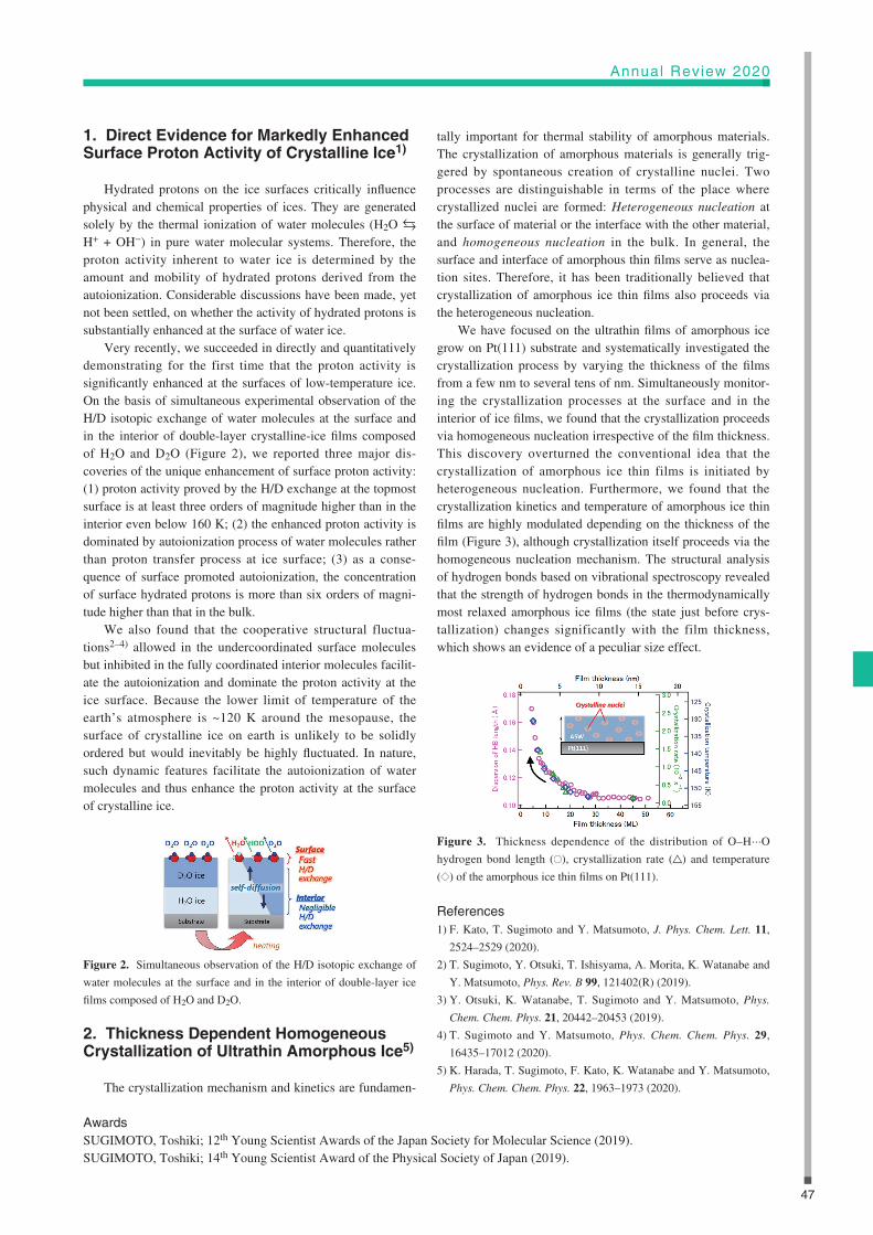
2. Thickness Dependent Homogeneous Crystallization of Ultrathin Amorphous Ice5)
The crystallization mechanism and kinetics are fundamen-
tally important for thermal stability of amorphous materials. The crystallization of amorphous materials is generally trig-gered by spontaneous creation of crystalline nuclei. Two processes are distinguishable in terms of the place where crystallized nuclei are formed: Heterogeneous nucleation at the surface of material or the interface with the other material, and homogeneous nucleation in the bulk. In general, the surface and interface of amorphous thin films serve as nuclea-tion sites. Therefore, it has been traditionally believed that crystallization of amorphous ice thin films also proceeds via the heterogeneous nucleation.
We have focused on the ultrathin films of amorphous ice grow on Pt(111) substrate and systematically investigated the crystallization process by varying the thickness of the films from a few nm to several tens of nm. Simultaneously monitor-ing the crystallization processes at the surface and in the interior of ice films, we found that the crystallization proceeds via homogeneous nucleation irrespective of the film thickness. This discovery overturned the conventional idea that the crystallization of amorphous ice thin films is initiated by heterogeneous nucleation. Furthermore, we found that the crystallization kinetics and temperature of amorphous ice thin films are highly modulated depending on the thickness of the film (Figure 3), although crystallization itself proceeds via the homogeneous nucleation mechanism. The structural analysis of hydrogen bonds based on vibrational spectroscopy revealed that the strength of hydrogen bonds in the thermodynamically most relaxed amorphous ice films (the state just before crys-tallization) changes significantly with the film thickness, which shows an evidence of a peculiar size effect.
Figure 3. Thickness dependence of the distribution of O–H···O
hydrogen bond length ( ), crystallization rate ( ) and temperature
( ) of the amorphous ice thin films on Pt(111).
References1) F. Kato, T. Sugimoto and Y. Matsumoto, J. Phys. Chem. Lett. 11,
2524–2529 (2020).
2) T. Sugimoto, Y. Otsuki, T. Ishisyama, A. Morita, K. Watanabe and
Y. Matsumoto, Phys. Rev. B 99, 121402(R) (2019).
3) Y. Otsuki, K. Watanabe, T. Sugimoto and Y. Matsumoto, Phys.
Chem. Chem. Phys. 21, 20442–20453 (2019).
4) T. Sugimoto and Y. Matsumoto, Phys. Chem. Chem. Phys. 29,
16435–17012 (2020).
5) K. Harada, T. Sugimoto, F. Kato, K. Watanabe and Y. Matsumoto,
Phys. Chem. Chem. Phys. 22, 1963–1973 (2020).
AwardsSUGIMOTO, Toshiki; 12th Young Scientist Awards of the Japan Society for Molecular Science (2019).SUGIMOTO, Toshiki; 14th Young Scientist Award of the Physical Society of Japan (2019).

48
RESEARCH ACTIVITIES
• M. Hiramoto, M. Kikuchi and S. Izawa, “Parts-per-Million-Level
Doping Effects in Organic Semiconductor Films and Organic
Single Crystals,” Adv. Mater. 30, 1801236 (15 pages) (2018).
[Invited Progress Report]
• M. Kikuchi, M. Hirota, T. Kunawong, Y. Shinmura, M. Abe,
Y.Sadamitsu, A. M. Moh, S. Izawa, M. Izaki, H. Naito and M.
Hiramoto, “Lateral Alternating Donor/Acceptor Multilayered Junc-
tion for Organic Solar Cells,” ACS Appl. Energy Mater. 2, 2087–
2093 (2019).
• S. Izawa, N. Shintaku, M. Kikuchi and M. Hiramoto, “Importance
of Interfacial Crystallinity to Reduce Open-Circuit Voltage Loss in
Organic Solar Cells,” Appl. Phys Lett. 115, 153301 (2019).
Selected Publications
Keywords Organic Semiconductors, ppm-Doping, Lateral Junction
MemberAssistant Professor
IZAWA, Seiichiro
Research FellowYABARA, Yusuke
Graduate StudentLEE, JihyunPALASSERY ITHIKKAL, Jaseela
SecretarySUGIHARA, HidemiNAKAMURA, Yuka
Education1984 B.E. Osaka University1986 Ph.D (Engineering) Osaka University
Professional Employment1984 Technical Associate, Institute for Molecular Science1988 Research Associate, Osaka University1997 Associate Professor, Osaka University2008 Professor, Institute for Molecular Science Professor, The Graduate University for Advanced Studies
Awards2017 Fellow Award of Japan Society of Applied Physics2006 Paper award, Molecular Electronics & Bioelectronics division,
Japan Society of Applied Physics2006 Research and Education Award, Osaka University2004 Editor Award, Japanese Journal of Applied Physics
HIRAMOTO, MasahiroProfessor[[email protected]]
Organic Solar CellsDepartment of Materials Molecular ScienceDivision of Molecular Functions
Organic solar cells have been intensively studied due to many advantages like flexible, printable, light, low-cost, fashionable, etc. Last year, we proposed a novel concept of the structure of organic solar cell, namely, a lateral multilayered junction (Figure 1). An essential point is that the photo-generated holes and electrons are laterally transported and extracted to the respective electrodes. We also investigated the reduction of open-circuit voltage loss in organic solar cells by using high-mobility organic semiconductors (Figure 2). On the other hand, we have been focused on the research on the ppm-level doping effects in organic semiconductor films and organic single crystals for organic solar cells. So far, we have reported complete pn-control, doping sensitization, ppm-level doping effects using an extremely low-speed deposition tech-nique reaching 10−9 nm s−1, in organic single crystals mea-sured by the Hall effect, which shows a doping efficiency of
24%, and enhancement of open-circuit voltage of organic solar cells by doping. These results can be regarded as a foundation for the construction of high efficient organic solar cells.
Figure 1. Lateral multilayered junction which can replace the
blended junction.

49
Annual Review 2020
1. Reduction of Open-Circuit Voltage Loss in Organic Solar Cells1)
Reducing the energy loss in output voltage is critically important for further enhancing the efficiency of organic solar cells. In this work, we showed that the organic solar cells with high mobility and highly crystalline donor and acceptor materials can reduce an open-circuit voltage (Voc) loss.
Two-layer cells consisting of C8-BTBT and Cn-PTCDI (Figure 2(a)), which acts as the donor and acceptor were fabricated. Voc increases in the order of C3 < C6 < C8 of Cn-PTCDI (Figure 2(a)) and reached to thermodynamic (Shockley–Queisser) limit (Figure 2(b), red dots). Simulta-neously, electron mobility increases by the suppression of molecular vibration of π-stacking due to the increase of crystallinity by fastener effect by increasing the chain length (C3 < C6 < C8). Voc increase can be reasonably explained by the suppression of non-radiative (vibrational) recombination from CT (charge transfer) exciton (D+/A−) to ground state (D/A) (Figure 2(c)).
By using the high mobility (band conductive) organic semiconductors, high efficient organic solar cells would be realized by reducing Voc loss.
Figure 2. (a) J–V characteristics of C8-BTBT/Cn-PTCDI cells. (b)
Voc vs. CT state energy. (c) Non-radiative recombination from CT
state to ground state.
2. Ultra-Thick Blended Layer up to 10 µm in Organic Solar Cells2)
Blended layer thickness of organic solar cells made with small molecules has limitation up to the order of a few hundred nm which is still not enough to absorb whole solar light. In this work, we succeeded to operate the organic solar cells having 10-µm-thick photoactive blended layer, consisting of zinc phthalocyanine (ZnPc), and fullerene (C60).
A method of co-evaporant induced crystallization3) was used for the deposition of ZnPc:C60 codeposited films. Co-evaporant molecule (polydimethylsiloxane; PDMS) acts as a liquid in the vacuum, which induces the crystallization and phase separation of the codeposited film. The cross-sectional image of co-deposited films with a co-evaporant show the columnar structures of ZnPc and C60 which offer the vertical transport routes for holes and electrons (Figure 3, inset).
With co-evaporant, short-circuit photocurrent (Jsc) and fill factor (FF) showed almost constant value up to the surprising blended layer thickness of 10 µm (Figure 3, blue diamonds). However, without co-evaporant, they steeply decreased within several hundred nm (Figure 3, green crosses). Photocurrent density of 20 mAcm−2 and the conversion efficiency (PCE) of 4.3% were observed. Whole solar light absorption by ultra-thick blended layer fabricated by co-evaporant will open the way to realize the high efficient small-molecular type organic solar cells.
Figure 3. Dependence of cell performance on blended layer thick-
ness. Inset: Phase separated columnar structure made by co-evaporant
induced crystallization.
References1) S. Izawa, N. Shintaku, M. Kikuchi and M. Hiramoto, Appl. Phys.
Lett. 115, 153301 (2019).
2) M. Katayama, T. Kaji, S. Nakao and M. Hiramoto, Front. Energy
Res., Section Solar Energy 8:4, 1–12 (2019).
3) T. Kaji, M. Zhang, S. Nakao, K. Iketaki, K. Yokoyama, C. W. Tang
and M. Hiramoto, Adv. Mater. 23, 3320–3325 (2011).
(a)
(b)
(c)

50
RESEARCH ACTIVITIES
• N. Uekama, T. Aoki, T. Maruoka, S. Kurisu, A. Hatakeyama, S.
Yamaguchi, M. Okada, H. Yagisawa, K. Nishimura and S. Tuzi,
“Influence of Membrane Curvature on the Structure of the Mem-
brane-Associated Pleckstrin Homology Domain of Phospholipase
C-d1,” Biochim. Biophys. Acta, Biomembr. 1788, 2575–2583 (2009).
• T. Iijima and K. Nishimura, “2H Quadrupolar Carr-Purcell-
Meiboom-Gill NMR for Paramagnetic Solids,” Chem. Phys. Lett.
514, 181–186 (2011).
• K. Yazawa, F. Suzuki, Y. Nishiyama, T. Ohata, A. Aoki, K.
Nishimura, H. Kaji and T. Asakura, “Determination of Accurate 1H
Positions of Alanine Tripeptide with Anti-Parallel and Parallel
β-Sheet Structures by High Resolution 1H Solid State NMR and
GIPAW Chemical Shift Calculation,” Chem. Commun. 48, 11199–
11201 (2012).
• M. Tanio and K. Nishimura, “Intramolecular Allosteric Interaction
in the Phospholipase C-d1 Pleckstrin Homology Domain,” Biochim.
Biophys. Acta, Proteins Proteomics 1834, 1034–1043 (2013).
• M. Yagi-Utsumi, K. Kato and K. Nishimura, “Membrane-Induced
Dichotomous Conformation of Amyloid β with the Disordered
N-Terminal Segment Followed by the Stable C-Terminal β Struc-
ture,” PLoS One 11, 0146405 (10 pages) (2016).
• N. Ousaka, F. Mamiya, Y. Iwata, K. Nishimura and E. Yashima,
“‘Helix-in-Helix’ Superstructure Formation through Encapsulation
of Fullerene-Bound Helical Peptides within a Helical Poly(methyl
methacrylate) Cavity,” Angew. Chem., Int. Ed. 56, 791–795 (2017).
Selected Publications
Keywords Solid State NMR, Biomolecules, Developments
MemberSecretary
YOKOTA, Mitsuyo
NISHIMURA, KatsuyukiAssociate Professor[[email protected]]
Education1994 B.S. Himeji Institute of Technology (University of Hyogo)1999 Ph.D. Himeji Institute of Technology (University of Hyogo)
Professional Employment1999 Postdoctoral Fellow, National High Magnetic Field Laboratory,
Florida State University2001 Assistant Professor, Yokohama National University2006 Associate Professor, Institute for Molecular Science Associate Professor, The Graduate University for Advanced
Studies
Award2002 The Young Scientist Poster Award, The Nuclear Magnetic
Resonance Society of Japan
Solid-State NMR for Molecular ScienceDepartment of Materials Molecular ScienceDivision of Molecular Functions
In order to elucidate functions of molecules, characteri-zation of the molecule is the first step. There is a variety of important molecules, which are insoluble in any solvents and functional at amorphous state. Solid-state NMR enables us to obtain a variety of information at atomic resolution without damage to molecules and significant restrictions. Thus, solid-state NMR is one of the essential tools for the characteri-zations of those molecules.
We have been working on methodology and hardware developments of solid-state NMR and their application to structural biology and materials science. We study characteri-zation of membrane proteins and peptides, organic materials, natural products and synthetic polymers. Characterization of those molecules based on solid-state NMR is underway through collaborations with several research groups.
Figure 1. Outline of our studies.

51
Annual Review 2020
1. Development of Solid-State NMR Probe
We have built a variety of solid-state NMR probes such as static and MAS 1H-X double resonance probes for 400 MHz NMR, and a variable temperature 1H-X double resonance MAS probe for 920 MHz ultra-high field NMR so far. Most of these probe buildings were achieved through major modifi-cations of commercial probes. During the past few years, we have been working on building an original solid-state NMR probe which is fully compatible with commercial instruments currently used.
We have built original narrow bore solid-state NMR 1H-X (“X” indicates variable resonant frequency) double resonance magic angle spinning (MAS) probe for 2.5 mm outer diameter (O.D.) sample tube used for 400 MHz (9.4 T) NMR spectrom-eter. The developed probe was built with originally designed parts except for spinning and spinning rate detection modules which were purchased from NMR company. The capacitive matching network design composed of commercially available non-magnetic variable capacitors was used. Balun type elec-tric circuit was incorporated into 1H channel, in which reduces to half the effective voltage of tuning capacitor and also minimize antenna effect of rf coil and rf inhomogeneity, especially at high field. Low frequency X channel was enabled to change largely its tunable frequency range to observe various nuclei by exchanging additional non-magnetic capaci-tors from bottom of the probe. The used network design may be compatible at higher fields by changing the parts related to resonant frequency.
Currently, the designs of individual parts are further updated and parts positions in the probe are further optimized to improve performance of the probe and access to the parts for the maintenance of the probe. 1H-13C-15N triple resonance MAS probe was re-designed based on improved 1H-X double reso nance probe and is under building. We would like to replace two NMR modules purchased from NMR company to original ones. Therefore, we are currently attempting to design original spinning module for 4 mm sample tube in which a little bit easier than that for 2.5 mm sample tube. Probe developments enable to reduce cost for acquiring probes and open up pos sibilities to design new experiments which are tightly related to specifically designed hardware. In near future, we would like to incorporate special functions into our original probes.
2. Structural Characterization of Amyloid β Protein Oligomer Promoted on Lipid Bilayers Using Solid-State NMR
Amyloid β (Aβ) protein is disordered in solutions under diluted conditions, however it conforms insoluble amyloid fibrils, which are found in senile plaque as a hallmark of Alzhei mer’s disease. Although molecular structures of amy-
loid fibrils have been determined, its molecular process for fibrillation in vivo has not been clarified yet. However, accu-mulated evidences suggest that the fibrillation process may be promoted on neuronal cell membrane. Especially, it has been reported that Aβ specifi cally interacts with ganglioside GM1 which is one of the key lipids in lipid raft. Therefore, GM1 embedded into lipid bilayers composed of neutral lipid DMPC may be regarded as the most simplified model neuronal cell membrane. In order to clarify the role of GM1 in the fibril-lation process, first, we have successfully determined the oligomeric structure of Aβ (1-40) induced on DMPC bilayers based on solid-state NMR.1) We have been collaborated with Prof. Kato group in IMS for those Aβ studies.
In the current study, Aβ (1-40) oligomer induced on lipid bilayers consisting of GM1 and DMPC have been attempted to characterize using solid-state NMR. All of essential solid-state NMR experiments such as 13C-homonuclear- and, 13C-15N heteronuclear correlation experiments for signal assignments and dipolar coupling based 13C-homonuclear correlation experiments to obtain distance information were completed.
As reported in last report, analysis of secondary structure of Aβ based on the chemical shifts of assigned signals revealed that disordered N-terminus followed by two β-sheet structures from middle region to C-terminus, in which differ from the one induced on DMPC bilayers.
During a year, the signal assignments were reconfirmed and dipolar coupling based 13C-homonuclear correlation experiments were performed for the sample of [U-13C,15N] Aβ diluted with natural abundant Aβ at various mixing times to differentiate intra- and intermolecular correlations. Then intra- and intermolecular distance information was extracted through the analyses of those NMR data. By considering the result of paramagnetic relaxation enhancements (PRE) experiments as reported last year, promising intermolecular packing model was successfully obtained from the NMR data.
Currently, precise molecular structure of Aβ together with intermolecular packing configuration is under investigations based on NMR data with combination of computational sci-ence through collaboration with Prof. Okumura group in IMS.
3. Structural Characterizations of Molecular Materials Using Solid-State NMR
We have also been working on collaboration works with two other research groups, Prof. Yoshito Tobe in Osaka university and Prof. Nobuyuki Nishi in Aichi university of education for the characterizations of newly designed molecular materials based on solid-state NMR. Those projects are underway.
Reference1) M. Yagi-Utsumi, K. Kato and K. Nishimura, PLoS One 11,
0146405 (10 pages) (2016).

52
RESEARCH ACTIVITIES
Study on Ion Conductive Materials for Novel Energy Storage/Conversion Devices
Department of Materials Molecular ScienceDivision of Molecular Functions
Chemical energy conversion/storage using electrochemical devices such as fuel cells and batteries will become increas-ingly important for future sustainable societies. As ion trans-port in solids is key for determining the performance of these devices, an improved understanding of the characteristics of existing electrode and electrolyte materials is required. For example, crystal structure, thermal stability, and reaction mechanisms are important to enhancing battery performance. Furthermore, the development of novel ion conduction phe-nomena through the synthesis of a new class of substances will be expected to lead to the creation of novel battery systems. In this context, I have concentrated my research efforts into two main areas: (i) Studies into the reaction mechanisms of cathodic materials for lithium secondary bat-teries; and (ii) The synthesis of new materials exhibiting hydride ion (H–) conductivity and the development of a novel
battery system utilizing both the H– conduction phenomenon and the H–/H2 redox reaction.
• G. Kobayashi, Y. Hinuma, S. Matsuoka, A. Watanabe, M. Iqbal, M.
Hirayama, M. Yonemura, T. Kamiyama, I. Tanaka and R. Kanno,
Science 351, 1314–1317 (2016).
• G. Kobayashi, Y. Irii, F. Matsumoto, A. Ito, Y. Ohsawa, S.
Yamamoto, Y. Chui, J.-Y. Son and Y. Sato, J. Power Sources 303,
250–256 (2016).
• A. Watanabe, G. Kobayashi, N. Matsui, M. Yonemura, A. Kubota,
K. Suzuki, M. Hirayama and R. Kanno, Electrochemistry 85(2),
88–92 (2017).
• Y. Iwasaki, N. Matsui, K. Suzuki, Y. Hinuma, M. Yonemura, G.
Kobayashi, M. Hirayama, I. Tanaka and R. Kanno, J. Mater. Chem.
A 6, 23457–23463 (2018).
• F. Takeiri, A. Watanabe, A. Kuwabara, H. Nawaz, N. Ayu, M.
Yonemura, R. Kanno and G. Kobayashi, Inorg. Chem. 58, 4431–
4436 (2019).
• N. Matsui, G. Kobayashi, K. Suzuki, A. Watanabe, A. Kubota, Y.
Iwasaki, M. Yonemura, M. Hirayama and R. Kanno, J. Am. Ceram.
Soc. 102, 3228–3235 (2019).
• H. Nawaz, F. Takeiri, A. Kuwabara and M. Yonemura and G.
Kobayashi, Chem. Commun. 56, 10373–10376 (2020).
Selected Publications
Keywords Solid State Ionics, H− Conductor, Battery
MemberAssistant Professor
TAKEIRI, Fumitaka
Graduate StudentNAWAZ, HaqOKAMOTO, KeiUCHIMURA, TasukuAYU, Nur Ika Puji*
Technical FellowIMAI, YumikoKUBOTA, Akiko
SecretarySUZUKI, Ai
Education2006 B.E. Kanazawa University2008 M.E. Tokyo Institute of Technology2010 D.S. Tokyo Institute of Technology
Professional Employment2010 Postdoctoral Fellow, Tokyo Institute of Technology2011 Assistant Professor, Kanagawa University2012 JST-PRESTO Researcher (Additional post)2013 Research Associate Professor, Institute for Molecular
Science2018 Associate Professor, Institute for Molecular Science Associate Professor, The Graduate University for Advanced
Studies
Awards2010 ACerS Spriggs Phase Equilibria Award, American Ceramics
Society2011 Tejima Doctoral Dissertation Award, Tokyo Institute of
Technology2018 The 39th Honda Memorial Young Researcher Award, The
Honda Memorial Fundation2018 The 7th Ishida Award, Nagoya University2019 Tagawa Solid State Chemistry Awards, Division of Solid
State Chemistry, The Electrochemical Society of Japan2019 Morino Foundation for Molecular Science
KOBAYASHI, GenkiAssociate Professor[[email protected]]
Figure 1. The concept of our research toward the realization of new
energy storage/conversion devices.

53
Annual Review 2020
1. Study on H– Conductive Oxyhydrides1–5)
Ionic charge carriers include a variety of species, such as Li+, H+, Ag+, Cu+, F–, and O2–, and their conductors have found applications in energy devices such as fuel cells and batteries. A hydride ion (H−,) is an attractive charge carrier because it exhibits promising features for fast ionic con-duction; namely, monovalence, suitable ionic size similar to that of F− and O2−, and high polarizability. Furthermore, its strong reducing properties with a standard redox potential of H−/H2 (−2.3 V) which is close to that of Mg/Mg2+ (−2.4 V) may be applied in energy storage/conversion devices with high energy densities. In 2016, we synthesized a series of K2NiF4-type oxyhydrides, La2−x−ySrx+yLiH1−x+yO3−y, which are equipped with anion sublattices that exhibit flexibility in the storage of H−, O2−, and vacancies (Figure 2 upper) and demonstrated pure H− conduction properties in the oxy-hydrides. The Li-based oxyhydrides acted as solid electrolytes and the all-solid-state Ti/La2−x−ySrx+yLiH1−x+yO3−y/TiH2 cell showed a redox reaction based on hydrogen storage/desorption on the electrodes, which is a first battery reaction using H− conduction phenomena.
More recently, we synthesized novel H− conductive oxy-hydrides, Ba2MHO3 (M = Sc, Y), with the K2NiF4-type struc-ture and confirmed its unique site selectivity for H−. In accord-ance with the electrostatic valence rule, it was found that the hydride ions in Ba2MHO3 selectively occupied the rock-salt layer, in contrast to those of the observed isostructural Li- based oxyhydrides that preferentially occupy the perovskite layer (Figure 2). The cation size at the octahedral center
influences the anion arrangement within the rock salt layer, and in the Y-oxyhydride, the complete ordering of H/O led to the formation of the [Ba2H2] layers. These results indicate that anion ordering in oxyhydrides could be tuned by appropriate element substitutions, which is a new insight for designing H− conducting materials.
2. High-Performance of Li-Rich Layered Cathode Materials through A2O3-Surface Modification6)
Controlling the cathode/electrolyte interface by surface modification of cathode materials with metal oxides or phos-phate is being explored as a possible strategy for improv ing the electrochemical performance of such materials. We syn-thesized Al2O3-coated Li[Li0.2Ni0.18Co0.03Mn0.58]O2 and inves-tiated the crystal structure, the chemical bonding state from bulk to surface, and the influence of the surface modification on the electrochemical performance by X-ray diffraction, hard X-ray photoelectron spectroscopy (HAXPES), and galva-nostatic charge/discharge reaction. It revealed that the surface-modification layer was composed of Li-Al oxides and Al oxides and that a LiM1–xAlxO2 (M = transition metals) inter-layer was formed between the modification layer and the Li[Li0.2Ni0.18Co0.03Mn0.58]O2 particles (Figure 3). The cycling performance of the Li-rich layered oxide was enhanced by the surface modification with Al2O3. A discharge capacity of more than 310 mA h–1 and excellent cycling stability at 50 °C were achieved by the combination of the gradual Li-insetion/de-insertion process (stepwise precycling treatment) and the surface-modification.
Figure 3. Schematic illustration and Al 1s HAXPES spectra of the 2
wt% Al2O3-coated Li[Li0.2Ni0.18Co0.03Mn0.58]O2 particle.
References1) H. Nawaz, G. Kobayashi* et al., Chem. Commun. 56, 10373–10376
(2020).
2) F. Takeiri, G. Kobayashi* et al., Inorg. Chem. 58, 4431–4436
(2019).
3) N. Matsui, G. Kobayashi* et al., J. Am. Ceram. Soc. 102, 3228–
3235 (2019).
4) A. Watanabe, G. Kobayashi* et al., Electrochemistry 85, 88–92
(2017).
5) G. Kobayashi*, R. Kanno* et al., Science 351, 1314–1317 (2016).
6) G. Kobayashi* et al., J. Power Sources 303, 250–256 (2016).
AwardKOBAYASHI, Genki; Morino Foundation for Molecular Science (2019).
* carrying out graduate research on Cooperative Education Program of IMS with High Energy Accelerator Research Organization
Figure 2. Crystal structures of H− conductive oxyhydrides La2−x−ySrx+y
LiH1−x+yO3−y (x = 0, y = 0, 1, 2) and Ba2MHO3 (M = Sc, Y).

54
RESEARCH ACTIVITIES
Surface and interface are the places for energy conversion processes in phys ics, chemistry, and biology. The detailed mechanism of such energy conversion processes is yet to be clari-fied. The reaction at the interface be-tween the electrode and electrolyte of rechargeable batteries are a typical sys-tem of the energy conversion. At the interface in lithium ion battery, which
is most widely used rechargeable battery,1) the energy transfer is proceeded by the insertion and extraction of the carrier ions at the interface during the charge and discharge reactions. Also, at the interface, the carrier ions, counter ions, solvents, additives, and crystal structures of the electrode surface forms characteristic structures that induce different properties from the material bulk. Traditionally, the reaction mechanism at the interface has been discussed based on electrochemical mea-surements. However, the nature of the interface is still unclear due to the difficulty of the analysis of the buried interface.
Scanning probe microscopy (SPM) is an excellent tech-nique to analyze the geometric, mechanical, electric, and electronic properties at the surfaces and interfaces. Thus, it has been employed for the analysis of the energy conversion processes. SPM can directly access to the buried interfaces such as the interface between electrode and electrolyte and has abilities to provide important information to clarify the reac-tion mechanism. In 2020, two specially designed SPMs have been launched at the instrument center of the Institution for Molecular Science. One is designed for the analysis of the
geometric, mechanical, electric, and electronic properties at the surface and interface under photo excitation or magnetic field with high resolution. The another is solely designed for the analysis under electrochemical reactions (Figure 1). Using the two SPM systems, both the physical properties and the reaction mechanism of the energy conversion processes at the surface and interface could be clarified.
In electrochemical devices, the local distribution of the reactions at the interface between the electrode and electrolyte strongly influences the device performances. The direct obser-vation of the local distribution at the interface provides impor-tant information that aids the understanding of the reaction mechanism of the system. Using the SPM system with the specially designed cantilever having Pt tip, the local distri-bution of the electrochemical reactions was visualized. Figure 2 shows an example of the mapping of the electrochemical reaction on SiN/Pt in hexamine RuCl3 electrolyte. The bright area shows the area with higher Faraday current than the dark area. The higher Faraday current is caused by the redox reactions of Ru ions in the electrolyte. From the simultaneous observation of the mechanical properties of the electrode surface using the SPM, the bright and dark areas are assigned to the regions of Pt and SiN, respectively. The results reveal the high reactivity of the Pt in the reaction. Employing this technique in the present and innovative battery systems,1–3) the local distribution of the reactions in the batteries can be visualized. This will avail more vital information for the design of new energy devices.
References1) T. Minato and T. Abe, Prog. Surf. Sci. 92, 240–280 (2017).
2) T. Minato, H. Konishi, A. Celik Kucuk, T. Abe and Z. Ogumi,
Ceramics Japan 54, 637 (2019).
3) H. Konishi, R. Takekawa, T. Minato, Z. Ogumi and T. Abe, Chem.
Phys. Lett. 755, 137785 (2020).
Clarification of the Energy Conversion Mechanism at the Surface and Interface by Scanning Probe Microscopy
Instrument Center
MINATO, TaketoshiSenior Researcher
Figure 1. The scanning probe microscope system for the analysis of
electrochemical reactions installed in the instrument center of Institute
for Molecular Science.
Figure 2. A Faraday current mapping on SiN/Pt obtained by scanning
electrochemical microscopy in 5 mM hexamine RuCl3 and 0.1 M
KNO3 aqueous solution. Cantilever: Pt; counter electrode: Pt, refer-
ence electrode: Ag, sample potential: −0.1 V (vs reference), image
size: 25 µm × 6 µm.

55
Annual Review 2020
Visiting Professors
Visiting ProfessorKISHINE, Jun-ichiro (from The Open University of Japan)
Theoretical Studies on Chiral Material ScienceWe focus on chirality-induced phenomena in solids. (1) Magnetic response of a highly nonlinear
soliton lattice in a monoaxial chiral helimagnet: We presented a theory of nonlinear magnetic response of a chiral soliton lattice state in a monoaxial chiral helimagnet under an oscillating magnetic field. (2) Tensile deformations of the magnetic chiral soliton lattice probed by Lorentz transmission electron microscopy:
We considered the case of a chiral soliton lattice subjected to uniaxial elastic strain. We found that the strain induced anisotropies give rise to three distinct non-trivial spin textures, depending on the nature of the strain, and we show how these states may be identified by their signatures in Lorentz transmission electron microscopy (TEM). (3) Chirality-Induced Spin-Polarized State of a Chiral Crystal: Chirality-induced spin transport phenomena are investigated at room temperature without magnetic fields in a monoaxial chiral dichalcogenide CrNb3S6. We found that spin polarization occurs in these chiral bulk crystals under a charge current flowing along the principal c axis.
Visiting Associate ProfessorFURUKAWA, Ko (from Niigata University)
Advanced ESR Study of Molecule-Based Functional MaterialsTo develop the high-efficiency molecule-based device, it’s vital to clarify the mechanism of the
functional molecules/materials. We investigate the mechanism of the solid-state functional materials in terms of advanced electron spin resonance (ESR) spectroscopy such as high-field/high-frequency ESR, time-resolved ESR, pulsed-ESR and so on. Recently, our themes are following three (I) spin dynamics
study of molecule-based materials with the complex function combined to photoconductivity and photo-induced magnetic properties, (II) operand ESR study of the alternative catalyst for oxygen reduction reaction (ORR) in fuel cell, and (III) The ESR study aimed to investigate the paddy soil environments and to identify the rice cultivar from the trace metal in the rice bran.
Visiting Associate ProfessorOSHIMA, Yugo (from RIKEN)
Microscopic Studies of the Bilayer-Type Molecular Ferromagnet (Et-4BrT)[Ni(dmit)2]2 by ESRRecently, a novel type of ferromagnet has been developed by Kusamoto Group and Yamamoto Group
in IMS. The novel molecular ferromagnet (Et-4BrT)[Ni(dmit)2]2, where dmit is 1,3-dithiol-2-thiole-4,5-dithiolate and Et-4BrT is ethyl-4-bromothiazolium, takes a bilayer structure, and becomes ferromagnetic below 1 K. We are considering that this ferromagnet is the first realization of the Nagaoka-Penn ferromagnetism, where the ferromagnetism is achieved by the light hole-doping of the insulating Ni(dmit)2
layer owing to the internal dipole moment of the monovalent cation Et-4BrT. In collaboration with Kusamoto Group and Yamamoto Group, we have investigated the microscopic electronic state of (Et-4BrT)[Ni(dmit)2]2 by high-frequency ESR. We have found that ESR lineshape largely changes below 30 K, which is probably due to the effect of doping from the cation site. We are now developing an ESR sample holder for field-effect transistors (FETs), so that we can control precisely the electrical doping of (Et-4BrT)[Ni(dmit)2]2 by means of FET structure, and investigate its change of magnetic properties by ESR spectroscopy.