
Repurposing the anti-epileptic drug sodium valproate as an ...
15
RESEARCH ARTICLE Repurposing the anti-epileptic drug sodium valproate as an adjuvant treatment for diffuse intrinsic pontine glioma Clare L. Killick-Cole 1 *, William G. B. Singleton 1,2 , Alison S. Bienemann 1 , Daniel J. Asby 1 , Marcella J. Wyatt 1 , Lisa J. Boulter 1 , Neil U. Barua 1,2 , Steven S. Gill 1,2 * 1 Functional Neurosurgery Research Group, School of Clinical Sciences, University of Bristol, Learning & Research Building, Southmead Hospital, Bristol, United Kingdom, 2 Department of Neurosurgery, North Bristol NHS Trust, Bristol, United Kingdom * [email protected] (SG); [email protected] (CKC) Abstract Targeting epigenetic changes in diffuse intrinsic pontine glioma (DIPG) may provide a novel treatment option for patients. This report demonstrates that sodium valproate, a histone deacetylase inhibitor (HDACi), can increase the cytotoxicity of carboplatin in an additive and synergistic manner in DIPG cells in vitro. Sodium valproate causes a dose-dependent decrease in DIPG cell viability in three independent ex vivo cell lines. Furthermore, sodium valproate caused an increase in acetylation of histone H3. Changes in cell viability were con- sistent with an induction of apoptosis in DIPG cells in vitro, determined by flow cytometric analysis of Annexin V staining and assessment of apoptotic markers by western blotting. Subsequently, immunofluorescent staining of neuronal and glial markers was used to deter- mine toxicity in normal rat hippocampal cells. Pre-treatment of cells with sodium valproate enhanced the cytotoxic effects of carboplatin, in three DIPG cell lines tested. These results demonstrate that sodium valproate causes increased histone H3 acetylation indicative of HDAC inhibition, which is inversely correlated with a reduction in cell viability. Cell viability is reduced through an induction of apoptosis in DIPG cells. Sodium valproate potentiates car- boplatin cytotoxicity and prompts further work to define the mechanism responsible for the synergy between these two drugs and determine in vivo efficacy. These findings support the use of sodium valproate as an adjuvant treatment for DIPG. Introduction Whilst great advances have been made in the characterisation of the molecular changes in DIPG, the clinical challenges that oppose treatment strategies remain set in place. Previous clinical trials have been reliant on the assumption of similar genetics between adult high grade glioma and DIPG, however, recent discoveries of H3F3A and AVCR1 mutations have led to the emergence of several preclinical studies with DIPG targeted therapies [1–6]. The current DIPG treatment regime consists of radiotherapy that provides only a palliative response for patients and it is well known that cranial radiation alone in children can cause neurological PLOS ONE | https://doi.org/10.1371/journal.pone.0176855 May 25, 2017 1 / 15 a1111111111 a1111111111 a1111111111 a1111111111 a1111111111 OPEN ACCESS Citation: Killick-Cole CL, Singleton WGB, Bienemann AS, Asby DJ, Wyatt MJ, Boulter LJ, et al. (2017) Repurposing the anti-epileptic drug sodium valproate as an adjuvant treatment for diffuse intrinsic pontine glioma. PLoS ONE 12(5): e0176855. https://doi.org/10.1371/journal. pone.0176855 Editor: Marta M. Alonso, Universidad de Navarra, SPAIN Received: October 6, 2016 Accepted: April 18, 2017 Published: May 25, 2017 Copyright: © 2017 Killick-Cole et al. This is an open access article distributed under the terms of the Creative Commons Attribution License, which permits unrestricted use, distribution, and reproduction in any medium, provided the original author and source are credited. Data Availability Statement: All relevant data are within the paper. Funding: This study was funded by the Functional Neurosurgery Research Fund, held by Southmead Hospital Charities. SG and NB are advisers to Renishaw PLC. Neither Functional Neurosurgery Research Fund nor Renishaw had any role in study design, data collection and analysis, decision to publish, or preparation of the manuscript.
Transcript of Repurposing the anti-epileptic drug sodium valproate as an ...

RESEARCH ARTICLE
Repurposing the anti-epileptic drug sodium
valproate as an adjuvant treatment for diffuse
intrinsic pontine glioma
Clare L. Killick-Cole1*, William G. B. Singleton1,2, Alison S. Bienemann1, Daniel J. Asby1,
Marcella J. Wyatt1, Lisa J. Boulter1, Neil U. Barua1,2, Steven S. Gill1,2*
1 Functional Neurosurgery Research Group, School of Clinical Sciences, University of Bristol, Learning &
Research Building, Southmead Hospital, Bristol, United Kingdom, 2 Department of Neurosurgery, North
Bristol NHS Trust, Bristol, United Kingdom
* [email protected] (SG); [email protected] (CKC)
Abstract
Targeting epigenetic changes in diffuse intrinsic pontine glioma (DIPG) may provide a novel
treatment option for patients. This report demonstrates that sodium valproate, a histone
deacetylase inhibitor (HDACi), can increase the cytotoxicity of carboplatin in an additive and
synergistic manner in DIPG cells in vitro. Sodium valproate causes a dose-dependent
decrease in DIPG cell viability in three independent ex vivo cell lines. Furthermore, sodium
valproate caused an increase in acetylation of histone H3. Changes in cell viability were con-
sistent with an induction of apoptosis in DIPG cells in vitro, determined by flow cytometric
analysis of Annexin V staining and assessment of apoptotic markers by western blotting.
Subsequently, immunofluorescent staining of neuronal and glial markers was used to deter-
mine toxicity in normal rat hippocampal cells. Pre-treatment of cells with sodium valproate
enhanced the cytotoxic effects of carboplatin, in three DIPG cell lines tested. These results
demonstrate that sodium valproate causes increased histone H3 acetylation indicative of
HDAC inhibition, which is inversely correlated with a reduction in cell viability. Cell viability is
reduced through an induction of apoptosis in DIPG cells. Sodium valproate potentiates car-
boplatin cytotoxicity and prompts further work to define the mechanism responsible for the
synergy between these two drugs and determine in vivo efficacy. These findings support the
use of sodium valproate as an adjuvant treatment for DIPG.
Introduction
Whilst great advances have been made in the characterisation of the molecular changes in
DIPG, the clinical challenges that oppose treatment strategies remain set in place. Previous
clinical trials have been reliant on the assumption of similar genetics between adult high grade
glioma and DIPG, however, recent discoveries of H3F3A and AVCR1 mutations have led to
the emergence of several preclinical studies with DIPG targeted therapies [1–6]. The current
DIPG treatment regime consists of radiotherapy that provides only a palliative response for
patients and it is well known that cranial radiation alone in children can cause neurological
PLOS ONE | https://doi.org/10.1371/journal.pone.0176855 May 25, 2017 1 / 15
a1111111111
a1111111111
a1111111111
a1111111111
a1111111111
OPENACCESS
Citation: Killick-Cole CL, Singleton WGB,
Bienemann AS, Asby DJ, Wyatt MJ, Boulter LJ, et
al. (2017) Repurposing the anti-epileptic drug
sodium valproate as an adjuvant treatment for
diffuse intrinsic pontine glioma. PLoS ONE 12(5):
e0176855. https://doi.org/10.1371/journal.
pone.0176855
Editor: Marta M. Alonso, Universidad de Navarra,
SPAIN
Received: October 6, 2016
Accepted: April 18, 2017
Published: May 25, 2017
Copyright: © 2017 Killick-Cole et al. This is an open
access article distributed under the terms of the
Creative Commons Attribution License, which
permits unrestricted use, distribution, and
reproduction in any medium, provided the original
author and source are credited.
Data Availability Statement: All relevant data are
within the paper.
Funding: This study was funded by the Functional
Neurosurgery Research Fund, held by Southmead
Hospital Charities. SG and NB are advisers to
Renishaw PLC. Neither Functional Neurosurgery
Research Fund nor Renishaw had any role in study
design, data collection and analysis, decision to
publish, or preparation of the manuscript.
deficits, further prompting the urgent need for the development of more efficacious and less
toxic treatment plans for these patients. Presently, median survival for DIPG patients is nine
months with mortality rates of 90% by 18 months from diagnosis [7, 8]. These statistics
authenticate the clear unmet clinical need for DIPG patients and the requirement for readily
translatable treatments. This study examines the in vitro effect of pre-sensitising DIPG cells by
epigenetic regulation with sodium valproate to enhance the cytotoxic effects of carboplatin.
One of the most frequent mutations occurring in DIPG arises in the H3F3A gene, which
encodes histone H3.3 [9, 10]. Other histone mutations occur in a mutually exclusive manner,
with alterations of HIST1H3B modifying histone H3.1. These mutations are considered the
driving force of tumorigenesis by reducing histone K27 methylation, which results in gene
expression alterations in cells of the developing brain stem [11].
Sodium valproate has been used clinically for a number of years and is a well-established
drug for the long-term treatment of epilepsy. More recently, it was proven to be a HDAC
inhibitor (HDACi) and exerts anti-tumour activity towards several different cancer types invitro [12–14]. Currently, epigenetic therapies are at the forefront of cancer research, with
HDACi in particular being approved for use against haematological malignancies, where it
has been shown to induce differentiation and apoptosis of leukaemia cells in vitro and in clini-
cal trials [15]. Providing a rationale for this study are the recent publications purporting the
efficacy of HDACi for the treatment of DIPG [16, 17]. To enable the rapid translation of
already approved drugs, we have identified sodium valproate as a potential adjuvant chemo-
therapeutic against DIPG, owing to its HDAC inhibition, anti-cancer activity demonstrated
in several tumour types in vitro and in vivo [12, 18, 19] and a well-known toxicity profile. Fur-
thermore, its ability to cross the blood brain barrier (BBB) favours its use as a treatment for
brain malignancies. We hypothesised that sodium valproate would not only cause cytotoxicity
to DIPG cells as a monotherapy, but that its HDAC inhibition would sensitise cells to DNA
intercalating chemotherapeutics, such as carboplatin. As such, our research focuses on estab-
lishing in vitro preclinical evidence to support the use of sodium valproate for the treatment of
DIPG.
Monotherapies using sodium valproate have shown limited success in clinical trials, with
only 5% of acute myelogenous leukaemia patients showing response to sodium valproate treat-
ment [15]. Given the mechanism of action, it is viable that sodium valproate treatment may
alter how DIPG cells respond to other chemotherapeutics, such as DNA intercalating agents.
Such combinations have been used in medulloblastoma and glioma studies, whereby sodium
valproate treatment was combined with topoisomerase inhibitors and enhanced the cytotoxic-
ity of topotecan and etoposide [20, 21].
The advent of convection enhanced delivery (CED) has provided a direct route for drug
administration to the brain, circumventing problems associated with drugs crossing the blood
brain barrier (BBB) [22]. CED has allowed discrete areas of the brain to be precisely targeted
for the delivery of treatments to brain tumours. Intermittent CED of carboplatin has been
used by our group to treat patients with DIPG and glioblastoma, utilising an implanted drug
delivery system that allows repeated infusions without the need for repeated surgery [23, 24].
Epigenetics are now of great importance in cancer biology, with the regulation of the epi-
genome by both HDAC and DNA Methyltransferase inhibitors being of keen interest. Here,
we present findings that demonstrate sodium valproate, a commonly used anti-epileptic drug,
as a potential adjuvant and neoadjuvant chemotherapeutic, especially when combined with
carboplatin, a drug recently used by CED for DIPG patients [23, 25]. These results support the
rationale for further investigation into the clinical efficacy of sodium valproate as a cancer
therapeutic.
Repurposing sodium valproate as an adjuvant treatment for DIPG
PLOS ONE | https://doi.org/10.1371/journal.pone.0176855 May 25, 2017 2 / 15
Competing interests: There are no patents,
products in development or marketed products to
declare. SG and NB’s employment by Renishaw
does not alter the authors’ adherence to PLOS ONE
policies on sharing data and materials.

Methods
Cell culture
Patient derived SF7761 and SF8628 DIPG cells, which both harbour a H3FRA gene mutation
(H3.3 K27M) were established from biopsies and obtained from Nalin Gupta (University of
California, US) [26]. Both cell lines were grown as adherent monolayers. SF7761 cells were
maintained in neurobasal A medium (NBA), penicillin streptomycin, B-27 supplement with-
out vitamin A, N-2 supplement, epidermal growth factor (EGF; Peprotech) and fibroblast
growth factor (FGF; Peprotech). SF8628 were maintained in Dulbecco’s modified eagle’s
medium supplemented with fetal calf serum (FCS). The DUB-D003 cell line was established
from a surgical biopsy and carries a HIST1H3B gene mutation (K27M H3.1), this cell line was
obtained from Chris Jones (Institute of Cancer Research, London, UK) and maintained in
NBA supplemented with EGF, FGF, platelet-derived growth factor (PDGF)-AA (Shenandoah
Biotech), PDGF-BB (Shenandoah Biotech), B-27 and heparin (StemCell Technologies). Pri-
mary rat hippocampal cells were isolated from surgically dissected hippocampi of day 18
embryonic rat brain and maintained in Neurobasal media, pen-strep, L-glutamine, B-27 sup-
plement and FCS. SF7761 and hippocampal cells were maintained as adherent cultures using
Laminin (Sigma) or poly-D-lysine (Sigma), respectively. All reagents were purchased from
Thermo Fisher Scientific, unless stated otherwise.
All ex vivo cell lines were authenticated by short tandem repeat profiling (Public Health
England, UK) (S1 Table) and were classified mycoplasma negative (in-house testing).
Cell viability assay
Cell culture treatments followed an 8-day schedule. Sodium valproate (Sigma) treatment alone
was assessed by a 72 hours of continuous drug exposure. Carboplatin (Accord Healthcare Ltd)
alone was assessed by two short six hour exposures on two consecutive days followed by
removal of media, fresh media added and incubated for an additional 72 hours. Sequential
short-term exposures were used to represent the drug infusion time in our clinical CED proto-
cols. Pre-conditioning of cells required exposure to 72 hours of sodium valproate followed by
six hours of carboplatin on two consecutive days. Drug was then removed and fresh media
containing sodium valproate added for 72 hours.
Assessment of the cytotoxicity of each drug was carried out by MTT (3-(4,5-Dimethylthia-
zol-2-yl)-2,5-Diphenyltetrazolium Bromide) assay. Briefly, cells were plated at 1000–3000
cells/well in 96-well plates. Following a defined drug incubation period, 10 μl MTT solution
was added to each well (5 mg/ml). Cells were then incubated for a further 3–4 hours at 37˚C.
Cell culture media was aspirated and DMSO:isopropanol was added to solubilise the precipi-
tate. Absorbance was then measured on a FluoSTAR Optima microplate reader (BMG Lab-
tech) at 590 nm, using a reference read at 730 nm. Samples were analysed in triplicate and
each experiment repeated a minimum of three times. Values were normalised against the
untreated control OD values and calculated as a percentage of control.
Synergy analysis
Analysis of drug synergy for pre-conditioning experiments was assessed using CompuSyn soft-
ware following the Chou & Talay method [27]. Fraction affected values were entered for 72
hours sodium valproate alone (drug A); two consecutive six hour doses of carboplatin alone
(drug B); and values for pre-treatment with sodium valproate prior to carboplatin exposure
(drug A and B in combination). Combination index (CI) values were extrapolated and are
indicative of the mode of action for the two drugs. CI values equal to one is indicative of
Repurposing sodium valproate as an adjuvant treatment for DIPG
PLOS ONE | https://doi.org/10.1371/journal.pone.0176855 May 25, 2017 3 / 15

additive effects of both drugs; a CI below one suggests synergy; and a CI above one indicates
antagonism between the two drugs.
Apoptosis assessment
Apoptosis was assessed using the Annexin V-FITC/7-AAD kit (BioLegend), following manu-
facturer’s instructions. Briefly, cells were plated at 20,000–40,000 cells/well in a 12-well plate,
incubated for 16 hours overnight and then dosed with sodium valproate for 72 hours. Cells
were then detached from the growing surface and collected. 7-AAD was added to staining
buffer at 4 μl per 100 μl of buffer, and FITC-conjugated Annexin V was added at 1 μl per
100 μl buffer. Cells were then diluted 1:1 in staining buffer containing both 7-AAD and
Annexin V. Single dyes and unstained cells were used for controls and compensation analysis.
All experiments were repeated on at least three occasions.
Western blotting
Cells were plated at 60,000–100,000 cells/well in a 6-well plate, prior to treating with sodium
valproate. Whole cell lysates were obtained from adherent cell cultures by washing cells with
ice-cold phosphate buffered saline (PBS) prior to collecting cells in PBS using a cell lifter and
added to conditioned media. Cells were then centrifuged at 1500 rpm for five minutes at 4˚C.
Supernatants were then discarded and pellets washed with PBS followed by a further five min-
ute centrifugation step. PBS was removed and pellets were re-suspended in 50–100 μl of RIPA
buffer (Thermo Fisher Scientific) containing protease and phosphatase inhibitors. Cells were
incubated on ice for 30 minutes to aid cell lysis. Samples were the centrifuged at 10,500 rpm
for five minutes at 4˚C to pellet cell particulate. Supernatants were then transferred to fresh
tubes and quantified using BCA assay (Pierce, Thermo Fisher Scientific). 10–20 μg of protein
were separated by 10% polyacrylamide gel electrophoresis under reducing conditions and
transferred to nitrocellulose membrane (Hybond). Membranes were blocked with 5% non-fat
dried milk in 1% tris-buffered saline containing 0.1% tween-20 (TBST) for one hour at room
temperature prior to probing with acetyl histone H3 antibody (1:5000, Cell Signaling Technol-
ogy #4353) and rabbit IgG against β-actin (1:50,000, Sigma #A3854) as a protein loading con-
trol. To detect apoptotic markers, cleaved caspase 3 (1:1000, Cell Signaling Technology #9664)
and cleaved PARP (1:2500, BD Biosciences #556494) antibodies were used. Horseradish perox-
idase-labelled secondary anti-rabbit IgG, anti-mouse IgG (Cell Signaling Technology) and
enhanced chemi-luminescence (Pierce, Thermo Fisher Scientific) was used to detect immuno-
reactive bands. Images and densitometry analysis were carried out using Image Lab software
(Bio-Rad).
In vitro neurotoxicity assessment by immunofluorescent staining
Immunofluorescent assays were performed on untreated and sodium valproate treated pri-
mary hippocampal cells. Experiments were performed on rat E18 hippocampal cultures grown
on poly-D-lysine coated glass coverslips after cell extraction had been performed using previ-
ously described protocols [28]. Sodium valproate toxicity was assessed at 72 hours post-treat-
ment. B3-tubulin (1:200 Millipore #MAB1637) and GFAP (1:300 Millipore #AB5804) primary
antibodies were used to detect the viability of neuronal and glial cells, respectively. Donkey
anti-mouse Alexa Fluor488 (Molecular Probes, Thermo Fisher Scientific) and Cy3 (Jackson
Labs) were used as secondary antibodies, respectively. The staining protocol required cells to
be washed with PBS, fixed using 4% paraformaldehyde and treated with 0.1% triton-X for five
minutes to permeabilise cell membranes. Samples were incubated in blocking solution (10%
donkey serum) for one hour at room temperature, followed by incubation overnight in
Repurposing sodium valproate as an adjuvant treatment for DIPG
PLOS ONE | https://doi.org/10.1371/journal.pone.0176855 May 25, 2017 4 / 15
primary antibodies at 4˚C. Samples were then washed with PBS and incubated with secondary
antibodies for one hour at room temperature. The coverslips were then washed three times
with PBS and mounted onto microscope slides using 2 μl VectaShield (Vector Labs) contain-
ing DAPI nuclear counter-stain. Fluorescently labelled cells were visualised with a Leica
DM5500 microscope and digital camera (Leica Microsystems).
Statistical analyses
For all in vitro experiments, data was collected from at least three independent experiments.
All statistical analyses were performed on GraphPad Prism1 (version 5) with IC50 values
being derived from logarithmic dose-response curves. The effect of sodium valproate pre-con-
ditioning on carboplatin response was calculated by one-way analysis of variance (ANOVA).
Statistical significance was determined using Bonferroni’s multiple comparison post hoc anal-
ysis. For comparisons of histone acetylation levels and apoptosis, a one-way repeated measures
ANOVA was used with Dunnett’s multiple comparison test. P-values <0.05 were considered
significant throughout.
Results
Sodium valproate reduces DIPG cell survival in vitro and correlates with
an increase in histone acetylation
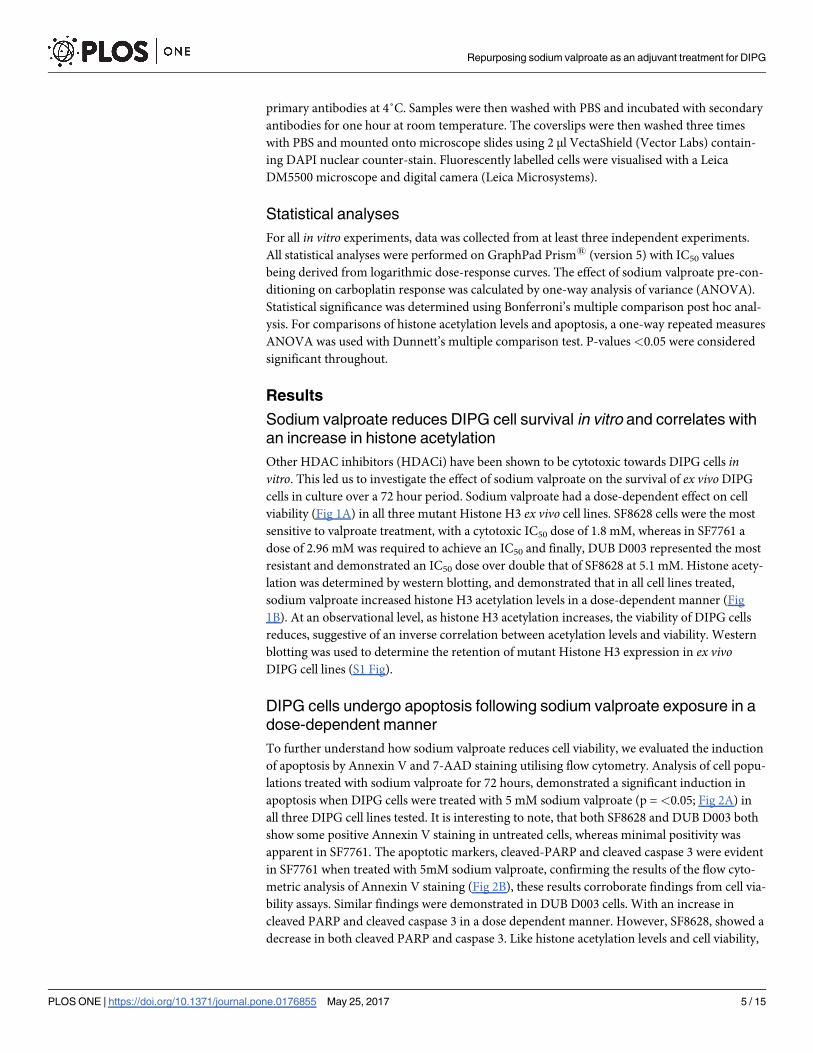
Other HDAC inhibitors (HDACi) have been shown to be cytotoxic towards DIPG cells invitro. This led us to investigate the effect of sodium valproate on the survival of ex vivo DIPG
cells in culture over a 72 hour period. Sodium valproate had a dose-dependent effect on cell
viability (Fig 1A) in all three mutant Histone H3 ex vivo cell lines. SF8628 cells were the most
sensitive to valproate treatment, with a cytotoxic IC50 dose of 1.8 mM, whereas in SF7761 a
dose of 2.96 mM was required to achieve an IC50 and finally, DUB D003 represented the most
resistant and demonstrated an IC50 dose over double that of SF8628 at 5.1 mM. Histone acety-
lation was determined by western blotting, and demonstrated that in all cell lines treated,
sodium valproate increased histone H3 acetylation levels in a dose-dependent manner (Fig
1B). At an observational level, as histone H3 acetylation increases, the viability of DIPG cells
reduces, suggestive of an inverse correlation between acetylation levels and viability. Western
blotting was used to determine the retention of mutant Histone H3 expression in ex vivoDIPG cell lines (S1 Fig).
DIPG cells undergo apoptosis following sodium valproate exposure in a
dose-dependent manner
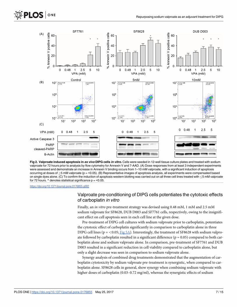
To further understand how sodium valproate reduces cell viability, we evaluated the induction
of apoptosis by Annexin V and 7-AAD staining utilising flow cytometry. Analysis of cell popu-
lations treated with sodium valproate for 72 hours, demonstrated a significant induction in
apoptosis when DIPG cells were treated with 5 mM sodium valproate (p = <0.05; Fig 2A) in
all three DIPG cell lines tested. It is interesting to note, that both SF8628 and DUB D003 both
show some positive Annexin V staining in untreated cells, whereas minimal positivity was
apparent in SF7761. The apoptotic markers, cleaved-PARP and cleaved caspase 3 were evident
in SF7761 when treated with 5mM sodium valproate, confirming the results of the flow cyto-
metric analysis of Annexin V staining (Fig 2B), these results corroborate findings from cell via-
bility assays. Similar findings were demonstrated in DUB D003 cells. With an increase in
cleaved PARP and cleaved caspase 3 in a dose dependent manner. However, SF8628, showed a
decrease in both cleaved PARP and caspase 3. Like histone acetylation levels and cell viability,
Repurposing sodium valproate as an adjuvant treatment for DIPG
PLOS ONE | https://doi.org/10.1371/journal.pone.0176855 May 25, 2017 5 / 15
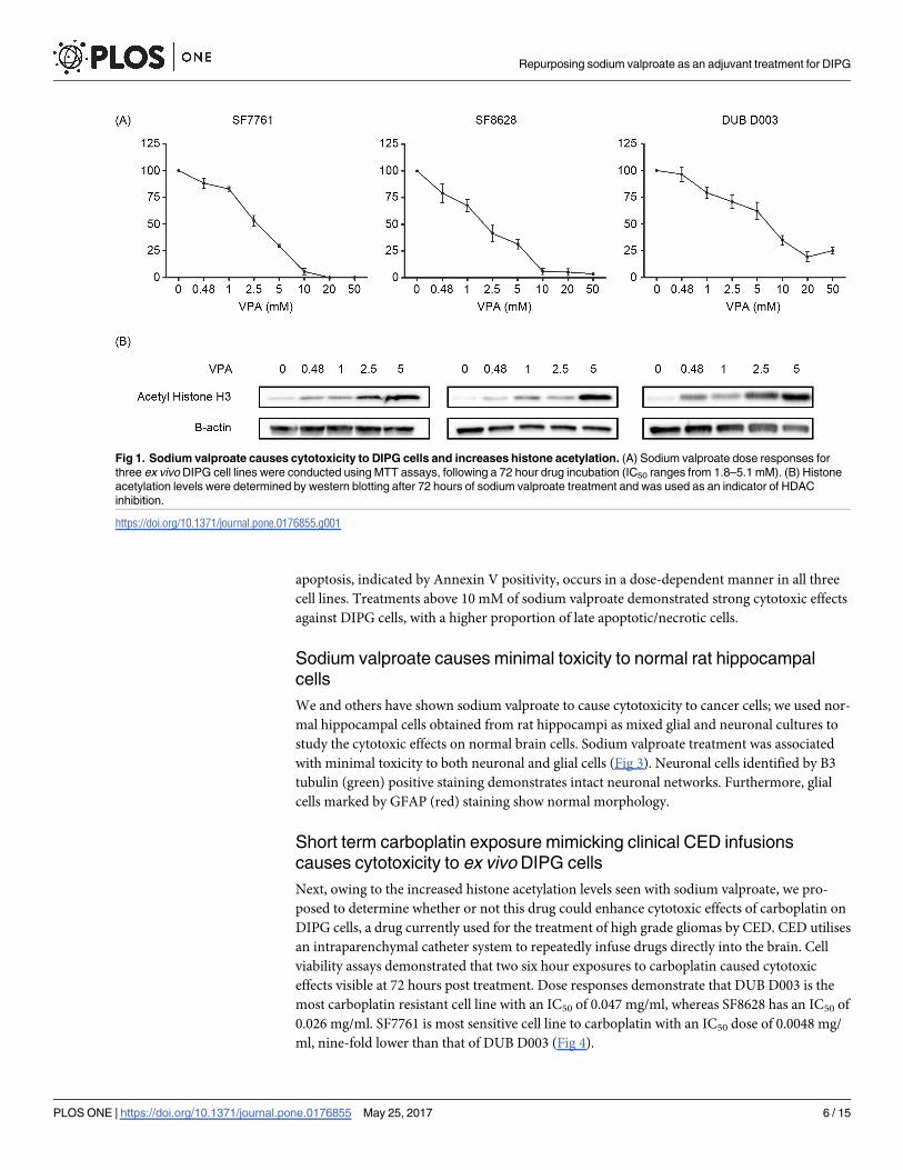
apoptosis, indicated by Annexin V positivity, occurs in a dose-dependent manner in all three
cell lines. Treatments above 10 mM of sodium valproate demonstrated strong cytotoxic effects
against DIPG cells, with a higher proportion of late apoptotic/necrotic cells.
Sodium valproate causes minimal toxicity to normal rat hippocampal
cells
We and others have shown sodium valproate to cause cytotoxicity to cancer cells; we used nor-
mal hippocampal cells obtained from rat hippocampi as mixed glial and neuronal cultures to
study the cytotoxic effects on normal brain cells. Sodium valproate treatment was associated
with minimal toxicity to both neuronal and glial cells (Fig 3). Neuronal cells identified by B3
tubulin (green) positive staining demonstrates intact neuronal networks. Furthermore, glial
cells marked by GFAP (red) staining show normal morphology.
Short term carboplatin exposure mimicking clinical CED infusions
causes cytotoxicity to ex vivo DIPG cells
Next, owing to the increased histone acetylation levels seen with sodium valproate, we pro-
posed to determine whether or not this drug could enhance cytotoxic effects of carboplatin on
DIPG cells, a drug currently used for the treatment of high grade gliomas by CED. CED utilises
an intraparenchymal catheter system to repeatedly infuse drugs directly into the brain. Cell
viability assays demonstrated that two six hour exposures to carboplatin caused cytotoxic
effects visible at 72 hours post treatment. Dose responses demonstrate that DUB D003 is the
most carboplatin resistant cell line with an IC50 of 0.047 mg/ml, whereas SF8628 has an IC50 of
0.026 mg/ml. SF7761 is most sensitive cell line to carboplatin with an IC50 dose of 0.0048 mg/
ml, nine-fold lower than that of DUB D003 (Fig 4).
Fig 1. Sodium valproate causes cytotoxicity to DIPG cells and increases histone acetylation. (A) Sodium valproate dose responses for
three ex vivo DIPG cell lines were conducted using MTT assays, following a 72 hour drug incubation (IC50 ranges from 1.8–5.1 mM). (B) Histone
acetylation levels were determined by western blotting after 72 hours of sodium valproate treatment and was used as an indicator of HDAC
inhibition.
https://doi.org/10.1371/journal.pone.0176855.g001
Repurposing sodium valproate as an adjuvant treatment for DIPG
PLOS ONE | https://doi.org/10.1371/journal.pone.0176855 May 25, 2017 6 / 15
Valproate pre-conditioning of DIPG cells potentiates the cytotoxic effects
of carboplatin in vitro
Finally, an in vitro pre-treatment strategy was devised using 0.48 mM, 1 mM and 2.5 mM
sodium valproate for SF8628, DUB D003 and SF7761 cells, respectively, owing to the insignifi-
cant effect on cell apoptosis seen in each cell line at the given dose.
Pre-treatment of DIPG cell cultures with sodium valproate prior to carboplatin, potentiates
the cytotoxic effect of carboplatin significantly in comparison to carboplatin alone in three
DIPG cell lines (p =<0.05; Fig 5A). Interestingly, the treatment of SF8628 with sodium valpro-
ate followed by carboplatin resulted in a significant difference (p = 0.05) compared to both car-
boplatin alone and sodium valproate alone. In comparison, pre-treatment of SF7761 and DUB
D003 resulted in a significant reduction in cell viability compared to carboplatin alone, but
only a slight decrease was seen in comparison to sodium valproate alone.
Synergy analysis of combined drug treatments demonstrated that the augmentation of car-
boplatin cytotoxicity by sodium valproate pre-treatment is synergistic, when compared to car-
boplatin alone. SF8628 cells in general, show synergy when combining sodium valproate with
higher doses of carboplatin (0.03–0.72 mg/ml), whereas the synergistic effects of sodium
Fig 2. Valproate induced apoptosis in ex vivo DIPG cells in vitro. Cells were seeded in 12-well tissue culture plates and treated with sodium
valproate for 72 hours prior to analysis by flow cytometry for Annexin V and 7-AAD. (A) Dose responses from at least 3 independent experiments
were assessed and demonstrate an increase in Annexin V binding occurs from 1–10 mM valproate, with a significant induction of apoptosis
occurring at doses of�5 mM valproate (p = <0.05). (B) Representative images of apoptosis analysis, all experiments were compensated based
on single dyes alone. (C) To confirm the induction of apoptosis western blotting was carried out on all three cell lines treated with�5 mM valproate
for 72 hours. * denotes statistical significance p = <0.05.
https://doi.org/10.1371/journal.pone.0176855.g002
Repurposing sodium valproate as an adjuvant treatment for DIPG
PLOS ONE | https://doi.org/10.1371/journal.pone.0176855 May 25, 2017 7 / 15

valproate and carboplatin are exerted at lower doses of 0.0001–0.003 mg/ml carboplatin for
SF7761 and 0.0001–0.03 mg/ml in DUB D003 cells (Fig 5B).
Discussion
Repurposing sodium valproate as an adjuvant therapy for DIPG carries many advantages over
other novel agents; namely its well characterised toxicity and pharmacokinetics profile. Here,
Fig 3. In vitro primary neuronal and glial cell toxicity analysis. Primary rat hippocampal cells were dosed
with valproate for 72 hours and assessed by immunofluorescent staining of neuronal (B3 tubulin) and glial
(GFAP) markers. Hippocampal cultures show intact neuronal networks at 5 mM valproate and glial cells with
normal morphology indicative of no significant toxicity.
https://doi.org/10.1371/journal.pone.0176855.g003
Fig 4. Anti-tumour effect of repeated 6h carboplatin exposures to DIPG cells. To mimic convection enhanced delivery infusion
times, DIPG cells were treated for two six hour periods with carboplatin followed by assessment of cell viability 72h later. DUB D003 is
the most resistant to short term carboplatin exposure with an IC50 of 0.047mg/ml, followed by SF8628 with an IC50 of 0.026 mg/ml whilst
SF7761 has an IC50 of 0.0048 mg/ml.
https://doi.org/10.1371/journal.pone.0176855.g004
Repurposing sodium valproate as an adjuvant treatment for DIPG
PLOS ONE | https://doi.org/10.1371/journal.pone.0176855 May 25, 2017 8 / 15

we have demonstrated the in vitro efficacy of sodium valproate as a cancer therapeutic and its
ability to enhance the cytotoxic effects of carboplatin on mutant Histone H3 DIPG cells invitro.
Anti-cancer activity of sodium valproate occurs in all three human DIPG cell lines tested invitro, with the IC50 ranging from 1.8–5.1 mM following a 72 hour drug exposure. These find-
ings are consistent with other in vitro cancer studies, which have demonstrated IC50 ranging
from 3–10 mM for ovarian and uterine sarcoma cells, and 4.5–8 mM for thoracic (oesophageal
and non-small cell lung) cancer cells [29, 30]. Much lower cytotoxic IC50 of 0.89–1.92 mM
have been noted in neuroendocrine tumours, but this required drug exposure times of up to
seven days [31].
The cell lines tested in this study all harboured histone H3 K27M mutations. Recent reports
have demonstrated no significant difference in the sensitivity of wild-type and mutant histone
H3 DIPG cells [16, 32]. These studies have used panobinostat, another HDAC inhibitor,
which is currently in clinical trials for DIPG treatment, and showed cell toxicity to occur inde-
pendently of histone H3 mutation status [16, 32].
We have confirmed that sodium valproate causes HDAC inhibition in DIPG cells, by dem-
onstrating increased histone H3 acetylation levels in a dose-dependent manner. Two studies
have suggested that HDAC inhibition in bladder cancer and colon cancer cells, causes an
induction in p21 expression, which results in the inhibition of cell growth and apoptosis [33,
Fig 5. Anti-tumour effect of carboplatin on DIPG cells pre-treated with sodium valproate. DIPG cells were treated with valproate for 72 hours
followed by two six hours carboplatin treatments and a final 72 hours of valproate. (A) Pre-treatment of SF7761, SF8628 and DUB D003 with sodium
valproate followed by carboplatin, bars represent mean of at least three independent experiments, error bars indicate standard error. * p<0.05,
assess by ANOVA. (B) To determine synergy, combination index (CI) values were calculated by CompuSyn software, and plotted against carboplatin
dose using GraphPad Prism software. CI values below 1 (dotted line through y-axis), represent synergistic combinations.
https://doi.org/10.1371/journal.pone.0176855.g005
Repurposing sodium valproate as an adjuvant treatment for DIPG
PLOS ONE | https://doi.org/10.1371/journal.pone.0176855 May 25, 2017 9 / 15

34]. Further investigation is required to determine whether HDAC inhibition by sodium
valproate leads to p21 induction and cell cycle arrest. We have shown an inverse correlation
between histone H3 acetylation and cell viability, however, we have not yet proven that HDAC
inhibition is the direct mechanism for the anti-cancer activity of sodium valproate. Interest-
ingly, Grasso and colleagues demonstrated that a dose-dependent increase in histone H3 acet-
ylation and increased H3K27-trimethylation following panobinostat treatment gave rise to a
partial rescue of the H3K27M-induced global hypotrimethylation phenotype [16]. Whilst dif-
ferences exist between the targets of panobinostat and valproate, it would be interesting to
establish if these detoxifying effects can also be achieved with valproate treatment, or are purely
a result of pan-histone acetylation with panobinostat.
We aimed to determine the mechanism of cell death induced by sodium valproate and as
such pursued further investigation into phosphatidylserine (PS) externalisation in DIPG cells,
which is an indicator of cells undergoing apoptosis. The externalisation of PS can be detected
by Annexin V binding. In all three DIPG cells, there was an increase in Annexin V binding in
sodium valproate treated cells, in a dose-dependent manner. Apoptosis was further confirmed
in two out of three cell lines by an induction of cleaved PARP and caspase 3. One cell line,
SF8628, showed the opposite effect on these apoptotic markers. One reason for this may be
due to 20% of cells labelling positive for Annexin V in untreated cells, and subsequent treat-
ment with sodium valproate pushes a higher proportion of cells through to late apoptosis and/
or primary necrosis. Alternatively, Annexin V staining could be a result of primary necrosis
and not apoptosis, as one report demonstrates that monocytic leukaemia cells that undergo
primary necrosis, stain positive for Annexin V prior to staining with propidium iodide [35].
HDACs are responsible for the removal of acetyl groups from core nucleosome histones
and as such, result in chromatin compaction and transcriptional repression. This reduction in
transcriptional activity could result in the silencing of pro-apoptotic genes, such as BAX and
BAK. Indeed, in the breast cancer cell line MCF7, treatment with valproic acid resulted in apo-
ptosis, which correlated with a down-regulation of the anti-apoptotic protein Bcl-2 and up-
regulation of Bak. Furthermore, treatment of chronic lymphocytic leukemia cells led to a
reduction in the Bcl-2/Bax ratio [36, 37].
HDAC inhibition is postulated to alter chromatin structure and as such, may allow DNA
intercalating agents such as carboplatin, greater access to the DNA. Owing to this, we have
studied the combined use of sodium valproate and carboplatin for the treatment of DIPG. Our
findings demonstrated that short-term sequential treatment of cells in vitro with carboplatin
causes significant cytotoxicity to DIPG cells. Collectively, we have also shown that pre-condi-
tioning of cells with sodium valproate prior to carboplatin dosing potentiates this cytotoxicity.
As an anti-seizure medication, it is not uncommon for brain tumour patients to be treated
with sodium valproate. A retrospective study of DIPG patients has shown that those given
sodium valproate in addition to standard therapy (radiotherapy, carboplatin and vincristine),
demonstrated 13.4 months overall survival compared to 7.8 months for those without valpro-
ate treatment [38], whilst in glioblastoma, the addition of valproate treatment was associated
with a 28% decrease in risk of death [39]. Furthermore, studies have demonstrated other
HDACi such as vorinostat can potentiate effects of carboplatin and paclitaxel [40], increasing
the response rate in patients with advanced solid malignancies to 53%, compared to 20–30%
for carboplatin-paclitaxel alone. Our observations have confirmed our hypothesis that sodium
valproate potentiates the cytotoxic effects of carboplatin on DIPG cells in vitro. Although, the
mechanism responsible for the synergy between these two drugs has not yet been determined,
one could surmise that HDAC inhibition by sodium valproate results in chromatin remodel-
ling, thereby relaxing the chromatin structure and allowing more carboplatin-DNA adducts to
form. Supporting this conjecture, HeLa cells treated with as little as 0.05 mM valproate for 1
Repurposing sodium valproate as an adjuvant treatment for DIPG
PLOS ONE | https://doi.org/10.1371/journal.pone.0176855 May 25, 2017 10 / 15

hour, showed a 48% decrease in condensed chromatin area, which was sustained over 24
hours with a 32% decrease at the longest time point [41]. Whilst breast cancer cells treated
with 2 mM sodium valproate demonstrated a down-regulation of structural maintenance of
chromatin protein 1 (SMC1), DNA methyltransferase 1 (DNMT1) and heterochromatin pro-
tein 1 (HP1), proteins known to maintain heterochromatin structures [42].
This study demonstrates the potential for sodium valproate to be used as a monotherapy
and as a sensitising agent for carboplatin. The synergy seen when sodium valproate is com-
bined with carboplatin, highlights the potential of sodium valproate to be used in combination
with many other drugs including temozolomide for treatment of glioblastoma. Interestingly,
studies have previously shown that valproic acid can re-sensitise cells to previously used cyto-
toxic chemotherapies, suggesting it could be used when a tumour has become resistant to a
particular therapy [43].
Extrapolated data from dose response experiments determined that at the highest clinically
achievable level of sodium valproate, specifically 1.5 mM [44], sodium valproate causes an
average of 20% reduction in viability across the three cell lines. Given that in vitro the average
IC50 dose across all DIPG cell lines tested is 2.8 mM, and assuming that only 15% of the clinical
serum level reaches the brain [45], it would seem unlikely to achieve therapeutic doses at the
tumour site through systemic drug administration without first incurring significant toxicity.
This advocates the need for other delivery methods to be used to achieve cytotoxic concentra-
tions of sodium valproate at the tumour site, if it were to be used as a monotherapy. Being a
water soluble drug, sodium valproate is a potential candidate for delivery via CED. We are cur-
rently undertaking preclinical work to ascertain its suitability for delivery by CED for the treat-
ment of malignant glioma.
In summary, we demonstrate in vitro anti-glioma activity of sodium valproate in mutant
histone H3 DIPG cells. Sodium valproate caused a reduction in cell viability, mainly through
the induction of apoptosis and an increase in histone H3 hyperacetylation, which is indicative
of HDAC inhibition. The cytotoxic effects of short-term carboplatin treatment in vitro, can be
potentiated by pre-conditioning DIPG cells with sodium valproate. Whilst the exact mecha-
nism is not known for the synergistic effects of these two drugs, this data clearly advocates fur-
ther study to test the efficacy in a relevant in vivo DIPG model and supports the use of sodium
valproate as an adjuvant chemotherapeutic.
Supporting information
S1 Fig. Confirmation of K27M mutant Histone H3 expression in ex vivo DIPG cells. West-
ern blotting was used to confirm if the ex vivo DIPG cells used in the study retained mutant
Histone H3 expression. The anti-Histone H3 (K27M mutant) antibody (Millipore, ABE419)
was used at a concentration of 1:1000 followed by 1:2500 dilution of secondary anti-rabbit.
(TIF)
S1 Table. Short tandem repeat (STR) profiling of ex vivo DIPG cell lines.
(DOCX)
Acknowledgments
We thank Chris Jones and Diana Carvalho (Institute of Cancer Research, London), Jane Pears
(Our Lady Children’s Hospital, Dublin), Michael Farrell and Jane Cryan (Beaumont Hospital,
Dublin), Darach Crimsons and John Caird (Children’s University Hospital, Dublin) for provi-
sion of the DUB-D003 cells.
Repurposing sodium valproate as an adjuvant treatment for DIPG
PLOS ONE | https://doi.org/10.1371/journal.pone.0176855 May 25, 2017 11 / 15

Author Contributions
Conceptualization: CKC AB SG NB.
Formal analysis: CKC WS.
Funding acquisition: SG.
Investigation: CKC LB.
Methodology: CKC.
Project administration: CKC AB.
Resources: CKC AB.
Supervision: SG NB AB.
Validation: CKC LB MW.
Visualization: CKC.
Writing – original draft: CKC.
Writing – review & editing: CKC NB WS AB DA.
References1. Buczkowicz P, Hoeman C, Rakopoulos P, Pajovic S, Letourneau L, Dzamba M, et al. Genomic analysis
of diffuse intrinsic pontine gliomas identifies three molecular subgroups and recurrent activating ACVR1
mutations. Nature genetics. 2014; 46(5):451–6. Epub 2014/04/08. PubMed Central PMCID:
PMCPMC3997489. https://doi.org/10.1038/ng.2936 PMID: 24705254
2. Castel D, Philippe C, Calmon R, Le Dret L, Truffaux N, Boddaert N, et al. Histone H3F3A and
HIST1H3B K27M mutations define two subgroups of diffuse intrinsic pontine gliomas with different prog-
nosis and phenotypes. Acta neuropathologica. 2015; 130(6):815–27. Epub 2015/09/25. PubMed Cen-
tral PMCID: PMCPMC4654747. https://doi.org/10.1007/s00401-015-1478-0 PMID: 26399631
3. Puget S, Philippe C, Bax DA, Job B, Varlet P, Junier MP, et al. Mesenchymal transition and PDGFRA
amplification/mutation are key distinct oncogenic events in pediatric diffuse intrinsic pontine gliomas.
PLoS One. 2012; 7(2):e30313. Epub 2012/03/06. PubMed Central PMCID: PMCPMC3289615. https://
doi.org/10.1371/journal.pone.0030313 PMID: 22389665
4. Taylor KR, Mackay A, Truffaux N, Butterfield YS, Morozova O, Philippe C, et al. Recurrent activating
ACVR1 mutations in diffuse intrinsic pontine glioma. Nat Genet. 2014; 46(5):457–61. Epub 2014/04/08.
PubMed Central PMCID: PMCPMC4018681. https://doi.org/10.1038/ng.2925 PMID: 24705252
5. Khuong-Quang DA, Buczkowicz P, Rakopoulos P, Liu XY, Fontebasso AM, Bouffet E, et al. K27M
mutation in histone H3.3 defines clinically and biologically distinct subgroups of pediatric diffuse intrinsic
pontine gliomas. Acta neuropathologica. 2012; 124(3):439–47. Epub 2012/06/05. PubMed Central
PMCID: PMCPMC3422615. https://doi.org/10.1007/s00401-012-0998-0 PMID: 22661320
6. Paugh BS, Zhu X, Qu C, Endersby R, Diaz AK, Zhang J, et al. Novel oncogenic PDGFRA mutations in
pediatric high-grade gliomas. Cancer research. 2013; 73(20):6219–29. Epub 2013/08/24. PubMed
Central PMCID: PMCPMC3800209. https://doi.org/10.1158/0008-5472.CAN-13-1491 PMID:
23970477
7. Donaldson SS, Laningham F, Fisher PG. Advances toward an understanding of brainstem gliomas.
Journal of clinical oncology: official journal of the American Society of Clinical Oncology. 2006; 24
(8):1266–72. Epub 2006/03/10.
8. Veldhuijzen van Zanten SE, Jansen MH, Sanchez Aliaga E, van Vuurden DG, Vandertop WP, Kaspers
GJ. A twenty-year review of diagnosing and treating children with diffuse intrinsic pontine glioma in The
Netherlands. Expert review of anticancer therapy. 2015; 15(2):157–64. Epub 2014/12/02. https://doi.
org/10.1586/14737140.2015.974563 PMID: 25435089
9. Schwartzentruber J, Korshunov A, Liu XY, Jones DT, Pfaff E, Jacob K, et al. Driver mutations in histone
H3.3 and chromatin remodelling genes in paediatric glioblastoma. Nature. 2012; 482(7384):226–31.
Epub 2012/01/31. https://doi.org/10.1038/nature10833 PMID: 22286061
Repurposing sodium valproate as an adjuvant treatment for DIPG
PLOS ONE | https://doi.org/10.1371/journal.pone.0176855 May 25, 2017 12 / 15

10. Wu G, Broniscer A, McEachron TA, Lu C, Paugh BS, Becksfort J, et al. Somatic histone H3 alterations
in pediatric diffuse intrinsic pontine gliomas and non-brainstem glioblastomas. Nat Genet. 2012; 44
(3):251–3. PubMed Central PMCID: PMCPMC3288377. https://doi.org/10.1038/ng.1102 PMID:
22286216
11. Hashizume R, Andor N, Ihara Y, Lerner R, Gan H, Chen X, et al. Pharmacologic inhibition of histone
demethylation as a therapy for pediatric brainstem glioma. Nat Med. 2014; 20(12):1394–6. https://doi.
org/10.1038/nm.3716 PMID: 25401693
12. Gottlicher M, Minucci S, Zhu P, Kramer OH, Schimpf A, Giavara S, et al. Valproic acid defines a novel
class of HDAC inhibitors inducing differentiation of transformed cells. The EMBO journal. 2001; 20
(24):6969–78. Epub 2001/12/18. PubMed Central PMCID: PMCPmc125788. https://doi.org/10.1093/
emboj/20.24.6969 PMID: 11742974
13. Cinatl J Jr., Cinatl J, Driever PH, Kotchetkov R, Pouckova P, Kornhuber B, et al. Sodium valproate inhib-
its in vivo growth of human neuroblastoma cells. Anti-cancer drugs. 1997; 8(10):958–63. Epub 1998/01/
22. PMID: 9436639
14. Kawagoe R, Kawagoe H, Sano K. Valproic acid induces apoptosis in human leukemia cells by stimulat-
ing both caspase-dependent and -independent apoptotic signaling pathways. Leukemia research.
2002; 26(5):495–502. Epub 2002/03/28. PMID: 11916526
15. Pilatrino C, Cilloni D, Messa E, Morotti A, Giugliano E, Pautasso M, et al. Increase in platelet count in
older, poor-risk patients with acute myeloid leukemia or myelodysplastic syndrome treated with valproic
acid and all-trans retinoic acid. Cancer. 2005; 104(1):101–9. Epub 2005/05/17. https://doi.org/10.1002/
cncr.21132 PMID: 15895376
16. Grasso CS, Tang Y, Truffaux N, Berlow NE, Liu L, Debily M-A, et al. Functionally defined therapeutic
targets in diffuse intrinsic pontine glioma. Nat Med. 2015; 21(6):555–9. http://www.nature.com/nm/
journal/v21/n6/abs/nm.3855.html#supplementary-information. https://doi.org/10.1038/nm.3855 PMID:
25939062
17. Halvorson KG, Barton KL, Schroeder K, Misuraca KL, Hoeman C, Chung A, et al. A high-thorughput in
vitro drug screen in a genetically engineered mouse model of diffuse intrinsic pontine glioma identifies
BMS-754807 as a promising therapeutic agent. PLoS One. 2015; 10(3):e0118926. https://doi.org/10.
1371/journal.pone.0118926 PMID: 25748921
18. Catalano MG, Fortunati N, Pugliese M, Costantino L, Poli R, Bosco O, et al. valproic acid induces apo-
ptosis and cell cycle arrest in poorly differentiated thyroid cancer cells. The journal of clinical endocrinol-
ogy & metabolism. 2004; 90(3):1383–9.
19. Gurvich N, Tsygankova OM, Meinkoth JL, Klein PS. Histone deacetylase is a target of valproic acid-
mediated cellular differentiation. Cancer research. 2004; 64(3):1079–86. Epub 2004/02/12. PMID:
14871841
20. Das CM, Aguilera D, Vasquez H, Prasad P, Zhang M, Wolff JE, et al. Valproic acid induces p21 and
topoisomerase-II (α/β) expression and synergistically enhances etoposide cytotoxicity in human glio-
blastoma cell lines. Journal of Neuro-Oncology. 2007; 85(2):159–70. https://doi.org/10.1007/s11060-
007-9402-7 PMID: 17534580
21. Wagner S, Van Gool S, Hau P, Pietsch T, Peters O, Wolff J. Valproic acid as a potent substance for
increasing efficacy of topoisomerase I inhibitors. Turk J Cancer. 2009; 39(5):104–9.
22. Barua NU, Gill SS, Love S. Convection-enhanced drug delivery to the brain: therapeutic potential and
neuropathological considerations. Brain pathology (Zurich, Switzerland). 2014; 24(2):117–27. Epub
2013/08/16.
23. Barua NU, Lowis SP, Woolley M, O’Sullivan S, Harrison R, Gill SS. Robot-guided convection-enhanced
delivery of carboplatin for advanced brainstem glioma. Acta Neurochirurgica. 2013; 155(8):1459–65.
https://doi.org/10.1007/s00701-013-1700-6 PMID: 23595829
24. Barua NU, Hopkins K, Woolley M, O’Sullivan S, Harrison R, Edwards RJ, et al. A novel implantable
catheter system with transcutaneous port for intermittent convection-enhanced delivery of carboplatin
for recurrent glioblastoma. Drug delivery. 2016; 23(1):167–73. Epub 2014/05/03. https://doi.org/10.
3109/10717544.2014.908248 PMID: 24786643
25. Singleton WGB, Barua NU, Morgan J, Bienemann AS, Killick-Cole CL, Asby DJ, et al. NS-21 Multi-cath-
eter intermittent convection-enhanced delivery of carboplatin as a treatment for Diffuse Intrinsic Pontine
Glioma (DIPG): Pre-clinical rationale and early clinical experience. Neuro-Oncology. 2016; 18(suppl 3):
iii131.
26. Hashizume R, Smirnov I, Liu S, Phillips JJ, Hyer J, McKnight TR, et al. Characterization of a diffuse
intrinsic pontine glioma cell line: implications for future investigations and treatment. J Neurooncol.
2012; 110(3):305–13. Epub 2012/09/18. https://doi.org/10.1007/s11060-012-0973-6 PMID: 22983601
Repurposing sodium valproate as an adjuvant treatment for DIPG
PLOS ONE | https://doi.org/10.1371/journal.pone.0176855 May 25, 2017 13 / 15

27. Chou TC, Talalay P. Quantitative analysis of dose-effect relationships: the combined effects of multiple
drugs or enzyme inhibitors. Advances in enzyme regulation. 1984; 22:27–55. Epub 1984/01/01. PMID:
6382953
28. Arshad A, Yang B, Bienemann AS, Barua NU, Wyatt MJ, Woolley M, et al. Convection-Enhanced Deliv-
ery of Carboplatin PLGA Nanoparticles for the Treatment of Glioblastoma. PLoS One. 2015; 10(7):
e0132266. Epub 2015/07/18. PubMed Central PMCID: PMCPMC4506141. https://doi.org/10.1371/
journal.pone.0132266 PMID: 26186224
29. Li Y, Liu T, Ivan C, Huang J, Shen DY, Kavanagh JJ, et al. Enhanced Cytotoxic Effects of Combined
Valproic Acid and the Aurora Kinase Inhibitor VE465 on Gynecologic Cancer Cells. Frontiers in oncol-
ogy. 2013; 3:58. Epub 2013/03/23. PubMed Central PMCID: PMCPMC3602963. https://doi.org/10.
3389/fonc.2013.00058 PMID: 23519775
30. Yeow WS, Ziauddin MF, Maxhimer JB, Shamimi-Noori S, Baras A, Chua A, et al. Potentiation of the
anticancer effect of valproic acid, an antiepileptic agent with histone deacetylase inhibitory activity, by
the kinase inhibitor Staurosporine or its clinically relevant analogue UCN-01. British journal of cancer.
2006; 94(10):1436–45. Epub 2006/05/18. PubMed Central PMCID: PMCPMC2361280. https://doi.org/
10.1038/sj.bjc.6603132 PMID: 16705314
31. Arvidsson Y, Johanson V, Pfragner R, Wangberg B, Nilsson O. Cytotoxic Effects of Valproic Acid on
Neuroendocrine Tumour Cells. Neuroendocrinology. 2016; 103(5):578–91. Epub 2015/10/28. https://
doi.org/10.1159/000441849 PMID: 26505883
32. Hennika T, Hu G, Olaciregui NG, Barton KL, Ehteda A, Chitranjan A, et al. Pre-Clinical Study of Panobi-
nostat in Xenograft and Genetically Engineered Murine Diffuse Intrinsic Pontine Glioma Models. PLOS
ONE. 2017; 12(1):e0169485. https://doi.org/10.1371/journal.pone.0169485 PMID: 28052119
33. Wilson AJ, Byun D-S, Popova N, Murray LB, L’Italien K, Sowa Y, et al. Histone Deacetylase 3 (HDAC3)
and Other Class I HDACs Regulate Colon Cell Maturation and p21 Expression and Are Deregulated in
Human Colon Cancer. Journal of Biological Chemistry. 2006; 281(19):13548–58. https://doi.org/10.
1074/jbc.M510023200 PMID: 16533812
34. Richon VM, Sandhoff TW, Rifkind RA, Marks PA. Histone deacetylase inhibitor selectively induces
p21WAF1 expression and gene-associated histone acetylation. Proceedings of the National Academy
of Sciences. 2000; 97(18):10014–9.
35. Sawai H, Domae N. Discrimination between primary necrosis and apoptosis by necrostatin-1 in Annexin
V-positive/propidium iodide-negative cells. Biochemical and biophysical research communications.
2011; 411(3):569–73. Epub 2011/07/19. https://doi.org/10.1016/j.bbrc.2011.06.186 PMID: 21763280
36. Fortunati N, Bertino S, Costantino L, Bosco O, Vercellinatto I, Catalano MG, et al. Valproic acid is a
selective antiproliferative agent in estrogen-sensitive breast cancer cells. Cancer letters. 2008; 259
(2):156–64. Epub 2007/11/17. https://doi.org/10.1016/j.canlet.2007.10.006 PMID: 18006146
37. Bokelmann I, Mahlknecht U. Valproic acid sensitizes chronic lymphocytic leukemia cells to apoptosis
and restores the balance between pro- and antiapoptotic proteins. Molecular medicine (Cambridge,
Mass). 2008; 14(1–2):20–7. Epub 2007/11/02. PubMed Central PMCID: PMCPMC2047630.
38. Felix FHC, Trompieri NM, de Araujo OL, da Trindade KM, Fontenele JB. Potential Role for Valproate in
the Treatment of High-Risk Brain Tumors of Childhood—Results from a Retrospective Observational
Cohort Study. Pediatric Hematology and Oncology. 2011; 28(7):556–70. https://doi.org/10.3109/
08880018.2011.563774 PMID: 21699466
39. Redjal N, Reinshagen C, Le A, Walcott BP, McDonnell E, Dietrich J, et al. Valproic acid, compared to
other antiepileptic drugs, is associated with improved overall and progression-free survival in glioblas-
toma but worse outcome in grade II/III gliomas treated with temozolomide. J Neurooncol. 2016; 127
(3):505–14. Epub 2016/02/03. https://doi.org/10.1007/s11060-016-2054-8 PMID: 26830093
40. Ramalingam SS, Parise RA, Ramanathan RK, Lagattuta TF, Musguire LA, Stoller RG, et al. Phase I
and pharmacokinetic study of vorinostat, a histone deacetylase inhibitor, in combination with carboplatin
and paclitaxel for advanced solid malignancies. Clinical cancer research: an official journal of the Ameri-
can Association for Cancer Research. 2007; 13(12):3605–10. Epub 2007/05/19.
41. Felisbino MB, Tamashiro WMSC, Mello MLS. Chromatin Remodeling, Cell Proliferation and Cell Death
in Valproic Acid-Treated HeLa Cells. PLoS ONE. 2011; 6(12):e29144. https://doi.org/10.1371/journal.
pone.0029144 PMID: 22206001
42. Marchion DC, Bicaku E, Daud AI, Sullivan DM, Munster PN. Valproic acid alters chromatin structure by
regulation of chromatin modulation proteins. Cancer research. 2005; 65(9):3815–22. Epub 2005/05/04.
https://doi.org/10.1158/0008-5472.CAN-04-2478 PMID: 15867379
43. Falchook GS, Fu S, Naing A, Hong DS, Hu W, Moulder S, et al. Methylation and histone deacetylase
inhibition in combination with platinum treatment in patients with advanced malignancies. Investigational
new drugs. 2013; 31(5):1192–200. Epub 2013/08/03. PubMed Central PMCID: PMCPMC3809091.
https://doi.org/10.1007/s10637-013-0003-3 PMID: 23907406
Repurposing sodium valproate as an adjuvant treatment for DIPG
PLOS ONE | https://doi.org/10.1371/journal.pone.0176855 May 25, 2017 14 / 15

44. Atmaca A, Al-Batran SE, Maurer A, Neumann A, Heinzel T, Hentsch B, et al. Valproic acid (VPA) in
patients with refractory advanced cancer: a dose escalating phase I clinical trial. British journal of can-
cer. 2007; 97(2):177–82. Epub 2007/06/21. PubMed Central PMCID: PMCPMC2360302. https://doi.
org/10.1038/sj.bjc.6603851 PMID: 17579623
45. Wieser HG. Comparison of valproate concentrations in human plasma, CSF and brain tissue after
administration of different formulations of valproate or valpromide. Epilepsy research. 1991; 9(2):154–
9. Epub 1991/07/01. PMID: 1794353
Repurposing sodium valproate as an adjuvant treatment for DIPG
PLOS ONE | https://doi.org/10.1371/journal.pone.0176855 May 25, 2017 15 / 15