Report on deaths and injuries to Grey-headed Flying- … shoot... · 1 Executive Summary For...
25
1 Institute of Wildlife Research, School of Biological Sciences, Heydon-Laurence Building (A08), The University of Sydney, Sydney, NSW, 2006 2 9 Albert Lane, Bronte, NSW, 2024 Report on deaths and injuries to Grey-headed Flying- foxes, Pteropus poliocephalus shot in an orchard near Sydney, NSW March 2009 Anja Divljan 1 , Kerryn Parry-Jones 1 , Peggy Eby 2
Transcript of Report on deaths and injuries to Grey-headed Flying- … shoot... · 1 Executive Summary For...

1Institute of Wildlife Research, School of Biological Sciences, Heydon-Laurence Building (A08), The
University of Sydney, Sydney, NSW, 2006 2 9 Albert Lane, Bronte, NSW, 2024
Report on deaths and injuries to Grey-headed Flying-
foxes, Pteropus poliocephalus shot in an orchard near
Sydney, NSW
March 2009
Anja Divljan1, Kerryn Parry-Jones
1, Peggy Eby
2

1
Executive Summary
For several years, animal ethics concerns have been raised over the practice of shooting
flying-foxes in commercial fruit orchards in Australia, and the role of government agencies in
licensing the kill. In New South Wales the practice is poorly monitored and insufficient
evidence has been available to assess ethical concerns. This study reports the first
systematically acquired data on flying-foxes shot under licence in NSW. In spring of 2007, a
licensee who believed themselves to be acting within the bounds of normal, acceptable
industry practice granted permission for flying-foxes to be collected from their orchard
during a short period of shooting for crop protection. A large number of animals were killed
in a two week period as a result of shooting, a high proportion of animals shot in the orchard
sustained significant injuries but were not killed and a high proportion of the adults shot were
lactating females whose dependent young would have died of starvation or predation in the
camp.
A total of 164 dead or injured flying-foxes were collected (n = 146) or observed (n = 18)
from an orchard in western Sydney between 9 November and 22 November 2007, after
shooting had occurred at the orchard to protect fruit crops. Deaths and injuries were caused
by shooting with a shotgun with No. 4 lead shot.
Of the 146 collected bats, detailed information was compiled on 136 animals. Another ten
bats (eight newborn pups and two sub-adults) were sent to wildlife organisations for
rehabilitation. The sex ratio (n = 136, 1.67:1) was strongly skewed towards female flying-
foxes, of which 22 (27%, excluding the newborn pups) were non-reproductive (had never had
a young) and 61 (73%) were reproductive. Of the reproductive females, the majority, 54
(65%) were lactating at the time, four were pregnant (5%) and three (4%) had bred before.
Thirteen lactating females were shot while carrying their newborn pup. Five of the young
died with their mother while eight were distributed to members of a wildlife rehabilitation
organisation. The dependent young of the remaining 41 lactating females would have died in
the camp as a result of the death of their mothers. Therefore, the total estimate of flying-foxes
killed due to shooting in the orchard over the two-week period was 205. The 48 adult males
were divided into 19 (40%) non-reproductive (1-3 years old) and 29 (60%) reproductive (≥3
years old) individuals.
Of the collected flying-foxes, 44 were alive at the time of collection (36 sub-adults/adults and
eight newborn pups). Thirty four of these animals were subsequently euthanased because of
their injuries. Six of the 18 animals that were observed but not collected were alive at the
time of observation.
All collected flying-foxes were permanently labelled with a metal band, sexed, measured and
examined; their obvious injuries were noted and in some cases photographed.
A total of 24 flying-foxes that had been collected dead were X-rayed and autopsied by a
qualified veterinary surgeon. Their injuries were noted and compared and in some cases their
autopsy was photographed.
Each of the 34 flying-foxes that had been euthanased were also X-rayed, autopsied, their
injuries were noted and compared, and in some cases their autopsy was photographed.

2
The most common cause of death of the animals that died before collection was attributed to
internal haemorrhaging in the thoracic and abdominal cavity. Haemorrhaging was associated
with non-vital organ damage, fracture of ribs and sternum and contusing of body wall and
muscle. Because autopsies were not performed on all bats that died before collection, the
exact proportion of these animals remains unknown. However, it is likely that since the vital
organs (brain, heart and both lungs) were not directly affected, bats had experienced pain for
some time before dying. In addition, 27 bats had skull and neck injuries. These had not
caused an instantaneous death in at least six bats which were alive at the time of collection.
This is likely a result of the shot impacting the mandible and not reaching the brain directly.
In three cases these injuries had been administered from above at close range, presumably
after the bat had been brought to the ground by another injury. These three cases are likely to
reflect actions taken by shooters to ensure the prompt death of injured animals as required
under the conditions of the shooting licence. If so, there is evidence that only 8% of injured
animals were located and killed by shooters.
All of the flying-foxes that were collected alive and later euthanased had major or multiple
injuries to their wings and considerable contusions. In the opinion of the veterinary surgeon
“If no intervention had taken place to euthanase these bats they may have suffered many days
before succumbing to predation, infection or dehydration and starvation”.
At least 27% of flying-foxes that were shot (not including newborn pups who were on their
mothers, but not directly injured) were alive hours and at times days after being shot. This is
in contravention of the definition of “humane killing” in the guidelines defined by the
Australian National Health and Medical Research Council (2004). Live, injured flying-foxes
have been opportunistically observed or collected after shooting at several other sites where
shooting has taken place, confirming that this outcome is not unique. Importantly, the Grey-
headed Flying-fox, Pteropus poliocephalus is a threatened native species and the killing of
reproducing females in crops must contribute to its declining numbers.

3
Introduction
The Grey-headed Flying-fox, Pteropus poliocephalus is a large endemic flying-fox, with a
distribution extending along the eastern coast of Australia, from mid Queensland to southern
coastal Victoria (Hall and Richards 2000). It preferentially feeds on the nectar and pollen
from native Myrtaceous species (for example species of Eucalyptus, Corymbia and
Melaleuca) but also eats various native and introduced fruits (Parry-Jones and Augee 2001).
At times flying-foxes feed on orchard fruit and historically, this behaviour resulted in them
being considered a pest species and the subject of various attempts at eradication (Ratcliffe
1931). Grey-headed Flying-foxes have been protected since 1986, however the orchardists
can apply to the Department of Environment and Climate Change (NSW) that issues licences
to harm or kill a limited number of flying-foxes under Section 120 of the National Parks and
Wildlife Act 1974 (Department of Environment and Conservation (NSW) 2005).
The Grey-headed Flying-fox is listed as vulnerable in NSW (Threatened Species
Conservation Act 1995) and under Federal Legislation (Environment Protection and
Biodiversity Act 1999). It has a population growth rate that is decreasing and estimates have
been made that the population is halving every 6.47 years (Divljan 2008). This decrease is the
result of high levels of adult mortality (Divljan 2008). The Grey-headed Flying-fox is a
species that has population dynamics characterised by a low reproductive rate and high
maternal investment in its young, balanced by long-lived adults (McIlwee and Martin 2002).
Adult flying-foxes can live into their 20s (Martin and McIlwee 2002) but recent work has
shown that only a very small percentage of the population survive beyond the age of 7
(Divljan 2008).
The high maternal investment may also be a factor in their decline. Grey-headed Flying-foxes
are generally three before they breed (Divljan 2008) and they have a maximum of one young
a year after a six month gestation (Ratcliffe 1931; Nelson 1965; Martin and McIlwee 2002).
They are highly seasonal in the production of their young with most births occurring in
October and November (Martin and McIlwee 2002). Juvenile flying-foxes are dependent on
their mothers for up to six months (Nelson 1965). Initially they are carried continuously by
their mothers for the first three weeks of life and then they are left at night in a roost site
(Nelson 1965) while the mother forages. They are suckled for up to six months (Nelson
1965). The death of the mother flying-fox generally results in the death of her young whether
she is carrying it or it is waiting for her at the camp site (Parry-Jones 2000).
In the Sydney area, flying-fox droppings have been shown to contain stone fruit (plums,
nectarines and peaches) over varying periods of time in November and December. In some
years the fruit is observed in the droppings over a couple of days, in other years it can be
found throughout a couple of weeks. The pattern is highly variable in both the time the fruit is
found and the percentage of droppings that contain stone fruit (Parry-Jones and Augee 2001).
This agrees with recent observations that the “damage and incidence of flying-foxes in
orchards in the Sydney Basin are highly variable from between nights, between years,
between orchards and between (fruit) varieties” (Dang et al. 2009, in preparation).
The Department of Environment and Climate Change (NSW) issues to owners of fruit
orchards licences to harm or kill a limited number of flying-foxes under Section 120 of the
National Parks and Wildlife Act 1974 (Department of Environment and Conservation (NSW)
2005). Shooting is the only method permitted to orchardists to protect their crops. Licences
are issued with the expectation that fruit growers will shoot to scare the animals, but that

4
some incidental harm is likely to result. The maximum number and species of flying-foxes
that may be harmed are specified in the licences as are various elements of the shooting
method. For example, a 12 gauge shotgun and No. 4 lead shot must be used and shooting
must be confined to the type of crop specified in the licence and the area of that crop. Only
persons authorised under the licence are permitted to shoot. Under the conditions of the
licence all authorised persons “… must locate each animal shot and promptly alleviate the
suffering of any injured flying-fox by gunshot to either the head or thorax of the animal.”
Most of the licences are issued to orchardists on the outskirts of Sydney (Department of
Environment and Climate Change (NSW) 2007) which is related to the fact that “shooting is
practiced by 75% of growers in Sydney Basin” (Dang et al. 2009, in preparation) in
comparison with the greater reliance on netting in other parts of the State. The number of
permits issued to shoot flying-foxes has decreased over the years, however there is an issue
with non-compliance: in the past 69% of orchardists have been reported as shooting without a
licence or outside licence provisions (Wahl 1994) and DECC (NSW) acknowledges that
insufficient resources are available to its field staff to adequately monitor compliance with
licence conditions (McLachlan 2002; Waples 2002). Although licensees are required to
provide reasonable access to their land for inspection by DECC officers, in the great majority
of cases neither the number of animals killed nor the prompt, humane killing of injured
animals is monitored.
There is considerable evidence that many injured flying-foxes are not subsequently killed by
orchardists and that the ethical guidelines suggested by NHMRC (2004) that is, that humane
killing should cause a rapid loss of consciousnesses until death occurs, are not being routinely
followed. This evidence consists of reports in various years of flying-foxes being found
injured or dead from shot-gun pellets outside orchards, in areas adjacent to orchards (WIRES
records) and of large numbers of dead and dying juveniles being found without their mothers
in camp sites within foraging range of orchards (Parry-Jones 2000).
Animal welfare and animal ethics concerns have been raised in various jurisdictions over the
practice of shooting flying-foxes in commercial fruit orchards in Australia, and the role of
government agencies in licensing the kill (AWAC 2008; Department of Environment and
Climate Change (NSW) (in prep); I. Temby Department of Sustainability and Environment
(Victoria), 2009 personal communication). In 2008, the Animal Welfare Advisory Committee
of Queensland advised the Minister for Primary Industries that shooting flying-foxes for crop
protection is inhumane (AWAC 2008).
Matters pertinent to these discussions include:
1. the unknown accuracy of shots fired at flying animals at night;
2. the capacity of shooters using shotguns to achieve instant death via a shot to vital
organs (brain or heart and lungs);
3. the extent of injuries sustained by animals that are not killed immediately, the fate of
those animals and the pain experienced;
4. the capacity of shooters to locate quickly and humanely kill injured animals; and
5. the fate and pain experienced by non-volant, dependent young whose mothers are
killed in crops while they remain in camps.
In New South Wales, shooting flying-foxes under licence for crop protection is poorly
monitored and only incidental evidence of the injuries sustained by flying-foxes has been

5
available (McLachlan 2002). The lack of evidence has prevented a balanced assessment of
animal ethics considerations.
This reports deals with the results of shooting by a licensed orchardist over two weeks within
an orchard in western Sydney in November 2007 during a fruit season in which, overall,
orchard damage by flying-foxes in the Sydney Basin was considered low (Dang et al. 2009,
in preparation). The aims of this report are to determine the number of animals shot, the
reproductive and physical condition and the age structure of the animals shot, the types of
injuries they received and the degree to which animal welfare and animal ethics guidelines
were followed with respect to the humane killing of the animals.
Methods
Study area
The study took place in a stone fruit orchard in the Lower Hawkesbury district west of
Sydney, New South Wales. The orchard was located on undulating land in a mixed landscape
of agricultural holdings, rural residential developments, small townships, small remnants of
native vegetation and expansive areas of native forest. It was within three kilometres of the
boundary of the Greater Blue Mountains World Heritage Area. Native food sources for
flying-foxes in the local area (within 10 km of the orchard) included 15 species of flowering
trees and at least nine species of rainforest fruit. The nearest flying-fox camp was six
kilometres from the orchard. Two additional camps were located within nightly foraging
distance of the orchard. Each of these camps was typically occupied every year from spring
to late autumn, although population size varies through time.
The study site contained approximately four hectares of mature nectarine trees, up to 3 m tall,
set out in two large blocks. Fruit trees were planted in parallel rows approximately 4.5 m
apart. This left a 1.5-2.0 m open space between rows for vehicle access. The nectarine crop
was ripening and being harvested progressively during the two-week study period. (However,
note that the harvest spanned a longer time period – and flying-foxes were not collected
through the entire duration of the likely time animals would have visited the crop).
The two blocks of mature plantings were separated by 1.6 hectares of cleared land containing
a small block of immature plantings less than one metre in height. A tall windrow of native
trees was located along the north west boundary of the study area and a 50-60 m strip of
remnant native vegetation ran along the northern boundary. The remaining balance of the
study area was surrounded by cleared land containing farm structures and scattered trees.
Shooting method
Shooting took place most nights from the time the first flying-fox entered the orchard until
either no more animals were observed or about 10.30 PM. Shooters used shotguns with No. 4
shot. There was evidence that the smaller, No. 7 shot was used on one night. Flying-foxes
were shot as they flew over the orchard. Care was taken to avoid shotgun pellets entering
fruit and therefore shots were never fired at stationary animals feeding in fruit trees. On
some, but not all nights, shots were fired at animals roosting in the windrow. Animals that

6
were shot on the wing either fell to the ground or glided to roost in nearby trees or on
structures such as fences.
Various methods of shooting were used. Typically, a single shooter stood at the edge of the
plantings in an elevated position with an open view over a section of the orchard. On some
nights more than one shooter was present. The positions taken by stationary shooters varied
between and within nights. Occasionally, shooters moved through the orchard either on foot
or on the back of a tray-back truck, disturbing animals that were feeding in fruiting trees until
they took flight.
Animal collection
Dead and injured flying-foxes were collected in the study area each morning from 9
November to 22 November, 2007. Two or three people commenced searching for animals at
about 7 AM and continued until the study area had been covered. The daily search area
included the orchard, windrow and immature plantings. Each row of trees was traversed and
the ground cover and canopy vegetation were searched thoroughly. A band around the
orchard approximately 15 m wide was also searched although not as thoroughly as other
areas.
Dead flying-foxes were collected, placed in individual bags and transferred to a freezer for
storage. Dead females were checked for the presence of live young. Live, injured animals
were captured and placed in small cages or cloth bags depending on the level of injury.
Injured bats were taken to a veterinarian and sedated with Zolatil prior to their injuries being
assessed. Where their injuries were deemed too severe to enable recovery to a standard
suitable for release into the wild, the animals were euthanased using Lethabarb. Where
rehabilitation was considered possible, animals were transferred to members of a licensed
wildlife rehabilitation organisation. Dependent young were left with injured mothers until a
veterinary assessment was made. In cases where a female carrying young did not survive, the
young was removed and transferred to a wildlife rehabilitation group.
Data recording and the assessment of dead individuals
Bodies of 136 dead flying-foxes were assessed in this report. Animals were sexed and banded
with the numbered metal bands for future identification; except dependent young (n = 5),
who were assessed and stored in the bags with their mothers. Body mass and forearm
measurement were recorded, and the general condition of each bat was subjectively scored
(Table 1). An index of body condition was also generated as a body mass-to-forearm ratio
which was assessed against the age of the animals, and compared to the lower 15% limit of
the mean indices obtained for 376 Grey-headed Flying-foxes living in the wild (Pinson, 2008
unpublished data). The 15% lower limit is likely to be a conservative estimate of the
malnutrition in P. poliocephalus, considering that the energetic costs reach the extremes in
some mammals (e.g. pinnipeds and ungulates), where males are known to stop feeding and
lose up to 40% of their mass (Deutsch et al. 1990). Still, current evidence from the bats
rescued by the wildlife rehabilitation organisations suggests that not many flying-foxes
survive with a body index that is 20-25% below the mean for a particular age class and sex
group (D. Pinson, 2009 personal comment).

7
Female reproductive status was assessed using five categories (Divljan 2008), animal: i) has
never lactated, ii) shows signs of „pseudo-lactating‟ (producing milk during the breeding
season, but does not have enlarged nipples), iii) is pregnant, iv) is lactating (nipples are
enlarged, producing milk and there is no fur around the nipple), v) animal has lactated before
but not at the time (nipples have somewhat regressed and fur around them has grown back).
The nipple elongation and loss of fur around the nipple is generally the direct consequence of
the offspring suckling and remaining attached to them for several months.
The left mandibular first premolar was extracted from each individual for accurate ageing
purposes (Divljan et al. 2006). Furthermore, the apparent external signs of injuries of every
bat were documented, noting any wing damage (bone fractures and membrane damage),
body, leg and head injuries (including fractures, pellet marks and external haemorrhaging).
Table 1. Subjective diagnostic for the general condition of dead flying-foxes. The condition scores
were based on palpation of the chest area: the level of fat deposits, pectoral muscle condition and
exposure of the sternum were considered.
Condition score Description
1 Underweight – low/no fat deposits, poor muscle, sternum easily felt
2 Satisfactory condition – some fat deposits and muscle, superior section of
sternum felt
3 Good condition – good fat deposits and muscle, sternum difficult to feel
Fifty eight of the dead flying-foxes were autopsied by an experienced veterinarian. Autopsies
were conducted on two separate occasions (n = 34 on 20 April 2008 and n = 24 on 8 February
2009). Animals were X-rayed to document the extent of bone damage and locate any shotgun
pellets, followed by a detailed external examination of each dead individual. Incisions were
then made in the anterior chest and abdominal wall, and the ribs and sternum were cut
through to expose the internal organs and assess the extent of internal body trauma.
Contusions of the body wall and muscle were documented, as well as the damage to any
organs, haemorrhaging and possible fractures. Any direct signs of pellets, round holes in
wing membranes, points of entry into the body cavity, and remnants of the embedded pellets,
were also recorded. Head and neck area were assessed for signs of cranial fractures,
haemorrhaging and/or contusion, but the brain was not directly exposed during the autopsy.
At times photographs were taken to show the extent of injuries.
Data analyses
Sustained injuries were assessed graphically. Two assumptions were made regarding these
injuries: 1) an extensive body trauma involving fracture and/or considerable haemorrhaging
would lead to the death of the animal, and 2) the time of death would depend on where the
injury was on an individual‟s body. Consequently, injuries were ranked based on the probable
time of death once the animal was shot (Table 2). For example, an animal with an extensive
head trauma and/or any other body and wing injuries was assumed to have died relatively
fast, as a result of the head trauma. Similarly an animal with internal abdominal/chest injuries
and/or wing damage, but no head injury was assumed to have died from those internal
injuries within several hours (Table 2).

8
Table 2. Animal injuries categorised by the probable time of death, once the animal was shot. The
ethical guidelines suggest that humane killing should cause a rapid loss of consciousnesses until death
occurs (NHMRC 2004). a – A direct shot into the heart should be included, however no autopsied
individuals showed signs of heart trauma.
Probable time to
death
Ethical assessment – loss of
consciousness until death
Description of injuries (pellets
may/may not be imbedded in the body
part)
Fast (minutes to
few hours)
Probable instantaneous loss of
consciousness
Head (and neck) trauma: bone fracture,
internal haemorrhaging a
Slower (several
hours)
Animal conscious and in pain for some
time until blood loss leads to decrease
in blood pressure
Chest and/or abdomen trauma: internal
haemorrhaging, organ damage and/or
rib/sternum fracture
Prolonged
(many hours-
possibly days)
Animal conscious and in pain (death
probably a result of stress and
dehydration)
Extensive wing damage (animal unable
to fly): fracture of arm and shoulder
bones, possible haemorrhaging
Results and discussion
Animal collection
A total of 164 dead or injured flying-foxes were collected or observed over the 14 day study
period (Table 3). The number of flying-foxes collected varied substantially between days.
The maximum number collected on a single day was 43 (combined dead and injured). No
flying-foxes were collected on three days during the study.
Of the 164 flying-foxes, 102 dead animals were found and collected in the orchard and in the
windrow (Table 3). Five dead newborn pups included in the sample were found on their dead
mothers, and no live pups were collected from dead females. No bats were found outside the
orchard and the windrow, but the typical locations of dead animals differed between the two
areas. All but one of the dead animals in the orchard were collected from the ground. The
other was found in a fruit tree. Twelve dead animals were observed in the canopy of windrow
trees and several dead animals were found on the ground beneath the windrow (a newborn
flying-fox was heard calling from the top of one of the casuarinas for four days near dead
mother). The bodies of animals in the windrow trees could not be retrieved and no further
data were collected from these individuals.
Forty four injured individuals were collected, of which 34 were sub-adults and adults that had
to be euthanased shortly after, given the extent of their injuries. Among the euthanased
animals were eight females with live newborn pups, who were removed and raised by a
licensed wildlife rehabilitation organisation (Table 3). One pup died a day after being
removed from its euthanased mother. Furthermore, one of the rescued pups was born to an
injured mother, highlighting an additional physiological stress to injured pregnant females.
Only two sub-adult animals with injuries went into care to recover and be released.
At least one injured live flying-fox was collected each day that animals were collected from
the orchard (n = 11 days). This suggests that at any time the likelihood of animals being
injured but not killed was independent of the number of shot flying-foxes. Injured animals
were mostly found roosting in the canopy of orchard trees, some were hanging low to the

9
ground, and five injured flying-foxes were collected from the ground. Animals in trees were
difficult to locate as they often roosted in dense foliage and did not move when approached.
While care was taken throughout the study to carefully search all of the fruit trees, it is
acknowledged that some injured animals may not have been located. A small number of
injured animals were also observed in casuarinas and other trees in the windrow, at heights
too high to be collected. These individuals were monitored through time so that they could be
collected if they moved within reach. However, the majority did not change their position in
the trees and six live animals were not retrieved. Injured animals that were able to glide after
being shot were sometimes found outside the orchard, roosting in unexpected locations such
as on fence posts or on plastic guards used to protect new plantings.
It is likely that the number of injured animals collected in this study is an underestimate. The
probability of locating live injured animals was low in tall trees. Additionally, it is probable
that some injured animals landed outside the perimeters of the search area and were not
observed. The reproductive behaviour of the species is likely to have resulted in the further
underestimate of the total number of the collected and observed bats. Dependent young, once
thermoregulating, are left behind at the camp site while mothers leave in the evening to
forage. This happens at about three weeks of age, and, given that females normally give birth
from late September through to late November (Martin and McIlwee 2002), it is expected
that some females foraging in the orchard during the study period had pups that were ≥3
weeks old. In fact, the assessment of the reproductive condition of the dead females showed
that 54 collected individuals were lactating at the time of death, which suggests that 41
dependent pups would have been left behind at the camp (i.e. 54-13 females collected with
pups, Table 3). These orphaned pups are unable to fly and forage for themselves, and are
destined to die of starvation and/or exposure to predators and other environmental factors.
Therefore the minimum total number of animals that would have died as a result of shooting
during this study is 205.
Table 3. The number of P. poliocephalus collected or directly observed during the two-week
study period in November 2007 in a stone fruit orchard (Lower Hawkesbury district west of
Sydney, New South Wales), and the minimum number of the newborn pups left behind in the
camp. a – One newborn pup was heard calling for four days next to dead mother high in
casuarinas. b – Seven pups were successfully raised by a wildlife rehabilitation organisation
(one pup died one day after being rescued from an euthanased mother). c – The minimum
number of dependent newborn pups left in the camp based on the number of collected dead
lactating females (and excluding the collected and/or observed newborn pups). It is possible
that more lactating females were present in the samples of adults that were not collected (12
and 5), which would increase the total number of pups left to die in the camp.
Newborn pups Sub-adults and adults
Dead collected 5 97
Dead not collected 12
Injured euthanased 34
Injured (or pup) not collected 1a 5
Injured (or pup) and taken into care 8b 2
Minimum no. left in the camp 41c
Total 55 150 205

10
Assessment of injuries
Scientific description of injuries and the animals affected
One hundred and thirty six dead animals were assessed for injuries resulting from shooting.
Four collected dead newborn pups had been shot in the body (signs of external
haemorrhaging were apparent at the site of impact) and one had a fractured skull (probable
result of the direct impact from a shot aimed at the mother). Therefore it is likely that 4/5
pups suffered in pain for at least some time, before succumbing to their injuries. Injuries of
sub-adult and adult animals (n = 131) were assessed at variable degrees of detail and
expertise and are described bellow (Table 4).
Table 4. Type of assessment of the body injuries in a sample of the collected dead individuals.
Sample (sample size) Type of assessment
Euthanased (n = 34) External assessment (scientific), X-ray and
autopsy (veterinarian)
Random sample of sub-adult/adult bats (n = 24) External assessment (scientific), X-ray and
autopsy (veterinarian)
Remaining dead sub-adult/adult bats (n = 73) External assessment only (scientific)
All dead bats showed external signs of body trauma and particularly some degree of wing
damage. Injuries included marked tissue penetration (round holes of variable size in the wing
membranes in 56% of dead animals, and puncture wounds where the pellets penetrated the
body wall in at least 62% of bats, Table 5). However, the most common external signs of
injuries (75%) were fractures (often compound) of the long bones in wings, particularly the
humerus, radius and ulna (Table 6, Figures 1 and 3). External haemorrhaging was generally
low, and depended on the size and nature of the open wound, but bleeding from the mouth
and nose was also noted in a small number of bats. Twenty seven individuals (21%) showed
signs of a direct head or neck trauma, characterised by fractures of the cranium and/or the
mandible, and haemorrhaging in the head and neck region. Pellets were still present in at least
six of these bats. If the brain were directly affected by these types of injuries (resulting in a
fast loss of consciousness), under the first assumption, these animals were more likely to have
died on impact or shortly thereafter. This would have been the case in three animals (8%) that
were missing parts of the cranium, indicative of a close-range shot to the head of an animal
on the ground (Figure 2). The observed proportion of head and neck trauma, however, is
likely to overestimate the number of animals who died on impact (or shortly after), as the
pellet fired from underneath a flying bat and hitting (and embedding in) the mandible, for
example, is unlikely to affect the brain directly. In fact, six animals that were collected alive
and injured (and had to be euthanased several hours after the shooting) had signs of serious
head and neck trauma (Figure 2). Still, this suggests that a large proportion of animals
(approximately 79%), unless hit directly in the heart (not observed), would have been
conscious and in pain before succumbing to their injuries at least several hours after being
shot.
11
Table 5. An overview of the documented injuries. All animals had multiple types of trauma; however
their severity and the subsequent probable time of death varied considerably.
Type of trauma Location (in the ascending order of
incidence)
Description (damage is primarily
associated with the high force
impact of the penetrating pellets,
and at times the impact upon
fall/crash landing)
Tissue penetration Muscle and body wall, wing
membrane
A hole left in the tissue as the high
force pellet passes through
Contusion Organs (e.g. eye, lungs), muscle
and body wall particularly in the
neck and chest area.
Damage to the capillaries in the
tissue often associated with more
serious trauma (fracture,
haemorrhaging)
Bone fracture (often
multiple)
Cranium, mandible, ribs, pectoral
girdle and limb bones
A broken bone - several ways
observed, e.g. closed (complete,
incomplete and comminuted) and
compound
Tissue damage Organ (e.g. liver and lungs), muscle
and body wall
Affected organ contused and/or
haemorrhaged
Haemorrhaging Externally (from mouth, nose, or
open wound), internally (head,
abdomen and chest area)
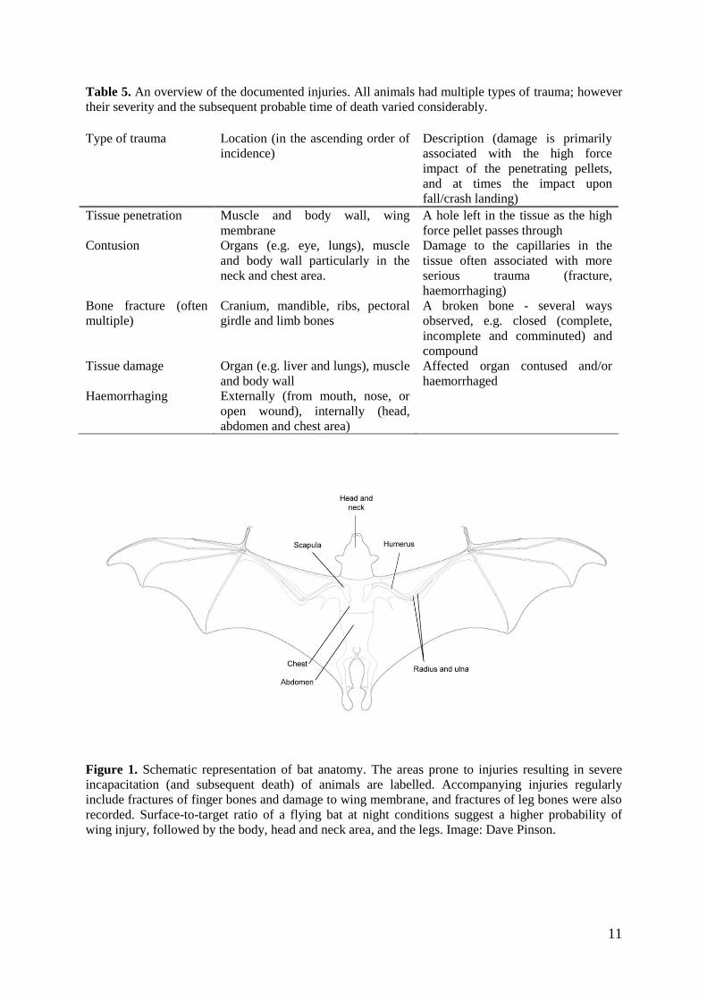
Figure 1. Schematic representation of bat anatomy. The areas prone to injuries resulting in severe
incapacitation (and subsequent death) of animals are labelled. Accompanying injuries regularly
include fractures of finger bones and damage to wing membrane, and fractures of leg bones were also
recorded. Surface-to-target ratio of a flying bat at night conditions suggest a higher probability of
wing injury, followed by the body, head and neck area, and the legs. Image: Dave Pinson.

12
Table 6. Summary of the number of flying-foxes in each sample with variable types of injuries. Each
animal had a unique combination of injuries and in its sample category could have contributed to any
of the columns (therefore the total for the columns in a row does not add up to the sample size for that
row). a – Possibly an underestimate since the number is based on a scientific external examination and
does not include probable serious internal injuries (rib fractures and internal haemorrhaging).
Sample n Injured body part
Head
and/or
neck
Body (chest
and/or
abdomen)
Wing Leg
Fracture of
arm and/or
shoulder
bones
Fracture
of hand
bones
Membrane
damage
Euthanased 34 6 20 31 15 25 5
Dead autopsied 24 7 18 19 10 20 5
Remaining dead 73 14 43a 48 19 28 5
Total 131 27 81 98 44 73 15
Percentage (%) 21 62 75 34 56 11
a) b)
Figure 2. a) External assessment of a cranial injury, showing a compound fracture of a large portion
of the cranium, exposing the brain. The nature and position of the injury indicates a close-range shot
of the bat on the ground (it was probably brought to the ground, but not immediately killed, by
another shot that shattered one wing). b) X-ray of a shot flying-fox with extensive body trauma
(internal haemorrhaging, rib and sternum fractures, and limb bone fractures), which was the likely
cause of death. Note, however, the pellet imbedded in the head, which fractured the left dentary on
impact, but caused no direct brain damage (cranium was intact).
13
Live, injured animals and euthanasia
The injuries of 34 of the 36 captured live, injured sub-adults/adults were deemed severe and
untreatable by an experienced veterinarian. The live, injured bats collected and observed
during this study provide direct evidence that some shot individuals (41 of the 150 collected
or sighted sub-adult/adult bats = 27%) do survive for many hours, even with severe body
trauma. Of the 34 euthanased flying-foxes most had severe wing trauma (Table 6) with
fractures of arm or shoulder bones, hand bones and membrane damage (Figure 3). These
animals would not have died directly from their injuries. In addition their behaviour
subsequent to being shot may have prolonged their life. For example one female with severe
breaks in her wing bones and a live young attached was observed dropping out of a fruit tree,
crawling across the ground to the casuarinas and climbing them. If the animal was able to
repeat this behaviour at night and forage in the orchard, it might have prolonged its life for
several days, extending the time it was under considerable stress and pain. She was
subsequently euthanased.
In addition, some euthanased animals had survived for a number of hours with head or body
trauma (Table 6). The type and degree of their injuries were variable (Table 6), which would
have contributed to their time of death (had they not been euthanased). Consequently, if the
injured individuals were not euthanased they may have suffered many days before
succumbing to their injuries, predation, infection or dehydration and starvation (Bellamy
2008).
a) b)
c) d)

14
Figure 3. Examples of wing related injuries in euthanased flying-foxes. a) A bat with right wing
damage: multiple membrane holes, fractures of the finger bones and the compound (open) fracture of
the humerus. The autopsy also revealed contusion of the right chest body wall. b) A bat with a
compound fracture of the left humerus and minor wing membrane damage. The autopsy revealed no
other signs of injury. c and d) Bats with closed complete fractures of the radius and ulna (the
forearm). No other injuries were documented. In all cases the fractures of long bones of the arm were
deemed severe and animals would not have been able to fly again.
Potential cause and time of death
Under the two assumptions (Table 2) the largest proportion (48%) of shot flying-foxes
probably died from internal haemorrhaging (Figure 4), within several hours after the impact.
These injuries were not always obvious in the external assessments, and it is therefore likely
that the number is an underestimate for the non-autopsied bats. The autopsies revealed that
these injuries were mainly associated with the presence of free blood in the body cavity,
fractures of the ribs and sternum and some damage to the organs (e.g. contusion of lungs). No
animals showed signs of vital organs (heart and/or both lungs) damage, suggesting that
instantaneous death from internal damage was unlikely. Contusions of the muscle and body
wall often surrounding the injury were also apparent in all autopsies, indicating a possible
further physiological damage to an individual as it falls/crash lands after being shot. In some
euthanised bats the crystals of euthanasia solution (Lethabarb) were observed in the body
cavity or liver. A relatively large proportion of bats (28%, Table 7) were also likely to have
died a slow, painful death as a result of extensive wing damage (in particular the fractures of
a) b)
c) d)

15
arm long bones and scapula, Figure 5 c and d). This is likely an overestimate and a number of
these bats in the non-autopsied sample were potentially suffering from an internal body injury
as well. Still, the figure is high (n = 16, almost 50% of the sample) for the euthanased bats,
supporting the argument that there is a very low rate of instantaneous death caused by
shooting.
Figure 4. An example of a body shot showing internal haemorrhaging in the chest. A pellet
penetrated the anterior chest body wall and muscle causing damage to the left lung and extensive
internal haemorrhaging. The animal would probably not have lost consciousness immediately, but
died in pain, sometime later.
Table 7. Probable time to death of flying-foxes after impact, based on the type and severity of
injuries. Note that animals might have endured multiple injuries from all three categories; however,
the one likely to have resulted in a faster loss of consciousness and/or loss of autonomous functions
(e.g. head trauma) was taken as the cause of death. For example, an animal with head or both the head
and wing injuries was assumed to have died relatively fast, compared to an individual with internal
body damage, and/or extensive wing damage alone (T. Bellamy, 2009 personal comment).
Sample n Probable time to death
Fast (minutes to few
hours)
Slower (several hours) Prolonged (many
hours-possibly days)
Euthanased 34 6 12 16
Dead autopsied 24 7 14 3
Remaining dead 73 14 37 18
Total 131 27 63 37
Percentage (%) 21 48 28

16
Figure 5. Examples of severe injuries resulting in a variable probable time to death.
a) A bat that has been shot in the head. The autopsy revealed haemorrhaging around the right eye
socket (the pellet is still embedded in the region), fractures of the right humerus and distal radius and
ulna, several holes in the wing membranes and contusion of the right lung. The animal had probably
died relatively fast as a result of a direct head shot.
b) A bat with no head injuries, but internal haemorrhaging in the chest area, fractured ribs and
contused lungs. The pellets found around the right shoulder have caused no fractures in the area, but
some contusion was apparent. Other injuries included multiple fractures of the right finger bones and
some contusion of the right abdominal wall. The animal has probably died from the internal chest
injuries some time after being shot.
c and d) Photographs of the external assessment and the X-ray of the same individual with a
compound (open) fracture of the left humerus. The bat also had some holes in the wing membrane and
contusions of the chest and abdomen body wall. It was collected alive and had to be euthanased, given
the extensive damage to the arm bones (a common injury seen in other shot individuals). The attached
newborn pup survived and was taken to a wildlife rehabilitation organisation.
a) b)
c)
d)

17
Assessment of the body condition and demography of shot flying-foxes
Body condition of animals and implications of feeding on commercial fruit
Collected flying-foxes had a range of body conditions with 35% of females and 63% of males
being classified as having a good condition (good fat deposits and muscle, Table 1). Only 20
(15%) of all collected sub-adult/adults had a body condition score of 1, suggesting that they
had very low body fat deposits and were generally underweight. The body mass/forearm ratio
provided a more detailed assessment of the overall condition of the shot animals, and the
general mean was 3.74 (ranging from 2.74 to 5.50). There was a weak trend of sub-adults (1-
2-year-olds) having a lower body mass/forearm ratio compared to adult animals irrespective
of the sex, which is expected given their smaller body size. However, the overlap in values
was large, particularly for the 2-year-olds. Males generally had a higher body condition index
in all age groups (statistical results not presented, Figure 6), again a consequence of their
larger body mass (and to a lesser extent their larger forearm lengths; Chapman et al. 1994). In
addition, the pregnant/lactating females would have been under increased physiological
stress, resulting from the elevation in basal metabolic rate and an increase in
thermoregulatory needs induced by their reproductive status (Welbergen 2005).
To maintain a healthy body condition, the high energetic demands of breeding females need
to be balanced by an increased nutritional input, both in terms of the quantity and
composition of food. However, this balance can be difficult to achieve (particularly in the
years of high environmental stress – e.g. food shortage) and females can struggle to meet
their energetic requirements, leading to a decrease in their body reserves, as residual
carbohydrates and fat reserves are exhausted (Duverge et al. 2000; Pinson 2009). This could
explain the observed pattern of reproductive females (≥3 years old) having a relatively low
body condition index, with 30% of females just below the proposed malnutrition mark
(Figure 6). Surprisingly, 53% of adult males were below the lower 15% limit of the expected
mean condition for male bats in summer. This indicates that a high proportion of shot
animals, both male and female, may not have been meeting their nutritional requirements.
18
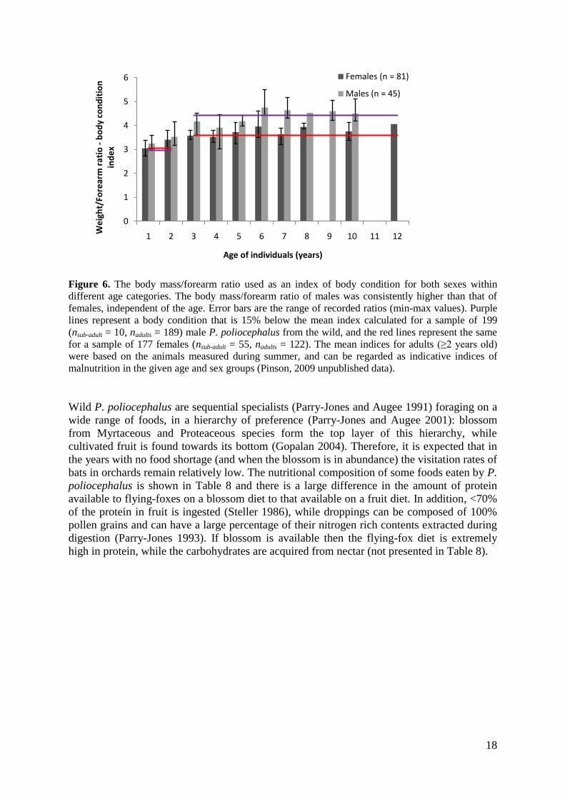
Figure 6. The body mass/forearm ratio used as an index of body condition for both sexes within
different age categories. The body mass/forearm ratio of males was consistently higher than that of
females, independent of the age. Error bars are the range of recorded ratios (min-max values). Purple
lines represent a body condition that is 15% below the mean index calculated for a sample of 199
(nsub-adult = 10, nadults = 189) male P. poliocephalus from the wild, and the red lines represent the same
for a sample of 177 females (nsub-adult = 55, nadults = 122). The mean indices for adults (≥2 years old)
were based on the animals measured during summer, and can be regarded as indicative indices of
malnutrition in the given age and sex groups (Pinson, 2009 unpublished data).
Wild P. poliocephalus are sequential specialists (Parry-Jones and Augee 1991) foraging on a
wide range of foods, in a hierarchy of preference (Parry-Jones and Augee 2001): blossom
from Myrtaceous and Proteaceous species form the top layer of this hierarchy, while
cultivated fruit is found towards its bottom (Gopalan 2004). Therefore, it is expected that in
the years with no food shortage (and when the blossom is in abundance) the visitation rates of
bats in orchards remain relatively low. The nutritional composition of some foods eaten by P.
poliocephalus is shown in Table 8 and there is a large difference in the amount of protein
available to flying-foxes on a blossom diet to that available on a fruit diet. In addition, <70%
of the protein in fruit is ingested (Steller 1986), while droppings can be composed of 100%
pollen grains and can have a large percentage of their nitrogen rich contents extracted during
digestion (Parry-Jones 1993). If blossom is available then the flying-fox diet is extremely
high in protein, while the carbohydrates are acquired from nectar (not presented in Table 8).
0
1
2
3
4
5
6
1 2 3 4 5 6 7 8 9 10 11 12
We
igh
t/Fo
rear
m r
atio
-b
od
y co
nd
itio
n
ind
ex
Age of individuals (years)
Females (n = 81)
Males (n = 45)

19
Table 8. Foods eaten by P. poliocephalus (Parry-Jones 1993) and their composition (Wills et al.
1987; Stace 1996). Nectar analysis is not presented.
Name Protein (%) Carbohydrates (%) Fats (%)
Pollen*
Banksia ericifolia 30.9 - -
Corymbia maculata 27.8 - -
Eucalyptus tereticornis 26.6 - -
Eucalyptus pilularis 21.8 - -
Melaleuca quinquenervia 22.7 - -
Fruit**
Apple 0.3 11.9 0.1
Fig 1.4 8.1 0.3
Nectarine 1.1 9.2 0.1
Peach 1.0 6.6 0.1
Pear 0.3 12.2 0.1
Plum 0.6 6.1 0.1
* Data from Stace 1996
** Data from Wills et al. 1987
It is likely that bats utilise an optimum foraging strategy in order to maximise their chances
for survival. In the absence of abundant natural food resources, a proportion of animals
(particularly those with high energetic needs, i.e. pregnant or lactating females and
particularly those in poor condition) may utilise the orchard in combination with scarce
native food resources. One shot bat, for example, had pollen on its fur, suggesting that it had
been feeding elsewhere, before coming to the orchard. Moreover, the commercial fruit may
offer a fast energy source (carbohydrates) which the animals may utilise in the energetically
costly flight to a preferred native food source.
Sex ratios, reproductive condition and ages
The total number of bats ≥1 year old was 131 (nfemales = 83, nmales = 48), and a significantly
higher number of females were shot (χ2
= 9.351, d.f. = 1, P = 0.0022). This is likely to reflect
sex bias in orchard visitation rates, and is to be expected (Tidemann et al. 1997; Duverge et
al. 2000), given the high energy requirements of females in the reproductive season due to
pregnancy/lactation and raising young. Twenty two (27%) females were not reproductive, of
which eight were showing signs of „pseudo-lactating‟. Sixty one (73%) of the total 83
females were reproductive, of which four (5%) were pregnant at the time (Figure 7), three
(4%) had lactated previously in their life-time but not in the study period and 54 (65%) were
lactating. Of the lactating females 13 were shot while carrying their newborn pup and of these
five died with their mother while eight were taken into care. Forty one females had therefore
left their dependent, ≥3-weeks-old young at the camp. This is consistent with the typical
breeding cycle for the species (Martin et al. 1995; Martin and McIlwee 2002). In addition, 59
(88%) of the females ≥3 years old (n = 67) were reproductive at the time of the study, and
females showed no signs of reproductive senescence, characteristic of other small mammals
and birds (Cluttonbrock 1984; Rockwell et al. 1993). This large proportion of reproductive
individuals is also indicative of favourable environmental conditions in 2007. Otherwise,
under adverse environmental conditions, the optimal strategy for female bats would have
been to allocate the resources to self maintenance instead of reproduction, resulting in lower

20
reproductive output (Eby 1999). The 48 males were divided into 19 (40%) non-reproductive
(<3 years old) and 29 (60%) reproductive (≥3 years old) individuals.
Figure 7. An X-ray of a pregnant female. The autopsy revealed anterior skull fractures (likely cause
of death, if the shown pellet has penetrated the brain) and damage to the right wing (three pellets can
still be seen on the X-ray).
The age structure of the collected sample is summarised in Figure 8. The age of animals of
both sexes varied from a newborn pup to an adult at least 12 years old, and 87% of the
sample were animals ≤6 years old. This apparent skewness towards younger adults groups
might appear surprising, given the reported longevity of the species to >20 years (Divljan et
al. 2006). In addition, there was an apparent lack of animals between 1-3 years old, which is
primarily driven by the age distribution of females (Figure 8). The same pattern was observed
in the age distributions of both a collection of dead individuals between 1999-2007 and a live
camp study in the Sydney Basin (Divljan 2008). The timeline of the studies combined, and
the close-to-maximum fecundity rates of females, suggest that the pattern is not likely an
effect of variable (and low) recruitment into the population, but a result of age-based
partitioning of the population (Divljan 2008) with the majority of 1-3-year-olds being outside
the Sydney Basin at the time.

21
Figure 8. Age distribution of the shot flying-foxes collected dead in November 2007, and dependent
pups that would have been left in the camp. The age distribution is driven primarily by the larger
sample of female bats, particularly in the ages 4-6.
Overall the age distribution of shot flying-foxes is consistent with the pattern observed for P.
poliocephalus sampled in Sydney Basin (Kolmogorov-Smirnov test: D = 0.2941, P = 0.387;
Divljan 2008), suggesting that bats foraging in orchards are a representative sample of
Sydney bats. This age structure, therefore, supports the results of an earlier, linear time-
invariant population model, based on such age distribution, which showed a negative
population growth rate (λ = 0.898) for the Sydney population (Divljan 2008). The model
further demonstrated a population halving time to be 6.47 years, which leads to the extinction
of this vulnerable species in the next 84 years, under constant (current) conditions involving
high mortality rates from anthropogenic causes including shooting (Divljan 2008).
Conclusion
A total of 164 flying-foxes were observed as having been shot during this study. This number
should be considered the minimum number of animals shot in the orchard, as it does not
include animals that were still able to fly out of the orchard having sustained minor or major
injuries that sooner or later would result in their death; and it does not include the young left
at the camp site.
The animals that were shot in the orchards were predominantly female and most of the
females were either lactating or pregnant. Overall, they were in less than optimal condition
which is probably the reason for their presence in the orchard. Additionally, the age structure
of the flying-foxes shot in the orchard was significantly similar to that found for bats in the
Sydney Basin, suggesting that the animals foraging in orchards are a representative sample of
the overall Sydney population.
Flying-foxes forage in orchards at night and most of the dark shape of a flying-fox flying
against the dark sky is made up of wing. Hence most of the shot flying-foxes had wing
injuries that alone would not have directly killed the animal. A smaller number of animals
0
5
10
15
20
25
30
35
40
45
50
0 1 2 3 4 5 6 7 8 9 10 11 12
Fre
qu
en
cy o
f in
div
idu
als
Age of individuals (years)
Dependent pups left in the camp (n = 41)
Females (n = 83)
Males (n = 48)

22
had body injuries and only a few had serious head injuries. These animals would have taken
varying times to die.
Almost a third of flying-foxes that were shot (not including newborn pups who were on their
mothers, but not directly injured) were alive hours and at times days after being shot. Of the
animals that were dead when collected, based on their injuries in comparison with the injuries
of the live animals, many would have taken a considerable length of time to die. Only three
animals had apparently been shot after falling to the ground after an initial injury in an
apparent attempt at euthanasia. Hence the treatment of shot flying-foxes as documented in
this study is in contravention of the definition of “humane killing” in the guidelines defined
by the Australian National Health and Medical Research Council (2004) and in contravention
of the DECC licence provisions that states that all authorised persons “… must locate each
animal shot and promptly alleviate the suffering of any injured flying-fox by gunshot to either
the head or thorax of the animal.” Live, injured flying-foxes have been opportunistically
observed or collected after shooting at several other sites where shooting has taken place,
confirming that this outcome is not unique. Importantly, the Grey-headed Flying-fox,
Pteropus poliocephalus is a threatened native species and the killing of reproducing females
in crops must contribute to its declining numbers.
References
AWAC. 2008. Animal Welfare Advisory Committee's Recommendations. Advice to the
Minister for Primary Industries and Fisheries, The Honourable Tim Mulherin QLD:
Animal Welfare Advisory Committee (Qld)
Bellamy, T., BVSc (Hons), DipWMEH, MENVSc, CAM. 2008. Interim Report on Non-
fatal Shooting Injuries Affecting Grey-headed Flying-foxes.
Chapman, A., Hall, L. S. and Bennett, M. B. 1994. Sexual dimorphism in the pelvic girdle
of Australian flying foxes. Australian Journal of Zoology 42: 261-265.
Cluttonbrock, T. H. 1984. Reproductive effort and terminal investment in iteroparous
animals. American Naturalist 123: 212-229.
Department of Environment and Climate Change (NSW). 2007. Annual Report on
Licences Issued Under the DECC Policy and Procedural Guidelines for the Mitigation
of Commercial Crop Damage by Flying-foxes for the 2006-07 Fruit Growing Season
Hurstville, NSW: Department of Environment and Climate Change (NSW).
Department of Environment and Conservation (NSW). 2005. DEC Policy for the
Mitigation of Damage to Commercial Fruit Crops by Flying-foxes, Updated June
2005. Hurstville, NSW: Department of Environment and Conservation (NSW).
Deutsch, C. J., Haley, M. P. and Le Boeuf, B. J. 1990. Reproductive effort of male
northern elephant seals: estimates from mass loss. Canadian Journal of Zoology 68:
2580-2593.
Divljan, A. 2008. Population ecology of the Grey-headed Flying-fox, Pteropus
poliocephalus: A study on the age-structure and the effects of mortality on a
vulnerable species. PhD thesis, University of Sydney.
Divljan, A., Parry-Jones, K. and Wardle, G. M. 2006. Age determination in the Grey-
headed flying fox. Journal of Wildlife Management 70: 607-611.
Duverge, P. L., Jones, G., Rydell, J. and Ransome, R. D. 2000. Functional significance of
emergence timing in bats. Ecography 23: 32-40.
Eby, P. 1999. Low reproductive output in Grey-headed Flying-foxes associated with a short
period of food scarcity. Australasian Bat Society Newsletter 14: 17-20.

23
Gopalan, P. 2004. Food preference of the grey-headed flying-fox Pteropus poliocephalus.
Honours thesis, University of Sydney.
Hall, L. S. and Richards, G. 2000. Flying Foxes: Fruit and Blossom Bats of Australia.
University of New South Wales: Sydney, NSW.
Martin, L., Kennedy, J. H., Little, L., Luckhoff, H. C., O'Brien, G. M., Pow, C. S. T.,
Towers, P. A., Waldon, A. K. and Wang, D. Y. 1995. The reproductive biology of
Australian flying-foxes (genus Pteropus). Symposia of the Zoological Society of
London 67: 167-184.
Martin, L. and McIlwee, A. P. 2002. The reproductive biology and intrinsic capacity for
increase of the Grey-headed Flying-fox Pteropus poliocephalus (megachiroptera), and
the implications of culling. Pp. 91-108. in Managing the Grey-headed Flying-fox as a
Threatened Species in New South Wales, edited by P. Eby and D. Lunney. Royal
Zoological Society of New South Wales, Mosman, NSW.
McIlwee, A. P. and Martin, L. 2002. On the intrinsic capacity for increase of Australian
flying-foxes (Pteropus spp. Megachiroptera). Australian Zoologist 32: 76-100.
McLachlan, B. 2002. NPWS Operational management of commercial crop damage by
flying-foxes – licensing in practice a far north coast perspective. Pp. 80-83. in
Managing the Grey-headed Flying-fox as a Threatened Species in NSW, edited by P.
Eby and D. Lunney. Royal Zoological Society of New South Wales, Mosman, NSW.
Nelson, J. E. 1965. Behaviour of Australian Pteropodidae (Megachiroptera). Animal
Behaviour 13: 544-557.
NHMRC. 2004. Australian Code of Practice for the Care and Use of Animals for Scientific
Purposes, 7th edition. Canberra, ACT: NHMRC - National Health and Medical
Research Council.
Parry-Jones, K. 1993. The movements of Pteropus poliocephalus in New South Wales. PhD
thesis, University of New South Wales.
Parry-Jones, K. 2000. Historical declines since the early 1900s, and current mortality factors
and abundance of the Grey-headed Flying-foxes in NSW. Pp. 56-65. in Proceedings
of a Workshop to Assess the Status of the Grey-headed Flying-fox in New South
Wales, edited by G. Richards. Available from:
<http://batcall.csu.edu.au/abs/ghff/ghffproceedings.pdf>. Accessed: 20 March, 2006.
Parry-Jones, K. and Augee, M. L. 1991. Food selection by Gray-headed Flying foxes
(Pteropus poliocephalus) occupying a summer colony site near Gosford, New-South-
Wales. Wildlife Research 18: 111-124.
Parry-Jones, K. and Augee, M. L. 2001. Factors affecting the occupation of a colony site in
Sydney, New South Wales by the Grey-headed Flying-fox Pteropus poliocephalus
(Pteropodidae). Austral Ecology 26: 47-55.
Pinson, D. 2009. The Flying-fox Manual, 2nd Edition. Murwillumbah, NSW: StickeeBatz
Publishing.
Ratcliffe, F. N. 1931. The flying fox (Pteropus) in Australia. Bulletin of CSIRO 53: 1-80.
Rockwell, R. F., Cooch, E. G., Thompson, C. B. and Cooke, F. 1993. Age and
reproductive success in female Lesser Snow Geese - Experience, senescence and the
cost of philopatry. Journal of Animal Ecology 62: 323-333.
Stace, P. 1996. Protein content and amino acid profiles of honeybee-collected pollens.
Available from: <http://www.honeybee.com.au/Library/Pollenindex.html>. Accessed:
3 March, 2009.
Steller, D. C. 1986. The dietary energy and nitrogen requirements of the Grey-headed
Flying-fox Pteropus poliocephalus (Temminck) (Megachiroptera). Australian Journal
of Zoology 34: 339-350.

24
Tidemann, C. R., Kelson, S. L. and Jamieson, G. 1997. Flying-fox damage to orchard fruit
in Australia - incidence, extent and economic impact. Australian Biologist 10: 179-
186.
Wahl, D. E. 1994. The management of flying foxes (Pteropus spp.) in New South Wales.
Degree of Master of Applied Science, University of Canberra.
Waples, K. 2002. Review of the NPWS policy on the mitigation of commercial crop damage
by flying-foxes. Pp. 39-46. in Managing the Grey-headed Flying-fox as a Threatened
Species in New South Wales, edited by P. Eby and D. Lunney. Royal Zoological
Society of New South Wales, Mosman, NSW.
Welbergen, J. A. 2005. The social organisation of the grey-headed flying-fox, P.
poliocephalus. PhD thesis, University of Cambridge.
Wills, R. B., Lim, J. S. and Greenfield, H. 1987. Composition of Australian foods. 40.
temperate fruits. Food Technology in Australia 39: 520-522.