REPORT OF THE INTENSIVE CARE FELLOWSHIP …REPORT OF THE INTENSIVE CARE FELLOWSHIP EXAMINATION...
49
1 REPORT OF THE INTENSIVE CARE FELLOWSHIP EXAMINATION MARCH/ / MAY 2013 This report is prepared to provide candidates, tutors and Supervisors of Training with information regarding the assessment of candidates’ performance in the General Fellowship Examination. Answers provided are not necessarily model answers but guides as to what was expected. Candidates should discuss the report with their tutors so that they may prepare appropriately for future examinations. The exam comprises a written section and an oral section. The written exam consists of two 2.5 hour papers of 15 ten-minute short answer questions each. Candidates are required to score at least 50% in the written section to be eligible to sit the oral section. The oral exam consists of eight interactive vivas and two separate clinical “hot cases”. The tables below provide an overall statistical analysis as well as information regarding performance in the individual sections. A comparison with data from the four previous exams is provided.
Transcript of REPORT OF THE INTENSIVE CARE FELLOWSHIP …REPORT OF THE INTENSIVE CARE FELLOWSHIP EXAMINATION...
1
REPORT OF THE INTENSIVE CARE FELLOWSHIP EXAMINATION
MARCH/ / MAY 2013
This report is prepared to provide candidates, tutors and Supervisors of Training with information regarding the assessment of candidates’ performance in the General Fellowship Examination. Answers provided are not necessarily model answers but guides as to what was expected. Candidates should discuss the report with their tutors so that they may prepare appropriately for future examinations. The exam comprises a written section and an oral section. The written exam consists of two 2.5 hour papers of 15 ten-minute short answer questions each. Candidates are required to score at least 50% in the written section to be eligible to sit the oral section. The oral exam consists of eight interactive vivas and two separate clinical “hot cases”. The tables below provide an overall statistical analysis as well as information regarding performance in the individual sections. A comparison with data from the four previous exams is provided.

2
Examination Statistical report
Candidates May-13 Oct-12 May-12 Oct-11
Presenting for written (including OTS) 27 43 41 55Carrying a pass from a previous attempt 7 13 11 11Total number presenting (written + carry + OTS) 34 56 52 66
Pathway to oral section May-13 Oct-12 May-12 Oct-11
Invited to orals (50% in written section) 18 29 26 45Total sitting oral section 25 42 37 56
Analysis of performance in individual sections May-13 Oct-12 May-12 Oct-11
18 29 26 4567% 67% 63% 82%
9 21 15 3936% 50% 41% 70%
7 10 7 2228% 24% 19% 39%15 36 22 44
60% 86% 59% 79%
Sectional pass rate - Vivas
Pass rateHighest
Individual Mark
Pass rateHighest
Individual Mark
Viva 1 48% 75% 90% 90%Viva 2 88% 81% 88% 75%Viva 3 56% 75% 40% 80%Viva 4 52% 76% 81% 93%Viva 5 60% 90% 98% 95%Radiology Viva 64% 83% 48% 90%Communication Viva 44% 88% 67% 100%Procedure Viva 20% 95% 71% 100%
Oral section pass rate May-13 Oct-12 May-12 Oct-11
11 24 19 3641% 83% 73% 80%13 31 20 43
52% 74% 54% 77%
Overall Pass Rate
Candidates who scored >50% in the written and passed the overall examAll candidates invited to the oral section and passed the overall exam (written + carry + OTS)
55% 38%
May-13
65%38%
Successful in the written section
Successful in the Viva section
Successful in both Hot Cases
Successful in the Hot Case section
Oct-12

3
4
EXAMINERS’ COMMENTS Written Section Twelve of the thirty questions had an overall pass rate of less than 50%. Topics covered by questions with a pass rate of less than 40% related to the role of daily interruption of sedation in ICU; Guillain Barre syndrome; clinical signs; intensive care outcome measures and regional citrate anticoagulation for CRRT. There was an apparent lack of understanding of commonly used drugs in ICU such as digoxin and nor-adrenaline. Candidates did not pass questions for one or more of the following reasons:
• Insufficient knowledge of the topic in question • Insufficient detail and/or depth of the answer • Poorly structured answer • Inadequate reference to supportive evidence • Failure to answer the question as asked • Fatal error / omission of key points (critical pass responses) • Omission of all or part of the question
It was noted that some answers relating to discussion or critical evaluation of a topic were not at the required level in terms of synthesis of information and depth of discussion. It also appears that candidates do not always read the questions carefully and thoroughly. Candidates who failed the written section passed an average of 11/30 questions compared with candidates scoring > 50% and gaining an invitation to the oral section, passing an average of 24/30 questions. Hot Cases Reasons for not achieving a pass mark in one or both Hot Cases included:
• Failure to address the question posed by the examiners • Poor examination technique • Missing important clinical signs • Disorganised presentation • Poor interpretation of investigations • Poor discussion and management plan
Candidates are reminded that they should not sit the General Fellowship Examination until they can confidently examine patients, present the relevant clinical findings and discuss management issues at the appropriate level, i.e. experienced senior registrar/junior consultant. Candidates are also encouraged to practice examination of individual systems.

5
Candidates who performed well in the Hot Cases demonstrated the following:
• Respect and consideration for the patient; • Competent and efficient examination technique; • The seeking of information that was relevant to the case; • Ability to interpret and synthesise their findings appropriately; • Presentation of their conclusions in a systematic fashion, addressing the issue in
question; • Discussion of management issues in a mature fashion, displaying confident and
competent decision-making • Overall performance at the expected level (competent Senior Registrar / Junior
Consultant) Vivas Three vivas had an overall pass rate of less than 50% - Viva 1 (management of influenza epidemic), Viva 7 (communication) and Viva 8 (procedure). Practical knowledge of aspects of temporary pacing was noted to be poor.

6
WRITTEN SECTION
GLOSSARY OF TERMS Critically evaluate: Evaluate the evidence available to support the hypothesis. Outline: Provide a summary of the important points. List: Provide a list. Compare and contrast: Provide a description of similarities and differences
(E.g. Table form). Management: Generic term that implies overall plan. Where appropriate, may
include diagnosis as well as treatment. Discuss: Explain the underlying key principles. Where appropriate, this
may include controversies and/or pros and cons Where laboratory values are provided, abnormal values are marked with an asterisk (*). Please note that in this report images from the SAQs have been removed. 1. Critically evaluate the role of a daily interruption of sedation for mechanically ventilated patients in the ICU. Introduction / Rationale A daily interruption of sedation is a strategy designed to reduce exposure to sedative agents, allow assessment of neurological status and assess readiness for extubation and to reduce duration of mechanical ventilation. Evidence Initial trials showed a marked reduction in duration of mechanical ventilation, and decreased duration of intensive care length of stay (e.g. Kress et al, NEJM 2000). It was notable that no sedation target nor protocol was used in the control group, thus this group may have been oversedated, analogous to the 12ml/kg TV group in the ARDSNET low TV trial. Subsequent studies have been somewhat conflicting:
• ABC study (Girard et al, Lancet 2008) showed improved outcomes (mortality, less time on mechanical ventilation, reduced ICU length of stay) in patients treated with a paired daily interruption of sedation and a spontaneous breathing trial compared to usual care plus a spontaneous breathing trial.
• SLEAP study (Mehta et al JAMA, 2012) showed no difference in outcomes comparing protocolised sedation to protocolised sedation plus daily interruption of sedation.
Disadvantages / Adverse effects / Limitations Potential adverse effects of daily interruption of sedation:
• Patient discomfort and risk of PTSD and other long term psychological issues • Dislodgment of ETT, CVC, arterial lines etc. • Increased nursing workload. • Cessation of sedation could lead to agitation which can be associated with
physiological instability, hypertension, tachycardia, ventilator dysynchrony and hypoxaemia, which could be associated with exacerbation of primary disease in certain conditions, e.g. myocardial ischaemia, brain injury. I.e. interruption of sedation contra-indicated in above patient groups.

7
Own Practice Any reasonable justifiable approach acceptable. Summary Daily interruption of sedation may have a role in physiologically stable patients in ICUs that do not routinely use protocolised sedation. 2. With regards to the use of high-flow nasal oxygen therapy in adults:
a) Describe the mechanisms by which high flow nasal oxygen therapy is believed to exert its beneficial effects.
b) List two potential adverse effects associated with the use of high-flow nasal
oxygen therapy.
c) List two relative contraindications to the use of high-flow nasal oxygen therapy.
a) Precise mechanism of benefit is yet to be elucidated. 1) Increased FiO2:
- Gas inlet flow limits secondary room air entrainment. - Provides anatomic oxygen reservoir in nasopharynx and oropharynx. - Rinsing of airway deadspace with oxygen.
2) CPAP effect: - decreases atelectasis and improves V-Q matching. - improves compliance. - decreases work of breathing by counteracting intrinsic PEEP.
3) Greater comfort: - Warmed and humidified oxygen can be better tolerated, therefore better compliance from patients.
b) Pressure areas in the nose. Epistaxis. Possible gastric distension. c) Nasal fracture. Upper airway haemorrhage. Base of skull fracture. Recent upper airway or aerodigestive tract surgery.

8
3. For each set of the following biochemical and arterial blood gas parameters:
a) Describe the abnormalities. b) Give one example of an associated clinical scenario.
Any reasonable scenario accepted that was both biochemically correct AND clinically likely. 3.1
Test Value Normal Adult Range Sodium 135 mmol/L 135 – 145 Potassium 4.0 mmol/L 3.2 – 4.5 Chloride 110 mmol/L 100 – 110 Bicarbonate* 3 mmol/L 24 – 32 pH* 7.10 7.35 – 7.45 pCO2* 10 mmHg (1.3 kPa) 35 – 45 (4.6 – 5.9) Increased anion gap and normal anion gap metabolic acidosis with appropriate respiratory compensation. Clinical scenario – diabetic ketoacidosis with ketonuria or DKA with N saline resuscitation. 3.2
Test Value Normal Adult Range Sodium 145 mmol/L 135 – 145 Potassium 4.0 mmol/L 3.2 – 4.5 Chloride* 91 mmol/L 100 – 110 Bicarbonate 30 mmol/L 24 – 32 pH* 7.62 7.35 – 7.45 pCO2* 30 mmHg (3.9 kPa) 35 – 45 (4.6 – 5.9) Increased anion gap, metabolic alkalosis and respiratory alkalosis. Clinical scenario – salicylate overdose. 3.3
Test Value Normal Adult Range Sodium 145 mmol/L 135 – 145 Potassium 4.0 mmol/L 3.2 – 4.5 Chloride* 96 mmol/L 100 – 110 Bicarbonate 25 mmol/L 24 – 32 pH 7.42 7.35 – 7.45 pCO2 40 mmHg (5.2 kPa) 35 – 45 (4.6 – 5.9) Increased anion gap metabolic acidosis and metabolic alkalosis. Clinical scenario – acute renal failure with vomiting .

9
3.4
Test Value Normal Adult Range Sodium 145 mmol/L 135 – 145 Potassium 4.0 mmol/L 3.2 – 4.5 Chloride 101 mmol/L 100 – 110 Bicarbonate* 34 mmol/L 24 – 32 pH* 7.2 7.35 – 7.45 pCO2* 90 mmHg (11.7 kPa) 35 – 45 (4.6 – 5.9) Acute respiratory acidosis with metabolic alkalosis. Clinical scenario – acute respiratory failure in COAD (Acute on chronic respiratory failure. 3.5
Test Value Normal Range Sodium 135 mmol/L 135 – 145 Potassium 4.0 mmol/L 3.2 – 4.5 Chloride 105 mmol/L 100 – 110 Bicarbonate* 22 mmol/L 24 – 32 pH* 7.60 7.35 – 7.45 pCO2* 23 mmHg (3.0 kPa) 35 – 45 (4.6 – 5.9) Acute respiratory alkalosis. Clinical scenario – Psychogenic hyperventilation. 4. With respect to the use of digoxin in the ICU:
a) What key cardiac effects are observed with acute digoxin toxicity? List two rhythm disturbances highly associated.
b) List three drugs known to enhance digoxin serum level. Provide a mechanism for each.
c) Other than drugs, what other factors are known to exacerbate digoxin toxicity?
d) With respect to the use of digoxin specific Fab fragments:
i) Outline your indications for use in suspected acute digoxin toxicity.
ii) Total serum digoxin level continues to remain high after the
administration of an appropriate dose of digoxin specific Fab fragments. What action would you take and why?

10
a)
Key cardiac features are increased automaticity combined with AV conduction block. Rhythms suggestive: PAT with variable block Accelerated junctional rhythms Bidirectional ventricular tachycardia (specific for Digoxin). Other (a variety are seen): SA node arrest, premature ventricular contractions, bradycardia, non paroxysmal junctional tachycardia, AV nodal blockade, ventricular tachycardia, ventricular flutter and fibrillation. Note: Features of digoxin effect (e.g. T wave flattening/ inversion) do not correlate well with toxicity.
b)
Verapamil, Diltiazem, Amiodarone via inhibition of P-glycoprotein (efflux pump that excretes many drugs, including Digoxin, into the intestine or proximal renal tubule) - effectively reducing renal and GI secretion. Erythromycin, omeprazole via increased Digoxin absorption.
c) Low potassium, magnesium, pH, high calcium.
d)
Early recognition of toxicity and prompt administration of Fab fragments essential for severe poisoning. The serum Digoxin concentration does not necessarily correlate with toxicity. Indications Include: Life threatening arrhythmia with cardiovascular instability Evidence of end organ dysfunction Hyperkalaemia (> 5.0 – 5.5 mEq/l) Ingestion of 10mg or more in total
After Fab administration free Digoxin levels are decreased to zero within minutes. Total Digoxin level will increase markedly since assays measure bound and free. Bound fraction rises due to an increase in Digoxin-Fab complex. These high levels have no correlation with toxicity and the serum level may be unreliable for several days and no action should be taken based on total level after digoxin-specific Fab fragments administration. 5. One of the serious complications of aneurysmal sub-arachnoid haemorrhage (SAH) is Delayed Cerebral Ischaemia (DCI). Briefly discuss DCI, including in your answer its assessment and management with reference to accepted and postulated strategies. DCI is a common (occurring in about 30%) and serious complication following SAH. Defined as any neurological deterioration presumed related to ischaemia that persists for more than an hour and cannot be explained by any other physiological abnormalities. It is mostly caused by vasospasm.

11
May be reversible but may develop into cerebral infarction. Its highest risk of occurrence is from day 3 to 14 after presentation. Aetiology still poorly understood Assessment of DCI Whilst up to 20% of patients can have a cerebral infarct despite being entirely asymptomatic, the mainstay of clinical monitoring is repeated clinical neurological examination. DSA is the gold standard for vasospasm but as a screening test, multimodal CT imaging with CT perfusion is accurate and less invasive. Transcranial Doppler has a high specificity but only moderate sensitivity. The other physiological modalities including EEG, brain tissue oxygen monitoring, cerebral microdialysis are less well established as monitoring modalities Management of DCI Aim is to prevent or minimise secondary injuries by haemodynamic management, drugs and endovascular procedures. Haemodynamic strategies Avoid hypotension. Triple H therapy:
• Induced hypertension improves CBF independent of blood volume. The most common agents used being Norad and phenylepherine. Secure aneurysm first.
• Hypervolaemia does not offer any benefit over euvolaemia, however it is important to avoid hypovolaemia.
• There is no place for haemodilution except for people with polycythaemia. Milrinone infusion has been used as alternative to Triple H. Pharmacological management
• Calcium-channel blockers The main intervention shown to be beneficial is the use of Nimodipine. Other CCBs have been shown to reduce vasospasm with beneficial effects on DCI but RCTs still needed.
• Intra-cisternal thrombolytics Used in some centres on selected patients
• Statins Evidence is conflicting. Awaiting results of STASH
• Magnesium sulphate MASH-II did not show a benefit compared with placebo
• Endothelin-1 antagonists Recent published studies evaluating clazosentan have shown no clinical benefit Other agents E.g. NO donors, EPO, enoxaparin, rho-kinase inhibitor either shown not to be beneficial or still being studied Endovascular procedures Angioplasty and/or Intra-arterial vasodilators may be used in addition to nimodipine and haemodynamic management if indicated and expertise available. Overall optimal triggers for escalating and de-escalating therapy not well defined

12
6. 6.1. The following arterial blood gas was taken from a female hospitalised for recurrent urinary tract infections. She was transferred to the ICU because of nosocomial pneumonia.
Test Value Normal Adult Range Barometric Pressure 760 mmHg (100kPa) FiO2 0.3 pH* 7.53 7.35 – 7.45 pCO2* 31 mmHg (4 kPa) 35 – 45 (4.6 – 5.9) pO2* 83 mmHg (11 kPa) Bicarbonate 25 mmol/L 24 – 32 Standard Base Excess* 3.3 mmol/L -2.0 – +2.0
a) Comment on the acid-base status. b) List two likely causes of the acid-base abnormality in this patient.
6.1 a)
Mixed respiratory and metabolic alkalosis 6.1 b)
Respiratory alkalosis from the hyperventilation due to the pneumonia. Metabolic alkalosis from vomiting or diuretic use.
6.2. A 29-year-old female is admitted to ICU extubated following an emergency Caesarian section at 38 weeks gestation for pre-eclampsia and failure to progress. The following data were taken on admission to ICU:
Test Value Normal Adult Range Barometric Pressure 760 mmHg (100 kPa) FiO2 0.4 pH* 7.31 7.35 – 7.45 pCO2 42 mmHg (5.6 kPa) 35 – 45 (4.6 – 5.9) pO2 110 mmHg (14.5 kPa) Bicarbonate* 20.5 mmol/L 24 – 32 Standard Base Excess* -4.9 mmol/L -2.0 – +2.0
a) Comment on this arterial blood gas report and explain the abnormalities.
6.2 a)
Increased A-aDO2 secondary to decreased FRC post surgery or possible collapse/consolidation or aspiration or pulmonary oedema. Acute respiratory acidosis on background compensated respiratory alkalosis of pregnancy. CO2 retention secondary to e.g. hypoventilation post anaesthetic.
13
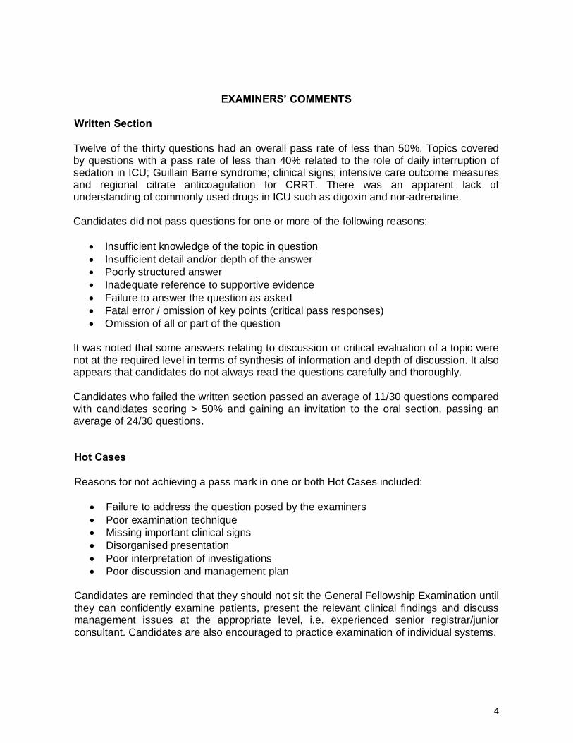
6.3. Following laparotomy for haemoperitoneum, a patient is transferred to ICU. Blood biochemistry and arterial blood gas analysis on admission to ICU are as follows:
Test Value Normal Adult Range Sodium* 147 mmol/L 135 – 145 Potassium 3.6 mmol/L 3.2 – 4.5 Chloride* 124 mmol/L 100 – 110 Haemoglobin* 106 G/L 115 – 155 pH* 7.32 7.35 – 7.45 pCO2* 32 mmHg (4.3 kPa) 35 – 45 (4.6 – 5.9) pO2* 63 mmHg (8.4 kPa) Bicarbonate* 16.0 mmol/L 24 – 32 Standard Base Excess* -9.0 mmol/L -2.0 – +2.0
a) Describe the acid-base status. b) What is the likely cause of this disturbance?
c) What is the underlying biochemical mechanism?
6.3 a)
Normal anion gap metabolic acidosis with appropriate respiratory compensation. 6.3 b)
Resuscitation with large volume saline infusion. 6.3 c)
ECF dilution by fluid with strong ion difference of zero. 6.4. A 33 year old female has Gram-negative bacteraemia and septic shock. The following are data from blood gas analysis.
Test Value Normal Adult Range Barometric pressure 760 mmHg (100 kPa) FiO2 0.3 pH 7.43 7.35 – 7.45 pCO2* 23 mmHg (3.1 kPa) 35 – 45 (4.6 – 5.9) pO2 107 mmHg (14.3 kPa) Bicarbonate* 15 mmol/L 24 – 32 Standard Base Excess* -8.6 mmol/L -2.0 – +2.0 Lactate* 23.0 mmol/L 0.2 – 2.5 Sodium* 147 mmol/L 137 – 145 Potassium* 6.7 mmol/L 3.2 – 4.5 Chloride* 95 mmol/L 100 – 110
a) Describe the acid-base abnormalities.
b) What are the possible mechanisms for a raised lactate in sepsis?

14
6.4 a)
Lactic acidosis with anion gap elevation (37 mEq/L). Metabolic alkalosis (Delta ratio >2). Respiratory alkalosis (PCO2 lower than predicted for HCO3).
6.4 b)
Tissue hypoperfusion and hypoxia Use of adrenaline (increased glycolytic flux) Down regulation of pyruvate dehydrogenase by inflammatory mediators Underlying ischaemic tissue
7. Discuss the role of nor-adrenaline in the management of hypotension post cardiac surgery. Introduction Hypotension following cardiac surgery is a common and important problem. Appropriate management includes correct diagnosis of the underlying problem and definitive treatment of the cause as well as supportive care. Rationale Primarily alpha agonist (vasoconstriction) with increasing beta actions (ino/chronotrophy) as dose increases. Has a short half-life and so given as continuous intravenous infusion and easily titratable. Increases blood pressure mainly by increase in diastolic BP and so increasing MAP and also increasing coronary artery perfusion pressure Reflex bradycardia with lower doses may also allow increased time for diastole and so increased ventricular filling and coronary artery perfusion Pros Appropriate for post-pump vasodilation / SIRS response to CBP – a common cause of post cardiac surgery hypotension There is less risk of the Beta adrenergic side effects of adrenaline such as tachyarrhythmias, hyperlactataemia and hyperglycaemia. It has the potential advantage of increasing DBP and myocardial perfusion and subsequently contractility in context of ischaemia. Familiar agent in ICU setting Cheap and widely available (not all countries e.g. South Africa) Cons Must be used with caution as a single agent in scenarios where preload or contractility is not optimal. I.e. exclude / correct hypovolaemia, tamponade and treat poor myocardial function appropriately

15
Excessive use can result in end-organ hypoperfusion. Own practice. Any reasonable practice and justification acceptable. Summary Nor-adrenaline is useful drug in this situation when hypotension due to vasodilation or reduced coronary perfusion pressure but important to ascertain and treat the underlying cause. 8. a) Define the following terms:
i. Intra-abdominal Hypertension (IAH) ii. Abdominal Compartment Syndrome (ACS) iii. Abdominal Perfusion Pressure (APP)
b) List the steps required to measure the intra-abdominal pressure (IAP) via a catheter inserted in the bladder.
c) List the adverse cardiorespiratory effects of an increase in IAP in a mechanically ventilated patient and outline the physiological mechanisms that account for these effects.
a) Definitions
(i) IAH is defined as a sustained IAP ≥12 mmHg
(ii) ACS is defined as a sustained IAP >20 mmHg (with or without APP <60 mmHg) that is associated with new organ dysfunction OR as IAH-induced new organ dysfunction without a strict IAP threshold.
(iii) IPP = MAP – IAP
b) Measuring IAP
• Patient is supine and no active abdominal muscle contractions • Clamp the urinary catheter, after ensuring it is freely flowing and not obstructed. • 25 ml of sterile saline is instilled into the bladder via a port in the urinary catheter
catheter and the catheter filled with fluid • A pressure transducer is connected to the urinary catheter, between the clamp and
the bladder • Allow 30-60 seconds after instillation of the saline so as to allow for bladder
detrusor muscle relaxation • Zero transducer at the mid-axillary line and at the level of the iliac crest • Measure pressure at end-expiration
c) Physiological mechanisms Cardiac Decreased cardiac output –
• Reduced venous return - due to intrabdominal venous compression and raised intrathoracic pressure.
• Increased systemic afterload – due to increased compression of intrabdominal arterial vessels, and PVR due to raised intrathoracic pressure.
• Decreased Right Ventricular output from raised intrathoracic pressure, raised PVR Increased CVP and LVEDP - Reduced compliance - due to elevation of diaphragm displacing the heart and increased afterload (see above).
16
Hypotension – decreased cardiac output. Respiratory
• Deteriorating O2 A-a gradients due to increased – raised diaphragm and atelectasis, increased intrapulmonary shunt / V/Q mismatch
• Hypercapnia – decreased chest wall and lung compliance. Increased airway pressure – altered respiratory compliance. 9. A 56-year-old male, with a previous splenectomy, presents with an altered mental state but a stable cardiorespiratory status. He is pyrexial with a temperature of 38.4oC. Blood cultures taken on admission show gram-positive cocci in both bottles.
a) What is the likely diagnosis and what other specific investigations would you order? b) Outline your specific treatment for this condition. c) List five factors that predispose to this condition. d) What follow-up treatment will you recommend for this man on hospital discharge?
a)
Pneumococcal bacteraemia – probably meningitis Investigations: CT head ± lumbar puncture PCR for pneumococcus Urine pneumococcal antigen (Routine bloods)
b)
Empiric antibiotics – ideally within 30 minutes 3rd generation cephalosporin + vancomycin (steroids may limit penetration to CSF) or rifampicin May change to penicillin if susceptible Alternative regime – any reasonable combination Dexamethasone before or with first dose of antibiotics
c)
Age <2 or >65 years Chronic lung disease Asplenic – functional or post splenectomy Immunosuppression Transplant recipient CSF leak Cochlear implants
d)
Vaccination and re-vaccinate at 5 year intervals Empiric antibiotics if develops temperature and consider life long antibiotic therapy in this patient.

17
10. With respect to the Guillain Barre Syndrome (GBS):
a) Outline how you would distinguish between GBS and Critical Illness Polymyoneuropathy (CIP).
b) What are the current treatment options in GBS? Briefly outline the supporting evidence
c) What is the prognosis of GBS and what factors are associated with a worse outcome?
a)
GBS CIP
History and Examination
Recent GI or resp illness. Progressive bilateral symmetric paralysis. Subtypes can be more localized e.g. MF opthalmoplegia and ataxia. Sensory involvement is common. Areflexic. Autonomic involvement may be present
Always occurs in association with a critical illness in particular severe sepsis. May have an association encephalopathy in early stages. It is a symmetrical weakness. May have muscle tenderness, hyporeflexic, diminished distal sensation Not associated with autonomic involvement
Investigations Blood
Albuminocytologic dissociation in CSF. Identification of infection with campylobacter, mycoplasma, EBV,Varicella, CMV. Elevated csf IGG levels and possible serum antiganglioside antibodies
Elevated CK which may be transient.
Nerve conduction studies and EMG
When demyelinating form is present, you get a reduction in conduction velocity as well as reduction in CMAP. In axonal forms however it is only the distribution of the findings that helps determine the diagnosis.
A axonal neuropathy resulting in a decreased CMAP without a reduction in conduction velocity

18
b)
Plasma exchange and IVIG are both better than no therapy in hastening recovery from an episode and are equally effective. No value in both being given routinely.
c)
2.5 - 5% mortality from medical complications in hospital. • Up to 20% of patients are still significantly disabled at 6 months and 15% still have
significant functional disability at 1 year. • Recurrence rate is 7% with the mean interval between recurrences being 7 years.
Poor prognosis associated with:
• Older age • Rapid onset (<7days) prior to presentation • Severe muscle weakness at presentation • Need for mechanical ventilation • EMG showing average distal motor response amplitude <20% normal • Preceding diarrhoeal illness (proven Campylobacter)
11. You are asked to review a 64-year-old male who has been brought to the Emergency Department having been burned in a house fire. He is drowsy and confused with a persistent cough and unable to give a coherent history. His heart rate is 120 beats/minute, blood pressure is 88/52 mmHg, respiratory rate is 28 breaths/min and oxygen saturation is 94% on high flow oxygen via a reservoir mask.
a) List the initial priorities in this patient’s management.
b) What features on history and examination would suggest a significant airway injury?
c) List the differential diagnoses for his altered mental state.
a)
• Resuscitation including primary and secondary survey • Assessment and management of potential airway burn (including early intubation, not cutting ETT, avoiding nasal tube) • Obtain large bore IV access and administration of fluid bolus (20 ml/kg) for probable hypovolaemic shock (mention groins are usually spared in burns and are a good site for clean skin vascath access • Look for signs of traumatic injury and assess extent of body surface area and depth of burn • Risk of hypothermia • Seek collateral history for past medical history, medication history and history of acute events
b)
• Burns occurring in a closed space • Cough, stridor, hoarseness of voice • Burns to face, lips, mouth, pharynx or nasal mucosa • Soot in sputum, nose or mouth • Hypoxaemia or dyspnea • Carboxyhaemoglobin levels >2%

19
• Acute confusional state or depressed conscious level (c)
• Traumatic brain injury • CO / Cyanide poisoning • Alcohol intoxication / drug overdose • Other pathology eg CVA, intracranial haemorrhage, seizure-related, hypoglycaemia
12. 12.1 A 21-year-old African American female has been admitted with mild dyspnoea and severe back and abdominal pain. Pulse oximetry on room air is 94%. Full Blood Count report is as follows:
Test Value Normal Adult Range White Cell Count* 16.52 x 10⁹/L 4.0 – 11.0 Haemoglobin* 82 g/L 115 – 155 Platelets* 529 x 109/L 150 – 400 Haematocrit* 0.23 0.40 – 0.54 Mean Corpuscular Volume 93.5 fL 79 – 99 Red Cell Count* 2.47 x 10¹²/L 4.5 – 6.5 Mean Corpuscular Haemoglobin 33.2 pg 27 – 34
Mean Corpuscular Haemoglobin Concetration 355 g/L 320 – 360
Neutrophils % 58% 40 – 80 Blood film comment: Target cells – occasional Spherocytes – occasional Sickle cells – occasional Neutrophilia with toxic granulation
a) Give the most likely explanation of her signs and symptoms. b) Briefly describe your management of this problem.
c) List three other conditions associated with the presence of target cells on the
blood film. a)
Sickle cell crisis b)
Ensure adequate ABC – in particular supplemental oxygenation to keep saturations above 96% IV fluids Adequate analgesia Investigate possible precipitants.
c)
Haemoglobinopathies Iron deficiency

20
Spleen removal Liver disease Thalassaemia Deficiency of enzyme lecithin cholesterol acyl transferase
12.2 A 72-year-old male has presented acutely unwell to the Emergency Department and found to be in acute renal failure. You have been asked to review him. His Full Blood Count report is as follows:
a) List four possible causes of this blood picture.
b) List four possible blood tests specific to aiding diagnosis in this case. a)
Alcohol abuse Liver disease Haemolysis Haemorrhage Folate deficiency Vitamin B12 deficiency Exposure to chemotherapy or other drugs Myelodysplasia Hypothyroidism
b)
Reticulocyte count Serum B12 Serum + red cell folate Serum EPG Immunoglobulins LFT
Test Value Normal Adult Range White Cell Count 6.45 x 10⁹/L 4.0 – 11.0 Haemoglobin* 97 g/L 130 – 180 Platelets 181 x 109/L 150 – 400 Haematocrit* 0.282 0.40 – 0.54 Mean Corpuscular Volume* 101.1 fL 79 – 99
Red Cell Count* 2.79 x 10¹²/L 4.5 – 6.5 Mean Corpuscular Haemoglobin* 34.8 pg 27 – 34
Mean Corpuscular Haemoglobin Concentration
344 g/L 320 – 360
Red Cell Distribution Width – Standard Deviation
53.2 fL
Red Cell Distribution Width – Coefficient Variation
14.4% 10.0 – 17.0
Neutrophils % 64.1%

21
Thyroid function tests Haptoglobins LDH
12.3 With respect to the peripheral blood film of an adult:
a) List four conditions in which plasma cells may appear. b) List four conditions in which nucleated red blood cells may appear.
c) Explain the significance of rouleaux formation.
a)
Multiple myeloma B cell lymphoma Plasmocytoma Spherocytosis Varicella zoster infection TB Leprosy Hepatitis Active/chronic HBV Serum sickness Plasma cell leukaemia MGUS Waldenstroms macroglobulinaemia
b)
Hyposplenism Compensatory erythropoeisis with anaemia Hypoxia Marrow replacement/ invasion Extramedullary haematopoeisis Other – uraemia, sepsis, liver disease, renal transplant, thermal injury chemotherapy.
c)
Rouleaux are stacked/clumped groups of red cells caused by the presence of high levels of circulating acute-phase proteins which increase red cell 'stickiness'. They are often an indicator that a patient has a high ESR and are seen in infections, autoimmune conditions, chronic inflammation, paraproteinaemia and myeloma.
13. Critically evaluate the timing of elective tracheostomy in adult Intensive Care patients. Introduction Tracheostomy is performed in critically ill adults requiring prolonged invasive ventilation as a strategy to reduce respiratory tract injury, improve patient confort and/or to facilitate weaning. Timing of tracheostomy has been a subject of debate and may be considered as “early” at <10 days or “late” >10 days although these definitions may vary. Rationale There has been debate as to whether “early” trache may confer advantages of reduced morbidity and mortality Disadvantages of tracheostomy include airway trauma, bleeding

22
and death and this may be increased by doing an “early” tracheostomy in patients who may otherwise die or be extubated before 10 days. Early tracheostomy is a consideration in patients with neurological issues (brain injury, GBS, CVA etc.) and shortens time on ventilator and time in ICU. Evidence Many studies and meta-analyses of variable quality have evaluated this issue. Methodological issues include differences in “early” and “late” timing, prediction of which patients will require “long-term” ventilation, exclusion/inclusion of specific patient groups and diagnosis of end-points such as VAP. Cochrane Review 2012 considered 4 studies (latest 2010) to meet inclusion criteria. Conclusions were that quality of evidence to date was poor and results conflicting. Recent RCT Tracman Study from UK – tracheostomy at 1-4 days v >10 days invasive ventilation. Early tracheostomy associated with shorter duration of sedation but increased number of procedures and associated complications with no beneficial effect on overall mortality not ICU/hospital LOS. Studies have evaluated patients with respiratory failure and not those intubated for neurological injury. Own Practice Any reasonable approach acceptable. Summary Lack of evidence to support early v late trache overall. Selected patients eg neurotrauma, GBS, stroke may benefit from early. Probably best decided on case by case basis. Involves invasive procedure with attendant risks and complications and needs appropriate expertise. 14. In each part of this question, list clinical examination findings for each of the two underlined conditions that would help you to distinguish between them:
a) A myopathy or a neuropathy as being the cause of weakness in all limbs of a patient
b) Aortic regurgitation or mitral stenosis as the cause of a patient’s diastolic
murmur.
c) Ulnar nerve or lower brachial plexus (C8/T1) injury as the cause of a patient’s hand weakness following prolonged cardiothoracic surgery two days previously.
a)
Myopathy • Wasting is late sign • Usually proximal weakness • Reflexes preserved until late • Normal sensory exam • May be muscle tenderness

23
Neuropathy • Wasting earlier • May be fasciculations • Often peripheral distribution • Loss of reflexes • May be abnormal sensory exam
b)
Aortic regurgitation • Collapsing pulse / wide pulse pressure • Decrescendo murmur heard over left 3rd intercostal space parasternally • Murmur loudest sitting forward in expiration • Signs associated with large pulse volume and peripheral vasodilation; eg Corrigans,
De Musets. Quinckes, Duroziez. • Evidence of associated conditions; Infective endocarditis, ankylosing spondylitis,
other seronegative arthropathies, Marfans. • Soft 2nd heart sound • 3rd heart sound • Displaced apex beat • Signs of LV failure
Mitral stenosis • Malar flush • Atrial fibrillation • Small pulse pressure • Loud 1st heart sound • Opening snap • Low-pitched, rumbling diastolic murmur over apex loudest in left lateral position • Pulmonary hypertension
c)
Ulnar nerve injury • Look for evidence of local trauma • Weak finger abduction and adduction, and thumb adduction • Sensory loss over little finger and medial half of ring finger Lower brachial plexus • All intrinsic muscles of hand weak (incl. LOAF muscles) • Sensory loss over little and ring finger, extending above the wrist, and up medial
aspect of arm • Horner’s syndrome

24
15. The following information relates to a patient being cared for in your ICU with an isolated severe traumatic brain injury. The patient is heavily sedated and unresponsive. A photograph of the patient bed-space and monitor, as well as the ventilator settings, arterial blood gas analysis, and plasma biochemistry results are provided. Based on these data, what therapeutic interventions would you perform in this patient? Give your reasons. Photographs of bed space and monitor omitted. Ventilator Settings: SIMV mode FiO2 0.3 Tidal Volume 450ml RR 18 PEEP 5
Parameter Result Normal Adult Range Barometric pressure 760 mmHg (100 kPa) pH 7.38 7.35 – 7.45 PCO2 45 mmHg (10.5 kPa) 35 – 45 (4.6 – 6.0) PO2 100 mmHg (5.0 kPa) Bicarbonate 26 mmol/L 22 – 27
Sodium* 150 mmol/L 134 – 146 Potassium 4.0 mmol/L 3.4 – 5.0 Chloride* 114 mmol/L 100 – 110 Urea* 10.1 mmol/L 3.0 – 8.0 Creatinine 104 µmol/L 50 – 120 Glucose* 15 mmol/L 3.0 – 7.0 Measured osmolality* 330 mOsm/Kg 280 – 300
• Head up – bed currently flat. Elevation to 15-30 degrees may be beneficial for ICP control.
• Remove cervical collar – jugular venous compression may elevate ICP • Assess ETT ties and if tight replace with alternative fixation method –same rationale • External ventricular drain is too high – should be lowered • Drainage of CSF given ICP of 35 • CPP is currently 45. Increase CPP (either by reducing ICP or increasing MAP) • Consider paralysis; paralysis indicated if patient coughing or posturing and to
prevent shivering with temperature control techniques. • Increase respiratory rate; CO2 is 45 – aim for 35-40mmHg • Control glucose – hyperglycaemia associated with worse outcome in head injury. • Temperature is 38.1 – fever associated with elevated ICP; Cool to normothermia
initially. • Consider osmotherapy with hypertonic saline targeting Na+ to 155

25
16. Discuss the potential role of corticosteroid administration as adjunctive treatment for septic shock. Controversial unresolved topic, conflicting evidence. Background: In septic shock, cytokines may suppress the cortisol response to adrenocorticotropin hormone and in almost half of the patients this causes poor adrenal activity. Tissues possibly become resistant to corticosteroids because of fewer corticosteroid receptors or receptors with lower affinity. Mechanism:
• Treatment of deficiency - concept of CIRCI possibly associated with increased mortality
• Pharmacological effects on immune system through nuclear factor kappa B • Up-regulation of receptors for vasopressors such as noradrenaline • A possible interaction with vasopressin treatment has been identified but this needs
further research Indications: Concept of CIRCI unclear and use of short Synacthen test to determine who may benefit from steroids is controversial Surviving Sepsis Campaign Guidelines 2012 reoommend 200mg hydrocortisone /24hr for septic shock only if fluid resuscitation and vasopressor therapy have been inadequate to restore haemodynamic stability (grade 2C). Further recommendations are not to use the short Synacthen test, to taper hydrocortisone when vasopressors are no longer required and not to use steroids in sepsis without shock Clear role for steroids in patients who are overtly hypoadrenal (as opposed to CIRCI) and Waterhouse-Friderichsen Syndrome Steroid treatment is recommended as early adjunctive treatment in bacterial meningitis but studies evaluating role of steroids in bacterial meningitis with septic shock are lacking and recommendations advise treatment as per septic shock Consider additional supplementation in patients already on steroid therapy Treatment effects: The use of low-dose corticosteroids in septic shock significantly reduces shock duration. No improvement in mortality has been shown except for a possible reduction in 28-day mortality in the subgroup of patients who received a prolonged course (defined as more than 5 days) of low-dose corticosteroids. Possible adverse effects include: impaired glucose control, increased fungal infection, myopathy and poor wound healing. Studies using low-dose corticosteroids have not shown an increase in the risk of gastrointestinal bleeding, superinfection, or acquired neuromuscular weakness compared with placebo, but may not have been adequately powered to observe significant differences in side effects. The use of corticosteroids may affect glucose metabolism and the need for insulin administration.

26
17. List the pharmacological characteristics of phenytoin and outline how these impacts on its clinical use.
Pharmacology Impact on clinical use Formulation: oral tablets or suspension or colourless solution (with a carrier agent). Intravenous preparation is stable in normal saline for a short time.
Administration: oral or slow intravenous injection (over 20 min). Should not be given IM as precipitates in tissue, with erratic absorption. Carrier agent thought to be responsible for some of the adverse effects of parenteral phenytoin and fosphenytoin may be preferable where available.
Good oral bioavailability (85%). Rate of absorption (although not total absorption) reduced by enteral feeding.
Enteral and intravenous doses are the same
Distribution: approximately 90% protein bound (to albumin).
Free drug concentration may be therapeutic in the face of low total drug concentration in hypoalbuminaemic patients.
Elimination predominantly hepatic Half life approx 22 hours (7-42 hours) Several days to reach a new steady state and
loading dose is required for patients in whom rapid attainment of therapeutic concentrations is required. Adjustments to maintenance doses should only be made every 4-7 days.
Kinetics: first order at low concentrations but zero order within the therapeutic range.
Changes in maintenance doses should be small.
Mode of action: Blocks voltage gated neuronal sodium channels.
Clinical use as antiepileptic drug predominantly for generalized or partial seizures (particularly status epilepticus) Also for prevention/treatment of torsades in long QT syndrome
Hepatic enzyme inducer Increased metabolism and lower drug concentrations of many drugs including other anti-epileptics, warfarin, benzodiazepines, cyclosporine, theophylline
Other interactions Phenytoin concentrations may be increased by fluconazole, omeprazole
Adverse effects: IV administration: heart block, hypotension Dose related neurotoxicity: slurred speech, impaired memory, headache, nystagmus, impaired coordination, confusion Haematological effects Blood dyscrasias Megaloblastic anaemia Acute hypersensitivity reactions - rare
Decrease rate of infusion, monitor blood pressure and ECG during infusion Check phenytoin concentration and reduce dose if appropriate Responds to folate supplementation Stop drug promptly with severe hypersensitivity reactions and avoid other aromatic

27
(anticonvulsant hypersensitivity syndrome [AHS} hepatotoxicity, DRESS, Stevens Johnson syndrome) Less important to mention: rash, acne, gingival hyperplasia
anticonvulsants.

28
18. Note: Images of the ECGs have been omitted. 18.1 The following ECG (labelled ECG 1) has been sent by fax from a doctor at a small rural hospital seeking advice. The ECG is that of a 78-year-old male presenting with a fractured neck of femur following a fall.
a) List the abnormalities shown on the ECG.
b) What cardiac complication may this patient develop?
c) What advice will you give the rural doctor? a)
Trifascicular block i.e. 1st degree heart block, left axis deviation, RBBB. b)
Complete heart block. c)
Establish cause of fall – mechanical or related to possible syncope. Continued cardiac monitoring. Referral to cardiology and transfer to centre with facilities for insertion TPW.
18.2 The following ECG (labelled ECG 2) is that of a haemodialysis patient presenting with pulmonary oedema
a) What test will you do to confirm the likely underlying diagnosis?
b) What is your immediate management for this condition? a)
Potassium level b)
Counteract cardiotoxic effects of hyperkalaemia • Calcium chloride • Sodium bicarbonate
Shift potassium into the cells • Dextrose and insulin • Beta agonists
Remove potassium (and water) • Urgent haemodialysis

29
18.3 The following ECG (labelled ECG 3.) is that of an 83-year-old female found by her neighbour collapsed on the bathroom floor.
a) List the abnormalities shown on the ECG.
b) What do these abnormalities indicate? a)
Profound bradycardia J (Osborn) waves Atrial fibrillation Shivering artefact LVH b)
Hypothermia 19. You are contacted by a rural physician seeking advice regarding a 35-year-old female who is 28/40 pregnant and has been intubated for acute respiratory failure with a possible diagnosis of H1N1 influenza A. Briefly discuss how you would confirm the diagnosis and outline the priorities regarding the immediate management of this case. Diagnosis Diagnosis of H1N1 is taken against background of pre-test probability (season, signs and symptoms of flu-like illness, possible exposure, other confirmed cases) and the sensitivity and specificity of laboratory test. rRT-PCR is most sensitive and specific. Rapid antigen tests and Immunofluorescent antibody testing are not specific for different flu A subtypes Serology useful for identifying patients post infection Confirmation of the diagnosis of H1N1 influenza A (CDC case definition) requires influenza-like illness with laboratory-confirmed H1N1 influenza A virus detection by real-time reverse transcriptase PCR or culture. Treat as positive in interim if test results uncertain Management Priorities Arrange transfer to appropriate centre with combined ICU/obstetric care Isolation, negative pressure room, contact precautions Antiviral treatment: antiviral oseltamivir – important not to delay treatment in pregnancy Obstetric input and counseling patient and partner. Pregnancy increases the likelihood of the development of severe disease. Early delivery likely either spontaneous or therapeutic so commence betamethasone/dexamethasone for foetal lung maturation

30
Supportive management: as indicated mechanical ventilation using lung protective strategies, vasopressor support if hypotensive etc. 20. 20.1 A 26-year-old male, admitted to the ICU 7 days ago following a traumatic brain injury, now has the following blood and urine results:
Test Value Normal Adult Range Sodium* 128 mmol/L 135 – 145 Potassium 3.8 mmol/L 3.2 – 4.5 Chloride* 94 mmol/L 100 – 110 Bicarbonate* 19 mmol/L 24 – 32 Glucose 5.5 mmol/L 3.0 – 6.0 Urea 7.8 mmol/L 2.7 – 8.0 Creatinine* 120 μmol/L 65 – 115 Measured Osmolality* 267 mosmol/Kg 275 – 290 Urine Sodium 76 Urine Potassium 10 Urine Chloride 96 Urine Osmolality 350 mosmol/Kg 50 – 1200
Give two possible diagnoses and the rationale for your answer. 20.1
SIADH Cerebral salt wasting. The presence of features that indicate hypovolaemia - low bicarb, high Cr and low plasma osmo and high anion gap . Plus those indicating an ADH response – low plasma and high urine osmo. Urine Na inappropriately high.
20.2 A 23-year-old female was found unconscious at home and subsequently admitted to the ICU. At admission she had the following results:
Test Value Normal Adult Range Sodium* 122 mmol/L 135 – 145 Potassium 3.8 mmol/L 3.2 – 4.5 Chloride* 91 mmol/L 100 – 110 Bicarbonate* 14 mmol/L 24 – 32 Glucose 4.0 mmol/L 3.0 – 6.0 Urea 6.8 mmol/L 2.7 – 8.0 Creatinine* 122 μmol/L 65 – 115 Measured Osmolality* 295 mosmol/Kg 275 – 290
Give the likely diagnosis and the rationale for your answer.

31
20.2 Methanol (or some other alcohol) toxicity. High anion gap acidosis, increased osmolar gap.
20.3 A 35-year-old male has presented to the Emergency Department with weakness and constipation. Whilst in the Emergency Department he had the following results:
Test Value Normal Adult Range Sodium 138 mmol/L 135 – 145 Potassium* 2.6 mmol/L 3.2 – 4.5 Chloride* 119 mmol/L 100 – 110 Bicarbonate* 10 mmol/L 24 – 32 Glucose 5.5 mmol/L 3.0-6.0 Urea 6.4 mmol/L 2.7 – 8.0 Creatinine 98 μmol/L 65-115 Urine Sodium 35 Urine Potassium 50 Urine Chloride 45
Give the likely cause of this disturbance and the rationale for your answer. 20.3
Distal (Type 1) RTA Hyperchloraemic, normal AG acidosis and severe hypoK, with normal renal function and positive urinary anion gap.
21. With respect to thrombotic thrombocytopaenic purpura (TTP):
a) List the classical clinical features of TTP.
b) Describe the underlying pathophysiological process
c) Plasma exchange has been used to treat TTP and Guillain Barre syndrome. Outline the important differences between the plasma exchange treatment regimens used for each condition
d) Explain the difference between plasmafiltration and plasmapheresis
e) Steroids and rituximab are two drug therapies commonly recommended as
adjunctive therapy in TTP. Outline the mechanism of action for each in treating TTP
a)
Thrombocytopaenia, Microangiopathic haemolytic anaemia, fever, neurological symptoms, renal failure.

32
b) A trigger such as infection, surgery, pancreatitis, pregnancy, produces endothelial activation. ADAMTS 13 is a von Willebrand factor cleaving protein. When endothelial activation occurs and ADAMTS 13 activity is low (often due to an autoantibody inhibitor), large vWF multimers accumulate causing microvascular thrombosis and haemolysis
c)
In TTP daily exchanges of 1.5 plasma volumes are used until remission has occurs (platelet count > 150). Replacement with cryodepleted plasma has been recommended as it has less vWF than standard FFP and adequate amounts of ADAMTS 13, but there is no clearly demonstrated clinical advantage over replacement with standard FFP. Replacement with 4% albumin would be inappropriate. In Guillain Barre Syndrome a course of treatment is given, typically 1.5 plasma volumes every second day for a total of 5 exchanges. The treatment is NOT continued until remission. Replacement with albumin has a lower complication rate than with FFP, and so is preferred.
d) With plasmafiltration, the patient’s blood is pumped through a circuit similar to a renal replacement therapy circuit. The membrane used filters plasma off. The filtered plasma is replaced with a colloid such as 4% albumin or FFP. With plasmapheresis, the patient’s blood drains into a reservoir where it is centrifuged into its compents. The non-plasma components are returned to the patient. The discarded plasma is replaced with a colloid such as 4% albumin or FFP
e) Steroids such as methylprednisolone or prednisolone bind to steroid receptors in the cytoplasm, and are then translocated into the nucleus where they interaction with specific DNA sequences to either enhance or suppress gene transcription. This causes widespread effects on both innate and acquired immunity, suppressing the immune response. Rituximab is an antibody against CD20 receptors on B cells, resulting killing of these cells, and hence suppressing the immune response.
22. A 60-year-old male presents 2 hours after the onset of vertigo and loss of consciousness. CT brain is performed and shows right basilar and vertebral occlusion with no evidence of infarction.
Discuss two possible definitive treatment strategies for this condition, including the indications and contra-indications of each. TWO of the following: Intravenous thrombolysis
The patient is within the suggested time window for thrombolysis and by current guidelines should receive intravenous rtPA. Overall this treatment reduces deaths and dependency but is associated with a risk of potentially fatal intracranial haemorrhage.
Indications include patients with acute ischaemic stroke presenting within the
appropriate time window (note to examiners – while initial guidelines suggested a time window of three hours there is data suggesting use up to 4.5 hours may be beneficial)

33
Contraindications include: Stroke or head trauma in previous 3 months Intracranial haemorrhage: past or present Major surgery in previous 14 days GI or urinary tract bleeding in previous 21 days MI in previous 3 months Non-compressible arterial puncture in previous 7 days Persistent severe hypertension Active bleeding or acute trauma Thrombocytopaenia
Intra-arterial thrombolysis
Although intra-arterial thrombolysis results in higher rates of re-cannulation there is no evidence that it reduces mortality or morbidity. However in patients who have undergone recent surgery (and therefore have a contra-indication to intravenous thrombolysis) or exceed the 6 hour time window for intravenous thrombolysis intra-arterial thrombolysis may be useful
Endovascular Thrombectomy This technique may be used in large vessel thrombus,especially if recanalisation has not occurred with intravenous thrombolysis, or the patient is outside the time window . It requires specialist expertise that may not be generally available, and carries the risk of vascular damage or dissection with potential worsening of symptoms. Indications would include ischaemic stroke in a large vessel in patients who have either failed thrombolysis or have a contraindication to it. Contraindications include: Tortuous vessels precluding angiographic access Pre exisiting coagulopathy Established infarct on imaging Contrast allergy 23. 23.1 A 55-year-old obese male with dysuria and hypotension was admitted to the ICU 12 hours previously. He had a femoral central venous catheter inserted in the Emergency Department on admission. Your registrar has reported that blood cultures collected through the CVC at the time of insertion growing Staphylococcus epidermidis.
a) What advice will you give the registrar regarding the blood culture result?
b) List two groups of patients in whom this result would be a concern.
23.1
a) Not to give additional antibiotics Consider removing / re-siting the femoral CVC depending on the patient’s condition

34
b) Immunocompromised patients with intravascular devices
Patients with surgical implants Patients high risk for endocarditis Low weight neonates and elderly
23.2 A 61-year-old male fisherman presented to the Emergency Department with hypotension, three days after falling on a coastal slipway and suffering extensive abrasions to both lower limbs. These abrasions are now progressing and transforming into haemorrhagic bullae. The patient is now admitted to the ICU for organ support.
a) What is the most likely causative organism? b) What is the specific treatment required in this case?
23.2
a) Vibrio vulnificus or V parahaemolyticus
b) Surgical debridement of necrotic tissue including amputation if indicated. Antibiotic therapy – 3rd generation cephalosporin + a tetracycline OR Ciprofloxacin OR Meropenem OR any reasonable antibiotic choice
23.3 A 56-year-old patient who has been on Meropenem and Fluconazole for six days for intra-abdominal sepsis has developed new fevers and blood cultures have shown a Gram negative organism. The sensitivities are given below:
Gentamicin Tobramycin Ampicillin Imipenem Ciprofloxacin Ticarcillin Gram
negative bacillus
R R R R R R
a) List three likely causative organisms for the new episode of sepsis. b) For each organism listed give an appropriate antibiotic.
23.3
i) Stenotrophomonas maltophilia • Co-trimoxazole
ii) Multi-resistant Acinetobacter • Amikacin / Colistin
iii) Multi-resistant pseudomonas spp • Amikacin
iv) Enterobacter spp • Amikacin
v) Proteus spp • Amikacin
35
Any other reasonable combination accepted 24. An 18-year-old male presents following a bicycle crash with obvious facial injuries. He has profuse bleeding from the mouth and nose and insists on sitting up at 90 degrees. He has bruising under both eyes, his face is significantly swollen and his mid-face is mobile. His breathing is “noisy”. His vital signs are as follows: HR 105 beats/minute; BP 115/60 mmHg; RR 30 breaths/min; SpO2 92% on room air. His GCS is 15. He has no cervical spine injury and no other significant injuries. List the possible techniques for securing the airway in this patient, and the pros and cons of each. Rapid Sequence Induction: Pro: Rapid technique - may be only option if patient peri – arrest No special expertise required May be best technique with ENT/Surgical backup at bedside to perform immediate tracheostomy if intubation fails. Con: Obscured / absent landmarks (potential to lose airway with RSI)
• Airway swelling • Haematomata and ongoing haemorrhage • Bony and soft tissue trauma
Co-existing upper airway / tracheal injuries Patient unable to lie flat Left lateral position may be preferred but increases degree of difficulty Limited respiratory reserve Pre-oxygenation, bag-mask ventilation problematic Likely to become haemodynamically unstable with sedation Cricothyroidotomy / Awake tracheostomy: Pro: Safe – no risk of losing airway Patient breathing throughout Con: May be difficult without sedation Positioning may be problematic May be technically challenging in the setting of local tissue damage and haemorrhage Fibreoptic intubation: Pro: No risk of losing airway Patient breathing throughout
36
Con: Likely to be extremely challenging in the setting of ongoing haemorrhage Attempted nasotracheal intubation could result in nasocranial passage of tube and/or severe nasal haemorrhage Need expert/experienced airway assistance Awake direct laryngoscopy / intubation: Pro: Quick – no time wastage Reduced risk of losing airway Patient breathing throughout Uses standard intubating equipment May be method of choice with senior operator Allows easy transition to a back-up technique Con: Technically challenging Needs adequate local anaesthesia Positioning patient problematic Credit given to discussion of any sensible technique and any relevant introductory or concluding statement giving a summary of the issues. 25. With reference to intensive care outcomes, discuss the advantages and limitations of each of the following endpoints as a measure of quality of care:
a) ICU mortality
b) Hospital mortality
c) 90 day mortality
d) 1 year functional outcome a)
ICU mortality Advantages:
• simple, single metric • concrete endpoint which is already available in hospital databases • death is an important endpoint • aggregation of a large number of diagnoses with a small number in each increases power to detect variation • variation over time may reflect institutional and organisational events or characteristics- budget cuts, bed pressure etc. and be able to detect true quality deficiencies • May be useful when combined as part of an overall quality program

37
Disadvantages: • Definition of ICU is very hospital specific which can influence mortality (e.g. non-ICU stepdown areas in some hospitals) • As a consequence can be ‘gamed’ e.g. transfers out to die in the ward or other units • Poor correlation between mortality and quality of care in some diagnoses - alternatives available e.g. diagnosis specific risk models e.g. EuroSCORE for CABG, APACHE SMR, trauma scores. Can mask problems in low volume diagnostic groups • Difficult to draw hospital comparisons and or allow league table construction • False conclusions can be drawn unless robust statistical methods used
b)
Hospital Mortality Compared to ICU mortality, avoids many problems of censoring at ICU discharge. Hospital mortality can often be 50% higher than ICU mortality, and is a reasonable surrogate (90%) for 90 days mortality. Advantages – gets over differences in definition of ICU and ICU discharge thresholds. Still a simple and robust endpoint which is easy to obtain from exisiting hospital databases. Disadvantages – can confound intensive care outcomes with deficiencies in ward or other post ICU care. Does not address in any way functional outcomes [so discharge from hospital to a nursing home in a vegetative state is counted as a positive outcome]
c)
90 day mortality Simple robust endpoint which addresses the issue of ongoing mortality after hospital discharge (though this difference is about 10% relative in recent large trials).
Advantages – simple robust endpoint; data may be available by linkage with external registries (e.g. Births, Deaths and Marriages) Disadvantages – still an arbitrary time point [while 28 days is clearly inadequate, 90 days may still be insufficient to accurately measure the attributable mortality from an episode of critical illness]. Problems with loss to follow up after ICU discharge. Ethical implications of contacting patients after discharge (especially for research studies).
d)
1 year functional outcome Advantages – a ‘POEM’ – (patient oriented endpoint that matters). Takes into account disability and true long-term consequences of critical illness. Disadvantages – no ideal soring tool available – existing tools all have problems; some measure particular functional domains well; problems with face validity. All functional outcome measures are time consuming to apply. Problems with loss to follow up. Time consuming, labour intensive, costly face to face vs. phone vs. mail follow up. Depending on disease may reflect more the natural history of the disease rather than the ICU care per se.

38
26. 26.1 A 69-year-old male, body mass index 17.5 kg/m2, is commenced on total parenteral nutrition (TPN) following surgery for a gastric malignancy. Four days later he develops increasing breathlessness and hypotension. Blood results are as follows:
Test Value Normal Adult Range Haemoglobin* 109 G/L 115 – 155 White Cell Count* 13.6 x 10⁹ /L 4.0 – 11.0 Platelets 178 x 109 /L 150 – 400 Urea* 10.3 mmol/L 3.0 – 8.0 Creatinine 84 µmol/L 45 – 90 Sodium 145 mmol/L 134 – 146 Potassium* 1.8 mmol/L 3.4 – 5.0 Chloride* 115 mmol/L 98 – 108 Bicarbonate* 14 mmol/L 22 – 32 Calcium (albumin adjusted)* 1.82 mmol/L 2.15 – 2.6
Albumin* 26 G/L 35 – 50 Magnesium* 0.41 mmol/L 0.7 – 1.1 Phosphate inorganic* 0.26 mmol/L 0.8 – 1.5 Glucose* 18.6 mmol/L 3.0 – 5.4
a) What is the likely diagnosis? b) Give four reasons that support your answer.
26.1
a) Refeeding syndrome (Nutritional recovery syndrome).
b) Clinical history Low PO4 Profound hypokalaemia Hypomagnesaemia
26.2 A 45-year-old male received an allogeneic bone marrow transplant for acute lymphatic leukaemia (ALL). Twenty-six days after the transplant he developed severe gastroenteritis and a maculopapular skin rash and respiratory insufficiency. The following investigations were performed:
Test Value Normal Adult Range
Haemoglobin* 94 G/L 110 – 150 WCC* 2.3 x 109/L 4.0 – 10.0 Platelets* 54 x 109/L 150 – 300 Sodium* 132 mmol/L 135 – 145 Potassium* 3.4 mmol/L 3.5 – 5.0 Urea* 8.2 mmol/L 4.0 – 6.0 Creatinine 100 µmol/L 40 – 120 Bilirubin* 67 µmol/L <25 ALP* 265 IU/L <125 AST 40 IU/L <40 ALT* 51 IU/L <40

39
Coagulation profile: Normal Stool:
Microscopy: WCC ++ Cultures: No growth C.difficile toxin: Not detected
a) List three possible diagnoses.
Over the next 3 weeks, he developed generalized oedema predominantly in the trunk and lower extremities. An ultrasound Doppler study of the abdomen revealed dilated portal vein and inferior vena cava and the following pressure measurements were obtained:
Test Value Normal Adult Range
Portal pressure* 18 mmHg 8 – 10 Infrahepatic IVC* 20 mmHg 9 – 11 Hepatic vein* 8 mmHg 9 – 10 Suprahepatic IVC 8 mmHg 7 – 8 Right atrium 6 mmHg 5 - 10
b) Give the likely explanation for these findings. c) List two treatment measures.
26.2
a) Sepsis CMV infection Graft v Host disease
b) Veno-occlusive disease of the liver
c) TIPS procedure Diuretics Fluid restriction
26.3 A previously healthy 34-year-old female is transferred to your hospital intubated and ventilated with a history of a prolonged generalized tonic-clonic convulsion. On arrival, she is deeply unconscious with a GCS of 3, fixed dilated pupils, absent tendon reflexes and bilateral up-going plantar reflexes. An admission CT scan shows bilateral intracerebral haemorrhages. A full blood count report is as follows:
Test Value Normal Adult Range
Haemoglobin* 78 G/L 115 – 155 White Cell Count* 14.5 x 10⁹ / L 4.0 – 11.0 Platelets* 43 x 109 /L 150 – 300
Blood picture: Thrombocytopaenia, fragmented red cells and reticulocytosis.

40
a) Based on the history, CT findings and full blood count report, give three possible diagnoses.
26.3
a) Eclampsia TTP HUS Meningococcal meningitis with DIC (big bleed less likely) Vasculitis
27.
a) Define tumour lysis syndrome (TLS). b) List the risk factors associated with the development of tumour lysis
syndrome.
c) List the strategies used for the prevention and/or treatment of tumour lysis syndrome and provide a rationale for the use of each strategy.
a)
Definition Tumor lysis syndrome (TLS) is an oncological emergency that is caused by massive tumor cell lysis with the release of large amounts of potassium, phosphate, and nucleic acids into the systemic circulation
b)
Risk factors Tumour-related factors:
High tumour cell proliferation rate or large tumour burden Chemo sensitivity of the malignancy Transformation to acute leukemia
Patient factors: Pre-treatment hyperuricemia or hyperphosphatemia A pre-existing reduction in renal function Volume depletion Surgery/Stress Steroid treatment
c)
Ongoing Intensive monitoring of electrolyte (K, calcium, phosphate, uric acid, urea creatinine) and fluid status important as part of both prevention and treatment Justification - significant changes in electrolytes expected - hyperkalemia, hypocalcamia and hyperphosphataemia and early identification of onset of TLS.
Prevention • Hydration to achieve urine output of at least 1 – 1.5 ml/kg (or 80 to 100 mL/m2) per
hour. Justification: To minimize the chance of uric acid precipitation in the renal tubules.
• Avoid potassium and calcium containing fluids and medications

41
Justification: To minimize risk of hyperkalemia and calcium phosphate deposits
• Allopurinol in doses ranging from 300 to 600 mg/day Justification: To decrease uric acid formation by blocking xanthine oxidase enzyme
• Rasburicase: this is recombinant urate oxidase enzyme that converts uric acid to
allantoin (5-10 times more soluble than uric acid). Justification: Conversion of uric acid to allantoin makes it more soluble. Rasburicase is particularly useful in patients with pre-existing hyperuricemia.
• Alkalinization of urine eg with ural – not a common strategy
Justification: To convert uric acid to a more soluble urate salt, thereby diminishing the likelihood of uric acid precipitation in the tubules. However, there are no data demonstrating the efficacy of this approach.
Treatment • Repeated dose of rasburicase
Justification as before
• Consideration of fluids + diuretic therapy Justification as before
• Specific management of hyperkalemia, hypocalcamia and hyperphosphataemia
Justification – avoid adverse effects and maintain normal physiology
• Haemodialysis, for standard indications; severe electrolyte abnormalities, oliguria, fluid overload, acidosis. Justification: Removes metabolites accumulated as a result of renal failure and also lowers uric acid levels very effectively
28. The following ECG trace was taken from a 68-year-old male, one hour following aortic valve replacement for aortic stenosis. Atrial and ventricular epicardial pacing wires are in place, and the pacing mode is DDD.
a) What problem is demonstrated?
b) Outline the steps that you would take to address the problem. The problem is resolved and normal DDD pacing resumes. One hour later another ECG trace is taken.

42
c) What new problem is demonstrated?
d) Briefly outline the underlying pathophysiological mechanism. a)
Intermittent failure of ventricular capture. b)
Increase the ventricular output Check the connections to the pacemaker and pacing connector leads Reverse the polarity of the pacing to the ventricle Replace pacemaker box and/or pacing connector leads Unipolar pacing with cutaneous pacing stitch Pharmacological therapy eg isoprenaline Alternative pacing method – transcutaneous, transvenous Open chest and replace epicardial wires
c)
Pacemaker mediated tachycardia. d)
The dual chamber pacemaker is forming part of a re-entry circuit. A ventricular ectopic has triggered retrograde conduction along the patient’s conducting system. The resulting P-wave has been sensed by the atrial lead of the pacemaker, and this has triggered ventricular pacing. The paced ventricular impulse has triggered retrograde conduction along the patient’s normal conducting system, and the cycle continues.
29. Regarding regional citrate anticoagulation for continuous renal replacement therapy (CRRT):
a) What is the mechanism by which citrate provides anticoagulation?
b) What is the metabolic fate of the citrate?
c) What are the features of citrate toxicity?
d) What conditions may increase the risk of citrate toxicity?
e) What alternative(s) to citrate could you use in a patient with severe HITS?

43
a)
Forms a calcium – citrate complex which drops the serum ionized calcium level. Calcium is necessary for IX → IXa, X → Xa and PT → thrombin.
b)
Citrate complexed with calcium is partially removed by the filter, but some enters the circulation. Citrate is largely metabolized in the liver, entering the tricarboxylic acid pathway (Krebs cycle) generating NADH. Also generates bicarbonate (at a rate of 3 bicarb per 1 citrate).
c)
2 sorts of problems:
First (most commonly described), due to inadequate calcium replacement, are those of low ionized calcium, i.e. chilliness, anxiety, perioral paraesthesias, carpopedal spasm, tetany, QT prolongation and arrhythmias. Associated with metabolic alkalosis from citrate metabolism, and possibly with sodium overload from the hypertonic sodium citrate. Secondly (less common), due to citrate load in excess of hepatic ability to metabolise it, i.e. accumulation of citrate-calcium complex. Metabolic acidosis with high anion gap from citrate; raised ratio of total to ionized calcium from complexed calcium in circulation (normal total:ionized ratio 1.9-2.2:1, toxic ratio > 2.5:1.
d)
Hypocalcaemia Liver failure Low cardiac output (i.e. poor hepatic perfusion)
e) Prostacyclin (PGI2) Argatroban Danaparoid Bivalirudin Fondaparinux Lepirudin
30.
a) Identify the item of equipment depicted in the image below. Image of Sengstaken-Blakemore tube omitted.
b) Outline how you would ensure correct position of the balloon labelled ‘A’ on
insertion.
c) List three complications of its use AND for each complication briefly outline the relevant precautions you would take.
a)
Sengstaken-Blakemore tube (Gastro-oesophageal balloon tamponade device or Minnesota tube acceptable)

44
b)
i) Estimate appropriate length of tube to be inserted for the patient
ii) Evaluation of compliance curve of gastric balloon pre-insertion by inflation of balloon with incremental 100ml aliquots of air to maximal recommended volume (usually 250 -300ml for SBT, 450-500ml for Minnisota) and notation of corresponding balloon pressure at each step.
If, post-insertion, balloon pressure on inflation with a given volume is >15 mmHg than the pre-insertion pressure, the balloon may be in the oesophagus and should be deflated and position checked
iii) Check balloon position with Xray or ultrasound post insertion
OR Any other acceptable technique.
E.g.: inflate gastric balloon with no more than 80 ml of air (or contrast) and confirm position on AXR or via gastroscope then inflate gastric balloon slowly to a volume of 250-300 ml (up to 450 for Minnesota tube) and clamp balloon inlet.
c)
Aspiration: • Use only in intubated patient and position patient head-up 30-45o
Oesophageal perforation: • Ensure both balloons completely deflated prior to insertion • Avoid inflation of oesophageal balloon • Ensure gastric balloon is correctly positioned during inflation
Pressure necrosis of gastric mucosa: • Do not leave SBT in situ for more than 24-36 hr • Avoid prolonged inflation of gastric balloon – deflate after 12 hr and reinflate if
ongoing bleeding
Upper airway obstruction secondary to balloon migration: • Avoid use in unintubated patient. If SBT in unintubated patient and develops
respiratory distress, immediately cut lumens for oesophageal and gastric balloons and remove tube

45
CLINICAL SECTION (HOT CASES) Royal Adelaide Hospital
• 24-year-old man, day 17 in ICU, multi-trauma and PEA arrest following 20m fall and new-onset sepsis. Injuries included traumatic brain injury, carotid dissection, chest and abdominal trauma. Candidates were asked to examine him and identify the current clinical concerns.
• 55-year-old woman, day 20 in ICU with influenza B infection, complicated by
Staphylococcal pneumonia and critical illness weakness. Candidates were asked to identify the reasons for failure to wean.
• 57-year-old man, day 16 in ICU post AVR and CABG. Post-operative course
complicated by cardiac failure and leg ischaemia. Candidates were asked to identify the nature of surgery, current issues and to formulate a management plan for the day.
• 83-year-old man, initially thought to present with respiratory failure secondary to
infective exacerbation of chronic airways disease and subsequently developed weakness and diagnosed with Guillain Barre syndrome. Candidates were told that the bedside nurse had noticed the patient was weak and were asked to examine him and determine whether the nurse’s concerns were justified.
• 17-year-old man, day 30 in ICU, following surgical correction of subglottic stenosis
(tracheostomy post MVA 2011) complicated by nosocomial pneumonia and failure to wean. Candidates were asked to examine him and determine the cause of his failure to wean.
• 46-year-old woman, day 47 in ICU, with multi-trauma (severe chest and abdominal
injuries) and nosocomial sepsis. Candidates were asked to determine the cause of her failure to wean and to formulate a management plan.
• 31-year-old woman, day 6 in ICU, with urosepsis and respiratory failure on a
background of SLE. Candidates were asked to assess her focussing on possible causes and management of her respiratory failure.
• 69-year-old man, day 13 in ICU, following multi-trauma with neck and chest injuries,
complicated by NSTEMI requiring percutaneous coronary intervention, nosocomial sepsis and failed extubation. Candidates were asked to examine him and suggest a plan for progress.
• 25-year-old woman, day 10 in ICU, with a severe traumatic brain injury post MVA
(GCS 3 at the scene) and a new fever. Candidates were asked to examine her with a view to assessing her prognosis,

46
Flinders Medical Centre
• 25-year-old man, 3 days post AVR and debridement of aortic root abscess, secondary to MSSA endocarditis following dental work with multiple emboli to brain, kidneys and spleen and low cardiac output state. Candidates were asked to determine the issues affecting his ability to wean.
• 75-year-old man, 3 days post out of hospital VF cardiac arrest with a recent history
of chest trauma following MVA. Candidates were asked to assess his suitability for extubation.
• 80-year-old woman, with a history of rheumatoid arthritis and colon cancer,
admitted with epigastric pain and referred to ICU following clinical deterioration with hypotension, hypoxia and oliguria. Candidates were asked to assess her and provide a differential diagnosis for her deterioration and a management plan.
• 73-year-old man, day 3 In ICU with septic shock and severe respiratory failure on a
background of urosepsis (previous nephrectomy) and left ventricular failure. Candidates were asked to perform a general examination and present a differential diagnosis.
• 40-year-old woman, 2 weeks in ICU, with a history of intravenous drug use,
presenting with PV bleeding and a septic spontaneous abortion, and now with right-sided endocarditis and lung abscesses and ongoing fever. Candidates were asked to assess her for causes of the fever.
• 74-year-old man with obesity, COPD and sleep apnoea, intubated for respiratory
failure associated with abdominal pain and distension. Candidates were asked to assess his suitability for extubation.
Queen Elizabeth Hospital
• 68-year-old man with Guillain Barre syndrome, with predominantly upper and lower limb motor weakness, ventilated on minimal pressure support and awake and obeying commands. Candidates were asked to assess his neurological function.
• 64-year-old woman, day 1 post VAT / decortication of empyema with chest drain in situ, intubated and ventilated with moderate oxygen and PEEP requirements. Candidates were asked to assess her respiratory status.
• 53-year-old man with liver failure admitted with increasing confusion. Candidates were asked to determine the cause of his altered state.
• 68-year-old woman, one month in ICU with ARDS and fluid overload, with ongoing
high ventilatory requirements, heavily sedated with tracheostomy in situ and critical illness weakness with absent reflexes. Candidates were asked to determine the reasons for her failure to wean.

47
VIVA SECTION Viva 1 A 50-year-old man has been admitted to your ICU with acute respiratory failure. He has a two-day history of fatigue, weakness, generalized myalgia, headache and fever; followed by dry cough and increasing shortness of breath. He is previously well and has recently returned from a business trip overseas. Physical examination reveals temperature of 390C, heart rate 120/min, blood pressure 140/75 mm Hg. He is being ventilated with Fi02 of 0.65 with oxygen saturations of 92%, and has bilateral crepitations audible on chest auscultation. He has been commenced on IV Ceftriaxone and Azithromycin as empirical antimicrobial therapy. Given the history, what infections will you consider as a cause for this man’s presentation? Viva 2 A 72-year-old lady with a long history of COPD is admitted to the ED semiconscious following a seizure. She does not have a history of epilepsy. She has recently had a URTI for which she was prescribed Azithromycin and Amoxicillin/Clavulanate. Her relatives claim that her compliance with medication is erratic. Her other current medications include:
• Salbutamol 2 puffs prn • Seretide 125/25 2 puffs bd • Theophylline SR 200 mg bd
On examination she was afebrile with a PR of 125/min in Sinus Rhythm. There was no neck stiffness. Her reflexes were brisk and Plantar responses were flexor. Her LOC prevented a more detailed neurological assessment. Give three possible causes for this patient’s presentation with a seizure and then briefly discuss your initial management of this patient. Viva 3 A 23 year old woman is the driver of a car involved in a high-speed motor vehicle crash and is brought to your hospital. She had been trapped for some time by her lower limbs, and at the time of extrication from the vehicle, she was talking and obeying simple commands. On arrival, she has obvious facial and scalp lacerations and localises bilaterally to pain, but does not open her eyes or vocalise. She has blood in her mouth, her breathing sounds noisy and obstructed with a respiratory rate of 22 per minute, and oxygen saturations are 91% on high flow oxygen. Her heart rate is 130/minute, systolic BP is 60mmHg. Paramedics have splinted her left lower leg, which was significantly deformed. Outline your immediate management.

48
Viva 4 A 36-year-old woman presents to the Emergency Department with a three (3) day history of dry cough and increasing shortness of breath. She is profoundly hypoxaemic and is intubated by ED staff for institution of mechanical ventilation. A chest x ray confirmed the position of the ET tube but showed bilateral diffuse opacities in the lungs. In the ICU, the patient is placed on 100% oxygen and 10 cm H2O PEEP. She is commenced on volume control mode of ventilation and the ICU registrar has set the ventilator to deliver 500 mls tidal volume at a rate of 12 breaths per min. A few minutes later, you are called by the bedside nurse as she is concerned by the ABG taken on 100% oxygen.
pH 7.29 pCO2 48 mmHg pO2 62 mmHg HCO3 20 mmol/L BE -4.3 Sat 88%
She weighs 60kg ideal body weight and is sedated and paralysed. How would you approach the ventilatory support of this patient? Viva 5 A 54-year-old man with type 2 diabetes mellitus and psoriatic arthritis on monoclonal antibody therapy presents with a two week history of progressive headache, nausea and vomiting, fever and an unsteady gait. He has had a recent episode of sinusitis which was treated with oral antibiotics. He has returned one week previously from a camping trip to North America and transited via Hong Kong for two days on his return to Australia. He was born in Taiwan and visits regularly. On examination he is febrile to 38ocelsius. He is stable from a cardiorespiratory point of view. His Glasgow Coma score is 15 with some slowness in answering questions. There is no neck stiffness. No cranial nerve abnormalities or other neurological deficits are noted. CT (non-contrast) of his Brain is normal apart from frontal sinusitis. What is your differential diagnosis for this man’s presentation?

49
Communication Viva You are taking over as the Intensivist on for the week and you meet with Chris who is the next of kin of 59 year old Pat, a patient in the ICU. Pat was admitted to hospital with dysarthria and limb weakness. Over the next four (4) days her neurological state deteriorated and she was intubated at a MET call for respiratory distress. One (1) week later in ICU she is quadriplegic and appears locked-in with only vertical eye movements and the ability to blink intact. She responds appropriately with these eye movements when questioned. Magnetic resonance imaging performed at the time of deterioration revealed extensive occlusion of the basilar and distal left vertebral arteries with acute pontine infarction. Chris wants to see you urgently as she/he is angry that Pat has not improved, despite a ward doctor telling her/him soon after the deterioration that she had a “90% chance of full recovery”. She/he has told the bedside nurse that she/he doesn’t understand what has happened and thinks people aren’t telling her/him the truth. Radiology Viva Candidates were asked to identify the salient abnormalities on four X-rays and three CT scans. Procedure Viva An obese patient with a history of emphysema has been admitted to Intensive Care with a HR of 23 and a BP of 80/40. He is drowsy but able to talk. He has been feeling very tired and washed out for two (2) days and had a fall after feeling dizzy. He has no injuries. Image of ECG showing complete heart block omitted. Comment on the ECG and outline your treatment options for this problem.