Detection of the Stator Inter-Turn Fault Using the Energy ...

1
EU Reference Laboratory for E. coli Department of Veterinary Public Health and Food Safety
Unit of Foodborne Zoonoses Istituto Superiore di Sanità
Report of the 9th inter-laboratory study on the detection of VTEC
belonging to the main pathogenic serogroups in seeds intended for sprout
production - 2012 1. INTRODUCTION The duties of the EU Reference Laboratory for E. coli (EU-RL VTEC) include the
development of methods for the detection of VTEC in food and animal samples and the
organization of proficiency tests (PT) on those methods for the designated National
Reference Laboratories (NRLs) for E. coli in the EU and EFTA Member States, EU
Candidate Countries, and certain third countries.
The EU-RL VTEC has coordinated a working group that has drafted a method for the
detection of VTEC in foodstuffs. On November 2012, this method has been published as
an ISO Technical Specification: ISO/TS 13136 “Microbiology of food and animal feed --
Real-time polymerase chain reaction (PCR)-based method for the detection of food-borne
pathogens -- Horizontal method for the detection of Shiga toxin-producing Escherichia coli
(STEC) and the determination of O157, O111, O26, O103 and O145 serogroups”.
Moreover, it had been recommended by EFSA for the detection of the main pathogenic
serogroups in food and animal samples (Guidance on the technical specifications for the
monitoring and reporting of VTEC on animals and food, EFSA Journal 2009; 7:1366).
The method had already been adopted and evaluated in four rounds of PT organized by
the EU-RL: PT3, carried out on bovine carcass swabs, PT4, carried out on milk samples,
PT7, carried out on vegetable samples, and PT8, carried out on water samples. The
reports of these PTs are available in the EU-RL web site (www.iss.it/vtec).
During the last years, a growing number of epidemic episodes have brought to the
attention the role of sprouts intended for direct consumption (ready to eat) in the
transmission of Salmonella and VTEC infections. In particular, sprouts have been
implicated as the source of the outbreak of VTEC O104:H4 infections occurring in Europe
in 2011 and an amendment to Regulation (EC) No 2073/2005 as regards microbiological
criteria for sprouts shall enter into force within 2013. Contaminated seeds represented the

2
source of sprout contamination in most of the epidemic episodes described in the
literature, and this underlines that testing of seed samples for the presence of VTEC
represents an important issue for food and public health laboratories. Therefore, the 9th PT
was focused on the detection of the presence of VTEC belonging to the “top 5” serogroups
involved in human infections (O157, O111, O26, O103, and O145) in seed samples. This
study complemented the previous studies conducted within the network of Reference
Laboratories for E. coli on matrices possibly involved in the control of the sprouts
production processes: vegetable matrices (PT7) and water (PT8). This document
represents the full evaluation report of the study.
2. DESIGN AND OBJECTIVES OF THE STUDY The study consisted in the assessment of the contamination of seed samples with VTEC
belonging to the “top 5” serogroups involved in human infections (O157, O111, O26,
O103, and O145). Although seeds are not included in the field of application of the
standard ISO/TS 13136 for the detection of VTEC in foodstuffs, this method was adapted
to this particular matrix. Two artificially contaminated seed samples were sent to the
laboratories that accepted to participate in the PT.
The objectives of the study were: i) to further train the NRLs in the use of the
international standard for the detection of VTEC; ii) to test the effectiveness of the
detection method when applied to a matrix not included in its field of application.
3. PARTICIPANTS Thirty-seven NRLs representing 26 EU Member States, Norway, Switzerland, Russia,
Turkey, and Egypt participated in the study. Each NRL received its own individual
laboratory numerical code, which was reported in the result tables.
The NRLs participating in the study were:
- Austria - Österreichische Agentur für Gesundheit und Ernährungssicherheit GmbH,
Bereich Humanmedizin Institut für Medizinische Mikrobiologie und Hygiene
- Belgium – Scientific Institute of Public Health, Direction Opérationnelle Maladies
Transmissibles et Infectieuses (also representing Luxembourg)
- Belgium – Veterinary and Agrochemical Research Centre (VAR-CODA-CERVA),
Operational Directorate Bacterial Diseases
- Bulgaria – National Diagnostic and Research Veterinary Institute

3
- Cyprus – Laboratory for the Control of Food of Animal Origin (LCFAO), Veterinary
Services
- Czech Republic – Veterinary Research Institute
- Denmark - National Food Institute, Technical University of Denmark, Division of Food
Microbiology and Risk Assessment
- Egypt – Ministry of Agriculture, Central Lab. Of Residue Analysis of Pesticides and
Heavy Metals in Foods, Giza
- Egypt – Ministry of Health and Population, Central Public Health Laboratories, Cairo
- Estonia – Veterinary and Food Laboratory
- Finland – Finnish Food Safety Authority, Evira, Microbiology Unit, Helsinki
- France – VetAgro Sup Campus Vétérinaire de Lyon, Laboratoire LMAP
- Germany – Federal Institute for Risk Assessment (BfR), Division “Microbial Toxins”
- Greece – National School of Public Health, Dept. of Microbiology
- Hungary – Central Agricultural Office Directorate Food and Feed Safety, Central Food
Microbiological Laboratory
- Hungary – Central Agricultural Office Directorate Food and Safety, National Reference
Laboratory for Feed Investigation
- Ireland – Central Veterinary Research Laboratory, Dept. of Agriculture & Food
Laboratories
- Italy – Istituto Superiore di Sanità (the laboratory staff involved in the analyses did not
have any role in the organization of the inter-laboratory study).
- Latvia – Institute of Food Safety, Animal Health and Environment “BIOR”, Laboratory of
Food and Environmental Investigation
- Lithuania - National Food and Veterinary Risk Assessment Institute, Molecular biology
and GMO Section
- Netherlands – National Inst. For Public Health and the Environment (RIVM), Centre for
Infectious Disease Control (Cib), Lab. for Zoonoses and Environmental Microbiology
- Norway – National Veterinary Institute, Section for food bacteriology and GMO
- Poland – National Institute of Public Health-National Institute of Hygiene, Dept. of Food
and Consumer Articles Research, Warsaw

4
- Poland - National Veterinary Research Institute, Dept. Hygiene of food of animal origin,
Pulawy
- Romania – Institute for Hygiene and Veterinary Public Health, Microbiology Dept.
- Russia - State Research Center for Applied Microbiology and Biotechnology, Obolensk
- Slovakia – State Veterinary and Food Institute Dolný Kubín, Dept. of Molecular Biology
- Slovakia – Public Health Authority of Slovak Republic, UVZSR, National Reference
Centre of Environmental Microbiology
- Slovenia – University of Ljubljana, National Veterinary Institute, Veterinary Faculty,
Laboratory for Bacteriology and Mycology
- Spain – Laboratorio Central de Sanidad Animal, Madrid
- Spain – Agencia Española de Seguridad Alimentaria y Nutrición, AESAN, Centro
Nacional de Alimentación, Servicio de Microbiología Alimentaria, Madrid
- Spain - University of Santiago de Compostela, Dept. Microbiology and Parasitology,
Faculty of Veterinary, Lugo
- Sweden – National Veterinary Institute, SVA, Dept. of Bacteriology
- Sweden – National Food Administration, SLV, Microbiology Division
- Switzerland – University of Zurich, Vetsuisse Faculty, Institute for food safety and
hygiene
- Turkey – Public Health Institution of Turkey, Microbiology Reference Laboratories Dept.
Nat. Ref. Lab. For Enteric Pathogens
- UK – Health Protection Agency Microbiology Services, Food Water and Environmental
Microbiology Laboratory, Preston (also representing Malta).
4. MATERIALS AND METHODS Two samples (samples A and B, each consisting of 50 g of beet seeds) potentially
contaminated with VTEC were sent in the blind to the NRLs. The laboratories were
requested to identify the presence of VTEC belonging to the 5 main pathogenic
serogroups (O157, O26, O103, O111 and O145) by examining the enrichment cultures for
the presence of their virulence genes (vtx1 group, vtx2 group, and eae) and, if positive for
at least one of the toxin genes and for eae, for the presence of serogroup-specific genes.
The PCR-positive samples were then subjected to a serogroup-specific enrichment
procedure, based on immuno-magnetic concentration, followed by the isolation and

5
characterization of the contaminating strains. In the absence of specific international
standards for water, the method developed for the detection of VTEC in foodstuffs
(ISO/TS 13136) was adapted to this particular matrix and included the mashing of the
seeds to allow the release of possibly internalized bacteria. The laboratory procedure,
provided by the EU-RL, is reported in the Annex.
4.1. Sample preparation The artificial contamination of the samples was obtained by soaking a number of beet
seeds in an exponential liquid culture of a VTEC strain positive for vtx1 and eae genes and
belonging to serogroup O157. The seeds were allowed to air dry under a laminar flow
hood overnight, then a single seed was placed in each stomacher bag containing the 50 g
of seeds representing the positive test samples. To evaluate the bacterial load, single
contaminated seeds were placed in 5 ml of PBS and thoroughly shaken. The bacteria
released were enumerated by plating serial dilutions of the PBS suspension on Sorbitol
MacConkey agar plates and the average contamination level recorded was 4 x 10⁶
CFU/seed. The characteristics of the samples are reported in Table 1 and were
considered as the gold standard.
Table 1: Characteristics of the water samples included in the study
Contaminant Sample A Sample B
VTEC O157, vtx1, eae 8 x 10⁴ CFU/g -
4.2. Assessment of the stability and homogeneity of the samples The stability and homogeneity of the samples were evaluated according to the
requirements of ISO 17043:2010.
The negative samples, corresponding to 50 g of seeds in stomacher bags, were prepared
on 13 November and 12 samples were tested for homogeneity on 14 November. Seeds
spiking was performed on 13 November and the test samples were assembled on 14
November. A total of 12 stomacher bags corresponding to sample B were randomly
selected for homogeneity testing and analyzed on 15 November. All the homogeneity tests
gave the expected results.
For the stability assessment, ad hoc artificially contaminated samples were prepared as
described in the sample preparation section, by assembling 50 g of seeds with a single

6
contaminated seed and testing the samples in a period of time of 17 days since their
preparation. Single seeds were spiked on 18 September 2012 and added to 50 g of
uncontaminated seeds on 19 September. Five aliquots were assembled and tested on 24
and 26 September, and on 1, 3 and 4 October. In all tests the contaminant VTEC strain
was detected as expected.
The test samples were labeled with randomly generated numerical codes different for
each NRL and transferred into safety packages that were shipped by a courier on 19
November. The NRLs were requested to start the analyses within 18 h upon receipt.
4.3. Collection and elaboration of the NRL results The results were submitted directly through an on-line system, using a dedicated page in
the “Restricted Area” of the EU-RL web site. The NRLs received their own user ID and
password for the log-in procedure and a step-by-step procedure for the submission of the
results. After the log in procedure, they had access to a dedicated section for submitting
the test results. This section also contained a Shipment form with the list of the samples to
be analyzed and fields to provide information on the arrival date, temperature and quality
of the sample, and the possibility to write notes to specify any problem with the sample
delivery/packaging. At the end of the study, the participants could print their own individual
reports directly from the web site.
4.4. Evaluation of the NRL performance The NRL performance was evaluated by calculating the following parameters:
- Agreement (Cohen’s kappa)
- Sensitivity
- Specificity
Cohen’s kappa and the relative confidence intervals refer to the level of association
between two measurements of the same item. K test estimates the level of agreement
beyond chance. The threshold values described by Fleiss J.L. (Statistical methods for
rates and proportions, 1981) were used for the evaluation of the value of Kappa. Therefore
Kappa point values > 0.75 were considered as excellent agreement, values between 0.4
and 0.75 as good agreement and values < 0.45 as poor agreement. Sensitivity was
defined as the proportion of positive results (detection of, vtx1, vtx2, eae and serogroup
associated genes for the PCR screening step, isolation of VTEC O157 for the isolation
step) correctly provided by the laboratory out of the total true positive results (gold

7
standard). Specificity was defined as the proportion of negative results (lack of vtx1, vtx2,
eae and serogroup associated genes for the PCR screening step, no isolation of VTEC
O157 for the isolation step) correctly provided by the laboratory out of the total true
negative results (gold standard). The 95 % confidence interval (95 %CI) was calculated for
all the above-mentioned parameters.
5. RESULTS The test samples were sent on 19 November 2012. A laboratory (L118) did not receive the
samples since it was not able to solve problems arisen with custom procedures. Another
laboratory (L22) did not perform the analyses for problems occurred in the PCR equipment
and that could not be solved in a suitable time. Therefore, the analytical results were
submitted by 35 laboratories. 5.1. Real-time PCR detection of the virulence and serogroup-associated genes in the enrichment cultures The Real-time PCR screening step was performed correctly by 33 (94%) of the 35 NRLs
that provided the results. These laboratories identified correctly the presence/absence of
all the target genes in the enrichment cultures of both samples (Table 2), with individual
sensitivity and specificity of 100% and a Cohen’s Kappa value (K) of 1, with confidence
limits 0.41-1.0.
A laboratory (L60) identified correctly the presence of virulence genes but did not perform
the tests for the serogroup-associated genes. Therefore, its individual sensitivity and
specificity were both 100% and the K value was 1, with confidence limits 0.20-1.0 due to
the lower number of assays carried out. Another NRL (L26) provided a negative result for
the positive sample A and, vice versa, identified the presence of vtx1, eae, and O157
genes in the negative sample B. This was likely due to an exchange of samples during the
PCR procedures. The individual sensitivity and specificity for L26 were 0% and 63%,
respectively, and the K value was 0, with confidence limits 0-0.28.
5.2. Isolation of the VTEC strains from the PCR-positive samples. All the 34 NRLs that had identified the presence of VTEC in sample A, including L60,
performed successfully the isolation step and identified correctly the genotype of the
isolated strain (Table 3).

8
Table 2. Detection of virulence and serogroup-associated genes in the enrichment cultures, by Real-time PCR. The green boxes highlight the correct results, the red boxes
the wrong results. The empty boxes indicate that the corresponding test has not been
performed, either appropriately (white boxes) or not appropriately (red boxes), in the case
the corresponding test should have been performed.
NRL Detection of genes in:
Sample A Sample B vtx1 vtx2 eae O26 O157 O103 O111 O145 vtx1 vtx2 eae O26 O157 O103 O111 O145
True value + - + - + - - - - - - - - - - -
L01 + - + - + - - - - - - L02 + - + - + - - - - - - L03 + - + - + - - - - - - L06 + - + - + - - - - - - L09 + - + - + - - - - - - L10 + - + - + - - - - - - L12 + - + - + - - - - - - L13 + - + - + - - - - - - L14 + - + - + - - - - - - L15 + - + - + - - - - - - L18 + - + - + - - - - - - L25 + - + - + - - - - - L26 - - - + - + - + - - - L31 + - + - + - - - - - - L33 + - + - + - - - - - - L35 + - + - + - - - - - - L36 + - + - + - - - - - - L38 + - + - + - - - - - - L41 + - + - + - - - - - - L47 + - + - + - - - - - - L48 + - + - + - - - - - - L50 + - + - + - - - - - - L53 + - + - + - - - - - - L54 + - + - + - - - - - - L58 + - + - + - - - - - - L60 + - + - - - L63 + - + - + - - - - - - L68 + - + - + - - - - - - L70 + - + - + - - - - - - L71 + - + - + - - - - - - L97 + - + - + - - - - - - L105 + - + - + - - - - - - L108 + - + - + - - - - - - L110 + - + - + - - - - - - L117 + - + - + - - - - - -

9
Table 3. Isolation and genotyping of VTEC strains from the seed samples. The green
boxes highlight the correct results, the red boxes the wrong results. The empty boxes
indicate that the corresponding test has not been performed, either appropriately (white
boxes) or not appropriately (red boxes), in the case the corresponding test should have
been performed.
NRL Isolation and genotyping of VTEC from:
Sample A Sample B VTEC Genotype VTEC Genotype
True value O157 vtx1 vtx2 eae None vtx1 vtx2 eae + - + - - -
L01 O157 + - + L02 O157 + - + L03 O157 + - + L06 O157 + - + L09 O157 + - + L10 O157 + - + L12 O157 + - + L13 O157 + - + L14 O157 + - + L15 O157 + - + L18 O157 + - + L25 O157 + - + L26 None L31 O157 + - + L33 O157 + - + L35 O157 + - + L36 O157 + - + L38 O157 + - + L41 O157 + - + L47 O157 + - + L48 O157 + - + L50 O157 + - + L53 O157 + - + L54 O157 + - + L58 O157 + - + L60 O157 + - + L63 O157 + - + L68 O157 + - + L70 O157 + - + L71 O157 + - + L97 O157 + - + L105 O157 + - + L108 O157 + - + L110 O157 + - + L117 O157 + - +

10
6. REMARKS 1. A total of 37 NRLs participated in the study. They included 31 NRLs representing 26
EU Member States, and the NRLs of Norway, Switzerland, Turkey, Russia, and Egypt
(2 NRLs). Two laboratories did not perform the analyses: one for problems occurred in
their PCR equipment that could not be solved in a suitable time; the other one was not
able to solve problems with custom procedure and did not receive the samples.
1. The participation in the PT confirmed that nearly all the European NRLs are now able to
perform the standard Real Time PCR-based method ISO/TS 13136 for the detection of
VTEC in food, regardless their serotype.
2. Of the 35 NRLs that carried out the Real-Time PCR screening step, 33 (94%) identified
correctly the presence of the target genes in the enrichment cultures and 34 (97%)
isolated correctly VTEC O157 from sample A. All of them performed correctly the
genotyping of the isolated strain.
3. The PCR-based standard method ISO/TS 13136 for the detection of VTEC in food was
adapted to analyze the seed samples, which are not included in its field of application,
and the results confirmed that it represents a robust method for the detection of VTEC
even in non-food matrices.

11
EU Reference Laboratory for E.coli Department of Veterinary Public Health and Food Safety
Unit of Foodborne Zoonoses Istituto Superiore di Sanità
9th inter-laboratory study on the detection of Verocytotoxin-producing E. coli (VTEC) belonging to the serogroups most involved in human infections (O157, O111, O26,
O103, and O145) in samples of seeds intended for sprout production
Laboratory Guideline
Introduction In the absence of specific international standards for the detection of VTEC in seeds, the
method developed for the detection of VTEC in foodstuffs, CEN/ISO TS 13136
“Microbiology of food and animal feed -- Real-time polymerase chain reaction (PCR)-
based method for the detection of food-borne pathogens -- Horizontal method for the
detection of Shiga toxin-producing Escherichia coli (STEC) and the determination of O157,
O111, O26, O103 and O145 serogroups”, has been adapted to this matrix. In particular,
the following aspects have been considered:
1. Seeds are generally contaminated at very low levels. Nonetheless, the sprouting
process is characterized by conditions (humidity, heat) favoring the pathogen’s
enrichment. Therefore, 50 gr of seeds are analyzed instead of the usual 25 gr of food
items, in order to increase the sensitivity of the assay.
− Seeds are generally dried. Therefore, the contaminating pathogens are supposed to be
stressed.
− The contamination may occur on the surface of the seeds as well as inside their body.
− The enrichment cultures of seeds may contain inhibitors of the DNA polymerase used
for the PCR screening of the samples.
The procedure comprises the following sequential steps:
- Smashing of the seeds to allow the release of possibly internalized bacteria;
- Transfer of the sample to the enrichment medium;
- Microbial enrichment;
- Nucleic acid extraction;

12
- Detection of virulence genes;
- Detection of serogroup-associated genes;
- Isolation from positive samples.
1. Treatment of the seed samples The samples are constituted by 50 gr of seeds placed in a stomacher bag.
1. The seeds are smashed directly in the stomacher bag, using a mortar with pestel or
other similar tools, before adding the enrichment broth. It is advisable to put the bag
with the sample into another sterile stomacher bag, to limit the possibility of spill over
due to cuts in the bag.
2. The smashed seeds are added with 450 ml BPW and incubated for 24 hrs at 37°C
(either static or in agitation). Check carefully for the integrity of the stomacher bag after
smashing the seeds. In case of damages evidence transfer the smashed seeds
aseptically to a sterile container (flask or a new stomacher bag) before adding the
culture medium. This operation must be done under a Biohazard laminar flow hood.
3. A 5 ml aliquot of the enrichment culture is taken, mixed by vortex (in order to detach as
much as possible the bacteria possibly adhering to seeds), centrifuged at 500 X g 1
min to sediment the seeds’ debris.
4. One ml aliquot of the supernatant is taken at this stage and used for DNA preparation.
2. Nucleic acid extraction, detection of virulence and serogroup-associated genes, and isolation of VTEC This procedure is described in the Annex 1. Briefly, it is based on a Real-time PCR
screening of enrichment cultures to detect the presence of virulence genes (vtx1 and vtx2,
and eae) and serogroup-specific genes for O26, O103, O111 and O145, followed by the
isolation of VTEC from PCR-positive samples, accomplished by an immuno-concentration
enrichment step specific for the serogroups identified in the PCR step.
One ml aliquot of the enrichment culture performed and treated as described in the
previous paragraph is used for DNA extraction and purification, accomplished according
to the ISO 20837 “Microbiology of food and animal feeding stuffs - Polymerase chain
reaction (PCR) for the detection of foodborne pathogens - Requirements for sample
preparation for qualitative detection”. The remaining culture shall be stored at 4°C for the
isolation steps that will follow a positive PCR result.

13
To perform the Real-time PCR, the DNA sample is diluted 1:10 before use. In the case of
absence of amplification of the internal amplification control (IAC), the DNA template is
used at the dilution of 1:30.
The method is sequential:
Step 1: Detection of the genes vtx1, vtx2 and eae.
Step 2: Samples positive for both vtx and eae are tested for the serogroup-associated
genes (molecular serogrouping).
Step 3: Isolation of the VTEC strain; samples positive at the same time for vtx, eae, and at
least one of the serogroup-associated genes are submitted to a further step aimed
at isolation of the VTEC strain. This requires serogroup-specific enrichment based
on IMS or other immuno-capture suitable approaches. A guideline for the isolation
of the different VTEC serogroups is also included in the Annex 1.
Step 4: Characterization of the isolate i.e. identification, detection of vtx genes, the eae
gene and the serogroup gene.

14
Annex 1
EU Reference Laboratory for E.coli Department of Veterinary Public Health and Food Safety
Unit of Foodborne Zoonoses Istituto Superiore di Sanità
Laboratory procedure for the detection of Shiga toxin (Verocytotoxin)-producing Escherichia coli (STEC/VTEC) belonging to O157, O111, O26,
O103 and O145 serogroups in seed samples - Qualitative Method
Introduction Shiga toxin-producing Escherichia coli (STEC) cause severe disease in humans such as
haemorrhagic colitis and haemolitic uraemic syndrome (HUS). Although STEC may belong
to a large number of serogroups, those that have been firmly associated with severe
human disease, in particular HUS, belong to O157, O26, O111, O103, O145 (1), and
represent the targets of this Laboratory Procedure.
In this Laboratory Procedure, the wording Shiga toxin (Stx) is synonymous of
Verocytotoxin (Vtx).
The following nomenclature has been adopted throughout the text:
stx: Shiga toxin genes (synonymous of vtx)
Stx: Shiga toxin (synonymous of Vtx)
STEC: Shiga toxin-producing Escherichia coli (synonymous of VTEC: Verocytotoxin-
producing Escherichia coli).
1. Scope
This Laboratory Procedure describes a method for the detection of (i) the major virulence
genes of STEC (2,3), and (ii) the genes associated with the serogoups O157, O111, O26,
O103 and O145 (3,4).
In the case of detection of these genes, the isolation of the strain is attempted, to confirm
the simultaneous presence of the genes in the same live bacterial cell.

15
The analytical approach is based on the use of the Real-time PCR..
This Laboratory Procedure is applicable to enrichment cultures from seed samples.
2. Normative references
ISO/DIS 7218
General requirements and guidance for microbiological examinations
ISO/DIS 20837
Requirements for sample preparation for qualitative detection
ISO/DIS 20838
Requirements for amplification and detection of qualitative methods
ISO/DIS 22174
General method specific requirements
3. Terms and definitions
3.1 Shiga toxin-producing Escherichia coli (STEC): Microrganism possessing the Stx-
coding genes
3.2 Shiga toxin-producing Escherichia coli (STEC) potentially pathogenic to humans:
Microrganism possessing the Stx-coding genes and the intimin-coding gene eae.
3.3 Shiga toxin-producing Escherichia coli (STEC) highly pathogenic to humans:
Microrganism possessing the Stx-coding genes, the intimin-coding gene eae and
belonging to one of the serogroups in the scope of the present Laboratory Procedure.
4. Principle
4.1 General The detection of STEC and of the 5 serogroups comprises the following sequential steps:
1. Microbial enrichment
2. Nucleic acid extraction
3. Detection of virulence genes
4. Detection of serogroup-associated genes in samples positive to point 3
5. Isolation from samples positive to points 3 and 4.

16
A flow-diagram of the whole procedure is given in Annex A.
4.1.1 Microbial enrichment The number of STEC cells to be detected is increased by incubating the test portion in the
non-selective liquid nutrient medium Buffered peptone water (BPW).
The BPW is to be used to analyse the seeds samples since they are supposed to contain
stressed target bacteria (dryed matrix).
4.1.2 Nucleic acid extraction
Bacterial cells are separated from the enrichment medium and lysed. The nucleic acid is
then purified according to any protocol allowing the production of template DNA suitable
for Real Time PCR.
4.1.3 Target genes The purified nucleic acid is used for the detection of the following target genes:
- the main virulence genes for STEC: stx genes, encoding the Shiga toxins and the
eae gene, encoding a 90KDa protein, the intimin, involved in the attaching and
effacing mechanism of adhestion, a typical feature of the pathogenic STEC strains.
- the rfbE (O157), wbdl(O111), wzx(O26), ihp1(O145) and wzx(O103) genes, to
identify the corresponding serogroups.
4.1.4 Detection The Real-time PCR products are detected by light emission in a 5’ nuclease PCR assay.
4.1.5 Isolation Once a STEC is detected and the serogroup identified, in order to isolate the STEC strain,
a serogroup-specific enrichment is performed followed by plating onto the agar Trpytone-
bile-glucuronic medium (TBX) or onto a specific selective medium where available (see
note in Annex A).

17
5. Diluent, culture media and reagents 5.1 Culture media
5.1.1 Buffered peptone water (BPW) Peptone 10 g
Sodium chloride 5.0 g
Disodium phosphate (Na2HPO4) 3.5 g
Potassium dihydrogen phosphate (KH2PO4) 1.5 g
Water to 1000 ml
pH 7.2 ± 0.2
Preparation Dissolve the components or the dehydrated powder in the water. Adjust pH with a pH-
meter pH 7.4 +/- 0.2 at 25°C (6.8) and sterilize by autoclaving at 121°C 15 min (6.9).
5.2 Reagents for nucleic acid extraction The reagents to be used for nucleic acid extraction are not listed being dependent on the
method adopted (9.3)
5.3 Reagents for PCR See ISO 20838.
5.3.1 Oligonucleotides (primers) and detection probes Primers and probes for specific detection of the target gene sequences by Real time PCR
are listed in Annex E.
6. Equipment Usual microbiological laboratory equipment (see ISO 7218) and, in particular, the
following:
6.1 Water bath up to 100° C
6.2 Incubator according to ISO 7218, 37°± 1°C

18
6.3 Appatarus for nucleic acid extraction Appropriate equipment according to the method adopted. If needed.
6.4 Pipettes for volumes between 1µl and 1000 µl
6.5 Thin walled Real-Time PCR microtubes (0,2 ml /0,5 ml reaction tubes), multi-well
PCR microplates or other suitable light transparent disposable plasticware. 6.6 Thermal cycler Several brands of apparatus are available and can be chosen
according to the laboratory policies.
6.7 Apparatus for detection of the PCR product Light emission following 5’ nuclease PCR assay is detected by the Real-time PCR
apparatus
6.8 pH-meter capable of measuring to an accuracy of +/- 0.05 pH units and its resolution
shall be 0.01 pH units
6.9 Autoclave according to ISO 7218
6.10 Stomacher peristaltic blender with sterile bags possibly with a device for adjustin
speed and time
7. Sampling In the absence of specific International Standards dealing with sampling of the product
concerned, it is recommended that the parties concerned come to an agreement on this
subject.
8. Preparation of test sample Prepare the test sample in accordance with the specific International Standard dealing
with the product concerned where available, or other suitable guidelines.

19
9. Procedure 9.1 Test portion and initial suspension 9.1.1 General Use the necessary quantity of enrichment medium to yield a final dilution of 1/10 of the
original test portion (50 gr seeds + 450 ml BPW).
9.2 Enrichment 9.2.1 Incubation
Incubate the stomacher bag or the tube/bottle (9.1.2) at 37°C ± 1°C for 16 h to18 h.
9.2.2 Process control (for Real-time PCR)
Perform process control according to ISO 22174.
Guidance on internal amplification control (IAC) and process control are given in Annex D.
9.3 Nucleic acid preparation
Use an appropriate nucleic acid extraction procedure for Gram-negative bacteria. A
comprehensive collection of methods can be found in (7). Alternatively, commercial kits
may be used according to the manufacturers’ instructions.
9.4 Real-time PCR amplification 9.4.1 General Follow all requirements for the PCR amplification as described in ISO 20838 “Microbiology
of food and animal feeding stuffs – Polymerase chain reaction for the detection of food
pathogens – amplification and detection”.
Primers and detection probes for conducting the real-time PCR are described in Annex E.
9.4.2 PCR controls In accordance with ISO 22174, examples of PCR controls are given in Annex D.
9.4.3 Detection of PCR products Light emission is captured by the apparatus once generated during the amplification.

20
9.4.4 Interpretation of PCR results The PCR results obtained, including the controls specified in ISO 22174 and in Annex D,
are interpreted by the software linked to the apparatus. During amplification, the Software
monitors 5’-nuclease PCR amplification by analysing fluorescence emissions (Rn) of the
reporter dye for each sample. ΔRn was Rn minus the baseline reporter dye intensity
established in the first few cycles. At the end of the PCR, a reaction was considered
positive if its ΔRn curve exceeded the threshold, defined as 10 times the standard
deviation of the mean baseline emission calculated between the first few cycles. The cycle
threshold (Ct) was defined as the cycle number at which a sample’s ΔRn fluorescence
crossed the determined threshold value.
If the results are ambiguous, check the emission curves. Positive samples give a curve
with a clear increase in fluorescence, starting from a number of cycles corresponding to
the Ct.
If the controls yield unexpected results, repeat the procedure.
9.5 Strain isolation The pattern of virulence genes, exhibited by STEC which are considered to be pathogenic
to humans, is complex and it is possible that different strains presenting only part of the
virulence gene pattern may be present in the food sample at the same time. Therefore, the
isolation of the STEC strains is required to confirm that the positive PCR signals are
generated from genes that are simultaneously present in the same live bacterial cell.
A serogroup-specific enrichment followed by direct plating onto suitable solid media and
screening of the colonies for the presence of the virulence genesis are required.
The real-time PCR protocol described in this Laboratory Procedure may be used in order
to confirm the presence of the virulence genes in the isolated colonies. In alternative other
equivalent PCR protocols can be downloaded from the EU RL VTEC website
(http://www.iss.it/vtec/work/cont.php?id=152&lang=2&tipo=3).
STEC isolation is described in the flow chart of Annex B

21
Annex A
Flow-diagram of the screening procedure

22
Annex B Flow-diagram of the isolation procedure

23
Annex D Internal Amplification Control Three different internal amplification controls (IACs) can alternatively be used in Real-time
PCR:
• A commercially available TaqMan® Exogenous Internal Positive Control (Applied
Biosystems, Foster City, CA, USA). The reagent kit include all reagent necessary
(primers, a Vic™probe, IAC target DNA and blocking solution). The IAC target DNA
must be diluted 10 times to achieve a copy number of approximately 100 per PCR
reaction. The PCR product length is not declared to the customer.
• The open formula pUC 19 based internal amplification control IAC developed by
Fricker et al. (11). Approximately 100 copies of target DNA (pUC 19) should be
used per PCR reaction. The size of the IAC was 119 bp.
• A recombinant plasmid (named pIAC-STEC) can be used in the stx-specific real-
time PCR assay (Auvray et al. personal communication). This IAC contains the
following DNA fragment cloned into the EcoRI site of pUC19:5’-
ATTTTTGTTACTGTGACAGCTGAAGCTTTACGTGAATCGCCAGCGGCATCAGC
ACCTTGTCGCCTTGCGTATAGATGTTGATCTTACATTGAACTGGGGAATT-3’
(bold letters: stx1/stx2 forward and reverse primers binding sites sequences;
underlined sequence: IAC-probe binding site). The IAC is co-amplified with stx
genes using the same primers as stx (Annex E), under the same conditions and in
the same PCR tube (12). It is detected by a specific DNA probe (5’ [Red640]-
CAAGGCGACAAGGTGCTGATGCCG-[BHQ2] 3’) included in the PCR mix at a
concentration identical to that of the stx1 and stx2 DNA probes (both labelled with
[Fluorescein] and [BHQ1] at their 5’ and 3’ ends, respectively). 64 copies of the IAC
should be used per PCR reaction. The IAC PCR product is 96 bp-long. The
performance of the resulting stx-IAC real-time PCR assay has been shown using
artificially and naturally contaminated food samples (Auvray et al. personal
communication).
The last two systems may be also used as an extraction control by adding 100 copies of
the pUC 19 plasmid or of the pIAC-STEC to the sample aliquot prior to the DNA
purification step.
24
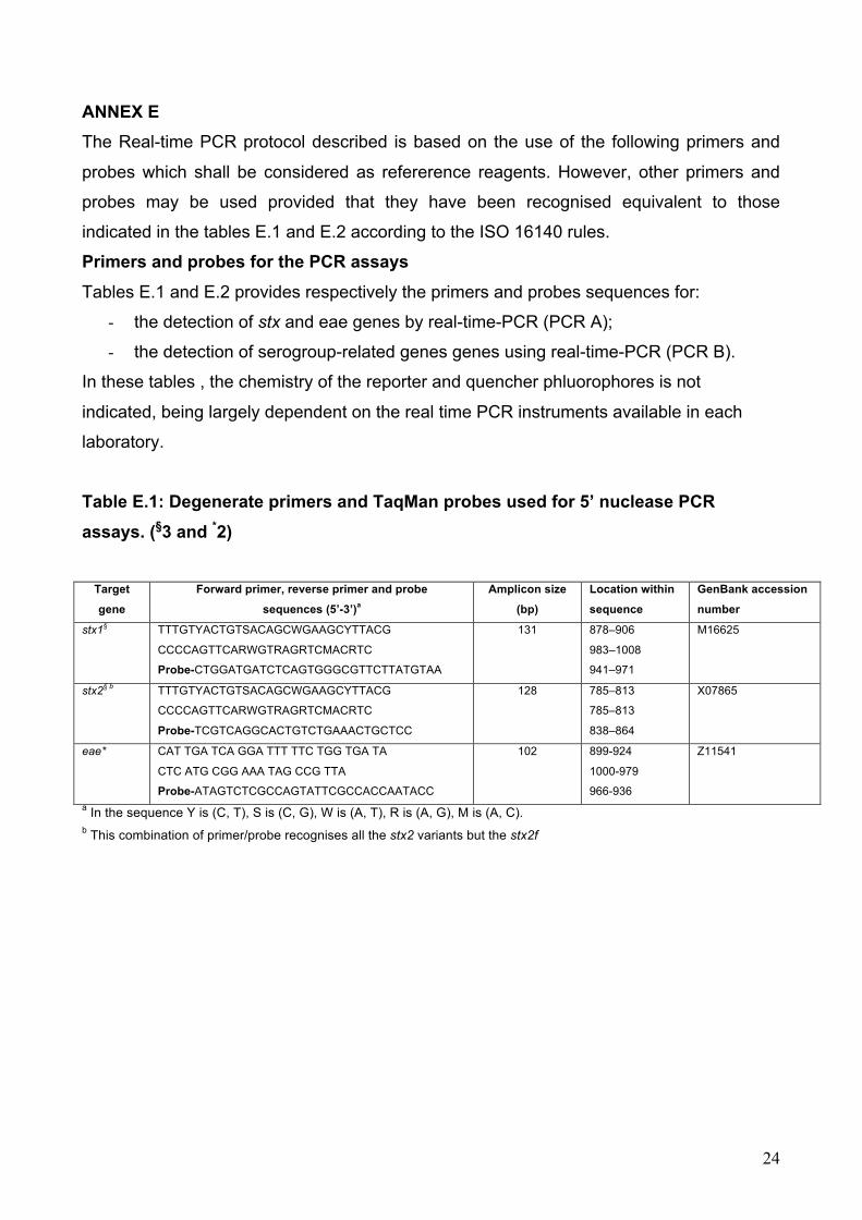
ANNEX E The Real-time PCR protocol described is based on the use of the following primers and
probes which shall be considered as refererence reagents. However, other primers and
probes may be used provided that they have been recognised equivalent to those
indicated in the tables E.1 and E.2 according to the ISO 16140 rules.
Primers and probes for the PCR assays Tables E.1 and E.2 provides respectively the primers and probes sequences for:
- the detection of stx and eae genes by real-time-PCR (PCR A);
- the detection of serogroup-related genes genes using real-time-PCR (PCR B).
In these tables , the chemistry of the reporter and quencher phluorophores is not
indicated, being largely dependent on the real time PCR instruments available in each
laboratory.
Table E.1: Degenerate primers and TaqMan probes used for 5’ nuclease PCR assays. (§3 and *2)
Target
gene Forward primer, reverse primer and probe
sequences (5’-3’)a Amplicon size
(bp) Location within
sequence GenBank accession
number stx1§ TTTGTYACTGTSACAGCWGAAGCYTTACG
CCCCAGTTCARWGTRAGRTCMACRTC Probe-CTGGATGATCTCAGTGGGCGTTCTTATGTAA
131 878–906 983–1008
941–971
M16625
stx2§ b
TTTGTYACTGTSACAGCWGAAGCYTTACG
CCCCAGTTCARWGTRAGRTCMACRTC
Probe-TCGTCAGGCACTGTCTGAAACTGCTCC
128 785–813 785–813 838–864
X07865
eae* CAT TGA TCA GGA TTT TTC TGG TGA TA
CTC ATG CGG AAA TAG CCG TTA
Probe-ATAGTCTCGCCAGTATTCGCCACCAATACC
102 899-924
1000-979
966-936
Z11541
a In the sequence Y is (C, T), S is (C, G), W is (A, T), R is (A, G), M is (A, C). b This combination of primer/probe recognises all the stx2 variants but the stx2f

25
Table E.2. Primers and probes used for amplification of O antigen specific genes in 5’ nuclease PCR assays. (§3 and *4)
Target gene
(serogroup) Forward primer, reverse primer and probe sequences
(5’-3’) Amplicon
size (bp) Location within
sequence GenBank accession
number §rfbE (O157) TTTCACACTTATTGGATGGTCTCAA
CGATGAGTTTATCTGCAAGGTGAT
Probe-AGGACCGCAGAGGAAAGAGAGGAATTAAGG
88 348–372
412–435 381–410
AF163329
§wbdI (O111) CGAGGCAACACATTATATAGTGCTTT
TTTTTGAATAGTTATGAACATCTTGTTTAGC
Probe-TTGAATCTCCCAGATGATCAACATCGTGAA
146 3464–3489
3579–3609
3519–3548
AF078736
§wzx (O26) CGCGACGGCAGAGAAAATT
AGCAGGCTTTTATATTCTCCAACTTT
Probe-CCCCGTTAAATCAATACTATTTCACGAGGTTGA
135 5648–5666
5757–5782
5692–5724
AF529080
§ihp1 (O145) CGATAATATTTACCCCACCAGTACAG
GCCGCCGCAATGCTT
Probe-CCGCCATTCAGAATGCACACAATATCG
132 1383–1408
1500–1514
1472–1498
AF531429
*wzx (O103) CAAGGTGATTACGAAAATGCATGT
GAAAAAAGCACCCCCGTACTTAT
Probe-CATAGCCTGTTGTTTTAT
99 4299–4323
4397–4375
4356–4373
AY532664

26
Annex F Isolation of STEC strains Follow the procedure described below to isolate STEC strains from real time PCR positive
samples:
1) Perform a serogroup-specific enrichment (SSE) on the remaining enrichment culture
(see Note 1)
2) Streak SSE onto TBX or other suitable medium (see note 2). Incubate for 18 to 24 hours
at 37°C
3) Pick up 10 to 50 colonies with E. coli morphology or with characteristic aspect (see Note
5) and point-inoculate on nutrient agar (NA) (see Note 3) and H2O (the colonies may be
pooled in water up to a number of ten per pool).
4) Perform the detection of the stx-coding gene and the eae gene on the isolated colonies
or the H2O pools (see Note 4).
5) If a pool is positive, go back to NA and assay the individual colonies forming the positive
pool in order to select one single positive colony.
6) Identify the colonies as E. coli and confirm the serogroup the sample was positive to in
the screening PCR assay (e.g. by PCR B in the Annex E), see Note 5.
7) Isolates may be sent to the a Reference Laboratory for further characterization.
NOTE 1: Serogroup-specific enrichment may be achieved by using immunocapture
systems such as immuno-magnetic separation (IMS) or equivalent. Generally, refer to the
instruction supplied by the manufacturer.
For O157 positive samples, use ISO 16654 or alternative methods validated according to
ISO 16140.
NOTE 2: For O157 positive samples, use ISO 16654 or alternative methods validated
according to ISO 16140. Sorbitol-fermenting E. coli O157 are susceptible to tellurite
contained in the CT SMAC medium indicated in ISO 16654. Therefore the use of a second
SMAC isolation plate without antibiotics is recommended. In the absence of Sorbitol-
negative colonies on the plates, the screening of Sorbitol-positive colonies is suggested.
For STEC O26 isolation, a differential solid media (MacConkey) containing Rhamnose
instead of lactose is commercially available (RMAC). It is very effective in distinguishing
STEC O26 strains, which do not ferment Rhamnose, from other E. coli.
NOTE 3: There are several types of nutrient agar media commercially available either
ready to use plates or prepared in house from dehydrated powders. Every type of non-

27
selective nutrient agar media (e.g. TSA) is suitable for the purpose of maintaining the
colonies for further characterisation. Enterohaemolysin Agar, can also be used. It gives the
advantage to detect the Enterohaemolysin production, which is a common feature of
STEC pathogenic to humans.
NOTE 4: The Real Time PCR described in this protocol may be adopted to confirm the
presence of the stx and eae in the isolated strains. Conventional PCR may be used as an
alternative (http://www.iss.it/vtec/work/cont.php?id=152&lang=2&tipo=3).
NOTE 5: Colony confirmation as E. coli may be achieved by using any commercial
biochemical multi-assay or by assessing the indole production. Confirmation of the
serogroup may be achieved either by PCR or by agglutination with commercial antisera.
Bibliography
[1] Monitoring of verotoxigenic Escherichia coli (VTEC) and identification of human
pathogenic VTEC types Scientific Opinion of the Panel on Biological Hazards
(Question No EFSA-Q-2007-036) The EFSA Journal (2007) 579, 1-61
[2] Nielsen EM, Andersen MT. Detection and characterization of verocytotoxin-
producing Escherichia coli by automated 5' nuclease PCR assay. 2003 J Clin
Microbiol. 41(7):2884-2893.
[3] Perelle S, Dilasser F, Grout J, Fach P. Detection by 5'-nuclease PCR of Shiga-toxin
producing Escherichia coli O26, O55, O91, O103, O111, O113, O145 and
O157:H7, associated with the world's most frequent clinical cases. 2004 Mol Cell
Probes. 18(3):185-192.
[4] Perelle S, Dilasser F, Grout J, Fach P. Detection of Escherichia coli serogroup
O103 by real-time polymerase chain reaction. 2005 J Appl Microbiol 98(5):1162-
1168.
[5] Vimont A, Delignette-Muller ML, Vernozy-Rozand C. Supplementation of
enrichment broths by novobiocin for detecting Shiga toxin-producing Escherichia
coli from food: a controversial use. Lett Appl Microbiol 2007 44(3):326-331.
[6] Uemura R, Sueyoshi M, Nagayoshi M, Nagatomo H. Antimicrobial susceptibilities of
Shiga toxin-producing Escherichia coli isolates from pigs with edema disease in
Japan. 2003 Microbiol Immunol 47(1):57-61.
[7] Sambrook and Russel, Molecular cloning, a laboratory manual. Cold Spring Harbor
Laboratory Press. third edition, 2001

28
[8] Paton AW, Paton JC. Detection and characterization of Shiga toxigenic Escherichia
coli by using multiplex PCR assays for stx1, stx2, eaeA, enterohemorrhagic E. coli
hlyA, rfbO111, and rfbO157. 1998J Clin Microbiol 36: 598-602.
[9] O'Brien AD, Newland JW, Miller SF, Holmes RK, Smith HW, Formal SB. Shiga-like
toxin-converting phages from Escherichia coli strains that cause hemorrhagic colitis
or infantile diarrhea. 1984 Science 226: 694-696.
[10] Perna N.T. et al. Genome sequence of enterohaemorrhagic Escherichia coli
O157:H7. 2001 Nature 409: 529-533.
[11] Fricker M, Messelhäusser U, Busch U, Scherer S, Ehling-Schulz M. Diagnostic real-
time PCR assays for the detection of emetic Bacillus cereus strains in foods and
recent food-borne outbreaks. 2007 Appl Environ Microbiol 73(6):1892-1898.
[12] Hoorfar J, Malorny B, Abdulmawjood A, Cook N, Wagner M, Fach P. Practical
considerations in design of internal amplification controls for diagnostic PCR
assays. 2004 J Clin Microbiol 42(5):1863-1868.
[13] Beutin L, Jahn S, Fach P. Evaluation of the 'GeneDisc' real-time PCR system for
detection of enterohaemorrhagic Escherichia coli (EHEC) O26, O103, O111, O145
and O157 strains according to their virulence markers and their O- and H-antigen-
associated genes. 2009. J Appl Microbiol. 106:1122-32.