Report of the 2nd RCM on Development of radiation ... · REPORTS BY PARTICPANTS IN THE COORDINATED...
234
INTERNATIONAL ATOMIC ENERGY AGENCY Report of the 2nd RCM on "Development of radiation-processed products of natural polymers for application in agriculture, healthcare, industry and environment" Reims, France 12-16 October 2009 Reproduced by the IAEA Vienna, Austria, 2010 NOTE The material in this document has been agreed by the participants and has not been edited by the IAEA. The views expressed remain the responsibility of the participants and do not necessarily reflect those of the government(s) of the designating Member State(s). In particular, neither the IAEA nor any other organization or body sponsoring this meeting can be held responsible for any material reproduced in the document. Working Material Working Document
Transcript of Report of the 2nd RCM on Development of radiation ... · REPORTS BY PARTICPANTS IN THE COORDINATED...
INTERNATIONAL ATOMIC ENERGY AGENCY
Report of the 2nd RCM on "Development of radiation-processed
products of natural polymers for application in agriculture, healthcare,
industry and environment"
Reims, France 12-16 October 2009
Reproduced by the IAEA Vienna, Austria, 2010
NOTE The material in this document has been agreed by the participants and has not been edited by the IAEA. The views expressed remain the responsibility of the participants and do not necessarily reflect those of the government(s) of the designating Member State(s). In particular, neither the IAEA nor any other organization or body sponsoring this meeting can be held responsible for any material reproduced in the document.
Working Material
Working Document

Working Material
Working Document

INTERNATIONAL ATOMIC ENERGY AGENCY
Report of the 2nd RCM on
"Development of radiation-processed products of natural polymers for application in agriculture, healthcare, industry and
environment"
Reims, France 12-16 October 2009
Reproduced by the IAEA
Vienna, Austria, 2010
NOTE The material in this document has been agreed by the participants and has not been edited by the IAEA. The views expressed remain the responsibility of the participants and do not necessarily reflect those of the government(s) of the designating Member State(s). In particular, neither the IAEA nor any other organization or body sponsoring this meeting can be held responsible for any material reproduced in the document.
Working Material
Working Document
EDITORIAL NOTE In preparing this publication for press, staff of the IAEA have made up the pages from the original manuscripts as submitted by the authors. The views expressed do not necessarily reflect those of the governments of the nominating Members States or of the nominating organizations. Throughout the text names of Member States are retained as they were when the text was compiled. The use of particular designations of countries or territories does not imply any judgment by the publisher, the IAEA, as to the legal status of such countries or territories, of their authorities and institutions or of the delimitation of their boundaries. The mention of names of specific companies or products (whether or not indicated as registered) does not imply any intention to infringe proprietary rights, nor should it be construed as an endorsement or recommendation on the part of the IAEA. The authors are responsible for having obtained the necessary permission for the IAEA to reproduce, translate or use materials from sources already protected by copyrights.
Working Material
Working Document

FOREWORD
Radiation processing offers a clean and additive-free method for preparation of value-added novel materials based on renewable, non-toxic and biodegradable natural polymers and natural polymer waste. The results of research work showed that depending on the irradiation conditions, natural polysaccharides (alginate, chitin/chitosan, carrageeneans, carboxylmethylcellulose, etc.) could be either degraded or crosslinked by radiation. This paved the way for development of many successful applications; some of them commercialized, for use in agriculture, health care and environmental protection. The inputs for the formulation of this CRP and the key issues that need to be addressed were provided by the Consultant’s Meeting on “Radiation Processing of Natural Polymers for Development of Finished Products for Health Care, Agriculture and Environment” held in Kuala Lumpur, Malaysia from 26 – 30 March 2007. The main objective was defined as wide-spread promotion and general application of radiation processed natural materials, by coupling radiation technology and end-users to derive additional benefits from these value-added natural materials. The first RCM of the CRP was convened in Vienna on 21-25 April 2008. The participants presented and discussed the status of the field, the needs for further research, and various application possibilities. The work plan formulated during the meeting focused on harmonization of procedures for characterization of irradiated polymers, and protocols for investigation of the functional properties of degraded natural polymer products and their field testing. A network for collaboration was also proposed. The Meeting Report was published and is available for all Member States. The second RCM of this CRP was held in Reims, France, on 12–16 October 2009. The meeting was attended by 14 participants who reported their individual research results obtained since the first RCM, as well as their further plans. This meeting report contains all these reports, as well as the reports by participants from Algeria, Japan and Thailand, who could not attend the meeting, but sent their contributions. The leaders of the harmonization exercise of the characterization procedures (Poland and UK) presented a detailed evaluation of the results and lessons learned, as well as the further plans. The report of Poland contains the summary of these findings. The detailed protocol for the determination of viscosity for chitosan solution, which was used by all participants, is given in the Annex. The IAEA wishes to thank all participants of the Meeting for their valuable contributions. The IAEA officer responsible for this Research Coordination Meeting was A. Safrany of the Division of Physical and Chemical Sciences.
Working Material
Working Document

Working Material
Working Document
CONTENTS
EXECUTIVE SUMMARY...................................................................................................... 1
1. BACKGROUND.................................................................................................................. 1
2. CRP OVERALL OBJECTIVE ............................................................................................ 3
3. MAIN RESEARCH OBJECTIVES AND COUNTRIES INVOLVED.............................. 4
4. SUMMARY OF PARTICIPANTS REPORTS ................................................................... 4
5. CONCLUSION .................................................................................................................. 17
5. RECOMMENDATIONS ................................................................................................... 18
REPORTS BY PARTICPANTS IN THE COORDINATED RESEARCH PROJECT ........ 21 Synthesis of PVA-Chitosan Hydrogels for wound dressing using gamma irradiation ........... 23
M. Mahlous, D. Tahtat, S. Benamer, A. Nacer Khodja, S. Larbi Youcef Radiation processing of marine algal polysaccharides for agriculture, as plant growth promoters & preservation of foods and health care products as instant hydrogel formulations for improved mucose adhesion and medical applications...................................................... 37
E.E. Smolko, M. Clozza, E.B.Giardina, F. Villela, M.D. Divo, L. Cerchietti Country report of Bangladesh on the activities on “modification of natural polymers by radiation processing for value added products” ................................................................ 47
M.E. Haque, R.A. Khan, N.C. Dafader, M.Z.I. Mollah
Ionizing radiation conversion of lignocellulosic biomass from sugarcane bagasse to production ethanol biofuel ................................................................................................. 55
C.L. Duarte, M.N. Mori, H. Oikawa1, H.R. Nagatomi, J. Fingueru, M. Célia, A. Galvão
Development of bioactive edible coatings and biodegradable packaging using gamma irradiation ............................................................................................................................... 65
Lacroix, M., Salmieri, S.
Radiation modification of natural polysaccharides for application in agriculture ................. 85 H.A. AbdEl-Rehim, El-S.A. Hegazy, A.M. El-Barbary
Thermoplastic starch with improved properties by blending lignins and radiation processing............................................................................................................................... 95
D. Zheng, S.Baumberger, P.-Y. Mikus, P. Dole, J. Soulestin, M.F.Lacrampe, C. Bliard, X. Coqueret
Preliminary studies on antifungal properties of radiation processed chitosan from crab shells............................................................................................................................. 101
F.C.K. Ocloo, A.Adu-Gyamfi, E.A. Quarcoo, Y. Serfor-Armah2, D. Asare
Working Material
Working Document

Current status of radiation processing of natural polymers in India .................................... 107 S.P. Ramnani
Modification of natural polymers by radiation-induced processing for versatile applications........................................................................................................................... 113
N. Nagasawa, A. Hiroki, M. Tamada Application of radiation degraded chitosan as plant growth promoter ................................ 121
K.Z.H.M. Dahlan, K. Hashim, K. Bahari, M. Mahmod, N. Yaacob, N. Talip, K. Hussein, A.S. Mardi, A.R. Harun, R. Yusof, S.Sitor, N. Q. Hien, T. Kume
Radiation processed materials from carrageenan for agricultural applications ................... 131
L.V. Abad, L.S. Relleve, C.T. Aranilla, A. M. De la Rosa Radiation chemistry and physical chemistry of chitosan and other polysaccharides fundamental studies and practical applications.................................................................... 145
J.M. Rosiak, R. Czechowska-Biskup, B. Rokita, A.K. Olejnik, P. Ulański Utilization of degraded chitosan for growth promoter and blossom blight disease controls in okra....................................................................................................................................... 153
P. Kewsuwan Radiation synthesis of superabsorbent polymers based on natural polymers ...................... 159
M. Sen, H. Hayrabolulu Characterisation and radiation modification of carrageenan in the solid state .................... 173
S. Gulrez, S. Al-Assaf , G. O. Phillips, Study of chemical treatment combined with radiation to prepare biotic elicitor for utilization in agriculture ........................................................................................................................ 187
N. Quoc Hien LIST OF PARTICIPANTS .................................................................................................. 195 ANNEX................................................................................................................................ 199 COLLABORATION TABLE.............................................................................................. 223
Working Material
Working Document
EXECUTIVE SUMMARY
1. BACKGROUND Radiation processing offers a clean and additive-free method for preparation of value-added novel materials based on renewable, non-toxic and biodegradable natural polymers. The results of research work in some member states showed that natural polysaccharides (alginate, chitin/chitosan, carrageeneans) could be either degraded or crosslinked by radiation, depending on the irradiation conditions. During a short span, many applications have been successfully developed and some of them commercialized in the areas of agriculture, health care and environment. The crosslinked products can be used as hydrogel wound dressings, face cleaning cosmetic masks, adsorbents of toxins and non-bedsore mats, while low molecular weight products show antibiotic, antioxidant and plant-growth promoting properties. These successes clearly indicate that radiation processing of natural polymers has emerged as an exciting area where the unique characteristics of these polymeric materials can be exploited for a variety of practical applications. Recognizing the potential benefits that radiation technology can offer for processing of natural polymers into useful products, the IAEA has implemented a number of activities in response to MS requests for TC projects and held consultants meetings on “Technical, Economical and Environmental Advantages of Radiation Processing of Cellulose” (2001), “Radiation Processing of Polysaccharides” (November 2003; the TECDOC 1422 was published in 2004), “Irradiation of Natural Resources for Marketable Products” (October 2004), “Radiation Processing of Natural Polymers for Development of Finished Products for Health Care, Agriculture and Environment” (March 2007). The designation of Malaysian Nuclear Agency (Nuclear Malaysia) as IAEA Collaborating Centre on Radiation Processing of Natural Polymers in October 2006 successfully helped the developments in this field and was extended for further 3 years.
This CRP started in the end of 2007 and aims to harmonize the procedures for both the evaluation methods for radiation-modified natural polymers, as well as the protocols for their field testing as plant elicitors. The first RCM was held in March 2008 in Vienna, where the work plan for both individual participants and collaborations were discussed and accepted, as reported in the Meeting Report published as IAEA Working Material (Blue Book). The second RCM was held between 12-16 October 2009 in Reims, France, and was attended by 14 participants (chief scientific investigators) and the representative of the HMC Company from Germany. The participants presented their research achievements since the first RCM, centred on the main objectives of this CRP: 1. Identification of methodologies and QA protocols for investigating structure-property relationship particularly with respect to radiation induced changes in natural polymers. Regarding this objective, at the first RCM it was decided, that the first level of characterization will be the determination of viscosity-average molecular weight (Mv) with a common and widely used method, a capillary viscometric measurements. An inter-laboratory programme was designed to examine the application of the proposed method to be tested by CRP participants following a detailed protocol. The programme was coordinated by UK and Poland with the assistance of HMC Company from Germany. HMC supplied well-characterized chitosan sample which was subjected to measurements before and after irradiation to various doses. In general the first phase of the harmonization exercise has been a success. Nine participants provided results obtained in their laboratories. Standard deviation of the measured intrinsic viscosity was 7 %, which, taking into account the complexity of the material and composite nature of the solvents, can be considered as fully acceptable. Data on radiation-induced degradation are also relatively uniform. Larger errors (SD of 17 %) than in the viscosity exercise were to be expected due to additional uncertainties resulting from irradiation and dosimetric procedures. Several parameters affecting the accuracy of the measurements have been identified and will be addressed with the potential assistance of HMC. The data were
1
Working Material
Working Document
further exploited to assess the G(S) values using the correct parameters needed. The obtained value will be useful for planning future experiments and predicting the doses necessary to achieve targeted molecular weight. Participants agreed to follow a uniform reporting procedure and to provide additional information related to their reported values. Participants who did not report their results for various reasons (material or equipment availability) agreed to submit their contribution in the near future. The exercise has shown that the results obtained in laboratories around the world are comparable. This is important for co-operation between groups working on chitosan, within this Project, but also in general, in the research community working on chitosan. The participants proposed to extend this programme on additional chitosan samples with different degrees of deacetylation which will be supplied by HMC. A revision of the protocol to deal with the samples will be provided by the coordinators. 2. Investigating the anti-oxidant properties of low molecular weight natural polymers and assessing suitability for preservation of food and allied products. Regarding this second objective, participants from Argentina, Egypt, Ghana, and France reported achievements in designing saccharide oligomers with optimal properties for plant protection and plant growth promotion. Radiation-processed alginates were investigated as plant growth promoters in different crops such as tomato, lettuce, spinach and cabbage on the different environmental conditions. Additionally, degraded alginate showed inhibitory effect on tobacco mosaic virus. Laboratory scale trials using radiation-degraded chitosan, of different molecular weights, controlled the post harvest tomato grape mold caused by Botrytis cinerea and enhanced fungal decay. In Canada, gamma irradiation was used for crosslinking of protein based films and coatings in order to enhance the physico-chemical properties of packaging films and to assure the preservation of coated fruits. These results showed that the film formulation used as a bioactive edible coating efficiently increased the shelf life of fresh strawberries and preserved their quality during storage. 3. Field-testing the potential of radiation-modified polysaccharides as plant growth promoters, soil conditioners and for enhancing fermentation of agro by-products. Regarding this third objective, Malaysia reported a successful establishment of a pilot scale production for oligochitosan using a continuous gamma irradiation plant at Nuclear Malaysia. In Vietnam, Bangladesh, Thailand, Egypt, and India, batch type gamma irradiation of chitosan aqueous solutions for production of oligochitosan have been developed. These samples were then used in field testing. The results of field tests have shown that oligochitosan and oligoalginate are very effective in promoting the growth and yield of rice, wheat, sugarcane, tomato, and vegetables like ammaranthus. In addition, oligochitosan suppressed fungal diseases on rice, wheat and tomatoes. These field tests are still in progress. Additional achievements reported by the participants include preparation of polysaccharide-based super absorbent hydrogels by radiation induced cross-links (Egypt, Turkey). The water absorption capacities of these natural polymer based super adsorbents can be controlled by changing the concentration of the natural polymer and of the synthetic monomers in the initial mixture. Due to their fast swelling, these natural/synthetic hybrid hydrogel systems can be considered as potential absorbents for water and body fluids such as urine and blood in personal care products. Additionally, they might be tuned for applications as soil conditioners to increase the water retention in arid environments. Brazil reported recent achievements in the facilitating of the production of bioethanol from non-food crops, by electron beam processing of the sugarcane bagasse. These results indicated that low-dose irradiation can cleave the external structure of sugarcane bagasse without destroying the cellulose, a desirable pre-treatment to the following enzymatic attack.
2
Working Material
Working Document

Under the framework of the 2nd RCM, one day open meeting was organized under the title “Sustainable plant management using biosourced polymers: when chemistry meets plant biology”. This meeting was attended by all RCM participants and more than 100 students, researchers and professors of the University of Reims and their collaborating partners from universities from The Netherlands, Germany and USA. Around 20 presentations were given, organized in following three sessions: Introduction, Plant defence mechanism, Use of biosourced polymers aiming at protecting crops against fungal disease. The presentations highlighted the current knowledge about plant defence mechanism, the pathogen-associated molecular pattern, molecular interactions and defence events by plant cells, as well as the various uses of polysaccharides for plant defence and protection. This event was of high value to all attendants, and a long discussion resulted about the importance and the possible ways of collaboration between chemists and plant biologists. Some participants of this CRP effectively utilized relevant National and Regional TC projects to fund scientific visits, fellowships and expert missions to help their joint efforts to achieve the objective of this CRP. 2. CRP OVERALL OBJECTIVE The overall objective of the proposed research project is the wide-spread promotion and general application of radiation processed natural materials, coupling radiation technology and end-users to derive enhanced benefits from these value-added natural materials. 3.1. Specific Research Objectives: • Identification of methodologies and QA protocols for investigating structure-property relationship particularly with respect to radiation induced changes in natural polymers. • Investigating the anti-oxidant properties of low molecular weight natural polymers and assessing suitability for preservation of food and allied products. • Field-testing the potential of radiation-modified polysaccharides as plant growth promoters, soil conditioners and for enhancing fermentation of agro by-products. The participant groups will address and contribute to one or more of the above topics. 3.2. Expected Research Outputs: • Harmonization of QA protocols for characterization of radiation-induced changes in natural polymers • Development of protocols for use of radiation-modified polysaccharides as plant growth promoters and soil conditioners on a field scale so as to produce marketable products. The results of the CRP will be presented in international conferences and published in scientific journals and as IAEA technical report.
3
Working Material
Working Document
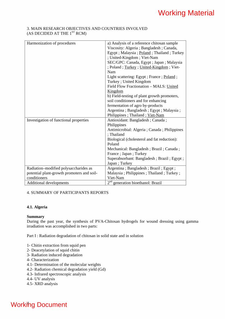
3. MAIN RESEARCH OBJECTIVES AND COUNTRIES INVOLVED (AS DECIDED AT THE 1ST RCM) Harmonization of procedures a) Analysis of a reference chitosan sample
Viscosity: Algeria ; Bangladesh ; Canada, Egypt ; Malaysia ; Poland ; Thailand ; Turkey ; United-Kingdom ; Viet-Nam SEC/GPC: Canada, Egypt ; Japan ; Malaysia ; Poland ; Turkey ; United-Kingdom ; Viet-Nam Light scattering: Egypt ; France ; Poland ; Turkey ; United Kingdom Field Flow Fractionation – MALS: United Kingdom b) Field-testing of plant growth promoters, soil conditioners and for enhancing fermentation of agro by-products Argentina ; Bangladesh ; Egypt ; Malaysia ; Philippines ; Thailand ; Viet-Nam
Investigation of functional properties Antioxidant: Bangladesh ; Canada ; Philippines Antimicrobial: Algeria ; Canada ; Philippines ; Thailand Biological (cholesterol and fat reduction): Poland Mechanical: Bangladesh ; Brazil ; Canada ; France ; Japan ; Turkey Superabsorbant: Bangladesh ; Brazil ; Egypt ; Japan ; Turkey
Radiation–modified polysaccharides as potential plant-growth promoters and soil-conditioners
Argentina ; Bangladesh ; Brazil ; Egypt ; Malaysia ; Philippines ; Thailand ; Turkey ; Viet-Nam
Additional developments 2nd generation bioethanol: Brazil 4. SUMMARY OF PARTICIPANTS REPORTS 4.1. Algeria Summary During the past year, the synthesis of PVA-Chitosan hydrogels for wound dressing using gamma irradiation was accomplished in two parts: Part I : Radiation degradation of chitosan in solid state and in solution 1- Chitin extraction from squid pen 2- Deacetylation of squid chitin 3- Radiation induced degradation 4- Characterization 4.1- Determination of the molecular weights 4.2- Radiation chemical degradation yield (Gd) 4.3- Infrared spectroscopic analysis 4.4- UV analysis 4.5- XRD analysis
4
Working Material
Working Document

Chitosan was degraded by gamma irradiation in both, solid state an in solution. The radiation chemical degradation yield was 12 to 31 times higher in solution than in solid state, depending on initial Mw and DD. UV and IR spectra revealed chemical changes in the polymer structure. Theses changes were attributed to the formation of carbonyl and carboxyl groups as a result of the degradation process. XRD analysis shows a strong decrease of crystallinity of the polymer irradiated in solid state at 250 kGy. Part II - Antibacterial activity of PVA/Chitosan hydrogel synthesized by gamma irradiation Preparation of the hydrogel PVA-Chitosan Swelling measurements Gel fraction Scanning Electron Microscopy (SEM) Infrared spectroscopy Antimicrobial tests PVA hydrogels containing moieties of chitosan exhibit antibacterial activity against both gram positive and gram negative bacteria. The release of the chitosan into the culture medium is governed by its molecular weight. The effect of chitosan content in the hydrogel on antibacterial activity is observed for low molecular weight chitosan. The results obtained in the viscosity measurement excersize are included in the report together with the results of other participants. Work Plan The antibacterial effect, of different molecular weight of chitosan incorporated in the hydrogel, will be investigated against Escherichia coli and Bacillus subtilis. The wound healing effect of chitosan contained in the hydrogel will be investigated with pharmacological and toxicological tests on Albino Wistar rats. Collaborations See attached Collaborations Table. 4.2. Argentina Summary Characterization of the bulk material for degradation and/or structure modification in grafting procedures during this period was continued. They were characterized by ultra-violet and visible (UV-vis) and Fourier transform infra- red spectroscopy (FTIR). After receiving the chitosan sample from Prof. Dr Rosiak we started the duty of harmonization procedures for the characterization of raw materials in terms of molecular weight determination by viscosity and to proceed with the measurements of samples of alginates and carrageenans. For investigation of radiation–processed alginates for agriculture applications degraded polymers were used as plant growth promoters, in different crops, such as tomato, lettuce, spinach and cabbage under different environmental conditions. By irradiation of alginates with various radiation doses (500, 750 and 1000 kGy gamma rays) degraded alginates have been obtained. They have been applied in horticultural species in 20 and 100 microgram/l concentrations, in solution state (under hydroponics cultivation condition) and by foliar spraying (under soil less culture). In a preliminary phase of this study, different alginate treatments (radiation doses and concentrations) were applied to determine the effect on seedlings quality, evaluated through leaf and root development, shoot/root ratio and photosynthetic activity. UV-VIS spectroscopy of alginate irradiated at very high doses ( up to 1000 kGy) showed an abrupt change in the macroscopic shape of the sample forming hard particles and shifting the absorbing band to a higher wavelength. Alginates irradiated in solution show an absorption band at 265 nm approximately which increases steadily with dose. Alginates irradiated in
5
Working Material
Working Document

solid state show also the absorption band at 265 nm which initially increases with the dose, but disappears at higher dose and shows a new wider band at 427 nm. Degradation was also observed accompanied by a color change to deep brown for highly degraded alginate. A conclusion from the color forming phenomena is that for lower doses the double bond formed after degradation can be established between the carbons 4 and 5 of the pyranose ring but for higher dose the double bond could be formed between the carbon 1 and the oxygen 1 of the glycoside bond giving a different terminal reactive species. In the grafting study of alginic acid-g-acrilic acid we consider highly probably demonstrated the modification of the structure by grafting for a new absorption band located at 1727 (1/cm) that corresponds also to a band found also in poly- methylmethacrylate and polyacrilic acid . Work Plan - Radiation grafting of alginate with acrylic monmer in paste state for drug delivery applications - Investigatin of radiatin degradatin of chitosan, carrageenan and alginate - Testing of radiation-degraded marine polysaccharides as plant growth promoters both under greenhouse and open field conditions - Participation in harmonization programme Collaborations See attached Collaborations Table. 4.3. Bangladesh Summary 1. Chitosan Bangladesh is a riverine country where a lot of shrimps and prawns are available. Moreover Bangladesh has around 750 kilometer coastal lines. A lot of hatcheries are growing in Bangladesh and now shrimps/prawns are exporting to Europe and America. But shrimp/prawn shells remain unutilized and causing a threat to the environment. Chitin and chitosan can be extracted from waste shrimp/prawn shells for better use. Chitosan was extracted from locally available resources for various applications. Effects of chitosan on the morphological properties on the plants/fruits were investigated by applying on various vegetables as plant growth promoter. Chitosan enhances the vegetative growth in terms of the average values of stem length, number of growing leaves, including leaf width and length etc. In this investigation, irradiated chitosan was sprayed on local vegetable (Ammaranthus Cruentus, Local name: Datasakh) and green chili plant. Films of chitosan were made and the effects of gamma radiation on the tensile properties of the film were investigated. The tensile strength of the irradiated films increases from 14 to 27 MPa by applying 200 krad of gamma radiation. The elongation at break increases for up to 100 krad but after that dose it reduces to about 7 MPA at 500 krad. The chitsan was modified with HEMA monomer for biomedical application. Further investigation on its properties is going on. The chitosan films were characterized by using TG, DTA and DTG. The TG curve shows two stage degradations. The onset temperature was about 55°C. The residue content was about 40% after 600°C. The maximum slope was obtained at 149ºC. DTA curve of chitosan shows endothermic peak at 273ºC. It depicts one predominant peak at 320ºC where the maximum degradation rate was 57.3 µg/min. From this investigation this is clear that chitosan started degradation at low temperature which is common for natural biopolymers but the maximum degradation happened at higher temperature which is a good sign of thermal stability of chitosan. The surface of the prepared chitosan films were investigated by SEM. It is found that the surface is quite smooth. 2. Alginate In Bangladesh, a lot of sea weeds are found in the coastal areas. Sodium alginate was extracted from brown algae (Sargassum).150 ppm degraded alginate shows a positive effect on the growth parameters of red amaranth with varying gamma doses (12.5 to 50.0 kGy). A positive result on the growth rate of the vegetable was found.
6
Working Material
Working Document
Work Plan 1. Chitosan - Investigation of the degree of deacetylation of chiotsan. - Determination of viscosity and molecular weight. - Effect of gamma radiation on the molecular weight of chitosan. - Modification of chitosan films using biocompatible monomers and gamma radiation. - Preparation of hydrogels using chitosan. - Application of irradiated chiotsan on different types of fruits and vegetables as preservatives (continued). - Effect of chitosan as plant growth promoter on various types of vegetables and rice (continued). 2. Alginate - Establishment of the extraction procedure of alginate from local resources. - Characterization of the extracted alginate - Application of radiation degraded alginate as plant growth promoter Collaborations See attached Collaborations Table. 4.4. Brazil Summary Sugarcane bagasse has been considered as a substrate for single cell protein, animal feed, and renewable energy production. Sugarcane bagasse generally contain up to 45% glucose polymer cellulose, 40% hemicelluloses, and 20% lignin. Pure cellulose is readily depolymerised by radiation, but in biomass, the cellulose is intimately bonded with lignin, that protect it from radiation effects. The objective of this study is the evaluation of the electron beam irradiation as a pre-treatment to enzymatic hydrolysis of cellulose in order to facilitate its fermentation and improves the production of ethanol biofuel. Samples of sugarcane bagasse were obtained in sugar/ethanol Iracema Mill sited in Piracicaba, Brazil, and were irradiated using Radiation Dynamics Electron Beam Accelerator with 1.5 MeV energy and 37kW, in batch systems. The applied absorbed doses of the fist sampling, Bagasse A, were 20 kGy, 50 kGy, 100 kGy and 200 kGy. After the evaluation the preliminary obtained results, it was applied lower absorbed doses in the second assay: 5 kGy, 10 kGy, 20 kGy, 30 kGy, 50 kGy, 70 kGy, 100 kGy and 150 kGy. The electron beam processing took to changes in the sugarcane bagasse structure and composition, lignin and cellulose cleavage. The yield of enzymatic hydrolyzes of cellulose increase about 75 % with 30 kGy of absorbed dose. Work Plan The HPLC/ELSD system now installed has very good specificity and linearity to carachterization and quantification of sugars and it will be useful to further experiments, to better understanding the action mechanism of ionizing radiation in the sugarcane bagasse structure. • Combination of irradiation with enzymatic and chemical hydrolysis of cellulose from sugarcane bagasse • Comparision and combination of irradiation with the pretreatment process of team explosion • Fermentation of the sugarcane bagasse after irradiation and after cellulose hidrolysis. Collaborations See attached Collaborations Table.
7
Working Material
Working Document

4.5. Canada Summary Gamma irradiation was used to cross-link milk proteins in order to enhance the physico-chemical properties of edible films made of calcium caseinate, whey protein isolate and glycerol. Fourier Transform Infrared analysis was used to characterize the conformation of proteins adopted after irradiation. The molecular weight of cross-linked proteins was measured by size-exclusion chromatography. Furthermore, the effect of the addition of methylcellulose to the irradiated protein matrix on the rheological properties (puncture strength, puncture deformation and water vapor permeability) of films was also studied. Moreover, cross-linking of polysaccharides under paste-like state was investigated and the cross-linking degree of the gel products was determined by gel fraction measurements and solubility percentage. In order to prepare bioactive coatings, several antifungal compounds were evaluated as bioactive compounds in order to select one of them to prepare an antimicrobial solution to spray onto strawberries or to encapsulate them in film formulations composed of milk proteins and methylcellulose based films. In addition, the bioactive coatings containing the antifungals were used to increase the radiosensitivity under air of moulds and total flora in strawberries and the relative sensitivity of selected formulations was calculated from their D10 value. The film formulation selected was used as a bioactive edible coating in order to determine their efficiency to increase the shelf life of fresh strawberries and to preserve their quality during storage. Work plan Future investigations will consist in evaluating the sensorial properties of antifungal agents added on strawberries in order to maintain acceptable attributes for consumers. Also, the development of MC-based edible coatings or packaging would be of first importance due to its remarkable rheological properties as compared to protein-based formulations. The gamma irradiation of MC should be developed in order to improve the functional properties of edible coatings applied onto strawberries. Moreover, the addition of lipids (fatty acids, acetylated monoglycerides, etc.), plasticizers (ex: polyols), surfactants, reinforcement agents (ex: nanocellulose) will be evaluated on the functional properties of MC-based coatings and operative conditions of film formation (homogenization parameters, drying conditions, etc.) will be optimized in order to increase the shelf life and improve the quality of strawberries during storage. Also, preliminary tests have demonstrated that the use of acrylic acid and irradiation has permitted grafting reaction of the monomer and an improvement of the physico-chemical properties of the polyvinyl alchool zein based films. Structural and physico-chemical properties of films are under going presently and results will be presented in our next report. Collaborations: See attached Collaborations Table. 4.6. Egypt Summary Controlling the degradation process of natural polymer like chitosan by gamma ray from a 60Co source at different doses in, powder form, and in presence of additives was investigated. The efficiency of these methods were verified by viscometric and GPC analysis through determination the average molecular weights of degraded natural polymers. The chemical-irradiation degradation method was much more appropriate from economical point of view. Characterization of degraded polymer by FTIR spectroscopy, UV-Vis spectroscopy, XRD, ESR and TGA analysis were done. The degraded sodium alginate with different doses was used for wheat plant. It was observed that spraying treated plants with alginate solutions improved plant growth and increasing its yields. Significant changes in amino acids content of the plants was observed as a result of treating them with irradiated alginate solutions. Irradiated Na alginate induced changes in plant DNA, which may be appear as increase in growth and yield characters. The degraded natural polymer chitosans were also tested as growth promoters for some plants like Zea maize and bean plant . It was found that the degraded chitosan has a great effect on the productivity and properties of these plants. Antiviral activity of irradiated
8
Working Material
Working Document

alginate against plant infection by tobacco mosaic virus was investigated. It is clear that the relatively high Molecular weigh alginate inhibits and reduces the plant infection. The effects of chitosan oligomers on gray mould caused by Botrytis cinerea in tomato was investigated. It was found that degraded chitosan controlled the gray mould disease caused by B. cinerea compared with control. Work Plan -Our objective work plan for the coming year will be focused on the degradation of modified -alginate and chitosan and the determination of the dose required for their degradation. Characteristics and some properties of the prepared irradiated modified natural polymers will be investigated. -Details studies on the evaluation of the radiation-induced degradation process of chitosan, and alginate, under varying irradiation conditions to reduce the dose required for degradation will be done. The chemically modified natural polymers will be irradiated and the comparison studies between the efficiency of the unmodified and chemically modified irradiated natural polymers for possible uses as growth promoter for different plants will be investigated. The low molecular weight natural polymer may be further treated with different metals necessary for the plant growth . -The use of oligosaccharides for inducing plant growth promotion or defence responses in some strategic plants, are excellent alternatives to agrochemical and pesticides. Because it is non toxic natural products and they do not cause economic developing and environmental injury. Our objective work plan for the coming year will be also aimed to utilize the radiation technology for agrochemical industries and shift them towards integrated pest management. In this respect, the use of radiation technology to degrade some polysaccharides which is a derivative of waste product from the shrimp and crab will be done. The irradiated products could be used for inducing defense responses in some plants which in turn might improve resistance to many plant diseases. This method settles nature and does not cause problems with resistances and endangers human well-being. - Continuation the agricultural field tests to fully demonstrate the envisaged benefits of the degraded natural products obtained by radiation .The synergistic effect of combining both degraded alginate and chitosan to increase plant performance and induce defense response will be studied . Collaboration: See attached Collaborations Table. 4.7. France Summary The biorefinery of lignocellulosics generates lignin-rich fractions, which are potential source of phenolic molecules for chemistry and polymeric materials. The aim of the LignoStarch project is to contribute to the development of original technologies producing biodegradable materials from biomass, using a combination of two different types of biomacromolecules, starch and lignins, with contrasting structures and properties, thus with poor mutual compatibility. To overcome the thermodynamic limitation to the formation of an alloy of materials with adjustable intermediate properties, a grafting process induced by high energy radiation processing, as a green method (solvent-free, with no or limited need for additive, no by-products) which is proposed to induce covalent linkages between the constituents. In one of its main research axes for improving the mechanical properties, the process will be driven in a way to induce major modifications in the molecular architecture of starch and/or of its orientation by exploiting the high potential of reactivity of lignins and lignin-like compounds. The judicious association of blending of properly selected constituents (nature, proportions), of chemical transformations (radiation induced, enzymatic), and of physical processing of conventional thermoplastics (thermal profiles control, orientation) is expected to overcome a number of limitations currently restricting the development of starch-based thermoplastics. A second research axis of the LignoStarch aims at using such lignin fractions to modify the bulk and the surface hydrophilicity of thermoplastic starch. Previous works suggested that the low-molar-mass phenolic compounds in technical lignin could be responsible for the reactivity of starch-lignin system under electron-beam irradiation and improvement of starch water resistance. A particular aspect of the current studies is focused on the role of lignin phenolic extractables and to investigate the different
9
Working Material
Working Document
chemical and physical parameters likely to impact the surface properties of starch-lignin materials. The present report is particularly focused on that latter objective of both basic and application-oriented interest. Precise knowledge of molecular structure of starting as well as modified materials is identified as a key point for establishing pertinent structure – properties relations. The various participating countries offer complementary and some distinct methods for characterization and testing. Harmonization of existing procedures based on current analytical methods as well as the spreading of newer methods of characterization will condition to a large part the success of the projects proposed within the frame of the CRP programme. Work Plan The work plan for the 2 years to come is organized as follows. A large part of this activity is included in the frame of the LignoStarch Project. 1- Advanced investigation at the molecular level of radiation-induced transformations taking place into irradiated starch and related model polysaccharides Study of maltodextrines and similar models of the linear polyglucose component of starch (amylose) Study of pullulanes and other ramified models of branched polyglucose component in starch (amylopectin) Effect of hydration, temperature and radiation dose on chemical modifications induced by irradiation: gas evolution, cross-linking, chain breaking, rearrangements, etc. Characterization of the changes affecting molecular architecture, quantification of chemical effects by spectrometric methods: SEC, Maldi-Tof, ESI MS, high resolution NMR. 2 - Advanced analytics for quantifying the transformations taking place in irradiated polysaccharides and in their blends Use of advanced methods in cooperation with the Center of Hydrocolloid research (UK) - SEC MALLS - Field Flow Fractionation - Viscometry - Rheological measurements Starch Effect of starch amorphization and degree of hydration on the changes of molecular characteristics induced by irradiation Spectroscopic measurements, viscosimetry, SEC measurements Starch blends Study of model blends: models of starch with organic additives Influence of additive nature and content on the modifications induced by radiation Influence of water on the reactivity of the blends Study of starch blended with selected organic additives Chemical effects due to irradiation 3 – Relation between molecular structure, radiation treatment after blending and mechanical properties Elaboration of thermoplastic starch lignin blends Elaboration of thermoplastic starch blended with aromatic low molecular additives Radiation treatment and characterization
10
Working Material
Working Document

Mechanical testing, structure-properties relationship 4 - Transverse analytical activities Molecular weight determination for polysaccharides: all participating countries Planned exchange of students with UK for complementing the methods available at the different research institutions Molecular characterization of polysaccharides by spectrometric methods (NMR, MS): Argentina, Philippines, Poland, United Kingdom, Viet-Nam 5 – Other projects 5.1 Radiation-induced formation of metallic nanoclusters by using chitosan derivatives as hydrocolloid stabilizers, project under preparation with Viet-Nam 5.2 Starch-Lignin separation and coupling (biofuels production and valorization of co-products), project under preparation with Brazil Collaboration See attached Collaborations Table. 4.8. Ghana Summary Chitosan extracted from sea crab shells was used to determine antifungal properties against Aspergillus niger. Chitosan powder irradiated at 100 kGy and dissolved in 1 % acetic acid (v/v) with pH adjusted to approximately 6.0 was used in preparing chitosan concentrations of 2 %, 1.5 %, 1 % and 0.5 %. The agar dilution method was used to test the antifungal activity of the various chitosan solutions at concentrations of 0.20 %, 0.15 %, 0.10 % and 0.05 %. Both media containing irradiated and unirradiated chitosan inhibited the mycelial growth of Aspergillus niger and the degree of inhibition was dependent on the concentration of the chitosan in the fungal growth medium. Results show that the media containing irradiated chitosan inhibited the mycelia growth of Aspergillus niger to a greater extent than the media containing unirradiated chitosan. Work Plan Investigating antioxidant properties of radiation processed chitosans Detailed study on antimicrobial properties of radiation processed chitosan. The use of the radiation processed chitosan as surface coating agents in some fruits and vegetables in Ghana. Field testing of radiation processed chitosan as plant growth promoter and soil conditioner. Collaborations See attached Collaborations Table. 4.9. India Summary Superabsorbent gel prepared by gamma irradiation method shows high absorbency of 460 g/g. Under the present CRP experiments were carried out to assess the performance of this gel as a soil conditioner. The results have shown that incorporation of 20 kg/ha in the soil enhance the yield almost by 10-15% in the wheat crop. The field work studies on the performance of radiation degraded
11
Working Material
Working Document

alginate and chitosan as plant elicitor has also been studied on three plants namely Triticum aestivum (Wheat), Vigna radiate (Beans) and Linum usitatissimum (Linseed) were selected for study. The unirradiated alginate and chitosan did not show any stimulatory effect on the seedling growth in all the three species. The seedling height with alginate, irradiated with different doses, ranged between 13.88 to 15.99 cm, 9.41 to 12.35 cm and 8.21 to 9.26 cm in wheat, mung and linseed, respectively whereas, with chitosan it was 14.18 to 15.07 cm, 10.82 to 11.20 cm and 6.78 to 8.89 cm in wheat, mung and linseed, respectively. The radiation degraded alginates and chitosan were found to be efficient in eliciting the defence response in terms of elevated activities of enzymes such as beta 1,3 glucanase and chitinase. Work Plan Attempts will be made to transfer the technology of the radiation degraded oligochitosan and oligoalginate to private company. The laboratory/field experiments shall be carried out on superabsorbent hydrogel in combination with radiation hygienized sludge to assess their performance as soil conditioner and organic fertilizer. The work on the use of radiation technique using to synthesize metal nanoparticles of gold and silver stabilized by natural polymer will be continued and their performance as antibacterial agent shall be envisaged. Collaborations See attached Collaborations Table. 4.10. Japan Summary Radiation-crosslinking mechanism of polysaccharide derivatives such as carboxymethyl cellulose (CMC), carboxymethyl chitosan (CMCts) is not clarified yet. Radicals in CMC formed by reaction with OH radical were studied by ESR in order to elucidate the radiation-crosslinking mechanism of CMC. ESR spectra implied that radicals were created on carboxymethyl groups linked to C2, C3 and C6 of glucose unit in CMC. The radicals of CMCts were also created on carboxymethyl groups linked to C3 and C6 of glusamine unit. For application of carboxymethyl cellulose (CMC) to a super water-adsorbent, gel fraction should be adjusted to the range of 50%. It was found that gel fraction of CMC could be control by the irradiation temperature of CMC aqueous paste-like state. Poly(L-lactic acid)(PLLA) could be crosslinked when it was irradiated with coexistence of crosslinker, triallyl isocyanurate. However, the crosslinked PLLA is thermally deformed under the stress at 70 °C. The thermo mechanical stability of PLLA was further improved by the adding SiO2 and the post heating at 90 °C. The storage modulus of the treated PLLA showed about 100 times higher than that of only crosslinked PLLA. The improved materials was applied to demonstration lens of eyewear. Work Plan - To improve the characteristics of crosslinked biodegradable products, the crosslinking processes which can control the gel fraction will be investigated. Super water-adsorbents will be developed by controlling the gel fraction of polysaccharide-based hydrogels for the effective treatment of livestock excrements. The thermostability of PLA will be further improved by controlling the gel fraction with additives and the post processing for the sake of fabricating optical materials based on PLA. - To spread the application fields of crosslinked biodegradable polymers, advanced fabrication processes will be developed, e.g., (1) polysaccharide blends will be crosslinked and (2) polysaccharides will be crosslinked with additives such as polymerizable monomers and crosslinkers. By using the former technique, the hydrogels having selective affinity against metal ion and those with controllable degradation period will be produced. By adopting the latter technique, the mechanical strength and elongation of hydrogels will be improved for their medical application. Collaborations See attached Collaborations Table.
12
Working Material
Working Document

4.11. Malaysia Summary The main objectives of this project are to establish the process and methodology of producing large scale radiation processed chitosan and to establish the protocols for the application of radiation processed chitosan (oligochitosan) as plant growth promoter for rice crops. This project was supported and funded by the Malaysian Government under the developmental budget of 9th Malaysian Plan (2006 -2010). Under this project, two main research activities have been carried out namely “pilot scale production of oligochitosan” and “field trial of radiation processed chitosan as plant growth promoter on 24 hectares of rice crops”. A pilot scale production of oligochitosan has been established using gamma irradiation. Partial degradation of chitosan powder of DDA 90% at 50 kGy was carried using gamma sterilization plant (SINARGAMA). Subsequently, 1,537 litre of aqueous solution of 3% irradiated chitosan powder in 2% lactic acids (3CL2) was prepared and irradiated using a continuous liquid gamma irradiation plant (RAYMINTEX) at a dose 12 kGy. A viscosity average molecular weight of ~10,000 of oligochitosan was obtained and subsequently used in the field trial of MR219 type of rice seeds on 24 hectares of rice plots. The protocols for production oligochitosan have been established. The germination test were carried out to study the effect of oligochitosan on the rice seeds germination and to determine treatment and soaking period that can give significant result on rice seeds germination. As the result, the rice seeds soaked 24hrs in water and 30 minutes in 200ppm oligochitosan were selected as the most effective method for germination of rice seeds prior to be seeding in the fields. First field trial was carried out in a wet season with the cooperation of a government link company, FELCRA (M) Berhad on 24 hectares of 8 plots of paddy fields with 3 replicates. Two control plots, 3 plots were sprayed with 20, 40 and 100ppm oligochitosan and another 3 plots of rice seeds treated with 200ppm oligochitosan were sprayed with 20, 40 and 100ppm oligochitosan. The rice plots that were sprayed with oligochitosan were found to have higher resistant towards blast diseases. Meanwhile, oligochitosan of 40ppm was found to be effective as fungicides and resulted in the increase of yield of rice of about 5%. The second field trial during dry season has been carried out and the results will be analyzed and presented in the next RCM. Further work on the effects of blast diseases on leaf and panicle and elicitation effects of rice crops treated with oligochitosan will also be carried out. Work Plan Collaborations See attached Collaborations Table. 4.12. Philippines Summary The radiation degradation yield (Gd) of - carrageenan was determined at different conditions. The computed Gd were as follows: 2.5, 1.7 and 1.2 for solid in atmosphere, solid in vacuum and at 1% aqueous solution, respectively. The values reveal that - carrageenan is more susceptible to radiation degradation than most other known polysaccharides. The conformation transition from coil to helix by addition of NaClO4 decreases both the rate constant reaction k of OH radical with - carrageenan and its Gd. Chemical structure of irradiated -carrageenan at different doses (1% aqueous and solid in air and in vacuum) was investigated using several methods (UV-Vis / FT-IR / NMR spectroscopy / reducing sugar analysis / sulfate and carboxylic acid analysis). The chemical and spectral analyses of the radiolytic products indicated increasing reducing sugars, carbonyl, carboxylic acids, and sulfates with increasing doses which reached a maximum level at a certain dose depending on the irradiation condition. FT-IR results indicate no structural modification 0f irradiated - carrageenan (in vacuum and in air) even up to a high dose of 100kGy. For 1% aqueous solutions, structural changes where already
13
Working Material
Working Document

observed starting at a dose of 10kGy. Maximum desulfation of 9-10% was observed at 50kGy for solid irradiation in air. NMR data also revealed an intact structure of the oligomer irradiated at 100 kGy (solid) in the specific fraction that contains an Mw = (3-10) kDa. Preliminary studies on the hydroxyl radical scavenging activity and reducing capacity of unirradiated carrageenan indicated high values for both the aqueous and ethanol seaweed extract. Maximum value was obtained in the methanol extract for the hydroxyl radical scavenging activity. Work Plan - field testing of locally available, radiation-degraded kappa carrageenan and chitosan as plant growth promoter for rice - investigation of antioxidant properties of irradiated kappa carrageenan - participation in the harmonization of molecular weight determination Collaborations See attached Collaborations Table. 4.13. Poland Summary Our activities this year were focused on planning, co-ordinating and performing the harmonization exercise aimed at establishing a common protocol for determination of intrinsic viscosity and viscosity-average molecular weight of chitosan, as one of the main polysaccharides used by the participating labs. The idea was to let all these labs to perform viscosity measurements on identical chitosan samples using the same solvents and conditions, according to a uniform protocol. The latter included also procedure for data handling and analysis. Furthermore, the labs were supposed to irradiate samples of identical chitosan under prescribed conditions, and analyze the resulting intrinsic viscosities and molecular weights. Details of these activities (selection of chitosan, selection of solvent and experiment conditions, procuring and distributing chtiosan samples, preparation and testing of the protocol, consultations and providing advice to participants) are described in the country report. Further activities incuded collection of data for participants, detailed analysis and providing recommendations for next steps. In general the first phase of the harmonization exercise has been a success. Nine participants provided results obtained in their laboratories. Standard deviation of the measured intrinsic viscosity is 7 %, which, taking into account the complexity of the material and composite nature of the solvent, can be considered as fully acceptable. The exercise has shown that the results obtained in labs around the world are comparable. This is important for co-operation between labs working on chitosan, within this Project, but also in general, in the research community working on chitosan. These results are also a proof that all the participating laboratories have considerable level of proficiency in performing viscometric analyses, and increase our confidence in the quality of our work. Data on radiation-induced degradation are also relatively uniform. Larger errors (SD of 17 %) than in the viscosity exercise were to be expected due to additional uncertainties resulting from irradiation- and dosimetric procedures. While, due to the nature of viscometric determination of average molecular weight, it is not possible to extract precise and absolute value of Gs from these data, the obtained relative value may be useful for planning future experiments and predicting the doses necessary to achieve targeted Mw. Due to non-uniform reporting format, there is still some uncertainty about the level of compliance to the supplied protocol and the potential influence of the deviations in procedure on the outcomes. This issue will be addressed by distributing a questionnaire to collect the missing details. The questionnaire will also serve as a feedback tool on the protocol itself. Work Plan Ad 1) Continuation of the harmonization activities of viscosity measurements of chitosan solutions performed at the laboratories participated in this Coordinated Research Project, including co-ordination of final steps in the first phase of tests, including data analysis, conclusions and preparation
14
Working Material
Working Document

of a report. Further activities will include expanding the exercise to other chitosan samples and broadening the study protocol by including determination of properties other than average molecular weight (insoluble content, degree of deacetylation. Ad 2) Studies on most efficient ways of regulating the molecular weight of chitosan, starch other polysaccharides and their derivatives. Basic studies in this area are of considerable importance in view of: increasing evidence on the dependence of chitosan’s activitiy or suitability for particular purposes on its molecular weight. In particular, we will try to compare various existing methods of degradation (in particular: irradiation at various conditions, ultrasound, chemical treatment), taking into account complexity, applicability, efficiency, rate, extent of side effects, reproducibility and ability to control the final properties. The methods of assessing degradation yields will be similar to those listed for topic 1. Our intention is that the results can act as a guide for practical chemists or biomaterial specialists on how to select appropriate method for reducing molecular weight of chitosan and starch for a particular purpose, or for cross-linking of polysaccharides and their derivatives. Ad 3) Application of radiation technique to obtain viscosupplementary preparations for medical purposes, based on polysaccharides and synthetic polymers). Contacts will be established with the local pharmaceutical industry (preliminary agreement has been reached) to study the applicability of the proposed product and technology for future large-scale application. Provided sufficient funding is obtained, further physicochemical studies, and animal studies on the elaborated preparation, which may lead to its further optimization. Collaborations See attached Collaborations Table. 4.14. Turkey Summary The experimental studies achieved in the first year of project mainly on the effect of radiation on the chemistry of natural polymers in different irradiation conditions and structure-property relationship particularly with respect to radiation induced changes on the molecular weight of natural polymers were summarized in the first part of the presentation. The experimental studies achieved in the second year of project mainly on the radiation synthesis of tara gum, guar gum and locust bean gum containing polyacrylic acid sodium salt super adsorbents was given in details in the second part of the presentation. The effect of natural polymer type and temperature on the swelling kinetic and maximum water adsorption capacity of hydrogels were explained. The effect of salt and urea solution and temperature on the swelling behavior of prepared hydrogels has been presented. Water absorption capacity of prepared super adsorbents under different loads in saline, urea aqueous solutions and distilled water are given in details. The swelling kinetics of the hydrogels was discussed in terms of the diffusion exponent "n" and the diffusion coefficient "D". Third part of the presentation the antioxidant properties of degraded sodium alginate (NaAlg) polymers were explained. The effect of G/M ratio and molecular weight of degraded NaAlgs on the antioxidant properties were compared by analyzing the hydroxyl (OH.) and Diphenylpicrylhydrazyl (DPPH) radical scavenging ability and Fe 2+ metal ion chelating ability Work Plan: We will continue to synthesis of super absorbent polymers based on natural polymers to be used as disposable diapers, soil conditioning materials in agriculture, horticulture and other super adsorbent using industries. The aqueous solution of natural polymers and/or their blends with a trace amount synthetic monomer will be irradiated in paste like conditions or aqueous solutions in the presence of cross-linking agents by gamma rays for the preparation of cross-linked hydrogel systems. The water absorption and deswellling capacity of prepared super adsorbents and retention capacity, absorbency under load, suction power, swelling pressure and pet-rewet properties will be determined in the third year of the project. Use of these materials instead of synthetic super absorbents will be examined by comparing the performance of finished products. The antioxidant properties of degraded natural polymer by radiation also will be investigate in the third year of the project. For the investigation of antioxidant properties of degraded natural polymers,
15
Working Material
Working Document

the hydroxyl (OH.), superoxide anion(O2.) and Diphenylpicrylhydrazyl (DPPH) radical scavenging
ability and Fe 2+ metal ion chelating ability will be determined. Use of degraded natural polymers LBG, TG, GG and NaAlg instead of antioxidant agents will be examined in the last year of the project by comparing the performance of finished products. Collaborations See attached Collaborations Table. 4.15. United Kingdom Summary There is an increasing interest in the modification of natural polymers (hydrocolloids) due to their biocompatibility and biodegradation. Several modification procedures such as chemical cross-linking, blending, grafting and irradiation have been proposed. It should be noted, however, that some polysaccharides can form gel in the presence of metal ions such as alginate, carragenan and LM pectin. For examples carrageenan, a gelling polysaccharide is obtained from different species of Rhodophyta: Gigartina, Chondrus crispus, Eucheuma and Hypnea. These polysaccharides are traditionally split into six basic forms: Iota (ι)-, Kappa (κ)-, Lambda (λ)-, Mu (μ)-, Nu (ν)- and Theta (θ)- carrageenan. They mainly consist of alternating 3-linked β-D-galactopyranose (G-units) and 4-linked α-D-galactopyranose (D-units) or 4-linked 3,6-anhydro-α-D-galactopyranose (DA-units), forming the disaccharide repeating unit of carrageenans. The κ-, ι- and λ-carrageenan dimers have one, two and three sulphate ester groups, respectively, resulting in correspondent calculated sulphate contents of 20%, 33% and 41% (w/w). Carrageenan in hot solution (above Tm) solution is present as random coil conformation. Upon cooling it transforms to rigid helical rods, which in presence of salt, further aggregate to form to form stable gels. Our work for the last year concentrated on the modification of carragenan using our radiation method for the modification of polysaccharides in solid state and in the presence of unsaturated alkyne gas (1,2). The objectives were to demonstrate the applicability of our method to produce novel material compared to those prepared using a gelling agent and to elucidate the possible cross-linking mechanism. Additionally, we have used another polysaccharide (gum arabic) to demonstrate and confirm the mechanism of radiation induced cross-linking by comparing with samples obtained using maturation (heat treated) acacia gum samples of the same molecular weight obtained by radiation modification. We have also been involved in the harmonisation programme for the development of a method for the characterisation of chitosan using capillary viscometric method and other related techniques. In particular we have developed GPC-MALLS method and CG-MALLS to further validate the value obtained from the capillary viscometric method. Our contribution to the methods of solution preparation and handling using the capillary viscometer is already given in our joint report with the Polish Group (Institute of Radiation Chemistry,Lodz, Poland). 1. Al-Assaf, S.; Phillips, G. O.; Williams, P. A. Food Hydrocolloids 2006, 20, 369. 2. Al-Assaf, S.; Phillips, G. O.; Williams, P. A.; du Plessis, T. A. Nuclear Instruments and Methods in Physics Research Section B: Beam Interactions with Materials and Atoms 2007, 265, 37. Work Plan The work plan for the third year will focus on extending the application of our radiation modification to other polysaccharides that are widely used in various industrial sectors. Xanthan, as an example of bacterial polysaccharide will be studied. The study will aim at developing a method for the determination of molecular weight distribution. Additionally, a number of commercially available xanthan will be subjected to radiation modification in the solid with the view of producing novel xanthan-based hydrogel materials and blends with other polysaccharides.
16
Working Material
Working Document
To continue with the harmonisation programme on chitosan samples as detailed in the conclusion of this RCM. Collaborations See attached Collaborations Table. 4.16. Vietnam Summary Chitosan was prepared from shrimp shell (alpha chitosan) and from squid pen (beta chitosan) with degree of deacetylation of about 70%. Degradation of chitosan in flake form by combined treatment with H2O2 and gamma Co-60 radiation was carried out. Results showed that combined treatment was highly effective for degradation of chitosan to obtain low molecular weight of 1-2 × 10⁵. Oligochitosan was prepared by irradiation of chitosan solution of 50g/l (5%, w/v). The dose required for oligochitosan with water soluble content of more than 70% was of 32kGy and 48kGy for beta and alpha chitosan, respectively. Synergic effect of degradation of chitosan in solution with 1% H2O2 and gamma Co-60 radiation was also investigated. The dose to obtain oligochitosan was reduced from 32kGy to 4kGy for beta chitosan and from 48kGy to 8kGy for alpha chitosan. The elicitation and growth promotion effect of oligochiotsan for sugarcane and rice were investigated. Results showed that oligochitosan (Mw~5,000-10,000) exhibited effective elicitation and growth promotion for plants. The optimum oligochitosan concentration by spraying was of 30 and 15ppm for sugarcane and rice, respectively. The disease index of Ustilgo scitaminea and Collectotrichum falcatum on sugarcane were reduced to 44.5 and 72.3% compared to control (100%). The productivity of sugarcane was increased about 13% (8tons/ha). The disease index of Pyricularia grisea on rice was reduced to 53.0% for leaf and 34.1% for neck of bloom compared to control (100%). The productivity of rice was increased for 11-26% (0.6-1.4 tons/ha). The obtained results indicated that oligochitosan is promising to use as a biotic elicitor for plant particularly for sugarcane and rice. Work Plan - Selection of suitable concentration of chitosan and hydrogenperoxide for irradiation to prepare oligochitosan elicitor - Selection of optimum dose for obtaining effective oligochitosan elicitor - Setting up procedure (200litters/batch) for production of oligochtosan by gamma irradiation in existing irradiator in VINAGAMMA centre. - Carrying out large field test of elicitation effect of produced oligochitosan elicitor on rice and sugarcane using optimum concentration. Collaborations See attached Collaborations Table. 5. CONCLUSIONS In general, this CRP showed a significant progress in achieving the objectives since the 1st RCM, both regarding the accomplishments in individual laboratories as well as in collaborative efforts. The properties of polysaccharides depend on the size, shape, structure, and functional groups (nature, position, distribution). Ability to determine these parameters is of paramount importance when elaborating, testing and applying modification techniques, as the radiation technique, aimed at changing the molecular weight of a polysaccharide to adjust it to the range required for a particular application. The first inter-laboratory programme for characterization of chitosan was a success. Standard deviation of the measured intrinsic viscosity was 7 %, and on radiation degradation 17 %, which,
17
Working Material
Working Document

taking into account the complexity of the material and composite nature of the solvent, as well as the uncertainties resulting from irradiation and dosimetric procedures is considered as fully acceptable. The design of saccharidic polymers / oligomers with optimal properties for plant protection and plant growth promotion require an advanced understanding of the physiological mechanism, where collaboration with plant biologists and plant physiologist would be beneficiary. This was evident at the seminar organized under this RCM, as well as from the non-conclusive results of investigations of radiation-processed oligosaccharides as plant growth promoters and plant protectors for different crops under different environmental conditions. It was concluded, that gamma irradiation can be used to produce protein based films and coatings in order to enhance the physico-chemical properties of packaging films and edible coatings, and to assure and preserve the high quality of coated fruits. Polysaccharides were successfully utilized for preparation of super absorbent hydrogels by radiation induced cross-linking. Since the water absorption/retention capacity of these gels can be tailored by changing the processing parameters, they can be considered as potential absorbents in various applications ranging from soil conditioners to personal care products. It was also concluded, that electron beam processing can successfully help in bioethanol production from non-food resources. It was shown, that the ionizing radiation with low doses can cleavage the external structure of sugarcane bagasse without destroying the cellulose or loose the sugar that is desirable as pre-treatment to enzymatic attack. Pilot scale production of oligochitosan has been established using a continuous gamma irradiation plant at Nuclear Malaysia. In other countries such as Vietnam, Bangladesh, Thailand, Egypt, India etc. batch type gamma irradiation of chitosan aqueous solutions for production of oligochitosan have also been developed. The parameters and conditions of the production of control molecular weight of oligochitosan have been determined. A protocol for the production of oligochitosan will be prepared. However, it depends on starting raw materials such as degree of deacytelation (DDA), molecular weight, viscosity and irradiation dose. It has been observed that synergistic effect of hydrogen peroxides play an important role to reduce the irradiation dose substantially. It was also concluded that the first stage of field tests were successful, as it showed that oligochitosan and oligoalginate are effective in promoting the growth and yield of rice, wheat, sugar cane, tomato, and vegetables like ammaranthus. In addition, oligochitosan shows strongly elicitation effect to suppress fungal diseases particular leaf and panicle blasts on rice, wheat and tomatoes. In the next stage a more detailed and harmonized tests needs to be conducted by the countries involved. 6. RECOMMENDATIONS The participants decided to: - further pursue the efforts on polysaccharide characterization with the programme on chitosan as a model, and to follow with the determination of the molecular weight and distribution, the chemical functionalities and end groups (2nd stage of harmonization exercise). - pursue the efforts on polysaccharide characterization with the programme on chitosan as a model, and extend the same approach to other types of natural polymers in order to exert a better control over their properties - expand the working relationship with the HEPPE Co - continue the development of polysaccharide-based hydrogels and plastics
18
Working Material
Working Document
- evaluate the biological activity of tailored formulations containing natural polymers in applications such as agriculture and food preservation: in vitro testing, green house experiments - develop joint programmes with plant physiologist - identify best practice for field testing of particular plants - explore ways for establishing various partnerships among interested stakeholders by using appropriate IAEA and EU mechanisms in the field of green chemistry for sustainable agriculture - organize the 3rd RCM at Glyndwr University (UK) or Lodz TU (Poland) in the spring of 2011.
19
Working Material
Working Document

20
Working Material
Working Document
REPORTS BY PARTICPANTS IN THE COORDINATED RESEARCH PROJECT
21
Working Material
Working Document

22
Working Material
Working Document
SYNTHESIS OF PVA-CHITOSAN HYDROGELS FOR WOUND DRESSING USING GAMMA IRRADIATION
PART I: RADIATION DEGRADATION OF CHITOSAN IN SOLID STATE AND IN SOLUTION
M. Mahlous, D. Tahtat, S. Benamer, A. Nacer Khodja, S. Larbi Youcef Nuclear Research Center of Algiers, Division of Nuclear Applications, BP-399 Alger-Gare, Algeria 1. INTRODUCTION Chitosan is a partially deacetylated product of chitin, a very abundant polysaccharide, existing in exoskeleton of crustaceans. It is a polymer consisting of glucosamine and N-acetylglucosamine units linked by β-1-4-glycosidic bonds. [1]
Chitosan, like others polysaccharides, such as cellulose derivatives, alginates and carrageenan is widely used in food, medicine and cosmetic fields. [2] Chitosan presents a variety of distinctive properties, such as biocompatibility, biodegradability, nontoxicity and nonantigenicity [2-3].
Chitosan obtained by the deacetylation of chitin has, generally, a high molecular weight, which limits its solubility in aqueous solvents. The reduction of its molecular weight by degradation is usually used in order to improve its water solubility [3]. Water-soluble chitosan exhibit some specific properties, such as antifungal activity [4], antimicrobial activity [5] and plant growth promotion [6].
Among the methods that have been tried to produce low molecular weight chitosan, radiation processing is the most promising one, since the process is simple, it is carried out at room temperature and no purification of the product is required after processing.
2. MATERIALS AND METHODS
2.1. Materials
Squid pens collected from an Algerian fishery company, were defrosted by washing with tap water, dried in an oven at 60°C, and then stored in polyethylene bags until their use. All chemicals were reagent grade; distilled water was used for solutions preparation.
2.2. Chitin extraction
The chitin was extracted from squid pens by elimination of the proteins. The procedure is as follows: Squid pens were cut to small pieces with a knife, and then 2mg were dropped in 100ml of 1M sodium hydroxide solution. The mixture was heated to 85°C and maintained at this temperature during 18 hours. The precipitate was filtered, washed with distilled water to neutrality and dried in vacuum. The obtained chitin was finely milled with coffee grinder. Due to low minerals content in squid pens, no demineralization treatment was applied.
2.3. Deacetylation of squid chitin
The deacetyltion was performed by the treatment of chitin with 50% NaOH (w/w) solution. In order to obtain different degrees of deacetylation, the alkaline treatment was conducted at different temperatures and reaction times (60 min at 60°C to obtain the chitosan noted CsD1 and 120 min at 100°C for CsD2). The obtained samples were rinsed to neutrality with distilled water.
The degree of deacetylation was determined by infrared spectroscopy method using the formula
A1315 /A1420 =1.5862 – 0.01045 DA (1)
Where A1315 and A1420 are the absorbance’s at 1315 cm-1 and 1420 cm-1 respectively, and DA, the degree of acetylation [7].
23
Working Material
Working Document

2.4. Radiation induced degradation
The obtained chitosans CsD1 and CsD2 were irradiated in solid state and in solution at 10g/l in CH3COOH 0.1M CH3COONa 0.2M with gamma rays on a 60Co source (dose rate: 59.11Gy/min-1) with various absorbed doses from 20 to 250 kGy for the samples in solid state and from 1kGy to 25kGy for the samples in solution.
2.5. Characterization
3. DETERMINATION OF THE MOLECULAR WEIGHTS
Powdered chitin was suspended in 40% (w/w) NaOH and kept at 4°C for 72 hours; the suspension was then mixed with finely crushed ice and stirred while being cooled in an ice bath. The result was a viscous solution containing 2.25 mg/ml of chitin and 10% (w/w) NaOH. The solution was filtered using 0.45μm membrane [8]. All measurements were performed within a maximum of 6 hours after the samples had been prepared.
The chitosan was dissolved in CH3COOH 0.1M CH3COONa 0.2M, then filtered with 0.45µm membrane [9]. The viscosity was measured using an Ubbelohde viscosimeter at 20°C and 25°C for the chitin and chitosan respectively.
The viscosity average molecular weight was determined by using the Mark Houwink equation:
[ ] aη =Κ×Μ (2)
Where [ ]η is the intrinsic viscosity, K and a are constants. For the chitin K=0.10 ml/g a=0.68 [8]. For the chitosan K and a were calculated according to the degrees of deacetylation of the samples:
K=1.64 ×10⁻³⁰ ×DD14 ml/g, and a=1.02×10⁻²×DD +1.82 [9]. DD is the degree of deacetylation of chitosan expressed in percentage.
The viscosity average molecular weight (Mv) was converted to number average molecular weight (Mn) by the following equation [10]:
(3)
Where Г (1+a) is the gamma function of 1+a.
3.1. Radiation chemical degradation yield (Gd)
Radiation chemical degradation yield was used to evaluate radiation sensitivity of polymer in solid state and in aqueous solution. Gd was determined by Charlesby-Pinner equation [9]
(4)
Where, Mn0 and Mn are the number average molecular weights of original and degraded chitosan respectively, at the absorbed dose D.
3.2. Infrared spectroscopic analysis
Chitin and chitosan samples were prepared in KBr pellets (1mg in 100mg of KBr ), infrared spectra were measured using an FTIR Nicolet 380 spectrophotometer. Spectra were recorded at a resolution of 4 cm-1 over the region of 400–4000 cm-1 and 64 scans were accumulated.
24
Working Material
Working Document

3.3. UV analysis
UV spectra of radiation degraded chitosan solutions were obtained on Varian 500 spectrophotometer. The scanning wavelength range was from 200 to 400nm and the interval was 1nm. All samples were prepared at the concentration of 2g/l CH3COOH 0.2M CH3COONa 0.1M solution.
3.4. XRD analysis
The structural changes were followed through the analysis of XRD spectra. The X-ray diffraction patterns were obtained using a Philips X-pert 2001 diffractometre with graphite monochromatized Cu-Kα radiation at 40 kV and 50 mA. The scattering angle 2θ was in the range of 5-45°.
4. RESULTS AND DISCUSSION
Samples deproteinised, decolored and rinsed to neutrality and dried at 60°C were reduced to powder. The resultant white insoluble powder is a chitin witch was characterised by FTIR. In Fig. 1 is given the IR spectrum of the chitin from squid pen.
569,
1
897,
4
1029
,110
70,9
1115
,011
56,4
1261
,913
16,3
1377
,41559
,316
30,6
1663
,6
2928
,3
3269
,5
30
35
40
45
50
55
60
65
70
75
80
85
90
95
100
105
%T
500 1000 1500 2000 2500 3000 3500 4000 Nombre d'onde (cm-1)
FIG. 1. IR spectrum of squid pen chitin.
4.1. Molecular weights and degrees of deacetylation
The viscosity average molecular weight MV of the chitin was evaluated to be of 774 000 Da. In a previous work [11] we reported, that at a fixed NaOH concentration (50%), the degree of deacetylation of chitosan increased as a function of temperature and time. Accordingly, as shown in Table 1, at the lower temperature and lower time (60°C, 6Omn), the chitosan obtained CsD1 was deacetylated at 70%. At higher temperature and time (100°C, 120mn), the DD of CsD2 was evaluated to be of 90%. These results are in accordance with those reported by P. Methacanon et al [12].
The molecular weight of the chitosan is dependent of the deacetylation conditions. Results from Methacanon et al. supported that, at constant NaOH concentration, temperature and time play a significant role in degradation. In Table I, is shown that, at a fixed NaOH concentration (50%), the Mv obtained at the lowest temperature and time was equal to 471 kDa, while at higher values of the parameters, Mv was evaluated to be of 101 kDa. By increasing temperature from 60 to 100°C and the time from 60 to 120 mn, the molecular weight decreased by about 80%.
25
Working Material
Working Document

TABLE 1. DEGREE OF DEACETYLATION AND MOLECULAR WEIGHT OF CHITOSAN OBTAINED WITH 50% NAOH AT DIFFERENT TEMPERATURES AND TIMES
Sample
Parameter
CsD1
CsD2
T°C, time min 60, 60 100, 120
DD % 70 90
Mv (kDa) 471 101
4.2. Radiation degradation of chitosan
The variation of viscosity average molecular weight of chitosan irradiated in solid state as a function of absorbed dose is represented in figure 2a. The degradation curves show a drastic reduction of Mv up to 150 kGy, and then gradually leveled off as the dose increased further.
The overall effects of irradiation on polymers in solid state are influenced by the lifetimes and mobilities of the reactive species formed in radiolysis processes. These factors will in turn depend on the degree of regularity of the structure, so that irradiation effects might be expected to differ in crystalline or amorphous regions of the material [13]. Generally, oxygen penetration to an amorphous region is faster than that to a crystalline one. In an amorphous polymer, radicals produced by irradiation, as a result of chain scission, react with oxygen and lead to the formation of carbonyl and carboxyl groups. The permeability of oxygen to the crystalline region is very small, so the radicals produced in this region form of conjugated double bonds and/or recombination reactions [14]. The amorphous region seems to be more radiation sensitive, so the degradation observed at low doses might take place mainly in this region. Wanvimol et al [15] attributed the significant reduction of molecular weight at low doses, to the fact that the degradation occurs mainly in the amorphous region.
Radiation degradation of chitosan in solution was investigated. In Fig 2b is shown the variation of Mν as a function of the dose, in the dose range of 0 to 25 kGy. It is observed that the molecular weight of chitosan was significantly reduced by irradiation.
In both irradiation conditions, the significant reduction of molecular weight is obtained at low doses. Comparing the degradation rates in solid state and in solution, for the chitosan with the lower DD (CsD1), one can see that the 47% reduction of Mν is obtained at the dose of 150 kGy in solid state is nearly equivalent to that achieved by only 2kGy when irradiation is performed in solution. Similar values are obtained for chitosan with the higher DD (CsD2); in solid state, the dose of 150 kGy is required to reduce the Mv by 52%, while in solution 2 kGy is sufficient to reduce Mν by 54%.
26
Working Material
Working Document

0 50 100 150 200 250
0
100000
200000
300000
400000
500000
M
v (D
a)
Doses (kGy)
DD:90% DD:70%
(a)
0 5 10 15 20 250
100000
200000
300000
400000
500000
Mv
(Da)
Doses (kGy)
DD:90% DD: 70%
(b)
FIG .2. Variation of viscosity average molecular weight (Mv) of chitosan irradiated in solid state (a) and in solution (b) as a function of irradiation dose.
In solid state, the degradation is mainly due to the direct effect of ionizing radiation, while in solution, the process occurs mainly through the indirect effect. The degradation in solid state, ascribed to the direct effect of ionizing radiation on the chitosan macromolecular chains, occurs through the formation of macro radicals R●, that produce low molecular weight polymeric chains R1, R2 etc. When the samples are in aqueous solution, the primary reactions occur with water, producing powerful oxidizing species, such as hydroxyl radicals OH●, that can attack the β-1-4 glycosidic bonds of chitosan [3]. Hence, the radiation processing of chitosan in solution could reduce significantly its molecular weight.
In order to evaluate the radiation sensitivity of chitosan, radiation chemical depolymerization yield was determined for both CsD1 and CsD2 samples, using Charlesby-Pinner equation and after converting the viscosity average molecular weight (Mv) to number average molecular weight (Mn). The results are represented in Fig 3. The values of Gd are seen to be for CsD1 0.28 and 3.3 in solid and in solution state respectively. For CsD2 these values are respectively 1.58 and 49.53. Therefore, the results obtained indicate that CsD2 is more sensitive to radiation than CsD1. The difference existing between CsD1 and CsD2 is that CsD2 has a higher DD and a lower initial molecular weight (Mν0). Therefore, the sensitivity of chitosan to gamma irradiation seems to be influenced by the degree of deacetylation; the higher is the DD, the higher is the Gd.
Concerning the influence of the initial molecular weight, it seems that, the lower is the Mν, the lower is the required irradiation dose for degradation. In our case, CsD2 has a lower Mν, so it is more sensitive to gamma irradiation. The degradation of polysaccharides under ionizing radiations involves chemical transformations. These transformations may be observed on UV and IR spectra.
27
Working Material
Working Document

0 5 10 15 20 25-0,00002
0,00000
0,00002
0,00004
0,00006
0,00008
0,00010
0,00012
0,00014
(1/M
n-1/
Mn 0)
Doses(kGy)
Gd=49,53
Gd=3,3
(b)
0 50 100 150 200 250
0,00000
0,00001
0,00002
0,00003
0,00004
0,00005
(1
/Mn-
1/M
n 0)
Doses (kGy)
Gd=1,58
Gd=0,28
(a)
FIG. 3. Influence of absorbed doses on the radiation chemical yield of degradation (Gd), (a): solid state, (b): aqueous solution.
4.3. FTIR spectral analysis
The FTIR spectra of chitosan before and after irradiation in solid state are shown on figure 4. A new shoulder appeared at the wave number of 1705 cm-1. This band is due to the formulation of carbonyl groups in irradiated samples. The intensity of this peak increased with increasing irradiation dose. An other peak, located at 1655 cm-1, increased remarkably with increasing dose. This peak is attributed to the formation of carbonyl or carboxyl groups in irradiated samples. These results are consistent with those of UV analysis.
-0,02
0,00
0,02
0,04
0,06
0,08
0,10
0,12
0,14
0,16
0,18
0,20
0,22
0,24
0,26
0,28
0,30
0,32
0,34
Abs
orba
nce
1200 1300 1400 1500 1600 1700 1800 1900 Nombre d'onde (cm-1)
1703 1655
c
b
a
FIG. 4. FTIR Spectra of chitosan: a-original sample, b-80kGy, c-250kGy. 4.4. UV analysis Radiation degradation process of chitosan was monitored by UV spectrophotometry and the absorption spectra are shown on Fig 5. Comparing the spectra of irradiated samples with that of non-irradiated one, we can observe appearance of new absorbance bands between 250 and 290 nm. The peak intensity at 249 nm and at 290 nm increased with increasing absorbed dose. The relationship between the peak intensity and the dose is represented inside figure 5. The peak at 290 nm could be attributed to a terminal carbonyl groups on C1 and C4 resulting from scission of glycosidic bonds. The absorption band at 249 nm may be due to C=O in COOH groups. Ulansky et al, observed two peaks at 247 and 290 nm, they attributed these peaks to the formation of carbonyl and carboxyl groups respectively.
28
Working Material
Working Document

200 250 300 3500,0
0,2
0,4
0,6
0,8
1,0
200 250 300 350 400
250 300 350 4000,0
0,2
0,4
0,6
0,8
1,0
1,2
250 300 350 400
0 1 2 3 4
0,0
0,1
0,2
0,3
0,4
0,5
0,6
0 1 2 3 4
0,0
0,1
0,2
0,3
0,4
0,5
0,6
0 50 100 150 200 250
0,0
0,1
0,2
0,3
0,4
0,5
0,6
0 50 100 150 200 250
0,0
0,2
0,4
0,6
0,8
Abs
orba
nce
Wavelength (nm)
0 kGy 20 kGy 60 kFy 80 kGy 100kGy 150 kGy 250 kGy
a
Wavelength (nm)
b
Abs
orba
nce
Wavelength (nm)
0 kGy 1 kGy 2 kGy 3 kGy 4 kGy
a
Wavelength (nm)
b
Abs
orba
nce
Doses (kGy)
Abs
orba
nce
Doses (kGy)
Abs
orba
nce
Doses (kGy)
Abs
orba
nce
Doses(kGy)
FIG. 5. UV spectra of original chitosan solid, liquid and radiation- degraded chitosan (a: DD=90%, b: DD = 70%).
4.5. XRD analysis The diffraction spectra of original chitosan and irradiated chitosan are illustrated in figure 6. It can be observed in the control sample a strong reflection at an angle 2-theta of 20°and small reflections at angle 2 theta of 36.1°, 39.5° and 43°. These reflection decreases in irradiated samples which looses its crystallinity when irradiated at 250 kGy.
29
Working Material
Working Document
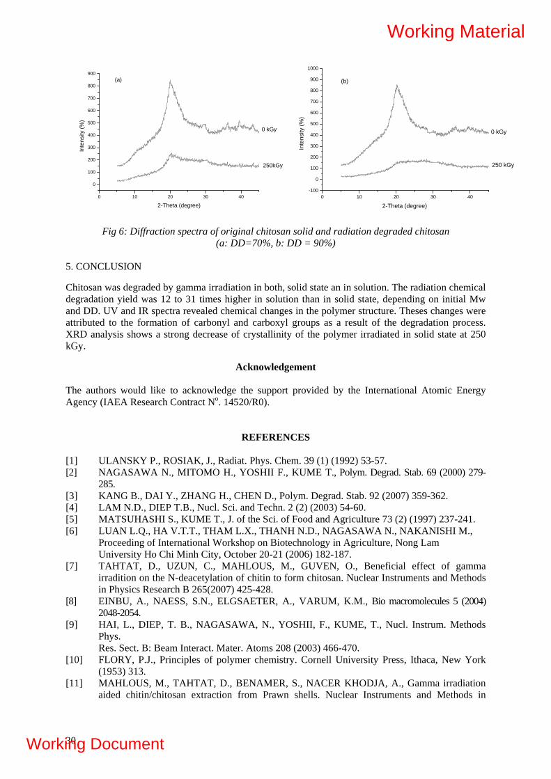
0 10 20 30 40-100
0
100
200
300
400
500
600
700
800
900
1000
Inte
nsity
(%)
2-Theta (degree)
250 kGy
0 kGy
(b)
0 10 20 30 40
0
100
200
300
400
500
600
700
800
900
Inte
nsity
(%)
2-Theta (degree)
250kGy
0 kGy
(a)
Fig 6: Diffraction spectra of original chitosan solid and radiation degraded chitosan (a: DD=70%, b: DD = 90%)
5. CONCLUSION
Chitosan was degraded by gamma irradiation in both, solid state an in solution. The radiation chemical degradation yield was 12 to 31 times higher in solution than in solid state, depending on initial Mw and DD. UV and IR spectra revealed chemical changes in the polymer structure. Theses changes were attributed to the formation of carbonyl and carboxyl groups as a result of the degradation process. XRD analysis shows a strong decrease of crystallinity of the polymer irradiated in solid state at 250 kGy.
Acknowledgement The authors would like to acknowledge the support provided by the International Atomic Energy Agency (IAEA Research Contract No. 14520/R0).
REFERENCES
[1] ULANSKY P., ROSIAK, J., Radiat. Phys. Chem. 39 (1) (1992) 53-57. [2] NAGASAWA N., MITOMO H., YOSHII F., KUME T., Polym. Degrad. Stab. 69 (2000) 279-
285. [3] KANG B., DAI Y., ZHANG H., CHEN D., Polym. Degrad. Stab. 92 (2007) 359-362. [4] LAM N.D., DIEP T.B., Nucl. Sci. and Techn. 2 (2) (2003) 54-60. [5] MATSUHASHI S., KUME T., J. of the Sci. of Food and Agriculture 73 (2) (1997) 237-241. [6] LUAN L.Q., HA V.T.T., THAM L.X., THANH N.D., NAGASAWA N., NAKANISHI M.,
Proceeding of International Workshop on Biotechnology in Agriculture, Nong Lam University Ho Chi Minh City, October 20-21 (2006) 182-187.
[7] TAHTAT, D., UZUN, C., MAHLOUS, M., GUVEN, O., Beneficial effect of gamma irradition on the N-deacetylation of chitin to form chitosan. Nuclear Instruments and Methods in Physics Research B 265(2007) 425-428.
[8] EINBU, A., NAESS, S.N., ELGSAETER, A., VARUM, K.M., Bio macromolecules 5 (2004) 2048-2054.
[9] HAI, L., DIEP, T. B., NAGASAWA, N., YOSHII, F., KUME, T., Nucl. Instrum. Methods Phys. Res. Sect. B: Beam Interact. Mater. Atoms 208 (2003) 466-470.
[10] FLORY, P.J., Principles of polymer chemistry. Cornell University Press, Ithaca, New York (1953) 313.
[11] MAHLOUS, M., TAHTAT, D., BENAMER, S., NACER KHODJA, A., Gamma irradiation aided chitin/chitosan extraction from Prawn shells. Nuclear Instruments and Methods in
30
Working Material
Working Document
Physics Research B 265(2007) 414-417. [12] METHACANON, P., PRASITSILP, M., POTHSREE, T., PATTARAARCHACHAI, J.,
Carbohydrate polymers 52 (2003) 119-123.
[13] MCLAREN, K.G., BRIT. J. Appl. Pms., 16 (1965) 185-193 [14] TORIKAI, A., SHIBATA, H., The Arabian Journal for Science and Engineering, 27, 1C
(2002) 11-24. [15] WANVIMOL, P., RANGRONG, Y., SUWABUN, C., 30th Congress on Science &
Technology of Thailand, October 2004.
PART II: ANTIBACTERIAL ACTIVITY OF PVA/CHITOSAN HYDROGEL SYNTHESIZED
BY GAMMA IRRADIATION
M. Mahlous, D. Tahtat, S. Benamer, A. Nacer Khodja, S. Larbi Youcef Nuclear Research Center of Algiers, Division of Nuclear Applications, BP-399 Alger-Gare, Algeria
1. INTRODUCTION
Poly(vinyl alcohol) (PVA) is a synthetic polymer used in a large range of medical, commercial, industrial and food applications, manufacture of paper products, surgical threads, wound care, and food-contact applications. It was recently used as a coating for dietary supplements and pharmaceutical capsules. Cross-linked PVA microspheres are also used for controlled release of oral drugs [1]. Chitin, a polysaccharide from which chitosan is derived, is the second most abundant natural polysaccharide after cellulose. Chitin is obtained from the exoskeletons (crab, shrimps and squid pen) fungi, insects, and some algae. Chitosan, a non toxic and biocompatible cationic polysaccharide, is produced by partial deacetylation of chitin; these properties of chitosan provide high potential for many applications [2]. Chitosan has been widely used in vastly diverse fields, such as in biomedical applications [3] drug delivery [4] in agriculture [5] metal ion sorption [6]. The most important characteristic of chitosan is the deacetylation degree (DD) which influences its physical and chemical behaviors [7]. Evaluation of DD can be carried out by FT-IR spectroscopy [8, 9] potentiometric titration [10], first derivative UV spectrophotometry, [11] 1H-NMR [12] and X-ray diffraction [13]. Chitosan extracted from squid pen chitin is inherently purer than crustacean chitosans, it does not contain large amounts of calcium carbonate, and it does contain large amounts of protein. The purity of squid pen chitosan makes it particularly suitable for medical and cosmetic application [14, 15]. Application of radiation for the formation of hydrogels for medical use offers a unique possibility to combine the formation and sterilization of the product in a single technological step. The main aim of this study is to synthesis poly(vinyl alcohol) hydrogels containing different moieties of chitosan by gamma irradiation at a dose of 25 kGy, and investigate the antibacterial effect of chitosan contained in the hydrogel.
1.1. Materials and Methods
1.1.1. Materials Chitosan CsD1 with degree of deacetylation (DD) of 70% and Mw = 471 kDa, and chitosan CsD2 with DD of 90% and Mw = 100.9 kDa, were produced locally from squid pens chitin (Loligo sp). Poly(vinyl alcohol) purchased from Fluka (Mw=205 kDa), with a degree of polymerization of 4200 and the degree of hydrolysis of 86%, polyethylene glycol (PEG 400) (Fluka) were used without any purification.
31
Working Material
Working Document

1.2. Preparation of the hydrogel PVA/ chitosan blended hydrogels were prepared by dissolving chitosan powder at different concentrations 0.25 to 1% in an aqueous solution of 2M acetic acid, PVA powder was added to the chitosan solution to a total polymer concentration of 5%, PEG was added at 1.5% to the mixture. The mixture was irradiated with gamma rays to a total dose of 25kGy witch represent a dose of cross-linking and sterilization. 1.3. Swelling measurements The swelling kinetics in deionized water was carried out at room temperature by measuring the water uptake of the hydrogel as a function of time. The degree of swelling (S %) was determined according to the following relationship:
S (%) = [(Wd-Ws)/Ws] ×100
Ws and Wd represent the weight of hydrogel at equilibrium after and prior to the immersion.
1.4. Gel fraction
The gel fraction, Gel (%) is defined as the ratio of the dry gel weight (Wdg) to the initial weight of the polymer (Wip).
Gel (%) = (Wdg/ Wip) × 100
1.5. Scanning Electron Microscopy (SEM)
The morphology structure of the blend hydrogels and pure PVA hydrogel were examined under a Philips XL 30 ESEM scanning electron microscopy. The samples were coated with gold (20 A). 1.6. Infrared spectroscopy
Infrared absorption spectra of PVA, chitosan and PVA-chitosan blend were recorded on Nicolet 380 FTIR spectrometer. The samples were prepared in KBr pellets (1 mg in 100 mg of KBr). IR spectra were recorded by accumulation of 1000 scans with a resolution of 4 cm-1 over the region of 400- 4000 cm-1. 1.7. Antimicrobial tests Antimicrobial tests were performed with Escherichia coli (Gram negative) ATCC 10536 with 165×107
cfu/ml and Bacillus subtilis (Gram positive) ATCC 27853 with 219×107 cfu/ml. The microorganisms were obtained from Pasteur Institute of Algeria. Fresh inoculants for the experimental assessment were prepared on nutrient agar at 30°C for 24 hours. Sterile discs of hydrogels PVA/chitosan (1cm) were deposited on agar medium (pH = 5.5) witch was previously inoculated with a suspension of bacteria. Samples were incubated at 30°C for 24 hours. 1.8. Statistical analysis All experiments were replicated at least three times. The results obtained were treated statistically by the calculation of the central tendency and dispersion parameters, and application the test of Student for the comparison between the various arithmetic means.
32
Working Material
Working Document

2. RESULTS AND DISCUSSION
2.1. Swelling measurements On Figures 1 and 2 are represented the swelling kinetics of PVA/Chitosan CsD1 and PVA/Chitosan CsD2 respectively. It can be observed from the curves that, the swelling ratio increases rapidly during the first 10 hours, then levels off to reach the equilibrium state after 48 hour. No dependence of the swelling behavior of the hydrogel on neither the degree of deacetylation nor the molecular weight was observed. But it depends on chitosan content in the hydrogel; the higher is the chitosan concentration, the higher is the equilibrium swelling ratio. PVA/Chitosan swell by absorption of water, which is kept in the free volume of cross-linked polymer; water uptake is the highest when the network is connected by relatively low number of intermolecular bonds and it decreases with cross-linking density increasing [16].
0 10 20 30 40 50 60 70
020406080
100120140160180200220240260280300
Time (h)
Deg
ree
of s
wel
ling
(%)
PVA 0.25 % 0.50 % 0.75 % 1.00 %
0 10 20 30 40 50 60 70
020406080
100120140160180200220240260280300
degr
ee o
f sw
ellin
g (%
)
Time (h)
PVA 0.25 % 0.50 % 0.75 % 1.00 %
FIG. 1. Variation of water uptake of PVA/Chitosane CsD1 versus time.
FIG. 2. Variation of water uptake of PVA/Chitosane CsD2 versus time.
2.2. Gel fraction
Variation of gel fraction and degree of swelling of PVA/Chitosan is shown in Fig. 3. The gel fraction is the insoluble part of the polymer caused by crosslinking. It was observed, that the gel fraction decreases with increasing chitosan content in blend hydrogels independently of neither the degree of deacetylation nor the molecular weight. The higher is the chitosan concentration in the hydrogel; the lower is the gel fraction. When the chitosan, concentration in the hydrogel increases, the cross-linking density decreases and, consequently, more water is absorbed by the hydrogel.
0,0 0,2 0,4 0,6 0,8 1,00
50
100
150
200
250
33
(S%) PVA-CsD1 (S%) PV 2A-CsD (G%) PV D1A-Cs (G%) PV D2
Concentration (%)
Deg
ree
of s
wel
ling
(S%
)
66
68
70
72
74
76
78
80
82
84
A-Cs
Gel fraction (G
%)
FIG. 3. Evolution of gel fraction and degree of swelling of hydrogel PVA/chitosan CsD1 and PVA/chitosan CsD2 versus concentration.
Working Material
Working Document

2.3. Scanning Electron Microscopy (SEM)
The surface texture of PVA and the PVA/chitosan blend is shown in Fig. 4. PVA showed a uniform and homogenous surface (Fig. 4a). In the blend hydrogel it is observed a heterogeneous texture induced by the chitosan content (Fig. 4b).
(b) (a)
FIG. 4. SEM photomicrographs: (a) PVA (b) hydrogels PVA/chitosan CsD1.
2.4. Infrared spectroscopy
FTIR spectra of PVA/chitosan blend, PVA, and chitosan are shown in Figure 5. It was observed new peaks at 1644 and 1574 cm-1 in the spectrum of PVA/chitosan blend. These peaks are assigned to C=O stretching of amide I band and N-H bending amide II band of the chitosan respectively. Another peak appeared in the blend at 2942 cm-1 which exists in neither PVA nor chitosan. The presence of these peaks in the spectrum of the blend indicates that the chitosan is present in the hydrogel, but does not denote any interaction between PVA and chitosan.
15
74
1644
2942
-0,05
-0,04
-0,03
-0,02
-0,01
0,00
0,01
0,02
0,03
0,04
0,05
0,06
0,07
0,08
0,09
0,10
0,11
0,12
Abso
rban
ce
1600 1800 2000 2200 2400 2600 2800 3000 Nombre d'onde (cm-1)
a
b
c
FIG. 5. FTRI spectrum (a) PVA/chitosan CsD11%, (b) PVA, (c) chitosan.
2.5. Antimicrobial tests
Microbiological test showed that the hydrogels containing moieties of chitosan exhibit antimicrobial activities against E. coli and B. subtilis. Both hydrogels, PVA/chitosan CsD1 and PVA/chitosan CsD2, showed more effective inhibition on Bacillus subtilis than on Escherichia coli. The effect may be attributed to their cell wall composition. Bacillus subtilis is a Gram positive bacterium, its cell wall is composed of peptide polyglycogen. The peptidoglycan layer is composed of networks with plenty of pores, which allow chitosan to come into the cell easily, whereas Escherichia coli is a Gram negative
34
Working Material
Working Document

bacterium, the cell wall of which is made up of a thin membrane of peptidoglycan and an outer membrane constituted of lipopolysaccharides and phospholipids, that constitute a barrier against chitosan [17].
It can be observed also, that the hydrogel containing CsD1 is more effective in inhibiting E. coli and B. subtilis than that containing CsD2 (Fig. 6). The Student test showed that this difference is significant. This effect may be attributed to the fact that the molecular weight of CsD1 is higher than that of CsD2. The diameter of inhibition zone for E. coli and B. subtilis in PVA/chitosan CsD1 does not change with increasing concentration (Fig. 6a); the Student test showed that there is no significant difference in diameters of inhibition zone for the different concentrations of chitosan CsD1, whereas for CsD2, there was a significant difference in diameters of inhibition zone with an increasing concentration of chitosan (Fig. 6b). This phenomenon is attributed to the molecular weight of the chitosan: when it is high, the release of chitosan from the hydrogel to the culture medium becomes lower. Figure 7 shows an example of inhibition zones of PVA/chitosan blends.
0,2 0,3 0,4 0,5 0,6 0,7 0,8 0,9 1,0 1,1
10
20
30
40
(b)
Dia
met
er o
f inh
ibiti
on a
rea
(mm
)
Concentration (%)
E. coli B. Subtilis
50
35
0,0 0,2 0,4 0,6 0,8 1,00
10
40
20
30
(a)
Dia
met
er o
f inh
ibiti
on a
rea
(mm
)
Concentration (%)
E. Coli B. subtilis
a
b
C
FIG. 6. Diameter of inhibition zone versus concentration of chitosan : (a) PVA/chitosan CsD1, (b) PVA/chitosan CsD2.
(a) (b)
(c) (d)
FIG. 7. Inhibition zone (a) E.coli with PVA/chitosan CsD1.(b) E.coli with PVA/chitosan CsD2. (c) B. subtilis with PVA/chitosanCsD1.(d) B. subtilis with PVA/chitosanCsD2.
Working Material
Working Document

36
3. CONCLUSION PVA hydrogels containing moieties of chitosan exhibit antibacterial activity against both gram positive and gram negative bacteria. The release of the chitosan into the culture medium is governed by its molecular weight. The effect of chitosan content in the hydrogel on antibacterial activity is observed for low molecular weight chitosan.
Acknowledgement
The authors would like to acknowledge the support provided by the International Atomic Energy Agency (IAEA Research Contract No 14520).
REFERENCES
[1] DE. MERLIS, C.C., SCHONEKER, D.R., Review of the oral toxicity of polyvinyl alcohol (PVA). Food Chem Toxicol. 41 (2003) 319–326.
[2] MUZZARELLI, R.A.A., Chitosan-based dietary foods. Carbohydrate polymers. 29 (1996) 309-316.
[3] PAUL, W., SHARMA, C.P., Chitosan and aliginate wound dressings: Short review. Trends Biomater. Artif. Organs. 18-1 (2004) 18-23.
[4] VAN DER LUBBEN, I.M., VERHOEF, J.C., BORCHARD, G., JUNGINGER, H.E., Chitosan and its derivative in mucosal drug and vaccine delivery. European Journal of Pharmaceutical Science. 14 (2001) 201-207.
[5] ZHANG, M., TAN, T., YUAN, H., RUI, C., Insecticidal and fungicidal activities of chitosan and oligo-chitosan. Journal of Bioactive and Compatible Polymers 18 (2003).
[6] GUIBAL, E., Interaction of metal ions with chitosan based sorbents: A review. Separation and Purification Technology 38 (2004) 43-74.
[7] MIMA, S., MIYA, M., IWAMOTO, R., YOSHIKAWA, S., Highly deacetylated chitosan and its properties. Journal of Applied Polymer Science 28 (1983) 1909-1917.
[8] TAHTAT, D., UZUN, C., MAHLOUS, M., GUVEN, O., Beneficial effect of gamma irradition on the N-deacetylation of chitin to form chitosan. Nuclear Instruments and Methods in Physics Research B 265 (2007) 425-428.
[9] BRUGNERTTO, J., LIZARDI, J., GOYCOOLEA, F.M., ARGUELLES-MONAL, W., DESBRIERES, J., RINAUDO, M., An infrared investigation in relation with chitin and chitosan characterisation. Polymer. 42 (2001) 3569-3580.
[10] JIANG, X., CHEN, L., ZHONG, W.A., New linear potentiometric titration method for the determination of deacetylation degree of chitosan. Carbohydrate Polymers 54 (2003) 457-463.
[11] TAN, S.C., KHOR, E., TAN, T.K., WONG, S.M., The degree of deacetylation of chitosan : advocating the first derivative UV spectrophotometry method of determination. Talanta. 45 (1998) 713-719.
[12] LAVERTU, M., XIA, Z., SERREQI, A.N., BERRADA, M., RODRIGUES, A., WANG, D., BUSCHMANN, M.D., GUPTA, A., A validation 1H-NMR method for the determination of the degree of deacetylation. J. Pha and biomedical Analysis. 32 (2003) 1149-1158.
[13] ZHANG, Y., XUE, C., XUE, Y., GAO, R., ZHANG, X., Determination of the degree of deacetylation of chitin and chitosan by X- ray powder diffraction. Carbohydrate Research. 340 (2005) 1914-1917.
[14] SHEPHERD, R., READER, S., FALSHAW, A., Chitosan functional properties. Glycoconjugate Journal 14 (1997) 535-542.
[15] TOLAIMATE, A., DESBRIERES, J., RHAZI, M., ALAGUI, A., VICENDON, M., VOTTERO, P., On the influence of deacetylation process on the physicochemical characterisation of chitosan from squid chitin. Polymer. 41 (2000) 2463-2469.
[16] BENAMER, S., MAHLOUS, M., BOUKRIF, A., MANSOURI, B., LARBI YOUEF, S., Synthesis and characterization of hydrogels based on Poly (Vinyl Pyrrolidone). Nuclear Instruments and Methods in Physics research B. 248 (2006) 284-290.
Working Material
Working Document
RADIATION PROCESSING OF MARINE ALGAL POLYSACCHARIDES FOR AGRICULTURE, AS PLANT GROWTH PROMOTERS & PRESERVATION OF FOODS AND HEALTH CARE PRODUCTS AS INSTANT HYDROGEL FORMULATIONS FOR IMPROVED MUCOSE ADHESION AND MEDICAL APPLICATIONS Eduardo E. Smolko, Mario Clozza*, Ernesto B.Giardina*, Fernando Villela*, Marta D.Divo*, Luciana Cerchietti Comisión Nacional de Energía Atómica, Centro Atómico Ezeiza, Laboratorio de Polímeros * Univ. de Buenos Aires, Fac.de Agronomía, Departamento de Producción Vegetal, Argentina. Summary Characterization of the bulk material for degradation and/or structure modification in grafting procedures during this period was continued They were characterized by ultra-violet and visible (UV-vis) and Fourier transform infra- red spectroscopy (FTIR). After receiving the chitosan sample from Prof. Dr Rosiak we started the duty of harmonization procedures for the characterization of raw materials in terms of molecular weight determination by viscosity and to proceed with the measurements of samples of alginates and carrageenans For investigation of radiation–processed alginates for agriculture applications degraded polymers were used as plant growth promoters, in different crops, such as tomato, lettuce, spinach and cabbage under different environmental conditions. By irradiation of alginates with various radiation doses (500, 750 and 1000 kGy gamma rays) degraded alginates have been obtained. They have been applied in horticultural species in 20 and 100 microgram/l concentrations, in solution state (under hydroponics cultivation condition) and by foliar spraying (under soil less culture). In a preliminary phase of this study, different alginate treatments (radiation doses and concentrations) were applied to determine the effect on seedlings quality, evaluated through leaf and root development, shoot/root ratio and photosynthetic activity. UV-vis spectroscopy of alginate irradiated at very high doses ( up to 1000 kGy) showed an abrupt change in the macroscopic shape of the sample forming hard particles and shifting the absorbing band to a higher wavelength. Alginates irradiated in solution shows an absorption band at 265 nm approximately and increases steadily with dose. Alginates irradiated in solid state shows also the absorption band at 265 and increases with doses but this band disappears at higher dose and shows a new wider band at 427 nm. Degradation was also observed accompanied by a color change to deep brown for highly degraded alginate.A conclusion from the color forming phenomena is that for lower doses the double bond formed after degradation can be established between the carbons 4 and 5 of the pyranose ring but for higher dose the double bond could be formed between the carbon 1 and the oxygen 1 of the glycoside bond giving a different terminal reactive species. In the grafting study of alginic acid-g-acrilic acid we consider highly probably demonstrated the modification of the structure by grafting for a new absorption band located at 1727 (1/cm) that corresponds also to a band found also in poly- methylmethacrylate and polyacrilic acid . 1. INTRODUCTION As it was recommended in the previous CRP Meeting, in alginate samples for different purposes such as DDS applications, plant growth promoting, biocide properties, antimicrobial, antioxidant, soil conditioners, etc., a better control of polymer structure after radiation processing was important. Characterization of the bulk material for degradation and/or structure modification in grafting procedures during this period was continued.
37
Working Material
Working Document

2. DEGRADATION STUDIES ON ALGINATES 2.1. Alginate UV-vis spectroscopy UV-vis spectra of the BERNA samples irradiated with 500, 750 and 1000 kGy are observed in the following figures. They consisted of samples of sodium alginate rich in sodium mannuronate as determined by FTIR. Irradiation took placed in close non-permeable polyethylene bags .The spectra was taken in a Shimadzu UV-vis spectrometer joined to a chart register. 2.2. Alginate FTIR spectroscopy Non irradiated samples from alginate BERNA, SIGMA–Aldrich or FMC showed the same peaks characteristic of a sample of sodium alginate rich in around 90 % in sodium mannuronate. Irradiation of alginate for grafting studies. For lower dosis ( 0.5 to 2 kGy ) as necessary for simultaneous grafting experiments, an AECL gammacell 220 unit was used. For post-irradiation grafting ( 10 to 30 kGy ) experiments the irradiation took place in the PISI installation. FTIR and UV-vis spectra of grafted alginate. The study of the grafting of acrylic acid monomer to alginate polymer was tried by both techniques 3. CHITOSAN INTRINSIC VISCOSITY MEASUREMENTS EXCERSIZE 1.1 Drying oven (Copreva Soc. Industrial y Comercial, Argentina.) Temperature 60.0 °C ± 0.1°C. 1.2 Simple thermometer for the range 0-100 °C. 1.3 Dessicator 1.4 Analytical balance Sartorius TE 153S (1mg accuracy). 1.5 Magnetic stirrer with stirrer bar 1.6 pH-meter with a pH electrode and a set of calibration buffers (0.01 units accuracy). 1.7 Adjustable automatic pipette Eppendorf for 1-5 mL and 1-10 mL. 1.8 Cannon -Fenske type glass viscometer ( IVA 120203 Code, 75 serie, 1.6-6.4 cSt, Vmax= 10 mL, Vmin=5mL). 1.9 Thermostatic water bath with transparent walls. 1.10 Precise thermometer (0.5 deg accuracy). 1.11 Stop-watch 1.12 Refrigerator 1.13 Computer with Statistix , Origin and Excel. Data processing software. 1.14 Disposable filtering units with a hydrophilic membrane of 0.45 micron pore size of Sartorius(cellulose nitrate filter 11306) All chemical reagents used were analytical grade. The closed vessels with chitosan were store in a desiccator until irradiation. 3.1. Irradiation Gamma irradiation was utilized Conditions: Lower doses ( 2 and 5 kGy ) were obtained from an AECL Gamma Cell 220. Higher doses (10, 25 and 35 kGy) from a commercial private irradiation firm called IONICS S.A. Dose was determined with potassium dichromate liquid dosimeters from the National Secondary Standards High Dose Dosimetry Laboratory (CNEA)
38
Working Material
Working Document
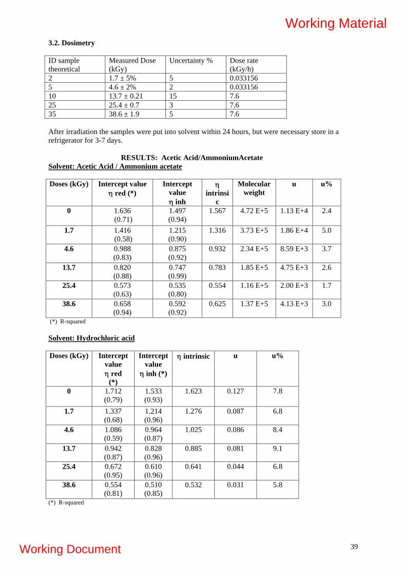
3.2. Dosimetry ID sample theoretical
Measured Dose (kGy)
Uncertainty % Dose rate (kGy/h)
2 1.7 ± 5% 5 0.033156 5 4.6 ± 2% 2 0.033156 10 13.7 ± 0.21 15 7.6 25 25.4 ± 0.7 3 7.6 35 38.6 ± 1.9 5 7.6 After irradiation the samples were put into solvent within 24 hours, but were necessary store in a refrigerator for 3-7 days.
RESULTS: Acetic Acid/AmmoniumAcetate Solvent: Acetic Acid / Ammonium acetate Doses (kGy) Intercept value
η red (*) Intercept
value η inh
η intrinsi
c
Molecular weight
u u%
0 1.636 (0.71)
1.497 (0.94)
1.567 4.72 E+5 1.13 E+4 2.4
1.7 1.416 (0.58)
1.215 (0.90)
1.316 3.73 E+5 1.86 E+4 5.0
4.6 0.988 (0.83)
0.875 (0.92)
0.932 2.34 E+5 8.59 E+3 3.7
13.7 0.820 (0.88)
0.747 (0.99)
0.783 1.85 E+5 4.75 E+3 2.6
25.4 0.573 (0.63)
0.535 (0.80)
0.554 1.16 E+5 2.00 E+3 1.7
38.6 0.658 (0.94)
0.592 (0.92)
0.625 1.37 E+5 4.13 E+3 3.0
(*) R-squared Solvent: Hydrochloric acid Doses (kGy) Intercept
value η red
(*)
Intercept value
η inh (*)
η intrinsic u u%
0 1.712 (0.79)
1.533 (0.93)
1.623 0.127 7.8
1.7 1.337 (0.68)
1.214 (0.96)
1.276 0.087 6.8
4.6 1.086 (0.59)
0.964 (0.87)
1.025 0.086 8.4
13.7 0.942 (0.87)
0.828 (0.96)
0.885 0.081 9.1
25.4 0.672 (0.95)
0.610 (0.96)
0.641 0.044 6.8
38.6 0.554 (0.81)
0.510 (0.85)
0.532 0.031 5.8
(*) R-squared
39
Working Material
Working Document
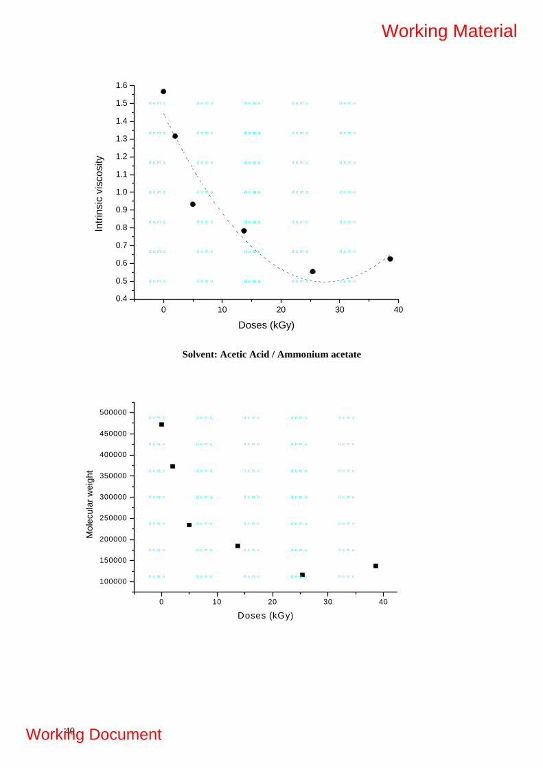
0 10 20 30 400.4
0.5
0.6
0.7
0.8
0.9
1.0
1.1
1.2
1.3
1.4
1.5
1.6In
trins
ic v
isco
sity
Doses (kGy)
d e m o d e m o d e m o d e m o d e m o
d e m o d e m o d e m o d e m o d e m o
d e m o d e m o d e m o d e m o d e m o
d e m o d e m o d e m o d e m o d e m o
d e m o d e m o d e m o d e m o d e m o
d e m o d e m o d e m o d e m o d e m o
d e m o d e m o d e m o d e m o d e m o
Solvent: Acetic Acid / Ammonium acetate
0 10 20 30 40
100000
150000
200000
250000
300000
350000
400000
450000
500000
Mol
ecul
ar w
eigh
t
Doses (kGy)
d e m o d e m o d e m o d e m o d e m o
d e m o d e m o d e m o d e m o d e m o
d e m o d e m o d e m o d e m o d e m o
d e m o d e m o d e m o d e m o d e m o
d e m o d e m o d e m o d e m o d e m o
d e m o d e m o d e m o d e m o d e m o
d e m o d e m o d e m o d e m o d e m o
40
Working Material
Working Document

Molecular Weight
DOSE (kGy) Acetic ac./ Ammonium
acetate Hydrochloric
acid 0 472000 494914 2 373000 357880 5 234000 266400
14 185000 218563 25 116000 141508 39 137000 110075
CHITOSAN VISCOSITY MOLECULAR WEIGHT
0
100000
200000
300000
400000
500000
600000
0 10 20 30 40 50
DOSE (kGy)
MO
LEC
ULA
R W
EIG
HT
Acetic Acid/ Ammonium acetateHydrochloric acid
INTRINSIC VISCOSITY OF CARRAGEENAN
Concentration ηred ηinh 0,555 1,103 0,860 0,625 1,056 0,811 0,714 1,015 0,763 0,833 1,013 0,735 1,000 0,990 0,688
41
Working Material
Working Document

CARRAGEENIN VISCOSITY
y = -0,3686x + 1,0462
y = -0,2273x + 1,2048
0,000
0,200
0,400
0,600
0,800
1,000
1,200
1,400
0,0 0,5 1,0 1,5 2,0 2,5
CONCENTRATION (g/dm3)
VIS
CO
SIT
Y (d
m3 /g)
REDUCED
INHERENT
4. APPLICATION OF RADIATION-PROCESSED ALGINATES Irradiation of alginates Dry matter production Leaf area estimation A model for each species is developed to estimate leaf area. In this way, growth analysis can be done avoiding periodically harvests (destructive measurement). Length and width of a representative number of leaves will be measured, and correlated to their area with an area meter (LI-COR Model 3100 Area Meter).
Photo 1. SPAD-502 meter
42
Working Material
Working Document
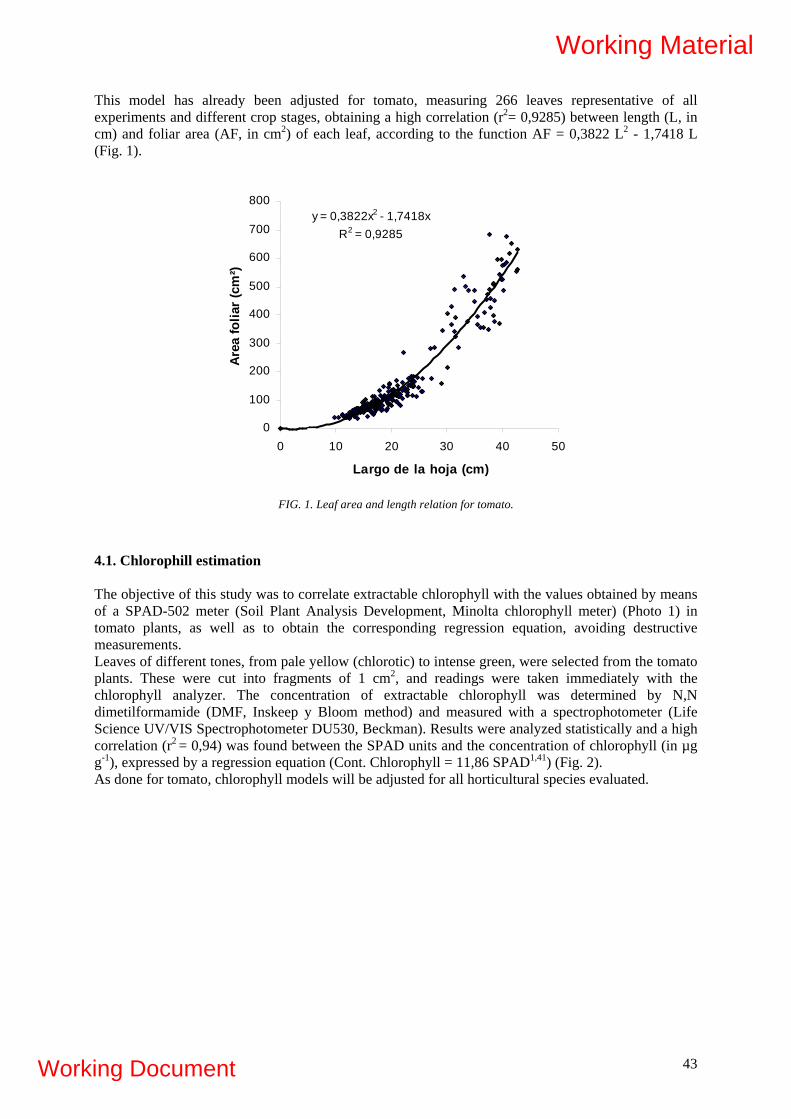
This model has already been adjusted for tomato, measuring 266 leaves representative of all experiments and different crop stages, obtaining a high correlation (r2= 0,9285) between length (L, in cm) and foliar area (AF, in cm2) of each leaf, according to the function AF = 0,3822 L2 - 1,7418 L (Fig. 1).
y = 0,3822x2 - 1,7418xR2 = 0,9285
0
100
200
300
400
500
600
700
800
0 10 20 30 40 50
Largo de la hoja (cm)
Area
folia
r (c
m²)
FIG. 1. Leaf area and length relation for tomato.
4.1. Chlorophill estimation The objective of this study was to correlate extractable chlorophyll with the values obtained by means of a SPAD-502 meter (Soil Plant Analysis Development, Minolta chlorophyll meter) (Photo 1) in tomato plants, as well as to obtain the corresponding regression equation, avoiding destructive measurements. Leaves of different tones, from pale yellow (chlorotic) to intense green, were selected from the tomato plants. These were cut into fragments of 1 cm2, and readings were taken immediately with the chlorophyll analyzer. The concentration of extractable chlorophyll was determined by N,N dimetilformamide (DMF, Inskeep y Bloom method) and measured with a spectrophotometer (Life Science UV/VIS Spectrophotometer DU530, Beckman). Results were analyzed statistically and a high correlation (r2 = 0,94) was found between the SPAD units and the concentration of chlorophyll (in µg g-1), expressed by a regression equation (Cont. Chlorophyll = 11,86 SPAD1,41) (Fig. 2). As done for tomato, chlorophyll models will be adjusted for all horticultural species evaluated.
43
Working Material
Working Document

Y = 11.86 X1.41
r2= 0.94
0
500
1000
1500
2000
2500
3000
3500
0 10 20 30 40 50 6Lectura de SPAD
Con
teni
dode
Clo
rofil
a(µ
g g-1
)
0
Y = 11.86 X1.41
r2= 0.94
0
500
1000
1500
2000
2500
3000
3500
0 10 20 30 40 50 6Lectura de SPAD
Con
teni
dode
Clo
rofil
a(µ
g g-1
)
0
FIG. 2. Concentration of chlorophyll and SPAD units relation for tomato.
Aereal biomass (g)
Spinach Cabbage Tomato
Treatment 20 mg 100 mg 20 mg 100 mg 20 mg 100 mg
0 0,62 b 0,95 b 7,83 a 8,90 a 9,3 a 7,94 a
500 1,26 b 1,36 b 8,00 a 8,61 a 9,47 a 8,88 a
750 1,20 b 1,64 a 7,20 ab 6,41 b 6,56 b 7,86 a
1000 1,70 a 0,74 b 7,55 ab 9,28 a 10,56 a 5,88 b
Control 1,89 a 7,08 ab 8,12 a
Shoot-root ratio
Spinach Cabbage Tomato
Treatment 20 mg 100 mg 20 mg 100 mg 20 mg 100 mg
0 0,319 a 0,403 a 0,836 a 0,927 a 0,840 a 0,853 a
500 0,156 b 0,195 b 0,828 a 0,851 a 0,907 a 0,668 a
750 0,156 b 0,264 ab 0,692 a 0,707 a 0,707 a 0,859 a
1000 0,268 ab 0,102 b 0,784 a 0,949 a 1,007 a 0,648 a
Control 0,273 ab 0,827 a 0,811 a
44
Working Material
Working Document
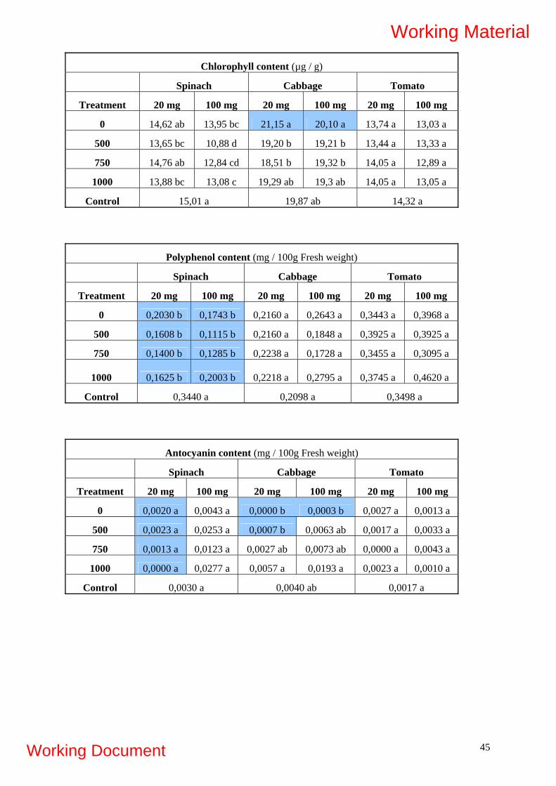
Chlorophyll content (µg / g)
Spinach Cabbage Tomato
Treatment 20 mg 100 mg 20 mg 100 mg 20 mg 100 mg
0 14,62 ab 13,95 bc 21,15 a 20,10 a 13,74 a 13,03 a
500 13,65 bc 10,88 d 19,20 b 19,21 b 13,44 a 13,33 a
750 14,76 ab 12,84 cd 18,51 b 19,32 b 14,05 a 12,89 a
1000 13,88 bc 13,08 c 19,29 ab 19,3 ab 14,05 a 13,05 a
Control 15,01 a 19,87 ab 14,32 a
Polyphenol content (mg / 100g Fresh weight)
Spinach Cabbage Tomato
Treatment 20 mg 100 mg 20 mg 100 mg 20 mg 100 mg
0 0,2030 b 0,1743 b 0,2160 a 0,2643 a 0,3443 a 0,3968 a
500 0,1608 b 0,1115 b 0,2160 a 0,1848 a 0,3925 a 0,3925 a
750 0,1400 b 0,1285 b 0,2238 a 0,1728 a 0,3455 a 0,3095 a
1000 0,1625 b 0,2003 b 0,2218 a 0,2795 a 0,3745 a 0,4620 a
Control 0,3440 a 0,2098 a 0,3498 a
Antocyanin content (mg / 100g Fresh weight)
Spinach Cabbage Tomato
Treatment 20 mg 100 mg 20 mg 100 mg 20 mg 100 mg
0 0,0020 a 0,0043 a 0,0000 b 0,0003 b 0,0027 a 0,0013 a
500 0,0023 a 0,0253 a 0,0007 b 0,0063 ab 0,0017 a 0,0033 a
750 0,0013 a 0,0123 a 0,0027 ab 0,0073 ab 0,0000 a 0,0043 a
1000 0,0000 a 0,0277 a 0,0057 a 0,0193 a 0,0023 a 0,0010 a
Control 0,0030 a 0,0040 ab 0,0017 a
45
Working Material
Working Document

46
Working Material
Working Document
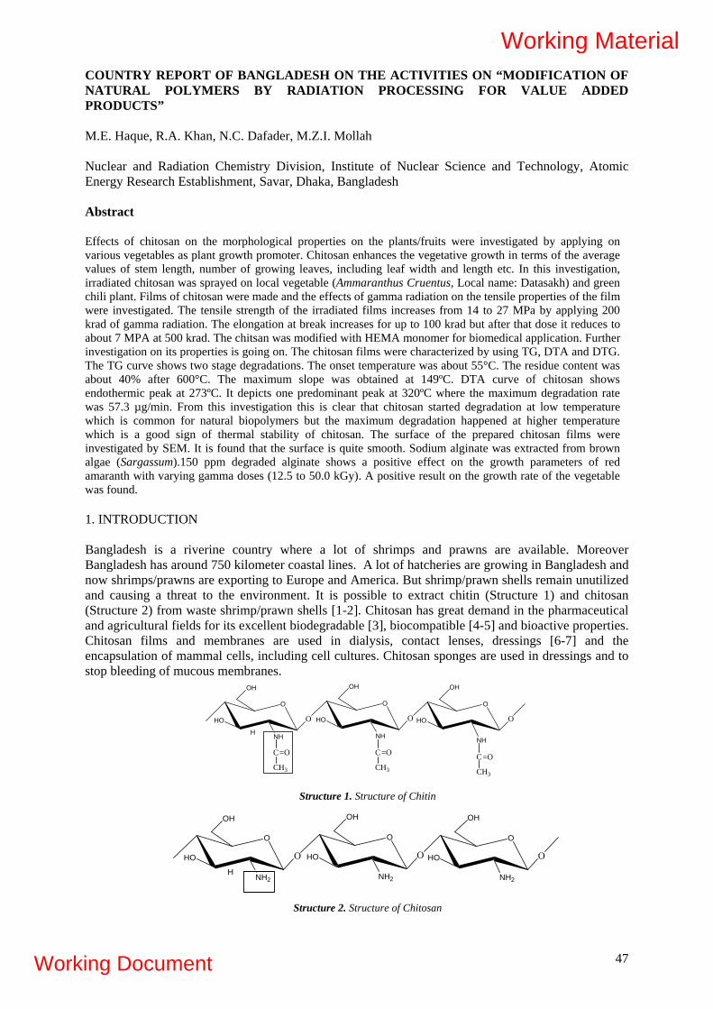
COUNTRY REPORT OF BANGLADESH ON THE ACTIVITIES ON “MODIFICATION OF NATURAL POLYMERS BY RADIATION PROCESSING FOR VALUE ADDED PRODUCTS” M.E. Haque, R.A. Khan, N.C. Dafader, M.Z.I. Mollah
Nuclear and Radiation Chemistry Division, Institute of Nuclear Science and Technology, Atomic Energy Research Establishment, Savar, Dhaka, Bangladesh Abstract Effects of chitosan on the morphological properties on the plants/fruits were investigated by applying on various vegetables as plant growth promoter. Chitosan enhances the vegetative growth in terms of the average values of stem length, number of growing leaves, including leaf width and length etc. In this investigation, irradiated chitosan was sprayed on local vegetable (Ammaranthus Cruentus, Local name: Datasakh) and green chili plant. Films of chitosan were made and the effects of gamma radiation on the tensile properties of the film were investigated. The tensile strength of the irradiated films increases from 14 to 27 MPa by applying 200 krad of gamma radiation. The elongation at break increases for up to 100 krad but after that dose it reduces to about 7 MPA at 500 krad. The chitsan was modified with HEMA monomer for biomedical application. Further investigation on its properties is going on. The chitosan films were characterized by using TG, DTA and DTG. The TG curve shows two stage degradations. The onset temperature was about 55°C. The residue content was about 40% after 600°C. The maximum slope was obtained at 149ºC. DTA curve of chitosan shows endothermic peak at 273ºC. It depicts one predominant peak at 320ºC where the maximum degradation rate was 57.3 µg/min. From this investigation this is clear that chitosan started degradation at low temperature which is common for natural biopolymers but the maximum degradation happened at higher temperature which is a good sign of thermal stability of chitosan. The surface of the prepared chitosan films were investigated by SEM. It is found that the surface is quite smooth. Sodium alginate was extracted from brown algae (Sargassum).150 ppm degraded alginate shows a positive effect on the growth parameters of red amaranth with varying gamma doses (12.5 to 50.0 kGy). A positive result on the growth rate of the vegetable was found. 1. INTRODUCTION Bangladesh is a riverine country where a lot of shrimps and prawns are available. Moreover Bangladesh has around 750 kilometer coastal lines. A lot of hatcheries are growing in Bangladesh and now shrimps/prawns are exporting to Europe and America. But shrimp/prawn shells remain unutilized and causing a threat to the environment. It is possible to extract chitin (Structure 1) and chitosan (Structure 2) from waste shrimp/prawn shells [1-2]. Chitosan has great demand in the pharmaceutical and agricultural fields for its excellent biodegradable [3], biocompatible [4-5] and bioactive properties. Chitosan films and membranes are used in dialysis, contact lenses, dressings [6-7] and the encapsulation of mammal cells, including cell cultures. Chitosan sponges are used in dressings and to stop bleeding of mucous membranes.
HO
O
NHH
OH
O HO
O
OH
O
OH
NH
O HO O
CH3
C=O
CH3
C=ONH
CH3
C=O
Structure 1. Structure of Chitin
47
HO
O
NH2H
OH
O HO
O
NH2
OH
O HO
O
NH2
OH
O
Structure 2. Structure of Chitosan
Working Material
Working Document

1.1. Characterization of Chitosan
Differential Thermal Analysis of the Extracted Chitosan: Figure 1 shows the Thermo-gravimetric (TG), Differential Thermal Analysis (DTA) and Differential Thermo Gravimetric (DTG) curves of the extracted chitosan powder. The black curve (top) is TG, green one (middle) is DTA and the red one (bottom) is DTG curves. The TG curve shows two stage degradations. The onset temperature was about 55ºC. The residue content was about 40% after 600ºC. The maximum slope was obtained at 149ºC. DTA curve of chitosan shows endothermic peak at 273ºC. DTG curve of chitosan depicts one predominant peak at 320ºC where the maximum degradation rate was 57.3 µg/min. From this investigation this is clear that chitosan started degradation at low temperature which is common for natural biopolymers but the maximum degradation happened at higher temperature which is a good sign of thermal stability of chitosan.
FIG. 1. Combined DSC-TGA-DTA spectrum of chitosan.
Scanning Electron Microscopic (SEM) investigation of the surface of Chitosan Films: The surface of the prepared chitosan films were investigated by SEM. It is found that the surface is quite smooth. Minor irregularities and bubbles are formed which may be from the PVC tray surface. From this analysis, it can be concluded that the surface of the films of chiotsan is almost uniform and can be used for further modification by using biocompatible monomers by gamma radiation.
FIG. 2. SEM image of the surface of chitosan film. 1.2. Application of Degraded Chitosan Solution as Plant Growth Promoter Chitosan has been used in agriculture as a coating material for seeds and vegetables, for controlled release of fertilizers, insecticide, fungicide etc. It is reported that irradiated chitosan solution is effective as plant growth enhancer. Chitosan enhances the vegetative growth in terms of the average values of stem length, number of growing leaves, including leaf width and length etc. In this
48
Working Material
Working Document
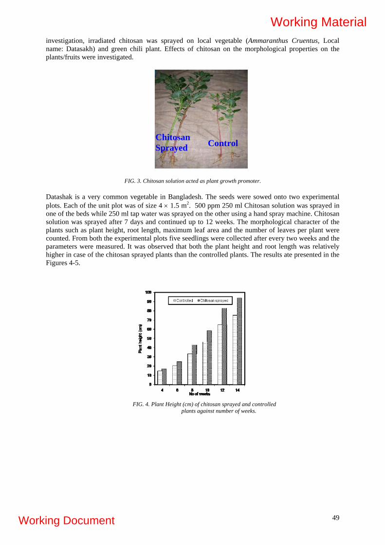
investigation, irradiated chitosan was sprayed on local vegetable (Ammaranthus Cruentus, Local name: Datasakh) and green chili plant. Effects of chitosan on the morphological properties on the plants/fruits were investigated.
Chitosan Sprayed Control
FIG. 3. Chitosan solution acted as plant growth promoter.
Datashak is a very common vegetable in Bangladesh. The seeds were sowed onto two experimental plots. Each of the unit plot was of size 4 × 1.5 m2. 500 ppm 250 ml Chitosan solution was sprayed in one of the beds while 250 ml tap water was sprayed on the other using a hand spray machine. Chitosan solution was sprayed after 7 days and continued up to 12 weeks. The morphological character of the plants such as plant height, root length, maximum leaf area and the number of leaves per plant were counted. From both the experimental plots five seedlings were collected after every two weeks and the parameters were measured. It was observed that both the plant height and root length was relatively higher in case of the chitosan sprayed plants than the controlled plants. The results ate presented in the Figures 4-5.
FIG. 4. Plant Height (cm) of chitosan sprayed and controlled plants against number of weeks.
49
Working Material
Working Document
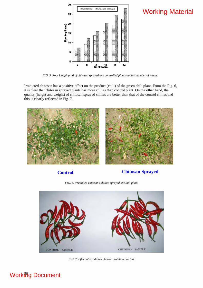
FIG. 5. Root Length (cm) of chitosan sprayed and controlled plants against number of weeks.
Irradiated chitosan has a positive effect on the product (chili) of the green chili plant. From the Fig. 6, it is clear that chitosan sprayed plants has more chilies than control plant. On the other hand, the quality (height and weight) of chitosan sprayed chilies are better than that of the control chilies and this is clearly reflected in Fig. 7.
Chitosan Sprayed Control
FIG. 6. Irradiated chitosan solution sprayed on Chili plant.
FIG. 7. Effect of Irradiated chitosan solution on chili.
50
Working Material
Working Document

1.3. Effect of Gamma radiation on the mechanical properties of the chitosan films Chitosan films were exposed to gamma radiation and the mechanical properties were investigated. The results are presented in Figures 8-9. It was found that tensile strength of the films reached 27 MPa at 200 krad of gamma dose. But further increase of the dose decayed the strength of the films. At 500 krad, the strength of the films reached to 19 MPa which is still higher than the control sample (14 MPa). Gamma radiation has also strong effect on the elongation properties of the chitosan films. Up to 100 krad of gamma dose, elongation at break of the films was found to improve but further increase of the dose, elongation at break decreased significantly.
5
10
15
20
25
30
0 100 200 300 400 500Total Dose (krad)
Tens
ile S
tren
gth
(MPa
)
FIG. 8. Effect of Gamma radiation on the tensile strength of the chitosan films.
4
8
12
16
0 100 200 300 400 500Total Dose (krad)
Elon
gatio
n at
Bre
ak (%
)
FIG.9. Effect of Gamma radiation on the elongation at break of the chitosan films.
1.4. Preparation and Characterization of Flexible Chitosan Films using HEMA monomer for
Biomedical Application The aim of this investigation was to prepare flexible chitosan films with excellent elongation properties for biomedical applications. It is reported that 2-Hydroxyethyl methacrylate (HEMA) is a biocompatible monomer. Chitosan films were immersed in the HEMA solution (1-50 wt%) along with
51
Working Material
Working Document
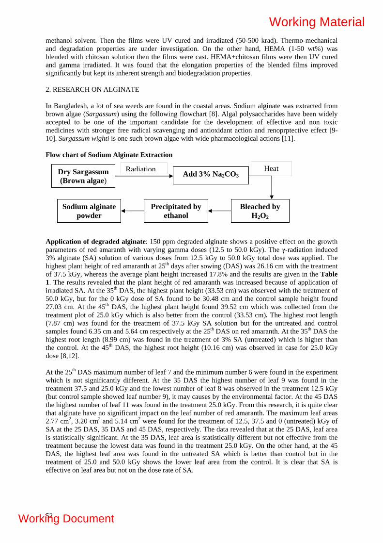
methanol solvent. Then the films were UV cured and irradiated (50-500 krad). Thermo-mechanical and degradation properties are under investigation. On the other hand, HEMA (1-50 wt%) was blended with chitosan solution then the films were cast. HEMA+chitosan films were then UV cured and gamma irradiated. It was found that the elongation properties of the blended films improved significantly but kept its inherent strength and biodegradation properties. 2. RESEARCH ON ALGINATE In Bangladesh, a lot of sea weeds are found in the coastal areas. Sodium alginate was extracted from brown algae (Sargassum) using the following flowchart [8]. Algal polysaccharides have been widely accepted to be one of the important candidate for the development of effective and non toxic medicines with stronger free radical scavenging and antioxidant action and renoprptective effect [9-10]. Surgassum wighti is one such brown algae with wide pharmacological actions [11]. Flow chart of Sodium Alginate Extraction
Dry Sargassum (Brown algae)
Add 3% Na2CO3
Bleached by H2O2
Precipitated by ethanol
Sodium alginate powder
Radiation Heat
Application of degraded alginate: 150 ppm degraded alginate shows a positive effect on the growth parameters of red amaranth with varying gamma doses (12.5 to 50.0 kGy). The γ-radiation induced 3% alginate (SA) solution of various doses from 12.5 kGy to 50.0 kGy total dose was applied. The highest plant height of red amaranth at 25th days after sowing (DAS) was 26.16 cm with the treatment of 37.5 kGy, whereas the average plant height increased 17.8% and the results are given in the Table 1. The results revealed that the plant height of red amaranth was increased because of application of irradiated SA. At the 35th DAS, the highest plant height (33.53 cm) was observed with the treatment of 50.0 kGy, but for the 0 kGy dose of SA found to be 30.48 cm and the control sample height found 27.03 cm. At the 45th DAS, the highest plant height found 39.52 cm which was collected from the treatment plot of 25.0 kGy which is also better from the control (33.53 cm). The highest root length (7.87 cm) was found for the treatment of 37.5 kGy SA solution but for the untreated and control samples found 6.35 cm and 5.64 cm respectively at the 25th DAS on red amaranth. At the 35th DAS the highest root length (8.99 cm) was found in the treatment of 3% SA (untreated) which is higher than the control. At the 45th DAS, the highest root height (10.16 cm) was observed in case for 25.0 kGy dose [8,12]. At the 25th DAS maximum number of leaf 7 and the minimum number 6 were found in the experiment which is not significantly different. At the 35 DAS the highest number of leaf 9 was found in the treatment 37.5 and 25.0 kGy and the lowest number of leaf 8 was observed in the treatment 12.5 kGy (but control sample showed leaf number 9), it may causes by the environmental factor. At the 45 DAS the highest number of leaf 11 was found in the treatment 25.0 kGy. From this research, it is quite clear that alginate have no significant impact on the leaf number of red amaranth. The maximum leaf areas 2.77 cm2, 3.20 cm2 and 5.14 cm2 were found for the treatment of 12.5, 37.5 and 0 (untreated) kGy of SA at the 25 DAS, 35 DAS and 45 DAS, respectively. The data revealed that at the 25 DAS, leaf area is statistically significant. At the 35 DAS, leaf area is statistically different but not effective from the treatment because the lowest data was found in the treatment 25.0 kGy. On the other hand, at the 45 DAS, the highest leaf area was found in the untreated SA which is better than control but in the treatment of 25.0 and 50.0 kGy shows the lower leaf area from the control. It is clear that SA is effective on leaf area but not on the dose rate of SA.
52
Working Material
Working Document

TABLE1. EFFECT OF IRRADIATED ALGINATE SOLUTION OF 150 PPM IN THE MORPHOLOGICAL CHARACTERS OF RED AMARANTH AT 1-5% LEVEL OF SIGNIFICANCE
Plant height average (cm)
Root length average (cm)
No. of leaf average Maximum leaf area (cm2) Treat-
ment 25 DAS
35 DAS
45 DAS
25 DAS
35 DAS
45 DAS
25 DAS
35 DAS
45 DAS
25 DAS
35 DAS
45 DAS
Control 17.02 27.03 33.53 5.64 7.98 7.51 6 9 9 1.80 3.22 b 4.13 Untreated 22.45 bc 30.48 bc 36.88 b 5.72bc 8.99 a 8.48 b 6 9 9 2.11 2.59 c 5.14a 12.5 kGy 24.89 ab 29.46 c 34.25 c 5.55 7.98 8.15 c 6 8 10 2.77 a 2.80 bc 4.90ab 25 kGy 21.13 c 31.59 ab 39.52 a 6.35 b 8.74 ab 10.16 a 7 9 11 2.22 c 2.58 c 3.74 37.5 kGy 26.16 a 35.36 bc 7.87a 8.25 bc 9.83 ab 7 9 10 2.27 bc 3.20 a 4.27b 50 kGy 24.13 b 33.53 a 35.36 bc 6.60ab 8.19 c 10 ab 7 9 10 2.34 b 2.65 c 3.00 ∂v 2.91 2.13 2.10 0.77 0.31 0.91 0.51 0.37 0.67 0.28 0.26 0.69 Note: In a column figures showing dissimilar letter (s) differ significantly according to Duncan Multiple Range Test (DMRT), all the data are mean value of three replications, the 18 number of independent samples. DAS: Days after sowing
REFERENCES [1] MAHLOUS, M., TAHTAT, D., BENAMER, S., NACER, A., KHODJA, Gamma irradiation-
aided chitin/chitosan extraction from prawn shells, Nucl. Instr. And Meth in Phys. Res. B; 265 (2007) 414-417.
[2] ENTSAR, S., ABDOU, KHALED, S.A., NAGY, MAHER, Z., ELSABEE, Extraction and characterization of chitin and chitosan from local sources, Bioresource Technology, 99 (2008) 1359-1367.
[3] MUZZARELLI, R., BALDASSARRE, V., CONTI, F., FERRARA, P., BIAGINI, G., GAZZANELI, G. AND VASI, V., Biological activity of chitosan: ultrastructural study. Biomaterials 9 (1988) 247-252.
[4] ELCIN, Y.M., DIXIT, V., GITNICK, G., Hepatocyte attachment on biodegradable modified chitosan membranes: in votro evaluation for the development of liver organiods. Artif Organs, 22 (1988) 837-46.
[5] NETTLES, D.L., ELDER, S.H., GILBERT, J.A., Potential use of chitosan as a cell scaffold material for cartilage tissue engineering. Tissue Eng., 8 (2002) 1009-16.
[6] ISHIHARA, M., NAKANISHI, K., ONO, M., KIKUCHI, M., SAITO, Y., Photocrosslinkable chitosan as a dressing for wound occlusion and accelerator in healing process. Biomaterials, 23 (2002) 833-40.
[7] JAYASREE, R.S., RATHINAM K AND SHARMA, C.P., Development of artificial skin (Template) and influence of different types of sterilization procedures on wound healing pattern in rabbits and guinea pigs, J. Biomater. Appl, 10 (1995) 144-132.
[8] MOLLAH, M.Z.I., MUBARAK, A., KHAN AND RUHUL, A. KHAN, Effect of gamma irradiated sodium alginate on red amaranth (Amaranthus cruentus L.) as growth promoter. Radiation Physics and Chemistry, 78 (2008) 61-64.
[9] ZANG, Q., LI, Z., XU, Z., ZHANG, H., Effect of fucoidan oin chromic renal failure in rats. Planta Med. 69 (2003) 537-541.
[10] ZHANG, Q., LI, N, LIU, X., ZHAO, Z., The structure of sulfated galactan from Porphyra haitanensis and its in vitro antioxidant activity, Carbohydr. Res. 339 (2004) 105-111.
[11] ANTHONY, J., GABAPATHY, A., COOTHAN, K.V., STREENIVASAN, P.P., ARJUNA, R., PALANI-NATHAN, V., Beneficial effects of sulfated polysaccharides from sargassum wighti against mitochondrial alterations induced by cyclosporine A in rat kidney. Mol. Nutr. Food Res. 51 (2007) 1413-1422.
[12] HIEN, N. Q., NAOTSUGU, N., LE., X. T., FUMIO, Y., VO., H.D., HIROSHI, M., KEIZO, M., TAMIKAZU, K., Growth-promotion of plants with depolynerized alginates by irradiation. J. Radiat. Phys. Chem, 59 (2000) 97-101.
53
Working Material
Working Document

54
Working Material
Working Document

IONIZING RADIATION CONVERSION OF LIGNOCELLULOSIC BIOMASS FROM SUGARCANE BAGASSE TO PRODUCTION ETHANOL BIOFUEL
C.L. Duarte1; M.N. Mori1; H. Oikawa1; H.R. Nagatomi; J. Finguerut2, M. Célia, A. Galvão2 1Instituto de Pesquisas Energéticas e Nucleares (IPEN / CNEN - SP) Radiation Technology Center Av. Professor Lineu Prestes 2242 05508-000 São Paulo, SP [email protected]
2Sugarcane Technology Center, CTC
Caixa Postal 162 Piracicaba SP Brazil 13400-970
Abstract
Sugarcane bagasse has been considered as a substrate for single cell protein, animal feed, and renewable energy production. Sugarcane bagasse generally contain up to 45% glucose polymer cellulose, 40% hemicelluloses, and 20% lignin. Pure cellulose is readily depolymerised by radiation, but in biomass, the cellulose is intimately bonded with lignin, that protect it from radiation effects. The objective of this study is the evaluation of the electron beam irradiation as a pre-treatment to enzymatic hydrolysis of cellulose in order to facilitate its fermentation and improves the production of ethanol biofuel. Samples of sugarcane bagasse were obtained in sugar/ethanol Iracema Mill sited in Piracicaba, Brazil, and were irradiated using Radiation Dynamics Electron Beam Accelerator with 1.5 MeV energy and 37kW, in batch systems. The applied absorbed doses of the fist sampling, Bagasse A, were 20 kGy, 50 kGy, 100 kGy and 200 kGy. After the evaluation the preliminary obtained results, it was applied lower absorbed doses in the second assay: 5 kGy, 10 kGy, 20 kGy, 30 kGy, 50 kGy, 70 kGy, 100 kGy and 150 kGy. The electron beam processing took to changes in the sugarcane bagasse structure and composition, lignin and cellulose cleavage. The yield of enzymatic hydrolyzes of cellulose increase about 75 % with 30 kGy of absorbed dose.
1. OBJECTIVE OF THE RESEARCH
The main objective of the project is to study the cleavage of lignocellulosic material from sugarcane bagasse using ionizing radiation from gamma source and industrial electron beam accelerator in order to facilitate its hydrolysis and fermentation and improves the production of ethanol bio-fuel. Specific Objectives:
1. To study the most adequate sugarcane bagasse and pre-treatments for irradiation processing; 2. To study the better irradiation systems in order to reach the expected results at different
radiation sources (gamma and electron beam) for irradiation processing optimization; 3. To study the modification of irradiated sugarcane bagasse fibber and the degradation of the
natural polymer cellulose, hemicelluloses and lignin in different varieties developed in Brazil by CTC;
4. Enzymatic and chemical hydrolysis and fermentation studies of irradiated sugarcane bagasse and consequently bio-fuel ethanol production;
5. Construction of pilot plant scale to produce cellulose ethanol bio-fuel by radiation processing for feasibility and economics studies of irradiated sugarcane bagasse in the bio-fuel ethanol production for industrial scale.
55
Working Material
Working Document

2. INTRODUCTION
Sugarcane bagasse generally contain up to 45% glucose polymer cellulose, much of which is in a crystalline structure, 40% hemicelluloses, an amorphous polymer usually composed of xylose, arabinose, galactose, glucose, and mannose and 20% lignin, which cannot be easily separated into readily utilizable components due to their recalcitrant nature. The remainder is lesser amounts of mineral, wax, and other compounds [1]. Cellulose is highly crystalline with comparatively rigid linear chains essentially free of side branching. The hydroxyl groups attached to the chains provide strong intermolecular bonding. Cellulose is a linear polymer of cellobiose repeating unit, and the degree of polymerization is normally 10 to 100 times greater than that of hemicelluloses. The lignin and hemicelluloses molecules are linked through ester linkages formed by the carboxyl groups in the lignin [2, 3]. The process to convert the sugarcane bagasse to ethanol biofuel consist to three steps, those are the delignification (liberation of cellulose and hemicelluloses from lignin named pre-treatment), depolymerisation of the carbohydrate polymer to produce free sugar (hydrolysis) and fermentation of mixed hexose and pentose sugars to produce ethanol. The Sugarcane bagasse pre-treatment’s are physical and chemical process that reduce the crystallinity, disrupt the hydrogen bonding of cellulose to more accessibility to hydrolytic depolymerisation reactions. Enzymatic hydrolysis of cellulose is a reaction carried out by cellulase enzymes, which are highly specific [4]. Several studies have shown that the irradiation of cotton cellulose deteriorated the mechanical parameters due to the chain scission reaction within the cellulose molecules. Literature studies have reported that the high-energy radiation cause a decrease in the degree of polymerization and an increase in the carbonyl content of cotton cellulose. Others studies have showed that, as a pre-treatment method, ionizing irradiation is equivalent in terms of increasing the digestibility to that of NaOH treatment, one of the most commonly used method in upgrading foliage digestibility [5, 6, 7, 8, 9].
The cleavage of polysaccharides from sugarcane bagasse using ionizing radiation from an industrial electron beam accelerator was studied, in order to facilitate enzymatic hydrolysis of cellulose. The main obstacle of cellulose hydrolysis by irradiation is destruction of the product, in this way it were applied absorbed doses as low as necessary to get cleavage in the lignin, but to avoid loss of glucose due to indiscriminatory degradation of cellulose and hemicelluloses.
3. MATERIALS AND METHODS
3.1. Sampling Two sugarcane bagasse samples from Iracema Mill (Piracicaba-SP) were collected. The first one, called “assay 1” were collected in the rainfalls and the bagasse was 15 days old and presented an average humidity of 50% and density 0.14 g/dm3; the second one, called “assay 2” was collected just after the milling with a humidity average of 60% and density 0.16 g/dm3. 3.2. Radiation Processing The electron beam irradiation was carried out using Radiation Dynamics Inc. USA Electron Beam Accelerator with 1.5 MeV energy and 37 kW, in batch systems. The irradiation parameters used were 2.8 cm sample width for assay 1 and 2.4 cm width for assay 2, 112 cm (94.1%) scan, and 6.72 m/min conveyor stream velocity. The preliminary applied absorbed doses were 20 kGy, 50 kGy, 10 0 kGy and 200 kGy. After obtained results evaluation, lower absorbed doses were applied in the second assay, such as 5 kGy, 10 kGy, 20 kGy, 30 kGy, 50 kGy, 70 kGy, 100 kGy, and 150 kGy (Figure 1).
56
Working Material
Working Document

FIG. 1. Samples irradiation at the electron beam accelerator in the IPEN.
3.3. Chemical Analysis After irradiation, sugarcane bagasse was separated by fibber size using sieves of 1.00 mm and 0.42 mm, this separation was important to remove the solids residues present due to sugarcane harvest process. After humidity rate determination, the bagasse was dried in an oven for 24 h at 60 oC and characterized by analysis of lignin, free sugar, and cellulose, using chemical methods adapted from literature [1, 5, 10]. Lignin was determinated by Klason method. To 0.3 g of sample it was added 3.0 mL of H2SO4, and 85 mL of destilled water and reflux for 1 h to 60oC. The preciptate was dried in an oven for 24 h at 100 oC, and the lignin was determinated gravimetrically. The phenol content was analyzed after extractions with acetonitrile, using an ultrasonic system per 30 minutes, and determined by gas chromatography in association with mass spectrometry, Shimadzu, model GC-MS QP-5000Klason Lignin were determinated by extraction of polissacarides with strong acids H2SO4 and determination of lignin gravimetricaly. The phenol content was analyzed after extraction with hexane/dichloromethane 1:1 v/v solvent, using an ultrasonic system per 30 minutes and determined by gas chromatography, Shimadzu, model GC-FID 17-A.
The low molecular weight carbohydrates (hemicelluloses and degraded cellulose) were determinate using the standard method (ASTM D1109, 1984). To 2.0 g of sample it was added 100 mL of NaOH 1% (p/v). The sample was filtered and the precipitate was dried for 24 h and determinate gravimetrically.
Hot solubility that represents the total free sugars was determinate by standard method (ASTM D1110, 1984). To 2.00 g of sample it was added 100 mL of distilled water and reflux for 3 h to 100 oC. The sample was filtered and the precipitate was dried for 24 h and determinate gravimetrically.
To cellulose total determination it was adopted the method described by Fengel and Wegener, 1989. To 5.0 g it was added a mixture of acetic acid 80% and nitric acid 70% in proportion 10:1, v/v, and reflux for 20 minutes at 60oC. The precipitate was filtered with the aid of suction and determinate gravimetrically.
57
Working Material
Working Document

Alpha and beta cellulose must be determinate on an extractive-free, lignin-free sugarcane bagasse sample, and were determinate using standard methods (ASTM D1103 and NBR14032). Alpha-Cellulose represents high molecular mass cellulose and beta-cellulose represents the degraded cellulose and hemicelluloses [1, 5, 10].
3.4. Infrared Spectroscopy The main difficult to infrared spectroscopy analyses was the sample preparation with KBr mixture. The ideal mixture was 0.0500 g of KBr and 0.0010 g (m/m) of sample. The analysis were made using an IV Espectrofotometric interferometric, BOMEM, model MB-SERIES. 3.5. Scanning Electronic Microscopy Electronic microcopies of the sugarcane bagasse from assay 2 were obtained using a Transmittance Electronic Microscopy from Phillips, to verify the material structural changes caused by the ionizing radiation.
3.6. Enzymatic hydrolysis The enzymatic hydrolysis of the non irradiated and irradiated bagasse was done at the CTC laboratories using samples from the Assay 2, for 24 and 48 h. It was used 50 of total mass in the reactor with 8% of solids charge (dry base). The enzyme concentrate used was a commercial Trichoderma reesei cellulose preparation (Celluclast 1.5 L), kindly supplied by Novozymes (Bagsvaerd, Denmark), with 5FPU/g-cellulose and Beta-glycosidase 0.5% (p/p).
4. RESULTS
After analysing the results of the first assay, it was evaluated that the applied radiation dose could be lower , because the obtained results of, e.g., free sugar, cellulose alpha and phenol after irradiation demonstraded that some modifications have ocurred in the sugarcane bagasse structure with absorved doses lower than 20 kGy. Considering this the applied irradiation doses in the second assay began from 5 kGy.
4.1. Chemical analysis The results of lignin concentration related to applied radiation dose for Assay 1 and 2 are presented in the Table.1. These results show that, considering the Klason method, the variations of the lignin concentration do not change with the applied absorbed doses in both assay. But the results of phenol, that is liberated by cleavage of lignin molecule, presented in the Figure 2 showed a increasing in phenol concentration related to absorbed doses until 100 kGy. After that it was observed a decreasing, that may be caused by the competition between the formation and degradation of phenol by radiation.
The low molecular weight carbohydrates (hemicellulose) showed a increase with the applied absorbed dose indicating a clevage of these polyssacharide, about 20% after irradiation with 100 kGy for samples from both assay(Table 1), but even with the low doses (5 kGy and 10kGy) a light increase can be notice in samples from the second assay.
58
Working Material
Working Document

TABLE 1. PERCENTUAL COMPOSITION OF SUGARCANE BAGASSE FROM TWO ASSAYS BEFORE AND AFTER IRRADIATION IN VARIOUS ABSORVED DOSES.
Dose Hemicellulose Lignin Free sugar Alpha cellulose Beta cellulose
(kGy) Assay 1 Assay 2 Assay 1 Assay 2 Assay
1 Assay
2 Assay
1 Assay
2 Assay 1Assay
2 0 31.27 36.70 25.99 29.54 11.31 8.19 90.88 71.52 22.72 24.06 5 n.a. 40.06 n.a. 33.61 n.a. 10.51 n.a. 56.49 n.a. 37.91
10 n.a. 40.67 n.a. 31.16 n.a. 10.80 n.a. 59.27 n.a. 37.57 20 34.75 40.63 23.64 33.96 9.32 11.08 47.68 20.96 60.32 74.17 30 n.a. 45.41 n.a. 35.96 n.a. 12.13 n.a. 18.25 n.a. 79.75 50 55.45 46.98 26.53 34.76 9.14 13.29 35.86 5.16 84.90 92.09 70 n.a. 50.49 n.a. 36.74 n.a. 13.66 n.a. 2.38 n.a. 94.43
100 55.45 54.42 22.41 37.17 11.84 14.32 2.70 0.00 99.55 96.43 150 63.95 60.38 21.70 34.30 15.51 17.49 0.24 0.00 147.76 94.45 200 71.42 n.a. 19.00 n.a. 21.51 n.a. n.a. n.a. 114.88 n.a.
Na= not applied
An increasing of the free sugar can be observed when absorbed doses higher than 100 kGy were applied in samples from both assay. These sugars are liberated by the total cleavage of cellulose and hemicellulose and are represented mainly by glicose and xylose, respectively.
FIG. 2. Phenol liberation after irradiation of sugarcane bagasse in various absorbed doses.
The sugarcane bagasse from Assay1 presented 46.9±0.6% of total cellulose and the bagasse from Assay 2 presented 49.8±0.8%. Cellulose presented higher sensibility to radiation than the other lignocelluloses, this can be observed by the results of alpha cellulose showed in the Table 1 for assay 1 (sample “A”) and the assay 2 (sample “B”). In both assays the alpha-Cellulose that is the portion insoluble in Noah 17.5% and represent high molecular mass cellulose, reduces about 99% with 100 key of absorbed doses. The radiation effect on the beta cellulose that is the solubilizated in NAOH 17.5% and precipitated in acid and represents the degraded cellulose and hemicellulose are presented in the Fig. 2. The behaviour of beta cellulose were similar in the two assays, showing a increasing of the degradation with dose until 100 kGy, and a decreasing after that suggesting that in higher absorved doses the molecules can recombine.
59
Working Material
Working Document

4.2. Infrared Spectroscopy Figure 3 presents the transmittance spectra of the samples from Assay 2. After irradiation the absorbance’s peak at the 3350 cm-1, 2927 cm-1, 1732 cm-1, 1592 cm-1, 1045 cm-1 and 896cm-1 are more evident and they are in the reference interview to cellulose characterization according to literature [9, 19, 23, and 25]. The absorption peak at 3350 cm-1 represents the hydroxyls in the molecule. The C-H is represented by the absorbance at 2927 cm-1, the carbonyl group (C=O) by the absorbance at 1732 cm-1, and the C-O-C by the absorbance at 1045 cm-1.
FIG. 3. Infrared spectra of sugarcane bagasse related to absorbed doses, with the identification of main organic function of cellulose structure.
4.3. Scanning Electronic Microscopy Electronic microscopy of the sugarcane bagasse from Assay 2 samples was obtained to verify the material structural changes caused by the ionizing radiation. The non irradiated sample exhibited rigid and highly ordered fibrils (Fig. 4). After irradiation some cleavage in the external layer can be observed, the material structure was modified resulting some free particles. This partial delaminating of the cell wall of the irradiated samples enhances the substrate accessibility to the enzymes because enzymatic hydrolysis requires direct physical contact between enzyme and substrate, the enzymes must diffuse from the bulk aqueous solution to the particle surface, diffuse through physical barriers such as lignin, adsorb on the substrate surface, and then catalyze the hydrolysis.
60
Working Material
Working Document
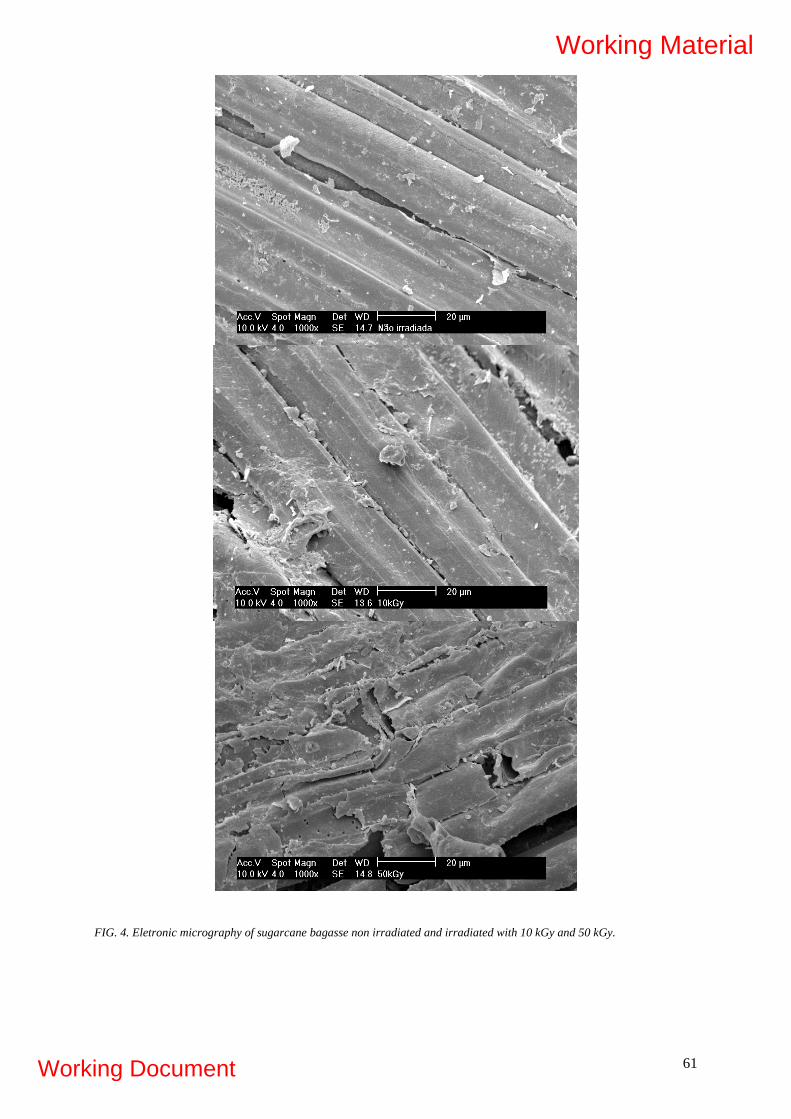
FIG. 4. Eletronic micrography of sugarcane bagasse non irradiated and irradiated with 10 kGy and 50 kGy.
61
Working Material
Working Document

4.4. Enzymatic hydrolyze The enzymatic hydrolyze were applied to samples from de second collect, and evaluate the convertion of cellulose to glucose by action of enzyme complex. After irradiation the glucose concentration increased after 24 h and also after 48h with absorbed doses from 5 to 30 kGy, then for higher doses there were a decreasing in the glucose concentration (Fig. 5). During the saccharification of lignocelluloses polysaccharides, aliphatic acids, e.g. acetic, formic and levulinic acid, furan derivatives, e.g. furfural and 5-hydroxyl-methyl-furfural (HMF), and phenolic compounds are formed in addition to the released sugars. These compounds might seriously inhibit the subsequent enzyme decreasing the hydrolyze yield [4, 11]. That can be investigated in next step of studies. With 20 kGy the conversion yield of cellulose to glucose increases from 8% to 14%, which represent 75% of efficiency. Even with higher doses the yield after irradiation was higher than the non irradiated sample, and the minimal were 10% for 150 kGy. In the Figure 6 is showed the results of humidity test wich accomplish the incubation for enzymatic hydrolysis. The difference among the non irradiated and the irradiated samples in all doses were not significative, that is an advantage because the maintenance of humidity is very important to the enzymatic process (Fig. 7).
FIG. 5. Convertion of cellulose to glicose by the enzymatic complex (CELUCLAST) related to absorbed doses.
62
Working Material
Working Document

FIG. 6. Humidity variation during the enzymatic process related to absorbed dose.
5. CONCLUSION
The obtained results from present work showed that the lignin did not degrade even in high absorbed doses (200kGy), but the increase of phenol compounds sugest a partial cleavage of the molecules. The liberation of free sugars have occurred after radiation processing. The decreasing of alpha-cellulose and the increasing of beta-cellulose means that a cleavage of cellulose occurred in relatively very low absorbed doses (lower than 20 kGy) considering other investigations in the literature. These results reflect that the ionizing radiation with low doses can cleavage the external structure of sugarcane bagasse without destroy the cellulose or lost the sugar, that is desirable as pretreatment to enzymatic attack. The stoked for 20 days and the just squeezed bagasse showed no significant variations on the results. The yield of enzymatic hydrolyzes of cellulose increased until 75 % with absorbed dose, and the decreasing after 20 kGy have to be investigated in future studies.
6. FUTURE PLAN OF PROJECT
• CHEMICAL ANALYSIS OF SUGARS
The HPLC/ELSD system now installed has very good specificity and linearity to carachterization and quantification of sugars and it will be useful to further experiments, to better understanding the action mechanism of ionizing radiation in the sugarcane bagasse structure.
• COMBINATION OF IRRADIATION WITH ENZYMATIC AND CHEMICAL HYDROLYSIS OF CELLULOSE FROM SUGARCANE BAGASSE
• COMPARISION AND COMBINATION OF IRRADIATION WITH THE PRETREATMENT PROCESS OF TEAM EXPLOSION
• FERMENTATION OF THE SUGARCANE BAGASSE AFTER IRRADIATION AND AFTER CELLULOSE HIDROLYSIS.
63
Working Material
Working Document

64
REFERENCES
[1] SUN, J. X., SUN, X. F., ZHAO, H., SUN, R.C., Isolation and characterization of cellulose from sugarcane bagasse. Polymer Degradation and Stability, 84 (2004) 331-339.
[2] GLASSER, W.G., KELLY, S.S., Encyclopedia of Polymer Science and Engeneering New York, Jonh Wiley & Sons, Inc. Lignin. (1987) 796-849.
[3] HASSUANI, S.J., Biomass power generation: sugar cane bagasse and trash/Suleiman José Hassuani, Manoel Regis Lima Verde Leal, Isaías de Carvalho Macedo – Piracicaba: PNUD-CTC (Série Caminhos para Sustentabilidade) (2005) 217.
[4] BRENNER, W., RUGG, B., ARNON, J. Radiation pretreatments for optimizing the sugar yield in the acid hydrolysis of waste cellulose. Radiat. Phys. Chem., 14 (1979) 299-308.
[5] FOLDVÁRY, Cs.M, TAKÁCS, E., WOJNÁROVITS, L. Effect of high-energy radiation and alkali treatment on the properties of cellulose Radiation Physics and Chemistry, 67 (2003) 505-508.
[6] HAN, Y.W., CATALANO, E.A., CIEGLER, A., Chemical and physical properties of sugarcane bagasse irradiated with γ rays. Journal of Agricultural and Food Chemistry, 311 (1983) 34-38.
[7] KHAN, F., AHMAD. S.R., KRONFLI, E., γ Radiation induced changes in the physical and chemical properties of lignocellulose. Biomacromolecules, 7, (2006) 2303-2309.
[8] MCLAREN, K.G., Degradation of cellulose in irradiated wood and purified celluloses. International Journal of Applied Radiation and lsotopes, 29 (1978) 631-635.
[9] SMITH, G.S., KIESLING, H.E., GALYEAN, M.L., BADER, J.R., Irradiation enhancements of biomass conversion. Radiation Physics Chemistry, 25 (1-3) (1985) 27-33.
[10] KHAN, F., AHMAD. S.R., KRONFLI, E., γ Radiation induced changes in the physical and chemical properties of lignocellulose. Biomacromolecules, 7 (2006) 2303-2309.
[11] KUMAKURA, M., KAETSU, I., Radiation-induced degradation and subsequent hydrolysis of waste cellulose materials. I. Journal of Applied Radiation and Isotopes, 30 (1978) 139-141.
[12] VITTI, M.S.S., MASTRO, N.L., KIKUCHI, O.K., NOGUEIRA, N.L., Irradiação de resíduos fibrosos com feixe de elétrons: efeitos na composição química e digestibilidade. Scientia Agrícola, 55 (2) (1998) 159-171.
Working Material
Working Document

DEVELOPMENT OF BIOACTIVE EDIBLE COATINGS AND BIODEGRADABLE PACKAGING USING GAMMA IRRADIATION Lacroix, M., Salmieri, S. INRS-Institut Armand-Frappier, Research Laboratories in Sciences Applied to Food Canadian Irradiation Center 531 des Prairies blvd. Laval, Quebec, Canada H7V 1B7 Abstract Gamma irradiation was used to cross-link milk proteins in order to enhance the physico-chemical properties of edible films made of calcium caseinate, whey protein isolate and glycerol. Fourier Transform Infrared analysis was used to characterize the conformation of proteins adopted after irradiation. The molecular weight of cross-linked proteins was measured by Size-Exclusion Chromatography. Furthermore, the effect of the addition of methylcellulose to the irradiated protein matrix on the rheological properties (puncture strength, puncture deformation and water vapor permeability) of films was also studied. Moreover, cross-linking of polysaccharides under paste-like state was investigated and the cross-linking degree of the gel products was determined by gel fraction measurements and solubility percentage. In order to prepare bioactive coatings, several antifungal compounds were evaluated as bioactive compounds in order to select one of them to prepare an antimicrobial solution to spray onto strawberries or to encapsulate them in film formulations composed of milk proteins and methylcellulose based films. In addition, the bioactive coatings containing the antifungals were used to increase the radiosensitivity under air of moulds and total flora in strawberries and the relative sensitivity of selected formulations was calculated from their D10 value. The film formulation selected was used as a bioactive edible coating in order to determine their efficiency to increase the shelf life of fresh strawberries and to preserve their quality during storage. 1. INTRODUCTION 1.1. Protein-Based Films and Coatings Proteins-based films and coatings have been applied with success using various proteins such as corn zein, wheat gluten, soy protein isolate, whey proteins isolate, and caseins [1,2]. However, the highly hydrophilic nature of protein coatings can limit their functional utilization [3]. While proteins have high film-forming properties primarily due to their ability to form hydrogen bonds, and hydrophobic or electrostatic interactions [4,5], cross-links formation is necessary to produce a strong, flexible film or coating. Physical-processed cross-linking of proteins can be achieved using thermocondensation (controlled heating) or gamma-irradiation [4]. Heat treatment creates disulfide bonds due to the oxidation of thiol groups from cystein residues. Gamma irradiation of proteins in aqueous solution allows creating intermolecular covalent cross-links issued from biphenolic products [4]. 1.2. Edible coatings Edible coatings are traditionally used to improve food appearance and conservation due to their environmental-friendly nature [6]. Coating films can act as barriers to moisture and oxygen during processing, handling and storage [7]. Moreover, they can retard food deterioration by inhibiting the growth of microorganisms, due to their natural intrinsic activity or to the incorporation of antimicrobial compounds [8]. Such edible films could also help to reduce food dehydration and therefore, improving the shelf life. However, the hydrophilic nature of these proteins limits their ability to provide desired edible film functions. Current approaches to extend functional and mechanical properties of these films, include (i) incorporation of hydrophobic compounds such as lipids to improve the water vapour permeability
65
Working Material
Working Document

resistance [2] (ii) optimization of the interaction between polymers (protein-protein interactions, charge-charge electrostatic complexes between proteins and polysaccharides) and (iii) formation of cross-links through physical, chemical, or enzymatic treatments [9]. Heat was essential for the formation of the intermolecular disulfide bonds in whey proteins [10]. It has been demonstrated that gamma-irradiation produces bityrosine bridges between protein chains, improve the mechanical, structural and barrier properties of edible coatings and biodegradable films [11]. Bioactive compounds can be added to such coatings in order to lengthen shelf life, to assure the innocuity, and to preserve nutritional values of foods [5]. Bioactive agents such as EOs are secondary metabolites of the plant and have long been used as flavouring agents in the food industry [12]. Moreover, EOs have been found to exhibit antimicrobial and antifungal properties, making them natural alternatives to combat foodborne pathogens and normal food decay caused by bacterial and mould growth. 1.3. Polysaccharides-Based Films and Coatings Polysaccharides are often used in films and coatings due to their ability to form stable and rigid gels [4]. While their hydrophilic nature can hamper their utilization, these types of films and coatings can preserve the sensorial qualities of food, limit racidification, prevent fat migration and may be antimicrobial [13]. Some common used polysaccharides include cellulose derivatives such as methyl-cellulose (MC), hydroxypropyl-cellulose (HPC) and carboxymethyl-cellulose (CMC), alginate, and chitosan. According to Ressouany et al. [14], addition of carboxymethylcellulose in the formulations improves the resistance of the films and an increase of the puncture strength of these films was observed with irradiation doses. 1.4. Active coating and radiosensitization Doses of 0.25-0.5 kGy can delay the ripening of certain tropical fruits. At doses > 1.75 kGy, inhibition of postharvest diseases and fungi can be observed [15]. However, from 1 to 3 kGy, depending on tolerance of the product, sensory deterioration such as taste and smell, as well as accelerated softening is seen in fruits and vegetables. The maximal irradiation dose tolerated in fresh products is around 2.25 kGy [16]. To control food contamination and quality loss, active edible coating or active biodegradable packaging has been recently introduced in food processing [5,17]. The immobilization of the active compounds in polymer can maintain high concentrations of the active compounds on the surface of foods for longer storage time [18]. Moreover, the combination of active edible coating application and the irradiation treatment had a synergistic effect on the irradiation sensitivity of pathogenic bacteria [19] and can reduce the dose necessary to eliminate pathogenic bacteria. The D10 (kGy) is the required irradiation dose necessary to inactivate 90% of a micro-organism population. An increase of the radiosensitization result of a reduction of the D10. Strawberries and ground beef represent interesting models for the development of active coating and packaging in combination with irradiation. Gamma-irradiation has been proven effective in reducing micro-organisms in fresh strawberries [20]. Gamma-irradiation has also been found to help reduce foodborn pathogens such as E. coli in meat products like ground beef [21-23]. In both cases a maximal dose of 2 kGy is acceptable before the texture of the cellular wall begins to change or a change of the flavour of meat [24,25]. As described previously, various active antimicrobial compounds can be added to reduce the required irradiation dose by increasing radioactivity sensitivity. Good results have been obtained by adding a mix of carvacrol (1 %) and tetrasodium pyrophosphate (0.1%) to ground beef, increasing the radiosensitivity of E. coli up to 55.5%. Also, both gamma-irradiation treatment and edible coating process significantly delayed (p ≤ 0.05) moulds growth in strawberries (Fragaria spp.). Edible coating based on irradiated caseinate was more effective than coating based on unirradiated caseinate. For example, at day 11, these coating reduced respectively by 48 and 23% the moulds appearance on strawberries [26].
66
Working Material
Working Document

1.5. Moulds contamination in strawberries Fresh fruits are prone to fungal contamination in the field, during harvest, transport, and from marketing to the consumer, and some pathogenic yeasts and moulds can grow and be responsible for infections and allergies [27]. The low pH of fruits makes them particularly susceptible to fungal spoilage due to competitive elimination of a bacterial part [28]. Various methods on controlling post-harvest microbial have been developed, such as low-temperature storage, controlled atmospheres, and fungicide applications to prolong the shelf life of fruits [29]. However, these commodities present some disadvantages due to the fact that some fruits are very sensitive to low temperature, involving severe injuries susceptible to microbial contamination and that some fungi could become resistant to commonly used pesticides that are classified as environment and health hazard chemicals [30]. Consequently, the restriction of synthetic fungicides and new developments of antifungal natural substances have changed the post-harvest storage conditions of fruits. Strawberries (Fragaria ananassa) are especially perishable fruits, being susceptible to mechanical injury, desiccation, decay and physiological disorders during storage. Tournas and Katsoudas [28] have examined mould and yeast growth in strawberries in order to isolate and identify predominant moulds after surface disinfection and incubation at room temperature to for up to 14 days. Botrytis cinerea (77%) and Rhizopus stolonifer (23%) were the two major storage pathogens observed in strawberries. Their high level of contamination, as compared to other type of fruits, was mainly due to a low pH, an optimal water activity for fungal growth, a high level of sugars and other nutrients, and a soft skin that can be easily ruptured, favouring microorgansims proliferation. B. cinerea (grey mould rot) is a ubiquitous pathogen which causes severe damages in many fruits, vegetables and ornamental crops in pre- and post-harvest. The pathogen infects leaves, stems, flowers and fruits [30] and is a major obstacle to long-distance transport and storage [7]. R. stolonifer is the causal agent of rhizopus rots disease of various fruits and vegetables. This fungus infects ripe fruit after harvest; unless fruit in the field has major injuries or split pits [31]. 1.6. Active natural antifungals Reddy et al. [32] showed that thyme essential oil (EO) from Thymus vulgaris at a concentration of 200 ppm exhibited strong antifungal activity against B. cinerea and R. stolonifer, with respective inhibition values of 90% and 66% by measuring the percentage of radial growth inhibition in Potato Dextrose Agar (PDA). The major constituents of oil were p-cymene (20.8%), thymol (18.1%), linalool (13.3%) and carvacrol (8.9%) and it has already been suggested that p-cymene and linalool may synergize the antimicrobial effect of thymol and carvacrol [33,34]. In addition of assays, an aqueous solution of thyme EO at 200 ppm was applied onto post-harvest strawberries in order to control the two pathogens during storage at 13°C, after inoculation in a suspension containing 2x10⁵ conidia/mL. Results showed that thyme EO was highly effective in reducing gray mould and soft rot incidence in fruits caused by B. cinerea and R. stolonifer respectively with a reduction in decay of 75% after 14 days of storage. Bouchra et al. [30] also reported that EOs from thyme (Thymus glandulosus; 43% thymol, 36% p-cymene) and oregano (Origanum compactum; 58% carvacrol) in solution at 100 ppm possessed a very strong in vitro antifungal activity against B. cinerea (100% inhibition), after mycelium radial growth assays in PDA. These results were emphasized with similar observations obtained with the main constituents (thymol and carvacrol) of the two EOs, as compared to other compounds, which is also in accordance with the finding of Reddy et al. [32]. The antifungal activity of grapefruit seed extract (GSE; Citricidal™) immobilized in chitosan based coating was evaluated in vitro and in vivo on grapes in order to control B. cinerea [7]. The shelf life study was conducted using grape clusters inoculated with 1x10⁶ conidia/mL of fungi. It was concluded that the GSE-chitosan combined treatment (composed of 1% chitosan and 0.1% GSE) allowed decreasing gray mould incidence in grapes significantly (p ≤ 0.05), in comparison with separated treatments, after 4 weeks of storage at 0-1°C (HR > 90%). Moreover, this combined treatment had a beneficial effect in delaying fruit dehydration and browning. These observations suggested that chitosan formed a film acting as a
67
Working Material
Working Document
barrier on the fruit surface against infection, reducing both conidial germination and mycelial growth of B. cinerea, and maintaining the keeping quality of fruits. 2. OBJECTIVES OF THE STUDY The objective of this study was 1) To develop new polymer formulations containing active compounds and produce crosslinking using irradiation to improve the physico-chemical properties of the films and of the coating; 2) Select antimicrobial compounds efficient against moulds responsible of strawberries contamination; 3) Develop an active edible crosslinked coating and packaging containing selected plant extracts; 4) Use a active biodegradable packaging and coating in combination with irradiation in order to increase the bacterial radiosensitization and the shelf life of strawberries. For the selection of antimicrobial compounds, an inhibitory in vitro test (antibiogram) was performed with selected antifungal natural compounds in order to evaluate their antimicrobial capacities against moulds and total flora in strawberries. Then, different formulations and irradiation treatments were verified on formulations tested in order to improve the physico-chemical properties of biodegradable films. The effect of the addition of different polysaccharides such as methylcellulose, carboxymethylcellulose and alginate on calcium caseinate and whey proteins based films on the rheological properties of films was evaluated. Rheological properties of films were provided by measurements of puncture strength, water vapour permeability (WVP), gel fraction and the percentage of polymer insolubility. In order to follow the crosslinking reactions during irradiation, the protein structure has been characterized using Fourier Transform Infrared (FTIR) spectroscopy and Size-Exclusion Chromatography (SEC).
3. MATERIALS AND METHODS
3.1. Materials and film preparation Materials Calcium caseinate (CC; Alanate 380, 93% of total protein mass; New Zealand Milk Product Inc., Santa-Rosa, CA, USA), whey protein isolate (WPI; BiPro 97.8% of total protein mass; Davisco Foods International Inc., Le Sueur, MN, USA) and glycerol (Gly; Laboratoire Mat, Beauport, QC, Canada) were used as main components of protein-based film formulations. Methyl cellulose (MC; 400 cP 2% solution; Sigma-Aldrich Canada Ltd, Oakville, ON, Canada), carboxymethyl cellulose (CMC; 1,500-3,000 cP 1% solution; high viscosity; Sigma-Aldrich) and alginate (ALG; low viscosity; 39% guluronic acid; Mw 12,000-80,000; Sigma-Aldrich) were compared as stabilizing agents by measuring their effect on the mechanical properties of films. Radiation cross-linking of proteins and preparation of hydrocolloid-based coating and films CC (2.5% w/v) and WPI (2.5% w/v) were solubilized in distilled water under constant stirring and Gly (2.5% w/v) was added to the solution. For cross-linking irradiation treatment, the CC/WPI/Gly solution was degassed under vacuum to remove air bubbles and flushed with nitrous oxide for 30 min. Solutions were irradiated in a 60Co irradiator (Gamma-Cell 220, MDS Nordion inc., Kanata, ON, Canada) at the Canadian Irradiation Center (CIC; Laval, QC, Canada) at a dose of 32 kGy. For each formulation containing CC, WPI, Gly, one polysaccharide among MC, CMC or ALG (0.25% w/v in final solution) was added separately. MC solubilization required cooling the solution in an ice bath under stirring. The final solution CC/WPI/Gly/polysaccharide was then heated at 80°C for 30 min in a water bath in order to cross-link proteins by thermocondensation. The solution was cooled at room temperature and cast in plastic Petri dishes (9 mL/Petri dish) and left to dry for 24 h in air at room temperature to allow film formation. Films were then peeled and conditioned for at least 48 h equilibration in desiccators containing a saturated NaBr solution ensuring a 59.1% relative humidity (RH) at 20°C, before further examination.
68
Working Material
Working Document
Hence, film formulations were prepared in order to evaluate the effect of polysaccharide addition combined with gamma irradiation of proteins on rheological properties of films. 3.2. ATR-FTIR Spectroscopy FTIR analysis was performed to characterize conformation changes in proteins after irradiation. FTIR spectra of the formulation 1 (non-irradiated proteins/Gly films) and formulation 3 (irradiated proteins/Gly films) were recorded using a Spectrum One spectrophotometer (Perkin-Elmer, Woodbridge, ON, Canada) equipped with an attenuated total reflectance (ATR) device for solids analysis and a high linearity lithium tantalate (HLLT) detector. Spectra were analyzed using the Spectrum software (version 3.02.01). Films were stored at room temperature for 48 h in a desiccator containing a saturated NaBr solution ensuring 56% RH. Films were then placed onto a zinc selenide crystal, and the analysis was performed within the spectral region of 650-4000 cm-1 with 128 scans recorded at a 4 cm-1 resolution. Measurements were done for 3 films of each formulation and the average spectrum was then calculated and analyzed. After attenuation of total reflectance and correction of the baseline, spectra were normalized with a limit ordinate of 1.5 absorbance units. The method of Cieśla et al. [35], based on analysis of the second derivative of the Amide I band (region of 1600-1700 cm-1), was applied in purpose to examine the effect of gamma radiation on the proteins conformation. 3.3. Size-exclusion chromatography Size-exclusion chromatography (SEC) was performed using a Waters 510 HPLC system (Waters Canada, Milford, MA, USA) equipped with a 490E multi-wavelength detector for the detection of the protein solution at 280 nm. One Supelco Progel TSK-PWH guard column and three main columns, TSKgel GMPW, Ultrahydrogel 2000 and Ultrahydrogel 500 series (Tosoh BioScience LLC, Montgomeryville, PA, USA) were used for the determination of the cross-linked proteins. The total molecular weight (Mw) exclusion limit was 25 × 106 Da based on linear polyethylene glycol. The eluant 0.02 M Tris buffer (pH = 8) was flushed in the columns at a flow rate of 0.8 mL/min. The Mw calibration curve was performed using a protein Mw markers kit (MW-GF-1000; Sigma-Aldrich Canada Ltd) ranging from 2 × 10⁶ to 29 kDa. All soluble protein solutions were prepared at a 2.5 mg/mL concentration and filtered on 45 μm prior to injection. 3.4. Mechanical Properties of biodegradable films Film Thickness Thickness was measured using a Mitutoyo digimatic Indicator (Mitutoyo MFG, Tokyo, Japan) at five random positions around the film, by slowly reducing the micrometer gap until the first indication of contact. Puncture strength and puncture deformation Puncture strength (PS) was carried out using a Stevens-LFRA texture analyzer (model TA-1000; Texture Technologies Corp., Scarsdale, NY), as described by Salmieri and Lacroix [5]. Films were fixed between two perforated Plexiglass plates (3.2 cm diameter), and the holder was held tightly with two screws. According to the resistance of films, a cylindrical probe (2 mm diameter; sensitivity 2V) or a spindle probe (sensitivity 1 V) was moved perpendicularly to the film surface at a constant speed (1 mm/s) until it passed through the film. Strength and deformation values at the puncture point were used to calculate the hardness and deformation capacity of the film. The PS values were divided by the thickness of the films in order to avoid any variation related to this parameter. PS was calculated using the equation:
PS (N/mm) = (9.81F)/x where F is the recorded force value (g), x is the film thickness (µm), and 9.81 m/s2 is the gravitational acceleration.
69
Working Material
Working Document

3.5. Water vapour permeability Thickness was measured as described for mechanical properties measurements. WVP tests were conducted gravimetrically using an ASTM procedure modified by Cieśla et al. [35]. The test films were mechanically sealed to Vapometer cells (No. 68-1; Kalamazoo Paper Chemicals, Richland, USA) containing 30 g of anhydrous calcium chloride (CaCl2). Tests were performed for 24 h, at 20°C in a desiccator containing a saturated sodium bromide (NaBr) solution in order to provide a relative humidity of 59.1 ± 0.5 % RH. The weight gain of CaCl2 was measured in order to determine the amount of absorbed water vapour through the film, up to a maximum of 10% weight gain. WVP was determined using the combined laws of Fick and Henry for gas diffusion through coatings and films [36], according to the following formula:
WVP (g.mm/m2.day.kPa) = w.x/A(P2-P1) where w is the weigh gain of the cell within 24 h (g/day), x is the film thickness (mm), A is the exposed area of the film (31.67 × 10⁻⁴ m2) and P2-P1 is the differential vapour pressure through the film (2.42 kPa) related to RH conditions in the desiccator. 3.6. Radiation cross-linking of cellulose derivatives The effect of gamma radiation on concentrated aqueous solutions of 20% methyl cellulose (MC), 20% (w/v) carboxymethyl cellulose (CMC) and 35% hydroxypropyl cellulose (HPC; Mw 80,000; Sigma-Aldrich) was evaluated by gel fraction and solubility measurements. These polysaccharide concentrations were established in order to obtain paste-like materials. Although it is well-established that polysaccharides are degrading-type polymers after radiation, our study consisted in verifying intermolecular γ-ray-induced cross-linking leading to the formation of insoluble gels in cellulose ethers. Gels were prepared according to the method of Wach et al. [37] in order to irradiate polysaccharides at paste-like conditions. Irradiation doses were performed according to the results obtained by Yoshii et al. [38]. Sample preparation and irradiation Each cellulose derivative was mixed with an appropriate amount of water and the material was kept overnight at room temperature to ensure complete dissolution and homogeneity of the specimen. High concentrated samples (20% solutions) were prepared in order to compare amount of gel content after gamma radiation. Irradiation was conducted with γ-rays generated from 60Co source by applying two different doses of 32 and 64 kGy at room temperature, at a dose rate of 20.74 kGy/h (0.3578 kGy/min) in a Underwater Calibrator-15A Research Irradiator and at a dose rate of 4.16 kGy/h (0.0718 kGy/min) in a Underwater Calibrator-15B Research Irradiator (MDS Nordion Inc., Kanata, ON, Canada). Prior to irradiation, samples were placed in high barrier bags (Deli One, 3 mil nylon/EvOH/polyethylene, 8 × 10; Winpak Division Ltd, St-Leonard, QC, Canada) and sealed under vacuum for air-free conditions. Gel content of MC hydrogels Amount of gel content was determined gravimetrically by measuring insoluble part after extraction in water for 24 h at room temperature. Gel fraction of non-irradiated vs irradiated cellulose ether-based pastes was calculated as follows:
Gel fraction (%) = (Gd/Gi) × 100 where Gi is the initial weight of polymer, Gd is the weight of the dried insoluble part after extraction. Hence i) the effect of 2 irradiation doses (32 and 64 kGy) and ii) the effect of air-vacuum before irradiation will be evaluated on the insolubility of gels.
70
Working Material
Working Document
Determination of insoluble matter The percentage of insoluble matter in gels was determined following the method of Salmieri and Lacroix [5]. The average dry weight of the gels was determined by drying them in an oven at 90 ºC until constant weight was achieved (72 h). Each type of gel was immersed into distilled water with mild stirring for 24 h at room temperature. Residual gels were then recovered by filtration (Whatman No. 1; Whatman Inc., Florham Park, NJ, USA) and dried as previously described. On the basis of dry matter, differences between the initial weight and the weight after treatment in water were calculated. Results were expressed as the recovery yield (RY, insoluble matter), which was calculated using the following equation:
RY (%) = (mf / mi) × 100 where mi is the initial weight of the gel and mf is the weight of the recovered gel after treatment in water. 3.7. Active edible coating and microbial radiosensitivity evaluation Culture of total mould and total bacterial floras Fresh strawberries were bought at a local supermarket and were inspected for no sign of disease and visual decay. In sterile conditions, 25 g of strawberries were placed in a bag containing 225 mL of peptoned water and homogenized for 2 min using a Lab-blender 400 stomacher (Laboratory Equipment, London, UK). From this mixture, serial dilutions up to 10-4 were achieved. A volume of 1 mL of the original solution was inoculated on PDA in pour plate in duplicate, 100 μL of each dilution were inoculated on the surface of PDA and then incubated at 25ºC for 72 h. Mould colonies were singled out of yeast colonies and inoculated in 36 mL of Tryptic soy Broth (TSB) at 25ºC for 48 h. Glycerol was added at a concentration of 20% (w/w) and 1 mL of the solution was placed in Cryovial® tubes that were frozen at -80°C for future utilization. For total bacterial culture, 25 g of strawberries were placed in a bag containing 225 mL of peptoned water and homogenized for 6 min with a stomacher. A volume of 1 mL of the original solution was inoculated in 36 mL of TSB and incubated at 25ºC for 24 h. Gly was added at a concentration of 25% (w/w) and 1 mL of the solution was placed in Cryovial® tubes that were frozen at -80°C for future utilization. Before each experiment, stock cultures were propagated through 2 consecutive growth cycles i) in Potato Dextrose Broth (PDB) for moulds (25°C for 72 h) and ii) in TSB for bacteria (37°C for 24 h) and washed twice in saline solution (0.85% w/v) to obtain working cultures containing approximately 108 CFU/mL. The optical density (OD) of the suspensions was measured at 600 nm at a 1/100 dilution. Inoculation procedure Strawberries were sterilized by immersion into a 2.5% sodium hypochlorite solution for 3 min, under mild stirring and washed twice with sterile water, prior to inoculation. Strawberries were then inoculated with working cultures of total moulds and total bacterial floras to obtain a final concentration of 104 CFU/g for moulds and 105 CFU/g for bacteria, respectively. A volume of 1 mL of the total bacterial flora was inoculated in 300 mL of TSB at 37ºC for 24 h and 1 mL of the total mould flora in 300 mL of PDB at 25ºC for 48 h. For each culture, the solution was then separated into two equal parts and placed in two 50 mL sterile conical tubes and centrifuged for 10 min at 1,500 g. The pellet was recovered, suspended in 10 mL of saline water, and centrifuged once again for 10 min at 1,500 g. Each pellet was suspended in 5 mL of saline water and both solutions were mixed together. The mould concentration was determined with a hematimeter. The bacterial concentration was determined by measuring the OD of the bacterial suspension at 600 nm. Under sterile conditions, 1L of sterile saline water was poured into a 4L sterile beaker. A volume of 1mL of the bath water was then inoculated in TSA at 37ºC for 24 hours and PDB at 25ºC for 48 h to verify water sterility. The required amount of total mould flora was added to obtain a total amount of 106 CFU/mL. The required amount of total bacterial flora was added to obtain a total amount of 107 CFU/mL. For each group (mould inoculation and bacteria inoculation), sstrawberries were immersed in the
71
Working Material
Working Document
corresponding inoculation baths under constant stirring for 5 min and then left to dry on sterile aluminum sheets for 30 min. Antimicrobial coatings on strawberries Limonene (LIM; Sigma-Aldrich) and peppermint essential oil (PM; Mentha piperita; BSA) were used as antimicrobial compounds. These compounds were selected on the basis of their acceptable organoleptic properties at a 0.1-0.2% (w/v) concentrated coating solution sprayed onto strawberries (results not shown). Before use, all chemicals were sterilized by using a 0.2 μm filter. The coating solutions were prepared by dispersing 0.2% (w/v) of antimicrobial and 0.4% Tween®80 in aqueous phase, under vigorous stirring, for 10 min at room temperature. In parallel, a modified alginate-based coating containing limonene was also prepared in order to evaluate the effect of a polysaccharide matrix on the coating efficiency. Alginate (65-70% guluronic acid; Sigma-Aldrich) was modified according to the method of Han et al. (2008). The alginate film-forming suspension was prepared by solubilizing 2% (w/v) modified alginate, 0.2% limonene and 0.4% Tween®80 in aqueous medium. The antimicrobial emulsions were sprayed in sterile conditions onto inoculated strawberries as described previously for shelf life analysis. Irradiation treatment For each strawberry groups depending on the coating treatment and the applied irradiation dose, 90g of strawberries were placed in a 0.5-mil metallized polyester-2-mil ethylene vinyl acetate copolymer bags (205 by 355 mm; Winpack, St-Leonard, QC, Canada) and left overnight at 4 ± 1°C before gamma irradiation. A UC-15A underwater calibrator (dose rate of 19.382 kGy/h; Canadian Irradiation Center; MDS Nordion) equipped with a 60Co source was used. Samples were irradiated in refrigerated conditions. D10 values were determined by applying 0.2 kGy interval doses from 0 to 2.4 kGy for moulds radiosensitization and 0.2 kGy interval doses from 0 to 1.0 kGy for bacteria radiosensitization. Samples were analyzed immediately after irradiation. Microbiological analysis Under sterile conditions, 25g of strawberries were added to 225 mL of peptoned water (0.1% w/v) and the mixture was homogenized in a Lab-blender 400 stomacher (Laboratory Equipment) for 2 min. Depending on the irradiation dose, various dilutions were made from this mixture. For the total mould flora, dilutions were pour-plated in PDB and incubated at 25ºC for 48 h. For the total bacterial flora, dilutions were pour-plated in TSA and incubated at 37ºC for 24 h. The detection minimal level was 10 CFU/g. Calculation of relative radiation sensitivity Relative radiation sensitivity (SR) was calculated using the following equation:
SR = D10 of control / D10 of sample treated with antimicrobial The D10 is defined as the radiation dose required for achieving a 90% reduction (1 log CFU) in viable microorganisms on inoculated strawberries and is obtained by calculating the reciprocal of the slope from the log CFU/g – Radiation dose (kGy) curve. 3.8. Evaluation of crosslinked polymer coating with or without active compounds on the shelf life of strawberries Shelf life of strawberries A shelf life study was prepared to verify the effect of a crosslinked coating containing or not an active compound on the mould growth on strawberries during storage at 4°C. The present parameters were
72
Working Material
Working Document

evaluated i) control without treatment ii) coated with crosslinked (irradiated protein solution at 32 kGy) protein solution (CC (2.5% w/v) and WPI (2.5% w/v) and Gly (2.5% w/v)) iii) coated with crosslinked protein solution in presence of limonene (LIM; Sigma-Aldrich). Fresh strawberries were bought at a local supermarket and were inspected for no sign of disease and visual decay. The Prot-Gly-MC film formulation was selected as the biopolymer-based coating for this test, since it was shown to possess the best rheological properties as an edible coating. The bioactive coating solution was prepared by dispersing 0.2% (w/v) LIM/0.4% Tween®80 in coating solutions, under vigorous stirring, for 10 min at room temperature. The coating was sprayed in sterile conditions onto fresh strawberries under a biological containment hood, at room temperature. Each side of each strawberry was sprayed twice. After spraying, strawberries were allowed to dry for 15 min on sterile aluminium sheets. Experimental design The strawberries were separated into 5 groups (20 strawberries/group) depending on the treatment: 1. Control (non-coated) 2. Irradiated Prot-Gly-MC 3. 3. Irradiated Prot-Gly-MC + LIM All groups were placed onto sterile aluminum foil-covered trays and covered over with another sterile aluminum foil sheet and were stored at 4°C. Visual decay of strawberries was then evaluated for 9 days (day 1 corresponded to the day of treatment) and the contamination percentage was calculated during storage. 3.9. Statistical analysis For each measurement, three samples in each replicate were tested. Analysis of variance and Duncan’s multiple-range tests were used to perform statistical analysis on all results, using SPSS Base 10.0 software (Stat-Packets statistical analysis software, SPSS Inc., Chicago, IL). Differences between means were considered to be significant when p ≤ 0.05.
73
Working Material
Working Document
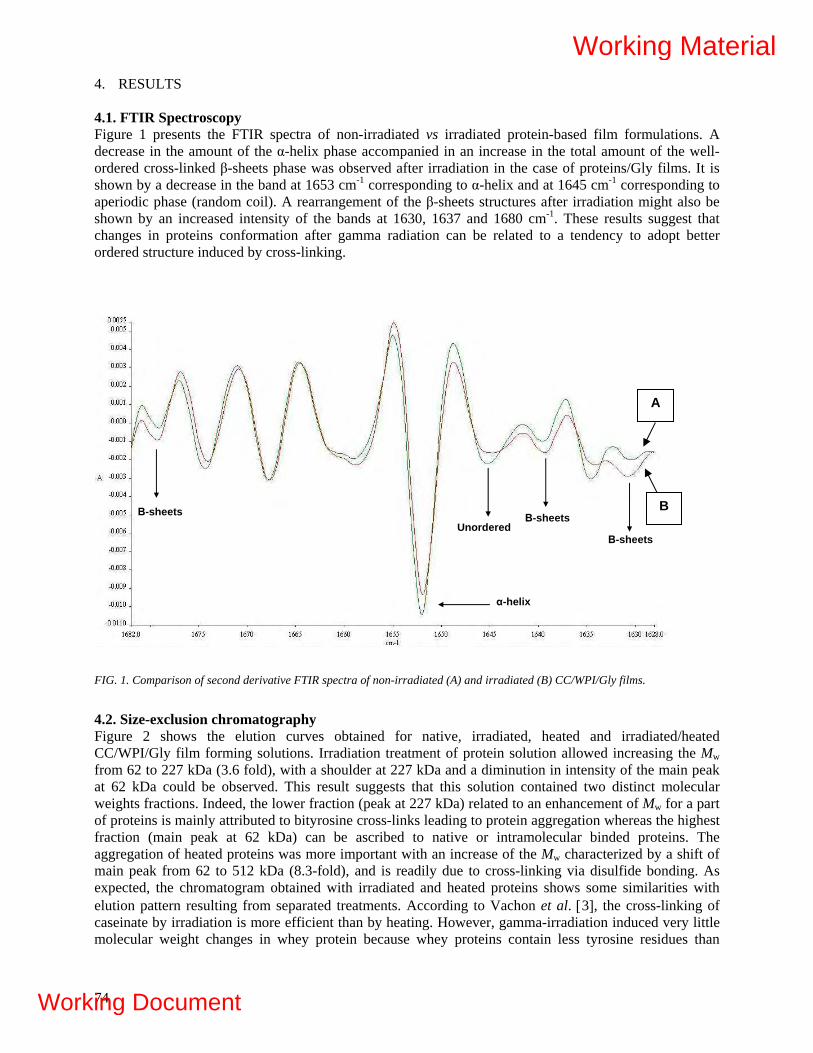
4. RESULTS 4.1. FTIR Spectroscopy Figure 1 presents the FTIR spectra of non-irradiated vs irradiated protein-based film formulations. A decrease in the amount of the α-helix phase accompanied in an increase in the total amount of the well-ordered cross-linked β-sheets phase was observed after irradiation in the case of proteins/Gly films. It is shown by a decrease in the band at 1653 cm-1 corresponding to α-helix and at 1645 cm-1 corresponding to aperiodic phase (random coil). A rearrangement of the β-sheets structures after irradiation might also be shown by an increased intensity of the bands at 1630, 1637 and 1680 cm-1. These results suggest that changes in proteins conformation after gamma radiation can be related to a tendency to adopt better ordered structure induced by cross-linking.
α-helix
Β-sheets Β-sheets Unordered
Β-sheets
A
B
FIG. 1. Comparison of second derivative FTIR spectra of non-irradiated (A) and irradiated (B) CC/WPI/Gly films.
4.2. Size-exclusion chromatography Figure 2 shows the elution curves obtained for native, irradiated, heated and irradiated/heated CC/WPI/Gly film forming solutions. Irradiation treatment of protein solution allowed increasing the Mw from 62 to 227 kDa (3.6 fold), with a shoulder at 227 kDa and a diminution in intensity of the main peak at 62 kDa could be observed. This result suggests that this solution contained two distinct molecular weights fractions. Indeed, the lower fraction (peak at 227 kDa) related to an enhancement of Mw for a part of proteins is mainly attributed to bityrosine cross-links leading to protein aggregation whereas the highest fraction (main peak at 62 kDa) can be ascribed to native or intramolecular binded proteins. The aggregation of heated proteins was more important with an increase of the Mw characterized by a shift of main peak from 62 to 512 kDa (8.3-fold), and is readily due to cross-linking via disulfide bonding. As expected, the chromatogram obtained with irradiated and heated proteins shows some similarities with elution pattern resulting from separated treatments. According to Vachon et al. [3], the cross-linking of caseinate by irradiation is more efficient than by heating. However, gamma-irradiation induced very little molecular weight changes in whey protein because whey proteins contain less tyrosine residues than
74
Working Material
Working Document

caseins. The globular whey proteins are more prone to intramolecular cross-linking, and readily underwent cross-linking via the formation of disulfide bonds during heating treatment. Comparison of aggregations reveals that cross-linking by heating is more efficient than by gamma irradiation. However, it is interesting to note a synergistic effect of combined treatments of irradiation/heating since a Mw of 875 kDa (14.1-fold) could be observed, suggesting that pre-irradiation treatment promoted the aggregation of proteins after heating. The results from heated and γ-irradiated protein solutions are consistent with those reported by Le Tien et al. [4] and Yu et al. [39].
15 20 25 30 35 40
Elution volume (mL)
875 kDa
Irradiated + heated
Heated
Abs
orba
nce
280
nm
512 kDa
227 kDa
Irradiated 62 kDa
Control
FIG. 2. Effect of gamma irradiation at 32 kGy and heating at 80°C on the aggregation of a CC/WPI/Gly solutions.
4.3. Mechanical Properties Table I and Figure 3 show the effect of polysaccharide addition and of γ-irradiation of proteins on the puncture strength of films. The γ-irradiation of proteins allowed improving the tensile strength significantly (p ≤ 0.05) of all film formulations regardless the added polysaccharide. Indeed, the puncture strength values were increased from 56, 73, 24 and 62 N/mm to 67, 81, 49 and 69 N/mm for control (proteins only), proteins/MC, proteins/CMC and proteins/ALG formulations. Concerning the effect of polysaccharide addition in non-irradiated formulations, only the presence of MC and ALG allowed increasing the puncture strength significantly (p ≤ 0.05) with puncture values from 56 to 73 and 62 N/mm for control, MC and ALG film formulations respectively. For irradiated formulations, only the presence of MC improved the resistance of films significantly (p ≤ 0.05), with respective values varying from 67 to 81 N/mm for control and MC film formulation. As a result, it can be concluded that MC addition was efficient to improve mechanical puncture strength of protein-based films. This observation could be related to the intrinsic functional properties (Mw, viscosity, chemical interactions with proteins, i.e.
75
Working Material
Working Document
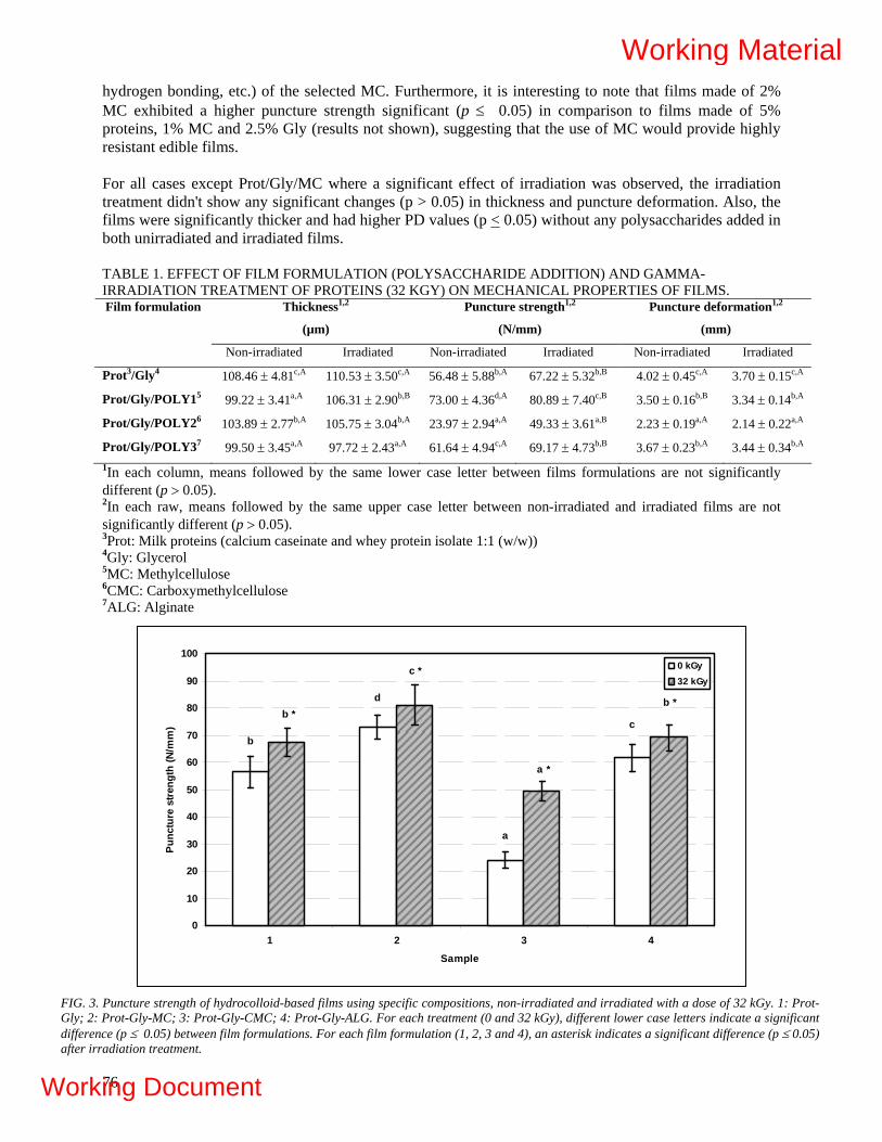
hydrogen bonding, etc.) of the selected MC. Furthermore, it is interesting to note that films made of 2% MC exhibited a higher puncture strength significant (p ≤ 0.05) in comparison to films made of 5% proteins, 1% MC and 2.5% Gly (results not shown), suggesting that the use of MC would provide highly resistant edible films. For all cases except Prot/Gly/MC where a significant effect of irradiation was observed, the irradiation treatment didn't show any significant changes (p > 0.05) in thickness and puncture deformation. Also, the films were significantly thicker and had higher PD values (p < 0.05) without any polysaccharides added in both unirradiated and irradiated films. TABLE 1. EFFECT OF FILM FORMULATION (POLYSACCHARIDE ADDITION) AND GAMMA-IRRADIATION TREATMENT OF PROTEINS (32 KGY) ON MECHANICAL PROPERTIES OF FILMS.
Thickness1,2
(µm)
Puncture strength1,2
(N/mm)
Puncture deformation1,2
(mm)
Film formulation
Non-irradiated Irradiated Non-irradiated Irradiated Non-irradiated Irradiated
Prot3/Gly4 108.46 ± 4.81c,A 110.53 ± 3.50c,A 56.48 ± 5.88b,A 67.22 ± 5.32b,B 4.02 ± 0.45c,A 3.70 ± 0.15c,A
Prot/Gly/POLY15 99.22 ± 3.41a,A 106.31 ± 2.90b,B 73.00 ± 4.36d,A 80.89 ± 7.40c,B 3.50 ± 0.16b,B 3.34 ± 0.14b,A
Prot/Gly/POLY26 103.89 ± 2.77b,A 105.75 ± 3.04b,A 23.97 ± 2.94a,A 49.33 ± 3.61a,B 2.23 ± 0.19a,A 2.14 ± 0.22a,A
Prot/Gly/POLY37 99.50 ± 3.45a,A 97.72 ± 2.43a,A 61.64 ± 4.94c,A 69.17 ± 4.73b,B 3.67 ± 0.23b,A 3.44 ± 0.34b,A 1In each column, means followed by the same lower case letter between films formulations are not significantly different (p > 0.05). 2In each raw, means followed by the same upper case letter between non-irradiated and irradiated films are not significantly different (p > 0.05). 3Prot: Milk proteins (calcium caseinate and whey protein isolate 1:1 (w/w)) 4Gly: Glycerol 5MC: Methylcellulose 6CMC: Carboxymethylcellulose 7ALG: Alginate
c
a
d
b
b *
a *
c *
b *
0
10
20
30
40
50
60
70
80
90
100
1 2 3 4
Sample
Punc
ture
stre
ngth
(N/m
m)
0 kGy32 kGy
FIG. 3. Puncture strength of hydrocolloid-based films using specific compositions, non-irradiated and irradiated with a dose of 32 kGy. 1: Prot-Gly; 2: Prot-Gly-MC; 3: Prot-Gly-CMC; 4: Prot-Gly-ALG. For each treatment (0 and 32 kGy), different lower case letters indicate a significant difference (p ≤ 0.05) between film formulations. For each film formulation (1, 2, 3 and 4), an asterisk indicates a significant difference (p ≤ 0.05) after irradiation treatment.
76
Working Material
Working Document
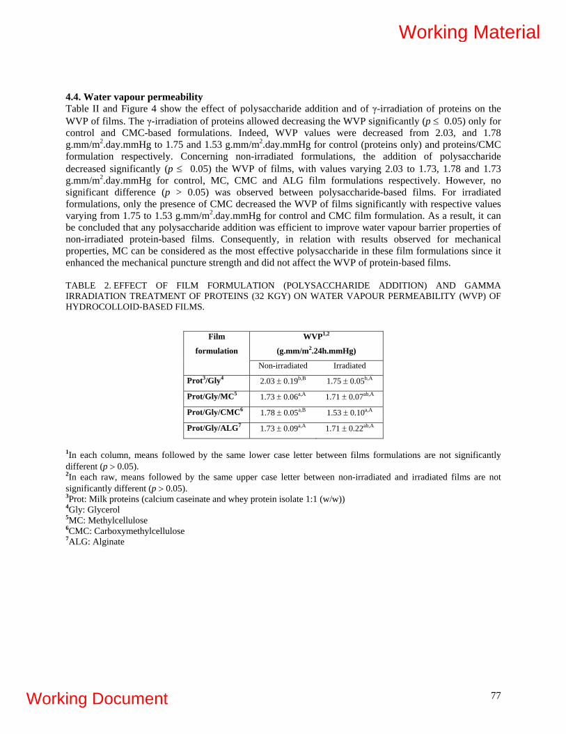
4.4. Water vapour permeability Table II and Figure 4 show the effect of polysaccharide addition and of γ-irradiation of proteins on the WVP of films. The γ-irradiation of proteins allowed decreasing the WVP significantly (p ≤ 0.05) only for control and CMC-based formulations. Indeed, WVP values were decreased from 2.03, and 1.78 g.mm/m2.day.mmHg to 1.75 and 1.53 g.mm/m2.day.mmHg for control (proteins only) and proteins/CMC formulation respectively. Concerning non-irradiated formulations, the addition of polysaccharide decreased significantly (p ≤ 0.05) the WVP of films, with values varying 2.03 to 1.73, 1.78 and 1.73 g.mm/m2.day.mmHg for control, MC, CMC and ALG film formulations respectively. However, no significant difference (p > 0.05) was observed between polysaccharide-based films. For irradiated formulations, only the presence of CMC decreased the WVP of films significantly with respective values varying from 1.75 to 1.53 g.mm/m2.day.mmHg for control and CMC film formulation. As a result, it can be concluded that any polysaccharide addition was efficient to improve water vapour barrier properties of non-irradiated protein-based films. Consequently, in relation with results observed for mechanical properties, MC can be considered as the most effective polysaccharide in these film formulations since it enhanced the mechanical puncture strength and did not affect the WVP of protein-based films. TABLE 2. EFFECT OF FILM FORMULATION (POLYSACCHARIDE ADDITION) AND GAMMA IRRADIATION TREATMENT OF PROTEINS (32 KGY) ON WATER VAPOUR PERMEABILITY (WVP) OF HYDROCOLLOID-BASED FILMS.
WVP1,2
(g.mm/m2.24h.mmHg)
Film
formulation
Non-irradiated Irradiated
Prot3/Gly4 2.03 ± 0.19b,B 1.75 ± 0.05b,A
Prot/Gly/MC5 1.73 ± 0.06a,A 1.71 ± 0.07ab,A
Prot/Gly/CMC6 1.78 ± 0.05a,B 1.53 ± 0.10a,A
Prot/Gly/ALG7 1.73 ± 0.09a,A 1.71 ± 0.22ab,A
1In each column, means followed by the same lower case letter between films formulations are not significantly different (p > 0.05). 2In each raw, means followed by the same upper case letter between non-irradiated and irradiated films are not significantly different (p > 0.05). 3Prot: Milk proteins (calcium caseinate and whey protein isolate 1:1 (w/w)) 4Gly: Glycerol 5MC: Methylcellulose 6CMC: Carboxymethylcellulose 7ALG: Alginate
77
Working Material
Working Document
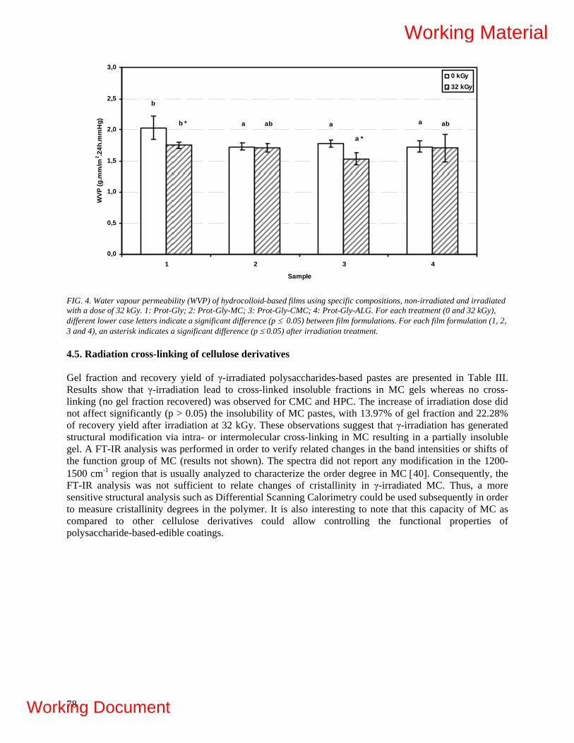
b
a a ab * ab
a *
ab
0,0
0,5
1,0
1,5
2,0
2,5
3,0
1 2 3 4
Sample
WVP
(g.m
m/m
2 .24h
.mm
Hg)
0 kGy32 kGy
FIG. 4. Water vapour permeability (WVP) of hydrocolloid-based films using specific compositions, non-irradiated and irradiated with a dose of 32 kGy. 1: Prot-Gly; 2: Prot-Gly-MC; 3: Prot-Gly-CMC; 4: Prot-Gly-ALG. For each treatment (0 and 32 kGy), different lower case letters indicate a significant difference (p ≤ 0.05) between film formulations. For each film formulation (1, 2, 3 and 4), an asterisk indicates a significant difference (p ≤ 0.05) after irradiation treatment. 4.5. Radiation cross-linking of cellulose derivatives Gel fraction and recovery yield of γ-irradiated polysaccharides-based pastes are presented in Table III. Results show that γ-irradiation lead to cross-linked insoluble fractions in MC gels whereas no cross-linking (no gel fraction recovered) was observed for CMC and HPC. The increase of irradiation dose did not affect significantly (p > 0.05) the insolubility of MC pastes, with 13.97% of gel fraction and 22.28% of recovery yield after irradiation at 32 kGy. These observations suggest that γ-irradiation has generated structural modification via intra- or intermolecular cross-linking in MC resulting in a partially insoluble gel. A FT-IR analysis was performed in order to verify related changes in the band intensities or shifts of the function group of MC (results not shown). The spectra did not report any modification in the 1200-1500 cm-1 region that is usually analyzed to characterize the order degree in MC [40]. Consequently, the FT-IR analysis was not sufficient to relate changes of cristallinity in γ-irradiated MC. Thus, a more sensitive structural analysis such as Differential Scanning Calorimetry could be used subsequently in order to measure cristallinity degrees in the polymer. It is also interesting to note that this capacity of MC as compared to other cellulose derivatives could allow controlling the functional properties of polysaccharide-based-edible coatings.
78
Working Material
Working Document

TABLE III. GEL FRACTION (%) AND RECOVERY YIELD (%) OF MC GELS (20% W/V) IRRADIATED AT 32 AND 64 KGY.
Gel fraction (%)1, 2 RY (%)1, 2
Treatment MC CMC HPC MC CMC HPC
Control ND ND ND ND ND ND
32 kGy 13.97 ± 1.67b ND ND 22.28 ± 2.90b ND ND
64 kGy 12.74 ± 0.36b ND ND 25.33 ± 2.78b ND ND
1In each column, means followed by the same lower case letter between treatments are not significantly different (p > 0.05). 2ND: Not determined (no gel formation).
4.6. Evaluation of the relative sensitivity Table IV present the D10 values and relative sensitivities related to radiosensitization of moulds and total flora in strawberries packed under air, with and without bioactive coating. Figures 5 and Figure 6 present the D10 curves that have been elaborated to characterize moulds and total flora radiosensitization respectively. For moulds radiosensitization (Table IV, Figure 5), results show that the presence of LIM and ALG-based coatings increased the radiosensitivity of moulds. Indeed, the addition of LIM and ALG+LIM increased the radiosensitization of moulds with respective relative sensitivities of 1.35 and 1.71 (D10 of 0.67 and 0.53) as compared to 1.00 (D10 of 0.91) for control. In comparison, the addition of PM did not seem to affect the radiosensitization of moulds with a lower relative sensitivity of 1.05. Moreover, the combination of ALG with LIM had a marked effect on the radiation dose necessary to eliminate moulds in strawberries, with a lethal dose of 2.2 kGy for ALG+LIM and 2.5 kGy for LIM as compared to 3.5 kGy for the control (figure 7). On the other hand, it can be observed that the presence of the PM or LIM-based coating allowed an increase of the total flora radiosensitization, with a relative sensitivity of 1.42 (D10 of 0.14) as compared to 1.00 for control (D10 of 0.19) (Table IV, Figure 6). The dose necessary to eliminate total flora in strawberries coated with PM and LIM is 0.8 kGy as compared to 1.0 kGy for ALG + LIM et 1.1 kGy for the control. According to Caillet et al. [41], the antimicrobial properties of the active compounds are responsible for this increased radiosensitivity, relatively to their phenolic or terpenic composition. TABLE IV. RADIATION SENSITIVITY OF TOTAL FLORA AND MOULDS IN STRAWBERRIES (FRAGARIA ANANASSA) COATED WITH BIOACTIVE COMPOUNDS (ALG+LIM, LIM, PM).
Treatment Slope D10 (kGy) Relative sensitivity
Moulds Control 1.10 0.91 1.00
ALG + LIM 1.88 0.53 1.71
LIM 1.49 0.67 1.35
PM 1.16 0.86 1.05
Total flora Control 5.19 0.19 1.00
ALG + LIM 5.26 0.19 1.01
LIM 4.99 0.20 1.42
PM 7.37 0.14 1.42
79
Working Material
Working Document
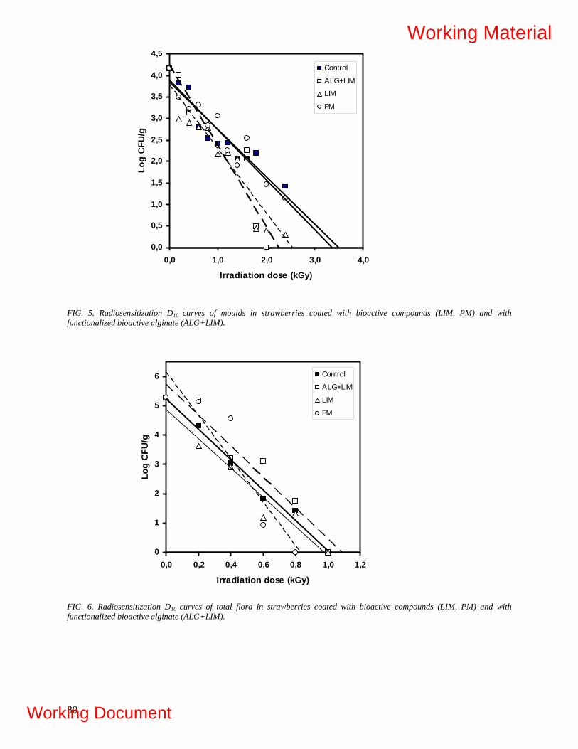
0,0
0,5
1,0
1,5
2,0
2,5
3,0
3,5
4,0
4,5
0,0 1,0 2,0 3,0 4,0
Irradiation dose (kGy)
Log
CFU
/g
Control
ALG+LIM
LIM
PM
FIG. 5. Radiosensitization D10 curves of moulds in strawberries coated with bioactive compounds (LIM, PM) and with functionalized bioactive alginate (ALG+LIM).
0
1
2
3
4
5
6
0,0 0,2 0,4 0,6 0,8 1,0 1,2
Irradiation dose (kGy)
Log
CFU/
g
Control
ALG+LIM
LIM
PM
FIG. 6. Radiosensitization D10 curves of total flora in strawberries coated with bioactive compounds (LIM, PM) and with functionalized bioactive alginate (ALG+LIM).
80
Working Material
Working Document

Mould Growth in Strawberries Results for mould growth on crosslinked milk protein-MC-based coatings (crosslinked with irradiation (32 kGy) and gamma irradiation of the coated fruits (1.5 kGy) are depicted in figure 7. Moulds started appearing on the untreated strawberries on day 8 with a 20% fruit loss and reached 65% contamination on day 17. Moulds appeared on day 10 for PI+Ir and PI+L+Ir with a contamination level of 10%. On day 14, mould contamination reached 15%. On day 17, mould contamination was 35% and 30% respectively for PI+Ir and PI+L+Ir.
0
10
20
30
40
50
60
70
1 7 10 14 17
Days
Per
cent
age
of d
ecay
(%)
Control
Coating + Irradiation
Active coating + Irradiation
FIG. 7. Mould growth on strawberries.
5. CONCLUSION A protein-polysaccharide-based film formulation was developed by evaluating the addition of polysaccharide and a treatment by gamma irradiation of proteins in aqueous solution. Gamma irradiation of proteins was characterized by FTIR spectroscopy to investigate conformation changes and SEC to evaluate Mw. FT-IR secondary derivative spectra exhibited changes in band intensities that were correlated to an increase of β-sheet structure adopted by proteins after irradiation and a decrease of α-helix and unordered fractions. SEC analysis determined a synergistic effect of combined treatments of irradiation/heating on the increase of Mw from 62 to 875 kDa, suggesting that pre-irradiation treatment promoted the aggregation of proteins after heating. The evaluation of puncture strength and WVP of films allowed optimizing the protein/glycerol-based film formulation by the addition of MC with a significant enhancement (p ≤ 0.05) of mechanical properties and no detrimental effect on WVP. In addition, the irradiation of concentrated MC solutions generated partially insoluble gels via cross-linking with a insolubility percentage of 22% at a dose of 32 kGy. Hence, these resulting rheological properties of MC are promising for the development of insoluble, highly resistant edible coatings or biodegradable packaging in various applications. The second part of this study consisted in evaluating the antimicrobial properties of natural extracts applied alone or incorporated in edible coatings in order to increase the shelf life of strawberries by
81
Working Material
Working Document

preserving their deterioration. The radiosensitization of moulds and total flora in strawberries was also evaluated in presence of PM and Lim incorporated in a functionalized alginate-based coating solution. The elaboration of D10 curves indicated that ALG and LIM were radiosensitizers for moulds whereas PM was less efficient for total flora in strawberries. Hence, it seems that a functionalized polymer-based edible coating could be an effective issue to develop in order to carry specific antifungal agents without detrimental effect on strawberries. In this context, future investigations will consist in evaluating the sensorial properties of antifungal agents added on strawberries in order to maintain acceptable attributes for consumers. Also, the development of MC-based edible coatings or packaging would be of first importance due to its remarkable rheological properties as compared to protein-based formulations. The gamma irradiation of MC should be developed in order to improve the functional properties of edible coatings applied onto strawberries. Moreover, the addition of lipids (fatty acids, acetylated monoglycerides, etc.), plasticizers (ex: polyols), surfactants, reinforcement agents (ex: nanocellulose) will be evaluated on the functional properties of MC-based coatings and operative conditions of film formation (homogenization parameters, drying conditions, etc.) will be optimized in order to increase the shelf life and improve the quality of strawberries during storage. Also, preliminary tests have demonstrated that the use of acrylic acid and irradiation has permitted grafting reaction of the monomer and an improvement of the physico-chemical properties of the polyvinyl alchool zein based films. Structural and physico-chemical properties of films are under going presently and results will be presented in our next report.
Acknowledgements The authors thank MDS Nordion’s for gamma radiation processing and Winpak Division Ltd for providing packaging material used for irradiated samples. They are also grateful to Asmaâ Imzilne, Elise Leroux and Jaejoon Han for their technical collaboration. The International Agency of Energy Atomic is acknowledged for his financial support through the IAEA's Co-ordinated Research Project in 'Development of radiation-processed products of natural polymers for application in agriculture, healthcare, industry and environment (CRP).
REFERENCES
[1] HERALD, T.J., HACHMEISTER, K.A., HUANG, S., BOWERS, J.R., Corn Zein Packaging
Materials for Cooked Turkey, J. Food Sci. 61 (1996) 415-418. [2] MCHUGH, T.H., KROCHTA, J.M., Milk-protein-based edible films and coatings, Food
Technol. 48 (1994) 97-103. [3] VACHON, C., YU, H.L., YEFSAH R., ALAIN R., ST-GELAIS D., LACROIX M.,
Mechanical and Structural Properties of Milk Protein Edible Films Cross-Linked by Heating and γ-Irradiation, J. Agric. Food Chem. 48 (2000) 3202-3209.
[4] LE TIEN C, LETENDRE M, ISPAS-SZABOS P, MATEESCU M A, DELMAS-PATTERSON G, YU H L, LACROIX M., Development of biodegradable films from whey proteins by cross-linking and entrapment of cellulose, J Agric Food Chem 48 (2000) 5566-5575.
[5] SALMIERI, S., LACROIX, M., Physicochemical properties of alginate/polycaprolactone-based films containing essential oils, J. Agric. Food Chem. 54 (2006) 10205-10214.
[6] KHWALDIA, K. PEREZ, C., BANON, S., DESOBRY, S., HARDY, J., Milk proteins for edible films and coatings, Crit. Rev. in Food Sci. and Nut. 44 (2004) 239-251.
[7] Xu, W.-T., Huang K.-L., Guo F., Qu W., Yang J-J., Liang Z-H., Luo Y-B., Postharvest grapefruit seed extract and chitosan treatments of table grapes to control Botrytis cinerea, Postharvest Biology and Technology 46 (2007) 86–94.
[8] CHA, D.S., CHINNAN, M.S., Biopolymer-based antimicrobial packaging: A review, Crit. Rev. in Food Sci. and Nut. 44 (2004) 223-237.
82
Working Material
Working Document
[9] LI J, CHEN H., Transglutaminase catalyzed crosslinking on functional properties of whey protein-based edible films. Book of Abstracts, IFT Annual Meeting July 24-28, Chicago, IL, USA (1999).
[10] HOFFMANN M A M, SALA G OLIEMAN C, DE KRUIF K G., Molecular mass distribution of heat-induced β-lactoglobulin aggregates, J Agric Food Chem. 45 (1997) 2949-2957.
[11] BRAULT D, D’APRANO G, LACROIX M., Formation of free-standing sterilized edible films from irradiated caseinates, J. Agric Food Chem. 45 (1997) 2964-2969.
[12] LACROIX, M., The Use of Essential Oils and Bacteriocins as Natural Antimicrobial and Antioxidant Compounds., Food 1 (2007) 181-192.
[13] KROCHTA, J.M. DE MULDER, C.L.C., Edible and biodegradable polymer films - challenges and opportunities (A Scientific Status Summary), Food Technol. 51 (1997) 61-74.
[14] RESSOUANY, M., VACHON, C., LACROIX, M., Irradiaiton dose and calcium effect on the mechanical properties of cross-linked caseinate films, J. Agric. Food Chem. 46 (1998) 1618-1623.
[15] KADER, 1986. [16] SOMMER AND FORTLAGE, 1966. [17] OUSSALAH, M., CAILLET, S., SALMIERI, S., SAUCIER, L., LACROIX, M.,
Antimicrobial effects of alginate-based film containing essential oils for the preservation of whole beef muscle, J. Food Prot. 69, 10 (2006) 2364-2369.
[18] OUATTARA, B., SABATO, S.F., LACROIX, M., Combined effect of antimicrobial coating and gamma irradiation on shelf life extension of pre-cooked shrimp (Penaeus spp.), Int. J. Food Microbiol. 68 (2001) 1-9.
[19] CAILLET, S., MILLETTE, M., SALMIERI, S., LACROIX, M., Combined effects of antimicrobial coating, modified atmosphere packaging, and gamma irradiation on Listeria innocua present in ready-to-use carrots (Daucus carota), J. Food Prot. 69 (2006a) 80-85.
[20] O’CONNOR R.E., MITCHELL G.E., Effect of irradiation on microorganisms in strawberries, Int.l J. Food Microbiol. 12 (1991) 247-255.
[21] DEMPSTER, J.F., HAWRYSH, Z.J., SHAND, P., LAHOLA-CHOMIAK, L., CORLETTO, L., Effect of low-dose irradiation (radurization) on the shelf life of beefburgers stored at 3°C, J. Food Technol. 20 (1985) 145–154.
[22] LACROIX, M., OUATTARA, B., Combined industrial processes with irradiation to assure innocuity and preservation of food products - A review, Food Res. Int. 33 (2000) 719–724.
[23] OUATTARA, B., GIROUX, M., SMORAGIEWICZ, W., SAUCIER, L., LACROIX, M., Combined effect of gamma irradiation, ascorbic acid, and edible coating on the improvement of microbial and biochemical characteristics of ground beef, J. Food. Sci. 65 (2002) 981–987.
[24] D’AMOUR J, GOSSELIN C, ARUL J, CASTAIGNE F, WILLEMOT C., Gamma-radiation affects cell wall composition of strawberries, J Food Sci 58 (1993) 182-185.
[25] BORSA, J., LACROIX, M., OUATTARA, B., CHIASSON, F., Radiosensitization: enhancing the radiation inactivation of foodborne bacteria, Rad. Phys. and Chem. 71 (2004) 135-139.
[26] VACHON, C., D'APRANO G., LACROIX, M., LETENDRE., M., Effect of edible Coating Process and Irradiation Treatment of Strawberry (Fragaria spp.) on Storage-keeping Quality, J. Food Sci. 68 (2003) 608-612.
[27] MONSO, E., Bronchial colonization in chronic obstructive pulmonary disease: What’s hiding under the rug, Archivos de Bronconeumologia. 40 (2004) 543-546.
[28] TOURNAS V.H.., KATSOUDAS E., Mould and yeast flora in fresh berries, grapes and citrus fruits, International Journal of Food Microbiology 105 (2005) 11-17.
[29] ECKERT, J.W., OGAWA, J.M., The chemical control of postharvest diseases: Subtropical and tropical fruits, Annual Review of Phytopathology. 23 (1988) 1031-1036.
[30] BOUCHRA C., ACHOURI M., IDRISSI HASSANI L.M., HMAMOUCHI M., Chemical composition and antifungal activity of essential oils of seven Moroccan Labiatae against Botrytis cinerea Pers: Fr., Journal of Ethnopharmacology 89 (2003) 165–169.
83
Working Material
Working Document

84
[31] BHASKARA REDDY, M.V., ANGERS P., GOSSELIN A. AND ARUL, J. Characterization and use of essential oil from Thymus vulgaris against Botrytis cinerea and Rhizopus stolonifer in strawberriy fruit, Phytochemistry 47, 1515-1520.
[32] REDDY, M.V.B., ANGERS, P., GOSSELIN, A., ARUL, J., Characterization and use of essential oil from Thymus vulgaris against Botrytis cinerea and Rhizopus stolonifer in strawberry fruits, Phytochem. 47 (1998) 1515-1520.
[33] CURTIS, O.F., SHETTY, K., CASSAGNOL, G., PELEG, M., Comparison of the inhibitory and lethal effects of synthetic versions of plant metabolites (anethole, carvacrol, eugenol and thymol) on a food spoilage yeast (Debaromyces hansenii), Food Biotechnol. 10 (1996) 55-73.
[34] KIM, J., MARSHALL, M.R., WEI, C.-I., Antibacterial activity of some essential oil component against five foodborne pathogens, J. Agric. Food Chem. 43 (1995) 2839-2845.
[35] CIESLA, K., SALMIERI, S., LACROIX, M., Modification of the properties of milk protein films by gamma radiation and polysaccharide addition, J. Sci Food Agric. 86 (2006) 908-914.
[36] DEBEAUFORT, F., MARTIN-POLO, M., ET VOILLEY, A., Polarity homogeneity and structure affect water permeability of model edible films, J. Food Sci. 58 (1993) 426-429
[37] WACH, R.A., MITOMO, H., NAGASAWA, N., YOSHII, F., Radiation crosslinking of methylcellulose and hydroxyethylcellulose in concentrated aqueous solutions, Nucl. Instr. and Meth. B. 211 (2003) 533-544.
[38] YOSHII, F., ZHAO, L., WACH, R.A., NAGASAWA, N., MITOMO, H., KUME, T., Hydrogels of polysaccharide derivatives crosslinked with irradiation at paste-like condition, Nucl. Instr. and Meth. B. 208 (2003) 320-324.
[39] YU, H., SABATO, S.F., D’APRANO, G., LACROIX, M., Effect of the addition of CMC on the aggregation behaviour of proteins, Rad. Phys. Chem. 71 (2004) 131-135.
[40] VELAZQUEZ, G., HERRERA-GOMEZ, A., MARTIN-POLO, M.O., Identification of bound water through infrared spectroscopy in methylcellulose, J. Food Eng. 59 (2003) 79-84.
[41] CAILLET, S., MILLETTE, M., TURGIS, M., SALMIERI, S. AND LACROIX, M., Influence of antimicrobial compounds and modified atmosphere packaging on radiation sensitivity of Listeria monocytogenes present in ready-to-use carrots (Daucus carota), J. Food Prot. 69 (2006) 221-227.
Working Material
Working Document

RADIATION MODIFICATION OF NATURAL POLYSACCHARIDES FOR APPLICATION IN AGRICULTURE
Hassan .A. AbdEl-Rehim, El-Sayed A. Hegazy, and Ahmed M. El-Barbary National Center for Radiation Research and Technology, NCRRT, P. O. Box 29, Nasr City, Cairo, Egypt. Abstract Controlling the degradation process of natural polymer like chitosan by gamma ray from a 60Co source at different doses in, powder form, and in presence of additives was investigated. The efficiency of these methods were verified by viscometric and GPC analysis through determination the average molecular weights of degraded natural polymers. The chemical-irradiation degradation method was much more appropriate from economical point of view. Characterization of degraded polymer by FTIR spectroscopy, UV-Vis spectroscopy, XRD, ESR and TGA analysis were done. The degraded sodium alginate with different doses were used for wheat plant. It was observed that spraying treated plants with alginate solutions improved plant growth and increasing its yields. Significant changes in amino acids content of the plants was observed as a result of treating them with irradiated alginate solutions. Irradiated Na alginate induced changes in plant DNA, which may be appear as increase in growth and yield characters. The degraded natural polymer chitosans were also tested as growth promoters for some plants like Zea maize and bean plant . It was found that the degraded chitosan has a great effect on the productivity and properties of these plants. Antiviral activity of irradiated alginate against plant infection by tobacco mosaic virus was investigated. It is clear that the relatively high Molecular weigh alginate inhibits and reduces the plant infection . The effects of chitosan oligomers on gray mould caused by Botrytis cinerea in tomato plants was investigated. It was found that degraded chitosan controlled the gray mould disease caused by B. cinerea compared with control.
KEYWORDS: Radiation, Degradation Natural polymer 1. INTRODUCTION Recently much attention has been paid to the utilization of radiation processing technology for degradation of natural polysaccharides in agricultural applications due to several reasons for instances: (1) the degradation process can be performed at room temperature. (2) The degraded polysaccharides can be used without purifications. (3) The simplicity of controlling the whole process. (4) Economic competitiveness to alternatives. As compared to the conventional techniques like acid or base hydrolysis or enzymatic methods, radiation processing offers a clean one step method for the formation of low molecular weight polysaccharides in aqueous solutions even at high concentrations. Low molecular weight polysaccharides and/or oligosaccharides can be produced by degradation of corresponding polysaccharides including marine polysaccharides such as alginate, chitin/chitosan and carrageenan [ 1-6].
A lot of studies have been carried out in countries like Japan, Vietnam, China, India and Philippines to investigate the plant growth promotion and plant protection effect of radiation processed polysaccharides in a variety of crops under different environmental conditions. Low molecular weight naturally occurring polysaccharides prepared by different methods possessed novel features such as growth-promotion of plants. Promotion of germination and shoot elongation stimulation of growth of bifidio-bacteria to resist infection of diseases for plants particularly oligochitosan in agriculture as biotic elicitor to enhance defense responses against diseases and suppression of heavy metals stress. The results of these studies have clearly shown that radiation processed polysaccharides even at very low concentrations of a few tens of ppm are very effective for use as plant growth promoter. This application offers tremendous opportunity to use them as highly effective organic fertilizers. Their biodegradability will be an additional advantage of using such materials as plant growth promoters.
In this respect, the present work is dealing with studying the effect of ionizing radiations on the degradation of some natural polymers such as, chitosan and alginate. Trials were made to control and reduce the irradiation dose required for the sodium alginate and Chitosan degradation. The degraded
85
Working Material
Working Document

natural polymers were tested as growth promoters and plant elicitors for some plants like wheat tomato bean. Antiviral activity of alginate against plant infection by tobacco mosaic virus was also investigated.
2. RESULTS AND DISCUSSION Determination the Molecular Weight and the Synergetic Effect The reduction in the molecular weights can be evaluated by determining the molecular weight. The intrinsic viscosity [η] of degraded natural was measured by an ubbelohde viscometer and used the Mark-Hownique equation to determining the viscosity average molecular weight. Tables (1) show the different average molecular weights of chitosan, after treating with ionizing radiation in the presence of APs and H2 O2 at different doses. The degradation of chitosan, increased by increasing the irradiation dose. It was observed that using 80 KGy irradiation dose reduces the average molecular weight of chitosan, from 9.2 x 10⁷ to 3.5 x 10⁶. Meanwhile, irradiation of chitosan, at 40 kGy in the presence of APS reduced the average molecular weight of chitosan, from 9.2 x 10⁷ to 8.5 x 10⁴. As seen from the Table, the rate of degradation reaction and the reduction in the molecular weight of investigated chitosan natural polymer is higher in presence of ammonium per-sulfate and hydrogen peroxide if compared with the molecular weight of irradiated natural polymers in absence of such additives
TABLE 1. NUMBER-AVERAGE MOLECULAR WEIGHTS OF CHITOSAN ON EXPOSURE TO GAMMA IRRADIATION AND THAT MIXED WITH DIFFERENT INITIATORS MEASURED BY GPC.
Irradiated chitosan Irradiated chitosan
+ APS Irradiated chitosan
+ H2O2 Radiation Dose (kGy)
After irradiati
on
After storing
After irradiati
on
After storing
After irradiati
on
After storing
0 9.2 x 10⁷ 9.2 x 10⁷ 9.2 x 10⁷ 9.2 x 10⁷ 9.2 x 10⁷ 9.2 x 10⁷ 20 1.7 x 10⁷ 1.58 x
107 1.1 x 10⁵ 9.8 x 10⁴ 4.77 x 10⁵ 7 x 10⁴
40 3.5 x 10⁶ 3.34 x 10⁶ 8.5 x 10⁴ 7.35 x
104 2.23 x
105 4.73 x
104 80 2.85 x
106 2.73 x
106 2.7 x 10⁴ 2.54 x 10⁴
1.85 x 10⁵ 1 x 10⁴
120 1.91 x 10⁶ 1.8 x 10⁶ 1.44 x
104 1.32 x
104 1.1 x 10⁵ 7.4x 10³
160 7.3 x 10⁵ 9.09 x 10⁴ 6.8 x 10³ 5.5 x 10³ 9.09 x
104 2.7 x 10³
200 6.9 x 10⁵ 7.5 x 10⁴ 4.17 x 10³
3.97 x 10³ 7.5 x 10⁴ 2.2 x 10³
Table(1) also shows the changes in the number-average molecular weights as a function of increasing the irradiation doses for pure chitosan, and chitosan mixed with H2O2 or APS when exposed to gamma irradiation and stored for 7 days. The storing has great effect on the molecular weight of irradiated chitosan mixed with H2O2. The degradation reactions of the polymer depend on the amount of free radicals formed during the irradiation process and the direct dissociation of H2O2. The decay rate of free radicals formed in the presence of H2O2 is very low and therefore, the degradation process was continuous with time.
2.1. Characterization of degraded chitosan FT-IR spectroscopy has been shown to be a powerful tool for the study of the physicochemical properties of polysaccharides. Curves a and b in Fig.1 shows the IR spectra of untreated chitosan and degraded water soluble irradiated chitosan. The main bands in the spectrum of original chitosan are as follows: Both the N–H stretching and O–H stretching vibrations can be characterized by a strong,
86
Working Material
Working Document
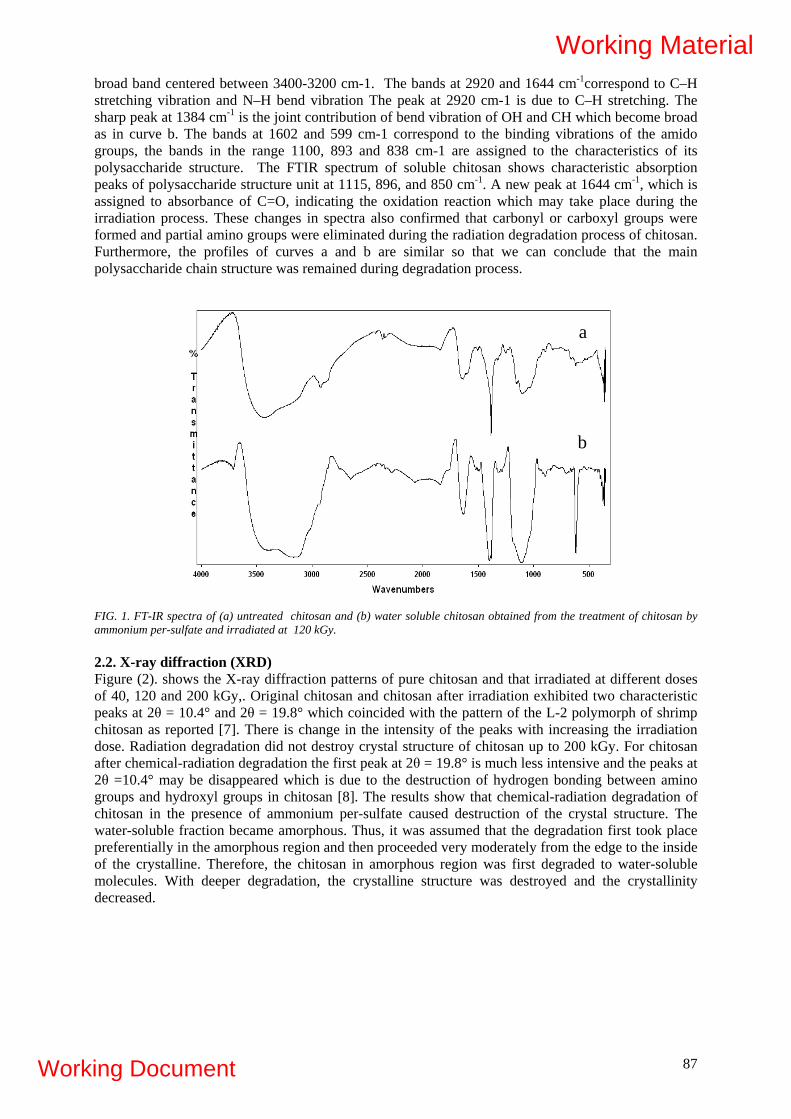
broad band centered between 3400-3200 cm-1. The bands at 2920 and 1644 cm-1correspond to C–H stretching vibration and N–H bend vibration The peak at 2920 cm-1 is due to C–H stretching. The sharp peak at 1384 cm-1 is the joint contribution of bend vibration of OH and CH which become broad as in curve b. The bands at 1602 and 599 cm-1 correspond to the binding vibrations of the amido groups, the bands in the range 1100, 893 and 838 cm-1 are assigned to the characteristics of its polysaccharide structure. The FTIR spectrum of soluble chitosan shows characteristic absorption peaks of polysaccharide structure unit at 1115, 896, and 850 cm-1. A new peak at 1644 cm-1, which is assigned to absorbance of C=O, indicating the oxidation reaction which may take place during the irradiation process. These changes in spectra also confirmed that carbonyl or carboxyl groups were formed and partial amino groups were eliminated during the radiation degradation process of chitosan. Furthermore, the profiles of curves a and b are similar so that we can conclude that the main polysaccharide chain structure was remained during degradation process.
a
b
FIG. 1. FT-IR spectra of (a) untreated chitosan and (b) water soluble chitosan obtained from the treatment of chitosan by ammonium per-sulfate and irradiated at 120 kGy. 2.2. X-ray diffraction (XRD) Figure (2). shows the X-ray diffraction patterns of pure chitosan and that irradiated at different doses of 40, 120 and 200 kGy,. Original chitosan and chitosan after irradiation exhibited two characteristic peaks at 2θ = 10.4° and 2θ = 19.8° which coincided with the pattern of the L-2 polymorph of shrimp chitosan as reported [7]. There is change in the intensity of the peaks with increasing the irradiation dose. Radiation degradation did not destroy crystal structure of chitosan up to 200 kGy. For chitosan after chemical-radiation degradation the first peak at 2θ = 19.8° is much less intensive and the peaks at 2θ =10.4° may be disappeared which is due to the destruction of hydrogen bonding between amino groups and hydroxyl groups in chitosan [8]. The results show that chemical-radiation degradation of chitosan in the presence of ammonium per-sulfate caused destruction of the crystal structure. The water-soluble fraction became amorphous. Thus, it was assumed that the degradation first took place preferentially in the amorphous region and then proceeded very moderately from the edge to the inside of the crystalline. Therefore, the chitosan in amorphous region was first degraded to water-soluble molecules. With deeper degradation, the crystalline structure was destroyed and the crystallinity decreased.
87
Working Material
Working Document

FIG. 2. XRD spectra of (a) pure chitosan and that treated with APS irradiated at different doses of (b) 40 kGy; (c) 120 kGy and (d) 200 kGy at the dose rate is 6.7 kGy/h.. 2.3. Antiviral activity of alginate against plant infected by tobacco mosaic virus Tobacco mosaic virus (TMV) is a rod-shaped virus. Composed of single-stranded RNA encapsulated in a coat Protein capsid. For the infection of TMV to take place in leaves, it is necessary that TMV penetrates directly into the cell and makes contact with materials in the plasma. The inhibitory effect of Na- Alginate on the infectivity of TMV was assayed by inoculating leaves with TMV alone and with TMV and Na- Alginate mixtures. The inhibitory activity of Na- Alginate was calculated as the percentage reduction in the number of local regions produced on leaves after 3 days as compared with their controls. It was found that sodium alginate had a high inhibitory activity against TMV infection. Figure 3 shows the effect of chitosan irradiated at different dose on the inhibitory effect of TMV. It is clear that alginates of high molecular weight have a great inhibitory effect on TMV. The lowest inhibition degree is noticed when the Mwt obtained of irradiated chitosan at 40kGy was used. The degree of inhibition increased with alginate concentration. The addition of Alginate caused TMV to form large aggregates. The antiviral activity of Alginate on infectivity of TMV may be caused by blocking the de-capsulation process of TMV protein on the cell membrane surface.
88
Working Material
Working Document

Dose kGy0 50 100 150 200
infe
ctio
n%
0
20
40
60
80
100
FIG. 3. Effect ofNa-Alg of different MWt on the TMV infection. 2.4. Growth and yield characters of wheat plants spraying with irradiated alginate In this search, sodium alginate was exposed to γ -rays and irradiated with doses (80,120 and 160 kGy). From each dose an aqueous solution of (1%) concentration was prepared for spraying wheat plants at different growth stages 1, 2,3 months). Spraying plants in different stages of growth, was effective in improving plant growth and increasing their yields as shown in Table (2). Regarding to plant height, number of branches ,spike length, number of spikes, total biomass mass of seeds/plant it was found that the plant treated with Na-alginate irradiated at 160 kGy gained the highest results if compared with control or other plant treated with irradiated alginate at ( 80 and 120 kGy). The increase in % of yield characters (wt. of spike and wt. of seeds/plant) was about 65%.
2.5.Growth and yield characters of bean and zea maze plants spraying with irradiated chitosan of different molecular weighs Table (3) describes plant height (cm), ear height (cm), ear length (cm), 100 grain weight (g), grain weight/ ear (g), biological yield fad. /(ardab), straw yield fad. /(ardab), grain yield fad. /(ardab), total protein (%) and total oil (%) of zea maize yield plants, spraying by 100 ppm of degraded chitosan,. It is obvious from that, the treatment by chitosan irradiated at different irradiation doses improved the grain yield (ardab/fad) of zea maize plants. The highest grain yield is obtained when the plant treated with chitosan irradiated at 120 kGy at which gives grain yield of 25.8. (ardab/fad) if compared with control plant which gives 18.6 (ardab/fad). Table (4) describes the properties of bean plant crop. It is obvious from Table (4) that the treatment of bean plants with chitosan irradiated at 120 kGy has positive effect on seed yields (ardab/fad), which gives bean seed yield of 10.79 (ardab/fad) and plant height of 125 cm if compared with control one which gives 7.7 (ardab/fad), and plant hight of 99.5 cm.
89
Working Material
Working Document

TABLE 2. GROWTH AND YIELD CHARACTERS OF CONTROL UNTREATED PLANTS (C),AND PLANTS TREATED WITH IRRADIATED NA ALGINATE SOLUTIONS WITH DOSES 80, 120 AND 160 KGY.
Harvest index
Mass of 100
seeds
Mass of
seeds
Total bio
-mass
Spikes mass
N0. of Spikes
Spike length
N0. of
branches
Plant height
Growth& yelid
Chara- cters
Treat- ments
38.59 4.52 7.32 18.52 10.07 3.33 11.67 4 90.06 Control 43.51 4.09 10.23 23.87 15.03 4.11 12.72 44.4 107.8
9 80
42.45 4.23 9.68 22.02 12.43 4.33 13.39 4.56 109.22
120
43.45 4.49 12.08 27.02 16.69 5.22 13.89 5.22 108.33
160
2.1 0.2 2.78 6.5 3.66 1.56 0.96 1.22 4.3 L.S.D.
TABLE 3. EFFECT OF SPRAYING 100 PPM OF CHITOSAN AND THAT MIXED BY 10% WT APS AT DIFFERENT IRRADIATION DOSES ON CROP YIELD OF ZEA MAIZE PLANTS.
Factor
tTreatmen
Plant height (cm)
Ear height (cm)
Ear length (cm)
100 grai
n Wt (g)
Grain wt/ear (g)
Biological yield fad. (ardab)
Straw yield fad. (ardab)
Grain yield/ fad. (ardab)
Total protein %
Total Oil %
Control
113.9
70.32
16.55
27.3
3
128.7
41.97
23.39
18.58
8.73
4.42
Blank
113.3
70.13
16.47
27.0
9
128.3
41.92
23.55
18.37
8.66
4.33
200kGy
113.7
70.21
16.51
27.3
1
128.1
41.71
23.27
18.44
8.47
4.15
With APS at 40 kGy
203.
85.31
17.99
30.4
5
141.8
45.06
24.19
20.87
9.02
4.5
With APS at 80 kGy
209
87.23
18.99
32.9
9
155.2
51.04
27.37
23.67
9.36
4.67
90
Working Material
Working Document

With APS at 120kGy
210.1
91.37
19.65
34.0
1
157.1
54.34
28.55
25.79
9.44
4.71
With APS at 160kGy
204.7
88.91
18.51
31.8
3
152.2
51.69
27.66
24.03
9.40
4.58
With APS at 200kGy
203.4
86.11
18.32
29.2
9
142.8
50.09
26.48
23.61
9.13
4.53
TABLE 4. EFFECT OF CHITOSAN EXPOSED TO DIFFERENT IRRADIATION DOSES ON THE PROPERTIES AND CROP YIELD OF FABA BEAN(SPRAY IN 100 PPM CONC.).
Factor
Treatment
Plant height (cm)
No. of Pods/ plant
No. of Pods/ Plant/m2
No. of plant / m2
100 seed wt (g)
Biological yield fad. (ardab)
Straw yield fad. (ardab)
Seed yield/ fad. (ardab)
Control
99.51
17.92
164
32.94
52.47
22.98
14.87
7.71
Blank
90.13
17.68
162
32.2
51.73
22.83
14.77
8.06
200kGy
90.24
17.7
162
32.27
51.79
22.82
14.71
8.11
With APS at 40 kGy
111.53
21.87
210
44.84
64.32
24.13
15.19
8.94
With APS at 80 kGy
121.61
23.85
233
50.79
70.25
28.58
18.71
9.87
With APS at 120kGy
125.18
24.55
241
52.9
72.35
32.34
21.62
10.72
With APS
117.91
23.12
225
48.6
68.07
41.31
31.69
9.62
91
Working Material
Working Document

at 160kGy
With APS at 200kGy
109.98
21.572
207
43.92
63.41
39.15
30.07
9.08
Radiation degradation of chitosan to enhance productivity and control tomato post-harvest gray mold caused by Botrytis cinerea The effect of different Mwt chitosans on the productivity of tomato plant was investigated and shown in table( 5). It is clear that the degradation of chitosan at different MWt enhances and increases the productivity of tomato plant The Suitable chitosan Mwt to obtain highest yield of tomato is 5.8 x 10⁴. Also, it was found That the degraded chitosan used for increase the productivity of Tomato plant, controls the gray mold fungus that infected the tomato plant fruit . Gray mold disease caused by fungus Botrytis cinerea is considered an important pathogen around the world. It induces decay on a large number of economically important fruit and vegetables during the growing season and during post harvest storage. It is also a major problem to long distance transport and storage. This fungal pathogen infects leaves, stems, flowers and fruit, either by direct penetration or through wounds caused by cultivation practices.The Control of this disease is especially important during storage because it develops at low temperatures (e.g. −0.5 °C) and spreads quickly among fruit and vegetables. From Table( 6 ) it can be noticed that the degraded chitosan enhance fungal decay when used at high concentration dose. After 3 days of treatment, when fruit were stored at 25 °C. Chitosan with a molecular weight of 5.8 × 10⁴ provided a good control of gray mold in tomato fruit among all different molecular weight chitosans.
TABLE 5. EFFECT OF CHITOSAN EXPOSED TO DIFFERENT IRRADIATION DOSES ON THE PROPERTIES AND CROP YIELD OF TOMATO PLANTS.
92
Working Material
Working Document

TABLE 6.
93
Working Material
Working Document

3. CONCLUSIONS It can be concluded that degraded chitosan of different Mwt could be used for growth promotion and increasing the productivity of many crops like Zea maize , wheat , bean plants . Also, they play important role in controlling some plant infection diseases like banana tobacco mosic and tomato gray mold.
REFERENCES [1] MATSUHASHI, S., KUME, T., Enhancement of antimicrobial activity of chitosan by
irradiation. J. Sci. Food Agric. 73 (1997) 237–241. [2] LAM, N.D., NAGASAWA, N., KUME, T., Effect of radiation and fungal treatment on
lignocelluloses and their biological activity. Radiat. Phys. Chem. 59 (2000) 393–398. [3] PARK, P.J., JE, J.Y., KIM, S.W. Free radical scavenging activities of different deacetylated
chitosans using an ESR spectrometer Carbohydrate Polymer, 55 (2004) 17–22. [4] WANICHPONGPAN P., SURIYACHAN K., CHANDRKRACHANG S., Effects of chitosan
on the growth of Gerbera flower plant (Gerbera jamesonii), in: T.Uragami, K. Kurita, T. Fukamizo (Eds.), Chitin and Chitosan in Life Science, Yamaguchi (2001)198–201. ISBN 4-906464-43-0.
[5] AKIYAMA H., ENDO T., NAKAKITA R., MURATA K., YONEMOTO Y., OKAYAMA K., BIOSCI, MOLLAH . M.Z.I., ;Biotech. Biochem. 56.
[6] MUBARAK, A, KHANAND RUHUL, A., KHAN, Effect of gamma irradiated sodium alginate on red amaranth (Amaranthus cruentus L.) as growth promoter. Radiation Physics and Chemistry. 78 (1) (2009) 61-64.
[7] HASEGAWA, M., ISOGAI, A., ONABE, F., USUDA, M., & ATALLA R. H., Characterization of cellulose–chitosan blend films. Journal of Applied Polymer Science, 45 (1992) 1873-1879.
[8] KIM, J. H., LEE, Y.M., Synthesis and properties of diethylaminoethyl chitosan. Polymer, 34 (9) (1993) 1952-1957.
94
Working Material
Working Document
THERMOPLASTIC STARCH WITH IMPROVED PROPERTIES BY BLENDING WITH LIGNINS AND RADIATION PROCESSING D. Zheng1), S.Baumberger1)
, P.-Y. Mikus2), P. Dole2), J. Soulestin3), M. F. Lacrampe3),
C. Bliard4) and X. Coqueret4) 1)UMR 206 Chimie Biologique AgroParisTech INRA - 78850 Thiverval-Grignon 2)UMR INRA FARE, Université de Reims Champagne Ardenne - 51687 Reims 3) Ecole des Mines de Douai, DTPCIM, 941 rue Charles Bourseul, BP 10838 - 59508 Douai 4)ICMR, UMR CNRS 6229, Université de Reims Champagne Ardenne - 51687 Reims
France
Abstract The biorefinery of lignocellulosics generates lignin-rich fractions, which are potential source of phenolic molecules for chemistry and polymeric materials. The LignoStarch project aims at using such fractions to functionalize a renewable material, starch, by a clean physical grafting process, without any synthetic chemical additive and without any by-products generation. Previous works suggested that the low-molar-mass phenolic compounds in technical lignin could be responsible for the reactivity of starch-lignin system under electron-beam irradiation and improvement of starch water resistance. A particular aspect of the current studies is focused on the role of lignin phenolic extractables and to investigate the different chemical and physical parameters likely to impact the surface properties of starch-lignin materials. 1. INTRODUCTION The biorefinery of lignocellulosics generates lignin-rich fractions, which are potential source of phenolic molecules for chemistry and polymeric materials. The aim of the LignoStarch project is to contribute to the development of original technologies producing biodegradable materials from biomass, using a combination of two different types of biomacromolecules, starch and lignins, with contrasting structures and properties, thus with poor mutual compatibility [1]. To overcome the thermodynamic limitation to the formation of an alloy of materials with adjustable intermediate properties, a grafting process induced by high energy radiation processing, as a green method (solvent-free, with no or limited need for additive, no by-products) which is proposed to induce covalent linkages between the constituents [2-4]. In one of its main research axes for improving the mechanical properties, the process will be driven in a way to induce major modifications in the molecular architecture of starch and/or of its orientation by exploiting the high potential of reactivity of lignins and lignin-like compounds. The judicious association of blending of properly selected constituents (nature, proportions), of chemical transformations (radiation induced, enzymatic), and of physical processing of conventional thermoplastics (thermal profiles control, orientation) is expected to overcome a number of limitations currently restricting the development of starch-based thermoplastics. A second research axis of the LignoStarch aims at using such lignin fractions to modify the bulk and the surface hydrophilicity of thermoplastic starch [5-7]. Previous works suggested that the low-molar-mass phenolic compounds in technical lignin could be responsible for the reactivity of starch-lignin system under electron-beam irradiation and improvement of starch water resistance. A particular aspect of the current studies is focused on the role of lignin phenolic extractables and to investigate the different chemical and physical parameters likely to impact the surface properties of starch-lignin materials. The present report is particularly focused on that latter objective of both basic and application-oriented interest.
95
Working Material
Working Document

2. GENERAL APPROACH OF THE PROJECT From a physico-chemical viewpoint, the bulk hydrophilicity of blended materials is reduced to the expectable extent depending on intrinsic hydrophobicity of the type of lignin that was used. Additionally, surface analyses showed that combining the presence of lignins with the treatment by ionizing radiation (electron beam) strongly reduces the material surface wettability by water, suggesting the formation of a particular hydrophobic structure at the surface. Various other aspects are being implemented in the frame of the LignoStarch project. At the molecular level, the efficiency of the grafting process under high energy radiation, as established from various types of analytical (HPLC of extracts form irradiated blends) and spectroscopic experiments (FTIR, 1H and 13C NMR, Maldi-Tof MS) is performed on blends natural starch and lignins, as well as on their model compounds. The important structural aspects of aging are also examined. The comparison between an irradiated sample aged for one year under standard conditions (RT, 55% relative humidity) and a reference blend non subjected to the ionising treatment clearly showed the absence of retrogradation in the first material, whereas the presence of crystallinity was revealed by new and intense lines in the X-ray diffractograms. Not only lignin and lignin like compounds thus contribute to these various positive effects but they were also proved to exert a protecting action against some detrimental effects of ionising radiation onto polysaccharides molecules and materials. Increasing the amount of lignins or lignin-like compounds in the blends was shown to allow stronger irradiation treatments without dramatic loss of elongation at break. Preliminary assessments of biodegrability showed a level of biodegradability comparable to starch samples (according to CO2 production under reference composting conditions), only is the process slowed to some extent, presumably as consequence of reduced hydrophilicity and higher resistance to disaggregating in the early stage of the composting test. 3. MATERIALS AND METHODS 3.1. Surface properties of starch films functionalized by lignins Previous works have shown that the EB irradiation of starch materials in the presence of alkali lignin improved the surface hydrophobicity of the material while preserving its biodegradability [8]. Ferulic acid present in alkali lignin was shown to be involved in the reactivity of starch-lignin system and supposed to be responsible for grafting and cross-linking reactions. However, no attention was paid to possible contribution of the surface physical state to hydrophobation. The objective of the present study was to better understand the hydrophobation mechanisms of starch-lignin systems by investigating the impact of the EB irradiation on the film surface topography and morphology, in parallel to wettability tests. Two different starches (wheat and potato) were implemented in order to assess the influence of the matrix structure on the system reactivity. An alkali lignin sample was selected as technical lignin available at industrial scale from agricultural crops and blended with starch by a thermo-mechanical process of extrusion-pressing. 3.2. Preparation of starch films Two different methods based on extrusion combined with compression moulding were implemented for the film preparation (Table 1). Method A: Starch-lignin films were obtained directly using a single-screw extruder (Rheocord 20). In this method the starch/glycerol blend was dried in oven before extrusion; Method B: Starch-lignin films were extruded from starch-lignin pellets obtained using a two
96
Working Material
Working Document

steps extrusion process. First, plasticized starch was obtained using a co-rotating twin screw-extruder (Clextral BC45). In a second step, a dry mix of obtained plasticized starch pellets and lignin powder was extruded on a single-screw extruder (Rheocord 40). Starch films were obtained by processing the obtained pellets on a single-screw extruder (Rheocord 40) equipped with a cast die and a calander. In both methods a compression molding step using Teflon sheet was included in order to reduce the film thickness and roughness. TABLE1. COMPARISON OF THE TWO DIFFERENT STARCH-LIGNIN FILM PREPARATION METHODS (ALL THE GLYCEROL, STARCH AND LIGNIN PROPORTION ARE EXPRESSED WITH RESPECT TO DRY WEIGHT). Method A Method B Extrusion 1. 85/15 starch/glycerol pre-
treated in an oven at 170°C for 45 min
plasticized starch
2. 80/20 plasticized starch/lignin processed on a single-screw extruder (3 heating zones: 100, 105, 110°C with cast die at 115°C at a screw speed of 50 rpm)
TPS-lignin films
1. 80/20 starch/glycerol extruded with twin-screw (5 heating zones: 105°C, 110°C, 110°C, 115°C and 115°C with slit die heated to 120°C at a screw speed of 40 rpm)
thermoplastic starch (TPS) pellets 2. 80/20 TPS/lignin extruded with twin-screw
(3 heating zones: 105, 110, 110°C at a screw speed of 40 rpm)
TPS-lignin pellets 3. TPS/lignin pellets processed on a single-screw
extruder (3 heating zones: 105, 110, 110°C, with cast die at 115°C at a screw speed of 40 rpm).
TPS-lignin films Pressing 120°C, 250 bars, 5 sec
~ 200 µm thick film 110°C, 160 bars, 3 min
~ 150 µm thick film 3.3. Film irradiation Electron beam irradiation was applied to starch lignin films with a 180 kV accelerator tot a total dose of 200 or 350 kGy in an inert atmosphere. The dose was applied in a single pass, which could lead to a slight temperature increase but avoid contact with air during the process. After irradiation, films were stored for two weeks at 58% HR and 23°C. 3.4. Physico-chemical characterization Film wettability. Wettability was assessed by the temporal change of shape of a sessile water drop deposited on the film surface. Values of the static contact angle at the air/liquid/solid-phase boundary were determined at 23 ±2 °C using a contact angle DSA10 Drop Shape Analysis system (Krüss, Germany). 3.5. Surface morphology Atomic force microscopy (AFM) in acoustic mode (PicoLE, Molecular Imaging, ScienTec, France) was used to characterize the topography of starch-based films in air at 22 C at a nano-scale level. Silicon nitrides gold-coated oxide-sharpened cantilever was equipped for analyse operation (ScienTec, France). Topographic and phase images were acquired at a resonant frequency of 69 KHz in fluid with a scanning rate of 0.5 nm/s. Film morphology was observed by scanning electron microscopy (SEMS-4500; Hitachi, Tokyo, Japan)
97
Working Material
Working Document

4. RESULTS AND DISCUSSION 4.1. Influence of the extrusion processing on the film wettability by liquid water To assess the influence of processing on the film surface wettability by a water droplet, two methods for film preparation were compared (methods A and B). The processing parameters of method B were chosen in order to improve the homogeneity of the blends, in particular via the use of two additional extrusion steps with twin-screw extruders known for its higher shear rate: one for the production of thermoplastic starch (TPS), the other for the production of TPS-lignin homogeneous blends. Moreover, the pre-heating step (170°C, 45 min) of the starch/glycerol mixture was suppressed in order to prevent starch thermal degradation and glycerol evaporation. The processing method did not significantly affect the initial contact angle values relative to starch-lignin films before compression moulding (MP= 6.3% difference, and MW= 12.6% difference; Figure 1), which suggests that this parameter is mainly governed by the chemical composition of the film rather than by the physical state of the matrix. However, all the films recovered from method B exhibit a slower drop absorption rate than the films from method A (drop still not absorbed 30 minutes after the deposition; Figure 1). This improved barrier property may be due to the higher homogeneity of the materials and is of particular relevance regarding applications in the field of packaging.
FIG. 1. Water wettability of films before compression moulding (contact angle measured at t0 and t30’ after drop deposition). MP: lignin/potato starch; MW: lignin/wheat starch film; A: method A; B: method B. 4.2 Compared hydrophobicity of potato and wheat starch based films Two types of starch commonly used for the investigation of starch-based biodegradable materials, wheat and potato starch, were used for the preparation of starch-lignin films by two different methods of extrusion combined with compression moulding. Potato starch was easier to process, leading to lower extrusion pressures than wheat starch. This different behaviour during extrusion was more particularly observed with the single-screw extruder of method A (maximum pressure of 350 bars reached with wheat starch-lignin blends). In agreement with previous results obtained on water-spin-coated glycerol/starch films [9], wheat starch lead to higher contact angle values than potato starch (Figure 1). This result is observed both before and after compression moulding and whatever the processing method. It reflects the more hydrophilic molecular properties of potato starch which contains phosphoryl groups and is exempt of lipids in contrast to wheat starch [10]. 4.3 Surface modification of the film by electron beam irradiation Non-pressed starch-lignin films recovered from method B were submitted to irradiation at two doses (200 and 350 kGy) slightly lower than the 400 kGy dose previously applied by Lepifre et al. on potato starch-lignin films [8]. We show here that whatever the starch matrix (wheat or potato) irradiation
98
Working Material
Working Document

dramatically improves the surface hydrophobicity of the materials. The contact angle values at t0 and t30’ for a water droplet both increase with increasing irradiation dose with maximum hydrophobicity reached at 350 kGy with wheat starch. Such an irradiation-mediated hydrophobation can be explained by structural chemical changes as previously proposed in view of experiments carried out on lignin and starch model compounds. However, it can also be suggested that the decrease in water wettability may result from a change in the film surface roughness. Indeed, surface roughness is a key parameter likely to govern wettability [11].
FIG. 2. Water wettability of native and irradiated non-pressed films (contact angle measured at t0 and t30’ after the water drop deposition). MP: lignin/potato starch; MW: lignin/wheat starch film; A: method A; B: method B. Both SEM and AFM observations (example given in Figure 3) indicate that irradiation may lead to a reduction of the roughness parameters, either by partial superficial fusion of the film during irradiation processing or by an ablation of the asperities. Since roughness has been shown to exacerbate the wettability of hydrophilic surfaces (contact angle < 90°) [12], the decrease in roughness due to irradiation could significantly contribute to the apparent surface hydrophobization of starch-lignin films.
5 µm
5 µm
5 µm
FIG. 3. SEM observation of the MPA film. From top to bottom: native thermoplastic starch-lignin film , film after irradiation at 200 kGy, film after irradiation at 350 kGy. In order to elucidate the contribution of roughness to the film behaviour, highly smooth films were compression molded after extrusion between Teflon sheets. This compression moulding step leads to very glossy materials, especially starch-lignin films recovered from method B. In agreement with the hypothesis that roughness enhances the film wettability by water, all the films recovered after compression moulding exhibit higher contact angle values that the non-pressed films (87° instead of 54° for MPB). Moreover, whatever the film processing method (A or B) irradiation applied to smooth
99
Working Material
Working Document
100
films did not dramatically increase the hydrophobicity of the films. Only a slight irradiation-mediated increase of contact angle values was observed for the wheat-starch based film recovered from method A (MWA). 5. CONCLUSIONS The results obtained in the frame of the LignoStarch project confirm the potential interest of radiation grafting for adjusting the properties of starch-based thermoplastic materials. The study of model blends allow us to assess quantitatively the conversion degree of the reactive additives added to amorphized starch and to model polysaccharides. The proposed structure for the adducts observed after irradiation of representative model compounds involves the reactivity of hydroxyl and cinnamyl functions of the unsaturated aromatic alcohol. This supports the occurrence of similar reactions in the blends of starch with industrial lignins. On-going experiments explore the potentialities of the controlled scission and coupling reactions taking place under radiation for improving the mechanical properties of thermoplastic starch formulated with lignin or with aromatic addiditives and subjected to moderate irradiation treatments. Radiation processing of thermoplastic starch-lignin blends is shown to reduce noticeably surface hydrophilicity and to impede starch retrogradation very efficiently. Controlling the surface hydrophobicity of agromaterials is one important challenge regarding the development of applications of thermoplastic starch in fields such as packaging. Electron beam irradiation can be seen as a “green” technology for modifying the surface of starch-based films since it does not require synthetic chemical reagents and avoid the production of waste or by-products. This study brings new original results concerning the factor governing the surface hydrophobicity of starch-based films functionalized by lignins. The botanical origin of starch as well as the nature of the lignin extracts used in the blends exert a strong influence with induction of distinct properties. In particular, the film roughness was shown as a primary parameter to take into account to understand and control the impact of the film processing, especially irradiation, on the surface wettability by water.
Acknowledgements This work was supported by French National Research Agency (ANR LignoStarch program). We thank Cédric Plessis (UMR 1211 AgroParisTech, INRA) for performing the irradiation experiments.
REFERENCES [1] BAUMBERGER, S., in “Chemical Modification, Properties, and Usages of Lignin”, T.Q. Hu,
Ed., Kluwer Academic/Plenum Publishers, New York, Chap. 1 (2002). [2] AMIDON, T.E., WOOD, C.D., SHUPE, A.M., WANG, Y., GRAVES, M., LIU, S., J. Biobased
Material and Bioenergy 2 (2) (2008) 100. [2] RUCKERT, D., CAZAUX, F., COQUERET, X., J. Appl. Polym. Sci. 73, (1999) 409. [3] OLIVIER, A., CAZAUX, F., COQUERET, X., Biomacromolecules (2000) 282. [4] OLIVIER, A., CAZAUX, F., GORS, C., COQUERET, X., Biomacromolecules (2001) 1260. [5] BAUMBERGER, S., LAPIERRE, C., MONTIES, B., J. Agric. Food Chem. 46 (1998) 2234. [6] BAUMBERGER, S., LAPIERRE, C., MONTIES, B., LOURDIN, D., COLONNA, P., Ind.
Crops Prod. 6 (1997) 253. [7] LEPIFRE, S., FROMENT, M., CAZAUX, F., HOUOT, S., LOURDIN, D., COQUERET, X.,
LAPIERRE, C., BAUMBERGER, S., Industr. Crops Res. 20 (2004) 219. [8] LEPIFRE, S., FROMENT, M., CAZAUX, F., LOURDIN, D., COQUERET, X., LAPIERRE,
C., BAUMBERGER, S., Biomacromolecules 5 (2004) 1678. [9] PROKOPOVICH, P., PERNI, S., J. Mater. Sci: Mater. Med. 20 (2009) 195. [10] ROBIN, J.P., MERCIER, C., DUPRAT, F., CHARBONNIERE, R., GUILBOT, A.,
Stärke/Starch. 27(2) (1975) 36. [11] YONG, X., ZHANG, L.T., Langmuir 25(9) (2009) 5045. [12] BICO, J., TORDEUX, C., QUERE, D., Europhys. Lett. 55(2) (2001) 214.
Working Material
Working Document

PRELIMINARY STUDIES ON ANTIFUNGAL PROPERTIES OF RADIATION PROCESSED CHITOSAN FROM CRAB SHELLS Fidelis C.K. Ocloo1, Abraham Adu-Gyamfi1, Emmanuel A. Quarcoo1, Yaw Serfor-Armah2, Daniel Asare1. 1 Biotechnology and Nuclear Agriculture Research Institute (BNARI) 2 National Nuclear Research Institute (NNRI) Ghana Atomic Energy Commission, Legon/GHANA Abstract Chitosan extracted from sea crab shells was used to determine antifungal properties against Aspergillus niger. Chitosan powder irradiated at 100 kGy and dissolved in 1 % acetic acid (v/v) with pH adjusted to approximately 6.0 was used in preparing chitosan concentrations of 2 %, 1.5 %, 1 % and 0.5 %. The agar dilution method was used to test the antifungal activity of the various chitosan solutions at concentrations of 0.20 %, 0.15 %, 0.10 % and 0.05 %. Both media containing irradiated and unirradiated chitosan inhibited the mycelial growth of Aspergillus niger and the degree of inhibition was dependent on the concentration of the chitosan in the fungal growth medium. Results show that the media containing irradiated chitosan inhibited the mycelia growth of Aspergillus niger to a greater extent than the media containing unirradiated chitosan.
1. INTRODUCTION The preservation of food by physical and chemical methods is an important area to the food industry. Food preservatives have to meet several regulatory standards, for example, they must be (i) efficient over a broad range of spoilage organism; (ii) tasteless and odorless, and (iii) non toxic, safe and inexpensive. When physical means of preservation are not available or desirable, chemical preservative(s) must be used, and the choice of one or more preservative is primarily based on the chemical composition of the food, its pH, and other characteristics. No preservative is universal, and the list of GRAS (generally recognized as safe) preservatives is short [1]. There is also a growing demand from the public for natural preservatives, and for these reasons, alternative sources of safe, effective, and acceptable preservatives need to be developed. Chitosan is the deacetylated form of the natural polymer chitin, a polysaccharide composed of N-acetyl D-glucosamine and D-glucosamine units, and extracted mainly from crustacean waste by demineralization and deproteinization. Over 60% deacetylation of chitin yields chitosan [2]. Recently, research on chitosan and its properties have shown its potential for use as a food preservative. Chang et al. (1989) [3] achieved a 10 day shelf life extension of “mulkimchi” (pickle type kimchi, i.e., chinese cabbage) at 5°C by incorporating 0.2% chitosan compared to control samples. Low molecular weight polysaccharides and/or oligosaccharides can be produced by degradation of corresponding polysaccharides including marine polysaccharides such as alginate, chitin/chitosan, and carrageenan. Chemical, enzymatic and radiation processing technologies can be applied for degradation process. Recently much attention has been paid to the application of radiation processing technology for degradation of natural polysaccharides. Research has shown that low molecular weight polysaccharides have antimicrobial properties. A number of pathogenic moulds such as Fusarium spp., Aspergillus spp, Penicillium spp and Rhizopus spp. have been reported as causal agents of foodborne disease and food spoilage [4]. Recently, there has been considerable interest in radiation-processed low molecular weight natural polysaccharides with antimicrobial properties. Matsuhashi and Kume [5] reported that irradiated chitosan having molecular weight of 105 to 3 x 10⁵ exhibited highly antimicrobial activities. Similarly, Ha et al. (1999) noted the enhancement of antifungal activity of irradiated chitosan for different fungi strains [6]. This study therefore is aimed at investigating the antifungal properties of radiation-processed chitosan from crab shells on Aspergillus niger.
101
Working Material
Working Document
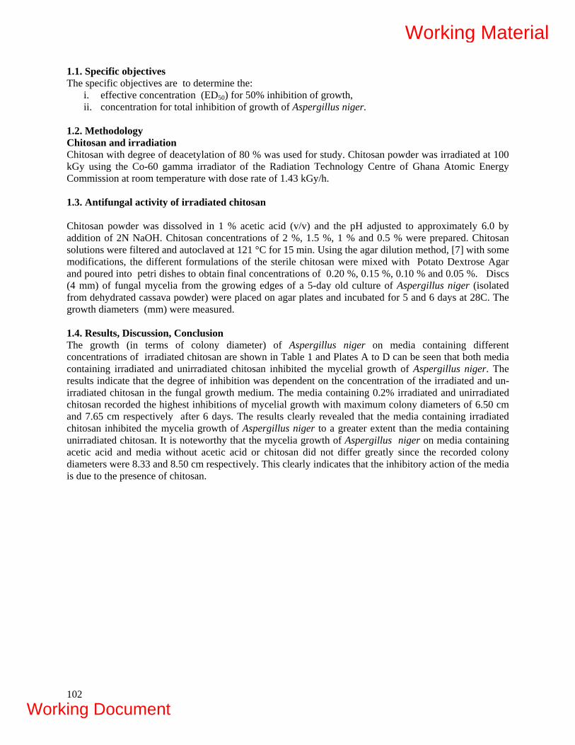
102
1.1. Specific objectives The specific objectives are to determine the:
i. effective concentration (ED50) for 50% inhibition of growth, ii. concentration for total inhibition of growth of Aspergillus niger.
1.2. Methodology Chitosan and irradiation Chitosan with degree of deacetylation of 80 % was used for study. Chitosan powder was irradiated at 100 kGy using the Co-60 gamma irradiator of the Radiation Technology Centre of Ghana Atomic Energy Commission at room temperature with dose rate of 1.43 kGy/h. 1.3. Antifungal activity of irradiated chitosan Chitosan powder was dissolved in 1 % acetic acid (v/v) and the pH adjusted to approximately 6.0 by addition of 2N NaOH. Chitosan concentrations of 2 %, 1.5 %, 1 % and 0.5 % were prepared. Chitosan solutions were filtered and autoclaved at 121 °C for 15 min. Using the agar dilution method, [7] with some modifications, the different formulations of the sterile chitosan were mixed with Potato Dextrose Agar and poured into petri dishes to obtain final concentrations of 0.20 %, 0.15 %, 0.10 % and 0.05 %. Discs (4 mm) of fungal mycelia from the growing edges of a 5-day old culture of Aspergillus niger (isolated from dehydrated cassava powder) were placed on agar plates and incubated for 5 and 6 days at 28C. The growth diameters (mm) were measured. 1.4. Results, Discussion, Conclusion The growth (in terms of colony diameter) of Aspergillus niger on media containing different concentrations of irradiated chitosan are shown in Table 1 and Plates A to D can be seen that both media containing irradiated and unirradiated chitosan inhibited the mycelial growth of Aspergillus niger. The results indicate that the degree of inhibition was dependent on the concentration of the irradiated and un-irradiated chitosan in the fungal growth medium. The media containing 0.2% irradiated and unirradiated chitosan recorded the highest inhibitions of mycelial growth with maximum colony diameters of 6.50 cm and 7.65 cm respectively after 6 days. The results clearly revealed that the media containing irradiated chitosan inhibited the mycelia growth of Aspergillus niger to a greater extent than the media containing unirradiated chitosan. It is noteworthy that the mycelia growth of Aspergillus niger on media containing acetic acid and media without acetic acid or chitosan did not differ greatly since the recorded colony diameters were 8.33 and 8.50 cm respectively. This clearly indicates that the inhibitory action of the media is due to the presence of chitosan.
Working Material
Working Document

TABLE 1. GROWTH (COLONY DIAMETER, CM) OF ASPERGILLUS NIGER ON POTATO DEXTROSE AGAR CONTAINING DIFFERENT CONCENTRATIONS OF IRRADIATED CHITOSAN. Concentrations 0 0 0.05% 0.1% 0.15% 0.2 % 0 0 0.05% 0.1 % 0.15 % 0.2 %
Sample DAY 5 DAY 6
Control 1 6.90 - - - - - 8.50 - - - - -
Control 2 - 6.75 - - - - - 8.33 - - - -
Irradiated
chitosan
- - 6.33 6.68 5.5 4.75 - - 8.00 7.50 7.50 6.50
Unirradiated
Chitosan
- - 6.9 6.5 6.35 5.83 - - 8.30 8.20 8.17 7.65
1 only PDA ; 2 contains 1% acetic acid of pH 6
103
Working Material
Working Document
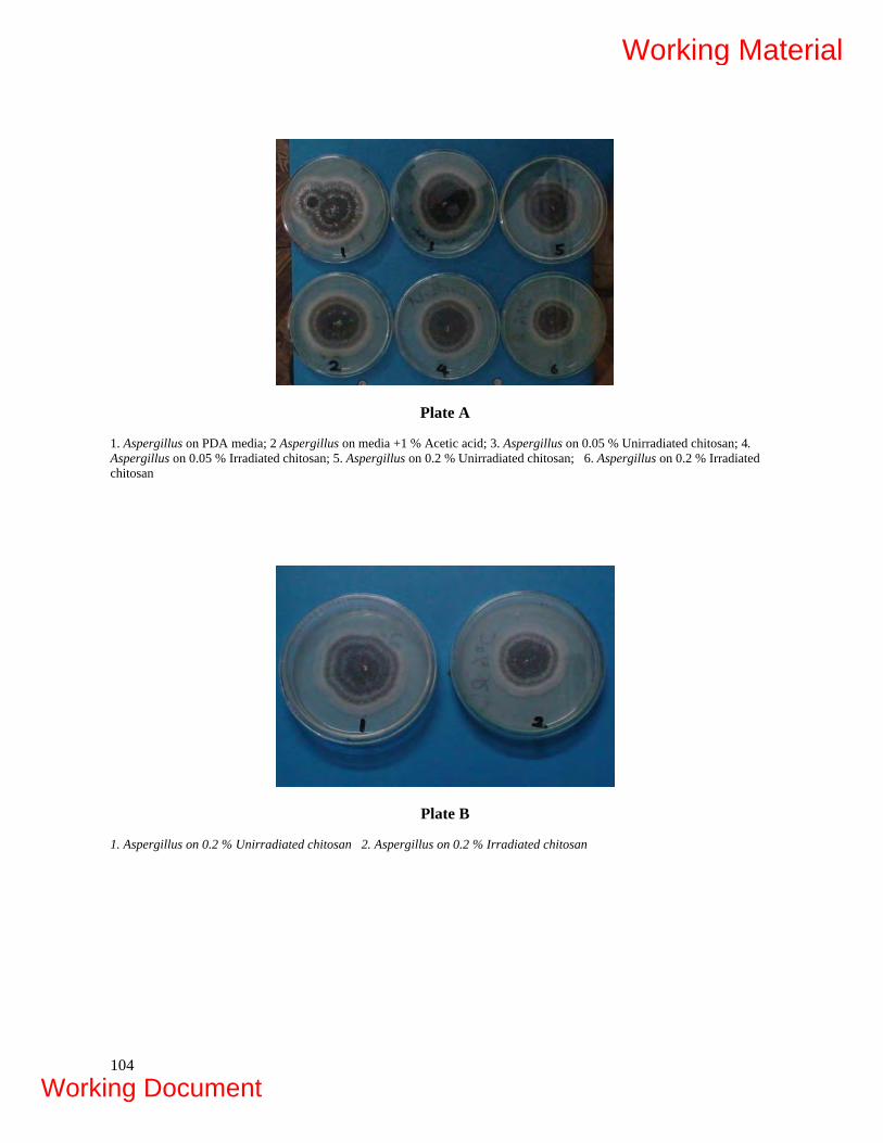
Plate A
1. Aspergillus on PDA media; 2 Aspergillus on media +1 % Acetic acid; 3. Aspergillus on 0.05 % Unirradiated chitosan; 4. Aspergillus on 0.05 % Irradiated chitosan; 5. Aspergillus on 0.2 % Unirradiated chitosan; 6. Aspergillus on 0.2 % Irradiated chitosan
Plate B
1. Aspergillus on 0.2 % Unirradiated chitosan 2. Aspergillus on 0.2 % Irradiated chitosan
104
Working Material
Working Document
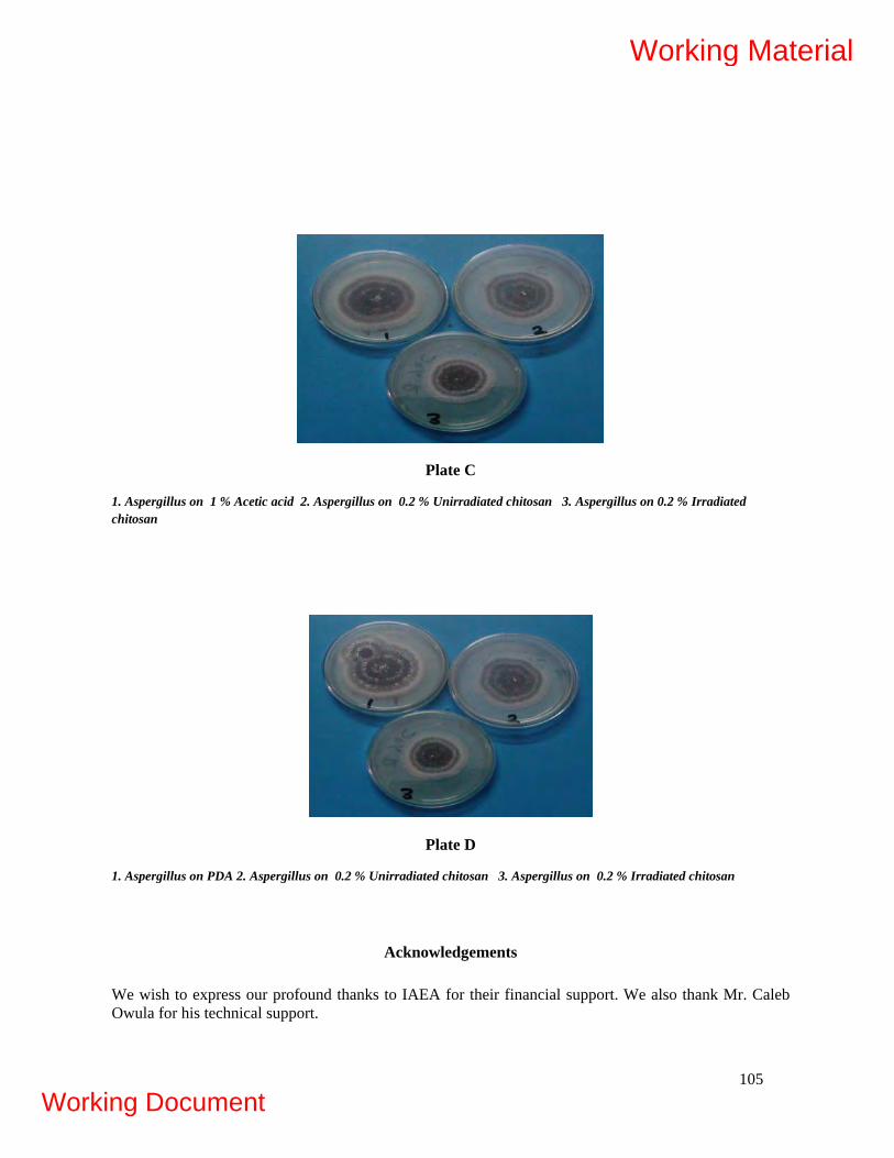
Plate C
1. Aspergillus on 1 % Acetic acid 2. Aspergillus on 0.2 % Unirradiated chitosan 3. Aspergillus on 0.2 % Irradiated chitosan
Plate D
1. Aspergillus on PDA 2. Aspergillus on 0.2 % Unirradiated chitosan 3. Aspergillus on 0.2 % Irradiated chitosan
Acknowledgements
We wish to express our profound thanks to IAEA for their financial support. We also thank Mr. Caleb Owula for his technical support.
105
Working Material
Working Document

106
Projections for the next 18 months • Investigating antioxidant properties of radiation processed chitosans • Detailed study on antimicrobial properties of radiation processed chitosan. • The use of the radiation processed chitosan as surface coating agents in some fruits and
vegetables in Ghana. • Field testing of radiation processed chitosan as plant growth promoter and soil conditioner.
REFERENCES
[1] VANDEMARK, P.J., BATZING, B.L., The microbes. An introduction to their nature and
importance. Benjamid Cummings Publishing Company Inc., Menlo Park, CA.(1987). [2] TSUGITA, T., Chitinkhitosan and their applications. In: Voigt, M.N. and Botta, R.J.,eds.
Advances in fisheries technology and biotechnology for increased profitability. Technornic Pub. Co., USA (1990) 287-298.
[3] CHANG, D.-S., CHO, H.-R., GOO, H.-Y., CHOE, W.-K. A development of food preservative with the waste of crab processing. Bull. Korean Fish. Soc., 22(2)(1989) 70-78.
[4] BETTS, G.D., LINTON, P., BETTERRIDGE, R.J., Food spoilage yeasts: Effect of pH, NaCl and temperature on growth. Food Control 10 (1), (1999) 27-33.
[5] MATSUHASHI, S., T., KUME, “Enhancement of antimicrobial activity of chitosan by irradiation” J. Sci. Food Agric. 73 (1997) 237 – 241.
[6] HA P. T. L. et al., “Antifungal activity of irradiated chitosan” Food Irradiation (in Japanese) 34 (1999) 43-46.
[7] FRATERNALE, D., GIAMPERI, L., RICCI, D. chemical composition and antifungal activity of Thymus mastichina L. J. Essent. Oil Res. 15 (2003) 278-281.
Working Material
Working Document
CURRENT STATUS OF RADIATION PROCESSING OF NATURAL POLYMERS IN INDIA
S.P. Ramnani
Radiation Technology Development Division, Bhabha Atomic Research centre Trombay, Mumbai 400 085, INDIA
1. INTRODUCTION
Radiation processing is being used on commercial basis in India since 1974 with the operation of ISOMED plant for radiation sterilization of medical products and Electron beam machine (ILU-6) in 1986. Since then many new products and processes have been investigated and some of them have culminated into useful applications. Many Indian industries produce and process natural polymers for local consumption and export. India exports about 120,000 tons of polysaccharides every year. Natural polysaccharides like guar gum, gum acacia, gum tora, agar, psylium husk etc are treated with gamma radiation mainly for controlling microbial contamination and sterilization. Radiation processing has also been used to reduce molecular weight of the polysaccharides in some applications. Recently, a few new applications have emerged where natural polysaccharides are used as additives and which play important role in controlling basic radiation chemistry reactions to produce useful products. Developed at BARC, radiation processed wound dressings, superabsorbent materials and low molecular weight chitosan are the products which have been used and could find potential applications in health care and agriculture sector. Radiation processed hydrogel wound dressings containing natural polysaccharides have already been commercialized. Some of the applications recently developed at BARC using natural polymers are briefly described below.
2. PVA-POLYSACCHARIDES HYDROGEL WOUND DRESSINGS (HDR) / HEALTHCARE
Use of hydrogel dressing is one of the desirable treatments for burns and ulcers. These dressing are normally produced by chemical methods which involve use of synthetic chemical initiators, preservatives, antimicrobials and other toxic chemicals. In radiation formation of HDR, an aqueous solution of polyvinyl alcohol (PVA) containing natural polysaccharide is exposed to gamma radiation at 25 kGy to produce sterile, elastic, transparent and mechanically strong HDR (1-2). Pure PVA aqueous solution, when exposed to gamma radiation at normal sterilization dose of 25 kGy, forms brittle hydrogel which are impractical for use as dressing. Moreover, the unreacted PVA has to be washed out and to be sterilized before use. In the present method of production of HDR, sterilization and gel formation are achieved in single stage irradiation process using natural polysaccharides. Natural polysaccharides have been observed to bring desirable characteristics to the HDR. Presence of polysaccharides like agar and carrageenan in formulations significantly affect mechanical properties and water absorption properties of HDR. These changes have been attributed to variation in the degree of crosslinking and network structure during irradiation brought about by the use of the polysaccharides. PVA mainly crosslinks and polysaccharides form interpenetrating network structure with PVA The extent of gelling power of the mixture of polysaccharides, charge on the polysaccharides and their high affinity for water in the formulation brings desirable characteristics to the dressing and eliminates use of synthetic plasticizers and humectants. Besides burn, these dressings have been observed to heal difficult wounds like leprosy, diabetic ulcers, bed sores and post surgical wounds. The dressings flatten fresh scars and are very useful on donor areas in plastic surgery. Burn injuries from firework, chemical, petrol, electrical appliances, road accidents, etc. can be treated using the HDR. The dressings can also be used on infected wounds by absorption of iodine from iodine solution (tincture of iodine etc.) as and when required. Other water soluble drugs could also be absorbed in the gel dressing and then applied to treat other skin indications. PVA is biodegradable and forms CO2 and water on incineration, helps in environment friendly disposal of the dressings after use. These dressings are now available under the brand name "Hi-Zel"(website: www.absmedicare.com).
107
Working Material
Working Document
Recent development to these dressing has been the use of nanotechnology to impart antibacterial properties to the dressing by in-situ generation of silver nanoparticles using radiation processing. Polysaccharides play an important role in stabilization of these particles as well as to their transport to the wound site. Modification of these polysaccharides by way of degradation and crosslinking and use in the product could bring diverse variations to the product property which could be possibly utilized for some applications. Some of these properties are being explored at our laboratories. 3. SUPERABSORBENT MATERIALS
Superabsorbent polymers can absorb very large amount of water and retain it even under pressure. These materials can absorb water many hundred times their dry weight. Due to their unique characteristics, they have found lot of application in personal hygiene products, agriculture and other specialized areas like controlled drug delivery system. Synthetic, semi-synthetic, or natural polymers are used separately or in combination for the synthesis of Superabsorbent polymers. In general, the Superabsorbent polymers have a structure in which water soluble polymers are made insoluble by some means of crosslinking. Carboxyl methyl cellulose (CMC) is an important water soluble derivative of cellulose. On exposure to high-energy radiation in aqueous solutions, it undergoes degradation. However, its irradiation in presence of monomers like acrylic acid (70% neutralized with NaOH) leads to the formation of three dimensionally crosslinked network structure that can absorb large quantities of water (about 460 g/g of dry gel) and thus act as a superabsorbing hydrogel. The effect of various additives such as divalent and trivalent metal ions, surfactants, organic molecules like urea and co-monomers such as hydroxyethyl methacrylate (HEMA) etc. on water absorbency were investigated. The results of all these experiments have shown that maximum water absorbency (800g/g) is obtained when surfactant such as Triton X-100 is incorporated into gel. This gel is at par with its properties when compared with the superabsorbent gel loaded in commercial diapers. In another independent development work on superabsorbent, a series of superabsorbent hydrogels were prepared from carrageenan and partially neutralized acrylic acid by gamma irradiation at room temperature (3). The gel fraction, swelling kinetics and the Equilibrium Degree of Swelling (EDS) of the hydrogels were studied. It was found that the incorporation of even 1% carrageenan (sodium salt) increases the EDS of the hydrogels from ~ 320g/g to ~ 800g/g. Thermal analysis were carried out to determine the amount of free water and bound water in the hydrogels. Under optimum conditions, poly (acrylic acid) – carrageenan hydrogels with high gel fraction (~80 %) and very high EDS (~800g/g) were prepared gamma radiolytically from aqueous solution containing 15 % partially neutralized acrylic acid and 1 to 5 % carrageenan. The hydrogels were also found to be sensitive to the pH and the ionic strength of the medium. In early stages of irradiation, acrylic acid undergoes polymerization and subsequently the polyacrylic acid chains undergo crosslinking reaction. During this process, presence of carrageenan reduces crosslinking density by scavenging OH/H radicals and probably physically screening the approach of radical sites that form crosslinks. The present study indicates that this effect practically gets saturated at about 1 % carrageenan concentration and further addition of carrageenan results in marginal increase of EDS. Presence of carrageenan could also modify the average pore size, the pore size distribution and the pore interconnections due to inter molecular interaction of carrageenan with polyacrylic acid during irradiation. These are important factors of the hydrogel matrix that determines its swelling characteristics and diffusion of solutes. There are always advantages of using fewer components in products for health care applications in radiation processing applications to avoid complex radiation degradation products. These materials developed in the laboratory were tested in the field as soil conditioners. The field experiments results indicated that incorporation of 20 Kg/ha in the soil enhance the yield by almost 10-15% in the wheat crop.
108
Working Material
Working Document

4. CHITOSAN DEGRADATION/CROSSLINKING
Chitosan finds many applications in food and pharmaceutical industry. Chitosan is an important natural polysaccharide derived from deacetylation of chitin which finds several applications in health care. When taken orally, it is known to lower cholesterol and lipids concentration in human blood. Low molecular weight chitosan has been reported to possess anti-tumor activity. Glucosamine is also known to show therapeutic effect for treating arthritis. Chitosan and alginates undergoes predominantly chain scission when exposed to ionizing radiation which results in lowering of its molecular weight. These low molecular weight products also known as oligomers have potential application in agriculture as plant growth promoter. At RTDD we have successfully demonstrated the application of these oligomers in plant growth promotion. For this three different crops namely Triticum aestivum (Wheat), Vigna radiate (Beans) and Linum usitatissimum (Linseed) were selected for study. Different treatments included in these studies were Dry seed treatment, Water presoaking treatment, Irrigation treatment and Foliar spray treatment. To determine the effective dose of gamma rays the dry seeds of wheat, mung and linseed were treated with 500 ppm of oligos of alginate and chitosan, produced by different doses of gamma rays. This experiment was performed specifically to determine the effective dose of gamma rays for the production of oligos of desired molecular weight with maximum biological activity. In this investigation gamma ray doses Viz., 20, 35, 66, 80 and 93 kGy were used to produce the oligos of alginate and 15, 35, 65, 77 and 89 kGy for production of oligo chitosan. All the doses of gamma rays were found to be effective in degrading the molecules of alginate and chitosan. The most effective dose of gamma ray was determined on the basis of stimulation in seedling growth, induced by oligos produced by a particular dose of gamma rays. The lowest dose of 20 kGy of gamma rays was marginally effective. However, the increase in the dose of gamma rays to 35 kGy and 66 kGy in alginate and 35 and 65 kGy in chitosan increased the growth stimulating activity of both the polysaccharides. Further increase in the dose of gamma rays up to 93 and 89 kGy led to decline in the stimulatory effect of alginate and chitosan, respectively. However, at the higher doses of gamma rays, alginate and chitosan still retained the stimulatory effect on the seedling height. In the water control, the seedling height in wheat, mung bean and Linum was 12.61, 9.50 and 8.40 cm, respectively. The unirradiated alginate and chitosan did not show any stimulatory effect on the seedling growth in all the three species. The seedling height with alginate, irradiated with different doses, ranged between 13.88 to 15.99 cm, 9.41 to 12.35 cm and 8.21 to 9.26 cm in wheat, mung and linseed, respectively whereas, with chitosan it was 14.18 to 15.07 cm, 10.82 to 11.20 cm and 6.78 to 8.89 cm in wheat, mung and linseed, respectivel In another attempt, chitosan was crosslinked using gamma radiation in presence of carbon tetrachloride as a sensitizer (4). The resultant product was insoluble in acid solution indicating that chitosan has crosslinked. Adsorption of Cr (VI) onto crosslinked chitosan synthesized by gamma irradiation has been investigated. The adsorption behavior of crosslinked chitosan (CRC) and its hydrolysis product (CRCH) has been compared with native chitosan. The maximum adsorption of Cr (VI) on crosslinked chitosan occurs at pH 3. The results obtained from equilibrium adsorption studies are fitted in various adsorption models such as Langmuir, Freundlich and Dubinin-Radushkevich (D-R) and the model parameters have been evaluated. Various thermodynamic parameters like H0, S0 and G0 for adsorption of Cr (VI) onto crosslinked chitosan have been estimated. The performance of crosslinked chitosan (CRC and CRCH) under flow conditions have also been studied and the results indicates that radiation crosslinked chitosan can be effectively used for treating wastewater containing Cr (VI). The most important aspect of using crosslinked chitosan for treating the wastewater containing Cr (VI) is that after the Cr (VI) is loaded; the column can be easily regenerated and efficiently reused.
5. GRAFTING OF POLYANILINEONTO RADIATION CROSSLINKED CHITOSAN
Among the conducting polymers, polyaniline has attracted attention of most of the researchers due to the combination of unique properties, good environmental stability, relatively high conductivity and low cost and also due to their wide spectrum of applications. We have chemically grafted polyaniline onto radiation
109
Working Material
Working Document
crosslinked chitosan already developed in the laboratory. The crosslinking of chitosan was carried out using 8 MeV electron beam and Co60 gamma rays in the presence of CCl4 as sensitizers [5]. The grafting of chitosan with polyaniline was carried using ammonium peroxydisulphate (APS) as an initiator [6]. Grafted polymer is characterized by dissolution, swelling, UV, VIS. NIR spectroscopy, Fourier transform infrared spectroscopy (FTIR), x-ray diffraction (XRD), SEM, thermogravimetric analysis, DC conductivity and nanoindentation studies. From the dissolution studies, grafting of PANI onto the crosslinked chitosan is confirmed and the weight of films before and after grafting gave the grafting percentage. Grafting percentage increases as the monomer concentration increases and decreases with increase in crosslinking. This may be due to the decrease in the penetration of the monomer onto the chitosan matrix with increase in crosslinking density. This is verified from SEM (cross-sectional view) of blend. UV-vis-NIR spectrum shows absorption peaks of PANI. Electrical property of grafted polymer is improved after doping with 1 M HCl. The change in volume conductivity is from 10-11 to 10-5 S/cm and surface conductivity from 10-10 to 10-2 S/cm. From TGA it is observed that grafted polymer and doped polymer are thermally stable. 6. ANTI-BACTERIAL COTTON FABRIC
Radiation grafting of vinylbenzyltrimethylammonium chloride (VBT) onto finished cotton fabric by mutual irradiation method has been carried out in aqueous medium to impart antibacterial activity (7). Additives such as metal salt and acid did not show enhancement on the grafting yield of VBT onto hydrophilic finished cotton substrate. The grafted cotton samples were tested for their antibacterial properties against test Escherichia coli and Staphylococcus aureus. The samples grafted to an extent of 5% start showing anti-bacterial activity and samples grafted to 13% are able to reduce the population of the organism by 4-5 log cycles within 2 hours. The anti-bacterial activity of grafted product is retained after many cycles of washing in commercial detergent powder. The VBT grafted material could be potentially used as dress material for high-risk group like hospital staffs.
7. CONCLUSION
India is a major producer of natural polymers. The recent work at BARC shows that use of natural polymers in radiation processing applications such as wound dressings, superabsorbent materials, radiation depolymerized alginates and chitosan and radiation crosslinked chitosan could bring highly desirable properties to the products for use in health care and agriculture sector. Development of methods of crosslinking of natural polymers using radiation would be desirable for development of new applications in waste water treatment.
Acknowledgement
Thanks are due to my colleagues Mr. C. V..Chaudhari, and Ms. Jayashree Biswal and all other colleagues from RTDD for their valuable contributions for the preparation of this report.
110
Working Material
Working Document

REFERENCES
[1] VARSHNEY, L., MAJMUDAR, A.A., Indian Patent No. 731/Bom, A process for manufacturing hydrogel wound dressing (1999).
[2] VARSHNEY, L., MAJMUDAR, A.A., A process for manufacture of hydrogels for burn and injury treatment, WO0130407, PCT application published on 3/5/2001.
[3] FRANCIS, S., KUMAR, M., VARSHNEY, L., Radiation synthesis of superabsorbent poly(acrylic acid)-carrageenan Hydrogels, Radiation Physics and Chemistry 69 (2004) 481-486.
[4] RAMNANI, S.P., CHAUDHARI, C.V., PATIL, N.D., SABHARWAL, S., Synthesis and characterization of crosslinked chitosan formed by γ-irradiation in the presence of carbon tetrachloride as a sensitizer. Journal of Polymer Science Part A: Polymer Chemistry, Volume 42, Issue 15 , 3897 – 3909.
[5] RAMAPRASAD, A.T., VIJAYLAXAMI RAO, PRAVEENA, M., GANESH SANJEEV, S., RAMNANI, P., SABHARWAL, S., Preparation of crosslinked chitosan by electron beam irradiation in the presence of CCl4. J. Appl. Polym. Sci, vol 111 (2009) 1063-1068.
[6] RAMAPRASAD, A.T., VIJAYLAXAMI RAO, PRAVEENA, M., GANESH SANJEEV, RAMNANI, S.P. SABHARWAL, S., Grafting of polyaniline onto radiation crosslinked chitosan. Synthetic Metals, 159 (2009) 1983-1990.
[7] KUMAR, V., BHARADWAJ, Y.K., RAWAT, K.P., SABHARWAL, S., Radiation-induced grafting of vinylbenyltrimethylammonium chloride (VBT) onto cotton fabric and study of its anti-bacterial activities. Radiation Physics and Chemistry (In print).
111
Working Material
Working Document

112
Working Material
Working Document

MODIFICATION OF NATURAL POLYMERS BY RADIATION-INDUCED PROCESSING FOR VERSATILE APPLICATIONS N. Nagasawa, A. Hiroki, M. Tamada Japan Atomic Energy Agency, Quantum Beam Science Directorate, Takasaki, Japan
Abstract Radiation-crosslinking mechanism of polysaccharide derivatives such as carboxymethyl cellulose (CMC), carboxymethyl chitosan (CMCts) is not clarified yet. Radicals in CMC formed by reaction with OH radical were studied by ESR in order to elucidate the radiation-crosslinking mechanism of CMC. ESR spectra implied that radicals were created on carboxymethyl groups linked to C2, C3 and C6 of glucose unit in CMC. The radicals of CMCts were also created on carboxymethyl groups linked to C3 and C6 of glusamine unit. For application of carboxymethyl cellulose (CMC) to a super water-adsorbent, gel fraction should be adjusted to the range of 50%. It was found that gel fraction of CMC could be control by the irradiation temperature of CMC aqueous paste-like state. Poly(L-lactic acid)(PLLA) could be crosslinked when it was irradiated with coexistence of crosslinker, triallyl isocyanurate. However, the crosslinked PLLA is thermally deformed under the stress at 70 °C. The thermo mechanical stability of PLLA was further improved by the adding SiO2 and the post heating at 90 °C. The storage modulus of the treated PLLA showed about 100 times higher than that of only crosslinked PLLA. The improved materials was applied to demonstration lens of eyewear. 1. OBJECTIVES OF THE RESEARCH Development of natural polymers including a polymer synthesized from fermented products has considerable attention from the viewpoint of carbon neutral in which carbon is recycled through the plant growth. The radiation processing, especially crosslinking, of natural polymers is regarded as unique technique to improve their physical, chemical, mechanical and thermal properties. Our objective focuses on the development of new functional materials by means of this crosslinking technique which can modify natural polymers. As natural polymers, the polysaccharide derivatives and aliphatic polyester were selected. It is expected from this research project that crosslinked natural polymers are widely applied in the fields of industry, medicine, agriculture and so on. 2. INTRODUCTION Japan Atomic Energy Agency (JAEA) was organized by consolidating of Japan Atomic Energy Research Institute and Japan Nuclear Cycle Development Institute in Oct. 2005. JAEA has 11 sites of research institutes in Japan. Takasaki site, JAEA carried out application R&D of radiation processing using of facilities such as gamma-ray irradiation, electron beam accelerator, and ion accelerators. Two divisions, environment and industrial materials research division and radiation-applied biology division, of quantum beam science directorate are located in Takasaki site. Environmental Polymer group in environment and industrial materials research division applied to this CRP in the research subject of "Modification of natural polymers by radiation-induced crosslinking for versatile applications". Environmental polymer group has researched radiation processing of polymer to apply the resulting materials to the fields of environmental preservation. Two major techniques for our research are crosslinking and graft polymerization which induced by high energy irradiation. In this CRP, the crosslinking of natural polymers which are degraded by irradiation has been investigated to explore new application field as shown in Figure 1. Furthermore, the properties of crosslinked natural polymer will be refined for technical transfer to end users. The polysaccharide derivatives and aliphatic polyester were selected as natural polymers. The following sections deals with crosslinking of carboxymethylated cellulose, starch, chitin and chitosan as polysaccharide derivatives and poly(lactic acid) as aliphatic polyester.
113
Working Material
Working Document

FIG. R&D in environmental polymer group, Takasaki site, JAEA.
3. CROSSLINKING OF CHEMICALLY MODIFIED POLYSACCHARIDES
3.1. Identification of CMC radical produced by reaction with OH radical In general, OH radical, which is one of main water radiolysis products and very oxidative species, is said to induce crosslinking reaction of polymer aqueous solution. The purpose of this study is to understand the radiation-crosslinking mechanism of CMC at a high concentrated aqueous solution. Identification of CMC radical produced by reaction with OH radical was introduced. CMC radical in an aqueous solution was observed directly by ESR method using photolysis of hydrogen peroxide as shown in Figure 2. The ESR spectra have a large doublet split and small triplet split like [Triplet x Doublet]. However, some species seemed to be overlapped on ESR spectra, because right-and-left triplet spectra are not equiform. Compared with some radicals, which have analogical structure to CMC, about hyperfine coupling constant, [Triplet x Doublet], assigned as radical on carboxymethyl group linked to C6, and [Doublet], assigned as radical on carboxymethyl group linked to C2 or C3, were isolated from ESR spectra of experimental results as shown in Figure 3. In conclusion, these spectra were identified as radicals located on secondary carbon of carboxymethyl groups[1,2].
114
Working Material
Working Document
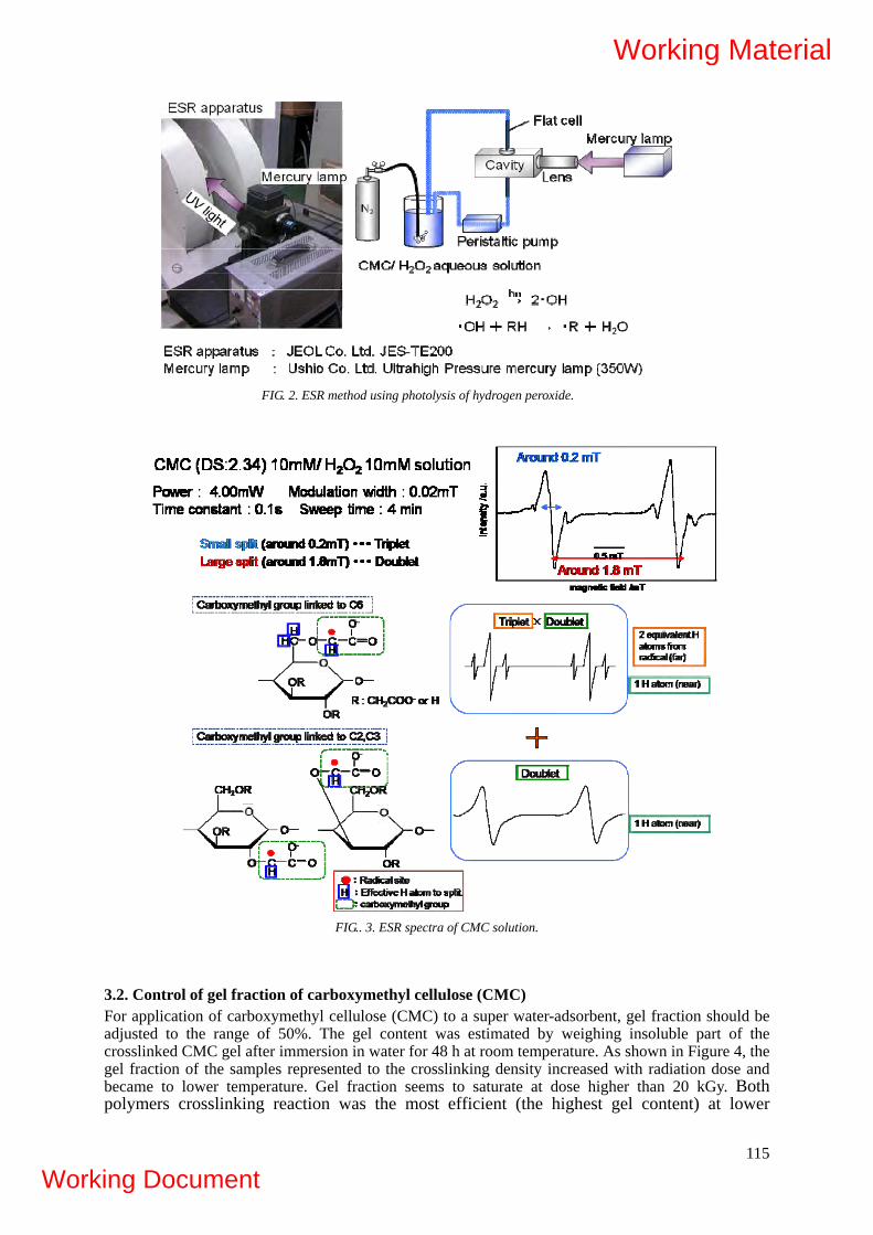
FIG. 2. ESR method using photolysis of hydrogen peroxide.
FIG.. 3. ESR spectra of CMC solution.
3.2. Control of gel fraction of carboxymethyl cellulose (CMC) For application of carboxymethyl cellulose (CMC) to a super water-adsorbent, gel fraction should be adjusted to the range of 50%. The gel content was estimated by weighing insoluble part of the crosslinked CMC gel after immersion in water for 48 h at room temperature. As shown in Figure 4, the gel fraction of the samples represented to the crosslinking density increased with radiation dose and became to lower temperature. Gel fraction seems to saturate at dose higher than 20 kGy. Both polymers crosslinking reaction was the most efficient (the highest gel content) at lower
115
Working Material
Working Document
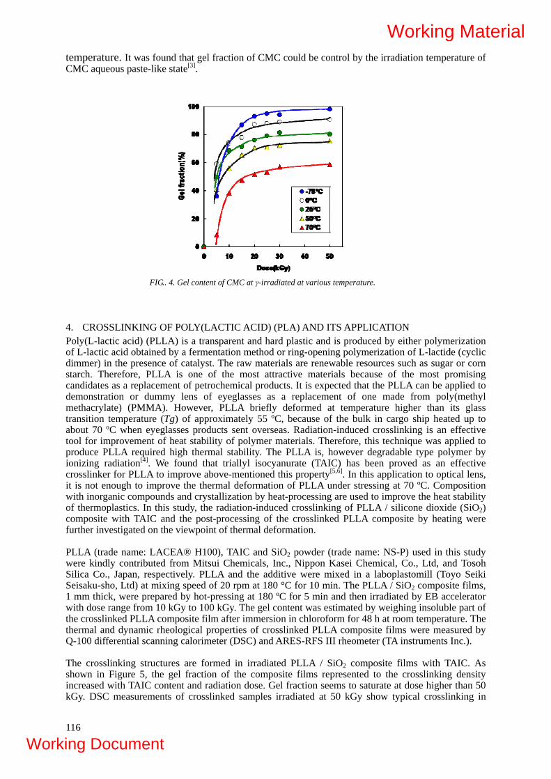
temperature. It was found that gel fraction of CMC could be control by the irradiation temperature of CMC aqueous paste-like state[3].
FIG.. 4. Gel content of CMC at γ-irradiated at various temperature.
4. CROSSLINKING OF POLY(LACTIC ACID) (PLA) AND ITS APPLICATION Poly(L-lactic acid) (PLLA) is a transparent and hard plastic and is produced by either polymerization of L-lactic acid obtained by a fermentation method or ring-opening polymerization of L-lactide (cyclic dimmer) in the presence of catalyst. The raw materials are renewable resources such as sugar or corn starch. Therefore, PLLA is one of the most attractive materials because of the most promising candidates as a replacement of petrochemical products. It is expected that the PLLA can be applied to demonstration or dummy lens of eyeglasses as a replacement of one made from poly(methyl methacrylate) (PMMA). However, PLLA briefly deformed at temperature higher than its glass transition temperature (Tg) of approximately 55 ºC, because of the bulk in cargo ship heated up to about 70 ºC when eyeglasses products sent overseas. Radiation-induced crosslinking is an effective tool for improvement of heat stability of polymer materials. Therefore, this technique was applied to produce PLLA required high thermal stability. The PLLA is, however degradable type polymer by ionizing radiation[4]. We found that triallyl isocyanurate (TAIC) has been proved as an effective crosslinker for PLLA to improve above-mentioned this property[5,6]. In this application to optical lens, it is not enough to improve the thermal deformation of PLLA under stressing at 70 ºC. Composition with inorganic compounds and crystallization by heat-processing are used to improve the heat stability of thermoplastics. In this study, the radiation-induced crosslinking of PLLA / silicone dioxide (SiO2) composite with TAIC and the post-processing of the crosslinked PLLA composite by heating were further investigated on the viewpoint of thermal deformation. PLLA (trade name: LACEA® H100), TAIC and SiO2 powder (trade name: NS-P) used in this study were kindly contributed from Mitsui Chemicals, Inc., Nippon Kasei Chemical, Co., Ltd, and Tosoh Silica Co., Japan, respectively. PLLA and the additive were mixed in a laboplastomill (Toyo Seiki Seisaku-sho, Ltd) at mixing speed of 20 rpm at 180 °C for 10 min. The PLLA / SiO2 composite films, 1 mm thick, were prepared by hot-pressing at 180 ºC for 5 min and then irradiated by EB accelerator with dose range from 10 kGy to 100 kGy. The gel content was estimated by weighing insoluble part of the crosslinked PLLA composite film after immersion in chloroform for 48 h at room temperature. The thermal and dynamic rheological properties of crosslinked PLLA composite films were measured by Q-100 differential scanning calorimeter (DSC) and ARES-RFS III rheometer (TA instruments Inc.). The crosslinking structures are formed in irradiated PLLA / SiO2 composite films with TAIC. As shown in Figure 5, the gel fraction of the composite films represented to the crosslinking density increased with TAIC content and radiation dose. Gel fraction seems to saturate at dose higher than 50 kGy. DSC measurements of crosslinked samples irradiated at 50 kGy show typical crosslinking in
116
Working Material
Working Document
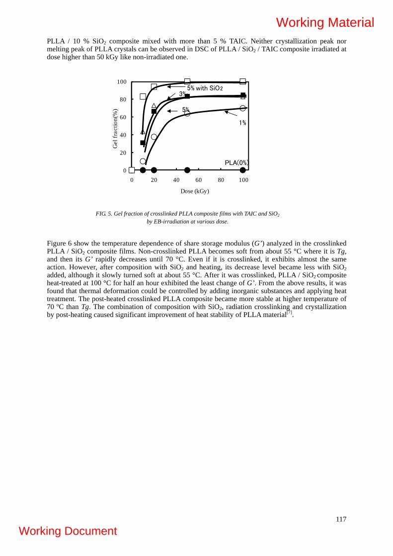
PLLA / 10 % SiO2 composite mixed with more than 5 % TAIC. Neither crystallization peak nor melting peak of PLLA crystals can be observed in DSC of PLLA / SiO2 / TAIC composite irradiated at dose higher than 50 kGy like non-irradiated one.
0
20
40
60
80
100
0 20 40 60 80 100
Gel
fra
ctio
n(%
)
Dose (kGy)
3%
PLA(0%)
5%
1%
5% with SiO2
FIG. 5. Gel fraction of crosslinked PLLA composite films with TAIC and SiO2
by EB-irradiation at various dose.
Figure 6 show the temperature dependence of share storage modulus (G’) analyzed in the crosslinked PLLA / SiO2 composite films. Non-crosslinked PLLA becomes soft from about 55 °C where it is Tg, and then its G’ rapidly decreases until 70 °C. Even if it is crosslinked, it exhibits almost the same action. However, after composition with SiO2 and heating, its decrease level became less with SiO2 added, although it slowly turned soft at about 55 °C. After it was crosslinked, PLLA / SiO2 composite heat-treated at 100 °C for half an hour exhibited the least change of G’. From the above results, it was found that thermal deformation could be controlled by adding inorganic substances and applying heat treatment. The post-heated crosslinked PLLA composite became more stable at higher temperature of 70 ºC than Tg. The combination of composition with SiO2, radiation crosslinking and crystallization by post-heating caused significant improvement of heat stability of PLLA material[7].
117
Working Material
Working Document

30 40 50 60 70 80 90Temperature(ºC)
G'(
Pa)
(a)
1011
1010
10 9
10 8
10 7
10 6
(b)
(c)
(d)
FIG. 6. Temperature dependence of share storage modulus (G’) of crosslinked PLLA films with 5 % TAIC by
EB-irradiation at 50 kGy and post processed sample. (a) Unirradiated PLLA, (b) Crosslinked PLLA, (c) Crosslinked PLLA / 10 % SiO2, (d) Crosslinked PLLA / 10 % SiO2. post-heated at 100 ºC for 0.5 h.
5. DEGARADATION OF MARINE POLYSACCHARIDES FOR PLANT GROWTH Radiation-induced degraded chitosan was applied to plant growth promoter for lawn grass, cyclamen, and vegetable such as dwarf pine needles by private company. 6. PROPOSED PLAN
Year 1st (2008): At the first stage of the development of functional materials based on biodegradable polymers using radiation processing, the irradiation processes are investigated for crosslinking of water-soluble polysaccharides and poly(lactic acid) (PLA), which are known as typical radiation-degradation polymers. In this research, carboxymethylcellulose (CMC), carboxymethylstarch (CMS), carboxymethylchitosan (CMCTS), carboxymethylchitin (CMCT) and hydroxylpropylcellulose (HPC) are used as water-soluble polysaccharides. By crosslinking of such polysaccharides, we develop hydrogels applicable to bedsore prevention mats, metal ion adsorbents and so on. Concerning PLA, its heat-resistance is improved for the application to thermally shrinkable tubes etc. Year 2nd (2009): To improve the characteristics of crosslinked biodegradable products, the crosslinking processes which can control the gel fraction are investigated. Super water-adsorbents are developed by controlling the gel fraction of polysaccharide-based hydrogels for the effective treatment of livestock excrements. The thermostability of PLA is further improved by controlling the gel fraction with additives and the post processing for the sake of fabricating optical materials based on PLA. Year 3rd (2010): To spread the application fields of crosslinked biodegradable polymers, advanced fabrication processes are developed, e.g., (1) polysaccharide blends are crosslinked and (2) polysaccharides are crosslinked with additives such as polymerizable monomers and crosslinkers. By using the former technique, the hydrogels having selective affinity against metal ion and those with controllable degradation period are produced. By adopting the latter technique, the mechanical strength and elongation of hydrogels are improved for their medical application.
118
Working Material
Working Document

REFERENCES
[1] SAIKI, S., NAGASAWA, N., HIROKI, A., MORISHITA, N., TAMADA, M., MUROYA, Y., KUDO, H., KATSUMURA, Y., “ESR study on carboxymethyl chitosan radicals in an aqueous solution”, Radiat. Phys. Chem., in press.
[2] SAIKI, S., NAGASAWA, N., HIROKI, A., MORISHITA, N., TAMADA, M., KUDO, H., KATSUMURA, Y., Radiat. Phys. Chem., in preparation.
[3] NAGASAWA, N., HA, P.T.L., YAGI, T., TAMADA, M., “Radiation crosslinking of CMC at various condition”, Carbohydr. Polym., submitted.
[4] NUGROHO, P., MITOMO, H., YOSHII, F., KUME, T., “Polymer degradation of poly(lactic acid) by γ-irradiation”, Polym. Degrad. Stab., 72, (2001) 337-343.
[5] MITOMO, H., KANEDA, A., QUYNH, T.M., NAGASAWA, N., YOSHII, F., “Improvement of heat stability of poly(L-lactic acid) by radiation-induced crosslinking”, Polymer, 46 (2005) 4695-4703.
[6] NAGASAWA, N., KANEDA, A., KANAZAWA, S., YAGI, T., MITOMO, H., YOSHII, F., TAMADA, M., “Application of poly(lactic acid) modified by radiation crosslinking”, Nucl. Instr. Meth. in Phys. Res. B (2005) 611-616.
[7] NAGASAWA, N., KASAI, N., YAGI, T., TAMADA, M., “Radiation-induced crosslinking and post-processing of poly(L-lactic acid) composite”, Radiat. Phys. Chem., submitted.
119
Working Material
Working Document

120
Working Material
Working Document

APPLICATION OF RADIATION DEGRADED CHITOSAN AS PLANT GROWTH PROMOTER A PILOT SCALE PRODUCTION AND FIELD TRIAL STUDY OF RADIATION PROCESSED CHITOSAN AS PLANT GROWTH PROMOTER FOR RICE CROPS
Khairul Zaman Hj Mohd Dahlan1, Kamaruddin Hashim1, Kamarudin Bahari1, Maznah Mahmod1 , Norzita Yaacob1, Norhashidah Talip1, Kamarolzaman Hussein1, Ahamad Sahali Mardi1, Abd Rahim Harun1, RohayaYusof2, Sauddi Sitor2, N. Q. Hien3 and T. Kume4 1 Malaysian Nuclear Agency, Bangi, 43000 Kajang, Selangor, Malaysia 2FELCRA (M) Berhad, 3 Vinagamma, Vietnam Atomic Energy Commission 4 Japan Atomic Energy Agency Abstract The application of radiation processed chitosan as plant growth promoter has been carried out in the 24 hectares of rice crops. For the field trial, a pilot scale production of oligochitosan was established using gamma irradiation for partial degradation of chitosan powder of DDA 90% and followed by gamma irradiation of aqueous solution of 3% irradiated chitosan powder in 2% lactic acids (3CL2). Radiation dose of 50 kGy was selected for initial degradation of chitosan powder and followed by 12 kGy irradiation of 3CL2. A viscosity average molecular weight of ~10,000 of oligochitosan was obtained and subsequently used in the field trial of MR219 type of rice seeds on 24 hectares of rice plots. The seedlings were carried out after the rice seeds were soaked 24hrs in water and 30 minutes in 200ppm oligochitosan. The rice plots that were sprayed with oligochitosan were found to have higher resistant towards blast diseases. Oligochitosan of 40ppm was found to be effective as fungicides and resulted in the increase of yield of rice seeds of about 5%. 1. INTRODUCTION
The oligosaccharides derived from depolymerization of polysaccharides through enzymatic hydrolysis have been reported to exhibit growth-stimulating activity like phytohormones that induce the promotion in germination, shoot and root elongation in variety of plants [1-3]. Radiation degraded polysaccharides also show similar effects as plant growth promoter. Radiation degraded alginate solution (4%) at 100 kGy has been shown to significantly enhance the growth of rice in hydrophonic system [4]. They showed that the suitable range of concentration of degraded alginate was between 20-100 ppm to impact 15-60% weight gain of rice and peanuts respectively. On the other hand, Tomoda et. al., showed that higher concentration, 1000ppm of degraded alginate prepared by degradation of enzymatic lyases was required to obtain the optimum growth-promotion of barley roots.
A similar study has been conducted by Relleve et.al., [5] using irradiated carrageenan for rice seedlings under non-circulating hydrophonic condition. The study showed that kappa carrageenan irradiated at 100 kGy gave maximum weight gained. However, iota carrageenan exhibited less growth promoting properties than kappa carrageenan and required higher irradiation dose to degrade at approximately the same molecular weight as kappa carrageenan. Both of irradiated iota and kappa carrageenan have caused the disappearance of fungi growth on the roots of rice.
In the case of irradiated chitosan (oligochitosan), it has been shown not only exhibit as plant growth promoter, but also exhibit highly anti-microbial activity. Irradiated chitosan either in solid state or in aqueous solutions are able to suppress the growth of various fungi strains and showed higher fungicidal effect than that of non-irradiated chitosan [6]. It has also been recognized as potent phytoalexin inducer (elicitor) to resist infection of diseases for plants.
121
Working Material
Working Document

The effects of radiation degraded polysaccharide on the growth promotion of plant is not well understood. However, radiation degraded polysaccharide can be control to certain size of molecular weight to produce oligosaccharides that are mobile and easy for uptake by the plant. Radiation has also been shown does not affect the functional groups of carrageenan (SO4-) and chitosan (NH2) but instead enhance the anti-microbial activity of the oligosaccharides that improved the plant resistivity towards fungi attacked. In this paper, the production of radiation processed oligochitosan at a pilot scale will be described. The oligochitosan properties are established and some results on its application on rice crops are reported.
2. MATERIAL AND METHOD Chitosan powder was purchased from China with the following properties, 90.6% of DDA, 10% water content and contain 0.1ppm As, 0.1ppm Cd, 0.8ppm Cu, 3.16ppm Ni and 0.1ppm Pb. Lactic acid (90%), NaOH solution (2M), Hydrogen Peroxide (30%) and Ethanol are all industrial grades and supplied by local companies. 2.1. Production of oligochitosan A Co-60 plant of ~ 800 kCi was used to partially degrade chitosan powder at a dose of 50 kGy and dose rate of 2.51 kGy/hr. Subsequently, 3% of radiation degraded chitosan in 2% lactic acid solution was prepared as follows Chitosan powder (10% moisture): 45 kg Lactic acid (90%): 33 L Water: 1,500 L Hydrogen Peroxide (30%): 4.5 L NaOH soln. (2M): 15 L Ethanol: 750 L TOTAL VOLUME = 2307 L (1.95 % chitosan ~ 19,500ppm) The chitosan solution was then irradiated by using liquid (Natural Rubber Latex) gamma irradiator of an activity, 94,818 Ci. The cycle time for 1500 L solution (56 strokes x 15 min/stroke) is ~14hrs for 12 kGy. Dose rate is 0.86 kGy/hr. 2.2. Characterization of chitosan and oligochitosan
The viscosities of 50 kGy irradiated chitosan powder solution and 12 kGy irradiated chitosan solution were determined at 3% chitosan solution in 2% lactic acid using spindle type viscometer model Brookfield DV II+.
The irradiated chitosan was purified by adding 2.5% ammonium hydroxides until the pH reached 6.5. The precipitated oligochitosan washed with ethanol and air dried. Subsequently, the purified oligochitosan at concentration 1.0 g/l was prepared in acetic acid/ammonium acetate buffer solution (0.20M/0.15M at 25 C) for intrinsic viscosity measurement using Ubbelohde viscometer capillary 531/10/I of Schott instrument, model AVS 440. Data of the viscosity average molecular weight was taken from the average of 3 measurements. pH of the solution were measured using pH meter Mettler Toledo.
2.3. Germination of rice seeds
Rice seeds of MR219 supplied by FELCRA (M) Berhad were used through out of this study. Three different treatments of rice seeds were performed namely:
i. Rice seeds (~ 100 seeds) were soaked in water for 24 hrs as control (C).
122
Working Material
Working Document

ii. Rice seeds were soaked in water for 24 hrs and followed by soaking in 200ppm oligochitosan for 15min , 30min, 60min, 90min and 120min.
iii. Rice seeds were soaked in 200ppm oligochitosan for 15 min, 30min, 60min, 90min and 120min.
Each sample was done in 3 replicates. The burnt-rice husk was used as the planting media in this experiment. All rice seeds were water sprayed 3 x a day for 5 days. No. of seeds germinated were counted and recorded every day.
2.4. Field trail preparation of rice seeds
Rice seeds, MR219 of 90 bags which is 1,800 kg (20 kg/bag) were supplied by FELCRA (M) Berhad. Eight Field Plots of 1.0 hectare size per plot were prepared in triplicates with a total size of 24 hectares as follows:
i. T1 and T2 (3 replicates for each plot) as control and were soaked in VITA CAMBAH (local growth promoter) for 24 hrs, tossed for another 24 hrs before sowing to the fields. These rice plots were given pesticides treatment as the normal local practises.
ii. T3, T4 and T5 (3 replicates for each plot) were soaked in water for 24 hrs, tossed for another 24 hrs before sowing to the fields. These rice plots were sprayed with oligochitosan at concentration of 20ppm, 40ppm and 100ppm at interval of 42 days and 72 days of planting.
iii. T6, T7 and T8 (3 replicates for each plot) were soaked in water for 24 hrs and followed by soaking in 200ppm oligochitosan for 30 min. Then, they were tossed for another 24 hrs prior to be sowing to the fields. These rice plots were sprayed with oligochitosan at concentration of 20ppm, 40ppm and 100ppm at interval of 42 days and 72 days of planting.
2.5. Collection of data from the field trial of rice crops
From each plot of 1 hectare, 5 quadrants of 1 square meter each of rice crops were identified for data collections on physical growth, fungus diseases, yields and rice seeds characterization.
3. RESULT AND DISCUSSION
3.1. Pilot scale production of oligochitosan
In the pilot scale production of oligochitosan, two step gamma irradiation were employed to suit the condition of Raymintex – latex gamma irradiator at Nuclear Malaysia. Currently, the latex gamma irradiator is set to deliver an irradiation dose of 12 kGy for natural rubber latex. Having this condition, the chitosan powder has to be partially degraded using another gamma irradiation plant at 50 kGy. The viscosities of chitosan powder (2% and 3% chitosan powder in 2% lactic acid) before and after irradiation are shown in Table 1.0. The viscosity of 3% chitosan in 2% latic acids after irradiation at 50 kGy is slightly higher than the viscosity of natural rubber latex and was selected for this study. The irradiated chitosan was in acidic condition and neutralized by using sodium hydroxide before being applied to the field as shown in Table 2.0.
123
Working Material
Working Document
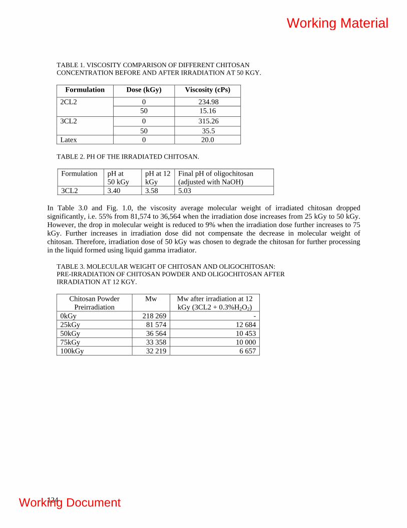
TABLE 1. VISCOSITY COMPARISON OF DIFFERENT CHITOSAN CONCENTRATION BEFORE AND AFTER IRRADIATION AT 50 KGY.
Formulation Dose (kGy) Viscosity (cPs)
0 234.98 2CL2 50 15.16 0 315.26 3CL2
50 35.5 Latex 0 20.0
TABLE 2. PH OF THE IRRADIATED CHITOSAN.
Formulation pH at
50 kGy pH at 12 kGy
Final pH of oligochitosan (adjusted with NaOH)
3CL2 3.40 3.58 5.03 In Table 3.0 and Fig. 1.0, the viscosity average molecular weight of irradiated chitosan dropped significantly, i.e. 55% from 81,574 to 36,564 when the irradiation dose increases from 25 kGy to 50 kGy. However, the drop in molecular weight is reduced to 9% when the irradiation dose further increases to 75 kGy. Further increases in irradiation dose did not compensate the decrease in molecular weight of chitosan. Therefore, irradiation dose of 50 kGy was chosen to degrade the chitosan for further processing in the liquid formed using liquid gamma irradiator.
TABLE 3. MOLECULAR WEIGHT OF CHITOSAN AND OLIGOCHITOSAN: PRE-IRRADIATION OF CHITOSAN POWDER AND OLIGOCHITOSAN AFTER IRRADIATION AT 12 KGY.
Chitosan Powder
Preirradiation Mw Mw after irradiation at 12
kGy (3CL2 + 0.3%H2O2) 0kGy 218 269 -25kGy 81 574 12 684 50kGy 36 564 10 45375kGy 33 358 10 000100kGy 32 219 6 657
124
Working Material
Working Document
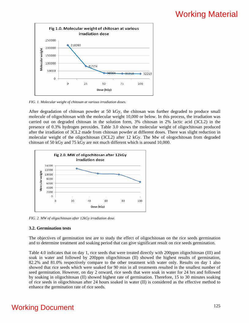
FIG. 1. Molecular weight of chitosan at various irradiation doses. After degradation of chitosan powder at 50 kGy, the chitosan was further degraded to produce small molecule of oligochitosan with the molecular weight 10,000 or below. In this process, the irradiation was carried out on degraded chitosan in the solution form, 3% chitosan in 2% lactic acid (3CL2) in the presence of 0.3% hydrogen peroxides. Table 3.0 shows the molecular weight of oligochitosan produced after the irradiation of 3CL2 made from chitosan powder at different doses. There was slight reduction in molecular weight of the oligochitosan (3CL2) after 12 kGy. The Mw of ologochitosan from degraded chitosan of 50 kGy and 75 kGy are not much different which is around 10,000.
FIG. 2. MW of oligochitosan after 12kGy irradiation dose. 3.2. Germination tests The objectives of germination test are to study the effect of oligochitosan on the rice seeds germination and to determine treatment and soaking period that can give significant result on rice seeds germination. Table 4.0 indicates that on day 1, rice seeds that were treated directly with 200ppm oligochitosan (III) and soak in water and followed by 200ppm oligochitosan (II) showed the highest results of germination, 82.2% and 81.0% respectively compare to the other treatment with water only. Results on day 1 also showed that rice seeds which were soaked for 90 min in all treatments resulted in the smallest number of seed germination. However, on day 2 onward, rice seeds that were soak in water for 24 hrs and followed by soaking in oligochitosan (II) showed highest rate of germination. Therefore, 15 to 30 minutes soaking of rice seeds in oligochitosan after 24 hours soaked in water (II) is considered as the effective method to enhance the germination rate of rice seeds.
125
Working Material
Working Document

TABLE 4. GERMINATION TESTS ON RICE SEEDS.
Treatment & Treatment
period
I Soak in water only
II Soak in water (24 hours)
followed by soak in Oligochitosan 200 ppm
III Soak in
Oligochitosan 200 ppm only
Germinated Seeds, % (Average) Day 1
15 mins. 60.0 81.0 81.7 30 mins 36.3 68.3 82.7 60 mins 58.3 68.7 79.7 90mins 59.7 44.7 68.7
Day 2 15 mins. 78.3 90.7 83.3 30 mins 65.7 75.7 86.7 60 mins 78.7 76.7 84.7 90mins 76.7 56.0 80.0
Day 3 15 mins. 82.0 92.7 84.0 30 mins 72.3 89.3 88.0 60 mins 84.7 86.0 86.7 90mins 81.3 75.7 85.7
Day 4 15 mins. 84.7 92.7 85.3 30 mins 75.7 90.7 88.7 60 mins 90.7 88.3 87.3 90mins 86.7 79.0 86.7
Day 5 15 mins. 90.3 93.7 86.7 30 mins 80.0 94.0 89.7 60 mins 91.0 92.0 87.7 90mins 90.0 80.7 90.3
3.3. Blast disease infection The result of the effects of oligochitosan on blast diseases of rice crops is given in Table 5.0. Analysis of variance showed that there is not significant different found between the treatments at 45 days. R square value (Coefficient Determination) is high indicating that it follows the model that was used. The Critical Value (CV) is considerably low around 35%. From T test LSD showed that the oligochitosan treatment gave lower number of blast infection as compare to control. However, the effect of oligochitosan at 45 days might not be strong enough to induce morphology and physiology of the plant to prevent biotic stress from outside. Analysis of variance on 72 days showed the probability of 0.075 where it is very close to 0.05. Therefore it is considered significant different between the treatments against the rice blast. T6, T5, T7 with oligochitosan treatment on seed and leaf have given the lower number of plant infection with blast, indicating that oligochitosan treatment has shown an effect to physiology of rice plant to prevent them from blast disease.
126
Working Material
Working Document

TABLE 5. OLIGOCHITOSAN EFFECTS ON RICE DISEASES – BLAST FUNGUS ON LEAVES AND PANICLES.
RESULT DAY 107th
TREATMENT & LOT NO.
DESCRIPTION SCORE DAY 45th
DAY 62nd
DAY 72nd LB PB
T1 (Lot 505, 520, 504)
Average : Control (without fungicide & without oligochitosan)
1 3 5
31 4
0.3
24 - -
- - -
1.3
-
T2 (Lot 511, 508, 521)
Average: Control (spray fungicide)
1 3 5
28 1.7 -
21 4.3 -
- - -
26.7
-
T3 (Lot 501, 510, 524)
Average: Spray oligochitosan on leaves (20 ppm)
1 3 5
35 3 -
39 9 -
- - -
6.7
-
T4 (Lot 502, 507, 518)
Average: Spray oligochitosan on leaves (40 ppm)
1 3 5
28 0.7 -
30 3.7 -
- - -
4.0
-
T5 (Lot 512, 513, 503)
Average: Spray oligochitosan on leaves (100 ppm)
1 3 5
30 1.0 -
6 1.3 0.7
- - -
2.7
-
T6 (Lot 515, 522, 516)
Average: Seed treatment with oligochitosan (200 ppm) + spray oligochitosan on leaves (20 ppm)
1 3 5
41 0.7 -
7 20 0.7 -
- - -
1.3
-
T7 (Lot 509, 506, 519)
Average: Seed treatment with oligochitosan (200 ppm) + spray oligochitosan on leaves (40 ppm)
1 3 5
36 2 -
10.3 1.3 -
7 0.7 -
- -
-
T8 (lot 514, 523, 517)
Average: Seed treatment with oligochitosan (200 ppm) + spray oligochitosan on leaves (100 ppm)
1 3 5
26 0.7 -
17 0.3 -
34 1.0 -
- - -
1.3
-
Note : LB – Leaves Blast, PB – Panicle Blast
127
Working Material
Working Document

FIG. 3. shows clearly the effects of oligochitosan on blast fungus at interval 45th, 72th and 107th days of planting. 3.4. Growth promotion Rice seeds were harvested on the 112 days after planting. The total yield of rice seeds per plot of experiments were given in Table 6.0. T1 and T2 plots that have no oligochitosans but sprayed with and without fungicides and were used as controls. However, all plots were sprayed with pesticides depending on the conditions of the rice crops during the trial periods. On the other hands, the trial plots, T3 – T8 were not sprayed with fungicides and only depend on oligochitosan to act as fungicides as well as growth promoter. During the trial periods, golden snails attacked and winds are the two mains phenomena that have affected certain plots and hence affected the yield of the rice crops. Table 5.0 indicates that highest yield of rice seeds is from the plot T4 and followed by the plot T7 in comparison with the control T1 and T2. The increase in yields is about 5.76% and 4.05% higher than control plot of T1 and T2 respectively. These increases are lower than what is expected which is around 10%. The yield for control T2 with fungicides is almost the same or lower than the yiled of plot that were treated with 40ppm oligochitosan and without fungicides, T4 and T7. This indicates that the fungicides treatment can be replaced by only using natural polymer of oligochitosan.
TABLE 6. YIELDS OF RICE SEEDS AS PER TRIAL PLOTS.
No. Plots Spraying with Oligo chitosan, ppm
Average Yields,
MT
% increase/ decrease in Yields as
compare to T1
% increase/ decrease in Yields as
compare to T2
T1 Control - No Fungicides and No Oligochitosan
- 6.08 100.00 -1.62
T2 Control - With Fungicides and No Oligochitosan
- 6.18 1.64 100.00
128
Working Material
Working Document
T3 Only Spraying with oligocitosan
20 6.15 1.15 -0.49
T4 Only Spraying with oligocitosan
40 6.43 5.76 4.05
T5 Only Spraying with oligocitosan
100 5.91 -2.80 -4.37
T6 Seed treatment with 200ppm
20 6.01 -1.15 -2.75
T7 Seed treatment with 200ppm
40 6.23 2.47 0.81
T8 Seed treatment with 200ppm
100 6.00 -1.32 -2.91
4. CONCLUSION The above pilot scale production of oligochitosan has been established using a continuous gamma irradiation plant at Nuclear Malaysia. The parameters and conditions of the production of control molecular weight of oligochitosan have been determined. Oligochitosan with molecular weight of 10,000, at 40ppm was shown effective to act as fungicides and enhance rice yields up to 5%. However, the above field trial was conducted during wet season and further study on the effectiveness of oligochitosan as plant elicitor and growth promoter will be carried out in a dry season.
REFERENCES [1] AKIYAMA, H., ENDO, T., NAKAKITA, R., MURATA, K., YONEMOTO, Y., OKAYAMA, K.,
“Effect of depolymerization alginates on the growth of Bifidobacteria”, Biosci. Biotech. Biochem., 56, (1992) 335-356.
[2] NATSUME, M., KAMO, Y., HIRAYAMA, M., ADACHI, J., “Isolation and characterization of alginate-derived oligosaccharides with root growth-promoting activities”, Carbohydrate Research, 256, (1994) 187-197.
[3] TOMODA, Y., UMEMURA, K., AND ADACHI, T., “Promotion of barley root elongation under hypoxic conditions by alginate lyase-lysate (A.L.L)”, Biosci. Biotech. Biochem, 58, (1994) 203-208.
[4] NGUYEN QUOC HIEN, NAGASAWA, N., LE XUAN THAM, YOSHII, F., VO HUY DANG, MITOMO, H., MAKAUUCHI, K., AND KUME, T., “Growth promotion of plants with depolymerization alginates by irradiation”, Radiation Physics and Chemistry, 59 (2000) 97-101.
[5] RELLEVE, L.S., ABAD, L.V., ARANILLA, C.T., ALIGANGA, A.K., DELA ROSA, A.M., YOSHII, F., KUME, T., AND NAGASAWA, N., “Biological activities of radiation degraded carrageenan”, Proceedings of a symposium on Radiation Technology in Emerging Industrial Applications, Beijing, China, Nov 6-10, IAEA-SM-365/24, (2000) 98-108.
[6] NGUYEN QUOC HIEN, “Radiation degradation of chitosan and some biological effects”, Radiation Processing of Polysaccharides, IAEA-TECDOC-1422, (2004) 67-73.
129
Working Material
Working Document

130
Working Material
Working Document

RADIATION PROCESSED MATERIALS FROM CARRAGEENAN FOR AGRICULTURAL APPLICATIONS Lucille V. Abad, Lorna S. Relleve, Charito T. Aranilla, Alumanda M. De la Rosa Philippine Nuclear Research Institute Commonwealth Ave., Diliman, Quezon City Philippines Abstract The radiation degradation yield (Gd) of κ- carrageenan was determined at different conditions. The computed Gd were as follows: 2.5, 1.7 and 1.2 for solid in atmosphere, solid in vacuum and at 1% aqueous solution, respectively. Chemical structure of irradiated κ-carrageenan at different doses (1% aqueous and solid in air and in vacuum) was investigated using several methods (UV-Vis / FT-IR / NMR spectroscopy / reducing sugar analysis / sulfate and carboxylic acid analysis). The chemical and spectral analyses of the radiolytic products indicated increasing reducing sugars, carbonyl, carboxylic acids, and sulfates with increasing doses which reach a maximum level at a certain dose depending on the irradiation condition. NMR data also revealed an intact structure of the oligomer irradiated at 100 kGy in the specific fraction that contains an Mw = (3-10) kDa.
1. INTRODUCTION Kappa carrageenan is a class of red seaweed from the Eucheuma species found abundantly in the Philippines. It is made of galactose residues made up of α - (1, 4) D-galactose-4-sulfate and β - (1,3) 3,6-anhydro-D-galactose units (Figure 1). Carrageenans are known to have valuable biological functions. Due to the superior gelling and high viscosity properties of the native carragenans, their utilization for biological applications is in most cases in the form of their oligomers. Oligomers of carrageenan can easily be prepared through depolymerisation either by chemical or enzymatic hydrolysis. Recently, degradation by radiation processing of the carrageenans has gained much attention due to its technological effectiveness in producing low molecular weight oligomers.
OH
OCH2OH
-O3SO
OH
O
H2CO
O
kappa-carrageenan
O
OH
OCH2OH
-O3SO
OH
O
H2CO
O
kappa-carrageenan
O
κ OH
OCH2OH
-O3SO
OH
O
H2CO
O
kappa-carrageenan
O
OH
OCH2OH
-O3SO
OH
O
H2CO
O
kappa-carrageenan
O
OH
OCH2OH
-O3SO
OH
O
H2CO
O
kappa-carrageenan
O
OH
OCH2OH
-O3SO
OH
O
H2CO
O
kappa-carrageenan
O
κ
FIG. 1. Idealized structure of κ- carrageenan.
Radiation modification of natural polymers like carrageenan has been a subject of research during the past years. This has resulted in the synthesis of value-added products such as plant growth promoter and plant protectors. Radiation processed carrageenan oligomers have been studied for plant growth promoter. Upon irradiation, carrageenans are depolymerized to form shorter fragments. When solution of irradiated κ-carrageenan is mixed with the growth medium for rice seedlings under hydroponics conditions, stimulation of growth is observed [1, 2]. This growth promoting activity of κ-carrageenan is maximum at 100 kGy. Plant growth promoter effect has also been tested in vegetables like bok-choi and mustard. Previous report discussed on some spectral analyses and dynamic behavior of irradiated κ-carrageenan with irradiated dose. Using dynamic light scattering technique, appearance of fast relaxation mode in characteristic time decay function is observed at doses of 75–150 kGy. Optimum peak intensity is found at 100 kGy which coincides with the optimum biological activity of κ-carrageenan. The approximate molecular weight of this oligomer is estimated to be 5 – 10 kDa [3]. This paper will report on the radiation degradation yield, some chemical and structural changes of κ-carrageenan with
131
Working Material
Working Document

132
irradiation dose. It will also determine the chemical structure of the oligomer (<10 kDa) obtained at 100 kGy by NMR. 2. METHODOLOGY 2.1. Molecular Weight Measurement GPC analyses were performed on a Tosoh chromatograph equipped with DP-8020 pump, CO-8020 column oven, RI-8020 refractive index detector and four TSK gel PWXL columns in series (G6000 PWXL, G4000 PWXL, G3000 PWXL and G2500 PWXL. Elution was carried out using 0.1M NaNO3 (to suppress electrostatic effects) as the mobile phase at a flow rate of 0.5 ml/min. The temperatures of the column and detector were both maintained at 40oC. A calibration curve was constructed using polyethylene oxide as standards. All molecular masses reported in this work are based on PEO standards and are not absolute. 2.2. Spectral Analyses FT-IR spectra of samples in KBr pellets (1mg / 100mg KBr) were measured using an FT-IR Nicolet Magna 550 at ambient temperature in the region of 4000-400cm-1. UV-visible spectroscopy of carrageenan solutions was performed using a Shimadzu spectrophotometer UV-265 FW at ambient temperature and at 0.025% (w/v) concentration. 2.3. Chemical Analyses The reducing group of the carrageenans was determined using the Nelson-Somogyi method of analysis with galactose as the standard [4]. The total acidity of the carrageenans was determined by acid–base titration method. Carrageenan solutions were titrated against standardized NaOH using a phenolphthalein indicator to determine end point. The acidity was reported as % H2SO4 in carrageenan. Samples of κ-carrageenan were washed three times in ethanol (95% purity) to remove the free sulfates. The samples were then freeze dried. Percent carboxylic acid was determined by acid-base titration with standardized NaOH of these washed samples. The % free sulfates was then computed as follows:
% Free Sulfates = % Total acidity– % Acidity due to COOH The percent cleaved sulfate was computed as follows:
% Desulfation = % free sulfates / 20.98 x 100 where the value 20.98 is the theoretical percent sulfate (as HSO3-) present in κ-carrageenan per mole of the repeating unit.
Working Material
Working Document

2.4. Fractionation of Irradiated κ-carrageenan Solid κ-carrageenan was irradiated by gamma irradiation at an absorbed dose of 100kGy. The sample was dissolved at 1% concentration and fractionated using an Amicon Pressure Filtration set up pressurized with N2O gas (not exceeding 1.5 kgf/cm2) using Millipore filters with molecular weight cut offs of >30 kDa, 10-30 kDa, and 3-10 kDa.2.5. NMR of Irradiated κ-Carrageenan Fractionated samples with molecular weight cut off of 3–10 kDa were dissolved in D2O at concentrations of 30-40 mg/ml. NMR analyses were carried out using a Broker 300 Ultra Shield equipment with 300MHz frequency at room temperature (25oC). The 1H spectra were recorded using 8 scans. 13C spectra were obtained using 2400 scans with 3-(Trimethylsilyl)-1-propanesulfonic acid (DDS) as internal standard. Neocarrabiose-4-O-Sulphate (DP=1, mol.wt = 449) and Neocarrahexaose-41,3,5-tri-O-suphate (DP=3, mol. wt = 1265) were also analyzed as reference materials. 3. RESULTS AND DISCUSSION 3.1. Radiation Degradation Yield of κ-Carrageenan Radiation degradation yield (Gd) is defined as radiation chemical yield which represents the number of radiolysis events caused by the absorption of 100 eV of radiation. Gd (mol/J) expresses the degradation susceptibility of the polymer during radiation and can be calculated according to the equation based on the theory of radiation degradation;
where Mn is the number-average molecular weight at absorption dose; Mn0 is the initial number average molecular weight; D is the absorbed dose in kGy. In aqueous solution, the concentration of the polymer has to be considered in the computation of the Gd since equation 1 is based on 1 kg polymer. Hence, the Gd in aqueous solution is as follows;
where c is the fractional weight of carrageenan solution. Figure 2 shows the change in molecular weight with dose of kappa carrageenan irradiated at different conditions. Expectedly, the molecular weight decreased drastically in aqueous solution as compared to solid due to the indirect effect of the free radicals from radiolysis of H2O. At 20kGy, no decrease in molecular weight was observed in aqueous conditions. Instead, a marked increase in radiolytic product appeared with increasing radiation dose as shown in the GPC profile in Figure 3. The Gd at 1% κ- carrageenan solution was thus obtained only by determining its molecular weight at low irradiation doses between 1 to 10kGy. The minimum molecular weight obtained by gamma degradation of κ-carrageenan was around 3,000 daltons at a dose of 50kGy. The decrease in molecular weight of κ- carrageenan irradiated in vacuum was lower than that in air. This result is again expected as some peroxy radicals are formed when irradiated in air. The computed Gd of κ- carrageenan at different conditions is as follows: 2.5, 1.7 and 1.2 for solid in atmosphere, solid in vacuum and at 1% aqueous solution, respectively. For κ- carrageenan whose known optimum biological activity is at 100kGy, an absorbed dose of approximately 2 kGy would be sufficient to obtain the desired molecular weight at 1% wt aqueous solution. Similar studies using Na+ type carrageenan indicate a lower radiation yield of 1.3 and 0.8 for solid in atmosphere and at 4% gel state
133
Working Material
Working Document
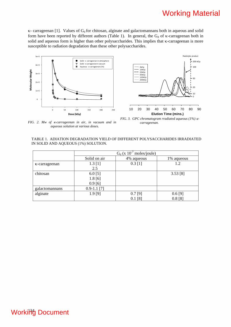
κ- carrageenan [1]. Values of Gd for chitosan, alginate and galactomannans both in aqueous and solid form have been reported by different authors (Table 1). In general, the Gd of κ-carrageenan both in solid and aqueous form is higher than other polysaccharides. This implies that κ-carrageenan is more susceptible to radiation degradation than these other polysaccharides.
FIG. 2. Mw of κ-carrageenan in air, in vacuum and in
aqueous solution at various doses.
FIG. 3. GPC chromatogram rradiated aqueous (1%) κ-
carrageenan.
10 20 30 40 50 60 70 80 90
0kGy 10kGy20kGy50kGy100kGy200kGy
Radiolytic product
Elution Time (mins.)
0kGy 10kGy20kGy50kGy100kGy200kGy
0kGy 10kGy20kGy50kGy100kGy200kGy
010
20
50
100
200 kGy
Dose (kGy)
0 50 100 150 200 250
Molecular Weight
0
1e+5
2e+5
3e+5
4e+5
5e+5
Solid κ‐ carrageenan in atmosphere
Soild κ‐carrageenan in vacuumAqueous κ‐carrageenan (1%)
TABLE 1. ADIATION DEGRADATION YIELD OF DIFFERENT POLYSACCHARIDES IRRADIATED IN SOLID AND AQUEOUS (1%) SOLUTION.
Gd (x 10-7 moles/joule)
Solid on air 4% aqueous 1% aqueous κ-carrageenan 1.3 [1]
2.5 0.3 [1] 1.2
chitosan 6.0 [5]
1.8 [6]
0.9 [6]
3.53 [8]
galactomannans 0.9-1.1 [7] alginate 1.9 [9] 0.7 [9]
0.1 [8] 0.6 [9] 0.8 [8]
134
Working Material
Working Document
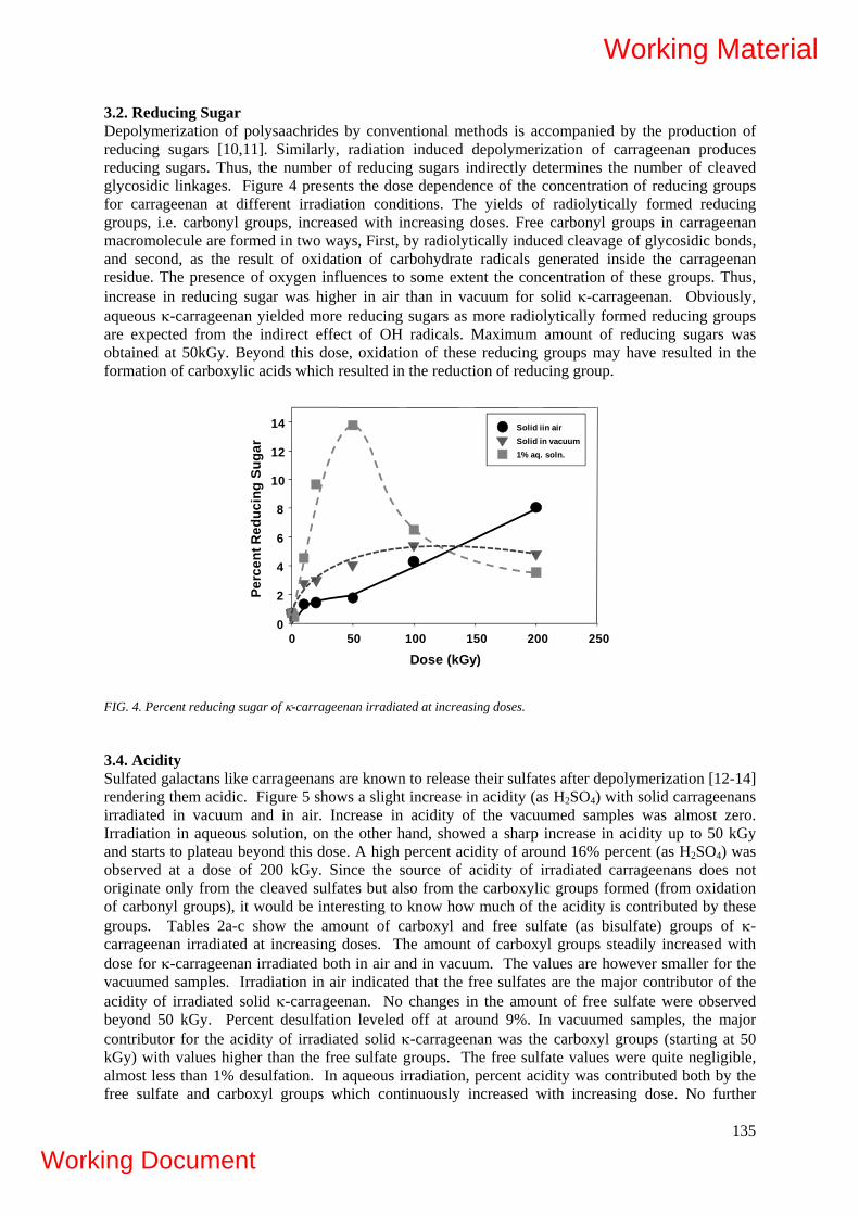
3.2. Reducing Sugar Depolymerization of polysaachrides by conventional methods is accompanied by the production of reducing sugars [10,11]. Similarly, radiation induced depolymerization of carrageenan produces reducing sugars. Thus, the number of reducing sugars indirectly determines the number of cleaved glycosidic linkages. Figure 4 presents the dose dependence of the concentration of reducing groups for carrageenan at different irradiation conditions. The yields of radiolytically formed reducing groups, i.e. carbonyl groups, increased with increasing doses. Free carbonyl groups in carrageenan macromolecule are formed in two ways, First, by radiolytically induced cleavage of glycosidic bonds, and second, as the result of oxidation of carbohydrate radicals generated inside the carrageenan residue. The presence of oxygen influences to some extent the concentration of these groups. Thus, increase in reducing sugar was higher in air than in vacuum for solid κ-carrageenan. Obviously, aqueous κ-carrageenan yielded more reducing sugars as more radiolytically formed reducing groups are expected from the indirect effect of OH radicals. Maximum amount of reducing sugars was obtained at 50kGy. Beyond this dose, oxidation of these reducing groups may have resulted in the formation of carboxylic acids which resulted in the reduction of reducing group.
Dose (kGy)0 50 100 150 200 250
Perc
ent R
educ
ing
Suga
r
0
2
4
6
8
10
12
14 Solid iin airSolid in vacuum1% aq. soln.
FIG. 4. Percent reducing sugar of κ-carrageenan irradiated at increasing doses.
3.4. Acidity Sulfated galactans like carrageenans are known to release their sulfates after depolymerization [12-14] rendering them acidic. Figure 5 shows a slight increase in acidity (as H2SO4) with solid carrageenans irradiated in vacuum and in air. Increase in acidity of the vacuumed samples was almost zero. Irradiation in aqueous solution, on the other hand, showed a sharp increase in acidity up to 50 kGy and starts to plateau beyond this dose. A high percent acidity of around 16% percent (as H2SO4) was observed at a dose of 200 kGy. Since the source of acidity of irradiated carrageenans does not originate only from the cleaved sulfates but also from the carboxylic groups formed (from oxidation of carbonyl groups), it would be interesting to know how much of the acidity is contributed by these groups. Tables 2a-c show the amount of carboxyl and free sulfate (as bisulfate) groups of κ-carrageenan irradiated at increasing doses. The amount of carboxyl groups steadily increased with dose for κ-carrageenan irradiated both in air and in vacuum. The values are however smaller for the vacuumed samples. Irradiation in air indicated that the free sulfates are the major contributor of the acidity of irradiated solid κ-carrageenan. No changes in the amount of free sulfate were observed beyond 50 kGy. Percent desulfation leveled off at around 9%. In vacuumed samples, the major contributor for the acidity of irradiated solid κ-carrageenan was the carboxyl groups (starting at 50 kGy) with values higher than the free sulfate groups. The free sulfate values were quite negligible, almost less than 1% desulfation. In aqueous irradiation, percent acidity was contributed both by the free sulfate and carboxyl groups which continuously increased with increasing dose. No further
135
Working Material
Working Document

release in sulfate was observed beyond 100 kGy. Percent desulfation at this dose was quite high which reached a value of around 40%. A similar study using 5% sodium type κ-carrageenan shows desulfation to cease beyond a radiation dose of 10 kGy. A maximum of 10% desulfation is observed [1].
0 50 100 150 2000
2
4
6
8
10
12
14
16In AirIn VacuumIn 1% Aqueous Solution
Dose (kGy)
% A
cidi
ty (a
s H
2SO
4)
TABLE 2A. ACIDIC COMPOSITION OF GAMMA IRRADIATED SOLID κ-CARRAGEENAN (IN AIR) WITH DOSE.
Radiation Dose (kGy)
% Acidity (as H2SO4)
%COOH Free Sulfates % HSO3
- % Desulfation
2 0.46 0.01 0.41 1.95 10 1.96 0.40 1.39 6.62 20 5.52 3.35 1.72 8.20 50 8.94 4.44 3.77 17.97
100 11.78 4.67 8.23 39.22 200 15.59 5.67 8.64 41.18
TABLE 2B. ACIDIC COMPOSITION OF GAMMA IRRADIATED SOLID κ-CARRAGEENAN (IN VACUUM) WITH DOSE.
Radiation Dose (kGy)
% Acidity (as H2SO4)
%COOH Free Sulfates % HSO3
- % Desulfation
10 0.07 0.02 0.05 0.24 20 0.14 0.06 0.07 0.33 50 0.17 0.15 0.01 0.05
100 0.21 0.19 0.01 0.05 200 0.33 0.27 0.04 0.19
FIG. 5. Percent Acidity of κ-carrageenan as a function of radiation dose at different conditions.
136
Working Material
Working Document

TABLE 2C. ACIDIC COMPOSITION OF GAMMA IRRADIATED κ-CARRAGEENAN (1% AQUEOUS SOLUTION) WITH DOSE.
Radiation Dose (kGy)
% Acidity (as H2SO4)
%COOH Free Sulfates (as % HSO3
-) % Desulfation
10 0.52 0.00 0.48 2.29 20 0.97 0.04 0.85 0.19 50 2.07 0.08 1.81 8.63
100 2.49 0.17 2.11 10.06 200 3.15 1.01 1.88 8.96
3.5. UV-Vis Spectrum It is already known that degradation of polysaccharides leads to some chemical changes such as the formation of carbonyl, carboxyl or double bonds [9, 15-16]. Likewise, it has also been reported that UV spectra of carrageenan show a new absorbance peak at around 260nm. This is attributed to the formation of carbonyl groups or double bond in the pyranose ring [1]. The current result also shows new absorbance peak at 260nm for κ-carrageenan with increasing peak intensities at higher doses. A plot of the increase in absorbance at 260 nm with radiation dose is shown in Figure 6. Irradiation in solid gave a linear curve. The slope of the curve was slightly higher in air than in vacuum (see inset). The reaction of peroxy radicals from O2 could have generated more carbonyl groups [17-19]. Increase in absorbance for1% aqueous carrageenan solution was very much higher than in solid. This is expected due to the indirect effect of radical species from the radiolysis of water. For aqueous κ-carrageenan, a decrease in the slope of the curve beyond 100 kGy was observed. Irradiating aqueous κ-carrageenan at a dose of as high as 100 kGy may have resulted in the decomposition of carbonyl/carboxyl peaks to probably CO2, thus, the decrease in the slope beyond 100 kGy. 3.6. FT-IR Spectrum FT-IR spectra in Figure 7 show the finger print functional groupings of κ-carrageenan [20] and some changes with irradiation. The spectra of κ-carrageenan irradiated in solid at atmospheric condition and in vacuum indicate that the functional groupings of carrageenan were entirely kept intact even at a high dose of 200 kGy (in air) and 100 kGy (in vacuum). Appearance of new absorption band at 1728 cm-1 was seen. This peak corresponds well to the
137
Working Material
Working Document

0 50 100 150 2000.0
0.1
0.2
0.3
0.4
0.5
A‐A
o(260nm
)
Absorbed Dose (kGy)
0 50 100 150 2000.000
0.005
0.010
0.015
0.020
0.025
0.030
1% aqueous in air
Solid in air
Solid in vacuum
FIG. 6. UV absorbance (260 nm) of irradiated κ-carrageenan at different conditions.
0kGy
200kGy
2 kGy
10 kGy
100 kGy
200kGy
100kGy
100kGy
600800100012001400160018002000
Wavenumber (cm‐1)
Relative Absorba
nce
Solid Powder in air 1% aq. soln. Solid Powder in vacuum
Carbon
yl
Polymer Bou
ndWater
Methylene
Group
CO, COH
Covalent
Sulfate
Galactose
Pyeano
sering
Glycosidic
linkage
3,6 anhydro
bridge
D‐galactose
4‐sulfate
FIG. 7. FT-IR spectra of κ-carrageenan irradiated in air, in vacuum and in 1% aqueous solution at different doses.
138
Working Material
Working Document
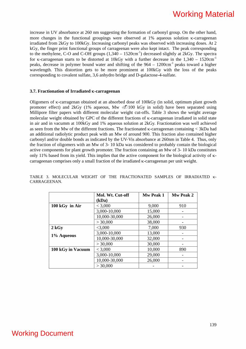
increase in UV absorbance at 260 nm suggesting the formation of carbonyl group. On the other hand, more changes in the functional groupings were observed at 1% aqueous solution κ-carrageenan irradiated from 2kGy to 100kGy. Increasing carbonyl peaks was observed with increasing doses. At 2 kGy, the finger print functional groups of carrageenan were also kept intact. The peak corresponding to the methylene, C-O and C-OH groups (1,340 – 1520cm-1) decreased slightly at 2kGy. The spectra for κ-carrageenan starts to be distorted at 10kGy with a further decrease in the 1,340 – 1520cm-1 peaks, decrease in polymer bound water and shifting of the 964 – 1200cm-1 peaks toward a higher wavelength. This distortion gets to be more prominent at 100kGy with the loss of the peaks corresponding to covalent sulfate, 3,6 anhydro bridge and D-galactose-4-sulfate. 3.7. Fractionation of Irradiated κ-carrageenan Oligomers of κ-carragenan obtained at an absorbed dose of 100kGy (in solid, optimum plant growth promoter effect) and 2kGy (1% aqueous, Mw 100 kGy in solid) have been separated using Millipore filter papers with different molecular weight cut-offs. Table 3 shows the weight average molecular weight obtained by GPC of the different fractions of κ-carrageenan irradiated in solid state in air and in vacumm at 100kGy and 1% aqueous solution at 2kGy. Fractionation was well achieved as seen from the Mw of the different fractions. The fractionated κ-carrageenan containing < 3kDa had an additional radiolytic product peak with an Mw of around 900. This fraction also contained higher carbonyl and/or double bonds as indicated by the UV-Vis absorbance at 260nm in Table 4. Thus, only the fraction of oligomers with an Mw of 3- 10 kDa was considered to probably contain the biological active components for plant growth promoter. The fraction containing an Mw of 3- 10 kDa constitutes only 11% based from its yield. This implies that the active component for the biological activity of κ-carrageenan comprises only a small fraction of the irradiated κ-carrageenan per unit weight. TABLE 3. MOLECULAR WEIGHT OF THE FRACTIONATED SAMPLES OF IRRADIATED κ-CARRAGEENAN.
Mol. Wt. Cut-off (kDa)
Mw Peak 1 Mw Peak 2
< 3,000 9,000 910 3,000-10,000 15,000 - 10,000-30,000 26,000 -
100 kGy in Air
> 30,000 38,000 - <3,000 7,000 930 3,000-10,000 13,000 - 10,000-30,000 32,000 -
2 kGy
1% Aqueous
> 30,000 30,000 - < 3,000 10,000 890 3,000-10,000 29,000 - 10,000-30,000 26,000 -
100 kGy in Vacuum
> 30,000 - -
139
Working Material
Working Document

TABLE 4. UV-VIS (λ = 260NM) OF FRACTIONATED IRRADIATED κ-CARRAGEENAN (0.025%).
Mol. Wt. Cut-off Fraction
Solid (100 kGy) 1% Aqueous (2 kGy)
Vacuum (100 kGy) 0.1%
> 30,000 - - - 10,000-30,000 0.005 0.006 0.047 3,000-10,000 0.004 0.028 0.051
<3,000 0.025 0.023 0.118
3.8. NMR of Fractionated Irradiated κ-carrageenan (Mwt Cut-off of 3-10kDa)
NMR spectroscopy gives valuable information about polysaccharide structures especially if the polysaccharide molecules are built of identical or related oligomeric blocks such as the case of carrageenans. It is quite helpful in the primary structure elucidation. Several data are now available for the NMR analysis of κ-carrageenan samples using both 1H and 13C NMR spectra [21-41]. It is known that the optimum plant growth promoter effect of κ-carrageenan is observed in oligomers with an Mw < 10kDa. Thus, it would be interesting to determine the structure of fractionated irradiated κ-carrageenan with a molecular weight cut-off of 3 to10kDa. NMR spectroscopy was performed on this specific fraction. Interpretation of the NMR spectra was made by comparing it with available model compounds such as neocarrabiose (DP-1) and hexacarrabiose (DP-3). 13C and 1H-NMR spectra of the fractionated oligomers from irradiated κ-carrageenan are shown in Figure 8. The 1H-NMR indicated a simple spectra with six 12 different protons. This may correspond to the protons of the 12 different carbons of the dimer unit of κ-carrageenan. The peaks in the spectra were rather broad which probably contains some overlapping unresolved peaks. This clearly indicates that the oligomers obtained were still large enough for the details of the proton to be seen by NMR. The spectra of these oligomers resemble more the spectra of a κ-carrageenan polymer rather than that of the neocarrabiose oligosaccharides. Based from the obtained MW from GPC experiments, these oligomers would have approximately a DP of 39. The proton NMR chemical shift data are shown in Table 5. The obtained δ were quite close to reference values taken from κ-carrageenan oligomers. 13C NMR spectra of radiation processed κ-carrageenan oligomer indicated only 11 different types of Carbons. Similar spectra are observed in the native κ-carrageenan polymer from references. Like the 1H-NMR, the 13C δ data of κ-carrageenan were quite close to reference values taken from the polymer and oligomer with DP4 (Table 6). Based on the 13C and 1H NMR spectra, the κ-carrageenan oligomers obtained from the polymer irradiated in solid in air and in vacuum and at 1% aqueous solution indicated still an intact repeating unit of the dimer with no changes in the chemical structure of the galacto-pyranose ring. This result could present further explanation on the optimum plant growth promoter effect found at 100kGy for solid irradiation.
140
Working Material
Working Document
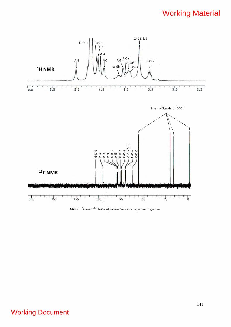
G4S
‐1A‐1
A‐3
A‐4
G4S
‐3A‐5
G4S
‐4A‐2 & A‐6
G4S
‐5
G4S
‐2G4S
‐6
Internal Standard (DDS)
1H NMR
G4S‐1A‐5
A‐4
G4S‐2
G4S‐3
G4S‐5 & 6
A‐1 A‐2A‐3A‐6a
A‐6a*A‐6b
D2O
13C NMR
FIG. 8. 1H and 13C NMR of irradiated κ-carrageenan oligomers.
141
Working Material
Working Document

TABLE 5. CHEMICAL SHIFTS OF THE PROTON NMR OF IRRADIATED κ-CARRAGEENAN OLIGOMERS.
Solid Vacuum Aqueous Reference Polymer
[39]
Reference LMW [25]
G-1 4.57 4.58 4.57 4.75 4.60 G-2 3.50 3.50 3.50 3.74 3.55 G-3 3.90 3.91 3.90 4.10 3.97 G-4 4.83 4.95 4.81 G-5 3.71 3.72 3.71 3.93 3.76 G-6 3.71 3.72 3.71 3.93 3.76 A-1 5.01 5.01 5.01 5.24 5.06 A-2 4.04 4.04 4.05 4.12 4.10 A-3 4.43 4.44 4.44 4.30 4.48 A-4 4.51 4.51 4.51 4.75 4.60 A-5 4.56 4.56 4.56 4.65 4.56 A-6a 3.98 3.98 3.98 4.15 4.01 A-6b 4.13 4.13 4.13 4.25 4.18 A-6a nr 3.94 3.94 3.94
TABLE 6. CHEMICAL SHIFTS OF THE C-13 NMR OF IRRADIATED κ-CARRAGEENAN OLIGOMERS.
Solid Vacuum Aqueous Reference (Polymer)
[22]
Reference (DP4) [22]
G-1 102.87 102.89 102.88 102.5 102.2 G-2 69.81 69.82 69.81 69.9 69.6 G-3 78.51 78.51 78.50 78.8 78.7 G-4 74.12 74.13 74.11 74.0 73.8 G-5 75.18 75.18 75.17 74.8 74.5 G-6 61.68 61.68 61.67 61.3 61.0 A-1 95.08 95.09 95.08 95.1 94.9 A-2 70.01 70.01 70.01 69.7 69.8 A-3 79.60 79.61 79.60 78.3 78.9 A-4 78.87 78.86 78.85 79.1 78.0 A-5 77.08 77.05 77.06 76.8 76.5 A-6 70.01 70.01 70.01 69.4 69.6
142
Working Material
Working Document

4. CONCLUSION The radiation degradation yields of κ-carrageenan in solid and in aqueous (1%) form at different conditions were found to be as follows: 2.5, 1.7 and 1.2 x 10⁻⁷ mol J-1 for solid in atmosphere, solid in vacuum and at 1% aqueous solution, respectively. Based on the Gd, 1% aqueous κ-carrageenan may be irradiated at a dose of as low as 2 kGy for optimum plant growth promoter effect. Chemical structure of irradiated κ-carrageenan at different doses (1% aqueous and solid in air and in vacuum) was investigated using several methods (UV-Vis / FT-IR / NMR spectroscopy / reducing sugar analysis / sulfate and carboxylic acid analysis). Like most other polysaccharides, increase in carbonyl groups, reducing sugar and acidity (free sulfates and carboxylic groups) with increasing doses were the observed radiolytic effects in κ-carrageenan. For solid irradiation, its chemical structure backbone was still kept intact even at a high dose of 200kGy. On the other hand, aqueous (1%) irradiation resulted in the destruction of the polymer backbone starting at a dose of 10kGy. The integrity of the chemical structure of κ-carrageenan irradiated in 1% aqueous solution at 2 kGy and solid (in vacuum and air) at 100 kGy (known to have optimum biological activity) has been proven by NMR. While irradiated κ-carrageenan may have some changes in functional groupings (from the chemical and spectral analyses), these groups may only be present as a small fraction of the entire molecular weight distribution. Most probably this is present in the fraction with an Mw < 3kDa as indicated by its carbonyl peak at 260nm of the UV-Vis spectra.
REFERENCES [1] RELLEVE, L., NAGASAWA, N., LUAN, L.Q., YAGI, T., ARANILLA, C., ABAD, L.,
KUME, T., YOSHII, F., DELA ROSA, A., Polym. Degrad. Stabil. 87 (2005) 403. [2] RELLEVE, L., ABAD, L., ARANILLA, C.A., ALIGANGA, A., DE LA ROSA, F., YOSHII,
KUME, T., NAGASAWA, N., “ Biological Activities of Radiation-Degraded Carrageenan” Proceedings of the Symposium on Radiation Technology in Emerging Industrial Applications, Beijing, People’s Republic of China, 6-10, November (2000).
[3] ABAD, L., NASIMOVA, I., RELLEVE, L., ARANILLA, C., DE LA ROSA, A., SHIBAYAMA, M., Int. J. Biol. Macromol. 34 (2004) 81.
[4] HODGE, J., HOFREITER, B., B.T. IN WHISTLER, R.L. & WOLFROM, M.L.(Eds.) Methods in Carbohydrate Chemistry, Academic Press: New York. Vol. 1 (1962) 380-394.
[5] CZECHOWSKA-BISKUP, ROKITA, R.B., ULANSKI, B., ROSIAK, P., J. Nucl. Instr. and Meth. in Phys. Res. B. 236 (2005) 383.
[6] HAI, L., DIEP, T.B., NAGASAWA, N., YOSHII, F., KUME,T., Nucl. Instr. and Meth. in Phys. Res. B., 208 (2003) 466.
[7] SEN, M., YOLACAN, B., GUVEN, O., Nucl. Instr. and Meth. in Phys. Res. B., 265 (2007) 429.
[8] WASIKIEWICZA, J., YOSHII, F., NAGASAWA, N., WACH, R., MITOMO, H.,Radiat. Phy. Chem., 73 (2005) 287.
[9] NAGASAWA, N., MITOMO, H., YOSHII, F., KUME, T., Polym. Degrad. Stab. 69 (2000) 279.
[10] T. HJERDE, O. SMIDSRØD, B. Christenesen, Carbohydr Res. 288 (1996) 175. [11] C. RIGOUIN, C. LADRAT, C. SINQUIN, S. COLLIEC-JOUAULT, M. DION, Carbohy.
Polym. 76 (2009) 279. [12] KARLSSON, A., SINGH, S., Carbohydr. Polym. 38 (1999) 7. [13] NAVARRO, D., FLORES, M., STORTZ, C., Carbohydr. Polym. 69 (2007) 742. [14] MILLER, I., BLUNT, J., Carbohydr. Res. 309 (1998) 39. [15] ULANSKI, P., ROSIAK, J., Radiat. Phy. Chem. 39 (1992) 53. [16] ZEGOTA, H., Food Hydrocolloids, 13 (1999) 51. [17] DZIĘDZIELA, W., KOTYŃSKA, D., Radiat. Phys. Chem. 23 (1984) 723. [18] AKHLAQ, M., BAGHDADI, S., VON SONNTAG, C., Carbohydr. Res.164 (1987) 71. [19] VON SONNTAG, C., BOTHE, E., ULANSKI, P., DEEBLE, J., Radiat. Phys. Chem. 46
1995) 527. [20] MATSUHIRO, B., Hydrobiologia. 326 (1996) 481.
143
Working Material
Working Document
144
[21] JIANG,Y., GUO, X., TIAN, X., Carbohydr. Polym. 61 (2005) 399. [22] ROCHAS, C., RINAUDO, M., VINCENDON, M., Int. J. Biol. Macromol. 5 (1983) 111. [23] KOLENDER, A., MATULEWICZ, M., Carbohydr. Res. 337 (2002) 57. [24] KNUTSEN, S., SLETMOEN, M., KRISTENSEN, T., BARBEYRON, T., KLOAREG, B.,
POTIN, P., Carbohydr. Res. 331 (2001) 101. 72. [25] JIANG, Y., GUO, X., Carbohydr. Polym. 61 (2005) 441. [27] VELDE, F., PEPPELMAN, H., ROLLEMA, H., R. Tromp, Carbohydr. Res. 331 (2001) 271. [28] USOV, A., Food Hydrocolloid. 12 (1998) 301. [29] USOV, A., YAROTSKY, S., SHASKOV, S.A., Biopolymers. 19 (1980) 977. [30] STORTZ, C., BACON, B., CHERNIAK, R., CEREZO, A., Carbohydr. Res. 261 (1994) 317. [31] MURANO, E., TOFFANIN, R., CECERE, E., RIZZO, R., KNUTSEN, S., Mar. Chem. 58
(1997) 319. [32] TOJO, E., PRADO, J.,Carbohydr. Polym. 53 (2003) 325. [33] VAN DE VELDE, F., ANTIPOVA, ROLLEMA, A.H., TATIANA, B., GRINBERG, N.,
PEREIRA, L., GILSENAN, P., TROMP, R., RUDOLPHE, B., GRINBERG, V., Carbohyd. Res. 340 (2005) 1113.
[34] VAN DE VELDE, F., PEREIRA, L., ROLLEMA, H., Carbohydr. Res. 339 (2004) 2309. [35] VAN DE VELDE, F., S., KNUTSEN, A., USOV, H., ROLLEMAY, A., CEREZO, Trends
Food. Sci. Tech. 13 (2002) 73. [36] CIANCIA, M., MATULEWICZ, M., FINCH, P., CEREZO, A., Carbohydrate Research, 238
(1993) 241. [37] KNUTSEN, S., GRASDALEN, H., Carbohydr. Res., 229 (1992) 233. [38] KNUTSEN, S., GRASDALEN, H., Carbohydr. Polym. 19 (1992) 199. [39] TURQUOIS, T., ACQUISTAPACE, S., ARCE VERA, F., WELTI, D., Carhohydr. Polym. 31
(1996) 269. [40] CAMPO, V., KAWANO, D., DA SILVA JR., D., CARVALHO, I., Carbohydr. Polym. 77
(2009) 167. [41] KOLENDER, A., MATULEWICZ, M., Carbohydr. Res. 339 (2004) 1619. [42] FALSHAW, R., FURNEAUX, R., WONG, H., Carbohydr. Res. 338 (2003) 1403.
Working Material
Working Document

RADIATION CHEMISTRY AND PHYSICAL CHEMISTRY OF CHITOSAN AND OTHER POLYSACCHARIDES. FUNDAMENTAL STUDIES AND PRACTICAL APPLICATIONS
Janusz M. Rosiak*, Renata Czechowska-Biskup, Bożena Rokita, Alicja K. Olejnik, Piotr Ulański Institute of Applied Radiation Chemistry, Technical University of Lodz, Wroblewskiego 15, 93-590 Lodz, Poland
Abstract
This report summarizes the second year of activities performed at the Institute of Applied Radiation Chemistry (IARC) within the framework of the CRP project. It consists of two parts. Part I is a brief account of the activities related to design, tests, sample procurement and characterization and formulation of “Protocol for determination of intrinsic viscosity of chitosan” designed to be the basis of the interlaboratory study on viscometric determination of chitosan molecular weight as well as on radiation degradation of chitosan in controlled conditions. Part II contains the text of the Protocol, and is given in the Annex. PART I. ACTIVITIES RELATED TO DESIGN, TESTS, SAMPLE PROCUREMENT AND CHARACTERIZATION AND FORMULATION OF “PROTOCOL FOR DETERMINATION OF INTRINSIC VISCOSITY OF CHITOSAN” 1. INTRODUCTION Any chemically distinct kind of a polysaccharide, such as chitosan, starch, cellulose, etc, may occur in a form of chains of various length, or various molecular weight, depending on the origin, processing and storage conditions. It has been demonstrated in numerous studies, that many properties of polysaccharides and in particular their suitability for particular applications do depend on the average molecular weight. Therefore this parameter is one of the most important characteristics of any polysaccharide material. Ability to determine average molecular weight is also of paramount importance when elaborating, testing and applying modification techniques, as the radiation technique, aimed at changing the average molecular weight of a polysaccharide to adjust it to the range required for a particular application. Precise determination of molecular weight of any polymer is a complex task; it becomes even more complex for biomolecules being at the same time charged macromolecules (polyelectrolytes), as chitosan. Well equipped research laboratories use sophisticated techniques and costly equipment to reach that goal; often not only an average molecular weight but molecular weight distributions are determined. Such studies require the use of multi-angle laser light scattering, gel permeation chromatography (preferentially with triple detector systems), asymmetric flow field fractionation or matrix-assisted laser desorption/ionization techniques. In routine work on polysaccharides or in research not aimed specifically at detailed molecular weight analysis but on, e.g., effects of radiation on polysaccharides, such methods are not always necessary; moreover, for many labs they are not affordable. A reasonable solution in such cases is to use the popular and less costly viscometry. While in general viscometric determination of molecular weight is a routine technique (when care is taken to follow the general rules and conditions regarding capillary selection, temperature stability, etc.), a number of issues has to be taken into account when using this method for measurements of molecular weights of charged biomolecules as chitosan. As typical one-component solvents as pure water are not suitable, multi-component solvents must * Head of Applied Radiation Chemistry Division, ph. (0048) 42 631 3196, fax (0048) 42 684 0043, e-mail [email protected], http://mitr.p.lodz.pl/biomat
145
Working Material
Working Document
be applied. The optimum choice of the components and their fractions in the mixture is not straightforward, which results in several tens of solvents for chitosan listed in the literature. For each solvent intended to be used for molecular weight determination, a set of Mark-Houwink parameters is needed. Literature values of the latter are determined by various methods and with various accuracies. Moreover, since chitosan is a copolymer of glucosamine and N-acetylglucosamine units, varying the proportions between them (i.e., varying the deacetylation degree, DD) results in changing physicochemical properties, including solubility and solution behaviour, as well as the values of Mark-Houwink parameters. This may lead to confusion or at least lack of confidence on whether the results reported by various labs, obtained using various solvents, different sets of Mark-Houwink parameters, samples of various DA, are really comparable. In order to harmonize the efforts being currently undertaken at many research and industrial labs worldwide to broaden our understanding of radiation effects on chitosan and to apply radiation treatment for controlled modification of this polysaccharide, where determination of molecular weight and its changes is very important, the partners co-operating within the current CRP set to perform an interlaboratory study on that issue.
2. THE IDEA OF INTERLABORATORY STUDY
The interlaboratory study was intended to be performed by each laboratory participating in the CRP which agreed to take part. The idea was to let all these labs to perform viscosity measurements on identical chitosan samples using the same solvents and conditions, according to a uniform protocol. The latter included also procedure for data handling and analysis. Furthermore, the labs were supposed to irradiate samples of identical chitosan under prescribed conditions, and analyze the resulting intrinsic viscosities and molecular weights. The main aim of this study is to check if analyses performed on identical samples and according to standardized protocol lead to consistent results, first of all in the terms of viscometry results, but also molecular weight vs. dose dependence. A positive outcome would increase confidence of the participating labs in the field of chitosan analysis and encourage the laboratories to exchange results and undertake common research. The results would also help to indentify sources of discrepancies, if any, and suggest appropriate corrective measures. Results will also help participating laboratories in performing other research tasks planned in the framework of their participation in the CRP. The idea of interlaboratory study as well as the procedure was elaborated in co-operation with prof. Saphwan Al-Assaf (Phillips Hydrocolloids Centre, Glyndwr University, UK)
3. CHOICE OF CHITOSAN
Chitosans available both as chemicals and as bulk-quantity raw materials vary in origin, purity, degree of deacetylation (DD) and molecular weight characteristics. Impurities typically involve proteins and insoluble solids. For obvious reasons, high-purity samples were required for the planned tests. Chitosan of high DD is typically used in most applications, mainly due to its high solubility and high content of ionizable free amine groups. Also most literature data, including information on solvent choice, Mark-Houwink constants etc. refers to high-DD samples. It was concluded that a value in the range 85 – 95 % would be suitable for the tests. Most chitosans are obtained by alkaline deacetylation of chitin, a process that involves also hydrolysis leading to a decrease in molecular weight. The typical values for the product, being the most common kind of chitosan on the market, are between 200 kDa and 600 kDa. While samples of other molecular weight are also available, it was considered that a sample of molecular weight being in the typical, above mentioned range would suit our purpose best,
146
Working Material
Working Document

being most common, allowing for precise molecular weight determination by viscometry, and providing the possibility to determine this parameter even after degradation caused by irradiation. We have approached the Heppe Medical Chitosan GmbH company (Halle, Germany) and asked for supplying us with chitosan sample of size, quality and characteristics suitable for the Project. The company agreed to provide such sample free of charge and expressed general interest in the Project. Characteristics of the provided sample, according to the certification of analysis provided by the Heppe company, is the following.
147
Working Material
Working Document
Sample was thoroughly mixed, and divided into portions for delivery to participating labs. Each portion (ca. 20 g) was sealed in two layers of PE foil and expedited via courier mail with a copy of certification of analysis, safety sheet and the protocol. Some labs which indicated also other type of measurements (GPC+FFF, LSI), were sent larger samples (ca. 50 g each).
4. CHOICE OF SOLVENT
It was decided to ask the participating laboratories to use two solvents for viscometric analyses on chitosan. The first solvent was selected using two basic criteria: literature indications to be probably the best (most recommended) typical solvent for determining physicochemical properties of chitosan and availability of Mark-Houwink constants for chitosan in the selected solvent, related to the value of deacetylation degree. After careful consideration and analysis of numerous recent publications, the first solvent was selected as 0.2 M acetic acid / 0.15 M ammonium acetate at 25.0 ºC. The Mark-Houwink parameters for chitosan of DD = 91.8 % have been reported as K = 9.66 × 10⁻⁵ dm3 / g, α = 0.742 . The solvent described above is a typical three-component solvent for chitosan. Water is the main dissolving medium, acid provides protonation of the amine groups of chitosan and thus allows for molecular dissolution of the polysaccharide and salt partly screens the strong electrostatic repulsive interactions between chain segments, allowing the chains to attain coiled conformation, desirable for most methods of chitosan characterization. However, preparation of three-component solvent is time consuming and the accuracy of concentration setting for each component is limited thus leading to accumulation of errors. It should be mentioned as well that even more complex four-component solvents have been tested as well. A new alternative is to use a simpler, two-component system, which can be actually purchased as ready solution from the major vendors of lab chemicals. Such systems, being tested in our lab, consist of water and strong acid. Concentration of the latter, definitely higher than stoichiometric for typical chitosan concentrations used in analysis (being in the range of 10 mM), serves two purposes: to fully protonate the amine groups of chitosan while the excess of acid over the stoichiometric concentration provides similar effect as salt in three component solvents – partial screening of charges on the chain and, in consequence, a coil-like conformation of macromolecules. Careful study of this issue allowed to propose 0.1 M HCl at 30.0 ºC. The Mark-Houwink parameters for chitosan in this newly proposed solvent are not yet known, and therefore the results obtained in the second solvent cannot be directly re-calculated into average molecular weights. Parallel study in two solvents on non-irradiated and radiation-degraded samples shall allow us to determine the Mark-Houwink parameters in the novel solvent.
* * *
Chitosan samples intended for the harmonization tests have been sent to 14 participants. Up to the time of the RCM held in Reims, 9 of these labs have sent their data. Seven partners performed their experiments in both recommended solvents, while other two limited the scope of measurements to the acetate solvent. In all cases but one the partners declared that they had adhered strictly to the provided protocol (albeit minor deviations could be noticed, mainly in the data recording and presentation); in one case a different type of viscometer (based on different measurement principle) was used, and this was most probably the cause of marked deviation, especially for the irradiated samples, therefore these data haven’t been taken into consideration. Preliminary results based on reports from 8 labs for the acetate solvent, in terms of intrinsic viscosity and viscosity-average molecular weight, are shown in Figs. 1 and 2, respectively. Standard deviation of the data obtained in individual labs amounts to 7.0 % of the mean value, which is considered as fully acceptable, taking into account complexity of the material and composite nature of the solvent. Figs. 3 and 4 illustrate the viscosity and molecular weight data obtained for samples which have been degraded by various doses of ionizing radiation. These data can be re-calculated into the radiation-chemical yields of chain scission, Gs. Values of the latter parameter obtained basing on the data from
148
Working Material
Working Document

the participating labs are shown in Fig. 5. Here the standard deviation is 16.6 % of the mean, i.e., somewhat higher than in the results for non-irradiated sample (as have been expected because of additional uncertainties resulting from irradiation and dosimetric procedures must be considered), but still the agreement is satisfactory. Since further sets of data are being expected to be sent by other laboratories, the above-presented analysis should be considered as preliminary. The final results, analysis, outcomes and conclusions will be included in the next report.
Algeria Japan MalaysiaPhilippines Poland Thailand Turkey U.K.0,0
0,2
0,4
0,6
0,8
1,0
1,2
1,4
1,6
IV /
dm3 g
-1
Country
1.2871.273
SD = 0.0891 (7.00 %)
95% Confidence Interval of the Mean = +- 0.0745 = +- 5.85 %
FIG. 1. Intrinsic viscosities of the non-irradiated chitosan sample in the acetate solvent as determined at eight of the participating laboratories.
149
Working Material
Working Document

Algeria Japan MalaysiaPhilippines Poland Thailand Turkey U.K.0
100
200
300
400M
v / k
Da
Country
357.0362.1
SD = 33.6 (9.41 %)
95% Confidence Interval of the Mean = +- 30.27 = +- 8.48 %
FIG. 2. Viscosity-average molecular weight of the non-irradiated chitosan sample as determined at eight of the participating laboratories.
0 10 20 300,0
0,5
1,0
1,5
40
IV a
ceta
te /
dm3 g
-1
Dose / kGy
Algeria Japan Malaysia Phillipines Poland Thailand Turkey
FIG. 3. Intrinsic viscosities of chitosan sample in acetate solvent as a function of irradiation dose, determined at eight of the participating laboratories.
150
Working Material
Working Document

0 10 20 300
100
200
300
400
40
Mv
/ kD
a
Dose / kGy
Algeria Japan Malaysia Philippines Poland Thailand Turkey
FIG. 4. Viscosity-average molecular weight of chitosan sample in acetate solvent as a function of irradiation dose, determined at eight of the participating laboratories.
Algeria Japan Malaysia Philippines Poland Thailand Turkey0,0
0,1
0,2
0,3
0,4
0,5
0,6
0,7
0,8
Gs
(um
ol /
J)
Country
0.635
0.628
SD = 0.104 (16.6 %)
95% Confidence Interval of the Mean = +- 0.097 = +- 15.3 %
FIG. 5. Values of the radiation-chemical yields of chain scission, calculated on the basis of data determined at eight of the participating laboratories.
151
Working Material
Working Document

152
Working Material
Working Document

UTILIZATION OF DEGRADED CHITOSAN FOR GROWTH PROMOTER AND BLOSSOM BLIGHT DISEASE CONTROLS IN OKRA Prartana Kewsuwan Thailand Institute of Nuclear Technology and Institute of Research Development Agricultural Production Science Abstract The experiment focus on the comparison of the effect of gamma radiation and chemical reagent on degradation of chitosan and preliminary test of degraded chitosan with different molecular weight as plant growth promoter of okra in potting experiment. 1. BACKGROUND The degraded chitosan1 can be prepared by acidic hydrolysis or enzymatic treatment. Chemical treatment is an easy, low cost process, but chemical waste and reproducibility are the main problems. Enzymatic hydrolysis is an effective way to achieve specific cleavage to chitosan oligomers. However, it requires multi-steps, particularly, enzyme preparation and purification of the product. Radiation can provide a useful tool for degradation of different polymers. In the reaction, no other chemical reagents are introduced and there is not a need to control the temperature, environment or additives. It was reported that low molecular chitosan can improve the biological activities and stimulate plant growth2,3. Therefore, it is interesting to use chitosan to improve the growth of okra because okra is an important export vegetable of Thailand especially to Japan, where demand amounts to 3,000 tons of fresh and frozen okra a year. This work will investigate the method of degradation of chitosan and the potential of degraded chitosan as plant growth promoter in okra in potting experiment. 2. EXPERIMENTAL 2.2. Preparation of degraded chitosan Commercial chitosan from shrimp shell was purchased from local company in Thiland and used without further purification. It was degraded using radiation and chemical method. For radiation degradation, chitosan was irradiated in powder and liquid form in air by gamma ray from 60Co Gammacell-220 at the dose rate of 10 kGy/h. Chitosan powder packaged in polyethylene bags was irradiated at doses of 0 – 200 kGy. For liquid irradiated chitosan, 5% of chitosan in 1% acetic acid was irradiated at dosees of 0 – 50 kGy. For chemical degradation, hydrogen peroxide (H2O2) in concentration from 0.04 – 2 % was used. 2.3. Fractionation of degraded chitosan A stirred ultrafiltration cell (model 8400, Millipore Co.) was employed for fractionation of degraded chitosan. A series of polysulfone ultrafiltration membranes, MW cut-off 10000, 30000, 50000 and 100000 were used. The degraded chitosan was dissolved in 1% acetic acid and then pour in the stirred ultrafiltration cell with different membranes. Each fraction collected was neutralized with 1 N NaOH to precipitate the degraded chitosan. After that, precipitated chitosan was transferred to a Buchner funnel and washed with distilled water until neutral pH 7 was reached. Collected chitosan then was dried at 50 °C in vacuum oven to get different molecular weight chitosan. 2.4. Molecular weight determination by intrinsic viscometric method Five different concentration (~ 0.01 – 1.0 %) solutions of chitosan in 0.1M acetic acid – 0.2 M NaCl) (1:1, v/v) were prepared. The solution was passed through a filter paper (Whatman # 4) to remove insoluble. The relative viscometry, η, of chitosan samples was measured using an ubbelohde capillary viscometer (Cannon-Fenske, No. OC) in a constant-temperature water circulating bath at 25°C. Three measurements were made on each sample. Viscosity average molecular weight of chitosan solutions was calculated based on Marke Houwink equation4,5: [η] = KMa , where K = 1.81 x 10⁻⁵ cm3/g and a = 0.93.
153
Working Material
Working Document

2.5. Molecular weight determination by gel permeation chromatography The molecular weights of degraded chitosans were determined by gel permeation chromatography (Waters 600E) on Ultralinearhydrogel column plus guard column (1000 – 20,000,000) using pullulans standard with molecular weights of 5,900 – 788,000. Standards and chitosans samples were introduced into the column at a flow rate of 0.6 ml min-1 using 0.5 M sodium acetate buffer as eluent. The operation was carried out at 30 °C and the peaks monitored by a refractive index detector. 2.6. Preliminary potting experiment of degraded chitosan as plant growth promoter of okra The effects of the molecular weight and concentration of chitosan of okra growth and production were studied. The growth promotion experiment was carried out under greenhouse conditions. Different molecular weight of degraded chitosan (12 kDa, 44 kDa and 60 kDa) was dissolved in 1% acetic acid in the concentration of 25, 50 and 100 ppm. The plants were sprayed every 2 weeks during 3 months period. Average number of flowers and pods, average length of pods, average of weight of fresh pods, of dry pods, of fresh plants, of dry plant, of plant height were determined. The experiment was carried out in four replicates. 3. RESULTS OBTAINED 3.1. Preparation of degraded chitosan with different molecular weight The irradiated chitosan was separated into 5 molecular weight fractions by using four kinds of ultrafiltration membranes; MWCO 10 kDa, 30 kDa, 50 kDa and 100 kDa. Fig 1 shows the comparision of molecular weight of separated fraction chitosan determined by viscosity method and GPC technique. The molecular weight of chitosan determined by GPC was significantly lower than that determined by intrinsic viscosity. The molecular weight value determined by intrinsic viscosity was used in this experiment.
TABLE 1 COMPARISON OF MOLECULAR WEIGHT OF CHITOSAN OBTAINED THROUGH DIFFERENT ULTRAFILTRATION MEMBRANE PORE SIZE DETERMINED BY INTRINSIC VISCOSITY AND GPC.
Ultrafiltration membrane size
Expected MW (kDa)
MW of chitosan by intrinsic
viscosity (kDa)
Mn of chitosan by GPC (kDa)
< 10 kDa < 10 - 5 30 kDa 10 – 30 12 6 50 kDa 30 – 50 44 9 100 kDa 50 - 100 60 22
> 100 kDa > 100 120 3.2. Effect of radiation dose and hydrogen peroxide on molecular weight of chitosan In general, the intrinsic viscosity of linear macromolecular substances is related to the molecular weight or degree of polymerization. Average molecular weights of radiation-treated solid chitosan, of radiation-treated liquid chitosan and of hydrogen peroxide-treated chitosan were calculated from measured intrinsic viscosity shown in Fig. 1, 2 and 3, respectively. Fig 1 and 2 show that irradiation in both dry powder and liquid of chitosan leads to the reduction of molecular weight. The irradiated degradation of chitosan in liquid stage use lower dose than that of in dry stage. The molecular weight decreased remarkably with increasing dosed up to 150 kGy in solid stage and up to 20 kGy in liquid stage. There was no significant change in molecular weight for higher dose.
154
Working Material
Working Document

0
100
200
300
400
500
600
700
800
900
0 50 100 150 200 250 300 350
D o s e ( k G y )
MW
of
ch
ito
sa
n (
kD
a)
0
100
200
300
400
500
600
700
800
0 0.05 0.1 0.15 0.2 0.25
H 2O 2 c o n c e n t r a t i o n ( M )
MW
of
ch
ito
sa
n (
kD
a)
0
100
200
300
400
500
600
700
800
0 10 20 30 40 50 6
D o s e ( k G y )
MW
of
ch
ito
sa
n (
kD
a)
0
FIG. 1. Degraded chitosan irradiated in solid form.
FIG. 3. Degradation of chitosan by H2O2.
FIG. 2. Degraded chitosan irradiated in liquid form.
Fig 3 shows that molecular weight of chitosan treated with hydrogen peroxide in acetic acid solution decreased as the hydrogen peroxide treatment increased up to 0.1 M. It can be seen that only small concentration of hydrogen peroxide causes rapid decrease of molecular weight. 3.3. Growth promotion test The effects of polymer size and concentration of chitosan on okra growth were studied. Parameters used for determination of plant growth promoter of chitosan were number of flower, number of pods, length of pod, weight of fresh pod, weight of dry pod, weight of fresh pod, weight of dry plant and average plant height. Fig 1 – 3 shows the effects of degraded chitosan (12, 44 and 60 kDa) at different
155
Working Material
Working Document

concentration of 25, 50 and 100 ppm, respectively, on the parameters. It was found that okra treated with degraded chitosan every molecular weights has no significantly affected the number of flowers, of pods, weight of dry pod, of dry plant and average plant height compared to those of the untreated control whereas okra treated with degraded chitosan has potential affected the weight of fresh pod and fresh plant compared to those of the untreated control.
0
200
400
600
800
number offlower
number ofpod
length ofpod
weight offresh pod
weight ofdry pod
weight offresh plant
weight ofdry plant
avergeplant
height
Control Conc. 25 ppm Conc. 50 ppm Conc. 100 ppm Chitosan 12 kDa
FIG. 1. Effect of chitosan concentration on the growth of okra after 90 days. The molecular weight is 12 kDa
0
200
400
600
800
number offlower
number ofpod
length ofpod
weight offresh pod
weight ofdry pod
weight offresh plant
weight ofdry plant
avergeplant
height
Control Conc. 25 ppm Conc. 50 ppm Conc. 100 ppm Chitosan 44 kDa
FIG. 2. Effect of chitosan concentration on the growth of okra after 90 days. The molecular weight is 44 kDa.
0
200
400
600
800
number offlower
number ofpod
length ofpod
weight offresh pod
weight ofdry pod
weight offresh plant
weight ofdry plant
avergeplant
height
Control Conc. 25 ppm Conc. 50 ppm Conc. 100 ppm
Chitosan 60 kDa FIG. 3. Effect of chitosan concentration on the growth of okra after 90 days.
The molecular weight is 60 kDa.
156
Working Material
Working Document
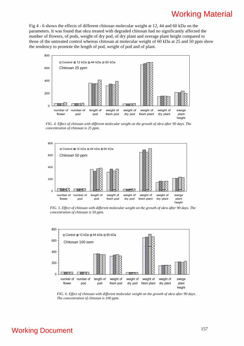
Fig 4 - 6 shows the effects of different chitosan molecular weight at 12, 44 and 60 kDa on the parameters. It was found that okra treated with degraded chitosan had no significantly affected the number of flowers, of pods, weight of dry pod, of dry plant and average plant height compared to those of the untreated control whereas chitosan at molecular weight of 60 kDa at 25 and 50 ppm show the tendency to promote the length of pod, weight of pod and of plant.
0
200
400
600
800
number offlower
number ofpod
length ofpod
weight offresh pod
weight ofdry pod
weight offresh plant
weight ofdry plant
avergeplant
height
Control 12 kDa 44 kDa 60 kDa
Chitosan 25 ppm
FIG. 4. Effect of chitosan with different molecular weight on the growth of okra after 90 days. The concentration of chitosan is 25 ppm.
0
200
400
600
800
number offlower
number ofpod
length ofpod
weight offresh pod
weight ofdry pod
weight offresh plant
weight ofdry plant
avergeplant
height
Control 12 kDa 44 kDa 60 kDa
Chitosan 50 ppm
FIG. 5. Effect of chitosan with different molecular weight on the growth of okra after 90 days. The concentration of chitosan is 50 ppm.
FIG. 6. Effect of chitosan with different molecular weight on the growth of okra after 90 days. The concentration of chitosan is 100 ppm.
Chitosan 100 ppm
0
200
400
600
800
number offlower
number ofpod
length ofpod
weight offresh pod
weight ofdry pod
weight offresh plant
weight ofdry plant
avergeplant
height
Control 12 kDa 44 kDa 60 kDa
157
Working Material
Working Document

158
4. CONCLUSIONS DRAWN Degradation of chitosan can be carried out by gamma radiation in solid powder, in liquid stage and by hydrogen peroxide treatment. Degrades chitosan has no significantly affect on okra growth. Only chitosan at the molecular weight of 60 kDa at 25 and 50 ppm show the tendency to promote the length of pod, weight of pod and of okra. 4.1. Problems The potting experiment was started in June. At that time okra were harvested. So infected okra disease hardly be found. Therefore, antimicrobial property of degraded chitosan is in process. 4.2. Work scheduled
- Study on effect of the combination of hydrogen peroxide and radiation on degradation of chitosan
- Investigation whether native and degraded chitosan with well defined molecular weight affecting on inhibition of growth of okra disease
- Further study of degraded chitosan on plant growth promoter and inhibition of okra disease by potting experiment
REFERENCES
[1] TAO FENG, YUMIN DU, YING HU AND JOHN F. KENNEDY, Enhancemint of antioxidant activity of chitosan by irradiation. Charbohydrate Polymers, (2008) 1-7 (article in press)
[2] LE XUAN THAM, ET.AL. Effect of radiation-degraded chitosan of plants stressed with vanadium. Radiation Physics and Chemistry, 61 (2001) 171 – 175.
[3] KUME, T., NAGASAWA, N. AND YOSHII, F. Utilization of carbohydrates by radiation processing. Radiation Physics and Chemistry, 63 (2002) 625 – 627.
[4] NO, H.K., LEE, S.H., PARK., AND MEYERS, S.P., Comparisonof physicochemical, binding, and antibacterial properties of chitosans prepared without and with deproteinization process. Journal of Agricultural and Food Chemistry, 51 (2003) 7659 – 7663.
[5] A.G., CHMIELEWSKI, ET.AL. Chemical-radiation degradation of natural oligoamino-polysaccharides for agricultural application, 76 (2007) 1840 – 1842.
Working Material
Working Document

RADIATION SYNTHESIS OF SUPERABSORBENT POLYMERS BASED ON NATURAL POLYMERS
Murat Sen, Hande Hayrabolulu Hacettepe University, Department of Chemistry, 06800, Beytepe, Ankara, TURKEY 1. OBJECTIVE OF THE RESEARCH
The objectives of proposed research contract were first synthesize superabsorbent polymers based on natural polymers to be used as disposable diapers and soil conditioning materials in agriculture, horticulture and other super adsorbent using industries. We have planned to use the natural polymers; locust beam gum, tara gum, guar gum and sodium alginate on the preparation of natural superabsorbent polymers(SAP). The aqueous solution of natural polymers and their blends with trace amount of monomer and cross-linking agents will be irradiated in paste like conditions by gamma rays for the preparation of cross-linked superabsorbent systems. The water absorption and deswellling capacity of prepared super adsorbents and retention capacity, absorbency under load, suction power, swelling pressure and pet-rewet properties will be determined. Use of these materials instead of synthetic super absorbents will be examined by comparing the performance of finished products. The experimental studies achieved in the second year of project mainly on the effect of radiation on the chemistry of sodium alginate polymers in different irradiation conditions and structure-property relationship particularly with respect to radiation induced changes on the molecular weight of natural polymers and preliminary studies on the synthesis of natural-synthetic hydride super adsorbent polymers were given in details. 2. INTRODUCTION Hydrophilic polymers when cross-linked chemically or physically forming three dimensional networks swell but do not dissolve in water. They are termed hydrogels when the amount of water retained is between 20-100% of the total weight, and when water content exceeds 100% these hydrogels are called super adsorbent hydrogels. Recently hydrogels have found a wide range of biomedical applications including controlled drug delivery systems, replacement blood vessels, wound dressing, soft tissue substitution, contact lenses and a variety of other related and potential uses. Hydrogels are generally found to be very well tolerated when implanted in vivo and can be easily tailored to suit the many functions of prosthetics in contact with blood or tissues. Hydrogels are typically synthesized by one of the two well established procedures: (a) polymerization and simultaneous or postpolymerization cross-linking of hydrophilic monomers, and (b) modification of hydrophilization of existing polymers with potential hydrogel properties. A comprehensive review of the chemistry and various synthetic approaches used in hydrogel preparation can be found in the compilation of [1]. A more recent review by [2] provides an in depth discussion on the methods of hydrogel synthesis. The inherent advantages of using high energy radiation in the synthesis of hydrogels for biomedical applications have been reviewed by [3]. The preparation of hydrogels by radiation treatment of aqueous solutions of hydrophilic monomers or polymers carries some advantages over the conventional techniques. It does not require initiators, cross-linkers and can be used practically with any vinyl monomer and both polymerization and cross-linking reactions can be initiated at ambient or sub-ambient temperatures. It is very well known that polysaccharides in dry form or in solution degrade when exposed to ionizing radiation [4,5]. The results reported so far on the radiation-induced degradation of polysaccharides indicated that chain scission yield strongly depends on the relative concentration of polymer.
159
Working Material
Working Document

Recently it has been found that when the concentration of polysaccharide is increased beyond viscous solution to paste-like condition, or in the presence of some additives like carbon tetrachloride cross-linking effect starts to dominate and typically chain scissioning polymers turn into cross-linking type[6-7]. In this project we will try to find general conditions for cross-linking of polysaccharide type natural polymers such as locust beam gum(LBG), tara gum(TG), guar gum(GG) obtained from plants sodium alginate from algea. We believe that development of radiation induced cross-linking of polysaccharides will enhance the development of radiation technology for natural polymers and producing of new finished products. World-wide, the demand for single-used disposable non biodegradable synthetic superabsorbent polymers has increased dramatically since 1986 and currently amounts to about 850.000 metric tons per year. The consumption is in the The United States (300.000 metric tons per year), followed by Europe at 250.000 metric tons and Asia and Oceania at 100.000 metric tones. Based on the trend, we expect demand for superabsorbent polymers to reach 1.000.000 metric tons per year within a few years. Use of biodegradable hydrogels based on natural superabsorbent polymers especially as diapers and soil conditioning material will be a new approach for the solution of an environmental problem faced in our world. Effect of radiation on the chemical structure of natural polymers LBG, TG and GG are given in details our first progress report of project. In this report we have tried to explain radiation-induced degradation of sodium alginate (NaAlg) having different G/M ratios. NaAlg samples were irradiated with gamma rays in air at ambient temperature in the solid state at low dose rate. Change in their molecular weights was followed by size exclusion chromatography (SEC). Changes in their rheological properties and viscosity values as a function of temperature, shear rate and irradiation dose were also determined. Chain scission yields, G(S), and degradation rates were calculated. It was observed that G/M ratio was an important factor controlling the G(S) and degradation rate and also antioxidant properties of sodium alginate NaAlg. The preliminary experimental studies on the synthesis of natural-synthetic hydride super adsorband polymers were given also in this report. 3. MATERIALS AND METHODS 3.1. Chemicals Sodium alginate samples were obtained from FCM Biopolymers Company, Norway and used as received. The initial molecular weights of NaAlgs, guluronic acids and mannuronic acid, (G)/(M) ratios and abbreviations used for these polymers are given in Table I. Commercially available locust bean gum (LBG), guar gum (GG) were obtained from Sigma-Aldrich and used as received, tara gum (TG) was obtained from domestic TGS Company. TABLE I. INITIAL MOLECULAR WEIGHTS AND G/M RATIOS OF NaALGS USED.
Polymer Mn Mw G/M LF120 340.0x10³ 670.0x10³ 70/30 LF200 310.0x10³ 690.0x10³ 50/50 LF240 275.0x10³ 500.0x⋅103 45/55
3.2. Irradiation of natural polymers Sodium alginate samples were placed in tightly closed containers, and irradiated at the required doses (2.5, 5, 10, 15, 20, and 25 kGy) in a Gammacell 220 type 60Co-gamma irradiator at room temperature in air. For the irradiation of galactomamnans, samples were placed in tightly closed containers and
160
Working Material
Working Document

irradiated to required doses (2.5, 5, 10, 20, 50, 100 and 150 kGy) in 60Co - gamma irradiator at room temperature in air. 3.3 Size Exclusion Chromatography Analyses Irradiated and unirradiated samples were analysed using a Waters Breeze model Gel Permeation Chromatograph. NaNO3 (0.1 M) was used as the eluting solvent. Water 2000-1000-500 hydrogel columns were used for molecular weight analyses, and universal calibration was constructed by using narrow molecular weight poly(ethylene oxide) standards. K and a constants for NaAlg were taken as 7.3 x 10⁻⁵ and 0.92, respectively [8]. 3.4 Rheological analyses Change in their viscosity with concentration and irradiation dose, and all rheological properties were measured by using a Thermo MARS rheometer equipped with a Controlled Test Chamber (CTC) for temperature control and using a cone–plate geometry (cone diameter 40mm and angle 4o). Viscosity measurements were performed in the shear rate range of 0.1–500 s-1, and at 30 oC. 3.5 Preparation of super adsorbent polymers
The super adsorbents were prepared by mixing 4 ml of 2 wt % of LBG, TG and GG solution with 0.4 ml of acrylic acid sodium salt and crosslinking agent N,N`-methylene bisacrylamide The amount of N,N`-methylene bisacrylamide in solution is 4 % by weight (weight of N,N`-methylene bisacrylamide/weight of acrylic acid sodium salt). The homogenous viscous solutions were placed in PVC straw of 3 mm diameter and irradiated to 12 kGy in air at ambient temperature. Irradiation was carried out in the Gamma cell 220 type, 60Co- gamma irradiator. Hydrogels obtained in long cylindrical shapes were grinded and dried in air. The hydrogels were stirred for one day in distilled water to remove the soluble fractions and were then dried first in air and then in vacuum oven at 40oC. 3.6 Absorption under load (AUL) measurement A macro porous sintered glass filter plate (d=100 mm, h=7 mm) was placed in a Petri dish and the dry hydrogel sample was uniformly placed on the surface of a polyester gauze located on the sintered glass. A cylindrical solid weight (d=80 mm, variable height) which could slip freely in a glass cylinder was used to apply the desired load (applied pressure=0.6 psi) to the dry hydrogel particles as shown in scheme 1. The sample was then covered by the solution such that the liquid level was equal to the height of the sintered glass filter. The dish and its contents were covered to prevent surface evaporation and probable change in the concentration. The swollen particles were weighed at regular time intervals and AUL was calculated by using equation (1). AUL (g/g) = W2 – W1 / W1 (1) Where, W1 and W2 represent the weight of dry and swollen hydrogel, respectively.
161
Working Material
Working Document
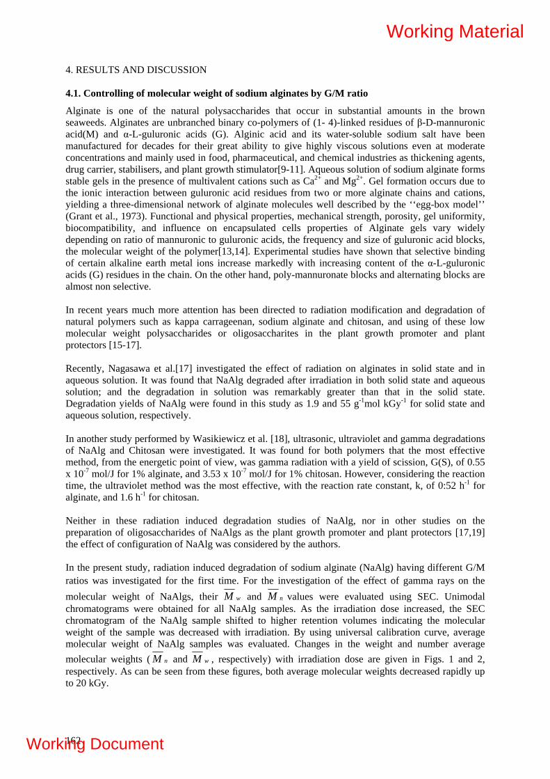
4. RESULTS AND DISCUSSION 4.1. Controlling of molecular weight of sodium alginates by G/M ratio
Alginate is one of the natural polysaccharides that occur in substantial amounts in the brown seaweeds. Alginates are unbranched binary co-polymers of (1- 4)-linked residues of β-D-mannuronic acid(M) and α-L-guluronic acids (G). Alginic acid and its water-soluble sodium salt have been manufactured for decades for their great ability to give highly viscous solutions even at moderate concentrations and mainly used in food, pharmaceutical, and chemical industries as thickening agents, drug carrier, stabilisers, and plant growth stimulator[9-11]. Aqueous solution of sodium alginate forms stable gels in the presence of multivalent cations such as Ca2+ and Mg2+. Gel formation occurs due to the ionic interaction between guluronic acid residues from two or more alginate chains and cations, yielding a three-dimensional network of alginate molecules well described by the ‘‘egg-box model’’ (Grant et al., 1973). Functional and physical properties, mechanical strength, porosity, gel uniformity, biocompatibility, and influence on encapsulated cells properties of Alginate gels vary widely depending on ratio of mannuronic to guluronic acids, the frequency and size of guluronic acid blocks, the molecular weight of the polymer[13,14]. Experimental studies have shown that selective binding of certain alkaline earth metal ions increase markedly with increasing content of the α-L-guluronic acids (G) residues in the chain. On the other hand, poly-mannuronate blocks and alternating blocks are almost non selective. In recent years much more attention has been directed to radiation modification and degradation of natural polymers such as kappa carrageenan, sodium alginate and chitosan, and using of these low molecular weight polysaccharides or oligosaccharites in the plant growth promoter and plant protectors [15-17]. Recently, Nagasawa et al.[17] investigated the effect of radiation on alginates in solid state and in aqueous solution. It was found that NaAlg degraded after irradiation in both solid state and aqueous solution; and the degradation in solution was remarkably greater than that in the solid state. Degradation yields of NaAlg were found in this study as 1.9 and 55 g-1mol kGy-1 for solid state and aqueous solution, respectively. In another study performed by Wasikiewicz et al. [18], ultrasonic, ultraviolet and gamma degradations of NaAlg and Chitosan were investigated. It was found for both polymers that the most effective method, from the energetic point of view, was gamma radiation with a yield of scission, G(S), of 0.55 x 10⁻⁷ mol/J for 1% alginate, and 3.53 x 10⁻⁷ mol/J for 1% chitosan. However, considering the reaction time, the ultraviolet method was the most effective, with the reaction rate constant, k, of 0:52 h-1 for alginate, and 1.6 h-1 for chitosan. Neither in these radiation induced degradation studies of NaAlg, nor in other studies on the preparation of oligosaccharides of NaAlgs as the plant growth promoter and plant protectors [17,19] the effect of configuration of NaAlg was considered by the authors. In the present study, radiation induced degradation of sodium alginate (NaAlg) having different G/M ratios was investigated for the first time. For the investigation of the effect of gamma rays on the molecular weight of NaAlgs, their wM and nM values were evaluated using SEC. Unimodal chromatograms were obtained for all NaAlg samples. As the irradiation dose increased, the SEC chromatogram of the NaAlg sample shifted to higher retention volumes indicating the molecular weight of the sample was decreased with irradiation. By using universal calibration curve, average molecular weight of NaAlg samples was evaluated. Changes in the weight and number average molecular weights ( nM and wM , respectively) with irradiation dose are given in Figs. 1 and 2, respectively. As can be seen from these figures, both average molecular weights decreased rapidly up to 20 kGy.
162
Working Material
Working Document

0 5 10 15 20 25
1618202224262830323436
LF120 LF200 LF240
Mn x
105
Dose (kGy)0 5 10 15 20 25
20
30
40
50
60
70
LF120 LF200 LF240
Mw x
105
Dose (kGy) FIG. 1. Change in the number average molecular weight ( nM ) of NaAlgs with dose.
FIG. 2. Change in the weight average molecular weight ( wM ) of NaAlgs with dose.
4.2 Determination of chain scission yield and degradation rate constant
The efficiency of radiation-induced events is expressed by the so-called G-value. The G-value, equal to the number of events per 100 eV of energy absorbed, has been customarily used to measure radiation chemical yield. Molecular weight values of NaAlgs are used for the determination of chain scission yield G(s) and degradation rate. If scission is the only mode of action of radiation then the radiation-chemical yield of degradation (scission) G(s) is determined from the Alexander-Charlesby-Ross equation [20] :
-7nD n01/M = 1/M + 1.04x10 G(s) D (2)
where the absorbed dose, D, is in kGy, and nDM and n0M are the number average molecular weights of the polymer before and after irradiation, respectively. Rapid decrease in nM and wM clearly indicated that the only mode of action of radiation on sodium alginate was degradation. For the determination of the G(s) values, 1/ nM was plotted against dose for all samples (Fig. 3). Then, G(s) values were calculated by using the intercepts. The calculated G(s) values were 0.71 ± 0.083, 0.97 ± 0.048, 1.05 ± 0.031 for LF120, LF200, and LF240 NaAlg, respectively. It was concluded that G(s) values were dependent on the structure (guluronic acids(G) and mannuronic acid(M) ratio) of the sample and followed the order LF240>LF200>LF120 or 70/30< 50/50< 45/55, G/M ratio. When the chemical structures of NaAlgs were examined it was seen that the properties of NaAlg varied widely depending on the composition of the alginate molecule (i.e., the ratio of mannuronic to guluronic acids, the frequency and size of guluronic acid blocks, the molecular weight of the polymer), and the concentrations of alginate and cation at the time of gelation. It could be concluded that the G(s) value increased with a decrease in the guluronic acids to mannuronic acid ratio. This increase was attributed to the decrease of radical-radical recombination reactions due to the decrease of stiff and extended nature of alginate chains. It is well known that the diaxial linkage in G-blocks results in a large, hindered rotation around the glycosidic linkage, which may account for the stiff and extended nature of alginate chains [21].
163
Working Material
Working Document

0 5 10 15 20 25
30
35
40
45
50
55
60
65
70
LF120 LF200 LF240
1/M
n x10
7
Dose (kGy) FIG. 3. Plot of 1/Mn versus dose for the determination of the G(S) values of NaAlgs.
The equation given by Jellinek [22] was modified and used in the determination of degradation rate. Where N is the average number of bond cleavages per original polymer molecule, this value can be calculated by:
n0
nD
MNM
= 1− (3)
where n0M and nDM are the nM before and after irradiation to a certain dose (D), respectively. N is also named as the degree of scission. Assuming the rate of chain scission is independent of chain length and position of the link in the chain, N is anticipated to be a linear function of irradiation dose (first-order reaction)
n0 n0
nD o
M MN 1 k mM
⎡ ⎤= − = ⎢ ⎥
⎣ ⎦D (4)
where k is the rate constant and mo is the molecular weight of a monomer unit. Eq. (4) can be rewritten as:
nD n0 o
1 1 k DmM M
⎛ ⎞− = ⎜ ⎟
⎝ ⎠ (5)
Degradation rate constants for NaAlgs were determined by using the (1/ nM )-(1/ n0M ) versus dose curves The determined degradation rate constants for NaAlgs are given in Table II. They followed the order LF240>LF200>LF120. These results clearly indicated that degradation rate was also dependent on the G/M ratio for these initial molecular weights.
TABLE II. DEGRADATION RATE CONSTANTS OF NAALGS.
Polymer G/M k(kGy-1) LF120 70/30 4.27x10⁻¹⁰ ± 3.2x10⁻¹¹ LF200 50/50 5.86x10⁻¹⁰ ± 2.95x10⁻¹¹
LF240 45/55 6.30x10⁻¹⁰ ± 1.98x10⁻¹¹
164
Working Material
Working Document
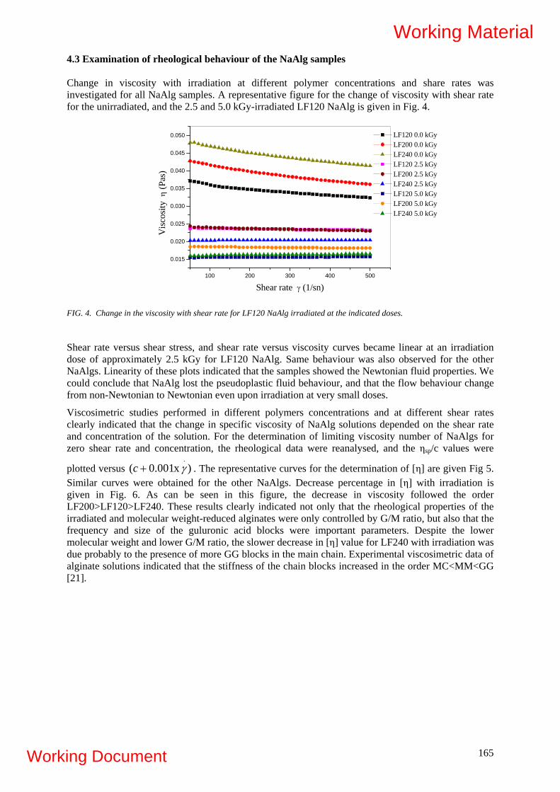
4.3 Examination of rheological behaviour of the NaAlg samples Change in viscosity with irradiation at different polymer concentrations and share rates was investigated for all NaAlg samples. A representative figure for the change of viscosity with shear rate for the unirradiated, and the 2.5 and 5.0 kGy-irradiated LF120 NaAlg is given in Fig. 4.
100 200 300 400 500
0.015
0.020
0.025
0.030
0.035
0.040
0.045
0.050 LF120 0.0 kGy LF200 0.0 kGy LF240 0.0 kGy LF120 2.5 kGy LF200 2.5 kGy LF240 2.5 kGy LF120 5.0 kGy LF200 5.0 kGy LF240 5.0 kGy
Vis
cosit
y η
(Pas
)
Shear rate γ (1/sn)
FIG. 4. Change in the viscosity with shear rate for LF120 NaAlg irradiated at the indicated doses.
Shear rate versus shear stress, and shear rate versus viscosity curves became linear at an irradiation dose of approximately 2.5 kGy for LF120 NaAlg. Same behaviour was also observed for the other NaAlgs. Linearity of these plots indicated that the samples showed the Newtonian fluid properties. We could conclude that NaAlg lost the pseudoplastic fluid behaviour, and that the flow behaviour change from non-Newtonian to Newtonian even upon irradiation at very small doses.
Viscosimetric studies performed in different polymers concentrations and at different shear rates clearly indicated that the change in specific viscosity of NaAlg solutions depended on the shear rate and concentration of the solution. For the determination of limiting viscosity number of NaAlgs for zero shear rate and concentration, the rheological data were reanalysed, and the ηsp/c values were
plotted versus . The representative curves for the determination of [η] are given Fig 5. Similar curves were obtained for the other NaAlgs. Decrease percentage in [η] with irradiation is given in Fig. 6. As can be seen in this figure, the decrease in viscosity followed the order LF200>LF120>LF240. These results clearly indicated not only that the rheological properties of the irradiated and molecular weight-reduced alginates were only controlled by G/M ratio, but also that the frequency and size of the guluronic acid blocks were important parameters. Despite the lower molecular weight and lower G/M ratio, the slower decrease in [η] value for LF240 with irradiation was due probably to the presence of more GG blocks in the main chain. Experimental viscosimetric data of alginate solutions indicated that the stiffness of the chain blocks increased in the order MC<MM<GG [21].
)x001.0(.γ+c
165
Working Material
Working Document
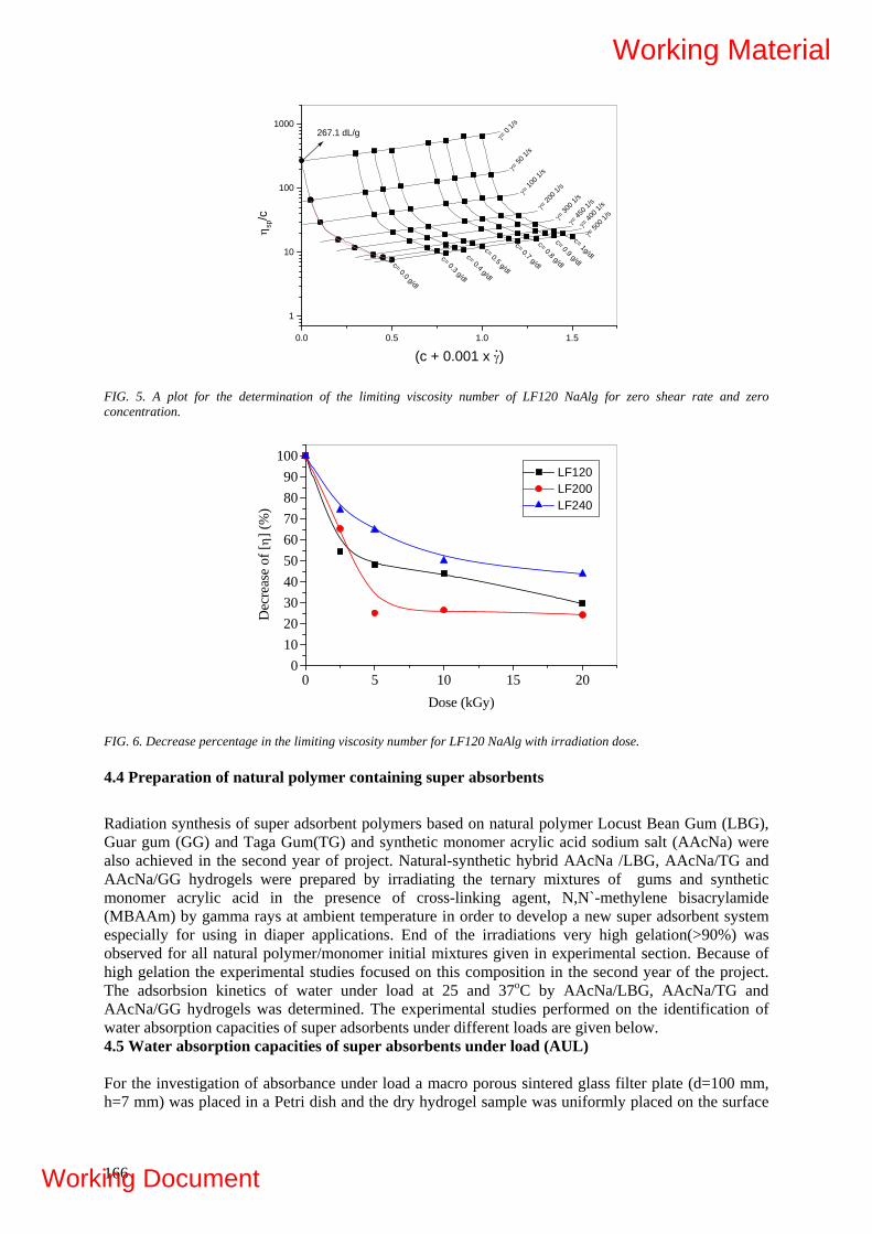
0.0 0.5 1.0 1.5
1
10
100
1000
.
267.1 dL/g
c= 0.3 g/dl
c= 0.0 g/dl
γ= 0
1/s
γ= 50
0 1/s
γ= 45
0 1/s
γ= 40
0 1/s
γ= 30
0 1/s
γ= 20
0 1/sγ=
100 1
/sγ= 50
1/s
c= 0.4 g/dl
c= 0.5 g/dl
c= 0.7 g/dl
c= 0.8 g/dl
c= 0.9 g/dl
c= 1g/dlη s
p/c
(c + 0.001 x γ)
FIG. 5. A plot for the determination of the limiting viscosity number of LF120 NaAlg for zero shear rate and zero concentration.
0 5 10 15 200
102030405060708090
100 LF120 LF200 LF240
Dec
reas
e of
[η] (
%)
Dose (kGy) FIG. 6. Decrease percentage in the limiting viscosity number for LF120 NaAlg with irradiation dose. 4.4 Preparation of natural polymer containing super absorbents
Radiation synthesis of super adsorbent polymers based on natural polymer Locust Bean Gum (LBG), Guar gum (GG) and Taga Gum(TG) and synthetic monomer acrylic acid sodium salt (AAcNa) were also achieved in the second year of project. Natural-synthetic hybrid AAcNa /LBG, AAcNa/TG and AAcNa/GG hydrogels were prepared by irradiating the ternary mixtures of gums and synthetic monomer acrylic acid in the presence of cross-linking agent, N,N`-methylene bisacrylamide (MBAAm) by gamma rays at ambient temperature in order to develop a new super adsorbent system especially for using in diaper applications. End of the irradiations very high gelation(>90%) was observed for all natural polymer/monomer initial mixtures given in experimental section. Because of high gelation the experimental studies focused on this composition in the second year of the project. The adsorbsion kinetics of water under load at 25 and 37oC by AAcNa/LBG, AAcNa/TG and AAcNa/GG hydrogels was determined. The experimental studies performed on the identification of water absorption capacities of super adsorbents under different loads are given below. 4.5 Water absorption capacities of super absorbents under load (AUL) For the investigation of absorbance under load a macro porous sintered glass filter plate (d=100 mm, h=7 mm) was placed in a Petri dish and the dry hydrogel sample was uniformly placed on the surface
166
Working Material
Working Document

of a polyester gauze located on the sintered glass. A cylindrical solid weight (d=80 mm, variable height) which could slip freely in a glass cylinder was used to apply the desired load (applied pressure=0.36 and 0.66 psi) to the dry grinded hydrogel particles[23]. The swelling kinetics of AAcNa/LBG, AAcNa/TG and AAcNa/GG hydrogels and pure AAcNa in distilled water at different temperatures are given in Figs.7-10.
0 5 10 15 20 25 30 35 40 45 50 55 60 650
2000
4000
6000
8000
10000
12000
14000
AAc/LBG 25 oC 0.0 psi AAc/LBG 25 oC 0.3 psi AAc/LBG 25 oC 0.6 psi
Sw
ellin
g (%
)
Time (min.)
(a)
0 10 20 30 40 50 60 70 80 90 1000
2000
4000
6000
8000
10000
12000
AAc/LBG 37 oC 0.0 psi AAc/LBG 37 oC 0.3 psi AAc/LBG 37 oC 0.6 psi
Sw
ellin
g (%
)Time (min.)
(b)
FIG. 7. Effect of temperature (a) 25 ºC, (b) 37 ºC and pressure on the swelling kinetics of AAcNa/LBG hydrogels.
0 5 10 15 20 25 30 35 40 45 50 55 60 65 700
2000
4000
6000
8000
10000
12000
14000
AAc/TG 25 oC 0.0 psi AAc/TG 25 oC 0.3 psi AAc/TG 25 oC 0.6 psi
Sw
ellin
g (%
)
Time (min.)
0 5 10 15 20 25 30 35 40 45 50 55 600
2000
4000
6000
8000
10000
AAc/TG 37 oC 0.0 psi AAc/TG 37 oC 0.3 psi AAc/TG 37 oC 0.6 psi
Sw
ellin
g (%
)
Time (min.) FIG. 8. Effect of temperature (a) 25 ºC, (b) 37 ºC and pressure on the swelling kinetics of AAcNa/TG hydrogels.
0 5 10 15 20 25 30 35 40 45 50 55 60 65 700
2000
4000
6000
8000
10000
12000
14000
AAc/GG 25 oC 0.0 psi AAc/GG 25 oC 0.3 psi AAc/GG 25 oC 0.6 psi
Sw
ellin
g (%
)
Time (min.)0 5 10 15 20 25 30 35 40 45 50 55 60
0
2000
4000
6000
8000
10000
12000
14000
AAc/GG 37 oC 0.0 psi AAc/GG 37 oC 0.3 psi AAc/GG 37 oC 0.6 psi
Sw
ellin
g (%
)
Time (min.) FIG. 9. Effect of temperature (a) 25 ºC, (b) 37 ºC and pressure on the swelling kinetics of AAcNa/GG hydrogels.
167
Working Material
Working Document

0 10 20 30 40 50 60
250500750
100012501500175020002250250027503000
0
AAc 25 oC 0 psi AAc 25 oC 0.36 psi AAc 25 oC 0.66 psi
Sw
ellin
g (%
)
Time (min)
0 5 10 15 20 25 30 35 40 45 50 550
250500750
1000125015001750200022502500
AAc 37 oC 0 psi AAc 37 oC 0.36 psi AAc 37 oC 0.66 psi
Sw
ellin
g (%
)
Time (min) FIG. 10. Effect of temperature (a) 25 ºC, (b) 37 ºC and pressure on the swelling kinetics of AAcNa hydrogels. Swelling kinetics of super adsorbents under different loads are also included in figures for comparison. As can be seen from these figures natural polymer containing super adsorbents exhibit higher water absorption character then pure AAcNa hydrogels. This changes is probably due to reducing of crosslink density ability of acrylic acid in the presence natural polymer during irradiation. The maximum or equilibrium swelling values of all prepared super adsorbents in different swelling conditions are compared in Fig 11. It is generally observed that the % swelling of hydrogels in water under 0.36, 0.66 psi load is lower than swelling in zero pressure. On the other hand, the effect of pressure is relatively low in AAcNa/TG hydrogels compared with the other hydrogel systems. It is seen from Fig. 11 that with increasing temperature of the swelling medium, the equilibrium degree of swelling values show a decrease and except GG/AAcNa system this effect becomes more pronounced under 0.66 psi pressure. The swelling kinetic figures show that the swelling increases with time continuously due to super absorbent character of these hydrogels, in which the samples almost reach to the equilibrium state after 5-10 minutes. Due to fast and anomalous water absorption capacities of AAcNa/LBG, TG, GG hydrogels we can conclude that the hydrogels prepared in this study can be considered as potential super adsorbents for diapers systems and may be used instead of 100 % synthetic super absorbent using diapers and feminine hygiene products in nowadays .
0
2000
4000
6000
8000
10000
12000
14000
0.66psi
0.36psi0psi
0.66psi
0.36psi
AAcNa/LBG 37 oC
Sw
ellin
g (%
)
AAc/LBG 25oC AAc/LBG 37oC
AAcNa/LBG 25 oC
0psi
(a)
0
2000
4000
6000
8000
10000
12000
14000
0.66psi
0.36psi0psi0.66psi0.36psi
AAcNa/TG 37 oC
Swel
ling
(%)
AAc/TG 25oC AAc/TG 37oC
AAcNa/TG 25 oC
0psi
(b)
168
Working Material
Working Document

0
2000
4000
6000
8000
10000
12000
14000
0.66psi
0.36psi0psi
0.66psi
0.36psi
AAcNa/GG 37 oC
Sw
ellin
g (%
) AAc/GG 25oC AAc/GG 37oC
AAcNa/GG 25 oC
0psi
(c)
0
500
1000
1500
2000
2500
0.66psi0.36psi
0psi
0.66psi
0.36psi
AAcNa 37 oC
Sw
ellin
g (%
)
AAc 25 oC AAc 37 oC
AAcNa 25 oC
0psi
(d)
FIG. 11. Variation of equilibrium swelling value of (a) AAcNa/LBG, (b) AAcNa/TG, (c) AAcNa/GG and (d) AAcNa hydrogels with temperature and pressure. Temperatures and pressures are indicated in figures. 5. CONCLUSIONS
In this report degrading effects of radiation have been considered from the point of views of controlling the molecular weights of sodium alginate. The effect of irradiation on the NaAlg samples was determined to be dependent on the chemical structure of the sample. As the guluronic acid to mannuronic acid ratio decreased, the effect of irradiation on the molecular weight and rheological properties of the sample became more pronounced. It could be concluded that the G/M ratio was one of the factors affecting chain scission yield of NaAlg. All NaAlg samples showed a pseudoplastic behaviour up to a certain dose, and a Newtonian flow behaviour above that dose. In this report radiation synthesis and characterization of swelling behaviour natural/synthetic hybrid super absorbents prepared by using LBG, TG, GG and acrylic acid sodium salt were given. Natural-synthetic hybrid hydrogels were prepared by irradiating the ternary mixtures of natural polymers LBG, TG or GG and synthetic monomer acrylic acid sodium salt in the presence of cross-linking agent, N,N`-methylene bisacrylamide (MBAAm) by gamma rays at ambient temperature in order to develop a new super adsorbent system especially for using in diaper applications. The swelling behaviours of the prepared hydrogels in distelled water were studied in different temperature and pressure. So far in most of the relevant literature, the swelling capacity values of super absorbent polymers are reported as free-swelling data, i.e. load free swelling. It is obvious that the swelling conditions, and hence the data, are not real, because in all of the super absorbent polymers applications (agricultural, hygienic, etc.), the swelling particles must absorb aqueous solutions while they are under pressure. It is generally observed that swelling capacity of all natural polymer/AAcNa hydrogels decrease by applying 0.36-0.66 psi pressure during swelling. On the other hand due to fast and anomalous swelling the natural/synthetic hybrid hydrogel systems prepared in this study can be considered as potential adsorbents for the body fluids such as urine and blood and may be used especially as diapers feminine hygiene products. FUTURE PLAN OF PROJECT
The overall objective of the proposed CRP is the wide-spread promotion and general application of radiation processed natural materials, coupling radiation technology and end-users to derive enhanced benefits from these value-added natural materials. The specific objectives are:
• Identification of methodologies and QA protocols for investigating structure-property relationship particularly with respect to radiation induced changes in natural polymers. • Investigating the anti-oxidant properties of low molecular weight natural polymers and assessing suitability for preservation of food and allied products. • Field-testing the potential of radiation-modified polysaccharides as plant growth promoters, soil conditioners and for enhancing fermentation of agro by-products.
169
Working Material
Working Document

In the first and second year of our project we investigated structure-property relationship particularly with respect to radiation induced changes in natural polymers. In the second year of the project we also started to synthesize of natural polymer containing super absorbents to be used as disposable diapers and soil conditioning materials in agriculture, horticulture and other super adsorbent using industries. In the third year of the project we will continue to synthesis of super absorbent polymers based on natural polymers to be used as disposable diapers, and other super adsorbent using industries. The aqueous solution of natural polymers and/or their blends with a trace amount synthetic monomer will be irradiated in paste like conditions or aqueous solutions and in the presence of cross-linking agents by gamma rays for the preparation of cross-linked hydrogel systems. The water absorption and deswellling capacity of prepared super adsorbents and retention capacity, absorbency under load, suction power, swelling pressure and pet-rewet properties will be determined also in the third year of the project. Use of these materials instead of synthetic super absorbents will be examined by comparing the performance of finished products. The antioxidant properties of degraded natural polymer by radiation also will be investigate in the third year of the project. For the investigation of antioxidant properties of degraded natural polymers, the hydroxyl (OH.), superoxide anion(O2
.) and Diphenylpicrylhydrazyl(DPPH) radical scavenging ability and Fe 2+ metal ion chelating ability will be evaluated. Use of degraded natural polymers LBG, TG, GG and NaAlg instead of antioxidant agents will be examined in the last year of the project by comparing the performance of finished products.
REFERENCES
[1] PEPPAS, N.A., Hydrogels in Medicine and Pharmacy, CRC Press, Boca Raton Vol. 3. (1987) 1. [2] MATHUR, M.A., SHAILENDER, K.M., SCRANTON, A., Methods for synthesis of hydrogel
networks: A review., J. Macromol. Sci., Rev. Macromol. Chem. Phys., C36, (1996) 405-408. [3] CARENZA, M., Recent achievements in the use of radiation polymerization and grafting for
biomedical applications, Radiat. Phys. Chem., 39, (1992) 485-489. [4] CHOI, W-S., AHN, K.-J., LEE, D.-W., BYUN, M.W., PARK, H.J., Preparation of chitosan
oligomers by irradiation, Polym. Deg. Stab., 78, (2002) 533-538. [5] WASIKIEWICZ, J.M., YOSHII, F., NAGASAWA, N., WACH, R.A., MITOMO, H.,
Degradation of chitosan and sodium alginate by gamma radiation, sonochemical and ultraviolet methods, Rad. Phys. Chem., 73, (2005) 287-295. [6] YOSHII, F., ZHAO, L., WACH, R.A., NAGASAWA, N., MITOMO, H., KUME, T., Hydrogels
of polysaccharide derivatives crosslinked with irradiation at paste-like condition, Nuclear Inst. Met. Phys. Res.B, 208, (2003) 320-324.
[7] RAMNAMI, S. P., CHAUDHARI, C. V., PATIL, N. D., SABHARWAL, S., Synthesis and characterization of crosslinked chitosan formed by gamma irradiation in the presence of carbontetrachloride as a sensitize, J. Polym. Sci., Part A. Polymer Chemistry, 42, (2004) 3897-3909.
[8] DERAMOS, C. M., IRWIN, A. E., NAUSS, J. L., STOUT, B. E.,13C NMR and Molecular modeling studies of alginic acid binding with alkaline earth metal ions, Inorganica Chimica Acta, 256, (1997) 69-75. [9] ØSTBERG, T, LUND, E.M., GRAFFNER, C., Calcium alginate matrices for oral unit
administration, IV. Release characteristics in different media, Int. J. Pharm., 112, (1994) 241–248.
[10] GARCIA, A.M., GHALY, E.S., Preliminary spherical agglomerates of water soluble drug using natural polymer and cross-linked technique, J. Control Rel., 40, (1996) 179-186.
[11] HIEN, N. Q., NAGASAWA, N., THAM, L. X., YOSHII, F., DANG, V. MITOMO, H. H., MAKUUCHI, K., KUME, T., Growth-promotion of plants with depolymerised alginates by irradiation, Radiation Physics and Chemistry, 59, (2000) 97-101.
170
Working Material
Working Document

[12] GRANT, G.T., MORRIS, E.R., REES, D.A., SMITH, P.J.C., THOM, D. Biological interactions between polysaccharides and divalent cations: the egg-box model, FEBS Lett ., 32, (1973) 195-198.
[13] SMIDSROD, O., Molecular basis for some physical properties of alginates in gel state, J Chem Soc Faraday Trans., 57, (1974) 263-74.
[14] KLOCK, G, FRANK, H, HOUBEN, R, ZEKORN, T, HORCHER, A, SIEBERS, U, WOHRLE, M, FEDERLIN, K, ZIMMERMANN, U., Production of purified alginates suitable for use in immunoisolated transplantation, Appl. Microbiol. Biotechnol., 40, (1994) 638-463.
[15] RELLEVE, L., NAGASAWA, N., LUANC, L.Q., YAGI, T., ARANILLA, C., ABAD L., KUME, T., YOSHII, F., DELA ROSA, A., Degradation of carrageenan by radiation Polymer Degradation and Stability, 87, (2005) 403-410.
[16] THAMA, L. X., NAGASAWA, N., MATSUHASHI, S., ISHIOKA, N. S., ITO, T., KUME, T., Effect of radiation-degraded chitosan on plants stressed with vanadium, Radiation Physics and Chemistry 61, (2001) 171-175.
[17] NAGASAWA, N., MITOMO, H., YOSHII, F., KUME, T., Radiation-induced degradation of sodium alginate, Polymer Degradation and Stability, 69, (2000) 279-285. [18] WASIKIEWICZ, J. M., YOSHII, F., NAGASAWA, N., WACH, R. A., MITOMO, H. Degradation of chitosan and sodium alginate by gamma radiation, sonochemical and ultraviolet methods, Radiation Physics and Chemistry, 73, (2005) 287–295. [19] KUME, T., NAGASAWA, N., YOSHII, F., Utilization of carbohydrates by radiation
processing, Radiation Physics and Chemistry, 63, (2002) 625-627. [20] CHARLESBY, A., Atomic Radiation and Polymers, Pergamon Press, New York, (1960). [21] SMIDSROD, O., GLOVER, R. M., WHITTINGTON, S. G., The relative extension of alginates having different chemical composition, Carbohydr. Res., 27, (1973) 107-118. [22] JELLINEK, H.H.G., Degradation of Vinyl Polymers, Wiley and Sons, New York, (1955). [23] RAMAZANI-HARANDI, M.J., ZOHURIAAN-MEHR, M.J., YOUSEFI A.A.,
ERSHAD-LANGROUDI, A., KABIRI, K., Rheological determination of the swollen gel strength of superabsorbent polymer hydrogels, Polymer Testing, 25 (2006) 470-474.
171
Working Material
Working Document

172
Working Material
Working Document

CHARACTERISATION AND RADIATION MODIFICATION OF CARRAGEENAN IN THE
SOLID STATE
S. Gulrez1, S. Al-Assaf 1*, G. O. Phillips1,2,
1Phillips Hydrocolloids Research Centre,
Glyndwr University,
Mold Road, Wrexham LL11 2AW, North Wales, UK 2Phillips Hydrocolloid Research Ltd,
45 Old Bond St., London W1S 4AQ, UK
Abstract This study reports the modification of kappa-carrageenan in the solid state using gamma radiation (in the dose
range of 1-25kGy) in the presence of unsaturated alkyne gas. The results showed maximum production of
hydrogel at 5kGy with nearly 80% of starting material being converted to hydrogel form in the absence of a
gellin agent. Higher irradiation doses at 25kGy resulted in reducing the hydrogel proportion to ~40% due to
degradation. The molecular weight and distribution was determined by GPC-MALLS and the results showed a
decrease in the mass recovery and molecular weight of the soluble fraction at 60C. The molecular weight results
were in agreement with hydrogel data determined from the filtration method. There was an optimum increase in
the viscosity, elasticity and mechanical strength at 5kGy which was followed by a decrease in the gel strength at
higher doses (25kGy). Our study demonstrates the potential production of novel hydrogel based carrageenan
obtained by irradiation in the absence of metal ions with possible new applications. A mechanism for the
radiation induced cross-linking to produce superhelical aggregates in the absence of a gelling agent is proposed.
1. INTRODUCTION
Carrageenan is a generic name for a family of polysaccharides, obtained by extraction from certain
species of red seaweeds (Rhodophyta). The word carrageenan is derived from the colloquial Irish
name for this seaweed, carrageen, which means ‘little rock’. The aqueous extraction of red seaweeds
to obtain these hydrophilic colloids is known in Ireland since 1810. All seaweeds that produce
carrageenan as their main cell-wall material belong to Rhodophyta [1].
Carrageenans are widely utilized in the food industry due to their excellent physical functional
properties, such as thickening, gelling and stabilizing abilities, and have been used to improve the
texture of cottage cheese, to control the viscosity and texture of puddings and dairy desserts, and as
binders and stabilizers in the meat-processing industry for the manufacture of patties, sausages and
low-fat hamburgers. Carrageenans are also used in various non-food products, such as pharmaceutical,
cosmetics, printing and textile formulations [2]. Carrageenans stabilize toothpaste preparations, absorb
173
Working Material
Working Document

body fluids when formulated in wound dressings and interact with human carotene to give soft skin
and silky hair in hand lotions and shampoos, respectively. They have proved to be useful as tableting
excipients due to the good compatibility, high robustness and persistent viscoelasticity of the tablet
during compression.
1.1 Chemical structure
Carrageenans are obtained from different species of Rhodophyta: Gigartina, Chondrus crispus,
Eucheuma and Hypnea. These polysaccharides are traditionally split into six basic forms: Iota (ι)-,
Kappa (κ)-, Lambda (λ)-, Mu (μ)-, Nu (ν)- and Theta (θ)- carrageenan [1]. Carrageenans belong to the
family of hydrophilic linear sulphated galactans. They mainly consist of alternating 3-linked β-D-
galactopyranose (G-units) and 4-linked α-D-galactopyranose (D-units) or 4-linked 3,6-anhydro-α-D-
galactopyranose (DA-units), forming the disaccharide repeating unit of carrageenans (FIG. 1).
FIG. 1. Schematic representation of the different structures of the repeating dimeric units of commercial carrageenans and related structures.
Three commercial most important carrageenans are Iota (ι-), Kappa (κ-) and Lambda (λ)-carrageenan.
The κ-, ι- and λ-carrageenan dimers have one, two and three sulphate ester groups, respectively,
resulting in correspondent calculated sulphate contents of 20%, 33% and 41% (w/w). The official
method for determining the sulphate content of carrageenans is based on the selective hydrolysis of the
sulphate ester by acid and subsequent selective precipitation of the sulphate ions as barium sulphate,
which is then measured by weighing or by turbidimetry as described by the FAO/WHO Joint Expert
Committee on Food Additives (JECFA) (Food and Agricultural Organization, 1992).
174
Working Material
Working Document
1.2. Functionalities
All carrageenan fractions are water soluble, being insoluble in organic solvents, oil or fats. However,
their water solubility depends essentially on the levels of sulphate groups (very hydrophilic) and on
their associated cations. The main ionisable cations found in carrageenans are sodium, potassium,
calcium and magnesium, but other ions can also occur at lower frequency. Consequently, the
proportion of sulphate fractions and the equilibrium of cations in the water solution determine the
viscosity of solutions or strength of gels formed by carrageenans, representing the major
characteristics explored by the food and pharmaceutical industries in the use of carrageenans as
thickening, gelling, and stabilizing agents [1].
1.3. Viscosity and gelling mechanism
The viscosity of carrageenans depends on concentration, temperature, the presence of other solutes,
the type of carrageenan and its molecular weight, and increases exponentially with the increase in their
concentration, being a typical behaviour of linear polymers with charge, or polyelectrolytes. However,
the increase of viscosity can occur by two different mechanisms: (i) interaction between the linear
chains, with a decrease of the free space or increase of the excluded volume; (ii) formation of a
physical gel caused by ‘cross-linking’ between chains. In the first case, the increase of the
macromolecule concentration allows a major interaction between the chains and the presence of salts
can decrease the viscosity by reducing the electrostatic repulsion among the sulphate groups. For
instance, this is what occurs for lambda fraction. The second mechanism of viscosity increase is
particular from kappa, iota and hybrid kappa- 2 fractions. For these carrageenans, in small
concentrations of salt and low temperature, the carrageenan solutions can gel, with increase of the
apparent viscosity. The viscosity of the carrageenan solution decreases reversibly with the increase of
temperature [1]. Among commercial carrageenans, κ- and ι- are gel-forming carrageenans, whereas λ-
carrageenan is characterized only as a thickener agent. The difference in rheological behaviour results
from the fact that the anhydrogalactose units (DA) of the gelling ones (kappa and iota) have 1C4
conformation (FIG. 2) and the D-galactopyranosil units (D) in k-carrageenan do not. The 1C4
conformation of the 3,6- anhydro-D-galactopyranosil units in κ- and ι-carrageenan allows a helicoidal
secondary structure, which is essential for the gel-forming properties.
Gelation of carrageenans, especially kappa, involves two separate and successive steps; coil-to-helix
transition upon cooling and subsequent cation-dependent aggregation between helices. The presence
of suitable cation, typically potassium or calcium, is an absolute requirement for gelation to proceed.
For both ι- and κ-carrageenans, the alkali metal ions (Li+, Na+, K+, Rb+, Cs+) are all capable of
inducing gelation, but K+ and Rb+ are considerably more effective than other ions in inducing gelation
at much lower concentrations of both the cation and the carrageenan [3].
175
Working Material
Working Document
FIG. 2. Representation of 1C4 and 4C1 conformations for β-D-galactopyranose; in structure 1, C-1 is above the plane and C-4
is below, and thus, the symbol is 1C4. Likewise, structure 2 is 4C1 [1].
Kappa- and iota-carrageenans form a network of three-dimensional double helices (FIG. 3), resulting
from the ‘cross-linking’ of the adjacent spiral chains that contain sulphate groups oriented towards
their external part. In λ-carrageenan, the 2-sulphate group is oriented towards the internal part, thus
avoiding this ‘cross-linking’ [1].
FIG. 3. Double helix model of ι-carrageenan; structure downloaded from Protein Data Bank (PDB code 1CAR)
(www.rcsb.org/pdb).
1.4. Modification of Carrageenan
Several chemical modifications have been proposed to modulate physicochemical properties of
carrageenans. Aqueous κ-carrageenan gels can display an undesirable large extent of syneresis (the
extraction of a liquid from a gel) when subjected to mechanical deformation or aging, a natural
process that affects the mechanical, magnetic and rheological properties of the gels. Gels prepared
from hydroxyalkyl κ-carrageenan derivatives demonstrated decreased syneresis and could contribute
to a wider industrial use of these polysaccharides [1]. Recently, Tari and Pekcan [4] demonstrated that
the association of κ-carrageenan with CaCl2 can change the swelling properties of carrageenan gels,
emphasizing the commercial relevance of carrageenans chemical modifications.
Bardajee, Pourjavadi et al. [5] prepared a κ-carrageenan hydrogel with a very high absorptivity in
saline through polyacrylamide cross-linking followed by alkaline hydrolysis. The hydrogel displayed
water absorbance properties similar to those of κ-carrageenan copolymerized with acrylic acid. By
176
Working Material
Working Document

using γ-irradiation as an initiator and cross-linking agent at the same time, the swelling behaviour of
these synthesized hydrogels could also be improved [1].
The copolymerization of κ-carrageenan with acrylic acid and 2-acrylamido-2-methylpropanesulfonic
acid led to the development of biodegradable hydrogels with potential use for novel drug delivery
systems [6]. These carrageenan hydrogels are very promising for industrial immobilization of enzymes
since the immobilization procedures result in increases of the storage and thermal stability of the
enzymes necessary for use in continuous systems [1].
Porous nanocomposites were prepared by coprecipitation of calcium phosphates into a κ-carrageenan
matrix, whose resulting porosity and morphology were suitable for application in bone tissue
engineering. The association between carrageenan, nanohydroxyapatite and collagen resulted in an
injectable bone substitute biomaterial, suitable for bone reconstruction surgery [7].
Yuan et al. [8] synthesized oversulphated, acetylated and phosphorylated derivatives of κ-carrageenan.
Sulphation was carried out using chlorosulphonic acid (HClSO3) in N,N-dimethylformamide (DMF),
acetylation employed acetic anhydride (Ac2O) and pyridine (Py) in DMF, and phosphorus oxychloride
(POCl3)/Py in formamide (FA) followed by hydrolysis were applied for phosphorylation. All
derivatives exhibited an antioxidant activity higher than that of κ-carrageenan, suggesting that
chemical modification can enhance antioxidant activity of carrageenans [1].
Low molecular weight carrageenans and their sulphated derivatives were prepared by
depolymerization in the presence of ferrous ions or ferrous ions plus ascorbic acid by [9]. All
compounds demonstrated anti-HIV activities and sulphation of κ-carrageenan also increased the
biological effects. In a later study these depolymerized carrageenans were acylated by carboxylic acid
anhydrides using DMF as solvent and 4-dimethylaminopyridine (DMAP)/tributylamine (TBA) as
catalysts [10]. The acylation of low molecular weight carrageenans resulted in potentiation of anti-
HIV activity although the anticoagulant activity of the compounds decreased [1].
Jiang and Guo [11] achieved the synthesis of an O-maleoyl derivative of κ-carrageenan by reaction of
tetrabutylammonium salt of the anionic polysaccharide fragments with maleic anhydride, 4-
dimethylaminopyridine and tributylamine under homogeneous conditions in N,N-dimethylformamide.
The design of the O-maleoyl derivative increased the density of the negative charge and rigidity of the
carrageenan molecule, two attributes that enhances the anti-HIV activity [1].
Zhai, Zhang et al. [12] prepared a series of blended hydrogels composed of poly-N-
isopropylacrylamide (polyNIPAAm) and Kappa carrageenan by γ-irradiation. In this work, κ-
carrageenan has been incorporated into polyNIPAAm hydrogels under the action of radiation. It was
expected that the incorporation of κ-carrageenan would overcome the low mechanical properties of
poly-NIPAAm hydrogels on one hand, and also extend the biomedical applications of polyNIPAAm
hydrogels on the other hand. κ-carrageenan solutions were prepared by dissolving Kappa carrageenan
in distilled water at 80oC for 2 h. The hot κ-carrageenan solution was mixed with NIPAAm, Bis
solution (bis-acrylamide), and then poured into a glass tube of diameter 15 mm. The final
177
Working Material
Working Document
concentrations in the mixture of κ-carrageenan, NIPAAm and Bis were 5, 5 and 0.1%, respectively.
The samples were cooled and then irradiated at room temperature by a 60Co-γ source. The hydrogels
obtained by this method were transparent and elastic and the gel strength was improved greatly.
Measurements showed that the polyNIPAAm/ κ-carrageenan blends were hydrogels, which not only
had pH and temperature sensitivities, but also exhibited a chloride ion-induced phase transition.
Abad, Relleve et al. [13] have reported the synthesis of hydrogel from varying concentrations of
polyvinyl pyrolidone (PVP) and kappa carrageenan (KC) using gamma radiation. PVP-KC hydrogels
were prepared by mixing predetermined amounts (3%, 6%, 9%, and 15%) of KC and dissolved PVP in
distilled water (3%, 5%, and 7%). The pasty polymer blends were packed in polyethylene films.
Samples were then gamma irradiated at a dose rate of 5.71 kGy/h. Physical properties such as gel
fraction and swelling behaviour was determined. Data revealed the presence of a network structure
whereby KC is physically entangled into the cross-linked PVP. TGA, X-RF and FT–IR analyses of the
gel fractions also indicated grafting and cross-linking of the PVP. The degree of grafting and cross-
linking depended on the concentrations of KC and PVP. Maximum grafting was obtained at higher KC
concentration and lower PVP. Gamma irradiation of PVP-KC blends would result in the simultaneous
cross-linking of PVP, degradation of KC, and grafting of KC to PVP. A combination of these
processes may form a PVP-KC hydrogel with a network structure of (a) a semi-interpenetrating
polymer network (SIPN) whereby KC is physically entangled within the cross-linked PVP and (b) a
grafted network whereby KC is grafted into the PVP backbone. Results showed that KC acts as a
cross-linking inhibitor thereby decreasing the gel fraction of the hydrogels with increasing KC
concentrations, but on the other hand grafting increases with increasing KC concentration.
Relleve, Nagasawa et al. [14] reported the irradiation of κ-, ι- and λ-Carrageenans in the solid state, gel
state or solution with various doses in air at ambient temperature. The molecular weights obtained
were in the range of 10.5 x 10⁴ to 0.8 x 10⁴ Da of narrow molecular weight distribution. The chemical
structural changes of carrageenans were accompanied by appearance of UV absorbance peak at 260
nm and a characteristic FT-IR band at 1728 cm-1. The structural changes corresponding to UV peak at
260 nm could be due to the formation of carbonyl group or double bond in the pyranose ring. Due to
radiation-induced desulfation, carrageenan oligomer has lower sulfate content. From comparison of
radiation degradation yield (Gd), susceptibility to degradation of the three types of carrageenans in
aqueous form follows the order of λ > ι > κ which could have been influenced by their conformational
state. The oligomer obtained from k-carrageenan with molecular weight of 1.0 x 10⁴ showed a strong
growth promotion effect on potato in tissue culture.
Similar study has been reported by Abad, Kudo et al. [13] whereby they found that radiation-induced
desulfation increased the acidity of carrageenans. The yields of radiolytically formed reducing groups,
i.e. carbonyl groups, increased with increasing doses for all types of solid carrageenan. The increase in
reducing end groups was almost the same for κ-, ι-, and λ-carrageenan. Free carbonyl groups in
178
Working Material
Working Document

carrageenan macromolecule are formed in two ways; First, by radiolytically induced cleavage of
glycosidic bonds, and second, as the result of oxidation of carbohydrate radicals generated inside the
carrageenan residue. The aqueous carrageenans yielded more reducing end groups as more
radiolytically formed reducing groups are expected from the indirect effect of OH radicals. Oligomers
prepared from radiation degradation have found concrete application in the acceleration of wound
healing process, reduction of cholesterol level in blood, some anti-cancer and tumor activity and also
in agriculture, as plant growth promoter [13].
The objective of this paper is to subject carrageenan (kappa form in particular; results on other form
are not shown here) to our novel radiation modification technique in the solid state in the presence of
unsaturated alkyne gas. The objectives were to demonstrate the applicability of our method to produce
novel material compared to those prepared using a gelling agent and to elucidate the possible cross-
linking mechanism.
2. EXPERIMENTAL
2.1. Materials
The carrageenan sample used in this study was kappa-type and was supplied by FMC biopolymer,
USA.
2.2. Irradiation
The method of irradiation and preparation of samples has been already reported [15].
2.3 Hydrogel estimation
For carrageenan control samples, the solutions were prepared by dissolving nearly 0.5-1.0 g of sample
in 20 ml of water and heated at 80oC for 30 minutes while continuous stirring to ensure the solubility
(as recommended by the supplier). The solutions were cooled and filtered at room temp. The same
method was applied on irradiated carrageenan solutions and hydrogel was estimated as % insoluble,
according to the following method:
A 70 mm glass fibre paper (Fisher, MF 200, pore size 1.2 micron,) was allowed to dry in an oven at
105oC for 1hour. The filter paper was transferred to a desiccator containing silica gel and left to cool.
Weight of the paper was taken (W1). About 0.5 g to 2 g of the sample (S) was taken and was dissolved
in 30-100 ml of distilled water. The solution was hydrated overnight and centrifuged for 2-5 minutes
at 2500 rpm prior to filtration (For sago starch sample, it was heated at 85oC for 20 minutes and hot
solution was filtered). The filter paper was then dried at 105oC for 1 hour and transferred to a
desiccator containing silica gel and left to cool. Weight of the paper (W2) was taken. % Insoluble
(termed as hydrogel in the whole thesis) was then calculated as follows:
179
Working Material
Working Document

100*lub% 12 ⎟⎠⎞
⎜⎝⎛ −
=S
WWleInso
2.4. GPC-MALLS
In order to determine the molecular weight and molecular weight distribution of carrageenan, the
samples were prepared at a concentration of 2mg/ml in eluent (0.1M NaNO3 containing 0.005%
NaN3). The solutions were heated at 60oC for 20 minutes in a teflon head-sealed vial and hot solution
was injected while maintaining the temperature of system at 60oC. Since carrageenan is gelling
polysaccharide, to avoid clustering of polymer chains and hence any blockade in the column and light
scattering cells, the temperature of GPC-MALLS system was maintained at 60oC. The rest of the
method was same as described previously [15].
2.5. Rheology
Carrageenan samples were prepared at 1% concentration in water and were hydrated overnight on
roller mixer followed by heating at 80oC for 30 minutes. The solutions were left overnight for cooling
and it became a gel. Since the gel-like solutions were not giving LVER (linear response of G’ and G”
on any oscillation frequency) at 500μm, gap was increased to 2000μm (required gap by British
pharmacopeia for gel rheology measurement as prescribed) [16] . Only 25kGy, being a thin solution
was measured at 500μm.
2.6. Mechanical Test
Cylindrical moulds (l=1cm, d=2cm) of carrageenan control and irradiated samples were prepared 10%
aqueous concentration. The slurry was heated at 80oC for 1h. Upon cooling, nice gels with excellent
strength and dimensional stability were obtained. The measurements were performed using was
Texture analyser (LFRA, Stevens, UK). The maximum load capacity of system is limited to 10N,
using a cylindrical probe made up of Teflon. The probe was set to move at a cross head speed of 0.5
mm/sec. The sample cross section at fracture, assuming cylindrical shape was used to determine the
strength of gel. The mould was placed under a cylindrical probe; an increasing load was applied to
compress the sample. Load per unit surface area gives the measure of stress while compression gives
the measure of strain%.
3. RESULTS AND DISCUSSION
Previous studies on the irradiation of carrageenan in solid state, gel state and in dilute solution,
resulted in degradation [13, 14]. The mechanism of degradation has been proposed by Abad,
Nasimova et al. [13] as shown in following FIG. 4. Upon irradiation, carrageenan degrades due
glycoside bond cleavage of molecular chains, and it result into low molecular weight material. Further
180
Working Material
Working Document
irradiation causes the ring opening and formation of reducing end groups, i.e. carbonyl groups which
increase with increasing the radiation doses for all types of carrageenan.
FIG. 4. Effect of radiation on structure of κ-carrageenan; initially causing degradation by glycosidic bond cleavage followed
by ring opening and formation of carbonyl groups [13].
The gel fraction data obtained after irradiating κ-carrageenan sample in a dose range of 1 to 25 kGy, is
listed in TABLE 1 which demonstrates that irradiation in acetylene results in the modification of base
material. Result shows that there is an increase in gel fraction with increased radiation dose up to
5kGy. The maximum amount of hydrogel obtained at this dose is ~78%. However, irradiation at
higher dose resulted in degradation as previously shown in case of other polymers such as CMC.
TABLE 1. % INSOLUBLE (HYDROGEL) DATA FOR Κ-CARARGEENAN IRRADIATED IN
SOLID STATE IN THE PRESENCE OF ACETYLENE TO VARIOUS DOSES.
Radiation dose Control 1kGy 5kGy 25kGy % Hydrogel 7 12 78.2 37.2
Furthermore, the effect of radiation on molecular weight of κ-carrageenan was also investigated. The
various control and irradiated solutions of κ-carrageenan samples were injected at 60oC as per the
method described above. Carrageenan on heating undergoes helix-coil conformational transition and
in heated state it acquires the conformation of random coil. The conformational transition temperature
of κ-carrageenan is nearly 70oC, and radiation has an effect of reducing it to 55oC at doses above
50kGy [13]. Injecting the control and irradiated samples of κ-carrageenan showed a shift towards low
molecular weight end as an effect of radiation (Figure 5). The molecular weight obtained for control
samples of κ-carrageenan is 8.4 x 10⁵, which is nearly half to that of reported by Abad, Nasimova et al.
[13], i.e. 1.1 x 10⁶ for κ-carrageenan. This could be due to the fact that they used the system
temperature of 25oC, where carrageenan in essentially in double helix conformation, while at
temperature used in this study (60oC), the dissociation of double helix to single strand random coil
would be taking place. The continuous fall in molecular weight is due to the fact that cross-linked
181
Working Material
Working Document

hydrogel portion is retained on the filter and only the soluble fraction is determined while injecting the
solution, thus causing a shift of molecular weight towards a lower end.
FIG. 5. Molecular weight distribution of control and irradiated κ-carrageenan.
TABLE 2. GPC-MALLS DATA FOR CONTROL AND IRRADIATED Κ-CARRAGEENAN. Radiation dose Parameters Control 1kGy 2kGy 5kGy 25kGy
Mw x 10-5 8.46 6.78 5.05 3.32 2.32 Mw /Mn 1.30 1.46 1.54 2.17 2.15 Rg (nm) 77.9 71.9 59.4 55.8 40.1
Additionally, the change in the viscoelastic behaviour of carrageenan due to irradiation was also
studied. The solution of various control and irradiated κ-carrageenan samples were prepared at
concentration of 2.5% aqueous concentration according to method described above. The viscosity data
at oscillation frequency ω=1 rad/s, for these samples are shown in FIG. 6. . The result showed a nearly
four times increase in dynamic viscosity at 5kGy due to formation of cross-linked swollen hydrogels.
However, at higher doses, there is a sharp fall in viscosity which could be attributed to degradation.
Despite degradation, there is very significant amount of hydrogel present at high doses (25kGy), but
still the solution viscosity falls down sharply at such doses. This could be due to formation of hydrogel
at high doses with high cross-link density. As a result they can not absorb and swell into solvent and
thus do not contribute to the solution viscosity. Similarly, the elastic modulus (G’) was found to be
increasing initially with increased radiation dose and reduces further at higher doses (Figure 7). The
initial increase is due to formation of elastic hydrogel network which can swell and contribute to
elastic modulus of system. However at high dose, G’ decreases due to degradation. It can also be
noticed that for irradiated carrageenan samples (up to 5kGy) G’ is almost independent of frequency
which is the characteristic of a gel behaviour.
10kGy 5kGy
control
2.5kG1kGy
y
a)
25kGy 5kGy
control
2kGy 1kGy a)
182
Working Material
Working Document

Also, it is interesting to observe that the gel formation and increase in viscoelasticity upon irradiation
of κ-carrageenan is achieved in this study without using a gelling agent.
0.01
0.1
1
10
100
1000
0.1 1 10
Frequency (Hz)
∠' (
Pa.s
)
control
1 kGy
5 kGy25 kGy
FIG. 6. Effect of radiation dose on dynamic viscosity of κ-carrageenan hydrogel.
0.001
0.01
0.1
1
10
100
1000
10000
0.1 1 10
Frequency (Hz)
G' (
Pa)
control1 kGy5 kGy25 kGy
FIG. 7. Effect of radiation dose on elastic modulus (G’) of κ-carrageenan gel.
In order to study the effect of radiation dose on the mechanical properties of carrageenan hydrogel, the
moulds were prepared as per method described above and subjected to measurement. The results
obtained are shown in Figure 8. The data shows that the strength of κ-carrageenan gels increased with
increased radiation dose and reaches to maximum at 5kGy. At this dose the hydrogel % also reached
to maximum (TABLE 1). However at higher doses (25 kGy), the gel strength starts to decrease. This
could be due to presence of degraded material in these moulds of high dose samples, as demonstrated
in GPC-MALLS result (see Table 2). It is also in agreement with the rheological measurement of
irradiated carrageenan samples where G’ was found to be decreasing at higher doses due to
183
Working Material
Working Document

degradation (as shown earlier in Figures 6 and 7). The production of smaller fragments due to
degradation of carrageenan on its irradiation has also been shown by dynamic light scattering study,
where intensity correlation function (ICF) shifted to a region of smaller relaxation times with
increasing radiation dose [13].
These gels measured here are the ones that are prepared without adding any gelling agent such as
potassium. The comparison of gel strength of these gels with those obtained by adding gelling agent
would be the future scope of this study.
0
0.01
0.02
0.03
0.04
0 5 10 15 20 25
Compression (%)
Stre
ss (N
/mm
2 )
cntrl1kGy5kGy25kGy
FIG. 8. Load bearing capacity of κ-carrageenan control and irradiated samples.
4. CONCLUSION
This study on carrageenan modification in the solid state demonstrated that a hydrogel form can be
produced in the absence of a gelling agent. The optimum dose range to achieve modification is ~5kGy
since at high dose degradation results in reduction of hydrogel %. Irradiation of carrageenan led to
production of nearly 78% hydrogel with an improvement in viscosity nearly four-fold to that of
control material. In this study also, the hydrogel formation and the increase in viscoelasticity upon
irradiation of κ-carrageenan is achieved without using a gelling agent. The texture analyser
measurements showed that radiation modified κ-carrageenan hydrogels are stronger than control
sample. We propose that the cross-linking mechanism achieved using radiation modification in the
solid results in a three dimensional hydrogel to produce superhelical rods which are responsible for the
increasing the viscosity, elasticity and mechanical properties as illustrated in Figure 9 below.
184
Working Material
Working Document

FIG. 9. Proposed cross-linking mechanism of k-carrageenan.
Control Radicals formation (random attack)
and/or and/or and/or
Possible combinations
γ-radiation and/or
cool cool
heat heat
irradiated κ-
carrageenan longer double helices with
increased hydrodynamic size aggregation of longer superhelical
rods into bundles
185
Working Material
Working Document

186
REFERENCES
[1] CAMPO, V. L.; KAWANO, D. F.; DA SILVA, D. B.; CARVALHO, I. Carbohydrate Polymers 2009, 77, 167.
[2] IMESON, A. P., In Handbook of hydrocolloids; Phillips, G. O.; Williams, P. A., Eds.; Woodhead Publishing Limited: Cambridge, England, 2000; pp 87.
[3] FUNAMI, T.; HIROE, M.; NODA, S.; ASAI, I.; IKEDA, S.; NISHIMARI, K. Food Hydrocolloids 2007, 21, 617.
[4] TARI, O.; PEKCAN, O. Macromolecular Symposia 2008, 265, 37. [5] BARDAJEE, G. R.; POURJAVADI, A.; SHEIKH, N.; AMINI-FAZL, M. S. Radiation Physics
and Chemistry 2008, 77, 131. [6] POURJAVADI, A.; ZOHURIAAN-MEHR, M. J. Starch-Starke 2002, 54, 482. [7] JAGETIA, G. C.; GANAPATHI, N. G.; VENKATESH, P.; RAO, N.; BALIGA, M. S.
Environmental and Molecular Mutagenesis 2006, 47, 490. [8] LI, Y. M.; XU, G. Y.; CHEN, A. M.; YUAN, S. L.; CAO, X. R. Journal of Physical Chemistry
B 2005, 109, 22290. [9] YOSHIMI, N.; KAWABATA, K.; HARA, A.; MATSUNAGA, K.; YAMADA, Y.; MORI, H.
Japanese Journal of Cancer Research 1997, 88, 1044. [10] YAMADA, T.; OGAMO, A.; SAITO, T.; UCHIYAMA, H.; NAKAGAWA, Y. Carbohydrate
Polymers 2000, 41, 115. [11] JIANG, Y. P.; GUO, X. K. Carbohydrate Polymers 2005, 61, 441. [12] LIU, P.; ZHAI, M.; LI, J.; PENG, J.; WU, J. Radiation Physics and Chemistry 2002, 63, 525–
528. [13] ALUPEI, I. C.; POPA, M.; HAMCERENCU, M.; ABADIE, M. J. M. European Polymer
Journal 2002, 38, 2313. [14] RELLEVE, L.; NAGASAWA, N.; LUAN, L. Q.; YAGI, T.; ARANILLA, C.; ABAD, L.;
KUME, T.; YOSHII, F.; DELA ROSA, A. Polymer Degradation and Stability 2005, 87, 403 [15] AL-ASSAF, S.; PHILLIPS, G. O.; WILLIAMS, P. A.; DU PLESSIS, T. A. Nuclear Instruments
and Methods in Physics Research Section B: Beam Interactions with Materials and Atoms 2007, 265, 37.
[16] Pharmacopeia, B., Drug Tarrif Technical Specification No. 50: Hydrogel Dressing, Amorphous, Sterile 1995.
Working Material
Working Document
STUDY OF CHEMICAL TREATMENT COMBINED WITH RADIATION TO PREPARE BIOTIC ELICITOR FOR UTILIZATION IN AGRICULTURE Nguyen Quoc Hien Research and Development Center for Radiation Technology, Vietnam Atomic Energy Institute 202A, Str. 11, Linh xuan Ward, Thu duc District, Ho Chi Minh City, Vietnam Phone: 84 8 62829159, Fax: 84 8 38975921, Email: [email protected]
Abstract Chitosan was prepared from shrimp shell (alpha chitosan) and from squid pen (beta chitosan) with degree of deacetylation of about 70%. Degradation of chitosan in flake form by combined treatment with H2O2 and gamma Co-60 radiation was carried out. Results showed that combined treatment was highly effective for degradation of chitosan to obtain low molecular weight of 1-2 × 10⁵. Oligochitosan was prepared by irradiation of chitosan solution of 50g/l (5%, w/v). The dose required for oligochitosan with water soluble content of more than 70% was of 32kGy and 48kGy for beta and alpha chitosan, respectively. Synergic effect of degradation of chitosan in solution with H2O2 and gamma Co-60 radiation was also investigated. The dose to obtain oligochitosan was reduced from 32kGy to 4kGy for beta chitosan and from 48kGy to 8kGy for alpha chitosan. The elicitation and growth promotion effect of oligochiotsan for sugarcane and rice were investigated. Results showed that oligochitosan with water soluble content of 70-80% (Mw~5,000-10,000) exhibited the most effective elicitation and growth promotion for plant. The optimum oligochitosan concentration by spraying was of 30 and 15ppm for sugarcane and rice, respectively. The disease index of Ustilgo scitaminea and Collectotrichum falcatum on sugarcane were reduced to 44.5 and 72.3% compared to control (100%). The productivity of sugarcane was increased about 13% (8tons/ha). The disease index of Pyricularia grisea on rice was reduced to 53.0% for leaf and 34.1% for neck of bloom compared to control (100%). The productivity of rice was increased for 11-26% (0.6-1.4 tons/ha). The obtained results indicated that oligochitosan is promising to use as a biotic elicitor for plant particularly for sugarcane and rice. The procedure for production of oligochitosan elicitor by γ- irradiation method was described. 1. INTRODUCTION Radiation technology based on the use of gamma Co-60 sources and electron beam has been increasingly applied in many countries. More than 30 years in the past, industrial application of radiation processing technology has been mainly focused to 1) crosslinking of wire and cable, heat shrinkable tube and sheet, vulcanization of rubber for tire production, 2) sterilization of medical supplies, cosmetics and pharmaceuticals, 3) food irradiation and more recently treatment of flue gas and waste water [1, 2]. Radiation technology has been considered as a useful tool for the production of new and value-added products. In addition, radiation technology is also considered as an environmentally friendly and energy saving technology. More recently, radiation processing of natural polysaccharides such as alginate, chitin/chitosan, carraginnane, modification of cellulose, starch and their derivatives is promissing to apply on large scale [3].
The effect of oligosaccharides on plants was studied since 1980 [4]. In 1985, the elicitation effect of oligosaccharides was for the first time reported [5] and then a series of research on this field was worked out [6-13]. However, up to now the product oligosaccharides as elicitor has been produced limitedly in some countries for instance “Enerplant” in Mexico and “Iodus” in France. In Vietnam, since 1996 research on radiation degradation of natural polysaccharides such as alginate used as plant growth promoter and chitosan used as fungicide for plants. In addition oligochitosan, an active exogenous elicitor, also exhibited growth promotion effect for plants.
In order to set up large scale production of biotic elicitor oligochitosan for application in agriculture, the research project “Study of chemical treatment combined with radiation for production of biotic elicitor for utilization in agriculture for rice and sugarcane” has been carried out.
187
Working Material
Working Document
2. EXPERIMENTAL
2.1. Chemicals Alpha-chitin, beta-chitin and absolute ethanol were products of Vietnam. Sodium hydroxide, lactic acid and hydroperoxide were purchased from Shanghai Chemical Agent Co., China. The others reagents were analytical grade and used as received.
2.2. Methods 2.2.1. Preparation of chitosan: Chitins from shrimp shells and squid pens were deacetylated with sodium hydroxide 50 and 30% respectively at room temperature. The obtained chitosan was washed by water and dried in force air oven at 600C.
2.2.2. Degradation of chitosan by hydroperoxide: Chitosan flake was degraded in aqueous solution of H2O2 with concentration of 1% for beta chitosan and 1.5% for alpha chitosan, pH9 at room temperature.
2.2.3. Degradation of chitosan by gamma Co-60 radiation: Chitosan flake was irradiated by gamma Co-60 radiation at the VINAGAMMA Center up to 50 kGy.
2.2.4. Preparation of oligochitosan: Chitosan solution 5% in dilute lactic acid with and without H2O2 was irradiated up to 50 kGy.
2.2.5. Determination of the content of water soluble oligomer (WSC): The yield of WSC was gravimetrically determined by neutralization of irradiated chitosan solution by NaOH to pH7. The WSC content (%) was calculated as follows:
WSC, % = 100 × (m0 - m)/m0
Where m0 and m is the weight of initial chitosan in solution and WSC, respectively [14].
2.2.6. Determination of degree of deacetylation (DDA): Based on infrared spectra of chitosan samples, DDA% value was determined as expression:
A1320/A1420 = 0.3822 + 0.0313 × (100 - DDA%)
Where A1320, A1420 were an intensive absorbency at 1320 and 1420 cm-1 peaks respectively [15].
2.2.7. Determination of molecular weight by viscosimeter (Mv): Mv of chitosan was measured by capillary vicosimeter and calculated as Mark-Houwink equation:
[η] = k × Mvα
Where k = 1.4 × 10⁻⁴, α = 0.83 [16].
2.2.8. Determination of molecular weight by gel permeation chromatograph (Mw): Mw of chitosan was measured by the gel permeation chromatograph method on HP-GPC 1100, using columns of Ultrahydrogel 250, 500 and refractive index detector. The standard polysaccharide was pullulan with molecular weight in the range 780 - 380,000Da and solvent was mixture solution of 0.25M CH3COOH/0.25M CH3COONa [16].
2.2.9. Field test of elicition effect of oligochitosan: Field test for sugarcane (variety VN84-4137) was carried out at the Institute of Sugarcane Research, Binh duong province and for rice (variety OM1490) at the Cuu long Delta Rice Research Institute, Can tho City. Field test was designed by random block with triplicate. Biotic elicitor oligochitosan was sprayed three times on sugarcane and rice plants for a crop. Disease index, productivity were investigated and data were processed by the method of analysis of variance (ANOVA).
188
Working Material
Working Document
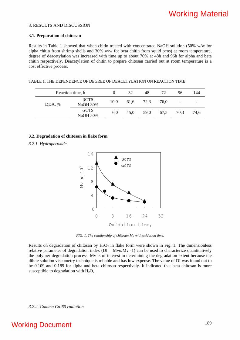
3. RESULTS AND DISCUSSION 3.1. Preparation of chitosan Results in Table 1 showed that when chitin treated with concentrated NaOH solution (50% w/w for alpha chitin from shrimp shells and 30% w/w for beta chitin from squid pens) at room temperature, degree of deacetylation was increased with time up to about 70% at 48h and 96h for alpha and beta chitin respectively. Deacetylation of chitin to prepare chitosan carried out at room temperature is a cost effective process. TABLE 1. THE DEPENDENCE OF DEGREE OF DEACETYLATION ON REACTION TIME
Reaction time, h 0 32 48 72 96 144 βCTS
NaOH 30% 10,0 61,6 72,3 76,0 - -
DDA, % αCTS
NaOH 50% 6,0 45,0 59,0 67,5 70,3 74,6
3.2. Degradation of chitosan in flake form
3.2.1. Hydroperoxide
16
Oxidation time,
Mv × 105
βCTS αCTS
12
8
4
0 0 328 16 24
FIG. 1. The relationship of chitosan Mv with oxidation time.
Results on degradation of chitosan by H2O2 in flake form were shown in Fig. 1. The dimensionless relative parameter of degradation index (DI = Mvo/Mv -1) can be used to characterize quantitatively the polymer degradation process. Mv is of interest in determining the degradation extent because the dilute solution viscometry technique is reliable and has low expense. The value of DI was found out to be 0.109 and 0.189 for alpha and beta chitosan respectively. It indicated that beta chitosan is more susceptible to degradation with H2O2. 3.2.2. Gamma Co-60 radiation
189
Working Material
Working Document

FIG. 2. The relationship of chitosan Mv with dose.
0 0
Dose, kGy
Mv × 105
βCTS
αCTS
16
12
8
4
488 16 24 32 40
esults on degradation of chitosan by gamma Co-60 radiation were shown in Fig. 2. Gs - value can be
.3. Preparation of oligochitosan
TABLE 2. THE CORRELATION OF WATER SOLUBLE CHITOSAN CONTENT WITH DOSE
Dose, kGy 4 8 12 16 24 32 48
Rused to estimate the susceptibility of polymer to radiation degradation. The Gs- value was calculated from Fig. 1 to be of 0.20 and 0.38 scissions/100eV for alpha and beta chitosan, respectively. That means beta chitosan is also more susceptible to radiation degradation as in the case with H2O2. 3
0 % H2O2 4 6 6 7- 5,0 - 1,9 9,8 8,2 - βCTS 1 % H2O2 74,9 86,0 93,9 100,0 - - -
0 % H2O2 - 21,3 - 29,2 - 4 74,9 3,4
WSC, %
αCTS 66,3 74,6 87,6 91,2 1 % H2O2 - - -
able 2 indicated that irradiation of chitosan solution containing 1% H2O2, the required
.4. Field test of elicitation effect of oligochitosan for sugarcane and rice
.4.1. Field test for sugarcane
Results in Tdose for obtaining 75% water soluble oligomer was reduced from 48kGy to 8kGy for alpha chitosan and from 32kGy to 4kGy for beta chitosan. The use of H2O2 together with radiation is very effective for degradation of chitosan in solution. 3 3
190
Working Material
Working Document
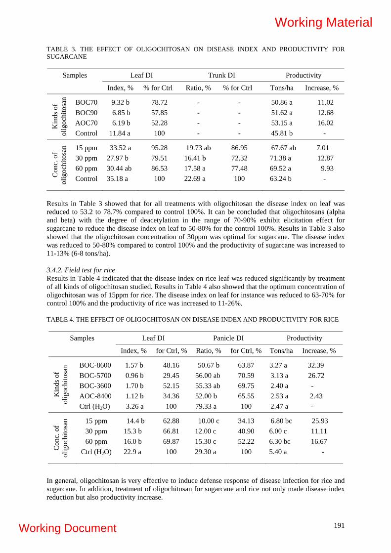
TABLE 3. THE EFFECT OF OLIGOCHITOSAN ON DISEASE INDEX AND PRODUCTIVITY FOR SUGARCANE
Leaf DI Trunk DI Productivity Samples
Index, % % for Ctrl Ratio, % % for Ctrl Tons/ha Increase, %
Kin
ds o
f ol
igoc
hito
san BOC70
BOC90 AOC70 Control
9.32 b 6.85 b 6.19 b
11.84 a
78.72 57.85 52.28 100
- - - -
- - - -
50.86 a 51.62 a 53.15 a 45.81 b
11.02 12.68 16.02
-
Con
c. o
f ol
igoc
hito
san 15 ppm
30 ppm 60 ppm Control
33.52 a 27.97 b 30.44 ab 35.18 a
95.28 79.51 86.53 100
19.73 ab 16.41 b 17.58 a 22.69 a
86.95 72.32 77.48 100
67.67 ab 71.38 a 69.52 a 63.24 b
7.01 12.87 9.93 -
Results in Table 3 showed that for all treatments with oligochitosan the disease index on leaf was reduced to 53.2 to 78.7% compared to control 100%. It can be concluded that oligochitosans (alpha and beta) with the degree of deacetylation in the range of 70-90% exhibit elicitation effect for sugarcane to reduce the disease index on leaf to 50-80% for the control 100%. Results in Table 3 also showed that the oligochitosan concentration of 30ppm was optimal for sugarcane. The disease index was reduced to 50-80% compared to control 100% and the productivity of sugarcane was increased to 11-13% (6-8 tons/ha).
3.4.2. Field test for rice Results in Table 4 indicated that the disease index on rice leaf was reduced significantly by treatment of all kinds of oligochitosan studied. Results in Table 4 also showed that the optimum concentration of oligochitosan was of 15ppm for rice. The disease index on leaf for instance was reduced to 63-70% for control 100% and the productivity of rice was increased to 11-26%. TABLE 4. THE EFFECT OF OLIGOCHITOSAN ON DISEASE INDEX AND PRODUCTIVITY FOR RICE
Leaf DI Panicle DI Productivity Samples
Index, % for Ctrl, % Ratio, % for Ctrl, % Tons/ha Increase, %
Kin
ds o
f ol
igoc
hito
san BOC-8600
BOC-5700 BOC-3600 AOC-8400 Ctrl (H2O)
1.57 b 0.96 b 1.70 b 1.12 b 3.26 a
48.16 29.45 52.15 34.36 100
50.67 b 56.00 ab 55.33 ab 52.00 b 79.33 a
63.87 70.59 69.75 65.55 100
3.27 a 3.13 a 2.40 a 2.53 a 2.47 a
32.39 26.72 - 2.43 -
Con
c. o
f ol
igoc
hito
san 15 ppm
30 ppm 60 ppm
Ctrl (H2O)
14.4 b 15.3 b 16.0 b 22.9 a
62.88 66.81 69.87 100
10.00 c 12.00 c 15.30 c 29.30 a
34.13 40.90 52.22 100
6.80 bc 6.00 c 6.30 bc 5.40 a
25.93 11.11 16.67
-
In general, oligochitosan is very effective to induce defense response of disease infection for rice and sugarcane. In addition, treatment of oligochitosan for sugarcane and rice not only made disease index reduction but also productivity increase.
191
Working Material
Working Document

3.5. Procedure for production of biotic elicitor oligochitosan - 10kg chitosan (DDA 65-75%, MW~ 100-200kDa) - Soaking with water in plastic tank - Adding 6kg lactic acid and stirring - Adding 4litters of H2O2 50% and water up to 200litters while stirring - Filtering through stainless steel net (~100mesh) - Packing in plastic can (25litter/can) - Irradiation with gamma Co-60 at 4kGy for beta chitosan (8kGy for alpha chitosan) - Neutralizing irradiated chitosan solution with NaOH to pH 5 - Adding ethanol with final concentration of 40% v/v for preservation - Quality control and packaging - Product: “biotic elicitor oligochitosan”. The characteristics of biotic elicitor oligochitosan product are as follows: - Concentration of oilgochitosan in solution: 30g/L - Content of water soluble oligomer pH7: 70-80% - Mw: 5,000 - 10,000 - Ethanol concentration: 40% v/v - Expire date: 2 years min. 4. CONCLUSIONS
- Preparation of alpha and beta chitosan with degree of degradation of about 70% and Mv in the range of 6 - 10 × 105 Da by deacetylation of chitin at room temperature.
- Reduction regulation of chitosan Mv to 1 - 2 × 10⁵ Da by degradation with H2O2. - Preparation of oligohitosan with molecular weight of 6 - 10 × 103 Da by irradiation of chitosan
solution at dose 48kGy for alpha chitosan and 32 kGy for beta chitosan. - Irradiation of chitosan solution containing 1% H2O2 to reduce the required dose to 8kGy for alpha
chitosan and 4kGy for beta chitosan. - Setting up production procedure for biotic elicitor oligochitosan with characteristics: oligochitosan:
30g/L; water soluble oligomer: 70-80%; Mw: 5,000-10,000; ethanol: 40% v/v, pH 5 and expire date: 2 years.
- Optimum concentration of biotic elicitor oligochitosan was of 30ppm for sugarcane and 15ppm for rice. The disease index (U. scitaminea and C. falcatum) for sugarcane was reduced to 45-72% compared to control 100% and sugarcane productivity was increased up to 13% (8 tons/ha). The disease index (P. grisea) for rice was reduce to 34-53% compared to control 100% and the productivity of rice was increased 11-26% (0.6-1.4 tons/ha).
REFERENCES
[1] HIEN, N.Q., Application of radiation technology in medicine and agriculture, Vietnam J. Sci.
Technol., 39 (2001) 57-64. [2] Emerging applications of radiation processing, IAEA-TECDOC-1386, IAEA, Vienna, Jan.
2004. [3] Radiation processing of polysaccharides, IAEA-TECDOC-1422, IAEA, Vienna, Nov. 2004. [4] HADWIGER, L.A., BECKMAN, J.M., Chitosan as a component of pea-fusarium solani
interaction, Plant Physiol., 66 (1980) 205-211. [5] ALBERSHEIM, P., DARVILL, A.G., Oligosaccharin, Scientific of America, 253 (1985) 44-55. [6] RYAN, C.A., Oligosaccharides as recognition signals for the expression of defensive genes in
plants, Biochemistry, 27 (1988) 8879-8883. [7] POSPIESZNY, H. et al., Induction of antiviral resistance in plants by chitosan, Plant Science, 79
(1991) 63-68.
192
Working Material
Working Document

[8] DARVILL, A. et al ., Oligosaccharins- Oligosaccharides that regulate growth, development and defense responses in plants, Glycobiology, 2 (1992) 181-198.
[9] JOHN, M. et al., Cell signaling by oligosaccharides, Trend in Plant Science, 2 (1997) 111-115. [10] VANDER, P., Comparison of the ability of partially N-acetylated chitosans and
chitooligosaccharides to elicit resistance reactions in wheat leaves, Plant Physiol., 118 (1998) 1353-1359.
[11] AKIMOTO, C., AOYAGI, H., TANAKA, H., Endogenous elicitor-like effects of alginate on physiological activities of plant cells, Appl. Microbiol. Biotechnol., 52 (1999) 429-436.
[12] KUME, T. et al., Utilization of carbohydrate by radiation processing, Radiat. Phys. Chem., 63 (2000) 625-627.
[13] VASYUKOVA, N.I. et al., Modulation of plant resistance disease by water soluble chitosan, Appl. Biochem. Microbiol., 37 (2001) 103-109.
[14] CHOI, W.S. et al, Preparation of chitosan oligomers by irradiation, Polym. Degr. Stab., 78 (2002) 533-538.
[15] BRUGNEROTTO J. et al., An infrared investigation in relation with chitin and chitosan characterization, Polymer, 42 (2001) 3569-3580.
[16] KNAUL J.Z. et al., Characterization of deacetylated chitosan and chitosan molecular weight review, Canadian J. Chem., 76 (1998) 1699-1706.
193
Working Material
Working Document

194
Working Material
Working Document
195
Participants List
2nd RCM on "Development of radiation-processed products of
natural polymers for application in agriculture, healthcare, industry and environment"
12-16 October 2009
Reims, France 1. Argentina Mr. Eduardo Esteban Smolko
Comisión Nacional de Energía Atómica (CNEA) Irradiation Processing Lab Avenida del Libertador 8250 C1429BNP Buenos Aires 0054 11 67798571 0054 11 67798322 [email protected]
2. Bangladesh Mr. M. Emdadul Haque Institute of Nuclear Science and Technology Atomic Energy Research Establishment Ganakbari, Savar P.O. Box 3787 Dhaka 100 88027790409 88001714328895 [email protected]
3. Brazil Ms. Celina Lopes Duarte Instituto de Pesquisas Energéticas e Nucleares IPEN-CNEN/SP Av. Lineu Prestes 2422 05508-900 São Paulo, SP [email protected]
4. Canada Ms. Monique Lacroix Monique Lacroix INRS - Institut Armand-Frappier 531 Blvd. des Prairies Laval city, Québec Canada H7V 1B7 001 450 6875010 4489 001 450 6875792 [email protected]
5. Egypt Mr. Hassan Ahmed AbdEl-Rehim Atomic Energy Authority (AEA); National Centre for Radiation Research and Technology (NCRRT) 3 Ahmed El-Zomor P.O. Box 29, Madinat Nas Cairo 0020 2 27492298 [email protected]
Working Material
Working Document

196
6. France Mr. Xavier Coqueret Université de Reims Champagne Ardenne Institut de Chimie moléculaire; UMR CNRS 6229 51687 Reims cedex 2 0033 3 26 913338 0033 3 26 913338 [email protected]
7. Ghana Mr. Fidelis Ocloo Ghana Atomic Energy Commission (GAEC) Biotechnology and Nuclear Agriculture Research Institute P.O. Box 80 Legon Accra 00233 21 402286 00233 21 402286 [email protected]
8. India Mr. Satish Parsran Ramnani Radiation Technology Development Section HIRUP Bhabha Atomic Research Centre Department of Atomic Energy Trombay Mumbai, Maharashtra 400 085 India [email protected]
9. Malaysia Mr. Khairul Zaman Haji Mohd Dahlan Division Of Radiation Processing, MINT Malaysia Nuclear Agency MOSTI Bangi, 43000 Kajang, Selangor Darul Ehsan, [email protected]
10. Philippines Ms. Lucille Abad Philippine Nuclear Research Institute (PNRI) Commonwealth Avenue, Diliman P.O. Box 213 Quezon City 1101 0063 2 9201655 0063 2 9201646 [email protected] [email protected]
11. Poland Mr. Piotr Ulanski Technical University of Lodz; Institute of Applied Radiation Chemistry Wroblewskiego 15 93-590 Lodz 0048 42 6313196 0048 42 6840043 [email protected]
Working Material
Working Document

197
12. Turkey Mr. Murat Sen Hacettepe University; Faculty of Science; Department of Chemistry Kimya Bolumu H.U Beytepe Kampusu, Beytepe 06532 Ankara 0090 312 2977989 0090 312 2987989 [email protected]
13. United Kingdom Mr. Saphwan Al-Assaf Reader in Chemistry North East Wales Institute of Higher Education Mold Road Wrexham, North Wales LL11 2AW 0044 1978 293321 0044 1978 293370 [email protected] [email protected]
14. Vietnam Mr. Quoc Hien Nguyen Research and Development Center for Radiation Technology, Vietnam Atomic Energy Commission 202A, Str. 11, Linh xuan Ward, Thu duc District Ho Chi Minh City 84 8 2829159 0084 8 8975921 [email protected]
Cost Free Observer
15. Germany Mr. Torsten Richter Heppe Medical Chitosan GmbH Heinrich-Damerow-Str. 1 D-06120 Halle(Saale) Tel: +49(0)345 27996 307 Fax: +49(0)345 27996 378 Web: www.medical-chitosan.com [email protected]
Scientific Secretary
IAEA Ms. Agnes Safrany Industrial Applications and Chemistry Section Division of Physical and Chemical Sciences Department of Nuclear Sciences and Applications P.O. Box 100 Wagramer Strasse 5 A-1400 Vienna Austria Telephone: +43-1-2600 ext. 21750 Facsimile: +43-1-26007 [email protected]
Working Material
Working Document

198
Working Material
Working Document

ANNEX
199
Working Material
Working Document

200
Working Material
Working Document

IAEA Co-ordinated Research Programme:
Development of Radiation-Processed Products of Natural Polymers for Application in Agriculture,
Healthcare, Industry and Environment
PROTOCOL FOR DETERMINATION OF INTRINSIC VISCOSITY OF CHITOSAN
Version 3.
Elaborated by:
• Janusz M. Rosiak, Piotr Ulański (Institute of Applied Radiation Chemistry,
Technical University of Lodz, Poland)
• Saphwan Al-Assaf (Phillips Hydrocolloids Centre, Glyndwr University, UK)
201
Working Material
Working Document

INTRODUCTION
According to the agreement reached at the 1st Meeting of the IAEA Co-ordinated Research
Programme “Development of Radiation-Processed Products of Natural Polymers for Application in
Agriculture, Healthcare, Industry and Environment”, a group of the participating laboratories has
undertaken to:
1. Perform analysis of identical chitosan samples by viscometry and, where available, by laser
light scattering and gel permeation chromatography. The analysis is focused at determining
the average molecular weight and molecular weight distribution.
2. Irradiate samples of this well characterized chitosan in similar conditions by electron beam or
gamma rays using with the same set of doses
3. Analyze the irradiated samples by the same methods and procedures as above.
Procedures described in this protocol are aimed at conducting a inter-laboratory test on determination
of intrinsic viscosity of a model chitosan sample. Each participating laboratory is provided with ca. 20
g of the same chitosan (originating from a single production batch) manufactured and donated for the
purpose of this study by Heppe company. The delivered amount should be sufficient for performing all
the analyses listed above on unirradiated and irradiated material, therefore it is advised not to use more
than 2 g for the analyses on unirradiated chitosan.
It is proposed that the viscometric measurements would be performed in two solvents. The first one
(0.2 M acetic acid / 0.15 M ammonium acetate) is considered by leading chitosan laboratories as
probably the best one being elaborated so far with respect to molecular dissolution of chitosan and
prevention of aggregation. The second one (0.1 M hydrochloric acid) is proposed as an alternative,
with the advantages of having less components (thus reducing the number of sources of error in
preparation) and being directly available in the ready-to-use form from the major suppliers.
202
Working Material
Working Document
1. BEFORE YOU START:
1.1. Familiarize yourself and all the involved staff with the Material Safety Data Sheets regarding
chitosan (enclosed) and regarding other chemicals listed below (available from the
manufacturers or suppliers), paying special attention to first-aid measures and exposure
controls / personal protection.
1.2. Make sure that you have appropriate equipment, glassware and chemicals, as specified in
points 1.3. – 1.5. below.
1.3. Equipment (asterisk* denotes an item which is not necessary and/or may be replaced)
1.3.1. Vacuum drying oven* OR, in unavailable, standard laboratory drying oven capable to
maintain constant temperature of 60 ºC with a +- 5 deg accuracy
1.3.2. Simple thermometer for the range of ca. 0 – 100 ºC, for checking the actual temperature
in the oven.
1.3.3. Dessicator*
1.3.4. Analytical balance (1 mg or better accuracy)
1.3.5. Magnetic stirrer* with a stirrer bar (coated by Teflon, other neutral polymer or glass)
OR a laboratory shaker
1.3.6. pH-meter with a pH electrode* with a set of calibration buffers OR pH test strips OR
universal indicator paper
1.3.7. Adjustable automatic pipette* for 1-5 ml or 1-10 ml with corresponding replaceable tips
OR a set of glass volumetric pipettes (the volumes depend on your viscometer; typically
fixed volume 5 ml and 10 ml, and graded 10 ml)
1.3.8. Ubbelohde-type glass viscometer, chosen according to the capillary diameter to provide
flow time for water at 25 ºC in the range 80 – 120 s. For example: Ubbelohde viscometer
531 10/I of Schott Instruments.
1.3.9. In case you don’t use any automatic viscometry system (see 1.3.9) but just the
viscometer alone, you would need a piece of tubing and a syringe (or rubber pipette
filler) OR any other arrangement to facilitate filling the viscometer capillary with the
studied liquid (i.e., to suck the liquid from the bottom reservoir into the capillary).
Caution: the tubing + syringe set can only be connected to the viscometer tube when
203
Working Material
Working Document
filling the capillary, but not during the measurement itself, since during the
measurement all viscometer tubes must be open.
1.3.10. Automatic viscometry system* (such as ViscoSystem AVS 370 of Schott Instruments),
allowing for automatic filling of the capillary and measurements of flow time in a pre-
programmed manner.
1.3.11. A thermostatic water bath with at least 1 transparent side wall, fitting in size to the
viscometer, capable of maintaining constant temperature with 0.1 deg or better accuracy.
Please note, that maintaining a constant temperature of 25.0 ºC and in most cases also
of 30.0 ºC requires both a thermostatically controlled heater and a cooling device (in a
simplest version – a metal tube loop with cold water running through).
1.3.12. Precise thermometer with a measuring range up to ca. 50 ºC and at least 0.1 deg
accuracy
1.3.13. A viscometer stand allowing to hold the viscometer in the water bath in a stable,
vertical position
1.3.14. Stop-watch (unless you use an automatic system for measuring flow time)
1.3.15. Refrigerator* (freezing capacity not needed), if you need to store your prepared
solvents and solutions for more than a day
1.3.16. Computer with any data processing software (Origin is a good choice, but, if it is not
available, Excel would suffice)
204
Working Material
Working Document

1.4. Glassware and disposables
1.4.1. Vessel(s) or vial(s) with tightly fitting lids. Glass vials with plastic screw-type caps may
be convenient for this purpose. The volume should allow to contain the amount of
chitosan which you need to dry and store (you may use several vials at a time). If you
have a desiccator, make sure that the vessel(s) size allows their storage in the desiccator.
1.4.2. Volumetric flasks for 250 ml and 100 ml.
1.4.3. Small beakers (50-100 ml capacity)
1.4.4. Glass vials of 10-20 ml capacity with tight lids
1.4.5. Single-use plastic syringes
1.4.6. Disposable filtering units (mountable on a syringe) with a hydrophilic membrane of
0.45 micron pore size. Recommended: Minisart NML (Cat. No. 16555 or 17598) of
Sartorius. Filtering units do not need to be sterile.
1.4.7. Parafilm* (a moisture-proof plastic film that molds to most surfaces, used to seal flasks,
etc., Cole-Parmer Cat. No. EW-06720-50)
1.5. Chemicals:
1.5.1. Distilled water or water of similar purity obtained by other technique
1.5.2. Acetic acid, preferably as a ready 1.0 M (equivalent to 1.0 N) or 0.5 M (0.5 N) solution
(for instance “Acetic acid solution, volumetric standard, 1.0 N in H2O”, Cat. No. 318590
of Sigma-Aldrich). If unavailable, use concentrated acetic acid of high purity (“p.a.” or
equivalent)
1.5.3. Ammonium acetate, high purity (“p.a.” or equivalent)
1.5.4. Hydrochloric acid, preferably a ready 0.1 M (equivalent to 0.1 N) solution (for instance
“Hydrochloric acid standard solution 0.1 N in H2O”, Cat. No. 318965 of Sigma-
Aldrich). If unavailable, use concentrated hydrochloric acid solution (typically 30 - 37
%) of high purity (“p.a.” or equivalent)
1.5.5. If you use a dessicator, you may also need fresh water-absorbing medium (a desiccant)
to fill the bottom of the dessicator (for instance: Drierite® desiccants of Cole-Parmer;
other simple desiccants are silica gel, calcium sulfate, calcium chloride)
205
Working Material
Working Document

1.5.6. You may need some other chemicals as well. For instance, before using a new
viscometer you may have to clean it using chemicals and procedure described in the
operating instructions of the viscometer.
2. DRYING AND STORAGE
2.1. Chitosan is a hygroscopic material which, when not dried and stored properly, in equilibrium
with regular lab air may contain even over 10 % of water. For molecular weight
measurements, concentration of chitosan solutions should be precisely known. It means that
for preparation of a solution either (a) chitosan must be carefully dried before determining its
weight, or (b) water content in a given sample must be known in order to calculate
appropriate correction. In our CRP programme it seems that method (a) is more suitable
(drying the chitosan and storing it in a dry state), since further steps in the programme
involve chitosan irradiation, which should be performed in the dry state, because moisture
does influence the yields of degradation.
2.2. Drying procedure:
2.2.1. Select appropriate vessel(s) or vials with lids (see above) for drying and storing your
chitosan. Wash the vessels, flush them twice with distilled water and dry completely.
2.2.2. Determine weight of your empty vessel with lid at the highest available accuracy (use
analytical balance, but make sure you do not overload it). In case you use many vessels,
mark each vessel and its lid clearly with matching numbers or letters and determine the
weight of each vessel + lid set.
2.2.3. Place your chitosan in the vessel(s) and determine the weight of filled vessel(s)
complete with lid(s). Calculate the weight of chitosan in each vessel.
If you have a vacuum drying oven:
2.2.4. Set the temperature in the oven to 40 ºC, wait until the temperature is reached and
stabilized (check it with a thermometer, it should not deviate by more than 5 deg), put
your samples in (without lids or with lids placed loosely or placed at an angle to the top
of the vessel to provide space for the water vapor to escape), and start the vacuum
operation according to the operating instructions of the oven.
206
Working Material
Working Document

2.2.5. Every 24 h open the oven according to the operating instructions, close the lid(s) of the
vessel(s), allow the vessel(s) to cool down to ambient temperature (preferably in a
desiccator) and check their weight (chitosan in vessels with closed lids). Calculate the
weight of chitosan in each vessel.
2.2.6. Continue drying until the difference in weight in subsequent measurements is lower
than 0.5 %.
If you don’t have a vacuum drying oven:
2.2.7. Set the temperature of an ordinary lab drying oven or oven to 60 ºC, wait until the
temperature is reached and stabilized (check it with a thermometer, it should not deviate
by more than 5 deg), put your samples in (without lids or with lids places at an angle to
the top of the vessel to provide space for the water vapor to escape).
2.2.8. Every 24 h open the oven, close the lid(s) of the vessel(s), allow the vessels to cool
down to ambient temperature (preferably in a desiccator) and check the weight (chitosan
in vessels with closed lids). Calculate the weight of chitosan in each vessel.
2.2.9. Continue drying until the difference in weight in subsequent measurements is lower
than 0.5 %.
2.3. Storage
2.3.1. Store the closed vessels with chitosan in a desiccator, if available. Alternatively, store at
room temperature in tightly closed vessels, preferably additionally sealed with Parafilm,
avoiding exposure to direct sunlight, away from sources of heat.
207
Working Material
Working Document

3. VISCOSITY MEASUREMENTS USING THE ACETIC ACID / AMMONIUM ACETATE
SOLVENT
3.1. Preparing the solutions
3.1.1. For viscosity measurements in the acetic acid / ammonium acetate system (0.2 M acetic
acid / 0.15 M ammonium acetate), prepare the solvent as follows. Charge 2.891 g of
ammonium acetate into a dry 250-ml volumetric flask. Add ca. 50 ml of distilled water.
Calculate the volume of your ready acetic acid solution or of your concentrated acetic
acid corresponding to 0.05 mol (3.00 g of pure acid) and transfer exactly that volume
into the volumetric flask. If you use concentrated acetic acid solution, read the
concentration (in %) and density in the label or specifications. If your concentrated acid
solution of a density d (in g / cm3) contains C% percent of pure acid, the volume of the
concentrated acid solution Vs (in cm3) which corresponds to 3.00 g of pure acid is:
%
%00.3Cd
Vs =100
×
Stir well, fill up to the mark with distilled water.
3.1.2. You may check the pH of the solvent, as one of the indications that the composition is
correct. To do so, take a small sample (10 – 15 ml) of the ready solvent into a small vial
or beaker and measure pH using a pH-meter (remember to calibrate it properly prior to
the measurement according to the operating instruction of your pH-meter) or pH test
strips or universal indicator paper. The correct pH of the acetic acid / ammonium acetate
solvent should be 4.5 +- 0.5.
3.1.3. If there is a necessity to store the solvent, keep it in refrigerator in a tightly closed
volumetric flask. The acetic acid / ammonium acetate solvent should be used within 3
days.
3.1.4. Using an analytical balance, weigh carefully ca. 0.100 g of dry chitosan (note the exact
weight) and transfer it into a 100-ml volumetric flask. Put in a stirrer bar. Add ca. 80 ml
of the chosen solvent, close the flask with a stopper and put it on a magnetic stirrer
(alternatively, you may use a lab shaker). Stir gently for 24 hours at room temperature.
Remove the stirrer bar and fill the flask with solvent to the mark. Shake gently.
3.1.5. Chitosan solutions should be in principle used for measurements within one day. If
necessary, store them in a refrigerator (do not freeze them) for not longer than 3 days.
208
Working Material
Working Document
3.1.6. Directly before charging the chitosan solution into the viscometer, the solution should
be filtered. Pour ca. 20 ml of the solution into a small vial or beaker, suck it into a 20-ml
plastic syringe (if you have a 10-ml syringe you will have to run the filtration twice),
while keeping the syringe upright (the orifice up) remove air bubbles by gently pressing
the plunger, fix the filtering unit (see 1.4.6.) to the syringe, still keeping the syringe
upright remove air from the filtering unit (by pressing the plunger gently until a drop of
liquid passes through the tip of the unit), and filter the contents into a glass vial after
discarding the first 1-2 ml to waste. Caution: remember that the filtering unit is a single-
use disposable material – never use it for a second time.
3.1.7. Close the glass vial with a lid. Use the contents immediately.
3.2. Determining flow times of the solvent
3.2.1. Wash your viscometer carefully, including the capillary (follow the cleaning procedure
suggested in the operation instructions), flush it several times with distilled water and let
it dry completely. It is advisable to do the washing a day before the measurements.
Caution: before using a new viscometer for the first time, clean it according to the
procedure described in operating instructions.
3.2.2. Place your viscometer in the water bath and fix the viscometer to the stand in such a
way that the capillary is in exactly vertical position and the whole viscometer except ca.
2 - 3 cm of the top parts of the glass tubes is submerged in water.
3.2.3. In the operating instructions of your viscometer check the minimum sample volume
(Vmin). Typically it is 10 ml (in some cases it may be 15 ml or some other value). Using
an automatic pipette or a glass pipette, fill the viscometer with the Vmin volume of the
acetic acid / ammonium acetate solvent through the filling tube (usually the widest one).
3.2.4. Set the temperature at your water bath controller at 25.0 ºC. Turn on the cooling system
(or, if you use a metal tube loop with cold water, start the cold water flow) and turn on
the heating system. Follow the bath temperature using a precise thermometer. Wait until
the temperature stabilizes at 25.0 +- 0.1 ºC. Wait further 10 minutes before you start the
measurements.
3.2.5. Follow the operating instructions of your viscometer (or automatic viscometer system)
in performing the measurements of flow time. Write down all the flow time readings in
the corresponding field (“t (individual) [s])”of Table 1 (see last two pages). Run the
measurements five times.
209
Working Material
Working Document

3.2.6. Calculate the average of these five flow times and check if every individual flow time
differs from the mean value by no more than 1.0 % if you use a stop watch for
measuring flow time or by no more than 0.5 % if you use an automatic device. If all five
values are within this limit, go to 3.2.8.
3.2.7. If any value deviates from the average by more than 1.0 % (or 0.5 %), cross this single
value out and make more measurements. The number of new measurements should be
equal to the number of values crossed out after the previous step. Go to 3.2.6.
3.2.8. When the measurements for the solvent are done, remove the viscometer from the water
bath, pour out the solvent, flush the viscometer several times with distilled water and let
it dry completely.
3.3. Determining flow times of a dilution series of chitosan solution
3.3.1. Find out in the operating instructions of your viscometer the maximum sample volume
(Vmax) suitable for measurements. Typically it is ca. 40 – 60 ml. If this value is not
provided, make a test yourself (when doing so please ensure that the level of the liquid at
your tested maximum volume is at least 2 cm below the lower exit of the capillary to
ensure free flow of the liquid from the capillary – do not overfill). Calculate the volume
increment for a concentration series of chitosan as ΔV = 0.25 × (Vmax – Vmin). For
practical reasons, especially when not using adjustable automatic pipettes, you may like
to round this value down to the nearest full ml or the nearest full 5 ml. For Ubbelohde
viscometer 531 10/I of Schott Instruments typically Vmin = 10 ml, Vmax = 50 ml, ΔV = 10
ml.
3.3.2. Fix the viscometer in the water bath as described before. Fill the viscometer with Vmin
volume of the chitosan solution in acetic acid / ammonium acetate solvent.
3.3.3. Proceed as in 3.2.4 until stabilization of temperature at 25.0 +- 0.1 ºC. Wait further 10
minutes before you start the measurements.
3.3.4. Follow the operating instructions of your viscometer (or automatic viscometer system)
in performing the measurements of flow times for chitosan solution. Write down all the
flow time readings in the corresponding field (“t (individual) [s])”of Table 1 (see two
last pages). Run the measurements five times.
3.3.5. Calculate the average of these five flow times and check if every individual flow time
differ from their mean value by no more than 1.0 % if you use a stop watch for
210
Working Material
Working Document

measuring flow time or no more than 0.5 % if you use an automatic device. If all five
values are within this limit, go to 3.3.7.
3.3.6. If any value deviates from the average by more than 1.0 % (or 0.5 %), cross this single
value out and make more measurements. The number of new measurements should be
equal to the number of values crossed out after the previous step. Go to 3.3.5.
3.3.7. Dilute the original solution by adding a ΔV volume of the acetic acid / ammonium
acetate solvent. Wait 15 minutes for complete equilibration of temperature and
concentration. Make the measurements of the flow time for chitosan solution, using the
same rules as described in 3.3.4.-3.3.6.
3.3.8. Repeat step 3.3.7. by adding subsequent ΔV volumes of the solvent and making flow
time measurements until the total volume of Vmax is reached and the flow times for this
sample volume are measured. In this way you should have collected five flow times for
each of five concentrations of chitosan.
211
Working Material
Working Document
3.4. Comments:
3.4.1. All the volumes should be measured accurately, using an adjustable automatic pipette or
a graded glass pipette.
3.4.2. Your measurement stand with the thermostatic water bath should be, if possible, located
in a place of relatively constant ambient temperature (avoid locating it close to the door
which are frequently opened, in direct sunlight, etc.) and free of dust.
3.4.3. During the measurements check frequently if temperature of water bath is within the
limits of 25.0 +- 0.1 ºC.
3.4.4. If your results for the solvent or for a given polymer concentration are not stable, this
may be caused by instability of temperature. Check if the temperature of the water bath
is stable within the defined limits. If there is a constant rising trend in the temperature,
check if your cooling is functioning well.
3.4.5. If, from time to time, you experience a flow time much higher than the average for a
given concentration, it may indicate a presence of an impurity (polymer aggregate, dust
particle, etc.) blocking the flow through the capillary. One should stop the
measurements, remove the solution, clean the viscometer carefully, dry it and re-start the
whole measurement procedure.
212
Working Material
Working Document

4. VISCOSITY MEASUREMENTS USING THE HYDROCHLORIC ACID SOLVENT
4.1. Preparing the solutions
4.1.1. The solvent in measurements described in Section 4 is aqueous solution of hydrochloric
acid where the HCl concentration is 0.100 M. It is referred to in the text as “hydrochloric
acid solvent”.
4.1.2. For viscosity measurements in the hydrochloric acid solvent, either use a ready-made
0.1 M aqueous hydrochloric acid standard solution (see 1.5.4.), or prepare such a
solution (250 ml) by appropriate dilution of a concentrated HCl solution of reasonable
purity (p.a. or equivalent). The amount of pure HCl corresponding to 0.025 mol is 0.912
g. If you use concentrated hydrochloric acid solution, read the concentration (in %) and
density in the label or specifications. If your concentrated acid solution of a density d (in
g / cm3) contains C% percent of pure acid, the volume of the concentrated acid solution
Vs (in cm3) which corresponds to 0.912 g of pure acid is:
%
%912.0Cd
Vs =100
×
4.1.3. You may check the pH of the solvent, as one of the indications that the composition is
correct. To do so, take a small sample of the ready solvent into a vial or a beaker and
measure pH using a pH-meter (remember to calibrate it properly prior to the
measurement according to the operating instruction of your pH-meter; make sure if the
operating ranges of your electrode and pH-meter cover pH 1) or pH test strips or
universal indicator paper. The correct pH of hydrochloric acid solvent it should be 1.0 +-
0.5.
4.1.4. If there is a necessity to store the solvent, keep it in refrigerator in tightly closed
volumetric flasks, for no longer than two weeks.
4.1.5. Using an analytical balance, weigh carefully ca 0.100 g of dry chitosan (note the exact
weight) and transfer it into a 100-ml volumetric flask. Put in a stirrer bar. Add ca. 80 ml
of the chosen solvent, close the flask with a stopper and put it on a magnetic stirrer
(alternatively, you may use a lab shaker instead of magnetic stirrer). Stir gently for 24
hours at room temperature. Remove the stirrer bar and fill the flask with solvent to the
mark. Shake gently.
4.1.6. Chitosan solutions in hydrochloric acid should be used for measurements within one
day after preparation.
213
Working Material
Working Document
4.1.7. Directly before charging the chitosan solution into the viscometer, the solution should
be filtered. Pour ca. 20 ml of the solution into a small beaker, suck it into a 20-ml plastic
syringe (if you have a 10-ml syringe you will have to run the filtration twice), while
keeping the syringe upright (the orifice at the top) remove air bubbles by gently pressing
the plunger, fix the filtering unit (see 1.4.6.) to the syringe, still keeping the syringe
upright remove air from the filtering unit (by pressing the plunger gently until a drop of
liquid passes through the tip of the unit), and filter the contents into a glass vial after
discarding the first 1-2 ml to waste. Caution: due to the limited resistance of the filtering
unit membrane to strongly acidic media, the contact time of the solution with the
filtering unit should not be longer than necessary. Remember that the filtering unit is a
single-use disposable material – never use it for a second time.
4.1.8. Close the glass vial with a lid.
4.2. Determining flow time of the solvent
4.2.1. Wash your viscometer carefully, including the capillary, flush it several times with
distilled water and let it dry completely.
4.2.2. Place your viscometer in the water bath and fix the viscometer to the stand in such a
way that the capillary is in exactly vertical position and the whole viscometer except 3-5
cm of the top parts of the glass tubes is submerged in water.
4.2.3. In the operating instructions of your viscometer check the minimum sample volume
(Vmin). Typically it is 10 ml (in some cases it may be 15 ml or some other value). Using
an automatic pipette or a glass pipette, fill the viscometer with the Vmin volume of the
hydrochloric acid solvent through the filling tube (usually the widest one).
4.2.4. Set the temperature at your water bath controller at 30.0 ºC. Turn on the cooling system
(or, if you use a metal tube loop with cold water, start the water flow) and turn on the
heating system. Follow the bath temperature using a precise thermometer. Wait until the
temperature stabilizes at 30.0 +- 0.1 ºC. Wait further 10 minutes before you start the
measurements.
4.2.5. Follow the operating instructions of your viscometer (or automatic viscometer system)
in performing the measurements of flow time. Write down all the flow time readings in
the corresponding field (“t (individual) [s])”of Table 1 (see last two pages). Run the
measurements five times.
214
Working Material
Working Document

4.2.6. Calculate the average of these five flow times and check if every individual flow time
differs from their mean value by no more than 1.0 % if you use a stop watch for
measuring flow time or no more than 0.5 % if you use an automatic device. If all five
values are within this limit, go to 4.2.8.
4.2.7. If any value deviates from the average by more than 1.0 % (or 0.5 %), cross this single
value out and make more measurements. The number of new measurements should be
equal to the number of values crossed out after the previous step. Go to 4.2.6.
4.2.8. When the measurements for the solvent are done, remove the viscometer from the water
bath, pour out the solvent, flush the viscometer several times with distilled water and let
it dry completely.
4.3. Determining flow times of a dilution series of chitosan solution
4.3.1. Find out in the operating instructions of your viscometer the maximum sample volume
(Vmax) suitable for measurements. Typically it is ca. 40 – 50 ml. If this value is not
provided, make a test yourself (when doing so please ensure that the level of the liquid at
your tested volume is well below the lower exit of the capillary to ensure free flow of the
liquid from the capillary – do not overfill). Calculate the volume increment for a
concentration series of chitosan as ΔV = 0.25 × (Vmax – Vmin). For practical reasons,
especially when not using adjustable automatic pipettes, you may like to round this value
down to the nearest full ml or the nearest full 5 ml. For Ubbelohde viscometer 531 10/I
of Schott Instruments typically Vmin = 10 ml, Vmax = 50 ml, ΔV = 10 ml.
4.3.2. Fix the viscometer in the water bath as described before. Fill the viscometer with Vmin
volume of the chitosan solution in hydrochloric acid solvent.
4.3.3. Proceed as in 4.2.4 until stabilization of temperature at 30.0 +- 0.1 ºC. Wait further 10
minutes before you start the measurements.
4.3.4. Follow the operating instructions of your viscometer (or automatic viscometer system)
in performing the measurements of flow times for chitosan solution. Write down all the
flow time readings in the corresponding field (“t (individual) [s])”of Table 1 (see last
two pages). Run the measurements five times.
4.3.5. Calculate the average of these five flow times and check if every individual flow time
differ from their mean value by no more than 1.0 % if you use a stop watch for
measuring flow time or no more than 0.5 % if you use an automatic device. If all five
values are within this limit, go to 4.3.7.
215
Working Material
Working Document

4.3.6. If any value deviates from the average by more than 1.0 % (or 0.5 %), cross this single
value out and make more measurements. The number of new measurements should be
equal to the number of values crossed out after the previous step. Go to 4.3.5.
4.3.7. Dilute the original solution by adding a ΔV volume of the hydrochloric acid solvent.
Wait 15 minutes for complete equilibration of temperature and concentration. Make the
measurements of the flow time for chitosan solution, using the same rules as described
in 4.3.4. – 4.3.6.
4.3.8. Repeat step 4.3.7 by adding subsequent ΔV volumes of the solvent and making flow
time measurements until the total volume of Vmax is reached and the flow times for this
sample volume are measured. In this way you should have collected five flow times for
each of five concentrations of chitosan.
216
Working Material
Working Document

4.4. Comments:
4.4.1. All the volumes should be measured accurately, using an adjustable automatic pipette or
a graded glass pipette.
4.4.2. Your measurement stand with the thermostatic water bath should be, if possible, located
in a place of relatively constant ambient temperature (avoid locating it close to the door
which are frequently opened, in direct sunlight, etc.) and free of dust.
4.4.3. During the measurements check frequently if temperature of water bath is within the
limits of 30.0 +- 0.1 ºC.
4.4.4. If your results for the solvent or for a given polymer concentration are not stable, this
may be caused by instability of temperature. Check if the temperature of the water bath
is stable within the defined limits. If there is a constant rising trend in the temperature,
check if your cooling is functioning well.
4.4.5. If, from time to time, you experience a flow time much higher than the average for a
given concentration, it may indicate a presence of an impurity (polymer aggregate, dust
particle, etc.) blocking the flow through the capillary. One should stop the
measurements, remove the solution, clean the viscometer carefully, dry it and re-start the
whole measurement procedure.
5. CALCULATIONS
5.1. Separate calculations should be made for viscometric measurements performed in each
solvent.
5.2. Calculate the arithmetic average (t (average)) of the five flow times
(t (individual)) determined for the solvent (Table 1) according to the rules given in 3.2.6 or
4.2.6 (don’t take under consideration any crossed-out values). This is the average flow time
of the solvent t0. Transfer this value to Table 2.
5.3. Calculate the so-called Hagenbach correction for your average flow time of the solvent. The
way and/or formula and/or table for calculating this correction should be provided in the
operating instruction for your viscometer. For Ubbelohde viscometer 531 10/I of Schott
217
Working Material
Working Document

Instruments the Hagenbach correction ΔtHC(0) is given by:
( )20)0( t
tHC =Δ9900
=
ttt =
5.4. The corrected average flow time of the solvent t0(corr) is calculated by subtracting the
Hagenbach correction ΔtHC(0) from the average flow time of the solvent t0.
Δ− )0(0)(0 HCcorr ttt
5.5. For each chitosan concentration calculate the arithmetic average (t (average)) of the five
individual flow times (t (individual)) determined according to the rules given in 3.3.5 or 4.3.5
(don’t take under consideration any crossed-out values) and write down this value in the
corresponding cell in the bottom row of Table 1. In this way you obtain the average flow
times for concentrations 1 - 5: t1, t2 … t5. Transfer these average values into the
corresponding column of Table 2.
5.6. For each average flow time for concentrations 1 - 5 calculate corresponding Hagenbach
corrections (see 5.2). Place each of them in a corresponding column of Table 2.
5.7. The corrected average flow time for a given chitosan concentration ti(corr) (where i is the
concentration number from 1 to 5) is calculated by subtracting the Hagenbach correction
ΔtHC(i) from the average flow time for that concentration ti.
Δ−
Place each of them in a corresponding column of Table 2.
)()( iHCicorri
5.8. For each chitosan concentration, calculate relative viscosity ηr according to:
)(0
)(
corr
corrir t
t=η
Place the calculated values in a corresponding column of Table 2.
5.9. For each chitosan concentration, calculate specific viscosity ηsp according to:
11)(0
)(
)(0
)(0)( −=−== rcorr
corri
corr
corrcorrisp tt
ηη− ttt
Place the calculated values in a corresponding column of Table 2.
5.10. For each chitosan concentration c, calculate reduced viscosity (viscosity number) ηred
according to:
218
Working Material
Working Document

csp
redη =η
Place the calculated values in a corresponding column of Table 2.
5.11. For each chitosan concentration c, calculate inherent viscosity ηinh as:
cr
inhηη ln
=
[ ]
Place the calculated values in a corresponding column of Table 2.
5.12. Using a data processing software of your choice, make a plot containing two sets of data:
reduced viscosity (ηred) and inherent viscosity (ηinh) as a function of chitosan concentration c.
5.13. Fit a straight line to each set of data (reduced viscosity vs. concentration and inherent
viscosity vs. concentration). Note the value of intercept of each of the fitted straight lines with
Y-axis.
5.14. Calculate the average of the two obtained intercept values. This is the intrinsic viscosity [η],
being the final result of your measurements.
5.15. The value of intrinsic viscosity for chitosan in some solvents (including the acetic acid /
ammonium acetate solvent) can be recalculated into the viscosity-average molecular weight
using the Mark-Houwink equation: α
if values of the K and α constants are known. For our chitosan (deacetylation degree 91.8 %)
in 0.2 M acetic acid / 0.15 M ammonium acetate solvent at 25.0 ºC the constants are K = 9.66
× 10-5 dm3 / g, α = 0.742 .
η MK ×=
5.16. Using the constants given in 5.14 and the value of intrinsic viscosity measured in the acetic
acid / ammonium acetate solvent, calculate the viscosity-average molecular weight of the
used chitosan sample:
[ ] αη⎟⎠⎞
⎜⎝⎛=
KM
1
219
Working Material
Working Document

TABLE 1. EXEMPLARY DATA FILLED IN FOR SOLVENT AND FIRST CONCENTRATION,
ASSUMING MANUAL TIME MEASUREMENTS.
Sample (solvent or
chitosan solution)
Solvent 1. 2. 3. 4. 5.
Polymer
concentration
0 1.012 ….. ….. ….. …..
t (individual) [s]
103.2
102.9
103.6
103.3
101.8
103.4
296.8
300.3
296.7
296.6
292.7
296.5
296.8
….. ….. ….. …..
t (average) [s]
103.28
(this is t0
value to be
transferred
to Table 2)
296.68
(this is t1
value to be
transferred
to Table 2)
….. ….. ….. …..
220
Working Material
Working Document

TABLE 2. EXEMPLARY DATA FILLED IN FOR SOLVENT AND FIRST CONCENTRATION, WITH
THE HAGENBACH CORRECTION CALCULATED ASSUMING USING CAPILLARY 531 10/I.
Sample
(solvent or
chitosan
solution)
Concentration
[g/dm3] t (average)
[s] ΔtHC
[s]
t(corr) [s]
ηr
[-] ηsp [-]
ηred [dm3/g]
ηinh [dm3/g]
Solvent 0 103.28
(this is t0 value
transferred
from
Table 1)
0.93
(this is
ΔtHC(0)
value)
102.35
(this is
t0(corr)
value)
- - - -
1. 1.012 296.68
(this is t1 value
transferred
from
Table 1)
0.11
(this is
ΔtHC(1)
value)
296.57
(this is
t1(corr)
value)
2.898 1.898 1.875 1.051
2. ….. ….. ….. ….. ….. ….. ….. …..
3. ….. ….. ….. ….. ….. ….. ….. …..
4. ….. ….. ….. ….. ….. ….. ….. …..
5. ….. ….. ….. ….. ….. ….. ….. …..
221
Working Material
Working Document

222
Working Material
Working Document

COLLABORATION TABLE
223
Working Material
Working Document

224
Working Material
Working Document

Country Research activities
Algeria Argentina Bangladesh Brazil Canada Egypt France Ghana India Japan Malaysia Philippines Poland Thailand Turkey UK Vietnam
Algeria Protein in chitosan Mw chitosan, hydrogel and viscosity
Protein in chitosan Mw, hydrogel and viscosity
Argentina Hydrogel, Alginate G/M ratio
Bangladesh Plant growth promoter, chitosan, super adsorbents
Hyrdogel (Fellow)
PGP (obtained samples)
Brazil Biofuel, Sugar cane pectin low MW
Lignin and Polysaccharides
Hydrogel (preparation)
Canada Proteins-PLS , rheology
Molding of natural polymer
Grafting
Egypt Mw characterization, plant growth promoter
Grafting Scientific Visit on characterization of Natural Polymers
Surface coating Fellowship on superabsorbents
Characterization, Mw, rheology
France Starch- viscosity Dynamic and static light scatt. Lignin’s, molecular char.
Lignin and polysaccharides
Scientific Visit on characterization of Natural Polymers
Elicitors Strach- viscosity Dynamic and static light scattering, Mw characterization
DA by NMR, nanoparticles
Ghana Chitosan Plant promoter, soil conditional
India PGP, superabsorbent, nanoparticles
Japan Hydrogel crosslink PLA
Super water absorbent
Hydrogel Plant elicitor and PGP
Carrageenan chracterization
Degradation of PLS
Grafting
Malaysia Mw chitosan-plant promoter
Scientific Visit on Rad. Proc on Natural Polymers
Scientific Visit on surface coating
Characterization of PS
Characterization of Natural Polymer & emulsion
PGP & Elicitor
Philippines Carrageenan- PGP, Food coating
Carrageenan chracterization
Poland Chitosan Mw validation, hydrogel
Hydrogels Degradation of PLS
Characterization of PS
Chitosan Mw validation, hydrogel
Thailand MW chitosan, plant promoter chıtosan, Hydrogel
Turkey Hydrogel, Mol. Weight, G/M ratio –alginate
Mw-, chitosan
Fellowship on superabsorbents
Hydrogel, Mw, G/M ratio –alginate
UK Hydrogel, Mol.Weight, rheology,
Protein in chitosan, Mw- Light Scattering
Strach- viscosity Dynamic and static light scattering, Mw characterization
Characterization of Natural Polymer & emulsion
Chitosan Mw validation, hydrogel
Mw Mw Characterization of Natural Polymers
Vietnam Plant growth promoter , chitosan
PGP DA by NMR, nanoparticles
Grafting PGP & Elicitor Characterization of Natural Polymers
Mw – Molecular weight Characterization; PGP – Plant Growth Promoter; PLA – Polylactic acid; PLS – Polysaccharide; DA – Degree of Acetylation
225
Working Material
Working Document

226
Working Material
Working Document