Report and Opinion, 2011;3(1) … · Report and Opinion, 2011;3(1) ... Nail, scalp and joint...
20
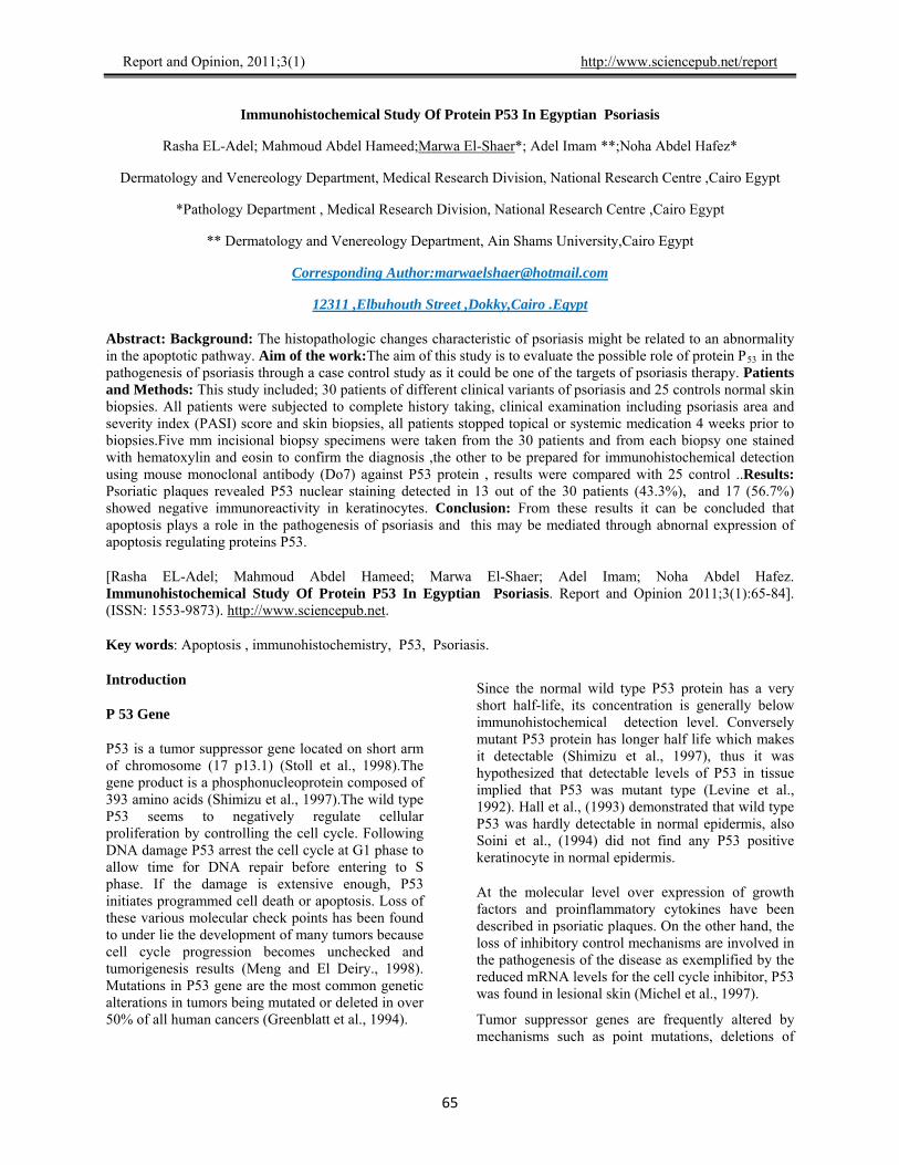
Report and Opinion, 2011;3(1) http://www.sciencepub.net/report Immunohistochemical Study Of Protein P53 In Egyptian Psoriasis Rasha EL-Adel; Mahmoud Abdel Hameed;Marwa El-Shaer *; Adel Imam **;Noha Abdel Hafez* Dermatology and Venereology Department, Medical Research Division, National Research Centre ,Cairo Egypt *Pathology Department , Medical Research Division, National Research Centre ,Cairo Egypt ** Dermatology and Venereology Department, Ain Shams University,Cairo Egypt Corresponding Author:[email protected] 12311 ,Elbuhouth Street ,Dokky,Cairo .Egypt Abstract: Background: The histopathologic changes characteristic of psoriasis might be related to an abnormality in the apoptotic pathway. Aim of the work:The aim of this study is to evaluate the possible role of protein P 53 in the pathogenesis of psoriasis through a case control study as it could be one of the targets of psoriasis therapy. Patients and Methods: This study included; 30 patients of different clinical variants of psoriasis and 25 controls normal skin biopsies. All patients were subjected to complete history taking, clinical examination including psoriasis area and severity index (PASI) score and skin biopsies, all patients stopped topical or systemic medication 4 weeks prior to biopsies.Five mm incisional biopsy specimens were taken from the 30 patients and from each biopsy one stained with hematoxylin and eosin to confirm the diagnosis ,the other to be prepared for immunohistochemical detection using mouse monoclonal antibody (Do7) against P53 protein , results were compared with 25 control ..Results: Psoriatic plaques revealed P53 nuclear staining detected in 13 out of the 30 patients (43.3%), and 17 (56.7%) showed negative immunoreactivity in keratinocytes. Conclusion: From these results it can be concluded that apoptosis plays a role in the pathogenesis of psoriasis and this may be mediated through abnornal expression of apoptosis regulating proteins P53. [Rasha EL-Adel; Mahmoud Abdel Hameed; Marwa El-Shaer; Adel Imam; Noha Abdel Hafez. Immunohistochemical Study Of Protein P53 In Egyptian Psoriasis. Report and Opinion 2011;3(1):65-84]. (ISSN: 1553-9873). http://www.sciencepub.net . Key words: Apoptosis , immunohistochemistry, P53, Psoriasis. Introduction P 53 Gene P53 is a tumor suppressor gene located on short arm of chromosome (17 p13.1) (Stoll et al., 1998).The gene product is a phosphonucleoprotein composed of 393 amino acids (Shimizu et al., 1997).The wild type P53 seems to negatively regulate cellular proliferation by controlling the cell cycle. Following DNA damage P53 arrest the cell cycle at G1 phase to allow time for DNA repair before entering to S phase. If the damage is extensive enough, P53 initiates programmed cell death or apoptosis. Loss of these various molecular check points has been found to under lie the development of many tumors because cell cycle progression becomes unchecked and tumorigenesis results (Meng and El Deiry., 1998). Mutations in P53 gene are the most common genetic alterations in tumors being mutated or deleted in over 50% of all human cancers (Greenblatt et al., 1994). Since the normal wild type P53 protein has a very short half-life, its concentration is generally below immunohistochemical detection level. Conversely mutant P53 protein has longer half life which makes it detectable (Shimizu et al., 1997), thus it was hypothesized that detectable levels of P53 in tissue implied that P53 was mutant type (Levine et al., 1992). Hall et al., (1993) demonstrated that wild type P53 was hardly detectable in normal epidermis, also Soini et al., (1994) did not find any P53 positive keratinocyte in normal epidermis. At the molecular level over expression of growth factors and proinflammatory cytokines have been described in psoriatic plaques. On the other hand, the loss of inhibitory control mechanisms are involved in the pathogenesis of the disease as exemplified by the reduced mRNA levels for the cell cycle inhibitor, P53 was found in lesional skin (Michel et al., 1997). Tumor suppressor genes are frequently altered by mechanisms such as point mutations, deletions of 65
Transcript of Report and Opinion, 2011;3(1) … · Report and Opinion, 2011;3(1) ... Nail, scalp and joint...
Report and Opinion, 2011;3(1) http://www.sciencepub.net/report
Immunohistochemical Study Of Protein P53 In Egyptian Psoriasis
Rasha EL-Adel; Mahmoud Abdel Hameed;Marwa El-Shaer*; Adel Imam **;Noha Abdel Hafez*
Dermatology and Venereology Department, Medical Research Division, National Research Centre ,Cairo Egypt
*Pathology Department , Medical Research Division, National Research Centre ,Cairo Egypt
** Dermatology and Venereology Department, Ain Shams University,Cairo Egypt
Corresponding Author:[email protected]
12311 ,Elbuhouth Street ,Dokky,Cairo .Egypt
Abstract: Background: The histopathologic changes characteristic of psoriasis might be related to an abnormality in the apoptotic pathway. Aim of the work:The aim of this study is to evaluate the possible role of protein P53 in the pathogenesis of psoriasis through a case control study as it could be one of the targets of psoriasis therapy. Patients and Methods: This study included; 30 patients of different clinical variants of psoriasis and 25 controls normal skin biopsies. All patients were subjected to complete history taking, clinical examination including psoriasis area and severity index (PASI) score and skin biopsies, all patients stopped topical or systemic medication 4 weeks prior to biopsies.Five mm incisional biopsy specimens were taken from the 30 patients and from each biopsy one stained with hematoxylin and eosin to confirm the diagnosis ,the other to be prepared for immunohistochemical detection using mouse monoclonal antibody (Do7) against P53 protein , results were compared with 25 control ..Results: Psoriatic plaques revealed P53 nuclear staining detected in 13 out of the 30 patients (43.3%), and 17 (56.7%) showed negative immunoreactivity in keratinocytes. Conclusion: From these results it can be concluded that apoptosis plays a role in the pathogenesis of psoriasis and this may be mediated through abnornal expression of apoptosis regulating proteins P53.
[Rasha EL-Adel; Mahmoud Abdel Hameed; Marwa El-Shaer; Adel Imam; Noha Abdel Hafez. Immunohistochemical Study Of Protein P53 In Egyptian Psoriasis. Report and Opinion 2011;3(1):65-84]. (ISSN: 1553-9873). http://www.sciencepub.net.
Key words: Apoptosis , immunohistochemistry, P53, Psoriasis.
Introduction
P 53 Gene
P53 is a tumor suppressor gene located on short arm of chromosome (17 p13.1) (Stoll et al., 1998).The gene product is a phosphonucleoprotein composed of 393 amino acids (Shimizu et al., 1997).The wild type P53 seems to negatively regulate cellular proliferation by controlling the cell cycle. Following DNA damage P53 arrest the cell cycle at G1 phase to allow time for DNA repair before entering to S phase. If the damage is extensive enough, P53 initiates programmed cell death or apoptosis. Loss of these various molecular check points has been found to under lie the development of many tumors because cell cycle progression becomes unchecked and tumorigenesis results (Meng and El Deiry., 1998). Mutations in P53 gene are the most common genetic alterations in tumors being mutated or deleted in over 50% of all human cancers (Greenblatt et al., 1994).
Since the normal wild type P53 protein has a very short half-life, its concentration is generally below immunohistochemical detection level. Conversely mutant P53 protein has longer half life which makes it detectable (Shimizu et al., 1997), thus it was hypothesized that detectable levels of P53 in tissue implied that P53 was mutant type (Levine et al., 1992). Hall et al., (1993) demonstrated that wild type P53 was hardly detectable in normal epidermis, also Soini et al., (1994) did not find any P53 positive keratinocyte in normal epidermis.
At the molecular level over expression of growth factors and proinflammatory cytokines have been described in psoriatic plaques. On the other hand, the loss of inhibitory control mechanisms are involved in the pathogenesis of the disease as exemplified by the reduced mRNA levels for the cell cycle inhibitor, P53 was found in lesional skin (Michel et al., 1997).
Tumor suppressor genes are frequently altered by mechanisms such as point mutations, deletions of
65
Report and Opinion, 2011;3(1) http://www.sciencepub.net/report
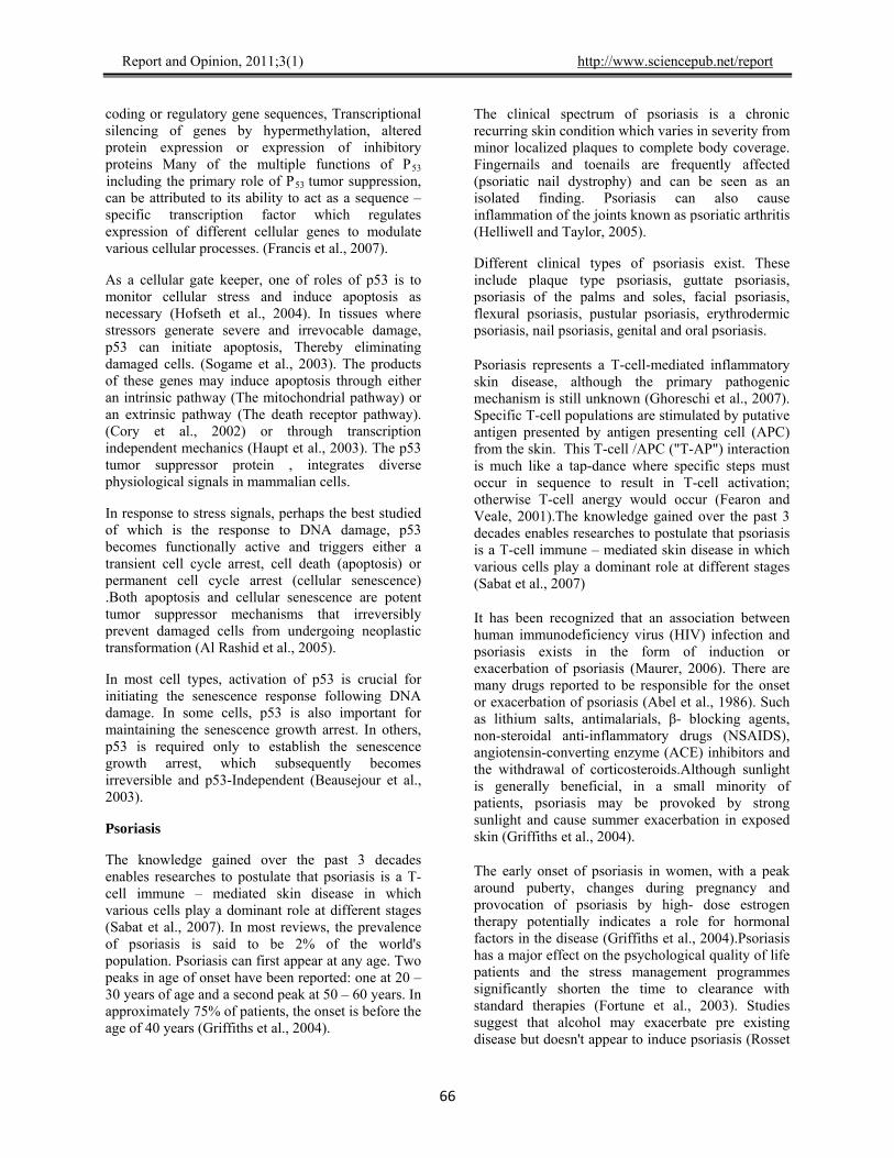
coding or regulatory gene sequences, Transcriptional silencing of genes by hypermethylation, altered protein expression or expression of inhibitory proteins Many of the multiple functions of P53
including the primary role of P53 tumor suppression, can be attributed to its ability to act as a sequence – specific transcription factor which regulates expression of different cellular genes to modulate various cellular processes. (Francis et al., 2007).
As a cellular gate keeper, one of roles of p53 is to monitor cellular stress and induce apoptosis as necessary (Hofseth et al., 2004). In tissues where stressors generate severe and irrevocable damage, p53 can initiate apoptosis, Thereby eliminating damaged cells. (Sogame et al., 2003). The products of these genes may induce apoptosis through either an intrinsic pathway (The mitochondrial pathway) or an extrinsic pathway (The death receptor pathway). (Cory et al., 2002) or through transcription independent mechanics (Haupt et al., 2003). The p53 tumor suppressor protein , integrates diverse physiological signals in mammalian cells.
In response to stress signals, perhaps the best studied of which is the response to DNA damage, p53 becomes functionally active and triggers either a transient cell cycle arrest, cell death (apoptosis) or permanent cell cycle arrest (cellular senescence) .Both apoptosis and cellular senescence are potent tumor suppressor mechanisms that irreversibly prevent damaged cells from undergoing neoplastic transformation (Al Rashid et al., 2005).
In most cell types, activation of p53 is crucial for initiating the senescence response following DNA damage. In some cells, p53 is also important for maintaining the senescence growth arrest. In others, p53 is required only to establish the senescence growth arrest, which subsequently becomes irreversible and p53-Independent (Beausejour et al., 2003).
Psoriasis
The knowledge gained over the past 3 decades enables researches to postulate that psoriasis is a T-cell immune – mediated skin disease in which various cells play a dominant role at different stages (Sabat et al., 2007). In most reviews, the prevalence of psoriasis is said to be 2% of the world's population. Psoriasis can first appear at any age. Two peaks in age of onset have been reported: one at 20 – 30 years of age and a second peak at 50 – 60 years. In approximately 75% of patients, the onset is before the age of 40 years (Griffiths et al., 2004).
The clinical spectrum of psoriasis is a chronic recurring skin condition which varies in severity from minor localized plaques to complete body coverage. Fingernails and toenails are frequently affected (psoriatic nail dystrophy) and can be seen as an isolated finding. Psoriasis can also cause inflammation of the joints known as psoriatic arthritis (Helliwell and Taylor, 2005).
Different clinical types of psoriasis exist. These include plaque type psoriasis, guttate psoriasis, psoriasis of the palms and soles, facial psoriasis, flexural psoriasis, pustular psoriasis, erythrodermic psoriasis, nail psoriasis, genital and oral psoriasis.
Psoriasis represents a T-cell-mediated inflammatory skin disease, although the primary pathogenic mechanism is still unknown (Ghoreschi et al., 2007). Specific T-cell populations are stimulated by putative antigen presented by antigen presenting cell (APC) from the skin. This T-cell /APC ("T-AP") interaction is much like a tap-dance where specific steps must occur in sequence to result in T-cell activation; otherwise T-cell anergy would occur (Fearon and Veale, 2001).The knowledge gained over the past 3 decades enables researches to postulate that psoriasis is a T-cell immune – mediated skin disease in which various cells play a dominant role at different stages (Sabat et al., 2007)
It has been recognized that an association between human immunodeficiency virus (HIV) infection and psoriasis exists in the form of induction or exacerbation of psoriasis (Maurer, 2006). There are many drugs reported to be responsible for the onset or exacerbation of psoriasis (Abel et al., 1986). Such as lithium salts, antimalarials, β- blocking agents, non-steroidal anti-inflammatory drugs (NSAIDS), angiotensin-converting enzyme (ACE) inhibitors and the withdrawal of corticosteroids.Although sunlight is generally beneficial, in a small minority of patients, psoriasis may be provoked by strong sunlight and cause summer exacerbation in exposed skin (Griffiths et al., 2004).
The early onset of psoriasis in women, with a peak around puberty, changes during pregnancy and provocation of psoriasis by high- dose estrogen therapy potentially indicates a role for hormonal factors in the disease (Griffiths et al., 2004).Psoriasis has a major effect on the psychological quality of life patients and the stress management programmes significantly shorten the time to clearance with standard therapies (Fortune et al., 2003). Studies suggest that alcohol may exacerbate pre existing disease but doesn't appear to induce psoriasis (Rosset
66

Report and Opinion, 2011;3(1) http://www.sciencepub.net/report
and Oki, 1971). Alcohol may also reduce the therapeutic compliance or may reflect a symptom of stress caused by severe psoriasis (Behnam et al., 2005).
Aim of the work
The aim of this study is to evaluate the possible role of protein P53 in the pathogenesis of psoriasis through a case control study as it could be one of the targets of psoriasis therapy.
Patients and Methods
This study included 30 patients of different clinical variants of psoriasis and 25 controls. The patients group consisted of 16 male and 12 females with their age ranging between 18 to 60 years. They were selected from dermatology outpatient clinic of Ain – Shams University Hospital and National research center during the period between August 2008 to August 2009.
Ethical considerations:
For confidentiality, the names were excluded and replaced with a numerical code.
A) Full history taking:
1. Personal history:
Name, age, sex, occupation and residence.
2. History of the disease:
• Age of onset, course and duration of the disease.
• Exacerbating and relieving factors.
• History of previous treatment.
• Family history of similar condition.
• Past history of other systemic affection
B)Clinical examination:
Site and severity of skin involvement.
Distribution of the lesion, generalized or localized.
Nail, scalp and joint affection.
Extent of skin involvement.
C) Clinical assessment:
Clinical assessment of psoriatic patients was performed through calculation of psoriatic area and severity index (PASI score) (Fredriksson and Pettersson., 1978).
PASI score:
For calculation of this index: the four main body areas were assessed: the head (h), the trunk (t), the upper extremities (u), and the lower extremities (I) corresponding to 10, 20, 30 and 40% of the total body area, respectively.
Three target symptoms namely erythema (E), infiltration (I) and desquamation (D) were assessed according to scale 0-4 where means complete lake of cutaneous involvement and 4 represents the severest possible involvement.
Investigation
5 mm incisional biopsies' were taken from sun unexposed lesions of psoriatic patients as well as from normal control. the biopsies were fixed in 10% neutral buffered formalin and paraffin embedded blocks were prepared
Two slides were made from each biopsy:
One slide was stained by hematoxylin and eosin stain and was examined by light microscopy to confirm the diagnosis.
The other slide was cut over positive charged coated slides for immunohistochemical staining using monoclonal antibody against p53 protein by immunoperoxidase technique
Immunohistochemical study: The kit used for immunohistochemical staining was “supersensitive ready to use immunodetection system" presented by clini lab. The kit contained P53 monoclonal antibody clone D07 which detects nuclear localization of P53 protein. This antibody reacts with both wild type and mutant type of P53 protein.
The staining procedure: • The paraffin embedded sections were cut
over positive charged coated slides produced by clini lab.
• The slides were then immersed in xylol over night.
•The slides were rehydrated by immersion in alcohol with descending concentrations (5 minutes in two changes of 100% ethanol, 5 minutes in two changes of 95% ethanol) to wash the xylol and remove any impurities, then they were washed with
67

Report and Opinion, 2011;3(1) http://www.sciencepub.net/report
water •They were immersed in buffer solution pH
7.4 for 5 minutes. The buffer was formed of sodium chloride + Potassium phosphate monobasic + potassium dibasic.
•Endogenous peroxidases were inhibited by treating the slides with 3% hydrogen peroxide in distilled water for 5 minutes.
• The slides were washed in buffer and were-wiped around the section.
•Antigen retrieval citra 10% (also produced by clini Lab) was then added to the sections to unmask hidden or weak antigens in tissues.
•The slides were put in a bath of water and microwaved for 5 minutes. They were removed from the microwave and inspected to see if the level of water around the slides has decreased. Water was then added to cover the slides if it was not enough and antigen retrieval was added to the sections once more, then they were remicrowaved for another 5 minutes.
• The slides were left to cool for 15 minutes. • The slides were washed with water then
with buffer. •The primary antibody was then added to the
slides which were monoclonal antibody clone D07 which reacts with both wild type and mutant form of P53 protein.
• The slides were incubated for one hour in a humid chamber.
• The slides were washed in buffer. •The link pre-diluted biotyonylated anti-
immunoglobulin was added for 20 minutes. •Then the slides were washed in buffer and
were wiped around the sections. •Slides were then labeled by peroxidase
conjugated streptavidin for 20 minutes. • Slides were washed again in buffer and
wiped around the sections •Liquid DAB (3,3 diaminobenzidine)
chromagen was then added with H2O2 (as substrate) for 20 minutes to visualize immunoreactivity sites.
•The slides were washed with distilled water and wiped around the sections.
•Counter stain was added (Mayer's hematoxylin) for 1 to 2 minutes.
•Slides were then washed in water and passed through rising concentrations of alcohol.
• They were then put in xylen for 5 minutes. • Finally Canada balsm was added then the
slides were cover. • All through the procedure slides were not
allowed to dry. • The slides were then read under the light
microscope. Interpretation of the results of
immunostaining: Positive staining appeared as brown coloration
of the nuclei of variable intensities (mild, moderate and strong).
Criteria of staining: A semi quantitative score was applied to
the sections as follows: • Negative (-): if no cells were stained. • Mild expression (+): if less than 10% of the
cells were stained. Moderate expression (++): if 10% to 50% of
the cells were stained. Strong expression (+++): if more than 50% of
the cells were stained. Results This study included 30 patients with different clinical variants of psoriasis. They were 16 males (53.3%) and 14 females (46.7%) and their age ranged between 18 to 60 years with mean + SD (36.3 + 9.7). The duration of psoriasis ranged between 1 to 16 years with mean + S.D (6.8 + 4.2). The psoriatic area and severity index (PASI) ranged between 3.3 to 67.6 with mean + S.D (22.6 + 22.2).Three of the 30 patients (10%) had positive family history of psoriasis . Thirteen psoriatic patients showed +ve P53 expression (Table 1). Skin biopsies from 25 apparently healthy individuals were taken and served as control after their consent. They were 15 males (60%) and 10 females (40%). Their age ranged between 16 to 64 years with mean +S.D (35.5 + 13.7), all were negative for P53 expression (Table 2).
Table (3) & figure (1) show no statistically significant difference between psoriatic group and control group as regards sex, as P > o.o5.Table (4) & figure (2) show no statistically significant difference between psoriatic group and control group as regards age , as P > o.o5
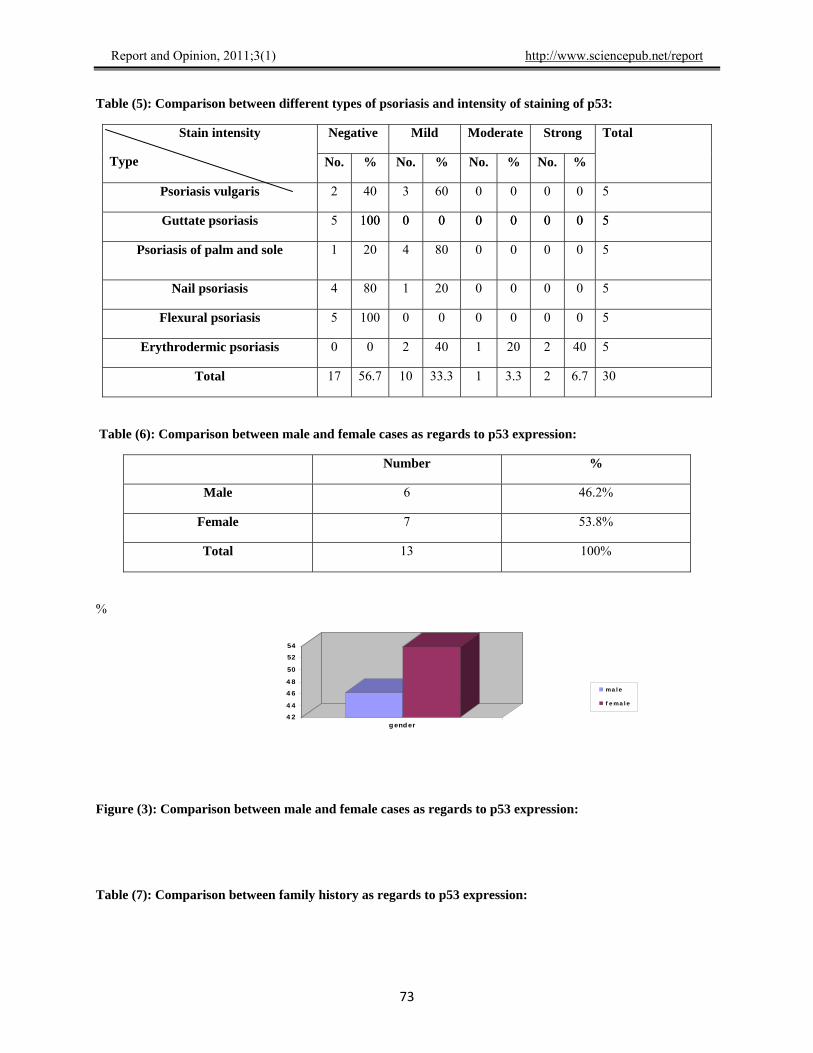
Table (5) shows a comparison between different clinical types of psoriasis and intensity of staining of P53. Five patients (16.6%) with psoriasis vulgaris, 2 of which (40%) show negative intensity of staining of P53 and 3 of which (60%) show mild P53 expression. Five patients (16.6%) with guttate type psoriasis all of them (100%) show negative P53 expression. Five patients (16.6%) with psoriasis of palm and sole, 1 of which (20%) show negative P53 expression and 4 of which (80%) show mild P53 expression. Five patients (16.6%) with nail psoriasis, 4 of which (80%) show negative P53 expression and 1 of which (20%) show mild P53 expression. Five patients (16.6%) with flexural psoriasis all of them (100%) show negative P53 expression. Five patients (16.6%) with erythrodermic psoriasis, 2 of which (40%) show
68
Report and Opinion, 2011;3(1) http://www.sciencepub.net/report
69
mild P53 expression and 1 patient (20%) shows moderate P53 expression and 2 patients (40%) show strong P53 expression. These findings mean that there is strong correlation between the severity of psoriasis and the intensity of staining of P53.
Table (6) & figure (3) showed that 6 (46.2%) psoriatic patients out of 13 (100%) psoriatic patients who showed +ve P53 expression were males while 7 (53.8%) psoriatic patients out of 13 (100%) showed + ve P53 expression were females. In figure (4 ), only 2 psoriatic patients out of 13 who were P53 +ve have +ve family history while 11(84.6%) psoriatic patients out of 13 who were P53 +ve have –ve family history.
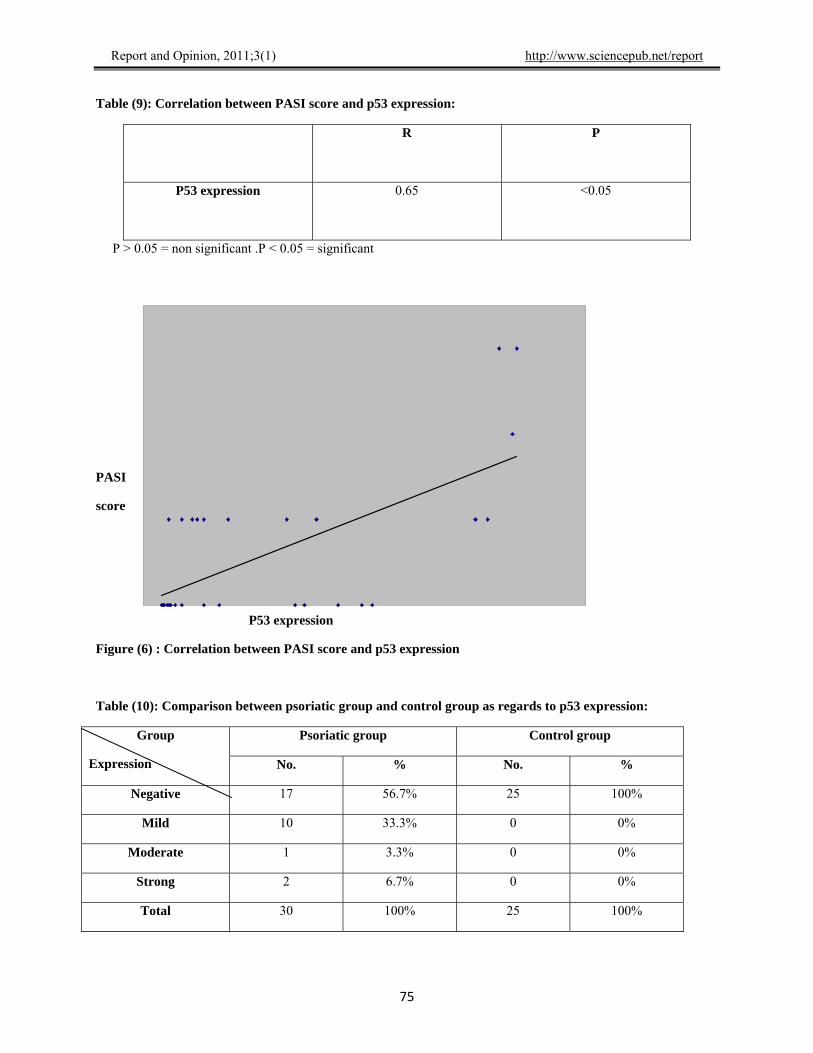
Table (8) & figure (5) show positive correlation between PASI score and the duration of psoriasis.Table (9) & figure (6) show a correlation between PASI score and P53 expression which was significant.
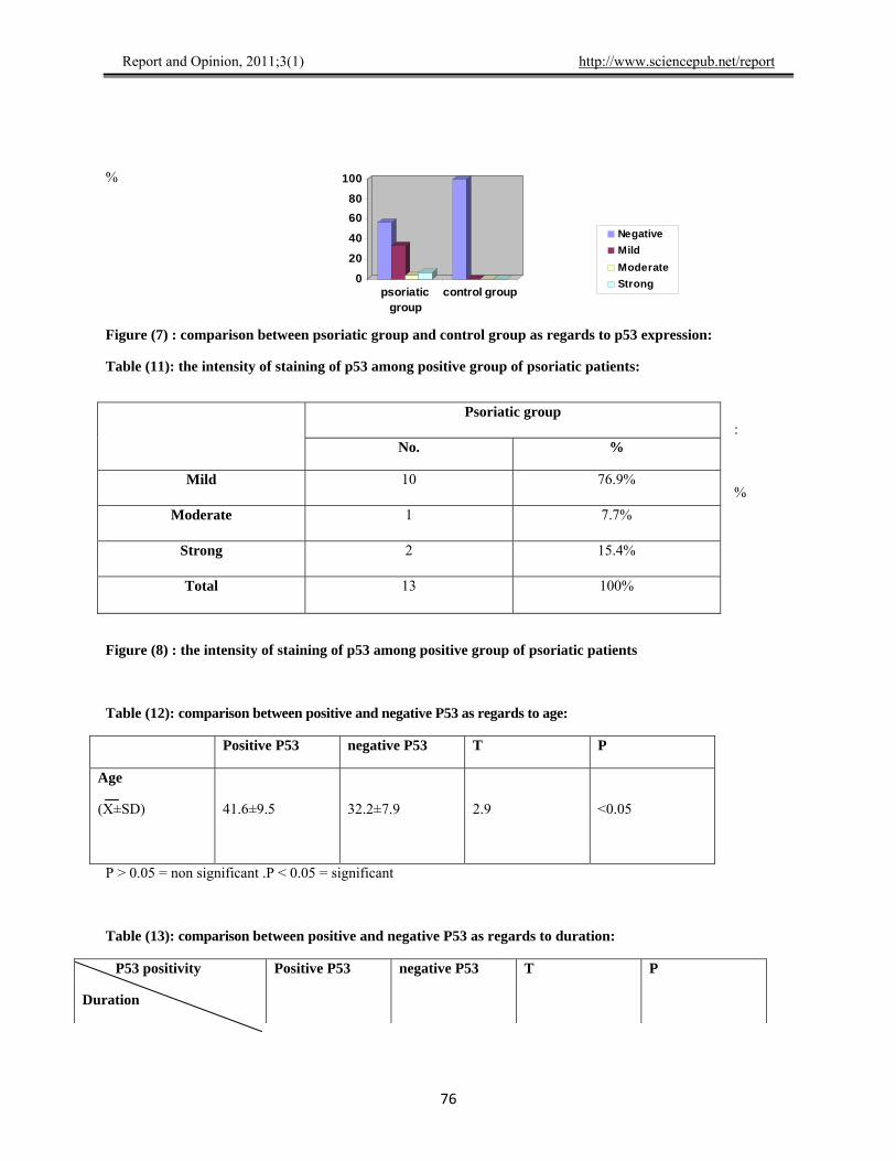
In table (10) & figure (7), the comparison between psoriatic group and control group as regards to P53
expression showed that all the 25 control healthy individuals were P53-ve, while among the psoriatic group, 17 (56.7%) showed negative P53 expression, while 10(33.3%) showed mild P53 expression, 1 (3.3%) showed moderate P53 expression, 2 (6.7%) showed strong P53 expression.
Table (11)& figure (8) Showed that there is 13 psoriatic patients had +ve intensity of P53 staining, 10 (76.9%) of which showed mild intensity of staining, 1 (7.7%) of which showed moderate intensity of staining, 2 (15.4%) of which showed strong intensity of staining.
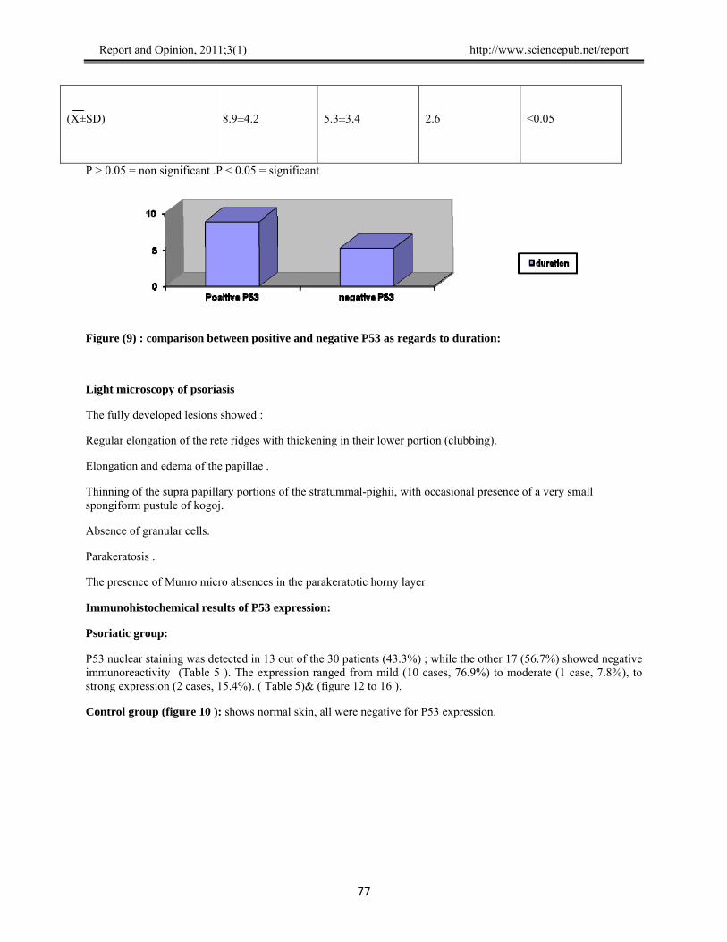
Table (12) show that the age of those patients with + ve P53 expression was higher compared to those with – ve P53 expression and this difference was statistically significant (P < o.o5 ). Table (13) & figure (9) showed that the duration of the disease was much higher among patients with +ve P53 expression compared to that with –ve P53 expression and the difference was statistically significant (P < o.o5 ).
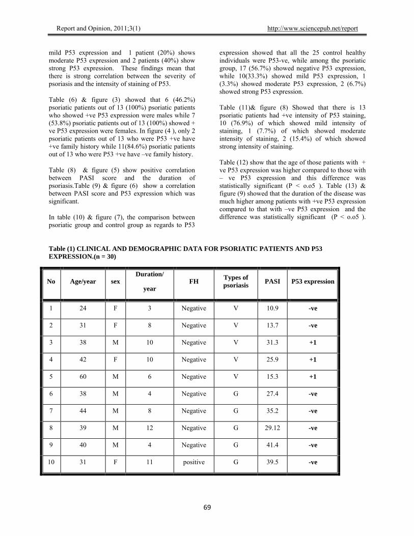
Table (1) CLINICAL AND DEMOGRAPHIC DATA FOR PSORIATIC PATIENTS AND P53 EXPRESSION.(n = 30)
No Age/year sex Duration/
year FH
Types of psoriasis
PASI P53 expression
1 24 F 3 Negative V 10.9 -ve
2 31 F 8 Negative V 13.7 -ve
3 38 M 10 Negative V 31.3 +1
4 42 F 10 Negative V 25.9 +1
5 60 M 6 Negative V 15.3 +1
6 38 M 4 Negative G 27.4 -ve
7 44 M 8 Negative G 35.2 -ve
8 39 M 12 Negative G 29.12 -ve
9 40 M 4 Negative G 41.4 -ve
10 31 F 11 positive G 39.5 -ve
Report and Opinion, 2011;3(1) http://www.sciencepub.net/report
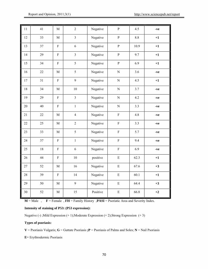
11 41 M 2 Negative P 4.5 -ve
12 33 M 3 Negative P 8.8 +1
13 37 F 6 Negative P 10.9 +1
14 29 F 3 Negative P 9.7 +1
15 34 F 5 Negative P 6.9 +1
16 22 M 5 Negative N 3.6 -ve
17 31 F 9 Negative N 4.5 +1
18 34 M 10 Negative N 3.7 -ve
19 29 F 3 Negative N 4.2 -ve
20 40 F 1 Negative N 3.3 -ve
21 22 M 4 Negative F 4.8 -ve
22 25 M 2 Negative F 3.3 -ve
23 33 M 5 Negative F 5.7 -ve
24 37 F 1 Negative F 9.4 -ve
25 18 F 6 Negative F 6.9 -ve
26 44 F 10 positive E 62.3 +1
27 52 M 16 Negative E 67.6 +3
28 39 F 14 Negative E 60.1 +1
29 50 M 9 Negative E 64.4 +3
30 52 M 15 Positive E 66.8 +2
M = Male , F = Female , FH = Family History ,PASI = Psoriatic Area and Severity Index.
Intensity of staining of P53: (P53 expression):
Negative (-) ;Mild Expression (+ 1);Moderate Expression (+ 2);Strong Expression (+ 3)
Types of psoriasis:
V = Psoriasis Vulgaris; G = Guttate Psoriasis ;P = Psoriasis of Palms and Soles; N = Nail Psoriasis
E= Erythrodermic Psoriasis
70

Report and Opinion, 2011;3(1) http://www.sciencepub.net/report
Table (2) = CONTROL GROUP AND P53 EXPRESSION(n = 25 )
No age sex P53 expression
1 22 M Negative
2 33 F Negative
3 16 M Negative
4 21 M Negative
5 24 M Negative
6 33 M Negative
7 32 M Negative
8 40 F Negative
9 52 F Negative
10 19 F Negative
11 33 M Negative
12 44 M Negative
13 22 F Negative
14 39 M Negative
15 21 M Negative
16 30 M Negative
17 23 M Negative
18 27 M Negative
19 31 M Negative
20 55 F Negative
21 57 F Negative
22 53 F Negative
23 47 F Negative
24 64 M Negative
25 49 F Negative
M= Male F=female
71

Report and Opinion, 2011;3(1) http://www.sciencepub.net/report
Table (3): Comparison between psoriatic group and control group as regards sex:
0
10
2 0
3 0
4 0
50
6 0
male f emale
pso r iat ic g roup
co nt ro l g ro up
3 4 .5
3 5
3 5.5
3 6
3 6 .5
psoriat ic g roup cont ro l g roup
a ge
Male Female Sex
Group No. % No. %
Total X2 P
psoriatic group 16 53.3% 14 46.7% 30
Control group 15 60% 10 40% 25
Total 31 56.4% 24 43.6% 55
0.3
>0.05
P > 0.05 = non significant .P < 0.05 = significant
%
Figure (1): Comparison between psoriatic group and control group as regards sex
Table (4): Comparison between psoriatic group and control group as regards age:
Group
Age
psoriatic group control group t P
Age
(X±SD)
36.3±9.7
35.4±13.7
0.3
>0.05
P > 0.05 = non significant .P < 0.05 = significant
Years
Figure (2): Comparison between psoriatic group and control group as regards age:
72
Report and Opinion, 2011;3(1) http://www.sciencepub.net/report
Table (5): Comparison between different types of psoriasis and intensity of staining of p53:
Negative Mild Moderate Strong Stain intensity
Type No. % No. % No. % No. %
Total
Psoriasis vulgaris 2 40 3 60 0 0 0 0 5
Guttate psoriasis 5 100 100 0 0 0 0 0 0 0 0 0 0 0 0 5 5
Psoriasis of palm and sole 1 20 4 80 0 0 0 0 5
Nail psoriasis 4 80 1 20 0 0 0 0 5
Flexural psoriasis 5 100 0 0 0 0 0 0 5
Erythrodermic psoriasis 0 0 2 40 1 20 2 40 5
Total 17 56.7 10 33.3 1 3.3 2 6.7 30
Table (6): Comparison between male and female cases as regards to p53 expression:
Number %
Male 6 46.2%
Female 7 53.8%
Total 13 100%
%
4 2
4 4
4 6
4 8
50
52
54
g ender
ma l e
f e ma l e
Figure (3): Comparison between male and female cases as regards to p53 expression:
Table (7): Comparison between family history as regards to p53 expression:
73

Report and Opinion, 2011;3(1) http://www.sciencepub.net/report
%
Figure (4) : Comparison between family history as regards to p53 expression
Table (8): Correlation between PASI score and duration of psoriasis
R P
Duration of psoriasis
0.75 <0.05
P > 0.05 = non significant .P < 0.05 = significant
PASI
score
Duration in years
Figure (5): Correlation between PASI score and duration of psoriasis
74
Report and Opinion, 2011;3(1) http://www.sciencepub.net/report
Table (9): Correlation between PASI score and p53 expression:
R P
P53 expression
0.65 <0.05
P > 0.05 = non significant .P < 0.05 = significant
PASI
score
P53 expression
Figure (6) : Correlation between PASI score and p53 expression
Table (10): Comparison between psoriatic group and control group as regards to p53 expression:
Psoriatic group Control group Group
Expression No. % No. %
Negative 17 56.7% 25 100%
Mild 10 33.3% 0 0%
Moderate 1 3.3% 0 0%
Strong 2 6.7% 0 0%
Total 30 100% 25 100%
75
Report and Opinion, 2011;3(1) http://www.sciencepub.net/report
%
0
20
40
60
80
100
psoriaticgroup
control group
Negative
Mild
Moderate
Strong
Figure (7) : comparison between psoriatic group and control group as regards to p53 expression:
Table (11): the intensity of staining of p53 among positive group of psoriatic patients:
:
%
Psoriatic group
No. %
Mild 10 76.9%
Moderate 1 7.7%
Strong 2 15.4%
Total 13 100%
Figure (8) : the intensity of staining of p53 among positive group of psoriatic patients
Table (12): comparison between positive and negative P53 as regards to age:
Positive P53 negative P53 T P
Age
(X±SD)
41.6±9.5
32.2±7.9
2.9
<0.05
P > 0.05 = non significant .P < 0.05 = significant
Table (13): comparison between positive and negative P53 as regards to duration:
P53 positivity
Duration
Positive P53 negative P53 T P
76
Report and Opinion, 2011;3(1) http://www.sciencepub.net/report
(X±SD)
8.9±4.2
5.3±3.4
2.6
<0.05
P > 0.05 = non significant .P < 0.05 = significant
Figure (9) : comparison between positive and negative P53 as regards to duration:
Light microscopy of psoriasis
The fully developed lesions showed :
Regular elongation of the rete ridges with thickening in their lower portion (clubbing).
Elongation and edema of the papillae .
Thinning of the supra papillary portions of the stratummal-pighii, with occasional presence of a very small spongiform pustule of kogoj.
Absence of granular cells.
Parakeratosis .
The presence of Munro micro absences in the parakeratotic horny layer
Immunohistochemical results of P53 expression:
Psoriatic group:
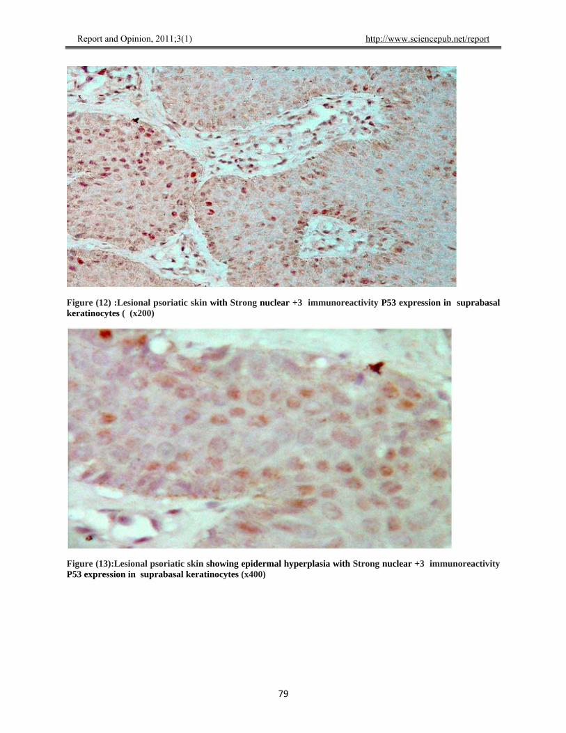
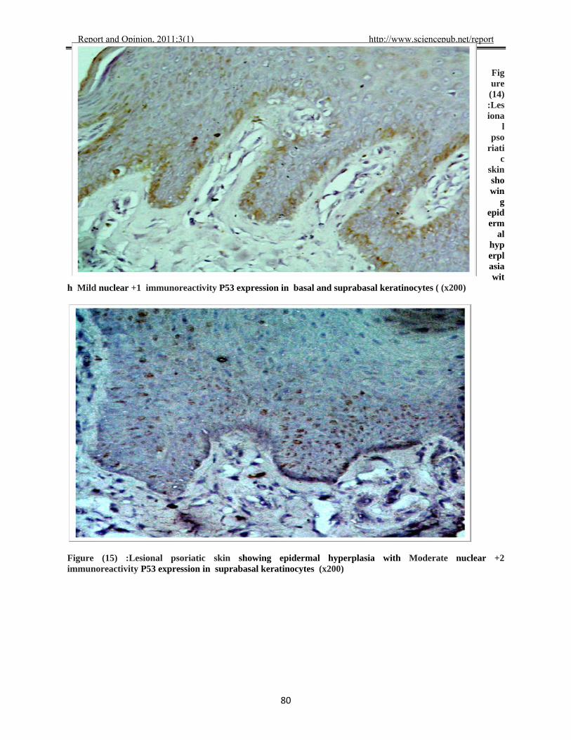
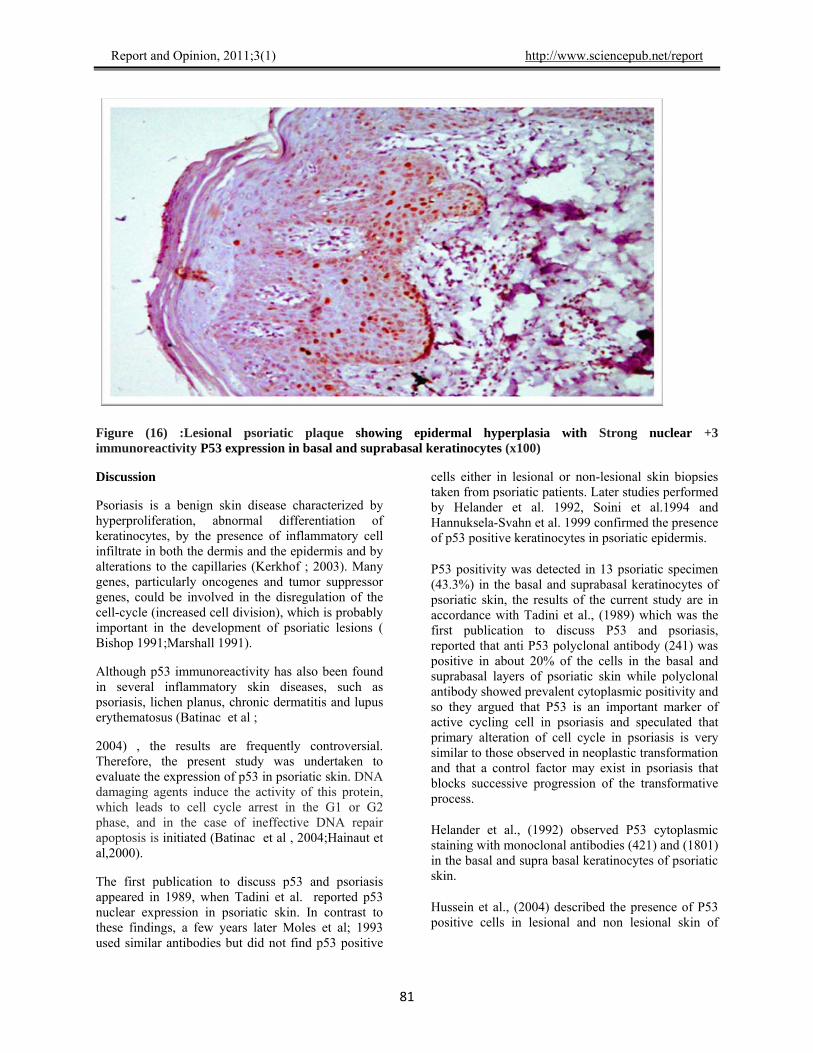
P53 nuclear staining was detected in 13 out of the 30 patients (43.3%) ; while the other 17 (56.7%) showed negative immunoreactivity (Table 5 ). The expression ranged from mild (10 cases, 76.9%) to moderate (1 case, 7.8%), to strong expression (2 cases, 15.4%). ( Table 5)& (figure 12 to 16 ).
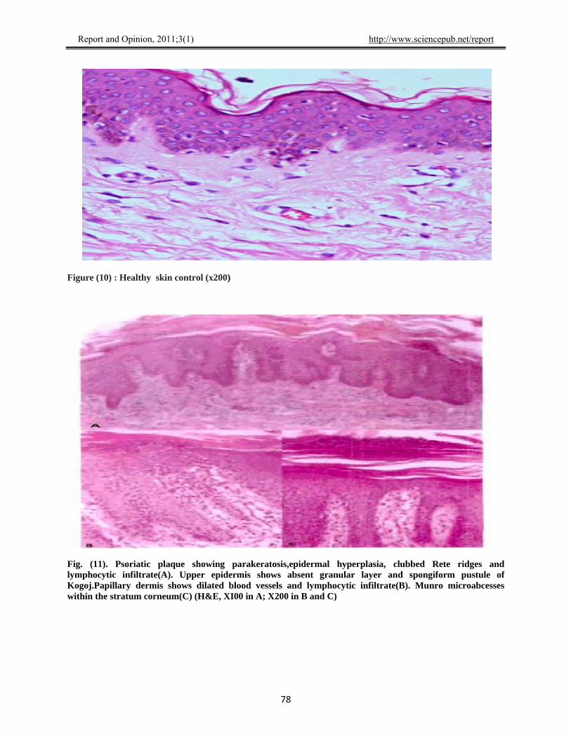
Control group (figure 10 ): shows normal skin, all were negative for P53 expression.
77
Report and Opinion, 2011;3(1) http://www.sciencepub.net/report
Figure (10) : Healthy skin control (x200)
Fig. (11). Psoriatic plaque showing parakeratosis,epidermal hyperplasia, clubbed Rete ridges and lymphocytic infiltrate(A). Upper epidermis shows absent granular layer and spongiform pustule of Kogoj.Papillary dermis shows dilated blood vessels and lymphocytic infiltrate(B). Munro microabcesses within the stratum corneum(C) (H&E, XI00 in A; X200 in B and C)
78
Report and Opinion, 2011;3(1) http://www.sciencepub.net/report
Figure (12) :Lesional psoriatic skin with Strong nuclear +3 immunoreactivity P53 expression in suprabasal keratinocytes ( (x200)
Figure (13):Lesional psoriatic skin showing epidermal hyperplasia with Strong nuclear +3 immunoreactivity P53 expression in suprabasal keratinocytes (x400)
79
Report and Opinion, 2011;3(1) http://www.sciencepub.net/report
80
u
ytes ( (x200)
Figre
(14):Lesiona
l pso
riatic
skin showin
g epiderm
al hyperplasia wit
h Mild nuclear +1 immunoreactivity P53 expression in basal and suprabasal keratinoc
Figure (15) :Lesional psoriatic skin showing epidermal hyperplasia with Moderate nuclear +2 immunoreactivity P53 expression in suprabasal keratinocytes (x200)
Report and Opinion, 2011;3(1) http://www.sciencepub.net/report
Figure (16) :Lesional psoriatic plaque showing epidermal hyperplasia with Strong nuclear +3 immunoreactivity P53 expression in basal and suprabasal keratinocytes (x100)
Discussion
Psoriasis is a benign skin disease characterized by hyperproliferation, abnormal differentiation of keratinocytes, by the presence of inflammatory cell infiltrate in both the dermis and the epidermis and by alterations to the capillaries (Kerkhof ; 2003). Many genes, particularly oncogenes and tumor suppressor genes, could be involved in the disregulation of the cell-cycle (increased cell division), which is probably important in the development of psoriatic lesions ( Bishop 1991;Marshall 1991).
81
Although p53 immunoreactivity has also been found in several inflammatory skin diseases, such as psoriasis, lichen planus, chronic dermatitis and lupus erythematosus (Batinac et al ;
2004) , the results are frequently controversial. Therefore, the present study was undertaken to evaluate the expression of p53 in psoriatic skin. DNA damaging agents induce the activity of this protein, which leads to cell cycle arrest in the G1 or G2 phase, and in the case of ineffective DNA repair apoptosis is initiated (Batinac et al , 2004;Hainaut et al,2000).
The first publication to discuss p53 and psoriasis appeared in 1989, when Tadini et al. reported p53 nuclear expression in psoriatic skin. In contrast to these findings, a few years later Moles et al; 1993 used similar antibodies but did not find p53 positive
cells either in lesional or non-lesional skin biopsies taken from psoriatic patients. Later studies performed by Helander et al. 1992, Soini et al.1994 and Hannuksela-Svahn et al. 1999 confirmed the presence of p53 positive keratinocytes in psoriatic epidermis.
P53 positivity was detected in 13 psoriatic specimen (43.3%) in the basal and suprabasal keratinocytes of psoriatic skin, the results of the current study are in accordance with Tadini et al., (1989) which was the first publication to discuss P53 and psoriasis, reported that anti P53 polyclonal antibody (241) was positive in about 20% of the cells in the basal and suprabasal layers of psoriatic skin while polyclonal antibody showed prevalent cytoplasmic positivity and so they argued that P53 is an important marker of active cycling cell in psoriasis and speculated that primary alteration of cell cycle in psoriasis is very similar to those observed in neoplastic transformation and that a control factor may exist in psoriasis that blocks successive progression of the transformative process.
Helander et al., (1992) observed P53 cytoplasmic staining with monoclonal antibodies (421) and (1801) in the basal and supra basal keratinocytes of psoriatic skin.
Hussein et al., (2004) described the presence of P53 positive cells in lesional and non lesional skin of
Report and Opinion, 2011;3(1) http://www.sciencepub.net/report
psoriatic patients with a statistically stronger expression in lesional skin.
Also the results of Soini et al., ( 1994) agree with our results, where they found aberrant expression of P53 in non-neoplastic inflammatory skin diseases as psoriasis but the percentage of P53 positive cells was lower than seen in malignant tumors. They also found increased proliferation of keratinocytes and they demonstrated a relationship between P53 positivity and the number of ki 67 positive cells and mitosis suggesting that P53 accumulation was enhanced by cell proliferation.
Hannuksela – Svahn et al., (1999) confirmed the presence of P53 positive keratinocytes in psoriatic epidermis and reported that the increase expression of P53 in psoriatic skin is a physiological reaction indicating the attempt to counteract the proliferation and to repair DNA errors, and is most often expressed as an increased number of mitosis.
El-Batawi et al., (2002) found higher P53 expression in lesional than in non lesional psoriatic skin using monoclonal antibody Do7. which is the same as our results. They concluded that the elevated levels of P53 in psoriatic plaques reflect an important role exerted by this tumor suppressor gene on the development of psoriatic plaques.
Kerkhof (2003) demonstrated that P53 is an important marker of active cycling cells in psoriasis and a large body of evidence indicates that the cell cycle time in psoriasis is normal and only increased recruitment of epidermal cells is responsible for the development of psoriatic lesions .
Batinac et al., (2004) examined P53 expression in lesional psoriatic skin, non lesional skin of psoriatic patients and skin samples from neoplastic diseases: basal cell carcinomas and squanous cell carcinomas. P53 immunoreativity was demonstrated in the epidermis of psoriatic skin in the basal and suprabasal layers and only in the basal layer in non lesional skin of psoriatic patients. The difference in P53 expression was however, not significant. In carcinomas (basal cell carcinoma, squamous cell carcinoma) P53 expression was significantly stronger.Our result is consistent with Batinac et al., (2004) who found P53 immunoreactivity was demonstrated in the epidermis of psoriatic skin in the basal and supra basal layers.
On the other hand our data disagree with Moles and associates (1993) who used three different antibodies; polyclonal antibody (CMI) and monoclonal
antibodies (Do7) and (1801) that recognize different epitopes for immunohistochemical analysis of P53 protein and they didn't observe nuclear staining in lesional and non lesional psoriatic skin. In addition, they did not detect any mutation in P53 gene using a combination of polymerase chain reaction and single strand conformation polymorphism techniques. Also Magee et al., (1994) found that, staining of P53 was not detected in psoriatic biopsies using anti-P53 Do7 antibodies.
Michel et al., (1996) demonstrated that P53 expression was decreased in lesional psoriatic skin. P53 transcription was clearly induced by antimicrobial substance FK506 (Tacrolimus) treatment, both post translational modification of P53 protein and transcriptional modulation of P53 gene which represent potential targets for the antipsoriatic action of the drug plus down modulation of IL – 8 receptor type A.
Dazard (2000) used immunofluorescence staining of cryosections of 13 psoriatic skin biopsies and could not find P53 positivity in this series of psoriasis cryosections when probed with a panel of specific antibodies.
The wide variation in the results obtained by different works can be attributed to either the difference in the sensitivity of the kits used or to the cross reactivity of anti P53 antibodies with other proteins like keratins (Moles et al., 1993), or due to racial differences.
In the current study P53 expression, either nuclear or cytoplasmic, could not be detected in any of twenty five specimens serving as control. Indicating the significance of P53 study in proliferative disorders, Helander et al., (1993) found a significant cytoplasmic reactivity for P53 was detected in normal cells of control group.
Some theories have been published by Qin et al., (2002) concerning the response of the keratinocytes to DNA damaging factors, which seems to be different in this instance to that in other cells of human body. Unique mechanisms are essential for the keratinocytes to preserve cutaneous homeostasis and maintain the structural integrity of the skin as a barrier between the organism and the environment.A mutation in the p53 gene has been found in most human tumor types. p53 protein is constitutively expressed in almost all cells, but due to the short span of its half-life it is extremely difficult to detect a “wildtype” p53 in standard immunohistochemistry. The activated or mutated form accumulates in cells and immunohistochemical detection is possible
82
Report and Opinion, 2011;3(1) http://www.sciencepub.net/report
Our results (which agree with several results and disagree with others) may suggest that the cell cycle is disturbed in psoriatic epidermis but further studies are needed to determine definitively the molecular basis of cell cycle regulation in epidermal cells, and to continue to broaden our knowledge of the pathogenesis of psoriasis.
Summary
The aim of this study was to identify the expression of P53 in psoriatic patients with different clinical types and severity to clarify its role in the pathogenesis of psoriasis and its correlation with the disease severity.
The patients group consisted of 16 males (53.3%) and 14 females (46.6%). Their age ranged between 18 to 60 years with mean + SD (36.3 + 9.7). The patients were divided into six subgroups according to the clinical types of psoriasis. Five patients (16.6%) with psoriasis vulgaris, 2 of which (40%) show negative intensity of staining of P53 expression and 3 of which (60%) show mild P53 expression. Five patients (16.6%) with guttate psoriaseis all of them (100%) show negative P53 expression. Five patients (16.6%) with psoriasis of palm and sole, 1 of which (20%) shows negative P53 expression and 4 of which (80%) show mild P53 expression. Five patients (16.6%) with Nail psoriasis, 4 of which (80%) show negative P53 expression and 1 of which (20%) shows mild P53 expression. Five patients (16.6%) with flexural psoriasis all of them show negative P53 expression. Five patients (16.6%) with erythrodermic psoriasis; 2 of which (40%) show mild P53 expression and 1 patient (20%) shows moderate P53 expression and 2 patients (40%) show strong P53 expression.
The PASI score of the patients ranged from 3.3 to 67.6 with mean + S.D (22.6+ 22.2) three of the 30 patients (10%) had positive family history, two of which (15.4%) had positive intensity of staining of P53.The expression ranged from mild (10 cases, 76.9%) to moderate (1 case, 7.8%), to strong expression (2 cases, 15.4%).The 25 control group all showed negative P53 expression.
The statistically significant elevation in P53 level with the clinical severity of psoriasis may raise the suspicion that P53 may have a role in determining disease severity and so we argued that P53 is an important marker of active cell cycling in psoriasis.
Conclusion
The results obtained from our study provide further evidence supporting the role of P53 in the pathogenesis of psoriasis as P53 nuclear staining was detected in 13 out of 30 patients (43.3%).
P53 is an important marker of active cycling cell in psoriasis and it reflects an important role exerted by this tumor suppressor gene in the development of psoriasis so P53 is recommended as a routine immunohistochemical marker for psoriasis.
Further studies on larger number of patients and on different clinical types of psoriasis are recommended.
Assessment of P53 levels is recommended after different therapeutic modalities.
There is certainly a hope in the near future that P53 protein comes from only a subject of academic research to being a therapeutic modality adopted in treating inflammatory and neoplastic disorders.
References
1-Abel EA, Dicicco LM, Orenberg EK, Fraki JE and Farber EM Drugs in exacerbation of psoriasis. J Am Acad Dermatol ; 15:1007,1986.
2- Al Rashid ST, Dellaire G, Cuddihy A, Jalali F, Vaid M, Coackley C, Folkard M, Xu Y and Chen BP Evidence for the direct binding of phosphorylated P53 to sites of DNA breaks in vivo. Cancer Res ; 65:10810 –10821,2005.
3-Batinac T, Gruber F, Lipozencic J et al. Protein p53 - structure, function, and possible therapeutic implications. Acta Dermatovenerol Croat; 11: 225–30,2003. 4- Batinac T, Zamolo G, Jonji} N, et al. p53 protein expression and cell proliferation in non-neoplastic and neoplastic proliferative skin diseases. Tumori; 90:120–7,2004.
5- Beausejour CM, Krtolica A, Galimi F, Narita M, Lowe SW, Yaswen P and Campisi J Reversal of human cellular senescence: roles of the P53 and p16 pathways. EMBO J; 22: 4212–4222,2003.
6- Behnam SM, Behnam SE and Koo JY Alcohol as a risk factor for plaque-type psoriasis. Cutis; 76(3):181,2005.
7-Bishop JM. Molecular themes in oncogenesis. Cell; 64: 235–48,1991.
8-Cory S and Adams J M The Bcl2 family: regulators of the cellular life-or-death switch. Nat Rev Cancer; 2:647–656,2002.
9- El- Batawi MM, Shahin Z, Ramadan S, El – Ramly AZ, El- Mahgoub D, El- Komy M H and Saba S Expression of Bcl2 and P53 in psoriasis. Egypt J Dermatol and Androl; 22(1): 13 – 17,2002.
83
Report and Opinion, 2011;3(1) http://www.sciencepub.net/report
84
10- Fearon and Veale D Pathogenesis of psoriatic arthritis. Clin exp Dermatol; Lab: 333–337,2001.
11- Fortune D G, Richards H L, Kirky B, Mc Elhone K, Markham T, Rogers S, Main C J and Griffiths C E Psychological Distress Impairs Clearance of Psoriasis in Patients Treated With Photochemotherapy . Arch Dermatol ; 139: 752,2003.
12- Francis Rodier, Judith Campisi and Dipa Bhaumik
Two faces of P53: aging and tumor suppression. Nucleic Acids Res; 35(22): 7475–7484,2007.
13-Ghoreschi K, Weigert C and Röcken M Immunopathogenesis and role of T cells in psoriasis. Clini Dermatol; 25: 574–580,2007.
14- Greenblatt MS, Bennet W P, Hollstein M and Harris CC Mutation of P53 tumor suppression gene: clues to cancer etiology and molecular pathogenesis. Cancer Res; 54 : 4855 – 4878,1994.
15-Griffiths CEM, Camp RDR and Barker JNWN PSORIASIS. In: Rook Text book of Dermatology. Burns T, Breathnach S. Cox N and Griffiths C (eds.). Severnth edition. Massachusetts, Oxford, Australia. Chapter 35, P.1 – 69,2004.
16- Hall PA, Mckee PA, Menage HP, Dover R and lane DP High levels of P53 protein in UV-irradiated normal human skin. Oncogene; 8: 203 – 207,1993.
17- Hannuksela-Svahn A, Paakko P, Autio P, et al. Expression of p53 protein before and after PUVA treatment in psoriasis. Acta Derm Venereol (Stockh); 79: 195–9,1999.
18- Haupt S, Berger M, Goldberg Z and Haupt Y Apoptosis-the P53 network. J Cell Sci; 116:4077–4085,2003.
19-Hainaut P, Hollstein M. p53 and human cancer the first ten thousand mutations. Adv Cancer Res; 77: 81–173,2000. 20- Helander SD, Peters MS, Pittelkow MR, et al.Expression of p53 protein in benign and malignant epidermal pathologic conditions. J Am Acad Dermatol; 29: 741–8,1993.
21- Helliwell PS and Taylor WJ Classification and diagnostic criteria for psoriatic arthritis. Ann Rheum Dis ; 64 : 3-82005.
22- Hofseth LJ, Hussain SP and Harris CC P53: 25 years after its discovery. Trends Pharmacol Sci; 25: 177-181,2004.
23- Hussein MR, Al-Badaiwy ZH, Guirguis MN et al. Analysis of p53 and bcl-2 protein expression in the non-tumorigenic, pretumorigenic, and tumorigenic keratinocytic hyperproliferative lesions. J Cutan Pathol; 31: 643–51,2004.
24- Kerkhof P. Texbook of psoriasis. Blackwell Publishing Ltd., Oxford; pp. 83–109,2003.
25- Levine A J and Momand E Tumor suppressor genes: the P53 and retinoblastoma sensitivity genes and gene products. Biochem Acta; 1032:119-136,1990.
26- Maurer LS, Jalian HR, starner TD and Modin RL A study of the incidence of AIDS in patients with psoriasis vulgaris . Dermatology ; 140 : 282,2006.
27- Marshall CJ. Tumor suppressor genes. Cell; 64: 313–26,1991.
28- Meng R D and El-Deiry WS Tumor suppressor Genes as Targets for cancer Gene Therapy. In : Gene Therapy of cancer, 1st ed, Lattime EC, and Gerson S L (eds.), Academic press, ch. 1, p. 8 – 10,1998.
29- Michel G, Mirmoham A and Olasz E Demonstration and Functional analysis of IL – 10 receptors in human epidermal Cells : decreased expression in psoriatic skin, down modulation by IL- 8 and upregulation by antipsoriatic glucocortisteroid in normal cultured keratinocytes. J Immunol; 159 (12): 6291 – 6297,1997.
30-. Moles JP, Theillet C, Basset-Seguin N, et al. Mutation of the tumor suppressor gene TP53 is not detected in psoriatic skin. J Invest Dermatol; 101:100–2,1993.
31- Qin JZ, Chaturvedi V, Denning MF et al. Regulation of apoptosis by p53 in UV-irradiated human epidermis, psoriatic plaques and senescent keratinocytes. Oncogene; 21: 2991–3002,2002.
32- Rosset M and Oki G Diseases in alcoholics. Q J Stud Alcohol; 32:1017,1971.
33-Sabat R Philipp S, Höflich C, Kreutzer S, Wailace E, Asadullah K, Volk HD, Sterry W and Wolk K Immunopathogenesis of psoriasis. Exp Derm; 16: 779-798,2007.
34- Shimizu T, Muto M, Murakami T and Furumoto H over expression of P53 protein associated with proliferative activity as evaluated by ki-67 immunostaining in well differentiated squamous cell carcinoma of the skin. Dermatol; 195 : 224 – 227, 1997.
35- Sogame N, Kim M and Abrans J M Drosophila P53 preserves genomic stability by regulating cell death. Proc Natl Acad Sci USA; 100: 4696–4701,2003.
36- Soini Y, Kamel D, Paakko P et al. Aberrant accumulation of p53 associates with Ki67 and mitotic count in benign skin lesions. Br J Dermatol; 131: 514–20,1994.
37- Stoll C, Baretton G and Iohrs U The influence of P53 and associated factors on the outcome of patients with oral squamous cell carcinoma. Virch Arch; 433: 427 – 433,1998.
38-. Tadini G, Cerri A, Crosti L, et al. P53 and oncogene expression in psoriasis. Acta Derm Venereol(Stockh); 146: 33–5,1989.
7/22/2010