Repeated Application of 4-Aminopyridine Provoke an...
12
ORIGINAL ARTICLE Repeated Application of 4-Aminopyridine Provoke an Increase in Entorhinal Cortex Excitability and Rearrange AMPA and Kainate Receptors Sa ´ndor Borbe ´ly • Da ´vid Cze ´ge ´ • Elek Molna ´r • Endre Dobo ´ • Andra ´s Miha ´ly • Ildiko ´ Vila ´gi Received: 22 September 2014 / Revised: 29 December 2014 / Accepted: 30 December 2014 Ó Springer Science+Business Media New York 2015 Abstract Entorhinal cortex is a highly epilepsy-prone brain region. Effects of repetitive seizures on ionotropic glutamate receptors (iGluRs) were investigated in rat en- torhinal cortex slices. Seizures were induced by daily administration of 4-aminopyridine (4-AP). Electrophysio- logical, pharmacological and histological investigations were carried out to determine changes in synaptic efficacy and in sensitivity of iGluRs due to recurring seizures. Repeated 4-AP-induced seizures increased the amplitude of evoked synaptic field responses in rat entorhinal cortical slices. While vulnerability to inhibition of AMPA receptors by the specific antagonist GYKI 52466 was slightly reduced, responsiveness to NMDA receptor antagonist APV remained unaffected. Testing of bivalent cation per- meability of iGluRs revealed reduced Ca 2? -influx through non-NMDA receptors. According to the semi-quantitative histoblot analysis GluA1–4, GluA1, GluA2, GluK5, GluN1 and GluN2A subunit protein expression differently altered. While there was a marked decrease in the level of GluA1–4, GluA2 and GluK5 receptor subunits, GluA1 and GluN2A protein levels moderately increased. The results indicate that brief convulsions, repeated daily for 10 days can increase overall entorhinal cortex excitability despite a reduction in AMPA/kainate receptor activity, probably through the alteration of local network susceptibility. Keywords Seizure Brain slice 4-Aminopyridine Entorhinal cortex Glutamate receptor Introduction The imbalance of excitatory (glutamatergic) and inhibitory (c-amino-butyric acid (GABA)-ergic) neurotransmission in the forebrain is a key feature of epilepsy. It is widely accepted that ionotropic glutamate receptors (iGluRs) are involved in the development of various forms of epilepsy. The iGluRs are glutamate-gated cation channels composed of four subunits (Traynelis et al. 2010). Based on their sequence homology, electrophysiological properties and pharmacological selectivity, iGluRs are subdivided into three main subtypes: a-amino-3-hydroxyl-5-methyl-4-iso- xazole-propionate (AMPA), kainate (KA) and N-methyl-D- aspartate (NMDA) type receptors (Traynelis et al. 2010). AMPA receptors (AMPARs) mediate fast synaptic trans- mission in the central nervous system (CNS) and they are key components of the modifiable synaptic response. AMPARs are composed of the GluA1–4 subunits, which can form functional heteromeric receptors. Most native AMPARs contain both the GluA2 subunit and either GluA1, GluA3 or GluA4 (Traynelis et al. 2010). Kainate receptors (KARs) are key players in the modulation of neuronal network activity throughout the CNS (Lerma 2003). While other AMPARs and NMDA receptors (NMDARs) operate mainly at postsynaptic sites, KARs are both presynaptic and postsynaptic, where they modulate S. Borbe ´ly (&) D. Cze ´ge ´ I. Vila ´gi Department of Physiology and Neurobiology, Institute of Biology, Eo ¨tvo ¨s Lora ´nd University, Pa ´zma ´ny Pe ´ter se ´ta ´ny 1/c, Budapest 1117, Hungary e-mail: [email protected]; [email protected] E. Molna ´r School of Physiology and Pharmacology, University of Bristol, Medical Sciences Building, University Walk, Bristol BS8 1TD, UK E. Dobo ´ A. Miha ´ly Department of Anatomy, Histology and Embryology, Faculty of Medicine, University of Szeged, Kossuth Lajos sgt. 40, Szeged 6724, Hungary 123 Neurotox Res DOI 10.1007/s12640-014-9515-7
Transcript of Repeated Application of 4-Aminopyridine Provoke an...
ORIGINAL ARTICLE
Repeated Application of 4-Aminopyridine Provoke an Increasein Entorhinal Cortex Excitability and Rearrange AMPAand Kainate Receptors
Sandor Borbely • David Czege • Elek Molnar •
Endre Dobo • Andras Mihaly • Ildiko Vilagi
Received: 22 September 2014 / Revised: 29 December 2014 / Accepted: 30 December 2014
� Springer Science+Business Media New York 2015
Abstract Entorhinal cortex is a highly epilepsy-prone
brain region. Effects of repetitive seizures on ionotropic
glutamate receptors (iGluRs) were investigated in rat en-
torhinal cortex slices. Seizures were induced by daily
administration of 4-aminopyridine (4-AP). Electrophysio-
logical, pharmacological and histological investigations
were carried out to determine changes in synaptic efficacy
and in sensitivity of iGluRs due to recurring seizures.
Repeated 4-AP-induced seizures increased the amplitude
of evoked synaptic field responses in rat entorhinal cortical
slices. While vulnerability to inhibition of AMPA receptors
by the specific antagonist GYKI 52466 was slightly
reduced, responsiveness to NMDA receptor antagonist
APV remained unaffected. Testing of bivalent cation per-
meability of iGluRs revealed reduced Ca2?-influx through
non-NMDA receptors. According to the semi-quantitative
histoblot analysis GluA1–4, GluA1, GluA2, GluK5, GluN1
and GluN2A subunit protein expression differently altered.
While there was a marked decrease in the level of
GluA1–4, GluA2 and GluK5 receptor subunits, GluA1 and
GluN2A protein levels moderately increased. The results
indicate that brief convulsions, repeated daily for 10 days
can increase overall entorhinal cortex excitability despite a
reduction in AMPA/kainate receptor activity, probably
through the alteration of local network susceptibility.
Keywords Seizure � Brain slice � 4-Aminopyridine �Entorhinal cortex � Glutamate receptor
Introduction
The imbalance of excitatory (glutamatergic) and inhibitory
(c-amino-butyric acid (GABA)-ergic) neurotransmission in
the forebrain is a key feature of epilepsy. It is widely
accepted that ionotropic glutamate receptors (iGluRs) are
involved in the development of various forms of epilepsy.
The iGluRs are glutamate-gated cation channels composed
of four subunits (Traynelis et al. 2010). Based on their
sequence homology, electrophysiological properties and
pharmacological selectivity, iGluRs are subdivided into
three main subtypes: a-amino-3-hydroxyl-5-methyl-4-iso-
xazole-propionate (AMPA), kainate (KA) and N-methyl-D-
aspartate (NMDA) type receptors (Traynelis et al. 2010).
AMPA receptors (AMPARs) mediate fast synaptic trans-
mission in the central nervous system (CNS) and they are
key components of the modifiable synaptic response.
AMPARs are composed of the GluA1–4 subunits, which
can form functional heteromeric receptors. Most native
AMPARs contain both the GluA2 subunit and either
GluA1, GluA3 or GluA4 (Traynelis et al. 2010). Kainate
receptors (KARs) are key players in the modulation of
neuronal network activity throughout the CNS (Lerma
2003). While other AMPARs and NMDA receptors
(NMDARs) operate mainly at postsynaptic sites, KARs are
both presynaptic and postsynaptic, where they modulate
S. Borbely (&) � D. Czege � I. Vilagi
Department of Physiology and Neurobiology, Institute of
Biology, Eotvos Lorand University, Pazmany Peter setany 1/c,
Budapest 1117, Hungary
e-mail: [email protected]; [email protected]
E. Molnar
School of Physiology and Pharmacology, University of Bristol,
Medical Sciences Building, University Walk, Bristol BS8 1TD,
UK
E. Dobo � A. Mihaly
Department of Anatomy, Histology and Embryology, Faculty of
Medicine, University of Szeged, Kossuth Lajos sgt. 40,
Szeged 6724, Hungary
123
Neurotox Res
DOI 10.1007/s12640-014-9515-7
neurotransmitter release and effectiveness of excitatory
neurotransmission (Jane et al. 2009; Lerma 2003).
Molecular cloning has identified five KAR subunits, named
GluK1–5 (Traynelis et al. 2010). Functional receptors are
usually formed from heteromeric combinations of subunits
and the most common postsynaptic KAR subunit combi-
nation is GluK2/GluK5 (Lerma 2003). NMDARs contrib-
ute to the late component of synaptic response and function
as coincidence detectors (Collingridge et al. 2004; Trayn-
elis et al. 2010). NMDARs are responsible for a wide range
of postsynaptic functions, including changes in synaptic
effectiveness when long-term potentiation (LTP) or long-
term depression (LTD) develops (Collingridge et al. 2004).
Excessive activation of NMDARs leads to pathological
processes (e.g. excitotoxic injury) in a number of acute and
chronic neurological disorders (Lau and Tymianski 2010).
NMDARs are obligate heterotetramers formed by the
assembly of GluN1 subunits with GluN2A–D and GluN3A/B
(Traynelis et al. 2010). Pathological glutamate receptor
function may underlie many neurological disorders (Lau and
Tymianski 2010).
Temporal lobe epilepsy (TLE) is one of the most com-
mon forms of intractable focal epilepsies (Chang and
Lowenstein 2003). Ex vivo brain slice experiments support
the idea that seizures are likely to be generated in the en-
torhinal cortex and subsequently spread to the hippocam-
pus in TLE (Armand et al. 1999). While several animal
models were introduced for the study of TLE, most widely
kainate or pilocarpine are applied to induce epileptiform
seizures. These convulsants provoke status epilepticus (SE)
soon after the treatment (Cavalheiro et al. 1991; Turski
et al. 1983). Kainate provokes SE and after a silent period
of 2–3 weeks, spontaneous motor limbic seizures are
generated (Nadler 2003). In the pilocarpine model, spon-
taneous seizure episodes appear following a 4–44 days-
long silent period as the consequence of neurodegenerative
processes (Scorza et al. 2009). Neuronal loss and rear-
rangement of synaptic connections are characteristic fea-
tures of these models (Choi et al. 2007; Fisher 1989; Nadler
et al. 1978).
A moderate dose of intraperitoneally applied 4-AP, a
potassium channel blocker, also induces acute seizures in
rats. In contrast with kainate and pilocarpine treatment, low
doses of 4-AP does not induce chronic spontaneous sei-
zures or cell damage (Vizi et al. 2004). The apparent lack
of neuronal loss following repeated 4-AP convulsions
renders this model suitable for the study of genuine in vivo
molecular alterations developing after repeated seizures.
On the other hand, 4-AP administered directly to the hip-
pocampus causes acute seizures and severe cell loss,
especially in the CA1 and CA3 hippocampal regions (Pena
and Tapia 2000). In this case, however, the local concen-
tration of 4-AP is relatively high. 4-AP applied directly to
brain slices ex vivo also induces seizure-like events that
can be inhibited by both NMDAR and AMPAR antagonists
(Doczi et al. 1999; Gulyas-Kovacs et al. 2002). Both
ex vivo and in vivo 4-AP treatment causes an elevation in
the transcription of certain immediate-early genes (such as
c-fos and c-jun) suggesting that seizures may cause chan-
ges in neuronal gene expression in the affected brain areas
(Borbely et al. 2006; Mihaly et al. 2005).
Previous ex vivo studies revealed brain region-specific
changes in glutamatergic neurotransmission in the rat
neocortex and hippocampus following repeated 4-AP-
induced seizures (Borbely et al. 2009; Vilagi et al. 2009).
In the present study, the effects of repeated 4-AP-induced
seizures on the excitability of the entorhinal cortex and
changes in iGluRs are investigated. Evoked field potentials
(EFPs) were tested to evaluate changes in the basic excit-
ability together with the pharmacological sensitivity to
specific AMPAR and NMDAR antagonists (GYKI 52466
and APV, respectively). Kainate-induced Co2?-uptake
assay was performed to evaluate the changes induced by
the repeated 4-AP treatment in the Ca2?-permeability of
AMPARs and KARs. Alterations in iGluR protein
expression levels were analysed by semi-quantitative his-
toblots (Kopniczky et al. 2005; Tonnes et al. 1999) using
selective antibodies to GluA1, GluA2, GluA1-4, GluK5,
GluN1 and GluN2 iGluR subunits. Our results indicate that
repeated brief convulsions lead to an increase of entorhinal
cortex excitability despite an overall reduction in AMPAR/
KAR activity.
Materials and Methods
Experiments were performed on adult, male Wistar rats
(150–280 g, Charles Rivers, Budapest, Hungary). All pro-
cedures were approved by the regional Animal Ethics
Committees in Budapest (p.n.: 22.1/829/003/2007) and
Szeged (p.n.: I./01508/2011) and by the European Com-
munities Council Directive of 1986 (86/609/EEC). Rats
were kept under constant 12 h light/dark cycle and con-
trolled temperature (22 ± 2 �C). Standard pellet food and
tap water were available ad libitum. All chemicals were
purchased from Sigma-Aldrich Ltd. (Budapest, Hungary)
or Merck Ltd. (Budapest, Hungary) unless it is stated
otherwise. GYKI 52466 was the kind gift of Dr. I. Tarnawa
(Richter Gedeon Plc, Budapest, Hungary).
Seizure Induction in Rats
4-AP, dissolved in physiological saline, was injected every
day intraperitoneally for 12 consecutive days. The starting
dose was 4.5 mg/kg. After the injection, the animals were
monitored for 2–2.5 h until they fully recovered from
Neurotox Res
123
seizures. Seizures were characterised according to the
widely used five-stage Racine scale (Racine 1972). If the
rats did not show a stage 5 seizure after the third day of
treatment, the dose was increased by 5 %, and it was fur-
ther increased by 5 % on any day when a stage 5 seizure
failed to develop on the previous day. The maximal dose
was 6.65 mg/kg. This group of animals is referred to as
seizured group. Control rats received physiological saline
intraperitoneally (0.5 ml) for 12 days, and were referred to
as normal group. The maximum injected volume of 4-AP
or saline was always less than 1 ml.
Slice Preparation and Electrophysiological Recording
Electrophysiological recordings were carried out on 48
slices from 18 rats, 8 slices were used in each experimental
group. Animals were decapitated in deep chloral-hydrate
anaesthesia (350 mg/kg), brains were quickly removed and
horizontal plane slices (400 lm thick) of the entorhinal
cortex were cut with a vibratome. Brain slices were left for
1 h to stabilise in the incubation solution containing N-2-
hydroxyethylpiperazine-N0-2-ethanesulfonic acid (HEPES)
buffer: (in mM): 120 NaCl, 20 NaHCO3, 2 KCl, 1.25
KH2PO4, 2 MgSO4, 2 CaCl2, 10 glucose, 6.7 HEPES, 2.6
Na-HEPES, pH 7.2–7.3). This solution was saturated with
carbogene (5 % CO2, 95 % O2) at room temperature
(20–22 �C). Then slices were transferred to a Haas-type
recording chamber (Experimetria Ltd., Budapest, Hungary)
which was perfused with standard artificial cerebrospinal
fluid (ACSF; 1.5 ml/min) using a peristaltic pump (Hei-
dolph PD 5101, Schwabach, Germany). The composition
of ACSF was (in mM) 126 NaCl; 26 NaHCO3; 1.8 KCl;
1.25 KH2PO4; 1.3 MgSO4; 2.4 CaCl2; 10 glucose, pH
7.1–7.3. The solution was saturated with carbogene at
33 ± 1 �C before application.
Glass microelectrodes filled with 1 M NaCl (8–10 MX)
were positioned as recording electrodes into layer III of the
lateral entorhinal cortex, while bipolar tungsten stimulating
electrodes were positioned below the recording electrodes
at the border of the white and grey matters. Duration of the
stimulation square voltage pulses was 100 ls and the
amplitude was gradually varied between threshold and
supramaximal values. Threshold (T) means the stimulation
strength when the evoked response just appears, and T was
introduced for better comparability of the recorded data.
Signals were amplified 1,000-fold with a preamplifier and
an Axoclamp 2A amplifier (Axon Instruments Inc., Union
City, CA), A/D converted and recorded with the SPEL
Advanced Intrasys computer program (Experimetria Ltd.,
Budapest, Hungary).
An evoked response was typically made up of an early
synaptic (referred to as N1) component, the latency of
which was always above 4.5 ms (Tolner et al. 2007). This
wave was followed by a series of longer latency peaks with
smaller amplitude referred to as late components (Fig. 1a).
Sometimes, but not always, a smaller, very early non-
synaptic peak (N0) also appeared, showing antidromic
activation. In the present evaluation, the amplitude of the
early (N1) synaptic component of EFP was systematically
analysed, while the late component of EFP (N2) was not
involved in detailed analysis due to its large variability.
The viability of each slice was tested before the begin-
ning of recording procedure by applying single pulse
stimulation and monitoring characteristic field responses. If
the absolute amplitude of N1 was smaller than 1 mV at
approximately 4T intensity, the slice was excluded from
further experiments. Slices were continuously stimulated
with 2T stimuli at a frequency of 0.1 Hz, to stabilise the
response amplitude. At least 15 min after placing the slices
into the recording chamber, the exact stimulus threshold
(1T) was determined. The recording session was started
only if all these tests were passed. At the beginning of
recording (0 min), an input–output curve (I–O curve,
stimulus intensity—amplitude of EFP) was measured from
1 to 4T stimulus intensity in 0.5T steps. During the sub-
sequent incubation phase, continuous stimulation was
applied with 0.1 Hz frequency and 2T intensity. After 10 or
30 min, another I–O curve was recorded. To analyse the
pharmacological sensitivity of particular iGluRs, we
applied either AMPAR antagonist (40 lM 1-(aminophe-
nyl)-4-methyl-7,8 methylendioxy-5H-2,3-benzodiazepine
(GYKI 52466)) for 30 min or NMDAR antagonist (25 lM
D-2-amino-5-phosphonopentanoic acid (D-APV)) for
10 min. The antagonists were dissolved in ACSF, the
applied concentration and the duration of treatments with
the antagonists was established by our previous studies
(Gulyas-Kovacs et al. 2002; Vilagi et al. 1996). Recorded
data were analysed using the SPEL Advanced Intrasys
computer program (Experimetria, Budapest, Hungary). In
each experiment, the amplitude of each EFPs was norma-
lised to the EFP evoked in 0 min at 4T stimulus intensity
for better comparability of the changes.
Co2?-Permeability of Non-NMDARs
Brain slices from normal (n = 9) and seizured (n = 10)
animals were used to test alterations in Co2? uptake. Co2?
can pass through activated non-NMDARs that are also
permeable to Ca2?, but not through NMDARs (Mayer and
Westbrook 1987).
Slice preparation was carried out in the same way as
described for electrophysiological recordings. The thick-
ness of the slices was 250 lm. From each rat, 8–10 en-
torhinal cortex slices were stained and evaluated. The
procedure was performed as described previously (Pruss
et al. 1991). Briefly, slices were incubated in a Ca2? free
Neurotox Res
123

incubation solution for 5 min before placement into a Co2?
uptake buffer (13 mM sucrose, 57.5 mM NaCl, 5 mM
KCl, 2 mM MgCl2, 1 mM CaCl2, 12 mM glucose, 10 mM
HEPES, pH 7.4) supplemented with 5 mM CoCl2 and
100 lM kainate. Incubation lasted for 20 min at room
temperature (20–22 �C). Control slices serving as staining
background were incubated in the same solution without
kainate. After the stimulated Co2? uptake, slices were
rinsed once in the uptake buffer and then incubated in the
same buffer containing 2 mM ethylenediaminetetraacetic
acid (EDTA) to remove non-specifically bound Co2?.
Slices were then rinsed twice in the uptake buffer, and
Co2? was precipitated through incubation in a 0.12 %
(NH4)2S solution for 5 min. During this procedure, dark
CoS precipitates were formed in the cells. Slices were then
fixed in 4 % paraformaldehyde for 30 min and mounted to
glycerine-coated slides for further computer analysis. The
Co2?-stained slices were analysed with an Olympus CH-2
microscope equipped with an Olympus Camedia C4040-
Zoom digital camera connected to a PC running AnalySIS
3.2 Docu software (Olympus Soft Imaging Solution
GmbH, Munster, Germany). Pictures were digitalised at a
fourfold magnification and converted to 8-bit grey scale
images. Staining intensity was measured in arbitrary units.
One data point corresponded to the mean of 50 recorded
points (509 300–600 pixels/point). Optical density (OD) of
a 50 9 100 pixel area in the corpus callosum was deter-
mined in each slice as background, and data were always
corrected with this OD value. The corrected OD values of
sham-treated normal slices were subtracted from the cor-
rected OD values of kainate-stimulated slices to reduce the
bias from non-specific staining. OD values of slices from
normal and seizured groups of animals were compared,
relative change was given as a percentage of OD value
originating from slices of normal group. For more accurate
comparison of the corresponding cortical layers, the dis-
tance between the pial surface and the subcortical white
matter was divided into 20 equal divisions which were
correlated to cortical layers using corresponding Nissl-
stained sections. The borders of the layers were as follows:
I: division 1–5, II: 6–7 division, III: 8–12 division, IV:
13–14 division, V: 15–17 division, VI: 18–20 division.
Histoblot Analysis of AMPAR and KAR Subunit
Proteins
Rats were killed following the final 4-AP treatment, and
used to determine changes in the distribution of different
AMPAR subunit proteins, using an in situ blotting tech-
nique (Gallyas et al. 2003; Kopniczky et al. 2005; Tonnes
et al. 1999). Animals were deeply anesthetised with diethyl
ether, decapitated and the brains were quickly frozen in
isopentane and stored at -80 �C until sectioning. Hori-
zontal plane cryostat sections (10 lm) were cut and
mounted on glass slides. The sections on the glass were
pressed against nitrocellulose membranes, which were
previously moistened with 48 mM Tris-base, 39 mM gly-
cine, 2 % (w/v) sodium dodecyl sulphate (SDS) and 20 %
(w/v) methanol for 15 min at room temperature
(20–22 �C). After blocking in 5 % (w/v) non-fat dry milk
in phosphate-buffered saline (PBS), nitrocellulose mem-
branes were DNase I-treated (5 U/ml), washed and incu-
bated in 2 % (w/v) SDS, 100 mM b-mercaptoethanol in
100 mM Tris–HCl (pH 7.0), for 60 min at 45 �C to remove
Fig. 1 Characteristic EFP in entorhinal cortex and changes in general
neuronal excitability. a Characteristic trace of evoked response from
entorhinal cortex. The non-synaptic N0 component was followed by
the early N1 component from seizured rat, which is thought to be the
first synaptic component of the evoked response, was determined as
the negative peak measured with at least 4.5 ms delay. The later
component (N2) of the evoked response was quite heterogeneous in
appearance, and lasted for up to several hundreds of milliseconds.
Small inset shows the N0 and N1 components. b The alteration of
general neuronal excitability as a measure of absolute EFP amplitude
in normal and seizured groups (n = 5 and n = 6 respectively). In
seizured group, the excitability was significantly higher than in
normal group. *p \ 0.05
Neurotox Res
123

adhering tissue residues. After excessive washing, blots
were reacted with affinity-purified subunit specific anti-
bodies (0.5 lg/ml) in blocking solution overnight at 4 �C.
The following primary antibodies were used: rabbit anti-
GluA1; rabbit anti-GluA2 (Chemicon; 1 lg/ml); rabbit
anti-GluA1–4 (pan-AMPAR), (Pickard et al. 2000); rabbit
anti-GluK5 (Upstate; 1:500). The bound primary antibod-
ies were detected with alkaline phosphatase-conjugated
anti-rabbit immunoglobulin G (IgG) secondary antibody
(Tonnes et al. 1999). To facilitate the identification of
structures and cell layers, adjacent cryostat sections were
stained with cresyl violet. Digital images were acquired by
scanning the membranes using a desktop scanner, and the
data were captured as grey scale images for evaluation.
Evaluation of images was carried out according to the
method described at cobalt uptake determination using the
same camera and image processing software.
Statistical Analysis
To compare I–O curve datasets obtained with control and
treated slices, two-sample unpaired Student’s t test was
used. Homogeneity of variances was examined by the two-
sample F test. The data are presented as mean ± SEM
(*p \ 0.05, **p \ 0.01, ***p \ 0.001).
Results
Seizure Behaviour
Intraperitoneal (i.p.) administration of 4-AP (4.5–6.21 mg/
kg) caused epileptiform seizures ranging from stage 3 to 5
on Racine’s scale (Racine 1972). Stage 5 generalised sei-
zures were induced on 5.0 ± 1.5 days (n = 27) out of
12 days, the development of seizures was random during
the 12-day treatment. When this stage developed, the
latency of the first seizure event was 26.6 ± 5.4 min
(n = 27), and sometimes the first seizure was followed by
a second generalised seizure within the first hour after the
4-AP injection. The mean delay between the two convul-
sions was 16.2 ± 4.9 min (n = 27). The symptoms of
4-AP seizures were described previously in detail (Mihaly
et al. 1990; Szakacs et al. 2003; Weiczner et al. 2008), and
analysed with electroencephalography (EEG) in our labo-
ratories (Kopniczky et al. 2005; Mihaly et al. 2005).
Increased General Neuronal Excitability
General neuronal excitability was characterised with the
amplitude of the EFP evoked by electrical stimulation,
where changes in the first component (N1 peak) were
measured in our experiments (Fig. 1a). The excitability
was tested at 2T stimulus intensity after the stabilisation of
EFP amplitude. Pronounced differences were detected in
evoked responses: the normal group showed a
1.05 ± 0.15 mV field response, while the seizured group
showed a 2.23 ± 0.31 mV response. (*p = 0.011, n = 10
and n = 11 for normal and seizured groups, respectively).
This indicates a 110 % higher excitability in the seizured
group compared to normal animals (Fig. 1b).
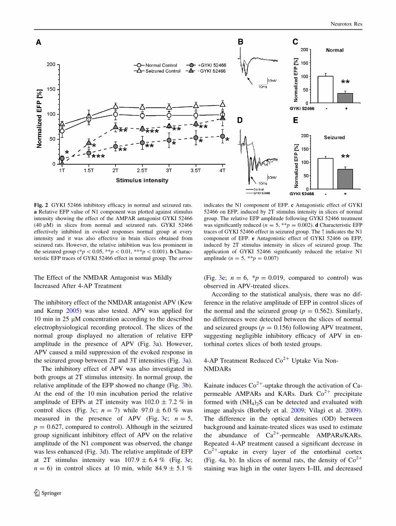
The Effect of the AMPAR Antagonist was Reduced
in the Seizured Slices
The pharmacological sensitivity of the N1 component of the
EFP was tested with the selective AMPAR antagonist, GYKI
52466 (Tarnawa et al. 1990). GYKI 52466 was applied in
40 lM final concentration for 30 min in the perfusion
solution. I–O curve and EFP amplitude at 2T stimulation
were determined in normal and seizured groups before and
immediately after finishing GYKI 52466 application. Each
EFP amplitude value was normalised for the EFP amplitude
of maximal stimulation intensity (4T) recorded at 0 min. The
slices of the normal group showed pronounced inhibition of
the EFP in the presence of AMPAR antagonist GYKI 52466
(Fig. 2a). The inhibitory effect of GYKI 52466 was also
significant in the seizured group, however, the observed
decrease of relative EFP was smaller than in slices of the
normal group, despite the fact that the control values of
relative EFP were similar in both animal groups (Fig. 2a).
GYKI 52466 was able to suppress the EFP at every stimulus
intensity in either normal or seizured groups.
The inhibitory effect of GYKI 52466 was further ana-
lysed in normal and seizured groups, at 2T stimulus
intensity. In the normal group, the amplitude of the N1
component of the EFP was reduced in the presence of
GYKI 52466 (Fig. 2b). At the end of the 30 min incubation
period the relative amplitude of EFPs at 2T intensity was
100.0 ± 10.6 % in control slices (Fig. 2c; n = 5) which
was significantly reduced to 35.6 ± 9.1 % relative ampli-
tude in the presence of GYKI 52466 (Fig. 2c; n = 5,
**p = 0.002, compared to control). In the seizured group,
GYKI 52466 also reduced the amplitude of the N1 com-
ponent of the EFP (Fig. 2d). The relative amplitude of
EFPs at 2T stimulus intensity was 115.0 ± 7.8 % (Fig. 2e;
n = 6) in control slices at 30 min, and reduced to
74.1 ± 8.9 % (Fig. 2e; n = 5, **p = 0.007, compared to
control) in GYKI 52466-treated slices.
The relative amplitude of EFPs showed no difference in
the control slices of the normal and the seizured group
(p = 0.275), however, there was a significant difference
between the GYKI 52466-treated slices of the normal and
the seizured animals (*p = 0.017). These results indicate
that the inhibition of AMPARs by GYKI 52466 was sig-
nificantly reduced in the seizured group.
Neurotox Res
123
The Effect of the NMDAR Antagonist was Mildly
Increased After 4-AP Treatment
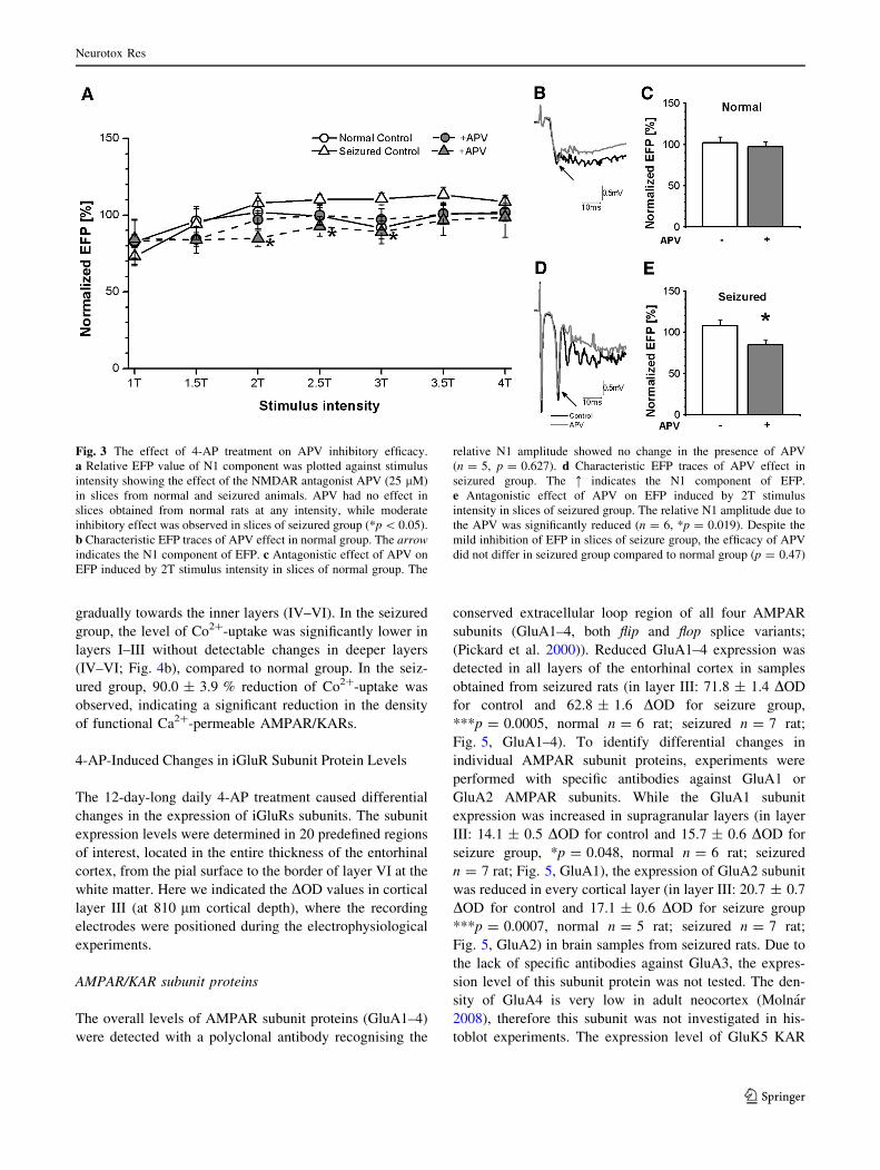
The inhibitory effect of the NMDAR antagonist APV (Kew
and Kemp 2005) was also tested. APV was applied for
10 min in 25 lM concentration according to the described
electrophysiological recording protocol. The slices of the
normal group displayed no alteration of relative EFP
amplitude in the presence of APV (Fig. 3a). However,
APV caused a mild suppression of the evoked response in
the seizured group between 2T and 3T intensities (Fig. 3a).
The inhibitory effect of APV was also investigated in
both groups at 2T stimulus intensity. In normal group, the
relative amplitude of the EFP showed no change (Fig. 3b).
At the end of the 10 min incubation period the relative
amplitude of EFPs at 2T intensity was 102.0 ± 7.2 % in
control slices (Fig. 3c; n = 7) while 97.0 ± 6.0 % was
measured in the presence of APV (Fig. 3c; n = 5,
p = 0.627, compared to control). Although in the seizured
group significant inhibitory effect of APV on the relative
amplitude of the N1 component was observed, the change
was less enhanced (Fig. 3d). The relative amplitude of EFP
at 2T stimulus intensity was 107.9 ± 6.4 % (Fig. 3e;
n = 6) in control slices at 10 min, while 84.9 ± 5.1 %
(Fig. 3e; n = 6, *p = 0.019, compared to control) was
observed in APV-treated slices.
According to the statistical analysis, there was no dif-
ference in the relative amplitude of EFP in control slices of
the normal and the seizured group (p = 0.562). Similarly,
no differences were detected between the slices of normal
and seizured groups (p = 0.156) following APV treatment,
suggesting negligible inhibitory efficacy of APV in en-
torhinal cortex slices of both tested groups.
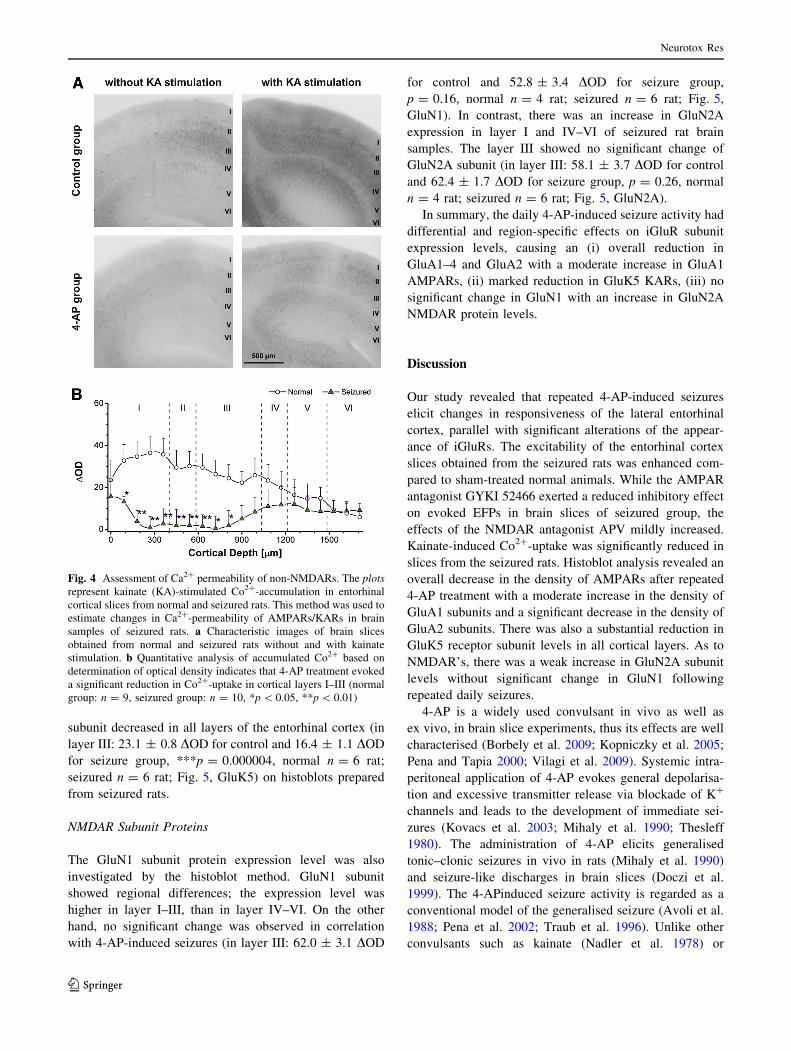
4-AP Treatment Reduced Co2? Uptake Via Non-
NMDARs
Kainate induces Co2?-uptake through the activation of Ca-
permeable AMPARs and KARs. Dark Co2? precipitate
formed with (NH4)2S can be detected and evaluated with
image analysis (Borbely et al. 2009; Vilagi et al. 2009).
The difference in the optical densities (OD) between
background and kainate-treated slices was used to estimate
the abundance of Ca2?-permeable AMPARs/KARs.
Repeated 4-AP treatment caused a significant decrease in
Co2?-uptake in every layer of the entorhinal cortex
(Fig. 4a, b). In slices of normal rats, the density of Co2?
staining was high in the outer layers I–III, and decreased
Fig. 2 GYKI 52466 inhibitory efficacy in normal and seizured rats.
a Relative EFP value of N1 component was plotted against stimulus
intensity showing the effect of the AMPAR antagonist GYKI 52466
(40 lM) in slices from normal and seizured rats. GYKI 52466
effectively inhibited in evoked responses normal group at every
intensity and it was also effective in brain slices obtained from
seizured rats. However, the relative inhibition was less prominent in
the seizured group (*p \ 0.05, **p \ 0.01, ***p \ 0.001). b Charac-
teristic EFP traces of GYKI 52466 effect in normal group. The arrow
indicates the N1 component of EFP. c Antagonistic effect of GYKI
52466 on EFP, induced by 2T stimulus intensity in slices of normal
group. The relative EFP amplitude following GYKI 52466 treatment
was significantly reduced (n = 5, **p = 0.002). d Characteristic EFP
traces of GYKI 52466 effect in seizured group. The : indicates the N1
component of EFP. e Antagonistic effect of GYKI 52466 on EFP,
induced by 2T stimulus intensity in slices of seizured group. The
application of GYKI 52466 significantly reduced the relative N1
amplitude (n = 5, **p = 0.007)
Neurotox Res
123
gradually towards the inner layers (IV–VI). In the seizured
group, the level of Co2?-uptake was significantly lower in
layers I–III without detectable changes in deeper layers
(IV–VI; Fig. 4b), compared to normal group. In the seiz-
ured group, 90.0 ± 3.9 % reduction of Co2?-uptake was
observed, indicating a significant reduction in the density
of functional Ca2?-permeable AMPAR/KARs.
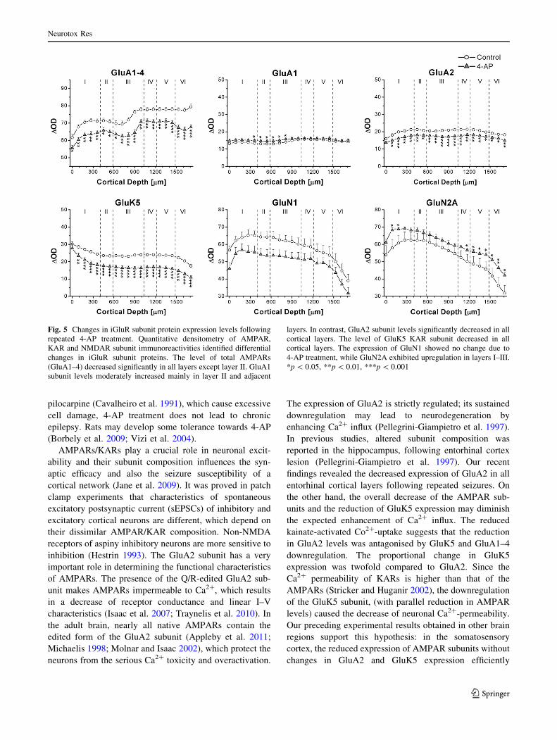
4-AP-Induced Changes in iGluR Subunit Protein Levels
The 12-day-long daily 4-AP treatment caused differential
changes in the expression of iGluRs subunits. The subunit
expression levels were determined in 20 predefined regions
of interest, located in the entire thickness of the entorhinal
cortex, from the pial surface to the border of layer VI at the
white matter. Here we indicated the DOD values in cortical
layer III (at 810 lm cortical depth), where the recording
electrodes were positioned during the electrophysiological
experiments.
AMPAR/KAR subunit proteins
The overall levels of AMPAR subunit proteins (GluA1–4)
were detected with a polyclonal antibody recognising the
conserved extracellular loop region of all four AMPAR
subunits (GluA1–4, both flip and flop splice variants;
(Pickard et al. 2000)). Reduced GluA1–4 expression was
detected in all layers of the entorhinal cortex in samples
obtained from seizured rats (in layer III: 71.8 ± 1.4 DOD
for control and 62.8 ± 1.6 DOD for seizure group,
***p = 0.0005, normal n = 6 rat; seizured n = 7 rat;
Fig. 5, GluA1–4). To identify differential changes in
individual AMPAR subunit proteins, experiments were
performed with specific antibodies against GluA1 or
GluA2 AMPAR subunits. While the GluA1 subunit
expression was increased in supragranular layers (in layer
III: 14.1 ± 0.5 DOD for control and 15.7 ± 0.6 DOD for
seizure group, *p = 0.048, normal n = 6 rat; seizured
n = 7 rat; Fig. 5, GluA1), the expression of GluA2 subunit
was reduced in every cortical layer (in layer III: 20.7 ± 0.7
DOD for control and 17.1 ± 0.6 DOD for seizure group
***p = 0.0007, normal n = 5 rat; seizured n = 7 rat;
Fig. 5, GluA2) in brain samples from seizured rats. Due to
the lack of specific antibodies against GluA3, the expres-
sion level of this subunit protein was not tested. The den-
sity of GluA4 is very low in adult neocortex (Molnar
2008), therefore this subunit was not investigated in his-
toblot experiments. The expression level of GluK5 KAR
Fig. 3 The effect of 4-AP treatment on APV inhibitory efficacy.
a Relative EFP value of N1 component was plotted against stimulus
intensity showing the effect of the NMDAR antagonist APV (25 lM)
in slices from normal and seizured animals. APV had no effect in
slices obtained from normal rats at any intensity, while moderate
inhibitory effect was observed in slices of seizured group (*p \ 0.05).
b Characteristic EFP traces of APV effect in normal group. The arrow
indicates the N1 component of EFP. c Antagonistic effect of APV on
EFP induced by 2T stimulus intensity in slices of normal group. The
relative N1 amplitude showed no change in the presence of APV
(n = 5, p = 0.627). d Characteristic EFP traces of APV effect in
seizured group. The : indicates the N1 component of EFP.
e Antagonistic effect of APV on EFP induced by 2T stimulus
intensity in slices of seizured group. The relative N1 amplitude due to
the APV was significantly reduced (n = 6, *p = 0.019). Despite the
mild inhibition of EFP in slices of seizure group, the efficacy of APV
did not differ in seizured group compared to normal group (p = 0.47)
Neurotox Res
123
subunit decreased in all layers of the entorhinal cortex (in
layer III: 23.1 ± 0.8 DOD for control and 16.4 ± 1.1 DOD
for seizure group, ***p = 0.000004, normal n = 6 rat;
seizured n = 6 rat; Fig. 5, GluK5) on histoblots prepared
from seizured rats.
NMDAR Subunit Proteins
The GluN1 subunit protein expression level was also
investigated by the histoblot method. GluN1 subunit
showed regional differences; the expression level was
higher in layer I–III, than in layer IV–VI. On the other
hand, no significant change was observed in correlation
with 4-AP-induced seizures (in layer III: 62.0 ± 3.1 DOD
for control and 52.8 ± 3.4 DOD for seizure group,
p = 0.16, normal n = 4 rat; seizured n = 6 rat; Fig. 5,
GluN1). In contrast, there was an increase in GluN2A
expression in layer I and IV–VI of seizured rat brain
samples. The layer III showed no significant change of
GluN2A subunit (in layer III: 58.1 ± 3.7 DOD for control
and 62.4 ± 1.7 DOD for seizure group, p = 0.26, normal
n = 4 rat; seizured n = 6 rat; Fig. 5, GluN2A).
In summary, the daily 4-AP-induced seizure activity had
differential and region-specific effects on iGluR subunit
expression levels, causing an (i) overall reduction in
GluA1–4 and GluA2 with a moderate increase in GluA1
AMPARs, (ii) marked reduction in GluK5 KARs, (iii) no
significant change in GluN1 with an increase in GluN2A
NMDAR protein levels.
Discussion
Our study revealed that repeated 4-AP-induced seizures
elicit changes in responsiveness of the lateral entorhinal
cortex, parallel with significant alterations of the appear-
ance of iGluRs. The excitability of the entorhinal cortex
slices obtained from the seizured rats was enhanced com-
pared to sham-treated normal animals. While the AMPAR
antagonist GYKI 52466 exerted a reduced inhibitory effect
on evoked EFPs in brain slices of seizured group, the
effects of the NMDAR antagonist APV mildly increased.
Kainate-induced Co2?-uptake was significantly reduced in
slices from the seizured rats. Histoblot analysis revealed an
overall decrease in the density of AMPARs after repeated
4-AP treatment with a moderate increase in the density of
GluA1 subunits and a significant decrease in the density of
GluA2 subunits. There was also a substantial reduction in
GluK5 receptor subunit levels in all cortical layers. As to
NMDAR’s, there was a weak increase in GluN2A subunit
levels without significant change in GluN1 following
repeated daily seizures.
4-AP is a widely used convulsant in vivo as well as
ex vivo, in brain slice experiments, thus its effects are well
characterised (Borbely et al. 2009; Kopniczky et al. 2005;
Pena and Tapia 2000; Vilagi et al. 2009). Systemic intra-
peritoneal application of 4-AP evokes general depolarisa-
tion and excessive transmitter release via blockade of K?
channels and leads to the development of immediate sei-
zures (Kovacs et al. 2003; Mihaly et al. 1990; Thesleff
1980). The administration of 4-AP elicits generalised
tonic–clonic seizures in vivo in rats (Mihaly et al. 1990)
and seizure-like discharges in brain slices (Doczi et al.
1999). The 4-APinduced seizure activity is regarded as a
conventional model of the generalised seizure (Avoli et al.
1988; Pena et al. 2002; Traub et al. 1996). Unlike other
convulsants such as kainate (Nadler et al. 1978) or
Fig. 4 Assessment of Ca2? permeability of non-NMDARs. The plots
represent kainate (KA)-stimulated Co2?-accumulation in entorhinal
cortical slices from normal and seizured rats. This method was used to
estimate changes in Ca2?-permeability of AMPARs/KARs in brain
samples of seizured rats. a Characteristic images of brain slices
obtained from normal and seizured rats without and with kainate
stimulation. b Quantitative analysis of accumulated Co2? based on
determination of optical density indicates that 4-AP treatment evoked
a significant reduction in Co2?-uptake in cortical layers I–III (normal
group: n = 9, seizured group: n = 10, *p \ 0.05, **p \ 0.01)
Neurotox Res
123
pilocarpine (Cavalheiro et al. 1991), which cause excessive
cell damage, 4-AP treatment does not lead to chronic
epilepsy. Rats may develop some tolerance towards 4-AP
(Borbely et al. 2009; Vizi et al. 2004).
AMPARs/KARs play a crucial role in neuronal excit-
ability and their subunit composition influences the syn-
aptic efficacy and also the seizure susceptibility of a
cortical network (Jane et al. 2009). It was proved in patch
clamp experiments that characteristics of spontaneous
excitatory postsynaptic current (sEPSCs) of inhibitory and
excitatory cortical neurons are different, which depend on
their dissimilar AMPAR/KAR composition. Non-NMDA
receptors of aspiny inhibitory neurons are more sensitive to
inhibition (Hestrin 1993). The GluA2 subunit has a very
important role in determining the functional characteristics
of AMPARs. The presence of the Q/R-edited GluA2 sub-
unit makes AMPARs impermeable to Ca2?, which results
in a decrease of receptor conductance and linear I–V
characteristics (Isaac et al. 2007; Traynelis et al. 2010). In
the adult brain, nearly all native AMPARs contain the
edited form of the GluA2 subunit (Appleby et al. 2011;
Michaelis 1998; Molnar and Isaac 2002), which protect the
neurons from the serious Ca2? toxicity and overactivation.
The expression of GluA2 is strictly regulated; its sustained
downregulation may lead to neurodegeneration by
enhancing Ca2? influx (Pellegrini-Giampietro et al. 1997).
In previous studies, altered subunit composition was
reported in the hippocampus, following entorhinal cortex
lesion (Pellegrini-Giampietro et al. 1997). Our recent
findings revealed the decreased expression of GluA2 in all
entorhinal cortical layers following repeated seizures. On
the other hand, the overall decrease of the AMPAR sub-
units and the reduction of GluK5 expression may diminish
the expected enhancement of Ca2? influx. The reduced
kainate-activated Co2?-uptake suggests that the reduction
in GluA2 levels was antagonised by GluK5 and GluA1–4
downregulation. The proportional change in GluK5
expression was twofold compared to GluA2. Since the
Ca2? permeability of KARs is higher than that of the
AMPARs (Stricker and Huganir 2002), the downregulation
of the GluK5 subunit, (with parallel reduction in AMPAR
levels) caused the decrease of neuronal Ca2?-permeability.
Our preceding experimental results obtained in other brain
regions support this hypothesis: in the somatosensory
cortex, the reduced expression of AMPAR subunits without
changes in GluA2 and GluK5 expression efficiently
Fig. 5 Changes in iGluR subunit protein expression levels following
repeated 4-AP treatment. Quantitative densitometry of AMPAR,
KAR and NMDAR subunit immunoreactivities identified differential
changes in iGluR subunit proteins. The level of total AMPARs
(GluA1–4) decreased significantly in all layers except layer II. GluA1
subunit levels moderately increased mainly in layer II and adjacent
layers. In contrast, GluA2 subunit levels significantly decreased in all
cortical layers. The level of GluK5 KAR subunit decreased in all
cortical layers. The expression of GluN1 showed no change due to
4-AP treatment, while GluN2A exhibited upregulation in layers I–III.
*p \ 0.05, **p \ 0.01, ***p \ 0.001
Neurotox Res
123

decreased the Ca2? permeability (Vilagi et al. 2009). On
the other hand, the CA1 region of the hippocampus dis-
played the decrease of GluA2, together with unchanged
GluA1–4 and GluK5, but the net result was a significant
increase in Ca2? permeability (Borbely et al. 2009). It has
been demonstrated that Co2?-uptake do not provide
information about the overall Ca2? intake, i.e. the perme-
ability changes of NMDARs, because these types of
receptors are impermeable to Co2? (Mayer and Westbrook
1987).
It was reported that other convulsants also cause alter-
ations in receptor number and subunit composition of
AMPARs (Pellegrini-Giampietro et al. 1997; Tolner et al.
2007). The common feature of this phenomenon was the
increased Ca2? permeability of AMPARs due to GluA2
downregulation (Rajasekaran et al. 2012). Rearrangement
of GluA1–4 subunit expression, parallel with a reduced
GluA2 subunit manifestation was observed in the CA3
region of rat hippocampus, following kainate-induced sta-
tus epilepticus (Friedman et al. 1994; Friedman and Ve-
liskova 1998). Similar observations were made in
pilocarpine-induced seizures (Lason et al. 1997; Rajasek-
aran et al. 2012). On the other hand, in early postnatal
seizures induced by pilocarpine, in the rat hippocampus an
elevated GluA1–3 subunits level and unchanged GluA4
was observed (Silva et al. 2005). Downregulation of GluA2
can be also induced in rat hippocampal CA1 region by
transient forebrain ischemia (Pellegrini-Giampietro et al.
1997), in layer V neocortical pyramidal neurons by in vivo
cortical lesion (Kharazia and Prince 2001) and in cortical
neurons by kainate administration (Jia et al. 2006). The
chronic treatment with pentylenetetrazole did not result in
significant overall reduction of AMPARs. In these experi-
ments, however, a significant decrease of KARs was
reported (Cremer et al. 2009), and the level of GluA2
subunit expression was reduced by 50 % (Cremer et al.
2009). Similarly, we also detected a significant 15 %
reduction of GluA2 subunit level.
GYKI 52466 is a non-competitive inhibitor binding to
an allosteric site of AMPARs (Tarnawa et al. 1990).
Therefore, its inhibitory effect is independent from gluta-
mate binding or extracellular glutamate levels (Wilding
and Huettner 1995). Therefore, the most likely explanation
of 4-AP treatment-related reduction in GYKI
52466-induced inhibition is that AMPAR/KAR levels and/
or subunit composition altered as a consequence of 4-AP
provoked seizures. GYKI 52466 has no preference to flip or
flop isoforms of AMPARs (Johansen et al. 1995), but
changes of the subunit composition may influence GYKI
52466 antagonism. GYKI 52466 is a less effective inhibitor
on homotetrameric receptors formed by GluA1 or GluA4
subunits than on heterotetramers of GluA1/GluA2 or
GluA2/GluA4. This suggests that the presence of GluA2
subunits in AMPARs increases the antagonistic effect
(Bleakman et al. 1996; Johansen et al. 1995). Since GYKI
52466 specifically inhibits AMPARs in the applied con-
centration (Bleakman et al. 1996), we can state that the
decrease of the GYKI 52466 effect was related to the
overall decrease of AMPARs and the decrease of the
GluA2 subunit expression.
Despite the fact that the expression of NMDAR subunits is
strictly regulated (Lujan et al. 2005), the alteration of subunit
composition can be induced by different convulsants. The
available experimental findings are, however, controversial.
Perinatal kainate-induced seizures result in upregulation of
GluN2A and GluN2B subunits (Gashi et al. 2007), while
their downregulation was observed after repeated pilocar-
pine-induced status epilepticus (Silva et al. 2005) in the rat
hippocampus. In adult rats pentylenetetrazole-kindling
reduced the expression of GluN2A without changes in
GluN1 and GluN2B levels (Zhu et al. 2004), suggesting that
GluN2A downregulation was necessary for the increase of
general excitability. Experimental data from human patients
with focal cortical dysplasia suggest that the upregulation of
NMDAR subunits is necessary for increased epileptogenesis
(Najm et al. 2000). We have detected a mild upregulation of
GluN2A subunit which may underlie the parallel reinforce of
APV inhibitory effect.
The number, molecular composition and functional
properties of receptors fundamentally influence the charac-
teristics of evoked field potentials in brain slices. In this
study, we identified an enhanced responsibility of the en-
torhinal cortex following repeated 4-AP-induced seizures.
Nevertheless, this was accompanied by an overall reduction
in the expression levels, Ca2? permeability and antagonist
sensitivity of AMPARs and KARs. However, in parallel a
moderate but detectable GluN2A receptor expression
increase was obtained. Our previous findings revealed sim-
ilar alterations of iGluR structure in the somatosensory
cortex (Vilagi et al. 2009), and in the CA3 region of the
hippocampus (Borbely et al. 2009) of seizured rats. But the
general excitability changes detected in ex vivo slice prep-
arations were not uniform: we measured decreased excit-
ability in the somatosensory cortex (Vilagi et al. 2009), and
increased excitability in the hippocampus (Borbely et al.
2009) and entorhinal cortex. The shape and the amplitude of
the evoked field response in slices from different cortical
regions are basically determined by the activation of excit-
atory and inhibitory neurons in the local networks. The
overall excitability depends on the balance of the activity of
excitatory and inhibitory cells, which strongly depends on
the actual receptor pool expressed. The inhibitory interneu-
rons also possess excitatory amino acid receptors. As these
types of neurons are more prone to overexcitation, so fol-
lowing the seizures the number of them may decrease, par-
allel decreasing the whole inhibition. So diminish of the
Neurotox Res
123

whole ionotropic excitatory amino acid receptors may result
in a net increase of EFP (Hestrin 1993). According to our
studies, long lasting 4-AP treatment mostly results in alter-
ations in non-NMDARs, the number of which may decrease
in all cell types. As a consequence, inhibition will be less
efficient in that brain area, which may alter the local network
activity, together with the regional sensitivity. Simulta-
neously, the moderately enhanced NMDAR activity may
explain an overall increase in excitability. We hypothesised,
that non-NMDAR-mediated processes are less sensitive, so
no spontaneous seizure activity develops following the
repetitive 4-AP application. Further investigations are nee-
ded to define the prominently affected cell types, synapses
and networks.
Acknowledgments This research was supported by Grant from the
Biotechnology and Biological Sciences Research Council, UK (Grant
BB/J015938/1 to E.M.) and from TAMOP (4.2.2/A-11/1/KONV-
2012-0052 to A.M.).
Conflict of interest The authors declare that they have no conflict
of interests.
References
Appleby VJ et al (2011) LTP in hippocampal neurons is associated with a
CaMKII-mediated increase in GluA1 surface expression. J Neuro-
chem 116:530–543. doi:10.1111/j.1471-4159.2010.07133.x
Armand V, Hoffmann P, Vergnes M, Heinemann U (1999) Epilep-
tiform activity induced by 4-aminopyridine in entorhinal cortex
hippocampal slices of rats with a genetically determined absence
epilepsy (GAERS). Brain Res 841:62–69
Avoli M, Perreault P, Olivier A, Villemure JG (1988) 4-Aminopyr-
idine induces a long-lasting depolarizing GABA-ergic potential
in human neocortical and hippocampal neurons maintained
in vitro. Neurosci Lett 94:327–332
Bleakman D et al (1996) Activity of 2,3-benzodiazepines at native rat
and recombinant human glutamate receptors in vitro: stereospec-
ificity and selectivity profiles. Neuropharmacology 35:1689–1702
Borbely S, Halasy K, Somogyvari Z, Detari L, Vilagi I (2006) Laminar
analysis of initiation and spread of epileptiform discharges in three
in vitro models. Brain Res Bull 69:161–167. doi:10.1016/j.
brainresbull.2005.11.018
Borbely S et al (2009) Modification of ionotropic glutamate receptor-
mediated processes in the rat hippocampus following repeated,
brief seizures. Neuroscience 159:358–368. doi:10.1016/j.neu
roscience.2008.12.027
Cavalheiro EA, Leite JP, Bortolotto ZA, Turski WA, Ikonomidou C,
Turski L (1991) Long-term effects of pilocarpine in rats:
structural damage of the brain triggers kindling and spontaneous
recurrent seizures. Epilepsia 32:778–782
Chang BS, Lowenstein DH (2003) Epilepsy. New Engl J Med
349:1257–1266. doi:10.1056/NEJMra022308
Choi YS, Cho KO, Kim SY (2007) Asymmetry in enhanced neurogen-
esis in the rostral dentate gyrus following kainic acid-induced
status epilepticus in adult rats. Arch Pharm Res 30:646–652.
doi:10.1007/bf02977661
Collingridge GL, Isaac JT, Wang YT (2004) Receptor trafficking and
synaptic plasticity. Nat Rev Neurosci 5:952–962. doi:10.1038/
nrn1556
Cremer CM, Palomero-Gallagher N, Bidmon HJ, Schleicher A,
Speckmann EJ, Zilles K (2009) Pentylenetetrazole-induced
seizures affect binding site densities for GABA, glutamate and
adenosine receptors in the rat brain. Neuroscience 163:490–499.
doi:10.1016/j.neuroscience.2009.03.068
Doczi J, Banczerowski-Pelyhe I, Barna B, Vilagi I (1999) Effect of a
glutamate receptor antagonist (GYKI 52466) on 4-aminopyri-
dine-induced seizure activity developed in rat cortical slices.
Brain Res Bull 49:435–440
Fisher RS (1989) Animal models of the epilepsies. Brain Res Rev
14:245–278
Friedman LK, Veliskova J (1998) GluR2 hippocampal knockdown
reveals developmental regulation of epileptogenicity and neuro-
degeneration. Mol Brain Res 61:224–231
Friedman LK, Pellegrini-Giampietro DE, Sperber EF, Bennett MV,
Moshe SL, Zukin RS (1994) Kainate-induced status epilepticus
alters glutamate and GABAA receptor gene expression in adult
rat hippocampus: an in situ hybridization study. J Neurosci
14:2697–2707
Gallyas F Jr, Ball SM, Molnar E (2003) Assembly and cell surface
expression of KA-2 subunit-containing kainate receptors. J Neu-
rochem 86:1414–1427
Gashi E, Avallone J, Webster T, Friedman LK (2007) Altered
excitability and distribution of NMDA receptor subunit proteins
in cortical layers of rat pups following multiple perinatal
seizures. Brain Res 1145:56–65
Gulyas-Kovacs A, Doczi J, Tarnawa I, Detari L, Banczerowski-
Pelyhe I, Vilagi I (2002) Comparison of spontaneous and evoked
epileptiform activity in three in vitro epilepsy models. Brain Res
945:174–180. doi:10.1016/s0006-8993(02)02751-8
Hestrin S (1993) Different glutamate receptor channels mediate fast
excitatory synaptic currents in inhibitory and excitatory cortical
neurons. Neuron 11:1083–1091
Isaac JT, Ashby MC, McBain CJ (2007) The role of the GluR2
subunit in AMPA receptor function and synaptic plasticity.
Neuron 54:859–871. doi:10.1016/j.neuron.2007.06.001
Jane DE, Lodge D, Collingridge GL (2009) Kainate receptors:
pharmacology, function and therapeutic potential. Neurophar-
macology 56:90–113. doi:10.1016/j.neuropharm.2008.08.023
Jia YH, Zhu X, Li SY, Ni JH, Jia HT (2006) Kainate exposure
suppresses activation of GluR2 subunit promoter in primary
cultured cerebral cortical neurons through induction of RE1-
silencing transcription factor. Neurosci Lett 403:103–108.
doi:10.1016/j.neulet.2006.04.027
Johansen TH, Chaudhary A, Verdoorn TA (1995) Interactions among
GYKI-52466, cyclothiazide, and aniracetam at recombinant
AMPA and kainate receptors. Mol Pharmacol 48:946–955
Kew JN, Kemp JA (2005) Ionotropic and metabotropic glutamate
receptor structure and pharmacology. Psychopharmacology
179:4–29. doi:10.1007/s00213-005-2200-z
Kharazia VN, Prince DA (2001) Changes of alpha-amino-3-hydroxy-
5-methyl-4-isoxazole-propionate receptors in layer V of epilep-
togenic, chronically isolated rat neocortex. Neuroscience
102:23–34
Kopniczky Z et al (2005) Lateral entorhinal cortex lesions rearrange
afferents, glutamate receptors, increase seizure latency and
suppress seizure-induced c-fos expression in the hippocampus of
adult rat. J Neurochem 95:111–124. doi:10.1111/j.1471-4159.
2005.03347.x
Kovacs A et al (2003) Seizure, neurotransmitter release, and gene
expression are closely related in the striatum of 4-aminopyri-
dine-treated rats. Epilepsy Res 55:117–129
Lason W, Turchan J, Przewlocka B, Labuz D, Mika J, Przewlocki R
(1997) Seizure-related changes in the glutamate R2 and R5
receptor genes expression in the rat hippocampal formation.
J Neural Transm 104:125–133
Neurotox Res
123
Lau A, Tymianski M (2010) Glutamate receptors, neurotoxicity and
neurodegeneration. Pflugers Arch 460:525–542. doi:10.1007/
s00424-010-0809-1
Lerma J (2003) Roles and rules of kainate receptors in synaptic
transmission. Nat Rev Neurosci 4:481–495. doi:10.1038/nrn1118
Lujan R, Shigemoto R, Lopez-Bendito G (2005) Glutamate and
GABA receptor signalling in the developing brain. Neuroscience
130:567–580
Mayer ML, Westbrook GL (1987) Permeation and block of N-methyl-
D-aspartic acid receptor channels by divalent cations in mouse
cultured central neurones. Journal Physiol 394:501–527
Michaelis EK (1998) Molecular biology of glutamate receptors in the
central nervous system and their role in excitotoxicity, oxidative
stress and aging. Prog Neurobiol 54:369–415
Mihaly A, Bencsik K, Solymosi T (1990) Naltrexone potentiates
4-aminopyridine seizures in the rat. J Neural Transm Gen Sect
79:59–67
Mihaly A et al (2005) Neocortical c-fos mRNA transcription in
repeated, brief, acute seizures: is c-fos a coincidence detector?
Int J Mol Med 15:481–486
Molnar E (2008) Molecular organization and regulation of glutamate
receptors in developing and adult mammalian central nervous
systems. In: Lajtha A, Vizi ES (eds) Handbook of neurochem-
istry and molecular neurobiology. Springer New York,
pp 415–441. doi:10.1007/978-0-387-30382-6_17
Molnar E, Isaac JT (2002) Developmental and activity dependent
regulation of ionotropic glutamate receptors at synapses. Scien-
tific World J 2:27–47. doi:10.1100/tsw.2002.74
Nadler JV (2003) The recurrent mossy fiber pathway of the epileptic
brain. Neurochem Res 28:1649–1658
Nadler JV, Perry BW, Cotman CW (1978) Intraventricular kainic acid
preferentially destroys hippocampal pyramidal cells. Nature
271:676–677
Najm IM et al (2000) Epileptogenicity correlated with increased
N-methyl-D-aspartate receptor subunit NR2A/B in human focal
cortical dysplasia. Epilepsia 41:971–976
Pellegrini-Giampietro DE, Gorter JA, Bennett MV, Zukin RS (1997)
The GluR2 (GluR-B) hypothesis: Ca(2?)-permeable AMPA
receptors in neurological disorders. Trends Neurosci 20:464–470
Pena F, Tapia R (2000) Seizures and neurodegeneration induced by
4-aminopyridine in rat hippocampus in vivo: role of glutamate-
and GABA-mediated neurotransmission and of ion channels.
Neuroscience 101:547–561
Pena F, Bargas J, Tapia R (2002) Paired pulse facilitation is turned
into paired pulse depression in hippocampal slices after epilepsy
induced by 4-aminopyridine in vivo. Neuropharmacology
42:807–812
Pickard L, Noel J, Henley JM, Collingridge GL, Molnar E (2000)
Developmental changes in synaptic AMPA and NMDA receptor
distribution and AMPA receptor subunit composition in living
hippocampal neurons. J Neurosci 20:7922–7931
Pruss RM, Akeson RL, Racke MM, Wilburn JL (1991) Agonist-
activated cobalt uptake identifies divalent cation-permeable
kainate receptors on neurons and glial cells. Neuron 7:509–518
Racine RJ (1972) Modification of seizure activity by electrical
stimulation. II. Motor seizure. Electroencephalogr Clin Neuro-
physiol 32:281–294
Rajasekaran K, Todorovic M, Kapur J (2012) Calcium-permeable
AMPA receptors are expressed in a rodent model of status
epilepticus. Ann Neurol 72:91–102. doi:10.1002/ana.23570
Scorza FA, Arida RM, Naffah-Mazzacoratti Mda G, Scerni DA,
Calderazzo L, Cavalheiro EA (2009) The pilocarpine model of
epilepsy: what have we learned? An Acad Bras Cienc 81:345–365
Silva AV, Regondi MC, Cipelletti B, Frassoni C, Cavalheiro EA,
Spreafico R (2005) Neocortical and hippocampal changes after
multiple pilocarpine-induced status epilepticus in rats. Epilepsia
46:636–642. doi:10.1111/j.1528-1167.2005.31604.x
Stricker NL, Huganir RL (2002) Ampa/kainate receptors. In: Moss SJ,
Henley J (eds) Receptor and ion-channel trafficking: cell biology
of ligand-gated and voltage-sensitive ion channels. Oxford
University Press, New York, pp 131–155. doi:10.1093/acprof:
oso/9780192632241.003.0006
Szakacs R, Weiczner R, Mihaly A, Krisztin-Peva B, Zador Z, Zador E
(2003) Non-competitive NMDA receptor antagonists moderate
seizure-induced c-fos expression in the rat cerebral cortex. Brain
Res Bull 59:485–493
Tarnawa I, Farkas S, Berzsenyi P, Patfalusi M, Andrasi F (1990) Reflex
inhibitory action of a non-NMDA type excitatory amino acid
antagonist, GYKI 52466. Acta Physiol Hung 75(Suppl):277–278
Thesleff S (1980) Aminopyridines and synaptic transmission. Neu-
roscience 5:1413–1419
Tolner EA, Frahm C, Metzger R, Gorter JA, Witte OW, Lopes da Silva
FH, Heinemann U (2007) Synaptic responses in superficial layers of
medial entorhinal cortex from rats with kainate-induced epilepsy.
Neurobiol Dis 26:419–438. doi:10.1016/j.nbd.2007.01.009
Tonnes J, Stierli B, Cerletti C, Behrmann JT, Molnar E, Streit P
(1999) Regional distribution and developmental changes of
GluR1-flop protein revealed by monoclonal antibody in rat brain.
J Neurochem 73:2195–2205
Traub RD, Borck C, Colling SB, Jefferys JG (1996) On the structure
of ictal events in vitro. Epilepsia 37:879–891
Traynelis SF et al (2010) Glutamate receptor ion channels: structure,
regulation, and function. Pharmacol Rev 62:405–496. doi:10.
1124/pr.109.002451
Turski WA, Cavalheiro EA, Schwarz M, Czuczwar SJ, Kleinrok Z,
Turski L (1983) Limbic seizures produced by pilocarpine in rats:
behavioural, electroencephalographic and neuropathological
study. Behav Brain Res 9:315–335
Vilagi I, Csucs G, Tarnawa I, Banczerowski-Pelyhe I (1996) An
increased intensity of N-methyl-D-aspartate (NMDA) but not
non-NMDA receptor activation may be responsible for the
enhancement of excitatory processes in the neocortex of two-
week-old rats: a brain slices study. Neurosci Lett 203:139–142
Vilagi I, Dobo E, Borbely S, Czege D, Molnar E, Mihaly A (2009)
Repeated 4-aminopyridine induced seizures diminish the effi-
cacy of glutamatergic transmission in the neocortex. Exp Neurol
219:136–145. doi:10.1016/j.expneurol.2009.05.005
Vizi S, Bagosi A, Krisztin-Peva B, Gulya K, Mihaly A (2004)
Repeated 4-aminopyridine seizures reduce parvalbumin content
in the medial mammillary nucleus of the rat brain. Mol Brain
Res 131:110–118. doi:10.1016/j.molbrainres.2004.08.022
Weiczner R, Krisztin-Peva B, Mihaly A (2008) Blockade of AMPA-receptors attenuates 4-aminopyridine seizures, decreases the
activation of inhibitory neurons but is ineffective against seizure-
related astrocytic swelling. Epilepsy Res 78:22–32. doi:10.1016/
j.eplepsyres.2007.10.004
Wilding TJ, Huettner JE (1995) Differential antagonism of alpha-
amino-3-hydroxy-5-methyl-4- isoxazolepropionic acid-prefer-
ring and kainate-preferring receptors by 2,3-benzodiazepines.
Mol Pharmacol 47:582–587
Zhu LJ, Chen Z, Zhang LS, Xu SJ, Xu AJ, Luo JH (2004)
Spatiotemporal changes of the N-methyl-D-aspartate receptor
subunit levels in rats with pentylenetetrazole-induced seizures.
Neurosci Lett 356:53–56
Neurotox Res
123