Laboratory tests of renal function Junfu Huang Southwestern Hospital TMMU.
Renal Function Tests WWW.RN.ORG®
Reviewed February, 2018, Expires February, 2020 Provider Information and Specifics available on our Website
Unauthorized Distribution Prohibited
©2018 RN.ORG®, S.A., RN.ORG®, LLC By Wanda Lockwood, RN, BA, MA
The purpose of this course is to explain the anatomy and physiology of the kidney and to outline the normal values and factors associated with increases and decreases in renal function tests.
Upon completion of this course, one should be able to:
• Describe the anatomy of the kidney and nephron.
• Explain the function of the kidneys and production of urine.
• Recognize reference values for renal function tests.
• Describe characteristics of common at least 4 renal diseases requiring renal function tests.
• Discuss estimated glomerular filtration rate (GFR) in relationship to creatinine and creatinine clearance tests.
• Describe at least 2 formulas used for estimation of GFR.
• Discuss at least 4 factors that effect BUN values.
• Discuss the relationship between serum and urine osmolality.
• Discuss the relationship between serum and urine sodium levels.
• List at least 5 causes of elevated serum uric acid.
• List 6 classes of nephrotoxic drugs and give examples for each class.
Introduction
Purpose
Course objectives
Properly functioning kidneys, the primary organs of the urinary system, are vital to maintain the homeostasis of the body. Millions of people in the United States suffer from kidney disease, but it is often a silent disease until irreparable damage has been done, resulting in acute or chronic renal failure. Because of this, the National Kidney Foundation recommends that routine screening on physical examination should include:
• Blood pressure measurement to assess hypertension, as this is a risk factor for renal disease.
• Urinary protein examination (dipstick) to check for albumin because healthy kidneys filter wastes from the blood but not proteins, and as kidney function deteriorates, proteins (primarily albumin) begin to filter into the urine.
• Calculation of estimated glomerular filtration rate of the kidneys to determine if kidney function is deteriorating.
If any abnormality is found in these three measures, or if there are symptoms suggestive of kidney disease, then further renal function tests should be done, including a routine urinalysis and specific tests to determine the extent and location of damage to the kidneys. A multitude of tests may be ordered if a diagnosis of kidney disease is made because kidney dysfunction affects the entire body; however, there are a number of tests that are specifically utilized as renal function tests (along with the standard urinalysis) and are used not only to diagnose kidney disease but also to monitor progress of the disease and response to treatment.
Review of kidney function and urine production
The kidneys filter unwanted waste materials from the blood and regulate the levels of water and chemicals in the body. The kidneys filter approximately 1200 mL of blood per minute. About 99% of the fluid circulating through the kidneys is reabsorbed with the remaining excreted as urine.
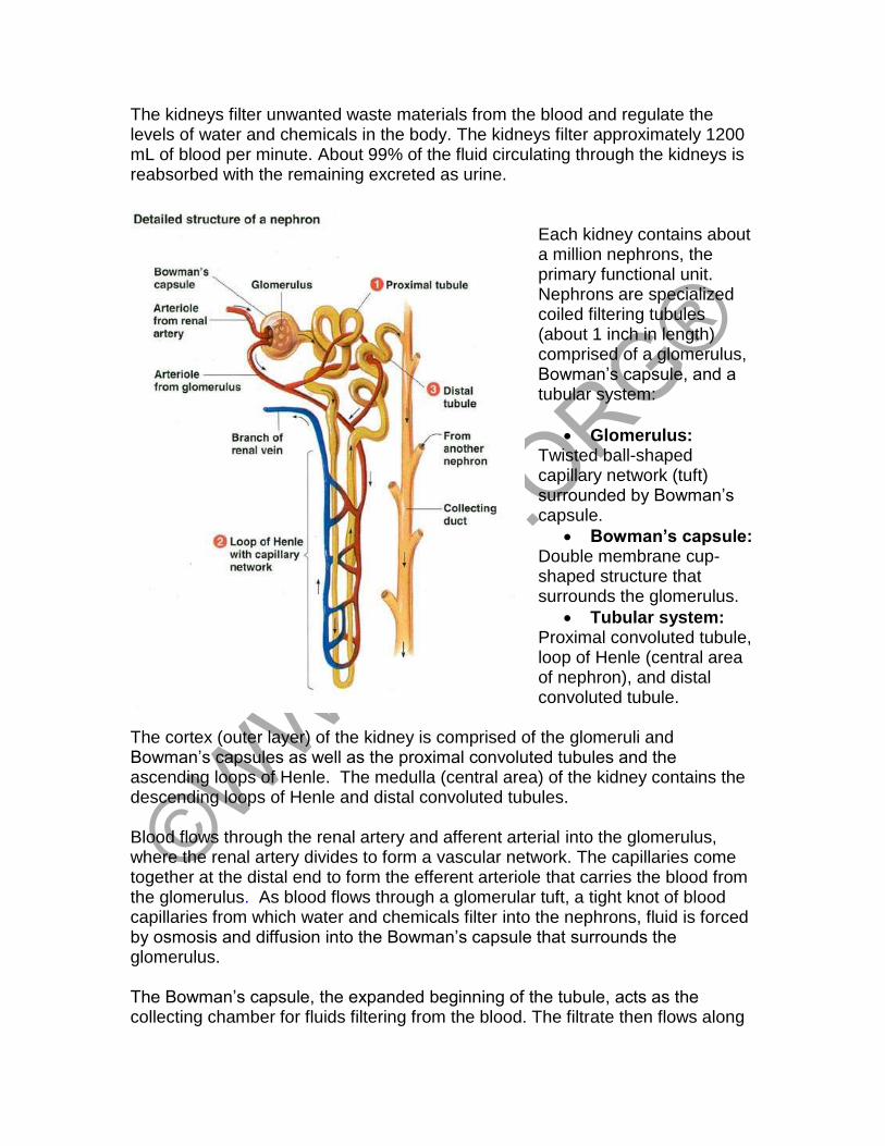
Each kidney contains about a million nephrons, the primary functional unit. Nephrons are specialized coiled filtering tubules (about 1 inch in length) comprised of a glomerulus, Bowman’s capsule, and a tubular system:
• Glomerulus: Twisted ball-shaped capillary network (tuft) surrounded by Bowman’s capsule.
• Bowman’s capsule: Double membrane cup-shaped structure that surrounds the glomerulus.
• Tubular system: Proximal convoluted tubule, loop of Henle (central area of nephron), and distal convoluted tubule.
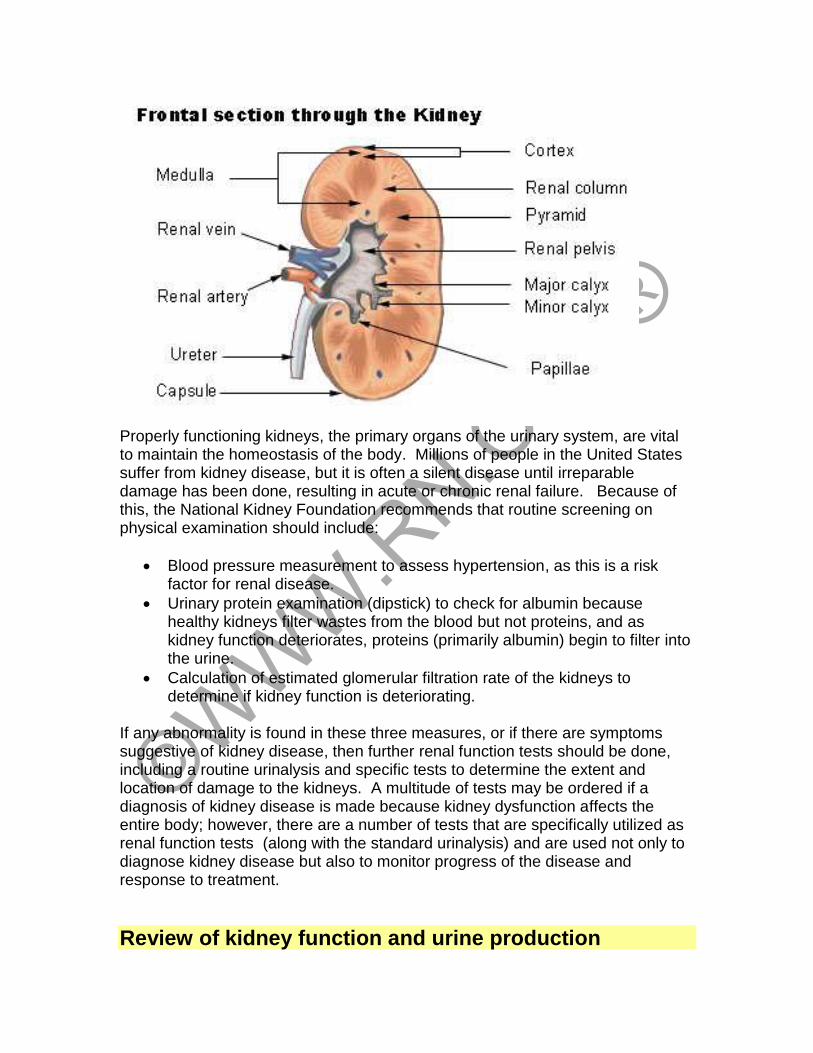
The cortex (outer layer) of the kidney is comprised of the glomeruli and Bowman’s capsules as well as the proximal convoluted tubules and the ascending loops of Henle. The medulla (central area) of the kidney contains the descending loops of Henle and distal convoluted tubules. Blood flows through the renal artery and afferent arterial into the glomerulus, where the renal artery divides to form a vascular network. The capillaries come together at the distal end to form the efferent arteriole that carries the blood from the glomerulus. As blood flows through a glomerular tuft, a tight knot of blood capillaries from which water and chemicals filter into the nephrons, fluid is forced by osmosis and diffusion into the Bowman’s capsule that surrounds the glomerulus. The Bowman’s capsule, the expanded beginning of the tubule, acts as the collecting chamber for fluids filtering from the blood. The filtrate then flows along
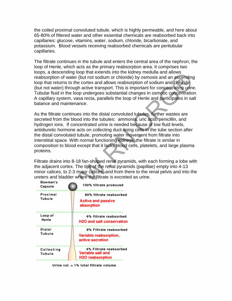
the coiled proximal convoluted tubule, which is highly permeable, and here about 65-80% of filtered water and other essential chemicals are reabsorbed back into capillaries: glucose, vitamins, water, sodium, chloride, bicarbonate, and potassium. Blood vessels receiving reabsorbed chemicals are peritubular capillaries. The filtrate continues in the tubule and enters the central area of the nephron, the loop of Henle, which acts as the primary reabsorption area. It comprises two loops, a descending loop that extends into the kidney medulla and allows reabsorption of water (but not sodium or chloride) by osmosis and an ascending loop that returns to the cortex and allows reabsorption of sodium and chloride (but not water) through active transport. This is important for concentrating urine. Tubular fluid in the loop undergoes substantial changes in osmotic concentration. A capillary system, vasa recta, parallels the loop of Henle and participates in salt balance and maintenance. As the filtrate continues into the distal convoluted tubules, further wastes are secreted from the blood into the tubules: ammonia, uric acid, penicillin, and hydrogen ions. If concentrated urine is needed because of low fluid levels, antidiuretic hormone acts on collecting duct-lining cells in the tube section after the distal convoluted tubule, promoting water movement from filtrate into interstitial space. With normal functioning kidneys, the filtrate is similar in composition to blood except that it lacks blood cells, platelets, and large plasma proteins. Filtrate drains into 8-18 fan-shaped renal pyramids, with each forming a lobe with the adjacent cortex. The tips of the renal pyramids (papillae) empty into 4-13 minor calices, to 2-3 major calices, and from there to the renal pelvis and into the ureters and bladder where the filtrate is excreted as urine.
When renal function is impaired, it is reflected in the blood pressure, blood chemistries, fluid balance, and general wellbeing. As kidney function is compromised, glomerular filtration and renal tubular reabsorption are impaired. This impairment is reflected in blood and urine tests. Renal function tests help to screen the individual for renal disease and to determine the extent or progression of rental disease
Review of common renal diseases requiring renal function tests
Acute glomerulonephritis is caused by an infective
agent, usually group A -hemolytic streptococcus, although other infective agents can cause glomerulonephritis (Staphylococcus aureus, varicella zoster virus, Epstein-Barr virus, and HIV). Glomerulonephritis results in an increase in the epithelial cells lining the glomeruli and infiltration of leukocytes. The glomerular filtration membrane begins to thicken and scar, decreasing the glomerular filtration rate (GFR). Hematuria is often present. As permeability of the glomerular membrane increases, proteinuria increases and urinary output decreases, causing BUN and serum creatinine to also increase. Chronic glomerulonephritis may result from repeated episodes of acute glomerulonephritis or other disorders that damage the glomeruli, such as hypertensive nephrosclerosis, hyperlipidemia, or glomerular sclerosis). The scarred glomeruli are replaced with scar tissue and the kidney can shrink to about 1/5th normal size with cortex of only 1-2 mm. The renal arteries thicken, and renal failure leads to end-stage renal disease.
Hemolytic uremic syndrome (HUS) is most common in infants, children, and
the elderly and usually follows a prodromal diarrheal or upper respiratory infection, most commonly Escherichia coli O157:H7 but other organisms, such as Shigella and Salmonella are also implicated. One type may be caused by the chemotherapeutic agent mitomycin C. Hemolytic uremic syndrome comprises a triad of disorders:
• Microangiopathic hemolytic anemia
• Thrombocytopenia
• Acute renal failure
HUS is characterized by hematuria and neurological complications (seizure, stroke, coma). About half of the people who survive the acute phase of the disease develop chronic renal failure and may require dialysis or kidney transplant. Bacterial toxins (usually from the intestines) invade the blood stream and damage blood and blood vessels, destroying platelets and causing severe anemia. As vessels in the kidneys are damaged, kidney failure occurs. Blood
Glomerulonephritis
Hemolytic uremic syndrome
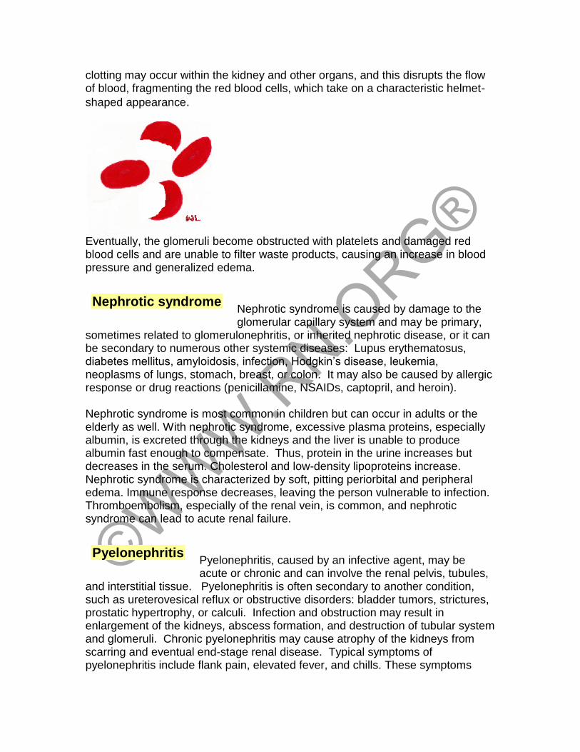
clotting may occur within the kidney and other organs, and this disrupts the flow of blood, fragmenting the red blood cells, which take on a characteristic helmet-
shaped appearance.
Eventually, the glomeruli become obstructed with platelets and damaged red blood cells and are unable to filter waste products, causing an increase in blood pressure and generalized edema.
Nephrotic syndrome is caused by damage to the glomerular capillary system and may be primary,
sometimes related to glomerulonephritis, or inherited nephrotic disease, or it can be secondary to numerous other systemic diseases: Lupus erythematosus, diabetes mellitus, amyloidosis, infection, Hodgkin’s disease, leukemia, neoplasms of lungs, stomach, breast, or colon. It may also be caused by allergic response or drug reactions (penicillamine, NSAIDs, captopril, and heroin). Nephrotic syndrome is most common in children but can occur in adults or the elderly as well. With nephrotic syndrome, excessive plasma proteins, especially albumin, is excreted through the kidneys and the liver is unable to produce albumin fast enough to compensate. Thus, protein in the urine increases but decreases in the serum. Cholesterol and low-density lipoproteins increase. Nephrotic syndrome is characterized by soft, pitting periorbital and peripheral edema. Immune response decreases, leaving the person vulnerable to infection. Thromboembolism, especially of the renal vein, is common, and nephrotic syndrome can lead to acute renal failure.
Pyelonephritis, caused by an infective agent, may be acute or chronic and can involve the renal pelvis, tubules,
and interstitial tissue. Pyelonephritis is often secondary to another condition, such as ureterovesical reflux or obstructive disorders: bladder tumors, strictures, prostatic hypertrophy, or calculi. Infection and obstruction may result in enlargement of the kidneys, abscess formation, and destruction of tubular system and glomeruli. Chronic pyelonephritis may cause atrophy of the kidneys from scarring and eventual end-stage renal disease. Typical symptoms of pyelonephritis include flank pain, elevated fever, and chills. These symptoms
Nephrotic syndrome
Pyelonephritis
differentiate pyelonephritis from cystitis, which usually does not have systemic symptoms.
Acute renal failure occurs when the glomerular filtration rate (GFR) decreases markedly, and kidneys fail to function. Urine volume may be normal
or non-existent, but oliguria (<400 mL/24 hr) is most common. Regardless of urinary output, the kidneys fail to filter waste products and azotemia (retention of metabolic wastes) occurs with increases in serum BUN and creatinine. Hyperkalemia may lead to cardiac dysrhythmias, metabolic acidosis may occur, and anemia is common because of decrease in erythropoietin production and increased destruction of red blood cells. There are 3 primary types of acute renal failure:
• Prerenal: Condition relates to failure of perfusion of the kidney rather than kidney abnormality. The GFR falls because of volume depletion, decreased cardiac output, or vasodilation. Typical laboratory findings include:
o Increased BUN and creatinine with 20:1 BUN/creatinine ratio. o Decreased urinary output and urine sodium (<20 mEq/L). o Normal sediment or few hyaline casts. o Increased urine osmolality (500 mOsmol). o Increased specific gravity.
• Intrarenal: Damage is intrinsic to the kidney itself, causing acute tubular necrosis and inability of the kidneys to filter. This may be caused by traumatic injury, burns, infections, or toxic agents. Transfusion reactions, rhabdomyolysis, and medications may also cause renal damage. Typical laboratory findings include:
o Increased BUN and creatinine. o Variable urinary output (usually decreased). o Increased urine sodium (>40 mEq/L). o Increased urinary sediment with abnormal casts. o Increased urine osmolality (350 mOsm) with similar serum level. o Decreased specific gravity (1.010).
• Postrenal: The kidneys are able to function, but obstruction distal to the kidneys (tumors, hypertrophic prostate, calculi, thrombus) causes increased pressure in the tubular system and distention of the kidney, resulting in decreased GFR. Typical laboratory findings include:
o Increased BUN and creatinine. o Variable urinary output, usually decreased or anuric.
o Variable urine sodium, often decrease (20 mEq/L) o Normal sediment. o Variable urine osmolality, may increase or equal serum level. o Variable specific gravity.
Renal failure, acute
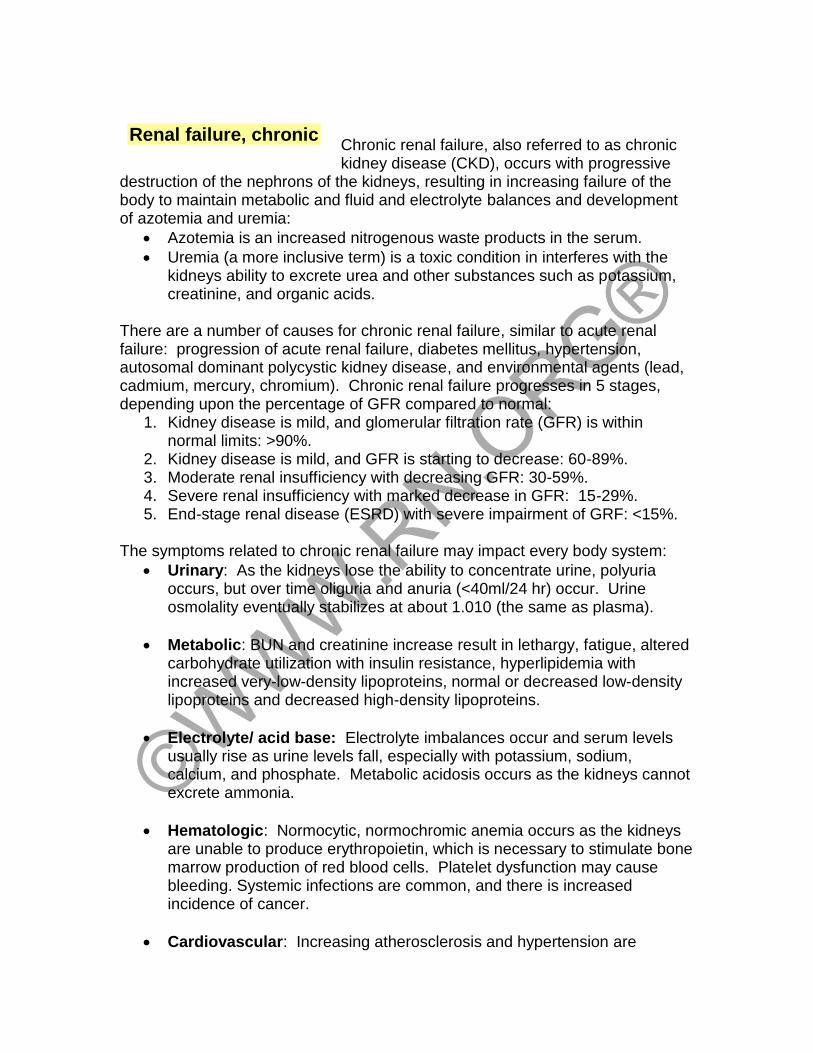
Chronic renal failure, also referred to as chronic kidney disease (CKD), occurs with progressive
destruction of the nephrons of the kidneys, resulting in increasing failure of the body to maintain metabolic and fluid and electrolyte balances and development of azotemia and uremia:
• Azotemia is an increased nitrogenous waste products in the serum.
• Uremia (a more inclusive term) is a toxic condition in interferes with the kidneys ability to excrete urea and other substances such as potassium, creatinine, and organic acids.
There are a number of causes for chronic renal failure, similar to acute renal failure: progression of acute renal failure, diabetes mellitus, hypertension, autosomal dominant polycystic kidney disease, and environmental agents (lead, cadmium, mercury, chromium). Chronic renal failure progresses in 5 stages, depending upon the percentage of GFR compared to normal:
1. Kidney disease is mild, and glomerular filtration rate (GFR) is within normal limits: >90%.
2. Kidney disease is mild, and GFR is starting to decrease: 60-89%. 3. Moderate renal insufficiency with decreasing GFR: 30-59%. 4. Severe renal insufficiency with marked decrease in GFR: 15-29%. 5. End-stage renal disease (ESRD) with severe impairment of GRF: <15%.
The symptoms related to chronic renal failure may impact every body system:
• Urinary: As the kidneys lose the ability to concentrate urine, polyuria occurs, but over time oliguria and anuria (<40ml/24 hr) occur. Urine osmolality eventually stabilizes at about 1.010 (the same as plasma).
• Metabolic: BUN and creatinine increase result in lethargy, fatigue, altered carbohydrate utilization with insulin resistance, hyperlipidemia with increased very-low-density lipoproteins, normal or decreased low-density lipoproteins and decreased high-density lipoproteins.
• Electrolyte/ acid base: Electrolyte imbalances occur and serum levels usually rise as urine levels fall, especially with potassium, sodium, calcium, and phosphate. Metabolic acidosis occurs as the kidneys cannot excrete ammonia.
• Hematologic: Normocytic, normochromic anemia occurs as the kidneys are unable to produce erythropoietin, which is necessary to stimulate bone marrow production of red blood cells. Platelet dysfunction may cause bleeding. Systemic infections are common, and there is increased incidence of cancer.
• Cardiovascular: Increasing atherosclerosis and hypertension are
Renal failure, chronic
common, accompanied by typical vascular changes and congestive heart failure. Electrolyte imbalances may trigger arrhythmias.
• Respiratory: Pulmonary edema causes increasing dyspnea, Kussmaul respirations, thick secretions, and decreased cough reflex.
• Gastrointestinal: Mucosal inflammation affects all parts of the system and can cause ulcerations, nausea, vomiting, diarrhea, or constipation.
• Neurological: Increasing azotemia, electrolyte imbalances, and metabolic acidosis combine to cause a wide range of neurological impairment, including lethargy, confusion, fatigue, seizures, coma, and peripheral neuropathy.
• Musculoskeletal: Electrolyte imbalances with calcium and phosphate lead to osteomalacia (lack of mineralization of new bone) and osteitis fibrosa (calcium in bones replaced with fibrotic tissue).
• Integumentary: Itching and discoloration (yellowish gray) of the skin occurs.
• Reproductive: Decrease in libido and increase in infertility is common.
• Endocrine: Hypothyroidism results in decreasing levels of thyroid hormones.
• Psychological: Depression, personality changes, and inability to concentrate are frequent.
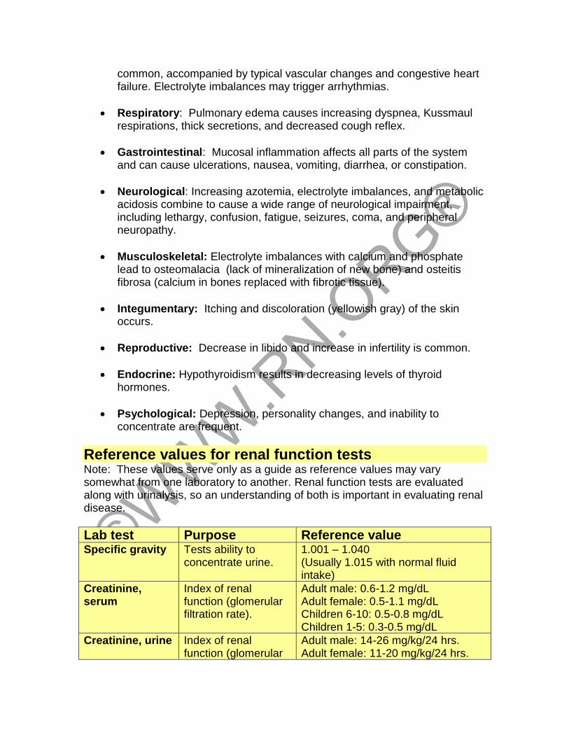
Reference values for renal function tests Note: These values serve only as a guide as reference values may vary somewhat from one laboratory to another. Renal function tests are evaluated along with urinalysis, so an understanding of both is important in evaluating renal disease.
Lab test Purpose Reference value Specific gravity Tests ability to
concentrate urine. 1.001 – 1.040 (Usually 1.015 with normal fluid intake)
Creatinine, serum
Index of renal function (glomerular filtration rate).
Adult male: 0.6-1.2 mg/dL Adult female: 0.5-1.1 mg/dL Children 6-10: 0.5-0.8 mg/dL Children 1-5: 0.3-0.5 mg/dL
Creatinine, urine
Index of renal function (glomerular
Adult male: 14-26 mg/kg/24 hrs. Adult female: 11-20 mg/kg/24 hrs.
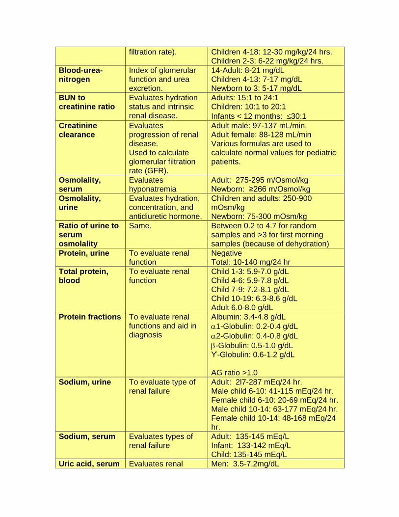
filtration rate). Children 4-18: 12-30 mg/kg/24 hrs. Children 2-3: 6-22 mg/kg/24 hrs.
Blood-urea-nitrogen
Index of glomerular function and urea excretion.
14-Adult: 8-21 mg/dL Children 4-13: 7-17 mg/dL Newborn to 3: 5-17 mg/dL
BUN to creatinine ratio
Evaluates hydration status and intrinsic renal disease.
Adults: 15:1 to 24:1 Children: 10:1 to 20:1
Infants < 12 months: 30:1
Creatinine clearance
Evaluates progression of renal disease. Used to calculate glomerular filtration rate (GFR).
Adult male: 97-137 mL/min. Adult female: 88-128 mL/min Various formulas are used to calculate normal values for pediatric patients.
Osmolality, serum
Evaluates hyponatremia
Adult: 275-295 m/Osmol/kg Newborn: ≥266 m/Osmol/kg
Osmolality, urine
Evaluates hydration, concentration, and antidiuretic hormone.
Children and adults: 250-900 mOsm/kg Newborn: 75-300 mOsm/kg
Ratio of urine to serum osmolality
Same. Between 0.2 to 4.7 for random samples and >3 for first morning samples (because of dehydration)
Protein, urine To evaluate renal function
Negative Total: 10-140 mg/24 hr
Total protein, blood
To evaluate renal function
Child 1-3: 5.9-7.0 g/dL Child 4-6: 5.9-7.8 g/dL Child 7-9: 7.2-8.1 g/dL Child 10-19: 6.3-8.6 g/dL Adult 6.0-8.0 g/dL
Protein fractions To evaluate renal functions and aid in diagnosis
Albumin: 3.4-4.8 g/dL
1-Globulin: 0.2-0.4 g/dL
2-Globulin: 0.4-0.8 g/dL
-Globulin: 0.5-1.0 g/dL ϒ-Globulin: 0.6-1.2 g/dL AG ratio >1.0
Sodium, urine To evaluate type of renal failure
Adult: 2l7-287 mEq/24 hr. Male child 6-10: 41-115 mEq/24 hr. Female child 6-10: 20-69 mEq/24 hr. Male child 10-14: 63-177 mEq/24 hr. Female child 10-14: 48-168 mEq/24 hr.
Sodium, serum Evaluates types of renal failure
Adult: 135-145 mEq/L Infant: 133-142 mEq/L Child: 135-145 mEq/L
Uric acid, serum Evaluates renal Men: 3.5-7.2mg/dL
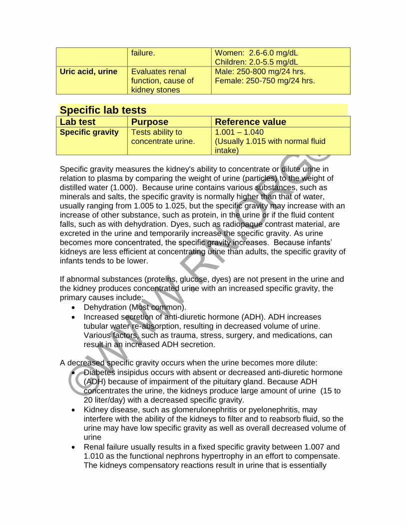
failure. Women: 2.6-6.0 mg/dL Children: 2.0-5.5 mg/dL
Uric acid, urine
Evaluates renal function, cause of kidney stones
Male: 250-800 mg/24 hrs. Female: 250-750 mg/24 hrs.
Specific lab tests Lab test Purpose Reference value Specific gravity Tests ability to
concentrate urine. 1.001 – 1.040 (Usually 1.015 with normal fluid intake)
Specific gravity measures the kidney's ability to concentrate or dilute urine in relation to plasma by comparing the weight of urine (particles) to the weight of distilled water (1.000). Because urine contains various substances, such as minerals and salts, the specific gravity is normally higher than that of water, usually ranging from 1.005 to 1.025, but the specific gravity may increase with an increase of other substance, such as protein, in the urine or if the fluid content falls, such as with dehydration. Dyes, such as radiopaque contrast material, are excreted in the urine and temporarily increase the specific gravity. As urine becomes more concentrated, the specific gravity increases. Because infants’ kidneys are less efficient at concentrating urine than adults, the specific gravity of infants tends to be lower. If abnormal substances (proteins, glucose, dyes) are not present in the urine and the kidney produces concentrated urine with an increased specific gravity, the primary causes include:
• Dehydration (Most common).
• Increased secretion of anti-diuretic hormone (ADH). ADH increases tubular water re-absorption, resulting in decreased volume of urine. Various factors, such as trauma, stress, surgery, and medications, can result in an increased ADH secretion.
A decreased specific gravity occurs when the urine becomes more dilute:
• Diabetes insipidus occurs with absent or decreased anti-diuretic hormone (ADH) because of impairment of the pituitary gland. Because ADH concentrates the urine, the kidneys produce large amount of urine (15 to 20 liter/day) with a decreased specific gravity.
• Kidney disease, such as glomerulonephritis or pyelonephritis, may interfere with the ability of the kidneys to filter and to reabsorb fluid, so the urine may have low specific gravity as well as overall decreased volume of urine
• Renal failure usually results in a fixed specific gravity between 1.007 and 1.010 as the functional nephrons hypertrophy in an effort to compensate. The kidneys compensatory reactions result in urine that is essentially
isotonic with plasma, regardless of time or day or fluid intake.
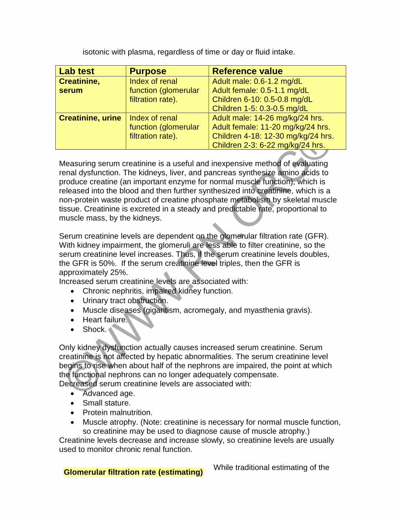
Lab test Purpose Reference value Creatinine, serum
Index of renal function (glomerular filtration rate).
Adult male: 0.6-1.2 mg/dL Adult female: 0.5-1.1 mg/dL Children 6-10: 0.5-0.8 mg/dL Children 1-5: 0.3-0.5 mg/dL
Creatinine, urine
Index of renal function (glomerular filtration rate).
Adult male: 14-26 mg/kg/24 hrs. Adult female: 11-20 mg/kg/24 hrs. Children 4-18: 12-30 mg/kg/24 hrs. Children 2-3: 6-22 mg/kg/24 hrs.
Measuring serum creatinine is a useful and inexpensive method of evaluating renal dysfunction. The kidneys, liver, and pancreas synthesize amino acids to produce creatine (an important enzyme for normal muscle function), which is released into the blood and then further synthesized into creatinine, which is a non-protein waste product of creatine phosphate metabolism by skeletal muscle tissue. Creatinine is excreted in a steady and predictable rate, proportional to muscle mass, by the kidneys. Serum creatinine levels are dependent on the glomerular filtration rate (GFR). With kidney impairment, the glomeruli are less able to filter creatinine, so the serum creatinine level increases. Thus, if the serum creatinine levels doubles, the GFR is 50%. If the serum creatinine level triples, then the GFR is approximately 25%. Increased serum creatinine levels are associated with:
• Chronic nephritis, impaired kidney function.
• Urinary tract obstruction.
• Muscle diseases (gigantism, acromegaly, and myasthenia gravis).
• Heart failure.
• Shock.
Only kidney dysfunction actually causes increased serum creatinine. Serum creatinine is not affected by hepatic abnormalities. The serum creatinine level begins to rise when about half of the nephrons are impaired, the point at which the functional nephrons can no longer adequately compensate. Decreased serum creatinine levels are associated with:
• Advanced age.
• Small stature.
• Protein malnutrition.
• Muscle atrophy. (Note: creatinine is necessary for normal muscle function, so creatinine may be used to diagnose cause of muscle atrophy.)
Creatinine levels decrease and increase slowly, so creatinine levels are usually used to monitor chronic renal function.
While traditional estimating of the
Glomerular filtration rate (estimating)
glomerular filtration rate (GFR), the rate at which the kidneys process blood through the glomerular system, has been done with of serum (as in estimate discussed above) and/or 24-hour urine creatinine, a newer method, the Modification of Diet in Renal Disease (MDRD) Study Equation, estimates the GFR based on the serum creatinine, age, race, and gender rather than serum creatinine alone. This equation is used for those 18-70. The equation does not use weight but reports results as normalized to 1.73 m2 (mass). The MDRD Study Equation has been found to be more effective than estimates based on serum creatinine alone or the 24-hour urine collection for most patients. The National Kidney Disease Education Program is promoting that labs provide an estimated GFR (based on the MDRD equation) with the results of serum creatinine since serum creatinine alone is a poor indicator of kidney function. However, use of the MDRD Study Equation is not advised for all patients. Those who should be evaluated with creatinine clearance and/or other formulas include:
• Non-adults under age 18. The Schwartz equation should be used for children.
• Those with unstable creatinine concentrations, such as women who are pregnant, those with severe co-morbid conditions, hospitalized patients, and those with acute renal failure.
• Those with extremes in muscle mass and diet (because of the normalized body measurement), including amputees, paraplegics, bodybuilders, the obese, people with muscular wasting or neuromuscular disease, those with malnutrition, vegetarians or those eating low meat diets, and those taking creatine dietary supplements.
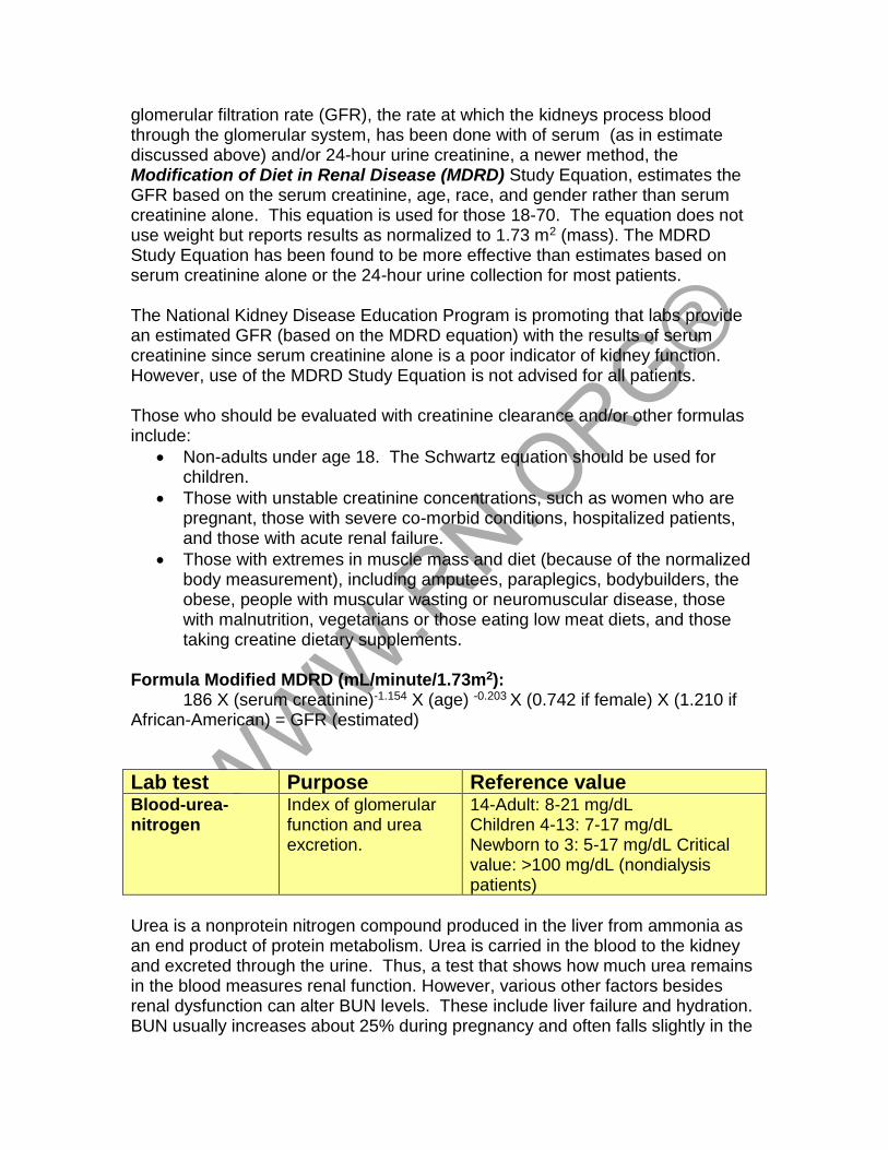
Formula Modified MDRD (mL/minute/1.73m2): 186 X (serum creatinine)-1.154 X (age) -0.203 X (0.742 if female) X (1.210 if African-American) = GFR (estimated)
Lab test Purpose Reference value Blood-urea-nitrogen
Index of glomerular function and urea excretion.
14-Adult: 8-21 mg/dL Children 4-13: 7-17 mg/dL Newborn to 3: 5-17 mg/dL Critical value: >100 mg/dL (nondialysis patients)
Urea is a nonprotein nitrogen compound produced in the liver from ammonia as an end product of protein metabolism. Urea is carried in the blood to the kidney and excreted through the urine. Thus, a test that shows how much urea remains in the blood measures renal function. However, various other factors besides renal dysfunction can alter BUN levels. These include liver failure and hydration. BUN usually increases about 25% during pregnancy and often falls slightly in the
elderly, as the ability to concentrate urine decreases with age. Thus, BUN and creatinine levels are often evaluated together. An increase in the BUN level results in azotemia, which can be caused by:
• Acute renal failure
• Congestive heart failure.
• Decreased renal perfusion.
• Chronic glomerulonephritis.
• Diabetes.
• Hypovolemia.
• Starvation/muscle wasting.
• Shock.
• Excessive protein intake or protein catabolism, hyperalimentation.
• Ketoacidosis.
• GI bleeding (digested blood produces urea).
• Neoplasms.
• Nephrotoxic agents. Conditions that result in decreased perfusion, such as hypovolemic shock or heart failure, cause BUN levels to increase. Additionally, severe dehydration increases BUN because the low fluid volume impairs the ability of the kidneys to excrete urea and other waste products. A GI hemorrhage of 1000 mL of blood may result in increased BUN of 40 mg/mL. A decreased BUN may be seen in:
• Protein malnutrition.
• Low protein/high carbohydrate diet.
• Anabolic steroid use
• Overhydration.
• Hepatic disease.
• Malabsorption syndromes
• Pregnancy.
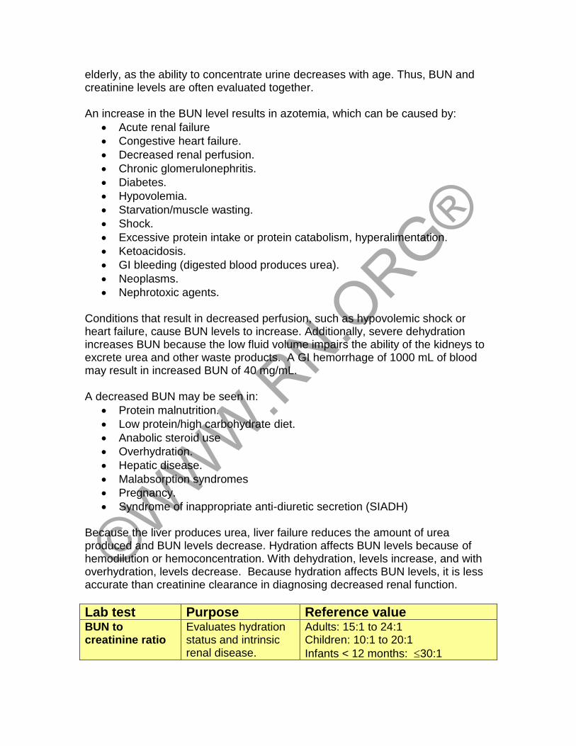
• Syndrome of inappropriate anti-diuretic secretion (SIADH) Because the liver produces urea, liver failure reduces the amount of urea produced and BUN levels decrease. Hydration affects BUN levels because of hemodilution or hemoconcentration. With dehydration, levels increase, and with overhydration, levels decrease. Because hydration affects BUN levels, it is less accurate than creatinine clearance in diagnosing decreased renal function. Lab test Purpose Reference value BUN to creatinine ratio
Evaluates hydration status and intrinsic renal disease.
Adults: 15:1 to 24:1 Children: 10:1 to 20:1
Infants < 12 months: 30:1
Serum creatinine and blood urea nitrogen (BUN) are often compared to evaluate renal function. Although the BUN can be affected by many factors, the serum creatinine level only increases with damage to the nephrons. The BUN to creatinine ratio is frequently used to evaluate dehydration. High BUN-to-creatinine ratios may occur with acute renal failure, related to shock or severe dehydration. A kidney stone or other blockage in the urinary tract can cause a high BUN-to-creatinine ratio. A very high BUN-to-creatinine ratio may result from bleeding in the digestive or respiratory tract.
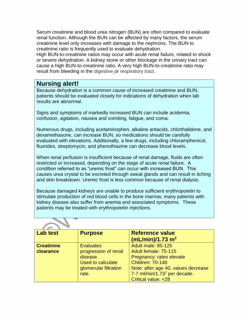
Nursing alert! Because dehydration is a common cause of increased creatinine and BUN, patients should be evaluated closely for indications of dehydration when lab results are abnormal. Signs and symptoms of markedly increased BUN can include acidemia, confusion, agitation, nausea and vomiting, fatigue, and coma. Numerous drugs, including acetaminophen, alkaline antacids, chlorthalidone, and dexamethasone, can increase BUN, so medications should be carefully evaluated with elevations. Additionally, a few drugs, including chloramphenicol, fluorides, streptomycin, and phenothiazine can decrease blood levels. When renal perfusion is insufficient because of renal damage, fluids are often restricted or increased, depending on the stage of acute renal failure. A condition referred to as “uremic frost” can occur with increased BUN. This causes urea crystal to be excreted through sweat glands and can result in itching and skin breakdown. Uremic frost is less common because of renal dialysis. Because damaged kidneys are unable to produce sufficient erythropoietin to stimulate production of red blood cells in the bone marrow, many patients with kidney disease also suffer from anemia and associated symptoms. These patients may be treated with erythropoietin injections.
Lab test Purpose Reference value (mL/min)/1.73 m2
Creatinine clearance
Evaluates progression of renal disease. Used to calculate glomerular filtration rate.
Adult male: 85-125 Adult female: 75-115 Pregnancy: rates elevate Children: 70-140 Note: after age 40, values decrease 7-7 ml/min/1.732 per decade. Critical value: <28
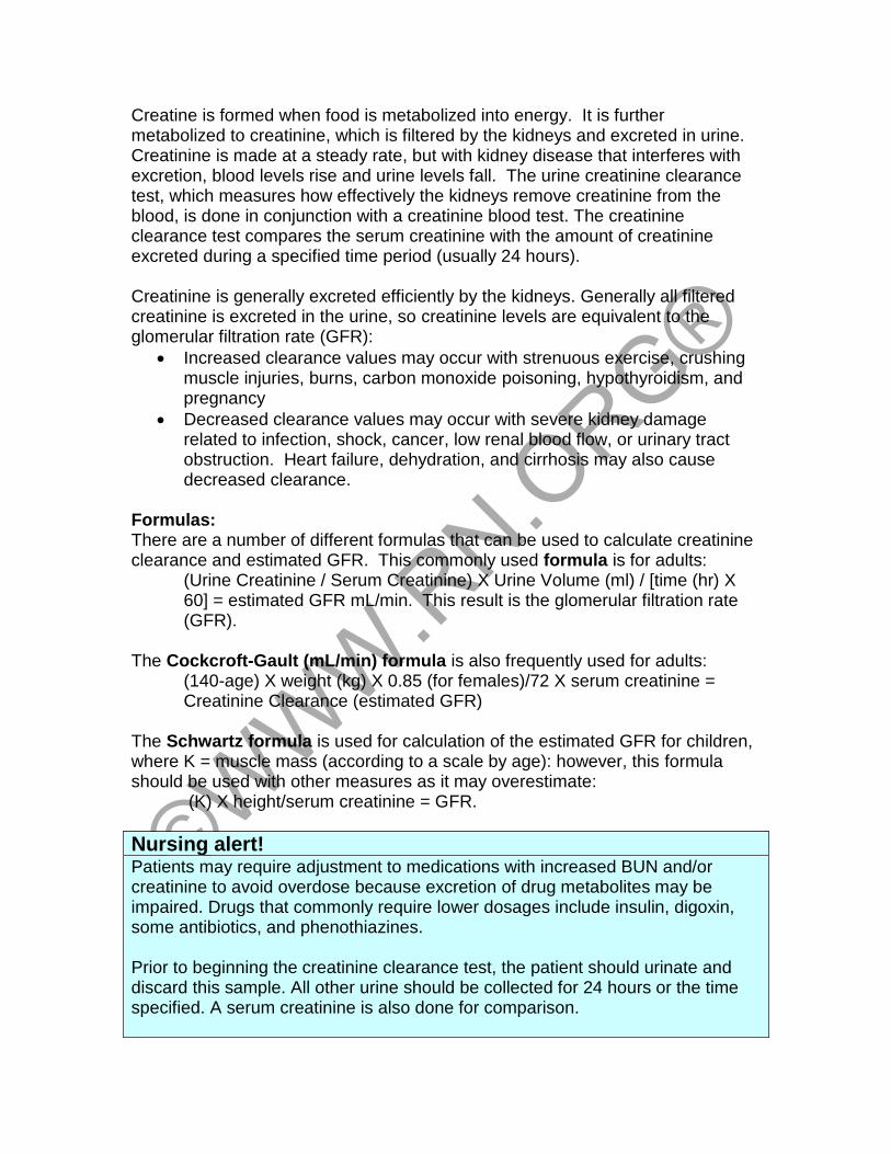
Creatine is formed when food is metabolized into energy. It is further metabolized to creatinine, which is filtered by the kidneys and excreted in urine. Creatinine is made at a steady rate, but with kidney disease that interferes with excretion, blood levels rise and urine levels fall. The urine creatinine clearance test, which measures how effectively the kidneys remove creatinine from the blood, is done in conjunction with a creatinine blood test. The creatinine clearance test compares the serum creatinine with the amount of creatinine excreted during a specified time period (usually 24 hours). Creatinine is generally excreted efficiently by the kidneys. Generally all filtered creatinine is excreted in the urine, so creatinine levels are equivalent to the glomerular filtration rate (GFR):
• Increased clearance values may occur with strenuous exercise, crushing muscle injuries, burns, carbon monoxide poisoning, hypothyroidism, and pregnancy
• Decreased clearance values may occur with severe kidney damage related to infection, shock, cancer, low renal blood flow, or urinary tract obstruction. Heart failure, dehydration, and cirrhosis may also cause decreased clearance.
Formulas: There are a number of different formulas that can be used to calculate creatinine clearance and estimated GFR. This commonly used formula is for adults:
(Urine Creatinine / Serum Creatinine) X Urine Volume (ml) / [time (hr) X 60] = estimated GFR mL/min. This result is the glomerular filtration rate (GFR).
The Cockcroft-Gault (mL/min) formula is also frequently used for adults: (140-age) X weight (kg) X 0.85 (for females)/72 X serum creatinine = Creatinine Clearance (estimated GFR)
The Schwartz formula is used for calculation of the estimated GFR for children, where K = muscle mass (according to a scale by age): however, this formula should be used with other measures as it may overestimate:
(K) X height/serum creatinine = GFR.
Nursing alert! Patients may require adjustment to medications with increased BUN and/or creatinine to avoid overdose because excretion of drug metabolites may be impaired. Drugs that commonly require lower dosages include insulin, digoxin, some antibiotics, and phenothiazines. Prior to beginning the creatinine clearance test, the patient should urinate and discard this sample. All other urine should be collected for 24 hours or the time specified. A serum creatinine is also done for comparison.
Strenuous exercise should be avoided for 48 hours prior to testing and meat or
other protein (especially beef) should be restricted to 8 oz. for 24 hours prior to beginning testing
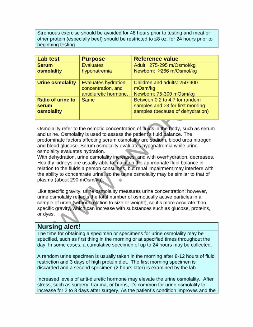
Lab test Purpose Reference value Serum osmolality
Evaluates hyponatremia
Adult: 275-295 m/Osmol/kg Newborn: ≥266 m/Osmol/kg
Urine osmolality Evaluates hydration, concentration, and antidiuretic hormone.
Children and adults: 250-900 mOsm/kg Newborn: 75-300 mOsm/kg
Ratio of urine to serum osmolality
Same Between 0.2 to 4.7 for random samples and >3 for first morning samples (because of dehydration)
Osmolality refer to the osmotic concentration of fluids in the body, such as serum and urine. Osmolality is used to assess the patient’s fluid balance. The predominate factors affecting serum osmolality are sodium, blood urea nitrogen and blood glucose. Serum osmolality evaluates hyponatremia while urine osmolality evaluates hydration. With dehydration, urine osmolality increases; and with overhydration, decreases. Healthy kidneys are usually able to maintain the appropriate fluid balance in relation to the fluids a person consumes, but renal impairment may interfere with the ability to concentrate urine, so the urine osmolality may be similar to that of plasma (about 290 mOsm/kg). Like specific gravity, urine osmolality measures urine concentration; however, urine osmolality reflects the total number of osmotically active particles in a sample of urine (without relation to size or weight), so it’s more accurate than specific gravity, which can increase with substances such as glucose, proteins, or dyes.
Nursing alert! The time for obtaining a specimen or specimens for urine osmolality may be specified, such as first thing in the morning or at specified times throughout the day. In some cases, a cumulative specimen of up to 24 hours may be collected. A random urine specimen is usually taken in the morning after 8-12 hours of fluid restriction and 3 days of high protein diet. The first morning specimen is discarded and a second specimen (2 hours later) is examined by the lab. Increased levels of anti-diuretic hormone may elevate the urine osmolality. After stress, such as surgery, trauma, or burns, it’s common for urine osmolality to increase for 2 to 3 days after surgery. As the patient’s condition improves and the
body experiences less tress, the ADH levels return to normal and diuresis occurs.
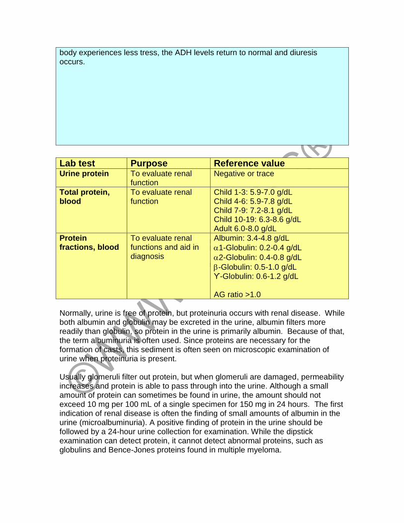
Lab test Purpose Reference value Urine protein To evaluate renal
function Negative or trace
Total protein, blood
To evaluate renal function
Child 1-3: 5.9-7.0 g/dL Child 4-6: 5.9-7.8 g/dL Child 7-9: 7.2-8.1 g/dL Child 10-19: 6.3-8.6 g/dL Adult 6.0-8.0 g/dL
Protein fractions, blood
To evaluate renal functions and aid in diagnosis
Albumin: 3.4-4.8 g/dL
1-Globulin: 0.2-0.4 g/dL
2-Globulin: 0.4-0.8 g/dL
-Globulin: 0.5-1.0 g/dL ϒ-Globulin: 0.6-1.2 g/dL AG ratio >1.0
Normally, urine is free of protein, but proteinuria occurs with renal disease. While both albumin and globulin may be excreted in the urine, albumin filters more readily than globulin, so protein in the urine is primarily albumin. Because of that, the term albuminuria is often used. Since proteins are necessary for the formation of casts, this sediment is often seen on microscopic examination of urine when proteinuria is present. Usually glomeruli filter out protein, but when glomeruli are damaged, permeability increases and protein is able to pass through into the urine. Although a small amount of protein can sometimes be found in urine, the amount should not exceed 10 mg per 100 mL of a single specimen for 150 mg in 24 hours. The first indication of renal disease is often the finding of small amounts of albumin in the urine (microalbuminuria). A positive finding of protein in the urine should be followed by a 24-hour urine collection for examination. While the dipstick examination can detect protein, it cannot detect abnormal proteins, such as globulins and Bence-Jones proteins found in multiple myeloma.

Proteinuria is described based on the following scale:
Description Mg/ 24 hours
Trace <150 mg
1+ 200-500 mg
2+ 500-1500 mg
3+ >2500 mg
4+ >3000 mg
Urine protein testing is done to evaluate kidney function, and assist in detection of Bence Jones proteins and diagnosis of myeloma, macroglobulinemia, lymphoma, and amyloidosis. Since proteins are necessary for the formation of casts, this sediment is often seen on microscopic examination of urine when proteinuria is present. Serum protein levels may remain within normal limits if protein loss is < 3g daily because the liver is able to compensate. Total protein increases (hyperproteinemia) in the presence of volume depletion, myeloma, sarcoidosis, and some types of chronic liver disease. Total protein decreases with chronic glomerulonephritis and nephrotic syndrome as well as other disorders, such as cirrhosis, burns, heart failure, starvation, and prolonged immobilization. For renal disease, the level of albumin is the most important. It usually does not increase with dehydration but decreases with nephrosis and nephritis.
Fractions are evaluated to determine the specific disorder. For example 1-
globulin increases with acute and chronic inflammatory disease: and -2 globulin
can increase with diabetes, pancreatitis, and hemolysis. However, 1-globulin
decreases in hereditary deficiency and -2 globulin decreases in nephrotic syndrome.
Lab test Purpose Reference value Urine sodium To evaluate type of
renal failure Adult: 2l7-287 mEq/24 hr. Male child 6-10: 41-115 mEq/24 hr. Female child 6-10: 20-69 mEq/24 hr. Male child 10-14: 63-177 mEq/24 hr. Female child 10-14: 48-168 mEq/24 hr.
Sodium, blood Evaluates types of renal failure
Adult: 135-145 mEq/L Infant: 133-142 mEq/L Child: 135-145 mEq/L
Sodium is an electrolyte that is critical to maintaining fluid balance within the body. Sodium levels are controlled by aldosterone, an adrenal hormone. About 85% of sodium is found in the blood and lymph. Both serum and urine levels of
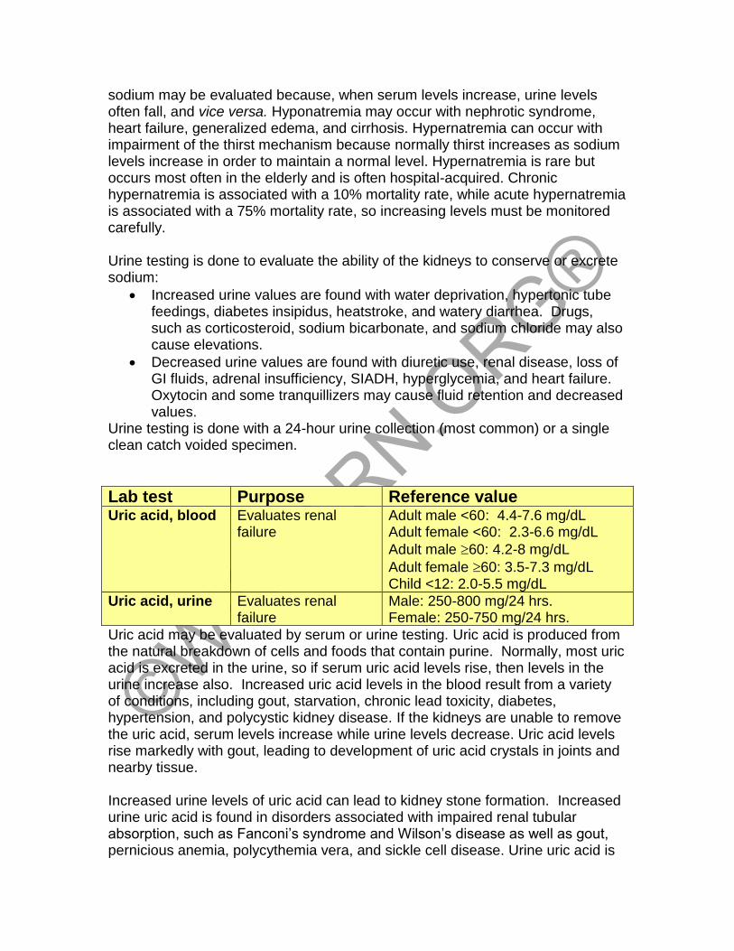
sodium may be evaluated because, when serum levels increase, urine levels often fall, and vice versa. Hyponatremia may occur with nephrotic syndrome, heart failure, generalized edema, and cirrhosis. Hypernatremia can occur with impairment of the thirst mechanism because normally thirst increases as sodium levels increase in order to maintain a normal level. Hypernatremia is rare but occurs most often in the elderly and is often hospital-acquired. Chronic hypernatremia is associated with a 10% mortality rate, while acute hypernatremia is associated with a 75% mortality rate, so increasing levels must be monitored carefully. Urine testing is done to evaluate the ability of the kidneys to conserve or excrete sodium:
• Increased urine values are found with water deprivation, hypertonic tube feedings, diabetes insipidus, heatstroke, and watery diarrhea. Drugs, such as corticosteroid, sodium bicarbonate, and sodium chloride may also cause elevations.
• Decreased urine values are found with diuretic use, renal disease, loss of GI fluids, adrenal insufficiency, SIADH, hyperglycemia, and heart failure. Oxytocin and some tranquillizers may cause fluid retention and decreased values.
Urine testing is done with a 24-hour urine collection (most common) or a single clean catch voided specimen.
Lab test Purpose Reference value Uric acid, blood Evaluates renal
failure Adult male <60: 4.4-7.6 mg/dL Adult female <60: 2.3-6.6 mg/dL
Adult male 60: 4.2-8 mg/dL
Adult female 60: 3.5-7.3 mg/dL Child <12: 2.0-5.5 mg/dL
Uric acid, urine Evaluates renal failure
Male: 250-800 mg/24 hrs. Female: 250-750 mg/24 hrs.
Uric acid may be evaluated by serum or urine testing. Uric acid is produced from the natural breakdown of cells and foods that contain purine. Normally, most uric acid is excreted in the urine, so if serum uric acid levels rise, then levels in the urine increase also. Increased uric acid levels in the blood result from a variety of conditions, including gout, starvation, chronic lead toxicity, diabetes, hypertension, and polycystic kidney disease. If the kidneys are unable to remove the uric acid, serum levels increase while urine levels decrease. Uric acid levels rise markedly with gout, leading to development of uric acid crystals in joints and nearby tissue. Increased urine levels of uric acid can lead to kidney stone formation. Increased urine uric acid is found in disorders associated with impaired renal tubular absorption, such as Fanconi’s syndrome and Wilson’s disease as well as gout, pernicious anemia, polycythemia vera, and sickle cell disease. Urine uric acid is
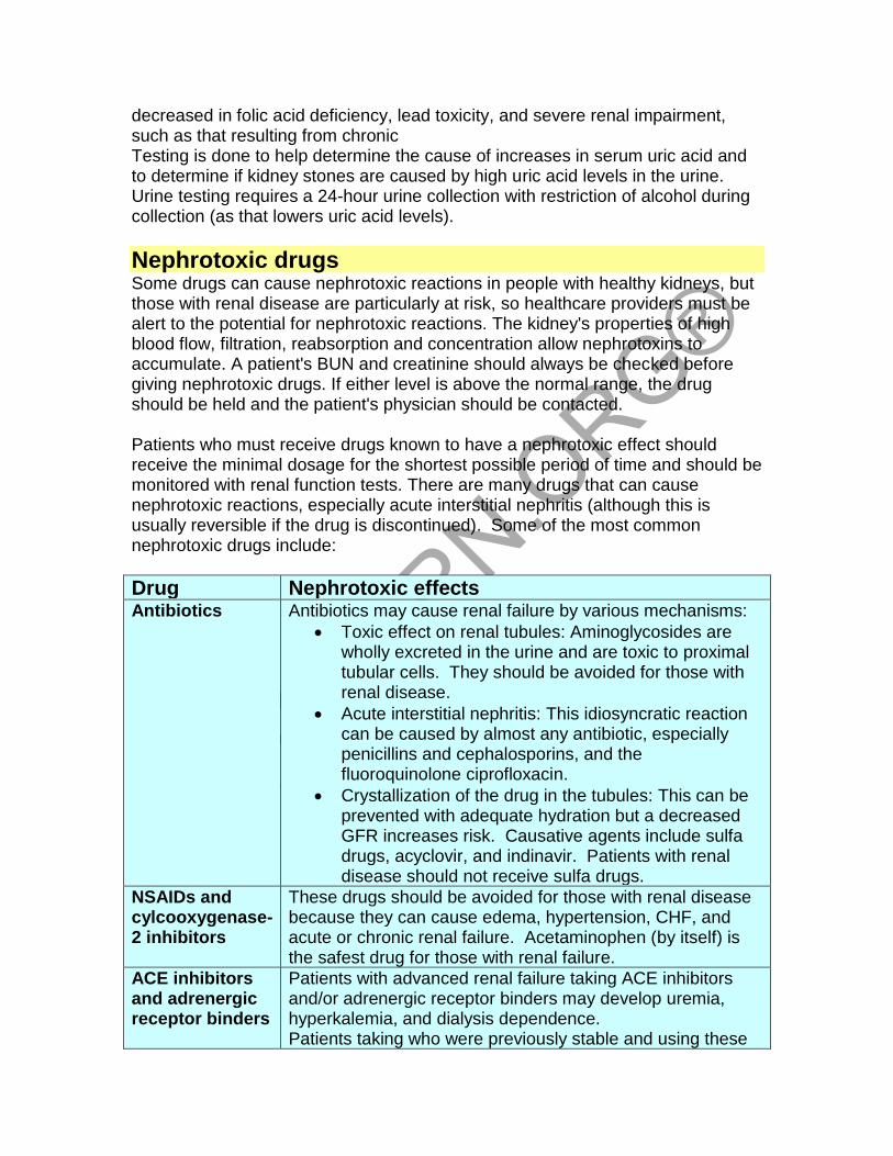
decreased in folic acid deficiency, lead toxicity, and severe renal impairment, such as that resulting from chronic Testing is done to help determine the cause of increases in serum uric acid and to determine if kidney stones are caused by high uric acid levels in the urine. Urine testing requires a 24-hour urine collection with restriction of alcohol during collection (as that lowers uric acid levels).
Nephrotoxic drugs Some drugs can cause nephrotoxic reactions in people with healthy kidneys, but those with renal disease are particularly at risk, so healthcare providers must be alert to the potential for nephrotoxic reactions. The kidney's properties of high blood flow, filtration, reabsorption and concentration allow nephrotoxins to accumulate. A patient's BUN and creatinine should always be checked before giving nephrotoxic drugs. If either level is above the normal range, the drug should be held and the patient's physician should be contacted. Patients who must receive drugs known to have a nephrotoxic effect should receive the minimal dosage for the shortest possible period of time and should be monitored with renal function tests. There are many drugs that can cause nephrotoxic reactions, especially acute interstitial nephritis (although this is usually reversible if the drug is discontinued). Some of the most common nephrotoxic drugs include:
Drug Nephrotoxic effects Antibiotics Antibiotics may cause renal failure by various mechanisms:
• Toxic effect on renal tubules: Aminoglycosides are wholly excreted in the urine and are toxic to proximal tubular cells. They should be avoided for those with renal disease.
• Acute interstitial nephritis: This idiosyncratic reaction can be caused by almost any antibiotic, especially penicillins and cephalosporins, and the fluoroquinolone ciprofloxacin.
• Crystallization of the drug in the tubules: This can be prevented with adequate hydration but a decreased GFR increases risk. Causative agents include sulfa drugs, acyclovir, and indinavir. Patients with renal disease should not receive sulfa drugs.
NSAIDs and cylcooxygenase-2 inhibitors
These drugs should be avoided for those with renal disease because they can cause edema, hypertension, CHF, and acute or chronic renal failure. Acetaminophen (by itself) is the safest drug for those with renal failure.
ACE inhibitors and adrenergic receptor binders
Patients with advanced renal failure taking ACE inhibitors and/or adrenergic receptor binders may develop uremia, hyperkalemia, and dialysis dependence. Patients taking who were previously stable and using these
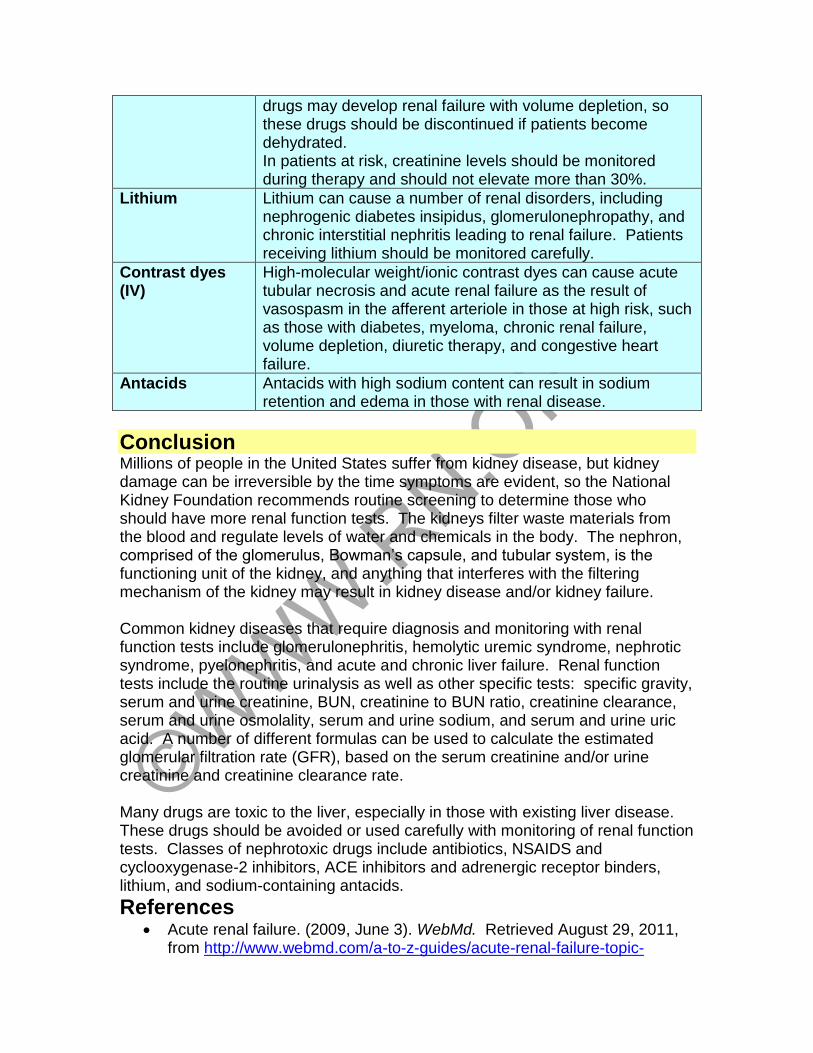
drugs may develop renal failure with volume depletion, so these drugs should be discontinued if patients become dehydrated. In patients at risk, creatinine levels should be monitored during therapy and should not elevate more than 30%.
Lithium Lithium can cause a number of renal disorders, including nephrogenic diabetes insipidus, glomerulonephropathy, and chronic interstitial nephritis leading to renal failure. Patients receiving lithium should be monitored carefully.
Contrast dyes (IV)
High-molecular weight/ionic contrast dyes can cause acute tubular necrosis and acute renal failure as the result of vasospasm in the afferent arteriole in those at high risk, such as those with diabetes, myeloma, chronic renal failure, volume depletion, diuretic therapy, and congestive heart failure.
Antacids Antacids with high sodium content can result in sodium retention and edema in those with renal disease.
Conclusion Millions of people in the United States suffer from kidney disease, but kidney damage can be irreversible by the time symptoms are evident, so the National Kidney Foundation recommends routine screening to determine those who should have more renal function tests. The kidneys filter waste materials from the blood and regulate levels of water and chemicals in the body. The nephron, comprised of the glomerulus, Bowman’s capsule, and tubular system, is the functioning unit of the kidney, and anything that interferes with the filtering mechanism of the kidney may result in kidney disease and/or kidney failure. Common kidney diseases that require diagnosis and monitoring with renal function tests include glomerulonephritis, hemolytic uremic syndrome, nephrotic syndrome, pyelonephritis, and acute and chronic liver failure. Renal function tests include the routine urinalysis as well as other specific tests: specific gravity, serum and urine creatinine, BUN, creatinine to BUN ratio, creatinine clearance, serum and urine osmolality, serum and urine sodium, and serum and urine uric acid. A number of different formulas can be used to calculate the estimated glomerular filtration rate (GFR), based on the serum creatinine and/or urine creatinine and creatinine clearance rate. Many drugs are toxic to the liver, especially in those with existing liver disease. These drugs should be avoided or used carefully with monitoring of renal function tests. Classes of nephrotoxic drugs include antibiotics, NSAIDS and cyclooxygenase-2 inhibitors, ACE inhibitors and adrenergic receptor binders, lithium, and sodium-containing antacids.
References • Acute renal failure. (2009, June 3). WebMd. Retrieved August 29, 2011,
from http://www.webmd.com/a-to-z-guides/acute-renal-failure-topic-
overview
• BUN. (2011, July 5). Lab Tests Online. Retrieved August 29, 2011, from http://www.labtestsonline.org/understanding/analytes/bun/glance.html
• Chronic renal insufficiency. (2008, February 7). The Foundation for IgA Nephropathy. Retrieved August 29, 2011, from http://www.igan.ca/id77.html
• Creatinine and creatinine clearance. (2006, August 21). WebMD. Retrieved August 29, 2011, from http://www.webmd.com/a-to-z-guides/creatinine-and-creatinine-clearance
• Drug prescribing in renal impairment. (2008, November 23). Patient UK. Retrieved August 29, 2011, from http://www.patient.co.uk/showdoc/40025092/
• Fischbach, F.T., & Dunning, M.B. (2005). Nurse’s Quick Reference to Common Laboratory & Diagnostic Tests. Philadelphia: J. B. Lippincott Company.
• GFR calculator for children. (2011, January 30). National Kidney Disease Education Program. Retrieved August 29, 2011, from http://www.nkdep.nih.gov/professionals/gfr_calculators/gfr_children.htm
• Glomerular filtration rate. (2011). National Kidney Foundation. Retrieved August 29, 2011, from http://www.kidney.org/kidneydisease/ckd/knowGFR.cfm
• Glomerular filtration rate. (2008, February 7). The Foundation for IgA Nephropathy. Retrieved August 29, 2011, from http://www.igan.ca/id86.htm
• Glomerular filtration rate, Estimated. (2011). ARUP National Reference Library. Retrieved August 29, 2011, from http://www.aruplab.com/guides/ug/tests/0020725.jsp
• Glomerulonephritis. (2011, April 2). MayoClinic.com. Retrieved August 29, 2011, from http://www.mayoclinic.com/health/glomerulonephritis/DS00503
• The kidneys and how they work. (2010, September 9). National Kidney and Urologic Information Clearinghouse. Retrieved August 29, 2011 from http://www.kidney.niddk.nih.gov/kudiseases/pubs/yourkidneys/
• Proteinuria. (2010, September 2). National Kidney and urologic Diseases Information Clearinghouse (KUDIC). Retrieved August 29, 2011, from http://kidney.niddk.nih.gov/kudiseases/pubs/proteinuria/
• Schrier, R. W. Diseases of the Kidney and Urinary Tract: Clinicopathologic Foundations of Medicine. Philadelphia: Lippincott, Williams, & Wilkins, 2007.
• Smeltzer, S. C., Bare, B., Hinkle, J.L., & Cheever, K. H. (2008). Brunner & Suddarth’s Textbook of Medical-Surgical Nursing, 11th ed. New York: Lippincott, Williams, & Wilkins.
• Sodium (Na) in urine. (2010, September 1). WebMD. Retrieved August 29, 2011, from http://www.webmd.com/a-to-z-guides/sodium-na-in-urine
• Tan, A.J. (2010, April 7). Hemolytic uremic syndrome. eMedicine. Retrieved August 29, 2011 from
http://emedicine.medscape.com/article/779218-overview
• Tofovic, S.P. (n.d.) Nephrotoxic drugs. [PowerPoint]. University of Pittsburgh School of Medicine.
• Uric acid in urine. (2010, August 9). WebMD. Retrieved August 29, 2011, from http://www.webmd.com/a-to-z-guides/uric-acid-in-urine
• Van Leeuwen, A.M., Poelhuis-Leth, D, & Bladh, M. (2011). Davis’s Comprehensive Handbook of Laboratory and Diagnostic Tests with Nursing Implications, 4th ed. Philadelphia: FA Davis Company