
Remodeling the inter-dental papilla for esthetic anterior implant restorations 嘉泉 醫大...
20
Remodeling the inter- dental papilla for esthetic anterior implant restorations 嘉嘉 嘉嘉 嘉嘉嘉 嘉嘉 嘉嘉嘉 R2 嘉 嘉 嘉
-
date post
18-Dec-2015 -
Category
Documents
-
view
233 -
download
2
Transcript of Remodeling the inter-dental papilla for esthetic anterior implant restorations 嘉泉 醫大...
Remodeling the inter-dental papilla for
esthetic anterior implant restorations
嘉泉 醫大 吉病院 齒科 補綴科 R2 김 세 웅

Implant treatment procedure
General historyExtraoral exam.
Intraoral exam.
Radiographic exam.Diagnostic cast
Evaluation of bone quantity&quality, soft tissueOcclusion analysisEvaluation of prosthetic space
Types of prostheticsNumbers, location, angulation
of implant
Augmentation of soft & hard tissue
Final evaluation of
Treatment plans

General Examinations 1. Systemic conditions 2. Causes of missing 3. Intraoral & extraoral examination 4. Occlusal & functional evaluation 5. X-ray examination 6. Examination of periodontium 7. Diagnostic cast 8. 외과학적인 술식과 위험성 9. Treatment time & cost 10. Informed consent

심미적 수복을 위한 검사1. Review of patient ; Expectations, Co-operations, Cleaning ability
2. Teeth ; Shape, angulation, occlusion, crown/impant ratio, periodontal disease, occlusal clearance, contact point3. Soft tissue ; Shape, smile line, depth, amount of keratinized gingiva, inter-dental papilla4. Bone ; Shape, quality, quantity, amount of horizontal or vertical resorption, depression of vestibule(undercut 의 존재 有無 )

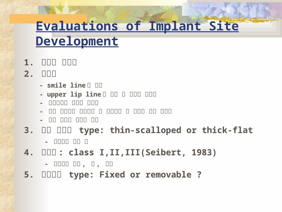
Evaluations of Implant Site Development
1. 환자의 필요성2. 심미성 - smile line 의 높이 - upper lip line 과 치아 및 치은의 노출도 - 인접치와의 치경선 조화도 - 정중 시상면을 기준으로 한 교합평면 및 치열의 좌우 대칭성 - 치간 유두의 형태와 높이
3. 치주 조직의 type: thin-scalloped or thick-flat - 연조직의 질과 양
4. 골결손 : class I,II,III(Seibert, 1983) - 잔존골의 높이 , 폭 , 형태
5. 보철물의 type: Fixed or removable ?
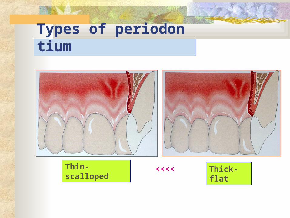
Types of periodontium
Thin-scalloped
Thick-flat
<<<<

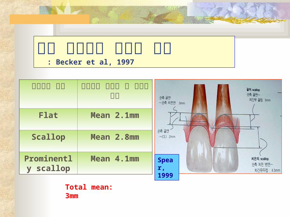
상악 전치부의 치조골 형태
: Becker et al, 1997
Spear, 1999
치조골의 형태 순측골과 치간부 골 사이의 거리
Flat Mean 2.1mm
Scallop Mean 2.8mm
Prominently scallop
Mean 4.1mm
Total mean: 3mm

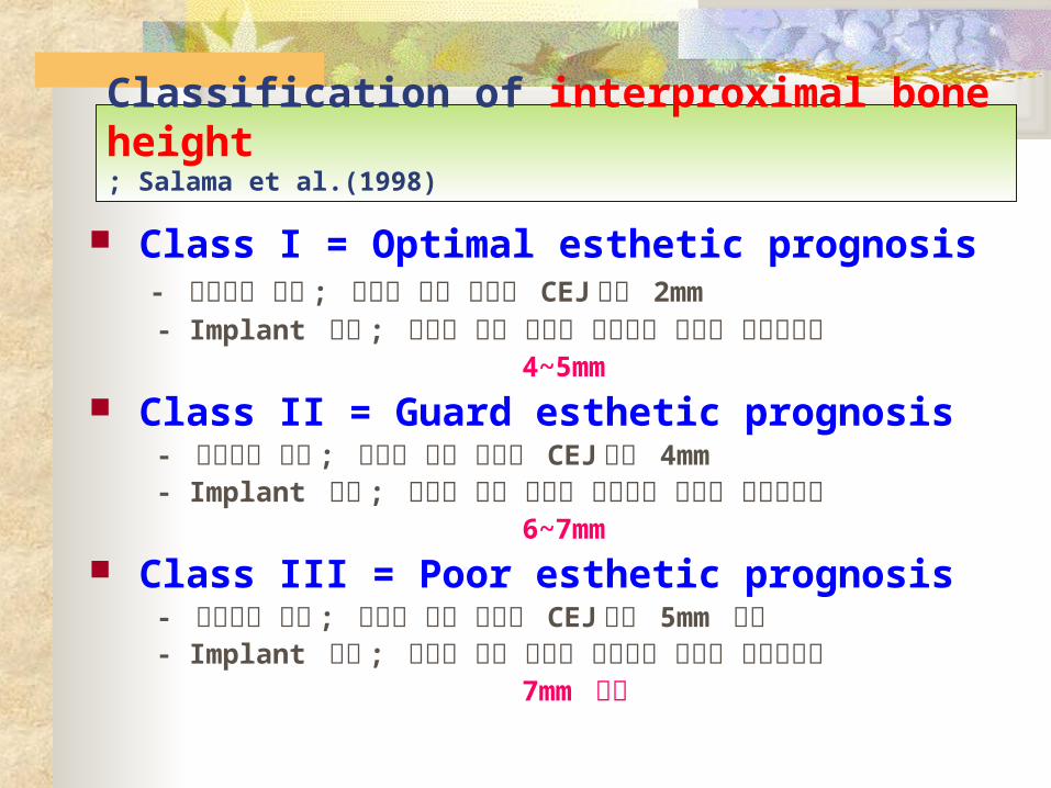
Classification of interproximal bone height; Salama et al.(1998)
Class I = Optimal esthetic prognosis - 자연치의 수복 ; 치간부 골의 높이가 CEJ 에서 2mm - Implant 수복 ; 치간부 골의 높이가 수복물의 접촉점 근첨측에서 4~5mm Class II = Guard esthetic prognosis - 자연치의 수복 ; 치간부 골의 높이가 CEJ 에서 4mm - Implant 수복 ; 치간부 골의 높이가 수복물의 접촉점 근첨측에서 6~7mm Class III = Poor esthetic prognosis - 자연치의 수복 ; 치간부 골의 높이가 CEJ 에서 5mm 이상 - Implant 수복 ; 치간부 골의 높이가 수복물의 접촉점 근첨측에서 7mm 이상
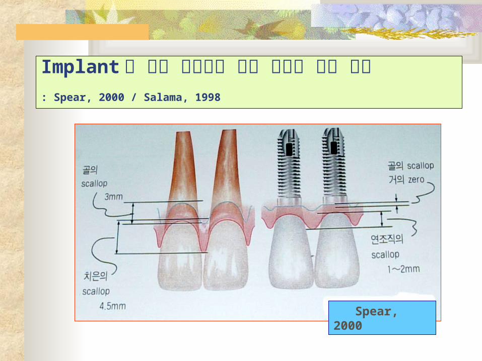
Implant 와 자연 치아에서 주위 조직의 형태 차이: Spear, 2000 / Salama, 1998
Spear, 2000

Width of natural tooth
치관 근원심 폭경 (mm)
CEJ 근원심 폭경 (mm)
CEJ 에서 2mm 근첨측의
근원심 폭경 (mm)
CEJ 의 협설측 폭경
(mm)
권장되는 implant 직경 (mm)
CILIC
PM1PM2M1M2
8.66.57.67.16.610.49.8
6.44.75.64.84.77.97.9
5.54.34.64.24.17.07.0
6.44.77.68.28.110.710.7
4.1/4.3/5.03.25/3.54.1/4.34.1/4.34.1/4.3
4.1/4.3/5.0/6.0
4.1/4.3/5.0/6.0

심미적인 implant 수복을 위한 필요조건(Jovanovic, 1997)
Sufficient bone volume : Horizontal, vertical volume & contour Stability of peri-implant soft tissue Esthetic soft tissue & interproximal papilla Proper implant position : Mesiodistal, buccolingual, vertical, angulation Natural emergence profile & cervical contour

Emergence Profile
<<<

Implant treatment procedure
General historyExtraoral exam.
Intraoral exam.
Radiographic exam.Diagnostic cast
Evaluation of bone quantity&quality, soft tissueOcclusion analysisEvaluation of prosthetic space
Types of prostheticsNumbers, location, angulation
of implant
Augmentation of soft & hard tissue
Final evaluation of
Treatment plans

General Examinations 1. Systemic conditions 2. Causes of missing 3. Intraoral & extraoral examination 4. Occlusal & functional evaluation 5. X-ray examination 6. Examination of periodontium 7. Diagnostic cast 8. 외과학적인 술식과 위험성 9. Treatment time & cost 10. Informed consent
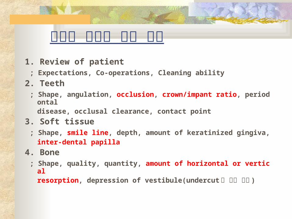
심미적 수복을 위한 검사1. Review of patient ; Expectations, Co-operations, Cleaning ability
2. Teeth ; Shape, angulation, occlusion, crown/impant ratio, periodontal disease, occlusal clearance, contact point3. Soft tissue ; Shape, smile line, depth, amount of keratinized gingiva, inter-dental papilla4. Bone ; Shape, quality, quantity, amount of horizontal or vertical resorption, depression of vestibule(undercut 의 존재 有無 )
Evaluations of Implant Site Development
1. 환자의 필요성2. 심미성 - smile line 의 높이 - upper lip line 과 치아 및 치은의 노출도 - 인접치와의 치경선 조화도 - 정중 시상면을 기준으로 한 교합평면 및 치열의 좌우 대칭성 - 치간 유두의 형태와 높이
3. 치주 조직의 type: thin-scalloped or thick-flat - 연조직의 질과 양
4. 골결손 : class I,II,III(Seibert, 1983) - 잔존골의 높이 , 폭 , 형태
5. 보철물의 type: Fixed or removable ?

Types of periodontium
Thin-scalloped
Thick-flat
<<<<
상악 전치부의 치조골 형태
: Becker et al, 1997
Spear, 1999
치조골의 형태 순측골과 치간부 골 사이의 거리
Flat Mean 2.1mm
Scallop Mean 2.8mm
Prominently scallop
Mean 4.1mm
Total mean: 3mm
Classification of interproximal bone height; Salama et al.(1998)
Class I = Optimal esthetic prognosis - 자연치의 수복 ; 치간부 골의 높이가 CEJ 에서 2mm - Implant 수복 ; 치간부 골의 높이가 수복물의 접촉점 근첨측에서 4~5mm Class II = Guard esthetic prognosis - 자연치의 수복 ; 치간부 골의 높이가 CEJ 에서 4mm - Implant 수복 ; 치간부 골의 높이가 수복물의 접촉점 근첨측에서 6~7mm Class III = Poor esthetic prognosis - 자연치의 수복 ; 치간부 골의 높이가 CEJ 에서 5mm 이상 - Implant 수복 ; 치간부 골의 높이가 수복물의 접촉점 근첨측에서 7mm 이상
다음으로 계속…… .


