RegulationofNT-PGC-1 SubcellularLocalizationand ... · RegulationofNT-PGC-1...
13
Regulation of NT-PGC-1 Subcellular Localization and Function by Protein Kinase A-dependent Modulation of Nuclear Export by CRM1 * □ S Received for publication, November 6, 2009, and in revised form, March 1, 2010 Published, JBC Papers in Press, March 29, 2010, DOI 10.1074/jbc.M109.083121 Ji Suk Chang ‡ , Peter Huypens ‡ , Yubin Zhang ‡§ , Chelsea Black ‡ , Anastasia Kralli ¶ , and Thomas W. Gettys ‡1 From the ‡ Laboratory of Nutrient Sensing and Adipocyte Signaling, Pennington Biomedical Research Center, Baton Rouge, Louisiana 70808, the ¶ Department of Chemical Physiology, Scripps Research Institute, La Jolla, California 92037, and the § Department of Biochemistry, China Pharmaceutical University, Nanjing 210009, China Peroxisome proliferator-activated receptor co-activator-1 (PGC-1) plays a central role in the regulation of cellular energy metabolism and metabolic adaptation to environmental and nutritional stimuli. We recently described a novel, biologically active splice variant of PGC-1 (NT-PGC-1, amino acids 1–270) that retains the ability to interact with and transactivate nuclear hormone receptors through its N-terminal transactiva- tion domain. Whereas PGC-1 is an unstable nuclear protein sensitive to ubiquitin-mediated targeting to the proteasome, NT-PGC-1 is relatively stable and predominantly cytoplasmic, suggesting that its ability to interact with and activate nuclear receptors and transcription factors is dependent upon regulated access to the nucleus. We provide evidence that NT-PGC-1 interacts with the nuclear exportin, CRM1, through a specific leucine-rich domain (nuclear export sequence) that regulates its export to the cytoplasm. The nuclear export of NT-PGC-1 is inhibited by protein kinase A-dependent phosphorylation of Ser-194, Ser-241, and Thr-256 on NT-PGC-1, which effec- tively increases its nuclear concentration. Using site-directed mutagenesis to prevent or mimic phosphorylation at these sites, we show that the transcriptional activity of NT-PGC-1 is reg- ulated in part through regulation of its subcellular localization. These findings suggest that the function of NT-PGC-1 as a transcriptional co-activator is regulated by protein kinase A-de- pendent inhibition of CRM1-mediated export from the nucleus. Many biological programs are regulated at the transcrip- tional level by interaction of transcription factors with tran- scriptional co-activators. For example, the peroxisome prolif- erator-activated receptor co-activator-1 (PGC-1) 2 family is integral to the coordinated activation of transcription com- plexes (1, 2) that regulate global responses such as mitochon- drial biogenesis (3–7) and tissue-specific responses such as adaptive thermogenesis (3), fatty acid oxidation (8, 9), hepatic gluconeogenesis (10 –14), fiber type switching in muscle (15), reactive oxygen species metabolism (16), and clock gene expression (17). PGC-1 contains a powerful autonomous transactivation domain at its N terminus (aa 1–200) that binds two other co- activators with acetyl transferase activity, SRC-1 and CBP/p300 (18). The leucine-rich motifs located downstream of the trans- activation domain allow the co-activator to interact with ligand-activated nuclear receptors, as well as various other transcription factors such as NRF1, Host Cell factor, MEF2C, and FOXO1 (4, 8, 19 –22). The central region of PGC-1 (aa 200 – 400) is required for interaction with p160 myb-binding protein (p160MBP), which acts as a repressor of PGC-1 (23), whereas the C-terminal end of PGC-1 (aa 400 –797) contains RNA recognition motifs and arginine/serine-rich domains characteristic of splicing factors (24). PGC-1 is regulated by signaling inputs from cAMP, cal- cium, nitro-oxide, and cGMP that induce its expression and/or increase its activity through post-translational modifications (10, 25–27). PGC-1 protein is relatively short-lived (2.3 h) due to rapid targeting to ubiquitin/proteasome-mediated pro- teolysis in the nucleus (28 –30). Various post-translational inputs are known to affect proteasomal targeting and stability, most notably p38 MAPK, which phosphorylates PGC-1 on Thr-262, Ser-265, and Thr-298 and enhances its stability (23, 28, 31). In addition, PGC-1 expression and/or stability are also modulated by AMP-dependent phosphorylation at residues Thr-177 and Ser-538 (32), Akt2/protein kinase B-dependent phosphorylation at Ser-570 (33), and glycogen synthase kinase 3-dependent phosphorylation at Thr-295 (30). In addition to phosphorylation, protein arginine methyltransferase 1-medi- ated methylation of PGC-1 at residues Arg-665, Arg-667, and Arg-669 activates transcriptional activity (34), whereas acetyla- tion by GCN5 decreases PGC-1 activity (35). We recently identified a novel, biologically active 270-aa iso- form of PGC-1 (NT-PGC-1) produced by alternative splic- ing that introduces a premature stop codon between exons 6 and 7 (36). NT-PGC-1 retains the N-terminal transactivation * This work was supported, in whole or in part, by National Institutes of Health Grants RO1 DK 074772 and P20-RR021945 (to T. W. G.), NIH Clinical Nutri- tion Research Unit Center Grant 1P30 DK072476, and NIH Grant DK064951 (to A. K.). □ S The on-line version of this article (available at http://www.jbc.org) contains supplemental Figs. S1 and S2. 1 To whom correspondence should be addressed: Pennington Biomedical Research Center, 6400 Perkins Rd., Baton Rouge, LA 70808. Tel.: 225-763- 3165; Fax: 225-763-0274; E-mail: [email protected]. 2 The abbreviations used are: PGC-1, peroxisome proliferator-activated receptor co-activator-1; PPAR, peroxisome proliferator-activated recep- tor; PPRE, PPAR response element; Bt 2 cAMP, dibutyryl cAMP; aa, amino acid(s); MAPK, mitogen-activated protein kinase; PKA, protein kinase A; CHO, Chinese hamster ovary; PBS, phosphate-buffered saline; GST, gluta- thione S-transferase; HA, hemagglutinin; NES, nuclear export sequence; LMB, leptomycin B; RXR, retinoid X receptor; BAT, brown adipose tissue. THE JOURNAL OF BIOLOGICAL CHEMISTRY VOL. 285, NO. 23, pp. 18039 –18050, June 4, 2010 © 2010 by The American Society for Biochemistry and Molecular Biology, Inc. Printed in the U.S.A. JUNE 4, 2010 • VOLUME 285 • NUMBER 23 JOURNAL OF BIOLOGICAL CHEMISTRY 18039 by guest on August 27, 2018 http://www.jbc.org/ Downloaded from
Transcript of RegulationofNT-PGC-1 SubcellularLocalizationand ... · RegulationofNT-PGC-1...
Regulation of NT-PGC-1� Subcellular Localization andFunction by Protein Kinase A-dependent Modulation ofNuclear Export by CRM1*□S
Received for publication, November 6, 2009, and in revised form, March 1, 2010 Published, JBC Papers in Press, March 29, 2010, DOI 10.1074/jbc.M109.083121
Ji Suk Chang‡, Peter Huypens‡, Yubin Zhang‡§, Chelsea Black‡, Anastasia Kralli¶, and Thomas W. Gettys‡1
From the ‡Laboratory of Nutrient Sensing and Adipocyte Signaling, Pennington Biomedical Research Center, Baton Rouge,Louisiana 70808, the ¶Department of Chemical Physiology, Scripps Research Institute, La Jolla, California 92037,and the §Department of Biochemistry, China Pharmaceutical University, Nanjing 210009, China
Peroxisome proliferator-activated receptor� co-activator-1�(PGC-1�) plays a central role in the regulation of cellular energymetabolism and metabolic adaptation to environmental andnutritional stimuli. We recently described a novel, biologicallyactive splice variant of PGC-1� (NT-PGC-1�, amino acids1–270) that retains the ability to interact with and transactivatenuclear hormone receptors through its N-terminal transactiva-tion domain. Whereas PGC-1� is an unstable nuclear proteinsensitive to ubiquitin-mediated targeting to the proteasome,NT-PGC-1� is relatively stable and predominantly cytoplasmic,suggesting that its ability to interact with and activate nuclearreceptors and transcription factors is dependent upon regulatedaccess to the nucleus. We provide evidence that NT-PGC-1�interacts with the nuclear exportin, CRM1, through a specificleucine-rich domain (nuclear export sequence) that regulates itsexport to the cytoplasm. The nuclear export of NT-PGC-1� isinhibited by protein kinase A-dependent phosphorylation ofSer-194, Ser-241, and Thr-256 on NT-PGC-1�, which effec-tively increases its nuclear concentration. Using site-directedmutagenesis to prevent ormimic phosphorylation at these sites,we show that the transcriptional activity of NT-PGC-1� is reg-ulated in part through regulation of its subcellular localization.These findings suggest that the function of NT-PGC-1� as atranscriptional co-activator is regulated by protein kinaseA-de-pendent inhibition of CRM1-mediated export from the nucleus.
Many biological programs are regulated at the transcrip-tional level by interaction of transcription factors with tran-scriptional co-activators. For example, the peroxisome prolif-erator-activated receptor � co-activator-1 (PGC-1)2 family isintegral to the coordinated activation of transcription com-
plexes (1, 2) that regulate global responses such as mitochon-drial biogenesis (3–7) and tissue-specific responses such asadaptive thermogenesis (3), fatty acid oxidation (8, 9), hepaticgluconeogenesis (10–14), fiber type switching in muscle (15),reactive oxygen species metabolism (16), and clock geneexpression (17).PGC-1� contains a powerful autonomous transactivation
domain at its N terminus (aa 1–200) that binds two other co-activators with acetyl transferase activity, SRC-1 andCBP/p300(18). The leucine-rich motifs located downstream of the trans-activation domain allow the co-activator to interact withligand-activated nuclear receptors, as well as various othertranscription factors such as NRF1, Host Cell factor, MEF2C,and FOXO1 (4, 8, 19–22). The central region of PGC-1� (aa200–400) is required for interaction with p160 myb-bindingprotein (p160MBP), which acts as a repressor of PGC-1� (23),whereas the C-terminal end of PGC-1� (aa 400–797) containsRNA recognition motifs and arginine/serine-rich domainscharacteristic of splicing factors (24).PGC-1� is regulated by signaling inputs from cAMP, cal-
cium, nitro-oxide, and cGMP that induce its expression and/orincrease its activity through post-translational modifications(10, 25–27). PGC-1� protein is relatively short-lived (�2.3 h)due to rapid targeting to ubiquitin/proteasome-mediated pro-teolysis in the nucleus (28–30). Various post-translationalinputs are known to affect proteasomal targeting and stability,most notably p38 MAPK, which phosphorylates PGC-1� onThr-262, Ser-265, and Thr-298 and enhances its stability (23,28, 31). In addition, PGC-1� expression and/or stability are alsomodulated by AMP-dependent phosphorylation at residuesThr-177 and Ser-538 (32), Akt2/protein kinase B-dependentphosphorylation at Ser-570 (33), and glycogen synthase kinase3�-dependent phosphorylation at Thr-295 (30). In addition tophosphorylation, protein arginine methyltransferase 1-medi-ated methylation of PGC-1� at residues Arg-665, Arg-667, andArg-669 activates transcriptional activity (34), whereas acetyla-tion by GCN5 decreases PGC-1� activity (35).We recently identified a novel, biologically active 270-aa iso-
form of PGC-1� (NT-PGC-1�) produced by alternative splic-ing that introduces a premature stop codon between exons 6and 7 (36). NT-PGC-1� retains the N-terminal transactivation
* This work was supported, in whole or in part, by National Institutes of HealthGrants RO1 DK 074772 and P20-RR021945 (to T. W. G.), NIH Clinical Nutri-tion Research Unit Center Grant 1P30 DK072476, and NIH Grant DK064951(to A. K.).
□S The on-line version of this article (available at http://www.jbc.org) containssupplemental Figs. S1 and S2.
1 To whom correspondence should be addressed: Pennington BiomedicalResearch Center, 6400 Perkins Rd., Baton Rouge, LA 70808. Tel.: 225-763-3165; Fax: 225-763-0274; E-mail: [email protected].
2 The abbreviations used are: PGC-1, peroxisome proliferator-activatedreceptor � co-activator-1; PPAR, peroxisome proliferator-activated recep-tor; PPRE, PPAR response element; Bt2cAMP, dibutyryl cAMP; aa, aminoacid(s); MAPK, mitogen-activated protein kinase; PKA, protein kinase A;CHO, Chinese hamster ovary; PBS, phosphate-buffered saline; GST, gluta-
thione S-transferase; HA, hemagglutinin; NES, nuclear export sequence;LMB, leptomycin B; RXR, retinoid X receptor; BAT, brown adipose tissue.
THE JOURNAL OF BIOLOGICAL CHEMISTRY VOL. 285, NO. 23, pp. 18039 –18050, June 4, 2010© 2010 by The American Society for Biochemistry and Molecular Biology, Inc. Printed in the U.S.A.
JUNE 4, 2010 • VOLUME 285 • NUMBER 23 JOURNAL OF BIOLOGICAL CHEMISTRY 18039
by guest on August 27, 2018
http://ww
w.jbc.org/
Dow
nloaded from
and nuclear receptor interaction domains of PGC-1� and isfunctionally active (36). In contrast to PGC-1�, NT-PGC-1� isrelatively stable and predominantly sequestered in the cyto-plasm (36). cAMP triggers nuclear accumulation of NT-PGC-1� and enhances the recruitment of NT-PGC-1� to thepromoters of consensus PGC-1� target genes such as UCP1andCPT1� (36). These findings suggest that nuclear transloca-tion ofNT-PGC-1� is a critical regulatory step in its response tocAMP-dependent signaling input, whereas cAMP signalingincreases PGC-1� activity in part by stabilizing the protein inthe nucleus. Sequestering transcription factors outside thenucleus is a commonmode of regulation. For example, NF-�B,FOXO1, c-Fos, and FKHRL1 are retained in the cytosol, andtheir activity is limited by binding partners that mask thenuclear localization signals or promote nuclear export. Thus, asseen with NT-PGC-1�, signaling input regulates their activityby regulating their nuclear translocation (37–41).In this report, we investigated how post-translational input
from PKA regulates the subcellular localization and function ofNT-PGC-1�.We found that NT-PGC-1� interacts with CRM1through the N-terminal CRM1-binding motif and is activelyexported to the cytoplasm. PKA-dependent phosphorylation ofNT-PGC-1� on Ser-194, Ser-241, and Thr-256 preventsnuclear export and allows a greater proportion of cellular NT-PGC-1� to remain in the nucleus. Using site-directedmutagen-esis to prevent PKA-dependent phosphorylation of these sites,we find a close relationship between the ability of PKA toincrease nuclear content of NT-PGC-1� and its ability toenhance transcriptional activation of the UCP1 and CIDEAgenes. These findings support PKA-dependent inhibition ofCRM1 mediated-NT-PGC-1� nuclear export as the primarymechanism through which PKA regulates the transcriptionalactivity of NT-PGC-1�. In addition, we show that nuclear NT-PGC-1� regulates genes that are both distinct from (e.g. Cox8b)and common to those that are regulated by PGC-1�. Thus, thisPKA-dependent activation of NT-PGC-1�would be importantto induction of the complete set of genes that are transcription-ally regulated by PGC-1� isoforms.
EXPERIMENTAL PROCEDURES
Cell Cultures and Adipocyte Differentiation—CHO-K1 celllines were maintained in culture in F-12K supplemented with10% fetal bovine serum and 1% penicillin/streptomycin(Invitrogen) and transfected using FuGENE 6 (Roche AppliedScience). An immortalized PGC-1�-deficient brown preadipo-cyte cell line (42) was a gift from Bruce Spiegelman (HarvardMedical School) and was maintained in Dulbecco’s modifiedEagle’s medium supplemented with 10% fetal bovine serum, 1%glutamine, and 1% penicillin/streptomycin. Preadipocytes weregrown to confluence in culture medium supplemented with 20nM insulin and 1 nM T3 (differentiation medium). Differentia-tion of adipocytes was induced (day 1) by incubating the cells indifferentiation medium supplemented with 0.5 mM isobutyl-methylxanthine, 0.5 �M dexamethasone, and 0.125 mM indo-methacin for 48 h. Thereafter, the cells were maintained indifferentiation medium until day 7.Plasmids—Expression plasmids for pcDNA3.1-NT-PGC-
1�-HA were described previously (36). Site-directed mutagen-
esis of pcDNA3.1-NT-PGC-1�-HA with the QuikChangemutagenesis system (Stratagene, La Jolla, CA) was performedto generate leucine to alanine mutations (NES1, NES2, andNES1/2) in the consensus CRM1 nuclear export sites,and serine/threonine to alanine or aspartate mutations inthe PKA phosphorylation sites (Ser-194, Ser-241, and Thr-256). pcDNA3.1-myc-CRM1 and pQE32-HIS6-RanQ69L werekindly provided by S. Meloche (Universite de Montreal). Toconstruct retroviral plasmids of pBABE-NT-PGC-1�-HA andpBABE-NT-PGC-1�-S194A/S241A/T256A-HA, BamHI andXhoI fragments of wild-type and mutant NT-PGC-1� weresubcloned into a BamHI- and SalI-digested pBABE-neo vector.Gal4-DBD fusions of wild-type and mutant NT-PGC-1� andPGC-1�were generated by subcloning into a pCMV-DBvector.(PPRE)3-TK-luc, pSV sport-RXR�, and pSV sport-PPAR�1were from Addgene Inc.Retroviral and Adenoviral Infection—To produce retrovirus,
GP-293 cells grown to 80% confluence were co-transfectedwith pVSV-G and pBABE-neo, pBABE-NT-PGC-1�-HA, orpBABE-NT-PGC-1�-S194A/S241A/T256A-HA by co-precipi-tation with calcium phosphate (Promega, Madison, WI). Fol-lowing transfection, the cells were incubated at 32 °C toincrease viral titer. Virus-containingmediumwas collected, fil-tered through the 0.45-�m low protein binding filter, and usedto infect target cells. Immortalized PGC-1�-deficient brownpreadipocyte cells (30–40% confluence) were infected with theviral supernatant supplemented with 8 �g/ml Polybrene by acentrifugation method (1800 rpm � 45 min). The mediumwasaspirated after 2 h and replaced with fresh viral supernatant,and the procedure was repeated. After 8 h of infection, the cellswere replaced with fresh Dulbecco’s modified Eagle’s mediumsupplemented with 10% fetal bovine serum, 1% glutamine, and1% penicillin/streptomycin. Selection was initiated with 800�g/ml G418 (Invitrogen) 48 h after infection.
Adenoviral infection of immortalized PGC-1�-deficientbrown preadipocyte or differentiated brown adipocyte cellswith NT-PGC-1�-FLAG was conducted using a poly-L-lysine(Mr 30,000–70,000)-assisted transduction procedure (43).Adenoviral supernatants were mixed in Opti-MEM containingpoly-L-lysine (0.5 �g/ml) and incubated for 1.5 h at room tem-perature. Cells were washed with PBS, and the adenoviral mix-ture was added and the mixture was incubated for 2 h at 37 °Cfollowed by addition of Dulbecco’s modified Eagle’s mediumand incubation overnight. The culture medium was replacedwith a fresh medium. Each experiment was performed at 48 hafter viral infection.Immunocytochemistry—Cells on coverslips were washed
with 1� PBS, fixed with 4% paraformaldehyde for 10 min, andpermeabilizedwith 0.5%TritonX-100. The fixed cells were firstincubated with the blocking buffer (1� PBS, 5% normal goatserum, and 1% bovine serum albumin) and then with a 1:4000diluted rabbit anti-HA antibody (Abcam) or a 1:1000 dilutedmouse anti-FLAG M2 antibody (Sigma) in the same buffer for2 h. After incubation with primary antibodies, cells werewashed three times with 1� PBS and incubated with an AlexaFluor 488-conjugated anti-rabbit or mouse immunoglobulinsecondary antibody for 1 h (Invitrogen). Cells were washedthree times with 1� PBS and mounted with Vectashield (Vec-
NT-PGC-1� by PKA and CRM1
18040 JOURNAL OF BIOLOGICAL CHEMISTRY VOLUME 285 • NUMBER 23 • JUNE 4, 2010
by guest on August 27, 2018
http://ww
w.jbc.org/
Dow
nloaded from
tor Labs). Nuclei were counterstained with 4,6-diamidino-2-phenylindole. Subcellular localization of NT-PGC-1� wasexamined with a Plan-Neofluar 40�/0.85 numerical apertureobjective on a Zeiss LSM510 confocal microscope. The relativeintensity of the fluorescence in the nucleus and cytoplasm wasquantified using the National Institutes of Health ImageJ pro-gram as described before (36, 44, 45). Briefly, the fluorescencesignals of NT-PGC-1� in the nucleus and entire cell were mea-sured using the NIH ImageJ in 150–200 (CHO-K1) or 20–60(brownpreadipocytes/adipocytes) randomly selected positivelytransduced cells, and the nuclear and whole cell areas wererecorded. The total nuclear fluorescence intensity was normal-ized for nuclear area. The total cytoplasmic fluorescence inten-sity (total cellular fluorescence minus nuclear fluorescence)was normalized for cytoplasmic area (whole cell area minusnuclear area). The relative nuclear/cytoplasmic ratio of fluores-cence intensity was determined by dividing the nuclear fluores-cence intensity per unit area by the cytoplasmic fluorescenceintensity per unit area.Western Blot and Immunoprecipitation—Cells were har-
vested, washedwith ice-cold PBS, and lysed in 50mMTris-HCl,pH 7.5, 1% Triton X-100, 1% sodium deoxycholate, 0.1% SDS,150 mM NaCl, 1 mM phenylmethylsulfonyl fluoride supple-mented with a protease inhibitor mixture (Roche Applied Sci-ence). Antibodies used were as follows: monoclonal anti-PGC-1� (36), anti-Myc (9E10), and anti-CRM1(H-300) (SantaCruz Biotechnology), anti-HA (Convance), and anti-tubulin(Abcam). Immunoprecipitations were performed in 20 mM
HEPES, pH 7.0, 150 mM NaCl, 0.2% Nonidet P-40 supple-mentedwith protease and phosphatase inhibitors. Lysates wereprecleared with protein G-agarose beads and immunoprecipi-tatedwith antibody-coated beads for 3 h at 4 °C.Afterwashings,immunoprecipitates were subjected to Western blot analysis.Recombinant GST Fusion Protein Purification and GST Pull-
down Assay—Overnight BL21 cultures were diluted and grownin LB�AMP at 30 °C to an A600 of 0.5. Protein expression wasinduced with isopropyl 1-thio-�-D-galactopyranoside for 3 h at30 °C. Cells were harvested, washed, resuspended in 1� PBSbuffer supplemented with 2.5 mM phenylmethylsulfonyl fluo-ride, 5 mM dithiothreitol, and a protease inhibitor mixture,lysed by sonication, and treated with 1% Triton X-100. Thelysates were then incubated with glutathione beads for 2 h at4 °C. GST, GST-PGC-1� (1–200 aa), and GST-NT-PGC-1�fusion proteins immobilized on the beads were incubated withthe CHO-K1 cell lysates expressingMyc-CRM1 or brown prea-dipocyte cell lysates for 3 h at 4 °C. After washings, the boundproteins were subjected to Western blot analysis.[32P]Orthophosphate Metabolic Labeling—CHO-K1 cells
transiently expressing pcDNA3.1, NT-PGC-1�-HA, or NT-PGC-1�-S194A/S241A/T256A-HA were washed three timeswith pre-warmed phosphate-free Dulbecco’s modified Eagle’smedium containing 1% glutamine, 0.1% bovine serum albu-min, and 1% (10 mM) Hepes, pH 7.4, and then incubated inthe same phosphate-free medium for 1 h. During the incuba-tion, H89 was added in some experiments. The medium wasthen replaced with fresh phosphate-free medium containing[32P]orthophosphate (0.25 mCi/ml, PerkinElmer Life Sciences)with the addition of Bt2cAMP (Sigma) alone or Bt2cAMP and
H89 (Calbiochem) and incubated for 3 h. Cells were washedthree times with ice-cold 0.15 MNaCl solution and lysed in coldlysis buffer (1% Nonidet P-40, 0.15 M NaCl, 20 mM Hepes, pH7.4) with protease (Roche Applied Science) and phosphatase(10 mM NaF, 0.1 mM Na3VO4) inhibitors. Lysates were pre-cleared with protein G-agarose beads and immunoprecipitatedwith anti-HA antibody-coated beads for 3 h at 4 °C. The immu-noprecipitates were washed eight times with the same bufferand subjected to autoradiography and Western blot analyses.In Vitro Phosphorylation of NT-PGC-1� by PKA—NT-PGC-
1�-HA and its PKA mutants were in vitro transcribed/trans-lated using TNT T7 Coupled Reticulocyte Lysate System (Pro-mega) and immunoprecipitated in immunoprecipitation buffer(50 mM Tris-HCl, pH 7.5, 150 mM NaCl, 0.2% Nonidet P-40, 5mM MgCl2, and protease inhibitors) with anti-HA antibodies.The immunoprecipitateswerewashed four timeswith the samebuffer and washed once with the kinase reaction buffer (25 mM
Tris-HCl, pH 7.5, 10 mM MgCl2, 2 mM dithiothreitol, 5 mM
�-glycerophosphate, 0.1 mM Na3VO4). Kinase reactions wereinitiated by addition of 100 �M ATP, 10 �Ci (0.74 MBq) of[�-32P]ATP (PerkinElmer Life Sciences), and 1 �g of affinity-purified bovine PKA in a final volume of 40 �l. After 30-minincubation at 30 °C, the immunoprecipitates were washed andsubjected to autoradiography and Western blot analyses.Luciferase Reporter Assay—CHO-K1 cells were transiently
transfected with (PPRE)3-TK-luc, pSV sport-RXR�, pSV sport-PPAP�1, pRL-SV40, and pcDNA3.1/NT-PGC-1�-HA/NT-PGC-1�-S194A/S241A/T256A-HA/NT-PGC-1�-S194D/S241D/T256D-HA using Fugene6 (Roche). 24 h after transfection, cellswere treated with vehicle, BRL49653/9-cis-RA or Bt2cAMP.48h later, cells were harvested for assay of luciferase activityusing a Promega Dual Luciferase assay kit (Promega). Fireflyluciferase activity was normalized using Renilla luciferase andprotein expression. Data representmean� S.D. of at least threeindependent experiments.Real-time PCR Analysis—Total RNA was isolated from dif-
ferentiated brown adipocyte cell lines using RNeasy kits (Qia-gen). For real-time PCR analysis, RNA samples were reversetranscribed using oligo(dT) primers and M-MLV reversetranscriptase (Promega), and 20 ng of cDNA was used inquantitative PCR reactions in the presence of a fluorescent dye(Cybergreen, Takara) on a Smart Cycler (Cepheid). Relativeabundance of mRNA was normalized to cyclophilin mRNA.The following primers were used: UCP1 (36), cyclophilin (36),CIDEA (42), CytC (42), Cox8b (46), and ERR� (46).
RESULTS
CRM1 Promotes Cytoplasmic Translocation of NT-PGC-1�—Proteins of�50 kDa are able to pass through nuclear pores andequilibrate across the nuclear membrane by passive diffusion(47, 48). Although NT-PGC-1� lacks the four consensusnuclear localization sequences identified in PGC-1� (20, 22), itspredicted size (�30 kDa) should not preclude its diffusionthrough the nuclear pore complex (36). However, our initialhypothesis that the proteinwould equilibrate across the nuclearmembrane was not supported by our finding that NT-PGC-1� was primarily localized to the cytosol (36). Two potentialexplanations for this unexpected finding are that 1) NT-
NT-PGC-1� by PKA and CRM1
JUNE 4, 2010 • VOLUME 285 • NUMBER 23 JOURNAL OF BIOLOGICAL CHEMISTRY 18041
by guest on August 27, 2018
http://ww
w.jbc.org/
Dow
nloaded from
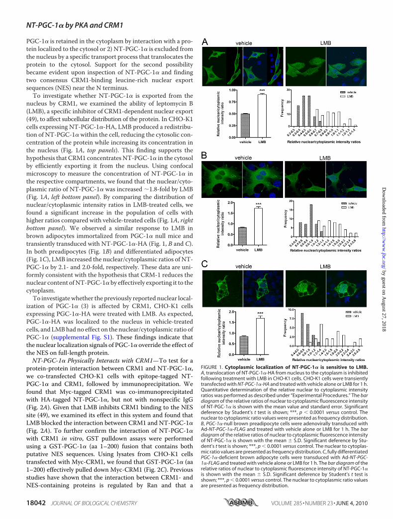
PGC-1� is retained in the cytoplasm by interaction with a pro-tein localized to the cytosol or 2) NT-PGC-1� is excluded fromthe nucleus by a specific transport process that translocates theprotein to the cytosol. Support for the second possibilitybecame evident upon inspection of NT-PGC-1� and findingtwo consensus CRM1-binding leucine-rich nuclear exportsequences (NES) near the N terminus.To investigate whether NT-PGC-1� is exported from the
nucleus by CRM1, we examined the ability of leptomycin B(LMB), a specific inhibitor of CRM1-dependent nuclear export(49), to affect subcellular distribution of the protein. In CHO-K1cells expressing NT-PGC-1�-HA, LMB produced a redistribu-tion of NT-PGC-1� within the cell, reducing the cytosolic con-centration of the protein while increasing its concentration inthe nucleus (Fig. 1A, top panels). This finding supports thehypothesis that CRM1 concentrates NT-PGC-1� in the cytosolby efficiently exporting it from the nucleus. Using confocalmicroscopy to measure the concentration of NT-PGC-1� inthe respective compartments, we found that the nuclear/cyto-plasmic ratio of NT-PGC-1� was increased �1.8-fold by LMB(Fig. 1A, left bottom panel). By comparing the distribution ofnuclear/cytoplasmic intensity ratios in LMB-treated cells, wefound a significant increase in the population of cells withhigher ratios compared with vehicle-treated cells (Fig. 1A, rightbottom panel). We observed a similar response to LMB inbrown adipocytes immortalized from PGC-1� null mice andtransiently transduced with NT-PGC-1�-HA (Fig. 1, B and C).In both preadipocytes (Fig. 1B) and differentiated adipocytes(Fig. 1C), LMB increased the nuclear/cytoplasmic ratios of NT-PGC-1� by 2.1- and 2.0-fold, respectively. These data are uni-formly consistent with the hypothesis that CRM-1 reduces thenuclear content ofNT-PGC-1� by effectively exporting it to thecytoplasm.To investigatewhether the previously reported nuclear local-
ization of PGC-1� (3) is affected by CRM1, CHO-K1 cellsexpressing PGC-1�-HA were treated with LMB. As expected,PGC-1�-HA was localized to the nucleus in vehicle-treatedcells, andLMBhadno effect on the nuclear/cytoplasmic ratio ofPGC-1� (supplemental Fig. S1). These findings indicate thatthe nuclear localization signals of PGC-1� override the effect ofthe NES on full-length protein.NT-PGC-1� Physically Interacts with CRM1—To test for a
protein-protein interaction between CRM1 and NT-PGC-1�,we co-transfected CHO-K1 cells with epitope-tagged NT-PGC-1� and CRM1, followed by immunoprecipitation. Wefound that Myc-tagged CRM1 was co-immunoprecipitatedwith HA-tagged NT-PGC-1�, but not with nonspecific IgG(Fig. 2A). Given that LMB inhibits CRM1 binding to the NESsite (49), we examined its effect in this system and found thatLMB blocked the interaction between CRM1 and NT-PGC-1�(Fig. 2A). To further confirm the interaction of NT-PGC-1�with CRM1 in vitro, GST pulldown assays were performedusing a GST-PGC-1� (aa 1–200) fusion that contains bothputative NES sequences. Using lysates from CHO-K1 cellstransfected with Myc-CRM1, we found that GST-PGC-1� (aa1–200) effectively pulled down Myc-CRM1 (Fig. 2C). Previousstudies have shown that the interaction between CRM1- andNES-containing proteins is regulated by Ran and that a
FIGURE 1. Cytoplasmic localization of NT-PGC-1� is sensitive to LMB.A, translocation of NT-PGC-1�-HA from nucleus to the cytoplasm is inhibitedfollowing treatment with LMB in CHO-K1 cells. CHO-K1 cells were transientlytransfected with NT-PGC-1�-HA and treated with vehicle alone or LMB for 1 h.Quantitative determination of the relative nuclear to cytoplasmic intensityratios was performed as described under “Experimental Procedures.” The bardiagram of the relative ratios of nuclear to cytoplasmic fluorescence intensityof NT-PGC-1� is shown with the mean value and standard error. Significantdeference by Student’s t test is shown; ***, p � 0.0001 versus control. Thenuclear to cytoplasmic ratio values were presented as frequency distribution.B, PGC-1�-null brown preadipocyte cells were adenovirally transduced withAd-NT-PGC-1�-FLAG and treated with vehicle alone or LMB for 1 h. The bardiagram of the relative ratios of nuclear to cytoplasmic fluorescence intensityof NT-PGC-1� is shown with the mean � S.D. Significant deference by Stu-dent’s t test is shown; ***, p � 0.0001 versus control. The nuclear to cytoplas-mic ratio values are presented as frequency distribution. C, fully differentiatedPGC-1�-deficient brown adipocyte cells were transduced with Ad-NT-PGC-1�-FLAG and treated with vehicle alone or LMB for 1 h. The bar diagram of therelative ratios of nuclear to cytoplasmic fluorescence intensity of NT-PGC-1�is shown with the mean � S.D. Significant deference by Student’s t test isshown; ***, p � 0.0001 versus control. The nuclear to cytoplasmic ratio valuesare presented as frequency distribution.
NT-PGC-1� by PKA and CRM1
18042 JOURNAL OF BIOLOGICAL CHEMISTRY VOLUME 285 • NUMBER 23 • JUNE 4, 2010
by guest on August 27, 2018
http://ww
w.jbc.org/
Dow
nloaded from
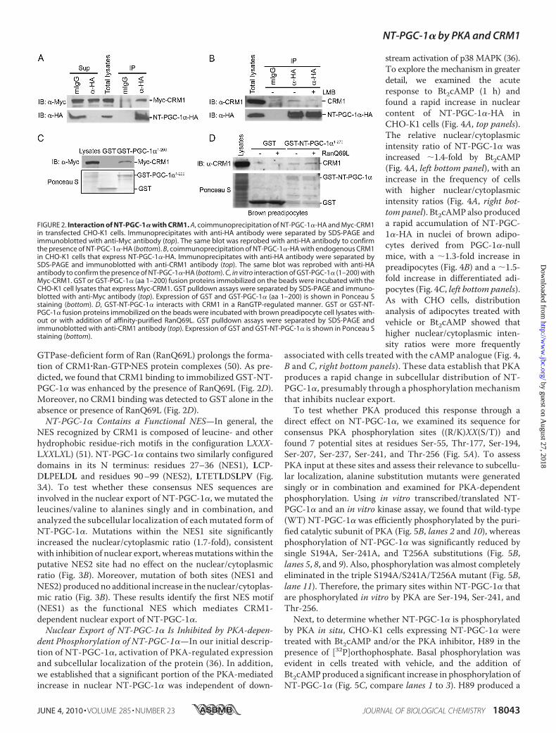
GTPase-deficient form of Ran (RanQ69L) prolongs the forma-tion of CRM1�Ran-GTP�NES protein complexes (50). As pre-dicted, we found that CRM1 binding to immobilized GST-NT-PGC-1� was enhanced by the presence of RanQ69L (Fig. 2D).Moreover, no CRM1 binding was detected to GST alone in theabsence or presence of RanQ69L (Fig. 2D).NT-PGC-1� Contains a Functional NES—In general, the
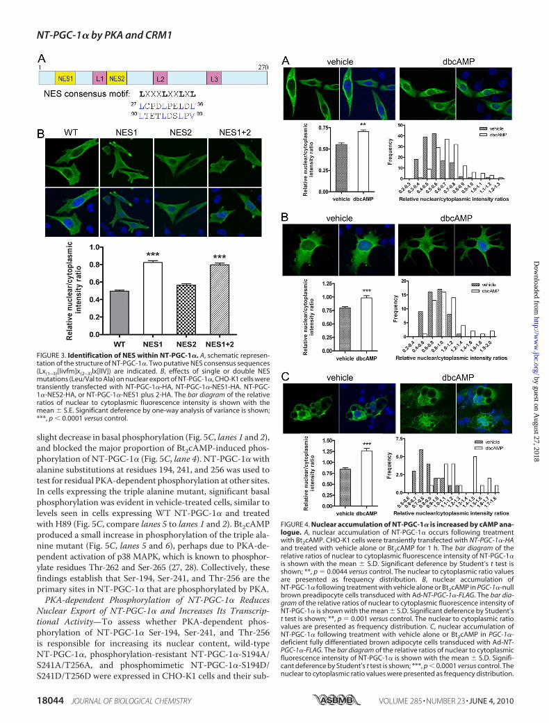
NES recognized by CRM1 is composed of leucine- and otherhydrophobic residue-rich motifs in the configuration LXXX-LXXLXL) (51). NT-PGC-1� contains two similarly configureddomains in its N terminus: residues 27–36 (NES1), LCP-DLPELDL and residues 90–99 (NES2), LTETLDSLPV (Fig.3A). To test whether these consensus NES sequences areinvolved in the nuclear export of NT-PGC-1�, we mutated theleucines/valine to alanines singly and in combination, andanalyzed the subcellular localization of eachmutated form ofNT-PGC-1�. Mutations within the NES1 site significantlyincreased the nuclear/cytoplasmic ratio (1.7-fold), consistentwith inhibition of nuclear export, whereasmutationswithin theputative NES2 site had no effect on the nuclear/cytoplasmicratio (Fig. 3B). Moreover, mutation of both sites (NES1 andNES2) producedno additional increase in the nuclear/cytoplas-mic ratio (Fig. 3B). These results identify the first NES motif(NES1) as the functional NES which mediates CRM1-dependent nuclear export of NT-PGC-1�.Nuclear Export of NT-PGC-1� Is Inhibited by PKA-depen-
dent Phosphorylation of NT-PGC-1�—In our initial descrip-tion of NT-PGC-1�, activation of PKA-regulated expressionand subcellular localization of the protein (36). In addition,we established that a significant portion of the PKA-mediatedincrease in nuclear NT-PGC-1� was independent of down-
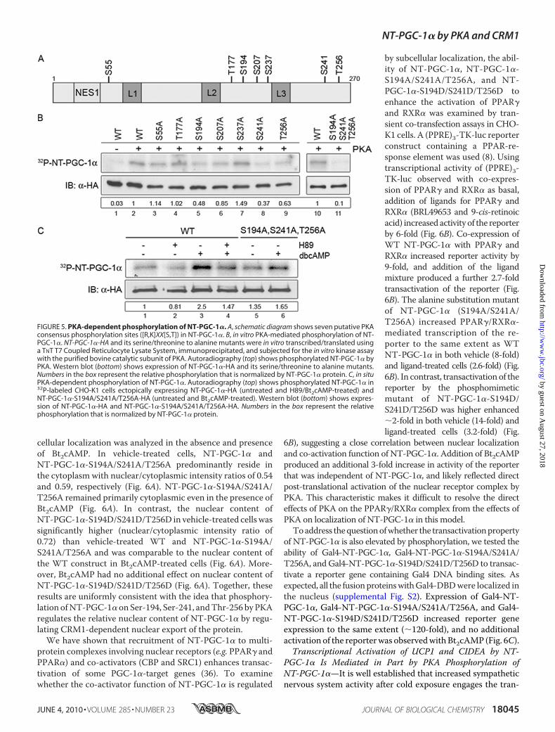
stream activation of p38 MAPK (36).To explore the mechanism in greaterdetail, we examined the acuteresponse to Bt2cAMP (1 h) andfound a rapid increase in nuclearcontent of NT-PGC-1�-HA inCHO-K1 cells (Fig. 4A, top panels).The relative nuclear/cytoplasmicintensity ratio of NT-PGC-1� wasincreased �1.4-fold by Bt2cAMP(Fig. 4A, left bottom panel), with anincrease in the frequency of cellswith higher nuclear/cytoplasmicintensity ratios (Fig. 4A, right bot-tom panel). Bt2cAMP also produceda rapid accumulation of NT-PGC-1�-HA in nuclei of brown adipo-cytes derived from PGC-1�-nullmice, with a �1.3-fold increase inpreadipocytes (Fig. 4B) and a �1.5-fold increase in differentiated adi-pocytes (Fig. 4C, left bottom panels).As with CHO cells, distributionanalysis of adipocytes treated withvehicle or Bt2cAMP showed thathigher nuclear/cytoplasmic inten-sity ratios were more frequently
associated with cells treated with the cAMP analogue (Fig. 4,B and C, right bottom panels). These data establish that PKAproduces a rapid change in subcellular distribution of NT-PGC-1�, presumably through a phosphorylationmechanismthat inhibits nuclear export.To test whether PKA produced this response through a
direct effect on NT-PGC-1�, we examined its sequence forconsensus PKA phosphorylation sites ((R/K)XX(S/T)) andfound 7 potential sites at residues Ser-55, Thr-177, Ser-194,Ser-207, Ser-237, Ser-241, and Thr-256 (Fig. 5A). To assessPKA input at these sites and assess their relevance to subcellu-lar localization, alanine substitution mutants were generatedsingly or in combination and examined for PKA-dependentphosphorylation. Using in vitro transcribed/translated NT-PGC-1� and an in vitro kinase assay, we found that wild-type(WT) NT-PGC-1� was efficiently phosphorylated by the puri-fied catalytic subunit of PKA (Fig. 5B, lanes 2 and 10), whereasphosphorylation of NT-PGC-1� was significantly reduced bysingle S194A, Ser-241A, and T256A substitutions (Fig. 5B,lanes 5, 8, and 9). Also, phosphorylation was almost completelyeliminated in the triple S194A/S241A/T256A mutant (Fig. 5B,lane 11). Therefore, the primary sites within NT-PGC-1� thatare phosphorylated in vitro by PKA are Ser-194, Ser-241, andThr-256.Next, to determine whether NT-PGC-1� is phosphorylated
by PKA in situ, CHO-K1 cells expressing NT-PGC-1� weretreated with Bt2cAMP and/or the PKA inhibitor, H89 in thepresence of [32P]orthophosphate. Basal phosphorylation wasevident in cells treated with vehicle, and the addition ofBt2cAMPproduced a significant increase in phosphorylation ofNT-PGC-1� (Fig. 5C, compare lanes 1 to 3). H89 produced a
FIGURE 2. Interaction of NT-PGC-1� with CRM1. A, coimmunoprecipitation of NT-PGC-1�-HA and Myc-CRM1in transfected CHO-K1 cells. Immunoprecipitates with anti-HA antibody were separated by SDS-PAGE andimmunoblotted with anti-Myc antibody (top). The same blot was reprobed with anti-HA antibody to confirmthe presence of NT-PGC-1�-HA (bottom). B, coimmunoprecipitation of NT-PGC-1�-HA with endogenous CRM1in CHO-K1 cells that express NT-PGC-1�-HA. Immunoprecipitates with anti-HA antibody were separated bySDS-PAGE and immunoblotted with anti-CRM1 antibody (top). The same blot was reprobed with anti-HAantibody to confirm the presence of NT-PGC-1�-HA (bottom). C, in vitro interaction of GST-PGC-1� (1–200) withMyc-CRM1. GST or GST-PGC-1� (aa 1–200) fusion proteins immobilized on the beads were incubated with theCHO-K1 cell lysates that express Myc-CRM1. GST pulldown assays were separated by SDS-PAGE and immuno-blotted with anti-Myc antibody (top). Expression of GST and GST-PGC-1� (aa 1–200) is shown in Ponceau Sstaining (bottom). D, GST-NT-PGC-1� interacts with CRM1 in a RanGTP-regulated manner. GST or GST-NT-PGC-1� fusion proteins immobilized on the beads were incubated with brown preadipocyte cell lysates with-out or with addition of affinity-purified RanQ69L. GST pulldown assays were separated by SDS-PAGE andimmunoblotted with anti-CRM1 antibody (top). Expression of GST and GST-NT-PGC-1� is shown in Ponceau Sstaining (bottom).
NT-PGC-1� by PKA and CRM1
JUNE 4, 2010 • VOLUME 285 • NUMBER 23 JOURNAL OF BIOLOGICAL CHEMISTRY 18043
by guest on August 27, 2018
http://ww
w.jbc.org/
Dow
nloaded from
slight decrease in basal phosphorylation (Fig. 5C, lanes 1 and 2),and blocked the major proportion of Bt2cAMP-induced phos-phorylation of NT-PGC-1� (Fig. 5C, lane 4). NT-PGC-1� withalanine substitutions at residues 194, 241, and 256 was used totest for residual PKA-dependent phosphorylation at other sites.In cells expressing the triple alanine mutant, significant basalphosphorylation was evident in vehicle-treated cells, similar tolevels seen in cells expressing WT NT-PGC-1� and treatedwith H89 (Fig. 5C, compare lanes 5 to lanes 1 and 2). Bt2cAMPproduced a small increase in phosphorylation of the triple ala-nine mutant (Fig. 5C, lanes 5 and 6), perhaps due to PKA-de-pendent activation of p38 MAPK, which is known to phosphor-ylate residues Thr-262 and Ser-265 (27, 28). Collectively, thesefindings establish that Ser-194, Ser-241, and Thr-256 are theprimary sites in NT-PGC-1� that are phosphorylated by PKA.PKA-dependent Phosphorylation of NT-PGC-1� Reduces
Nuclear Export of NT-PGC-1� and Increases Its Transcrip-tional Activity—To assess whether PKA-dependent phos-phorylation of NT-PGC-1� Ser-194, Ser-241, and Thr-256is responsible for increasing its nuclear content, wild-typeNT-PGC-1�, phosphorylation-resistant NT-PGC-1�-S194A/S241A/T256A, and phosphomimetic NT-PGC-1�-S194D/S241D/T256D were expressed in CHO-K1 cells and their sub-
FIGURE 4. Nuclear accumulation of NT-PGC-1� is increased by cAMP ana-logue. A, nuclear accumulation of NT-PGC-1� occurs following treatmentwith Bt2cAMP. CHO-K1 cells were transiently transfected with NT-PGC-1�-HAand treated with vehicle alone or Bt2cAMP for 1 h. The bar diagram of therelative ratios of nuclear to cytoplasmic fluorescence intensity of NT-PGC-1�is shown with the mean � S.D. Significant deference by Student’s t test isshown; **, p � 0.0044 versus control. The nuclear to cytoplasmic ratio valuesare presented as frequency distribution. B, nuclear accumulation ofNT-PGC-1� following treatment with vehicle alone or Bt2cAMP in PGC-1�-nullbrown preadipocyte cells transduced with Ad-NT-PGC-1�-FLAG. The bar dia-gram of the relative ratios of nuclear to cytoplasmic fluorescence intensity ofNT-PGC-1� is shown with the mean � S.D. Significant deference by Student’st test is shown; **, p � 0.001 versus control. The nuclear to cytoplasmic ratiovalues are presented as frequency distribution. C, nuclear accumulation ofNT-PGC-1� following treatment with vehicle alone or Bt2cAMP in PGC-1�-deficient fully differentiated brown adipocyte cells transduced with Ad-NT-PGC-1�-FLAG. The bar diagram of the relative ratios of nuclear to cytoplasmicfluorescence intensity of NT-PGC-1� is shown with the mean � S.D. Signifi-cant deference by Student’s t test is shown; ***, p � 0.0001 versus control. Thenuclear to cytoplasmic ratio values were presented as frequency distribution.
FIGURE 3. Identification of NES within NT-PGC-1�. A, schematic represen-tation of the structure of NT-PGC-1�. Two putative NES consensus sequences(Lx(1–3)[livfm]x(2–3)lx[lIV]) are indicated. B, effects of single or double NESmutations (Leu/Val to Ala) on nuclear export of NT-PGC-1�, CHO-K1 cells weretransiently transfected with NT-PGC-1�-HA, NT-PGC-1�-NES1-HA. NT-PGC-1�-NES2-HA, or NT-PGC-1�-NES1 plus 2-HA. The bar diagram of the relativeratios of nuclear to cytoplasmic fluorescence intensity is shown with themean � S.E. Significant deference by one-way analysis of variance is shown;***, p � 0.0001 versus control.
NT-PGC-1� by PKA and CRM1
18044 JOURNAL OF BIOLOGICAL CHEMISTRY VOLUME 285 • NUMBER 23 • JUNE 4, 2010
by guest on August 27, 2018
http://ww
w.jbc.org/
Dow
nloaded from
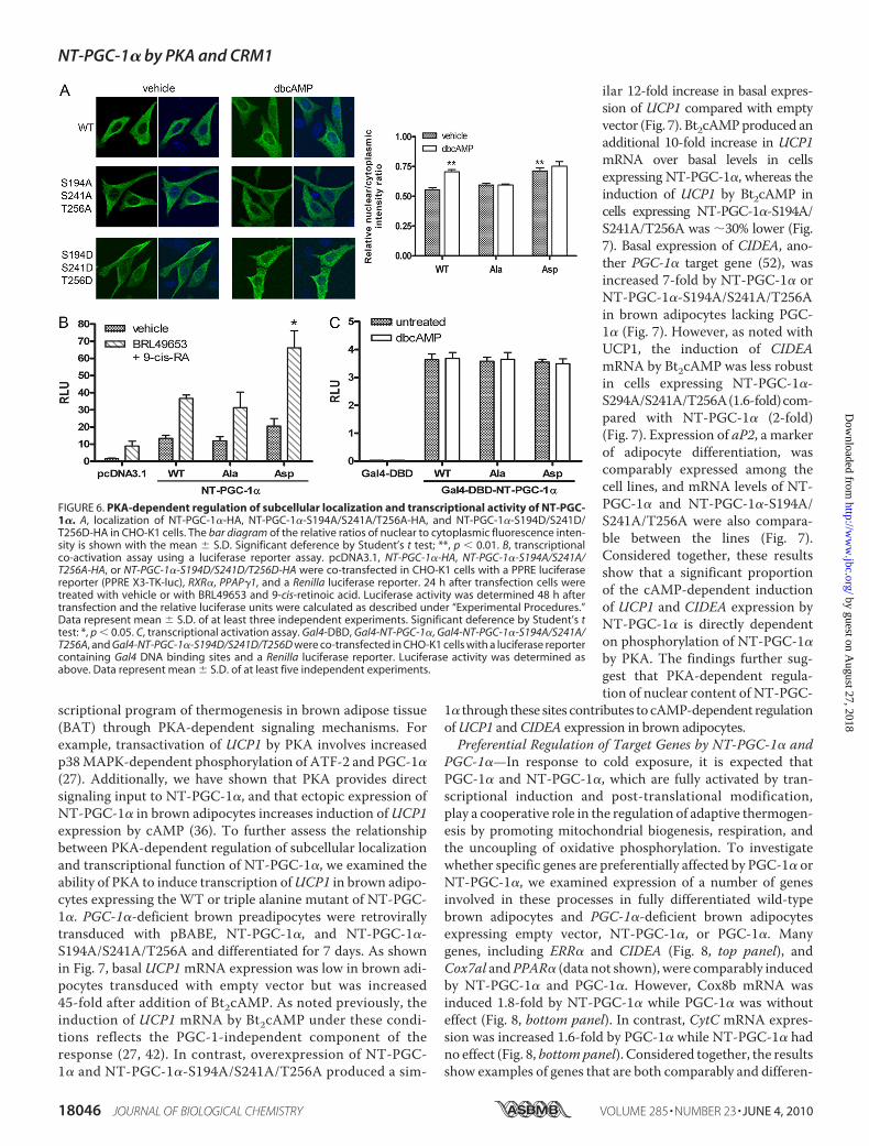
cellular localization was analyzed in the absence and presenceof Bt2cAMP. In vehicle-treated cells, NT-PGC-1� andNT-PGC-1�-S194A/S241A/T256A predominantly reside inthe cytoplasm with nuclear/cytoplasmic intensity ratios of 0.54and 0.59, respectively (Fig. 6A). NT-PGC-1�-S194A/S241A/T256A remained primarily cytoplasmic even in the presence ofBt2cAMP (Fig. 6A). In contrast, the nuclear content ofNT-PGC-1�-S194D/S241D/T256D in vehicle-treated cells wassignificantly higher (nuclear/cytoplasmic intensity ratio of0.72) than vehicle-treated WT and NT-PGC-1�-S194A/S241A/T256A and was comparable to the nuclear content ofthe WT construct in Bt2cAMP-treated cells (Fig. 6A). More-over, Bt2cAMP had no additional effect on nuclear content ofNT-PGC-1�-S194D/S241D/T256D (Fig. 6A). Together, theseresults are uniformly consistent with the idea that phosphory-lation ofNT-PGC-1�onSer-194, Ser-241, andThr-256 byPKAregulates the relative nuclear content of NT-PGC-1� by regu-lating CRM1-dependent nuclear export of the protein.We have shown that recruitment of NT-PGC-1� to multi-
protein complexes involving nuclear receptors (e.g. PPAR� andPPAR�) and co-activators (CBP and SRC1) enhances transac-tivation of some PGC-1�-target genes (36). To examinewhether the co-activator function of NT-PGC-1� is regulated
by subcellular localization, the abil-ity of NT-PGC-1�, NT-PGC-1�-S194A/S241A/T256A, and NT-PGC-1�-S194D/S241D/T256D toenhance the activation of PPAR�and RXR� was examined by tran-sient co-transfection assays in CHO-K1 cells. A (PPRE)3-TK-luc reporterconstruct containing a PPAR-re-sponse element was used (8). Usingtranscriptional activity of (PPRE)3-TK-luc observed with co-expres-sion of PPAR� and RXR� as basal,addition of ligands for PPAR� andRXR� (BRL49653 and 9-cis-retinoicacid) increased activity of the reporterby 6-fold (Fig. 6B). Co-expression ofWT NT-PGC-1� with PPAR� andRXR� increased reporter activity by9-fold, and addition of the ligandmixture produced a further 2.7-foldtransactivation of the reporter (Fig.6B). The alanine substitution mutantof NT-PGC-1� (S194A/S241A/T256A) increased PPAR�/RXR�-mediated transcription of the re-porter to the same extent as WTNT-PGC-1� in both vehicle (8-fold)and ligand-treated cells (2.6-fold) (Fig.6B). In contrast, transactivationof thereporter by the phosphomimeticmutant of NT-PGC-1�-S194D/S241D/T256D was higher enhanced�2-fold in both vehicle (14-fold) andligand-treated cells (3.2-fold) (Fig.
6B), suggesting a close correlation between nuclear localizationand co-activation function of NT-PGC-1�. Addition of Bt2cAMPproduced an additional 3-fold increase in activity of the reporterthat was independent of NT-PGC-1�, and likely reflected directpost-translational activation of the nuclear receptor complex byPKA. This characteristic makes it difficult to resolve the directeffects of PKA on the PPAR�/RXR� complex from the effects ofPKA on localization of NT-PGC-1� in this model.
Toaddress thequestionofwhether the transactivationpropertyof NT-PGC-1� is also elevated by phosphorylation, we tested theability of Gal4-NT-PGC-1�, Gal4-NT-PGC-1�-S194A/S241A/T256A, andGal4-NT-PGC-1�-S194D/S241D/T256D to transac-tivate a reporter gene containing Gal4 DNA binding sites. Asexpected, all the fusionproteinswithGal4-DBDwere localized inthe nucleus (supplemental Fig. S2). Expression of Gal4-NT-PGC-1�, Gal4-NT-PGC-1�-S194A/S241A/T256A, and Gal4-NT-PGC-1�-S194D/S241D/T256D increased reporter geneexpression to the same extent (�120-fold), and no additionalactivation of the reporterwas observedwithBt2cAMP (Fig. 6C).Transcriptional Activation of UCP1 and CIDEA by NT-
PGC-1� Is Mediated in Part by PKA Phosphorylation ofNT-PGC-1�—It is well established that increased sympatheticnervous system activity after cold exposure engages the tran-
FIGURE 5. PKA-dependent phosphorylation of NT-PGC-1�. A, schematic diagram shows seven putative PKAconsensus phosphorylation sites ([R,K]XX[S,T]) in NT-PGC-1�. B, in vitro PKA-mediated phosphorylation of NT-PGC-1�. NT-PGC-1�-HA and its serine/threonine to alanine mutants were in vitro transcribed/translated usinga TNT T7 Coupled Reticulocyte Lysate System, immunoprecipitated, and subjected for the in vitro kinase assaywith the purified bovine catalytic subunit of PKA. Autoradiography (top) shows phosphorylated NT-PGC-1� byPKA. Western blot (bottom) shows expression of NT-PGC-1�-HA and its serine/threonine to alanine mutants.Numbers in the box represent the relative phosphorylation that is normalized by NT-PGC-1� protein. C, in situPKA-dependent phosphorylation of NT-PGC-1�. Autoradiography (top) shows phosphorylated NT-PGC-1� in32P-labeled CHO-K1 cells ectopically expressing NT-PGC-1�-HA (untreated and H89/Bt2cAMP-treated) andNT-PGC-1�-S194A/S241A/T256A-HA (untreated and Bt2cAMP-treated). Western blot (bottom) shows expres-sion of NT-PGC-1�-HA and NT-PGC-1�-S194A/S241A/T256A-HA. Numbers in the box represent the relativephosphorylation that is normalized by NT-PGC-1� protein.
NT-PGC-1� by PKA and CRM1
JUNE 4, 2010 • VOLUME 285 • NUMBER 23 JOURNAL OF BIOLOGICAL CHEMISTRY 18045
by guest on August 27, 2018
http://ww
w.jbc.org/
Dow
nloaded from
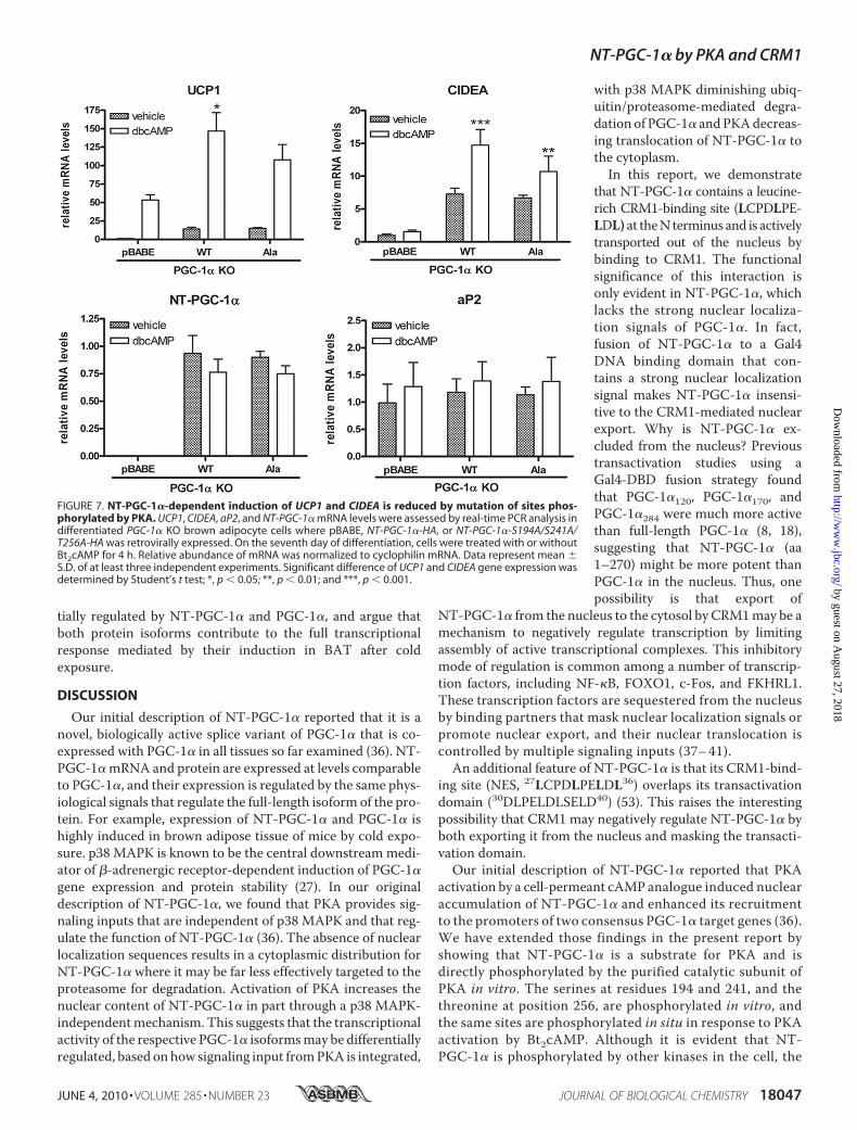
scriptional program of thermogenesis in brown adipose tissue(BAT) through PKA-dependent signaling mechanisms. Forexample, transactivation of UCP1 by PKA involves increasedp38MAPK-dependent phosphorylation of ATF-2 and PGC-1�(27). Additionally, we have shown that PKA provides directsignaling input to NT-PGC-1�, and that ectopic expression ofNT-PGC-1� in brown adipocytes increases induction ofUCP1expression by cAMP (36). To further assess the relationshipbetween PKA-dependent regulation of subcellular localizationand transcriptional function of NT-PGC-1�, we examined theability of PKA to induce transcription ofUCP1 in brown adipo-cytes expressing the WT or triple alanine mutant of NT-PGC-1�. PGC-1�-deficient brown preadipocytes were retrovirallytransduced with pBABE, NT-PGC-1�, and NT-PGC-1�-S194A/S241A/T256A and differentiated for 7 days. As shownin Fig. 7, basal UCP1mRNA expression was low in brown adi-pocytes transduced with empty vector but was increased45-fold after addition of Bt2cAMP. As noted previously, theinduction of UCP1 mRNA by Bt2cAMP under these condi-tions reflects the PGC-1-independent component of theresponse (27, 42). In contrast, overexpression of NT-PGC-1� and NT-PGC-1�-S194A/S241A/T256A produced a sim-
ilar 12-fold increase in basal expres-sion of UCP1 compared with emptyvector (Fig. 7). Bt2cAMPproduced anadditional 10-fold increase in UCP1mRNA over basal levels in cellsexpressing NT-PGC-1�, whereas theinduction of UCP1 by Bt2cAMP incells expressing NT-PGC-1�-S194A/S241A/T256A was �30% lower (Fig.7). Basal expression of CIDEA, ano-ther PGC-1� target gene (52), wasincreased 7-fold by NT-PGC-1� orNT-PGC-1�-S194A/S241A/T256Ain brown adipocytes lacking PGC-1� (Fig. 7). However, as noted withUCP1, the induction of CIDEAmRNA by Bt2cAMP was less robustin cells expressing NT-PGC-1�-S294A/S241A/T256A(1.6-fold)com-pared with NT-PGC-1� (2-fold)(Fig. 7). Expression of aP2, a markerof adipocyte differentiation, wascomparably expressed among thecell lines, and mRNA levels of NT-PGC-1� and NT-PGC-1�-S194A/S241A/T256A were also compara-ble between the lines (Fig. 7).Considered together, these resultsshow that a significant proportionof the cAMP-dependent inductionof UCP1 and CIDEA expression byNT-PGC-1� is directly dependenton phosphorylation of NT-PGC-1�by PKA. The findings further sug-gest that PKA-dependent regula-tion of nuclear content of NT-PGC-
1� through these sites contributes to cAMP-dependent regulationofUCP1 andCIDEA expression in brown adipocytes.Preferential Regulation of Target Genes by NT-PGC-1� and
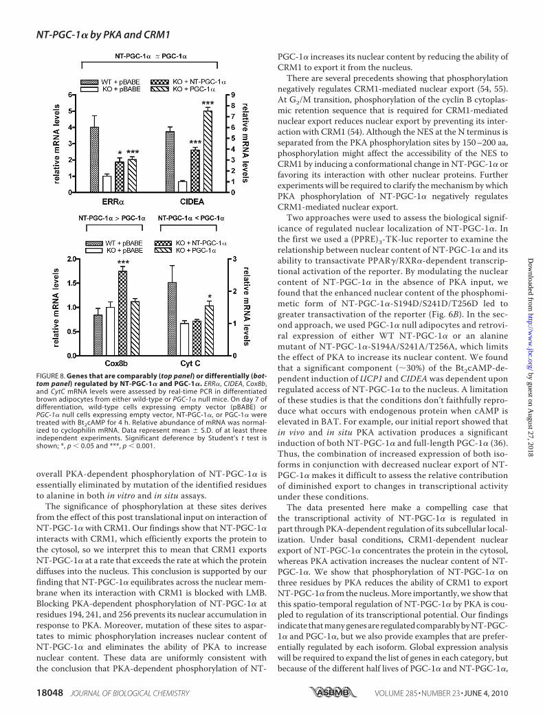
PGC-1�—In response to cold exposure, it is expected thatPGC-1� and NT-PGC-1�, which are fully activated by tran-scriptional induction and post-translational modification,play a cooperative role in the regulation of adaptive thermogen-esis by promoting mitochondrial biogenesis, respiration, andthe uncoupling of oxidative phosphorylation. To investigatewhether specific genes are preferentially affected by PGC-1� orNT-PGC-1�, we examined expression of a number of genesinvolved in these processes in fully differentiated wild-typebrown adipocytes and PGC-1�-deficient brown adipocytesexpressing empty vector, NT-PGC-1�, or PGC-1�. Manygenes, including ERR� and CIDEA (Fig. 8, top panel), andCox7al andPPAR� (data not shown), were comparably inducedby NT-PGC-1� and PGC-1�. However, Cox8b mRNA wasinduced 1.8-fold by NT-PGC-1� while PGC-1� was withouteffect (Fig. 8, bottom panel). In contrast, CytC mRNA expres-sion was increased 1.6-fold by PGC-1� while NT-PGC-1� hadno effect (Fig. 8, bottompanel). Considered together, the resultsshow examples of genes that are both comparably and differen-
FIGURE 6. PKA-dependent regulation of subcellular localization and transcriptional activity of NT-PGC-1�. A, localization of NT-PGC-1�-HA, NT-PGC-1�-S194A/S241A/T256A-HA, and NT-PGC-1�-S194D/S241D/T256D-HA in CHO-K1 cells. The bar diagram of the relative ratios of nuclear to cytoplasmic fluorescence inten-sity is shown with the mean � S.D. Significant deference by Student’s t test; **, p � 0.01. B, transcriptionalco-activation assay using a luciferase reporter assay. pcDNA3.1, NT-PGC-1�-HA, NT-PGC-1�-S194A/S241A/T256A-HA, or NT-PGC-1�-S194D/S241D/T256D-HA were co-transfected in CHO-K1 cells with a PPRE luciferasereporter (PPRE X3-TK-luc), RXR�, PPAP�1, and a Renilla luciferase reporter. 24 h after transfection cells weretreated with vehicle or with BRL49653 and 9-cis-retinoic acid. Luciferase activity was determined 48 h aftertransfection and the relative luciferase units were calculated as described under “Experimental Procedures.”Data represent mean � S.D. of at least three independent experiments. Significant deference by Student’s ttest: *, p � 0.05. C, transcriptional activation assay. Gal4-DBD, Gal4-NT-PGC-1�, Gal4-NT-PGC-1�-S194A/S241A/T256A, and Gal4-NT-PGC-1�-S194D/S241D/T256D were co-transfected in CHO-K1 cells with a luciferase reportercontaining Gal4 DNA binding sites and a Renilla luciferase reporter. Luciferase activity was determined asabove. Data represent mean � S.D. of at least five independent experiments.
NT-PGC-1� by PKA and CRM1
18046 JOURNAL OF BIOLOGICAL CHEMISTRY VOLUME 285 • NUMBER 23 • JUNE 4, 2010
by guest on August 27, 2018
http://ww
w.jbc.org/
Dow
nloaded from
tially regulated by NT-PGC-1� and PGC-1�, and argue thatboth protein isoforms contribute to the full transcriptionalresponse mediated by their induction in BAT after coldexposure.
DISCUSSION
Our initial description of NT-PGC-1� reported that it is anovel, biologically active splice variant of PGC-1� that is co-expressed with PGC-1� in all tissues so far examined (36). NT-PGC-1�mRNA and protein are expressed at levels comparableto PGC-1�, and their expression is regulated by the same phys-iological signals that regulate the full-length isoform of the pro-tein. For example, expression of NT-PGC-1� and PGC-1� ishighly induced in brown adipose tissue of mice by cold expo-sure. p38 MAPK is known to be the central downstreammedi-ator of �-adrenergic receptor-dependent induction of PGC-1�gene expression and protein stability (27). In our originaldescription of NT-PGC-1�, we found that PKA provides sig-naling inputs that are independent of p38 MAPK and that reg-ulate the function of NT-PGC-1� (36). The absence of nuclearlocalization sequences results in a cytoplasmic distribution forNT-PGC-1� where it may be far less effectively targeted to theproteasome for degradation. Activation of PKA increases thenuclear content of NT-PGC-1� in part through a p38 MAPK-independentmechanism. This suggests that the transcriptionalactivity of the respective PGC-1� isoformsmay be differentiallyregulated, based onhow signaling input fromPKA is integrated,
with p38 MAPK diminishing ubiq-uitin/proteasome-mediated degra-dation of PGC-1� andPKAdecreas-ing translocation of NT-PGC-1� tothe cytoplasm.In this report, we demonstrate
that NT-PGC-1� contains a leucine-rich CRM1-binding site (LCPDLPE-LDL) at theNterminus and is activelytransported out of the nucleus bybinding to CRM1. The functionalsignificance of this interaction isonly evident in NT-PGC-1�, whichlacks the strong nuclear localiza-tion signals of PGC-1�. In fact,fusion of NT-PGC-1� to a Gal4DNA binding domain that con-tains a strong nuclear localizationsignal makes NT-PGC-1� insensi-tive to the CRM1-mediated nuclearexport. Why is NT-PGC-1� ex-cluded from the nucleus? Previoustransactivation studies using aGal4-DBD fusion strategy foundthat PGC-1�120, PGC-1�170, andPGC-1�284 were much more activethan full-length PGC-1� (8, 18),suggesting that NT-PGC-1� (aa1–270) might be more potent thanPGC-1� in the nucleus. Thus, onepossibility is that export of
NT-PGC-1� from the nucleus to the cytosol by CRM1may be amechanism to negatively regulate transcription by limitingassembly of active transcriptional complexes. This inhibitorymode of regulation is common among a number of transcrip-tion factors, including NF-�B, FOXO1, c-Fos, and FKHRL1.These transcription factors are sequestered from the nucleusby binding partners that mask nuclear localization signals orpromote nuclear export, and their nuclear translocation iscontrolled by multiple signaling inputs (37–41).An additional feature of NT-PGC-1� is that its CRM1-bind-
ing site (NES, 27LCPDLPELDL36) overlaps its transactivationdomain (30DLPELDLSELD40) (53). This raises the interestingpossibility that CRM1 may negatively regulate NT-PGC-1� byboth exporting it from the nucleus and masking the transacti-vation domain.Our initial description of NT-PGC-1� reported that PKA
activation by a cell-permeant cAMP analogue induced nuclearaccumulation of NT-PGC-1� and enhanced its recruitmentto the promoters of two consensus PGC-1� target genes (36).We have extended those findings in the present report byshowing that NT-PGC-1� is a substrate for PKA and isdirectly phosphorylated by the purified catalytic subunit ofPKA in vitro. The serines at residues 194 and 241, and thethreonine at position 256, are phosphorylated in vitro, andthe same sites are phosphorylated in situ in response to PKAactivation by Bt2cAMP. Although it is evident that NT-PGC-1� is phosphorylated by other kinases in the cell, the
FIGURE 7. NT-PGC-1�-dependent induction of UCP1 and CIDEA is reduced by mutation of sites phos-phorylated by PKA. UCP1, CIDEA, aP2, and NT-PGC-1� mRNA levels were assessed by real-time PCR analysis indifferentiated PGC-1� KO brown adipocyte cells where pBABE, NT-PGC-1�-HA, or NT-PGC-1�-S194A/S241A/T256A-HA was retrovirally expressed. On the seventh day of differentiation, cells were treated with or withoutBt2cAMP for 4 h. Relative abundance of mRNA was normalized to cyclophilin mRNA. Data represent mean �S.D. of at least three independent experiments. Significant difference of UCP1 and CIDEA gene expression wasdetermined by Student’s t test; *, p � 0.05; **, p � 0.01; and ***, p � 0.001.
NT-PGC-1� by PKA and CRM1
JUNE 4, 2010 • VOLUME 285 • NUMBER 23 JOURNAL OF BIOLOGICAL CHEMISTRY 18047
by guest on August 27, 2018
http://ww
w.jbc.org/
Dow
nloaded from
overall PKA-dependent phosphorylation of NT-PGC-1� isessentially eliminated by mutation of the identified residuesto alanine in both in vitro and in situ assays.The significance of phosphorylation at these sites derives
from the effect of this post translational input on interaction ofNT-PGC-1� with CRM1. Our findings show that NT-PGC-1�interacts with CRM1, which efficiently exports the protein tothe cytosol, so we interpret this to mean that CRM1 exportsNT-PGC-1� at a rate that exceeds the rate at which the proteindiffuses into the nucleus. This conclusion is supported by ourfinding that NT-PGC-1� equilibrates across the nuclear mem-brane when its interaction with CRM1 is blocked with LMB.Blocking PKA-dependent phosphorylation of NT-PGC-1� atresidues 194, 241, and 256 prevents its nuclear accumulation inresponse to PKA. Moreover, mutation of these sites to aspar-tates to mimic phosphorylation increases nuclear content ofNT-PGC-1� and eliminates the ability of PKA to increasenuclear content. These data are uniformly consistent withthe conclusion that PKA-dependent phosphorylation of NT-
PGC-1� increases its nuclear content by reducing the ability ofCRM1 to export it from the nucleus.There are several precedents showing that phosphorylation
negatively regulates CRM1-mediated nuclear export (54, 55).At G2/M transition, phosphorylation of the cyclin B cytoplas-mic retention sequence that is required for CRM1-mediatednuclear export reduces nuclear export by preventing its inter-action with CRM1 (54). Although the NES at the N terminus isseparated from the PKA phosphorylation sites by 150–200 aa,phosphorylation might affect the accessibility of the NES toCRM1 by inducing a conformational change in NT-PGC-1� orfavoring its interaction with other nuclear proteins. Furtherexperiments will be required to clarify themechanism bywhichPKA phosphorylation of NT-PGC-1� negatively regulatesCRM1-mediated nuclear export.Two approaches were used to assess the biological signif-
icance of regulated nuclear localization of NT-PGC-1�. Inthe first we used a (PPRE)3-TK-luc reporter to examine therelationship between nuclear content of NT-PGC-1� and itsability to transactivate PPAR�/RXR�-dependent transcrip-tional activation of the reporter. By modulating the nuclearcontent of NT-PGC-1� in the absence of PKA input, wefound that the enhanced nuclear content of the phosphomi-metic form of NT-PGC-1�-S194D/S241D/T256D led togreater transactivation of the reporter (Fig. 6B). In the sec-ond approach, we used PGC-1� null adipocytes and retrovi-ral expression of either WT NT-PGC-1� or an alaninemutant of NT-PGC-1�-S194A/S241A/T256A, which limitsthe effect of PKA to increase its nuclear content. We foundthat a significant component (�30%) of the Bt2cAMP-de-pendent induction ofUCP1 and CIDEAwas dependent uponregulated access of NT-PGC-1� to the nucleus. A limitationof these studies is that the conditions don’t faithfully repro-duce what occurs with endogenous protein when cAMP iselevated in BAT. For example, our initial report showed thatin vivo and in situ PKA activation produces a significantinduction of both NT-PGC-1� and full-length PGC-1� (36).Thus, the combination of increased expression of both iso-forms in conjunction with decreased nuclear export of NT-PGC-1� makes it difficult to assess the relative contributionof diminished export to changes in transcriptional activityunder these conditions.The data presented here make a compelling case that
the transcriptional activity of NT-PGC-1� is regulated inpart throughPKA-dependent regulation of its subcellular local-ization. Under basal conditions, CRM1-dependent nuclearexport of NT-PGC-1� concentrates the protein in the cytosol,whereas PKA activation increases the nuclear content of NT-PGC-1�. We show that phosphorylation of NT-PGC-1� onthree residues by PKA reduces the ability of CRM1 to exportNT-PGC-1� from the nucleus.More importantly, we show thatthis spatio-temporal regulation of NT-PGC-1� by PKA is cou-pled to regulation of its transcriptional potential. Our findingsindicate thatmanygenesareregulatedcomparablybyNT-PGC-1� and PGC-1�, but we also provide examples that are prefer-entially regulated by each isoform. Global expression analysiswill be required to expand the list of genes in each category, butbecause of the different half lives of PGC-1� and NT-PGC-1�,
FIGURE 8. Genes that are comparably (top panel) or differentially (bot-tom panel) regulated by NT-PGC-1� and PGC-1�. ERR�, CIDEA, Cox8b,and CytC mRNA levels were assessed by real-time PCR in differentiatedbrown adipocytes from either wild-type or PGC-1� null mice. On day 7 ofdifferentiation, wild-type cells expressing empty vector (pBABE) orPGC-1� null cells expressing empty vector, NT-PGC-1�, or PGC-1� weretreated with Bt2cAMP for 4 h. Relative abundance of mRNA was normal-ized to cyclophilin mRNA. Data represent mean � S.D. of at least threeindependent experiments. Significant deference by Student’s t test isshown; *, p � 0.05 and ***, p � 0.001.
NT-PGC-1� by PKA and CRM1
18048 JOURNAL OF BIOLOGICAL CHEMISTRY VOLUME 285 • NUMBER 23 • JUNE 4, 2010
by guest on August 27, 2018
http://ww
w.jbc.org/
Dow
nloaded from
it seems likely that differences in target gene regulation mayextend to duration of transcriptional activation by the respec-tive protein isoforms. It is particularly interesting that the co-activation function of a novel isoform of PGC-1� is regulateddifferently from full-length PGC-1�. Two different modes ofregulation might be a way to coordinate the timing and fulltranscriptional potential of both PGC-1� isoforms in responseto physiological signals. For example, in BAT after cold expo-sure, cAMP increases the transcriptional induction of bothNT-PGC-1� and PGC-1�, andmediates the post-translational acti-vation of both proteins. Given that approximately half of thecold-induced PGC-1� total mRNA encodes for NT-PGC-1�,we predict that the post-translational input by PKA, which reg-ulates subcellular localization of NT-PGC-1�, is important notonly for achieving full PGC-1� activity in animals exposed tocold, but also for induction of the complete set of genes that aretranscriptionally regulated by PGC-1� isoforms.
Acknowledgments—We thank Dr. Bruce Spiegelman for providingcell lines from PGC-1� null mice. pcDNA3.1-myc-CRM1 andpQE32-HIS6-RanQ69L were kindly provided by S. Meloche (Uni-versite de Montreal). We thank Anik Boudreau, Jeho Shin, AaronAdamson, and David Burk for their technical contributions andadvice. We thank Anne Gooch for excellent administrativesupport.
REFERENCES1. Finck, B. N., and Kelly, D. P. (2006) J. Clin. Invest. 116, 615–6222. Handschin, C., and Spiegelman, B. M. (2006) Endocr. Rev. 27, 728–7353. Puigserver, P., Wu, Z., Park, C. W., Graves, R., Wright, M., and
Spiegelman, B. M. (1998) Cell 92, 829–8394. Wu, Z., Puigserver, P., Andersson, U., Zhang, C., Adelmant, G., Mootha,
V., Troy, A., Cinti, S., Lowell, B., Scarpulla, R. C., and Spiegelman, B. M.(1999) Cell 98, 115–124
5. Lehman, J. J., Barger, P. M., Kovacs, A., Saffitz, J. E., Medeiros, D. M., andKelly, D. P. (2000) J. Clin. Invest. 106, 847–856
6. Mootha, V. K., Lindgren, C.M., Eriksson, K. F., Subramanian, A., Sihag, S.,Lehar, J., Puigserver, P., Carlsson, E., Ridderstråle, M., Laurila, E., Houstis,N., Daly, M. J., Patterson, N., Mesirov, J. P., Golub, T. R., Tamayo, P.,Spiegelman, B., Lander, E. S., Hirschhorn, J. N., Altshuler, D., and Groop,L. C. (2003) Nat. Genet. 34, 267–273
7. Schreiber, S. N., Emter, R., Hock, M. B., Knutti, D., Cardenas, J., Podvinec,M., Oakeley, E. J., and Kralli, A. (2004) Proc. Natl. Acad. Sci. U.S.A. 101,6472–6477
8. Vega, R. B., Huss, J. M., and Kelly, D. P. (2000) Mol. Cell Biol. 20,1868–1876
9. Wang, Y. X., Lee, C. H., Tiep, S., Yu, R. T., Ham, J., Kang, H., and Evans,R. M. (2003) Cell 113, 159–170
10. Herzig, S., Long, F., Jhala, U. S., Hedrick, S., Quinn, R., Bauer, A., Rudolph,D., Schutz, G., Yoon, C., Puigserver, P., Spiegelman, B., andMontminy,M.(2001) Nature 413, 179–183
11. Yoon, J. C., Puigserver, P., Chen, G., Donovan, J., Wu, Z., Rhee, J., Adelm-ant, G., Stafford, J., Kahn, C. R., Granner, D. K., Newgard, C. B., andSpiegelman, B. M. (2001) Nature 413, 131–138
12. Kressler, D., Schreiber, S. N., Knutti, D., and Kralli, A. (2002) J. Biol. Chem.277, 13918–13925
13. Puigserver, P., and Spiegelman, B. M. (2003) Endo. Rev. 24, 78–9014. Rhee, J., Inoue, Y., Yoon, J. C., Puigserver, P., Fan, M., Gonzalez, F. J., and
Spiegelman, B. M. (2003) Proc. Natl. Acad. Sci. U.S.A. 100, 4012–401715. Lin, J., Wu, H., Tarr, P. T., Zhang, C. Y., Wu, Z., Boss, O., Michael, L. F.,
Puigserver, P., Isotani, E., Olson, E. N., Lowell, B. B., Bassel-Duby, R., andSpiegelman, B. M. (2002) Nature 418, 797–801
16. St-Pierre, J., Drori, S., Uldry, M., Silvaggi, J. M., Rhee, J., Jager, S., Hand-
schin, C., Zheng, K., Lin, J., Yang, W., Simon, D. K., Bachoo, R., andSpiegelman, B. M. (2006) Cell 127, 397–408
17. Liu, C., Li, S., Liu, T., Borjigin, J., and Lin, J. D. (2007) Nature 447,477–481
18. Puigserver, P., Adelmant, C., Wu, Z., Fan, M., Xu, J., O’Malley, B., andSpiegelman, B. M. (1999) Science 286, 1368–1371
19. Delerive, P., Wu, Y., Burris, T. P., Chin, W. W., and Suen, C. S. (2002)J. Biol. Chem. 277, 3913–3917
20. Michael, L. F., Wu, Z., Cheatham, R. B., Puigserver, P., Adelmant, G.,Lehman, J. J., Kelly, D. P., and Spiegelman, B. M. (2001) Proc. Natl. Acad.Sci. U.S.A. 98, 3820–3825
21. Lin, J., Puigserver, P., Donovan, J., Tarr, P., and Spiegelman, B. M. (2002)J. Biol. Chem. 277, 1645–1648
22. Puigserver, P., Rhee, J., Donovan, J., Walkey, C. J., Yoon, J. C., Oriente, F.,Kitamura, Y., Altomonte, J., Dong, H., Accili, D., and Spiegelman, B. M.(2003) Nature 423, 550–555
23. Fan, M., Rhee, J., St-Pierre, J., Handschin, C., Puigserver, P., Lin, J., Jaeger,S., Erdjument-Bromage, H., Tempst, P., and Spiegelman, B. M. (2004)Genes Dev. 18, 278–289
24. Monsalve, M., Wu, Z., Adelmant, G., Puigserver, P., Fan, M., andSpiegelman, B. M. (2000)Mol. Cell 6, 307–316
25. Handschin, C., Rhee, J., Lin, J., Tarr, P. T., and Spiegelman, B. M. (2003)Proc. Natl. Acad. Sci. U.S.A. 100, 7111–7116
26. Nisoli, E., Clementi, E., Paolucci, C., Cozzi, V., Tonello, C., Sciorati, C.,Bracale, R., Valerio, A., Francolini, M., Moncada, S., and Carruba, M. O.(2003) Science 299, 896–899
27. Cao, W., Daniel, K. W., Robidoux, J., Puigserver, P., Medvedev, A. V., Bai,X., Floering, L.M., Spiegelman, B.M., and Collins, S. (2004)Mol. Cell Biol.24, 3057–3067
28. Puigserver, P., Rhee, J., Lin, J., Wu, Z., Yoon, J. C., Zhang, C. Y., Krauss, S.,Mootha, V. K., Lowell, B. B., and Spiegelman, B. M. (2001) Mol. Cell 8,971–982
29. Sano, M., Tokudome, S., Shimizu, N., Yoshikawa, N., Ogawa, C.,Shirakawa, K., Endo, J., Katayama, T., Yuasa, S., Ieda, M., Makino, S.,Hattori, F., Tanaka, H., and Fukuda, K. (2007) J. Biol. Chem. 282,25970–25980
30. Olson, B. L., Hock, M. B., Ekholm-Reed, S., Wohlschlegel, J. A., Dev, K. K.,Kralli, A., and Reed, S. I. (2008) Genes Dev. 22, 252–264
31. Knutti, D., Kressler, D., and Kralli, A. (2001) Proc. Natl. Acad. Sci. U.S.A.98, 9713–9718
32. Jager, S., Handschin, C., St-Pierre, J., and Spiegelman, B. M. (2007) Proc.Natl. Acad. Sci. U.S.A. 104, 12017–12022
33. Li, X., Monks, B., Ge, Q., and Birnbaum, M. J. (2007) Nature 447,1012–1016
34. Teyssier, C., Ma, H., Emter, R., Kralli, A., and Stallcup, M. R. (2005)GenesDev. 19, 1466–1473
35. Rodgers, J. T., Lerin, C., Haas, W., Gygi, S. P., Spiegelman, B. M., andPuigserver, P. (2005) Nature 434, 113–118
36. Zhang, Y., Huypens, P., Adamson, A. W., Chang, J. S., Henagan, T. M.,Boudreau, A., Lenard, N. R., Burk, D., Klein, J., Perwitz, N., Shin, J.,Fasshauer, M., Kralli, A., and Gettys, T. W. (2009) J. Biol. Chem. 284,32813–32826
37. Biggs,W.H., 3rd,Meisenhelder, J., Hunter, T., Cavenee,W.K., andArden,K. C. (1999) Proc. Natl. Acad. Sci. U.S.A. 96, 7421–7426
38. Brunet, A., Kanai, F., Stehn, J., Xu, J., Sarbassova, D., Frangioni, J. V., Dalal,S. N., DeCaprio, J. A., Greenberg,M. E., and Yaffe,M. B. (2002) J. Cell Biol.156, 817–828
39. Hayden, M. S., and Ghosh, S. (2004) Genes Dev. 18, 2195–222440. Sasaki, T., Kojima, H., Kishimoto, R., Ikeda, A., Kunimoto, H., and Naka-
jima, K. (2006)Mol. Cell 24, 63–7541. Roux, P., Blanchard, J. M., Fernandez, A., Lamb, N., Jeanteur, P., and Piec-
haczyk, M. (1990) Cell 63, 341–35142. Uldry, M., Yang, W., St-Pierre, J., Lin, J., Seale, P., and Spiegelman, B. M.
(2006) Cell Metab. 3, 333–34143. Bereziat, V., Moritz, S., Klonjkowski, B., Decaudain, A., Auclair, M., Eloit,
M., Capeau, J., and Vigouroux, C. (2005) Biochimie 87, 951–95844. Zhang, Y., and Xiong, Y. (2001) Science 292, 1910–191545. Qu, L., Huang, S., Baltzis, D., Rivas-Estilla, A. M., Pluquet, O., Hatzoglou,
NT-PGC-1� by PKA and CRM1
JUNE 4, 2010 • VOLUME 285 • NUMBER 23 JOURNAL OF BIOLOGICAL CHEMISTRY 18049
by guest on August 27, 2018
http://ww
w.jbc.org/
Dow
nloaded from
M., Koumenis, C., Taya, Y., Yoshimura, A., and Koromilas, A. E. (2004)Genes Dev. 18, 261–277
46. Villena, J. A., Hock, M. B., Chang, W. Y., Barcas, J. E., Giguere, V., andKralli, A. (2007) Proc. Natl. Acad. Sci. U.S.A. 104, 1418–1423
47. Pante, N., and Aebi, U. (1996) Crit. Rev. Biochem. Mol. Biol. 31,153–199
48. Cyert, M. S. (2001) J. Biol. Chem. 276, 20805–2080849. Kudo, N., Matsumori, N., Taoka, H., Fujiwara, D., Schreiner, E. P., Wolff,
B., Yoshida, M., and Horinouchi, S. (1999) Proc. Natl. Acad. Sci. U.S.A. 96,9112–9117
50. Sachdev, S., Bagchi, S., Zhang, D. D.,Mings, A. C., andHannink,M. (2000)
Mol. Cell Biol. 20, 1571–158251. Stade, K., Ford, C. S., Guthrie, C., and Weis, K. (1997) Cell 90,
1041–105052. Hallberg, M., Morganstein, D. L., Kiskinis, E., Shah, K., Kralli, A., Dil-
worth, S. M., White, R., Parker, M. G., and Christian, M. (2008) Mol.Cell Biol. 28, 6785–6795
53. Sadana, P., and Park, E. A. (2007) Biochem. J. 403, 511–51854. Yang, J., Bardes, E. S., Moore, J. D., Brennan, J., Powers, M. A., and Korn-
bluth, S. (1998) Genes Dev. 12, 2131–214355. Kilstrup-Nielsen, C., Alessio, M., and Zappavigna, V. (2003) EMBO J. 22,
89–99
NT-PGC-1� by PKA and CRM1
18050 JOURNAL OF BIOLOGICAL CHEMISTRY VOLUME 285 • NUMBER 23 • JUNE 4, 2010
by guest on August 27, 2018
http://ww
w.jbc.org/
Dow
nloaded from
Thomas W. GettysJi Suk Chang, Peter Huypens, Yubin Zhang, Chelsea Black, Anastasia Kralli and
Kinase A-dependent Modulation of Nuclear Export by CRM1 Subcellular Localization and Function by ProteinαRegulation of NT-PGC-1
doi: 10.1074/jbc.M109.083121 originally published online March 29, 20102010, 285:18039-18050.J. Biol. Chem.
10.1074/jbc.M109.083121Access the most updated version of this article at doi:
Alerts:
When a correction for this article is posted•
When this article is cited•
to choose from all of JBC's e-mail alertsClick here
Supplemental material:
http://www.jbc.org/content/suppl/2010/03/29/M109.083121.DC1
http://www.jbc.org/content/285/23/18039.full.html#ref-list-1
This article cites 55 references, 30 of which can be accessed free at
by guest on August 27, 2018
http://ww
w.jbc.org/
Dow
nloaded from