regulation synthesis gonadotrophin placenta
11
Role of oestradiol-17\g=b\ in the regulation of synthesis and secretion of human chorionic gonadotrophin by first trimester human placenta S C Sharma, P Purohit and A J Rao Department of Biochemistry and Center for Reproductive Biology and Molecular Endocrinology, Indian Institute of Science, Bangalore 560012, India (Requests for offprints should be addressed to A J Rao) ABSTRACT Inhibition of aromatase, a key enzyme in the biosynthesis of oestradiol-17\g=b\, by the addition of 1,4,6-androstatrien-3,17-dione resulted in a signifi- cant increase in the levels of immunoreactive human chorionic gonadotrophin (hCG) in the medium and tissue. This increase was partially reversed by the simultaneous addition of oestradiol-17\g=b\. These effects on the levels of immunoreactive hCG were also reflected by the increased levels of mRNA specific for the \g=a\ and \g=b\ subunits of hCG following the addition of the aromatase inhibitor. However, addition of tamoxifen resulted in a drastic decrease in the levels of both the messages. Based on these results, it is suggested that the synthesis of hCG is negatively modulated by oestradiol-17\g=b\ in the human placenta. Journal of Molecular Endocrinology (1993) 11, 91—101 INTRODUCTION Human chorionic gonadotrophin (hCG) is a glycoprotein hormone produced by the human placenta (Talamantes & Ogren, 1988). It shares structural and functional homology with pituitary luteinizing hormone (LH) (Birken, 1984). Both positive and negative regulation of LH, respectively by gonadotrophin-releasing hormone (GnRH) and the gonadal steroids progesterone and oestradiol- 17ß (OE2), are well documented in the literature (McCann, 1974; Conn et al. 1987). It is possible that, as the placenta is also known to produce GnRH and steroids like progesterone and OE2, hCG may also be subject to similar regulation by GnRH and steroids. Using minced first trimester human placenta we have shown in an earlier study (Mathialagan & Rao, 1986) that GnRH stimulates the synthesis and secretion of hCG, thus providing support for the suggestion that hCG is also subject to positive modulation by GnRH, as in the case of pituitary LH. However, while it has been reported by Belleville et al. (1978) that the addition of progesterone or OE2 had no effect on hCG secretion by first trimester human placenta, Maruo et al. (1986), using cDNA probes for the a and ß subunits of hCG and an in-situ hybridization technique, have shown that the addition of progesterone (5—20 pg/ml) to cultures of normal first trimester human placenta results in a decrease in the cellular levels of hCG a and ß mRNA. OE2 at lower concentrations had no effect on the immunoreactive levels of the hormone in the medium. One of the main problems associated with studies of the role of OE2 or progesterone in the regulation of the synthesis and secretion of hCG in the placenta is that placental tissue is already exposed to high concentrations of endogenous steroids and it is difficult to observe the effects of added steroids. In the present study we have examined the effect of OE2 on the synthesis and secretion of hCG under conditions where the influence of endogeneous OE2 is minimal. MATERIALS AND METHODS First trimester human placentae (6—8 weeks) were collected from cases of medical termination of pregnancy from the local hospital. Placentae were collected after obtaining written consent from the subjects. The tissue was collected in cold Earl's balanced salt solution (EBSS; pH 7-4) and immedi¬ ately brought to the laboratory on crushed ice and
Transcript of regulation synthesis gonadotrophin placenta

Role of oestradiol-17\g=b\in the regulation of synthesis andsecretion of human chorionic gonadotrophin by firsttrimester human placentaS C Sharma, P Purohit and A J RaoDepartment of Biochemistry and Center for Reproductive Biology and Molecular Endocrinology,
Indian Institute of Science, Bangalore 560012, India
(Requests for offprints should be addressed to A J Rao)
ABSTRACT
Inhibition of aromatase, a key enzyme in thebiosynthesis of oestradiol-17\g=b\,by the addition of1,4,6-androstatrien-3,17-dione resulted in a signifi-cant increase in the levels of immunoreactive humanchorionic gonadotrophin (hCG) in the medium andtissue. This increase was partially reversed by thesimultaneous addition of oestradiol-17\g=b\.Theseeffects on the levels of immunoreactive hCG werealso reflected by the increased levels of mRNA
specific for the \g=a\and \g=b\subunits of hCG followingthe addition of the aromatase inhibitor. However,addition of tamoxifen resulted in a drastic decreasein the levels of both the messages. Based on theseresults, it is suggested that the synthesis of hCGis negatively modulated by oestradiol-17\g=b\in thehuman placenta.Journal of Molecular Endocrinology (1993) 11, 91—101
INTRODUCTION
Human chorionic gonadotrophin (hCG) is a
glycoprotein hormone produced by the humanplacenta (Talamantes & Ogren, 1988). It sharesstructural and functional homology with pituitaryluteinizing hormone (LH) (Birken, 1984). Bothpositive and negative regulation of LH, respectivelyby gonadotrophin-releasing hormone (GnRH) andthe gonadal steroids progesterone and oestradiol-17ß (OE2), are well documented in the literature(McCann, 1974; Conn et al. 1987). It is possiblethat, as the placenta is also known to produceGnRH and steroids like progesterone and OE2,hCG may also be subject to similar regulation byGnRH and steroids. Using minced first trimesterhuman placenta we have shown in an earlier study(Mathialagan & Rao, 1986) that GnRH stimulatesthe synthesis and secretion of hCG, thus providingsupport for the suggestion that hCG is also subjectto positive modulation by GnRH, as in the case ofpituitary LH. However, while it has been reportedby Belleville et al. (1978) that the addition ofprogesterone or OE2 had no effect on hCG secretionby first trimester human placenta, Maruo et al.(1986), using cDNA probes for the a and ß subunitsof hCG and an in-situ hybridization technique,
have shown that the addition of progesterone(5—20 pg/ml) to cultures of normal first trimesterhuman placenta results in a decrease in the cellularlevels of hCG a and ß mRNA. OE2 at lowerconcentrations had no effect on the immunoreactivelevels of the hormone in the medium. One of themain problems associated with studies of the role ofOE2 or progesterone in the regulation of thesynthesis and secretion of hCG in the placenta isthat placental tissue is already exposed to highconcentrations of endogenous steroids and it isdifficult to observe the effects of added steroids. Inthe present study we have examined the effect ofOE2 on the synthesis and secretion of hCG underconditions where the influence of endogeneous OE2is minimal.
MATERIALS AND METHODS
First trimester human placentae (6—8 weeks) were
collected from cases of medical termination ofpregnancy from the local hospital. Placentae were
collected after obtaining written consent from thesubjects. The tissue was collected in cold Earl'sbalanced salt solution (EBSS; pH 7-4) and immedi¬ately brought to the laboratory on crushed ice and
processed at 4 °C. The tissue was washed exten¬sively with cold EBSS until the washing liquid was
clear, and villous tissue was separated by visualexamination; a wet weight of 2-5 g was recorded.The villous tissue was finely minced with surgicalscissors, washed twice with EBSS and suspendedin a known volume of buffer. About 80—lOOmgtissue (wet weight) were dispensed into 3 ml tubes.The total volume of incubation was 0-5 ml.
Tamoxifen (TMX) was a gift from ImperialChemical Industries Ltd, London, U.K. Rabbitreticulocyte lysate (N.90 lysate) was obtainedfrom Amersham International pic, Amersham,Bucks, U.K. [a-32P]dCTP (3000 Ci/mmol) and[35S]methionine (800 Ci/mmol) were obtained fromBhaba Atomic Research Center, Bombay, India.Unlabelled OE2 and l,4,6-androstatrien-3,17-dione(aromatase inhibitor; AI) were obtained fromSteroloids Inc., Wilton, NH, U.S.A. EBSS was
obtained from Hi-Media, Bombay, India.Nitrocellulose membrane filters were obtained
from Bio-Rad Laboratories, Richmond, CA, U.S.A.Whatman filter paper no. 3 was purchased fromWhatman Ltd, Maidstone, Kent, U.K. Trizmabase, formamide, oligo(dT) cellulose, potassiumacetate, dithiothreitol and sodium dodecyl sulphate(SDS) were purchased from Sigma Chemical Co.,St Louis, MO, U.S.A. DNA markers (1 kb ladder)were obtained from Bethesda Research Laborato¬ries, Life Technologies, Inc., Gaithersburg, MD,U.S.A. All other chemicals used were obtainedlocally and were of analytical grade. Clones for hCGa subunit cDNA and ß subunit cDNA cloned at theampicillin PstI site in the pBR322 vector were
obtained as a gift from Dr I. Boime (Department ofPharmacology, Washington University, St Louis,MO, U.S.A.). These were transformed in the HB101 strain of Escherichia coli, and the 0-7 kb insertfor a and 1 kb insert for ß hCG were isolated bydigestion with PstI. These probes were labelledwith [a- P]dCTP by nick translation. The specificactivity of the probes was around 5x10 c.p.m./pg.
Effect of addition of AI on the levels of OE2 inthe placental tissue and mediumMinced placental tissue (80-100 mg) was incubatedin the presence or absence of different concen¬
trations (5, 10, 15 and 30 pM) of AI under an at¬
mosphere of 95% oxygen and 5% carbon dioxide for4 h. Following this, the tissue was separated bycentrifugation (2000 ¿*) and washed three times withEBSS, homogenized and extracted with ether. Theether extract was used for the estimation of OE2and progesterone by specific radioimmunoassays(RIAs).
Estimation of hormoneshCG in the tissue homogenate and medium was
estimated by plastic tube RIA as describedpreviously (Murthy et al. 1989). Inter- andintra-assay coefficients of variation were 12-3 and86% respectively, and the minimum detectablequantity of hCG was 2 ng/ml. The assay was
specific for whole hCG, and assays were carried outin the presence of (0-01%) lima bean trypsininhibitor. OE2 and progesterone in the ether extractof the tissue homogenate and medium were
estimated by specific RIAs as described previously(Jagannadha Rao et al. 1984). The inter- andintra-assay coefficients of variation were 103 (n=l0)and 6-5% (w=8) respectively for progesterone and8-66 (n=9) and 5-2% (n = 8) respectively for OE2.
Total protein in the tissue homogenate was
estimated by the method of Lowry et al. (1951)using bovine serum albumin as a standard, and allvalues are expressed per mg protein.
Effect of AI, TMX or OE2 on immunoreactivehCG levels in first trimester human placentaMinced placental tissue in EBSS (1 g for theisolation of RNA or 80-100 mg for the estimation ofhCG) were incubated in triplicate in 3 ml tubes or
25 ml conical flasks with or without AI (15 pM),TMX (5 pM) or OE2 (10 nM-7-5 pM) for 4 h at 37 °Cunder 95% oxygen and 5% carbon dioxide. Mediumand tissue were separated by centrifugation at2000 gat 4 °C.
Effect of the addition of AI with or withoutTMX or OE2 on a and ß hCG mRNA levels infirst trimester human placentaMinced first trimester human placental tissue (1 gwet weight) was incubated in 10 ml EBSS in a 25 mlconical flask with or without 15pM AI (aconcentration of AI at which the maximum decreasein OE2 levels in the tissue was noticed after 4 h) for30 min at 37 °C under 95% oxygen and 5% carbondioxide. Following this, both groups of mincedtissue were washed twice with 3 vols EBSS and usedfor further studies. While control tissue was
incubated without any additions, Al-treated tissuewas resuspended in a medium containing 15 pM AIand incubated in the presence or absence of 10 nM
OE2 or 5 pM TMX for 4 h under the conditionsdescribed above. Following incubation, the tissueand medium were separated by centrifugation andthe tissue was processed for the isolation of totalRNA according to the procedure of Boime et al.(1976). Total RNA was quantitated by monitoringthe optical density at 260 nm. Poly(A) RNA was

isolated by chromatography on oligo(dT) celluloseas described by Aviv & Leader (1972).
Quantitation of mRNAmRNA quantitation was performed by dot blothybridization and also after resolving RNA byagarose gel electrophoresis.RNA dot blot hybridizationTotal cytoplasmic RNA (20 pg) from each group wasimmobilized on nitrocellulose filters and hybridizedseparately to nick-translated cDNA probes for a(0-7 kb) and ß (1 kb) hCG subunits according to theprocedure described by Thomas (1980). The nitro¬cellulose filters were washed with 2 x SSC (1 x SSCis 8765 g NaCl, 4-41 g sodium citrate in one litre ofdouble-distilled water, pH 70) four times at room
temperature for 20 min each, followed by 1 x SSCfour times at 65 °C for 20 min each and 0-2 x SSCthree times at 65 °C for 20 min each. Filters weredried between sheets of Whatman no. 3 filter paper,air-dried and exposed to X-ray film for 2—5 days.The hybridization spots were subjected to laser beamdensitometric scanning.
Electrophoretic analysis of RNAElectrophoresis of RNA (20 pg) on 1% agarose gelscontaining 2-2 M formaldehyde was carried outaccording to the procedure described by Maniatis etal. (1982). RNA was transferred to nitrocellulosefilters as described by Southern (1975). Followingtransfer, the nitrocellulose filters were baked in a
vacuum oven at 80 °C and hybridized withnick-translated probe. Hybridization and washingconditions were the same as those used in the dotblot procedure. mRNA levels were quantitated byscanning the autofluorograms with a laser beamscanner (LKB Model 2001) and also by monitoringthe radioactivity associated with the hybridizationspots.
In-vitro translation
Poly(A) RNA (1-2 pg) was translated using rabbitreticulocyte lysate. Equal quantities of trichloro-acetic acid-precipitable counts from individualsamples were immunoprecipitated by a specifichCG anti-serum raised against highly purified hCG(CR 123, 13 000IU/mg; a gift from NIAMDD,NIH, Bethesda, MD, U.S.A.) in the rabbit. Theantiserum was highly specific to hCG, with mini¬mal cross-reactivity with a and ß hCG subunits;it did not show any cross-reactivity with otherglycoprotein hormones and thus was found tobe suitable for immunoprecipitation studies. In
addition, minced placental tissue was incubatedin the presence or absence of modulators with
Sjmethionine. hCG was precipitated from thelabelled proteins using the specific antiserumdescribed above. The immunoprecipitate was
analysed by 10% SDS-polyacrylamide gel electro¬phoresis (PAGE) and the gels were processed forautofluorography and scanned in a LKB laser beamscanner.
GeneralThe results (means ± s.E.) of at least three separateobservations presented here are from a representa¬tive experiment. However, each experiment was
repeated at least three times with different batchesof placental tissue. The gestational ages of theplacentae were determined from the time of the lastmenstrual period, with the gestational age rangingfrom 6 to 10 weeks. Due to problems involved incollecting tissues of the same gestational age on a
single day, placentae collected from gestations ofbetween 6 and 10 weeks on the same day were
pooled. Although the absolute values variedbetween experiments due to variability in the healthof the subjects and their exact gestational ages, thepattern of response was comparable in eachexperiment. The results were analysed for statisticalsignificance using Student's /-test and all P valuesbelow 005 were considered to be significant.
RESULTS
Effect of the addition of AI on OE2 andprogesterone levels in the placental tissueA dose-dependent decrease in tissue OE2 levels was
seen following the addition of AI. Although themaximum decrease was seen with 30 pM AI (Table1), as the inhibition at 15 pM (71-5%) was notsignificantly different from that seen at 30 pM(76-8%) in all studies 15 pM was used. However, theaddition of AI had no effect on the levels ofprogesterone, thus establishing the specificity of theinhibitor at the concentration used.
Effect of the addition of AI, TMX or OE2 on
immunoreactive hCG levels in the tissue andmedium of minced first trimester humanplacentaeAnalysis of the tissue homogenate and incubationmedium for immunoreactive hCG indicated a
significant increase in the levels of hCG in the tissueand a marginal increase in the medium after theaddition of AI (Fig. 1). This increase was partiallyreversed when OE2 was added along with AI,

table 1. Effect of the addition of aromatase inhibitor onthe levels of oestradiol-17ß and progesterone in mincedplacenta: oestradiol and progesterone were estimated byspecific radioimmunoassays
Oestradiol Progesterone(ng/g tissue) (ng/g tissue)
Aromataseinhibitor (um)Control 2-8 ±0-31 125 ±2-5
5 10 ±012** 120 ±15-010 1-3 ±0-20** 128 ±3-015 0-8 ±007** 118 ±10-030 0-65 ±0-10** 113 ±17-0
Results are means ± S.E. of triplicate experiments.**P=001 compared with control (Student's r-test).
although the reversal was much more evident in themedium (Fig. la and b). However, OE2 alone was
not effective in suppressing the levels of hCG ineither tissue or medium.
In a separate experiment, the effect of theaddition of higher concentrations of OE2 on hCGlevels in the tissue and medium was examined (Fig.
#
(a) ** (b)20 ~ f^xl so° " tÉ
1 10 - T x^ll I 25°- HIT
ABCDE ABCDEfigure 1. Effect of the addition of aromatase inhibitor(AI) on immunoreactive levels of human chorionicgonadotrophin (hCG) in the tissue and medium of firsttrimester human placenta. Each value is the mean ± s.E.of three observations. Minced first trimester humanplacenta was incubated with no additions (A), 10 nM
oestradiol-17ß (OE2) (B), 1 pM AI (C), 10 pM AI (D) or
10 nM OE2 and 10 pM AI (E) for 4 h at 37 °C under 95%02 and 5% C02. hCG was quantitated both in mediumand tissue by plastic tube radioimmunoassay. Forexperimental details refer to the Materials and Methods;(a) hCG levels in tissue, (b) hCG levels in medium.*P<005, **P<0-001 compared with control; fP<002,ttP<001 compared with group treated with 10 pM AIalone.
A B C D BCDfigure 2. Effect of the addition of oestradiol-17ß (OE2)on immunoreactive levels of human chorionicgonadotrophin (hCG) in minced first trimester humanplacenta and medium. Each value is the mean ± s.E. ofthree observations. Minced first trimester humanplacenta was incubated with or without OE2 (A, 0 pM;B, 0-074 pM; C, 0-74 pM; D, 3-7 pM; E, 7-4 um) for 4 h at37 °C under 95% 02 and 5% C02. hCG was quantitatedboth in medium and tissue by plastic tuberadioimmunoassay. For experimental details refer to theMaterials and Methods; (a) hCG levels in tissue, (b)hCG levels in medium. **P<0001 compared withcontrol.
2). A significant decrease in tissue levels was seen
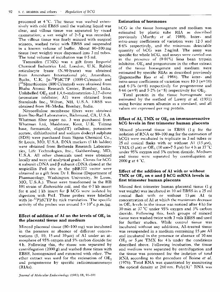
only at 3-7 and 7-4 pM OE2 (Fig. 2a). In contrast,TMX was found to be effective even at lowconcentrations (01 pM) in suppressing hCG levelsin tissue (Fig. 3a), while a decrease in the hCGlevels in the medium (Fig. 36) could be seen only athigh concentrations (1 and 5 pM).
Effect of the addition of AI, TMX, OE2,AI+OE2 or AI+TMX on the levels of ß and ahCG subunit specific mRNAsAddition of AI resulted in an increase in the levelsof ß hCG subunit mRNA by 110% over the controllevels (Fig. 4, panel I). In contrast, the additionof TMX, which was used as an antagonist to blockthe action of OE2, resulted in a decrease of 70%in the levels of hCG ß mRNA. While OE2 hadno discernible effect at concentrations of 10 and
(«) (b)
10 - 2- j
rirrh¡U / /o //O. //M # //
Is- j| - |kABCD ABCD
figure 3. Effect of the addition of tamoxifen (TMX)on immunoreactive levels of human chorionicgonadotrophin (hCG) in minced first trimester humanplacenta and medium. Each value is the mean ± s.E. ofthree observations. Minced first trimester humanplacenta was incubated with or without TMX (A, 0 pM;B, 0-1 pM; C, 1 pM; D, 5 pM) for 4 h at 37 °C under95% 02 and 5% C02. hCG was quantitated both inmedium and tissue by plastic tube radioimmunoassay.For experimental details refer to the Materials andMethods; (a) hCG levels in tissue, (b) hCG levelsin medium. *P<0002, **P<0001 compared withcontrol.
100 nM, it exerted a marginal (28%) inhibitory effectat a concentration of 500 nM. It may be pertinent topoint out that TMX is known to have both agonisticand antagonistic effects and was perhaps acting in thepresent case as an agonist exerting effects that weresimilar to the addition of high concentrations of OE2.
The results of the studies carried out on theeffect of the addition of TMX and OE2 along withAI are presented in Fig. 4, panel II. It can once
again be seen that the inhibition of oestrogensynthesis in minced first trimester human placentaby the addition of AI resulted in an increase in thelevels of ß hCG subunit mRNA levels by 78%over the control values. Addition of OE2 with AImaintained the levels of ß mRNA at control levels,while the addition of TMX with AI resulted in adrastic decrease in the levels of ß hCG subunitmRNA.
It can also be seen from the results presented inFig. 4, panel III, that, as was the case for ß hCGmRNA levels, the addition of AI resulted in anincrease of over 90% in the levels of the a messageand TMX caused a drastic decrease (70%).
Interestingly, the effect of AI on the levels of amRNA was also partially reversed by the addition ofOE2 along with AI (Fig. 4, panel IV) . However, theaddition of TMX along with AI completelyreversed the stimulatory effect of AI and decreasedthe levels to below basal values.
These results are further supported by thescanning of hybridization spots as well as by thequantitation of radioactivity in the hybridizationspots (data not shown). Thus, while there were
230 and 310% increases in the radioactivityrecovered in the spots following the addition ofAI for ß and a subunits respectively, these valuesreturned to control levels with the addition ofOE2. However, with TMX, the amount of radio¬activity recovered decreased drastically in the casesof both a and ß subunits. It can also be seen thatthe effect of TMX was much more pronouncedwhen compared with OE2, in that it drasticallydecreased ß hCG mRNA well below control levels.However, the addition of OE2 resulted in levels ofß hCG subunit mRNA which were near controlvalues.
Northern blot analysisThe effects of the addition of AI, TMX or OE2 on
the levels of ß hCG subunit mRNA, as judged byNorthern blot hybridization analysis, are presentedin Fig. 5a and b. It is evident that, based on theDNA size markers, the signal seen in the controllane is of 105 kb, which corresponds to the size ofmRNA for ß hCG reported in the literature(Ringler et al. 1989). It can also be seen thataddition of AI resulted in a very clear increase inthe levels of mRNA specific for ß hCG; thisincrease was nearly 88% greater than the control,as assessed by scanning (Fig. 5b). The addition ofOE2 alone had no discernible effect, while theaddition of TMX resulted in a drastic decrease(over 80%) in the level of ß hCG mRNA. Asexpected, the addition of OE2 along with AIprevented the increase induced by AI, whileaddition of TMX along with AI not onlyprevented the increase but also resulted in a
decrease in the level of ß hCG message. Theseconclusions are evident not only from the intensityof the signals but also from scanning the autofluo-rogram (Fig. 5b). Analysis of 28S and 18S RNAsbefore transfer to nitrocellulose revealed no differ¬ences (data not shown) in the quantities of RNAloaded from various groups. This establishes thatthe observed differences in the levels of mRNA are
not due to differences in the quantities of RNAloaded.
(b)250 (4) II
200 (3)
<
114- O
CO "-^
5Q.
150
100 h
<z^II« o
1--S °3SÏ
50
0
250
200
150
100
50 \-
(3)(3) (3)
1(2)
(2)
.Í112 3 4 5 6
III
(4)
I
(3) (3) (3) (2)
(2)
(3)(3)JL
(3)
12 3 4
IV
(2) (3)X
(3)
12 3 4 5 6 12 3 4

In-vitro translation, immunoprecipitation andSDS-PAGE analysisIn addition, RNA isolated in the above experimentwas translated in vitro, the product was immunopre-cipitated using hCG antiserum and the precipitatewas subjected to SDS-PAGE and autofluorography.It can also be seen from the autofluorograms (Fig.6a and b) that the increases seen in the levels of aand ß hCG mRNA following the addition of AIwere also reflected in the translation product,namely a and ß hCG (62% increase in the case of aand 50% increase in the case of ß over the control;Fig. 6, panel I). A decrease of nearly 50% wasobserved following the addition of TMX (Fig. 6,panel II), indicating a decrease in the levels of botha and ß hCG mRNA. Results of studies carried outusing [35S]methionine to monitor the synthesis ofhCG in the presence or absence of modulators andimmunoprecipitation of radioactive hCG usingantiserum to whole hCG are also supportive of thenegative modulation of hCG by OE2 (Fig. 6, panelIII).
DISCUSSION
Over the years it has become well established thatpituitary gonadotrophins are subject to negativeregulation by gonadal steroids, and these conclu¬sions have been confirmed by studies usingmolecular biological approaches (Counis et al. 1983;Nilson et al. 1985; Papavasiliou et al. 1986; Gharibet al. 1987, 1990). It is pertinent to point out that, inthe whole animal model, the effects of added OE2can be studied in the relatively complete absence ofthe influence of endogenous steroids by performingcastration or ovariectomy. In the case of humanplacenta, in which the same cell, namely thesyncytiotrophoblast, produces hCG as well as OE2,the influence of endogenous steroids cannot becompletely eliminated. In fact, it has been suggestedthat, as the placenta is already exposed to largequantities of OE2, the effects of added steroidswhich are seen are over and above those due to
endogenous hormone (Joel et al. 1961). In thepresent study, the influence of endogenous OE2 hasbeen eliminated as far as possible by the use of theAI; the inhibition of the enzyme aromatase by thiscompound to the extent of 81% is well documentedin the literature (Schwarzel et al. 1973). In our
studies, a clear dose-dependent decrease in OE2concentration in the placenta was observed follow¬ing the addition of AI. This suggests that theplacenta is capable of synthesizing OE2 fromendogenous substrate even though no precursorshave been provided in the medium. It should benoted that Wunsch et al. (1986) have recentlyreported that term placental cells produce increasedquantities of OE2 over a period of 96 h in theabsence of any added precursor. Although a cultureof purified cells would have been the ideal system,in view of the very low yield of purified cells fromfirst trimester human placenta (6—10 weeks, weigh¬ing only 5-10 g), we have restricted our studies to aminced system. We have established the functionalviability of the minced tissue in an earlier study(Mathialagan & Rao, 1986), for a period of 4-6 hunder in-vitro conditions.
The results of the present study using the AIshow that the synthesis of both a and ß subunits isunder the negative control of OE2. This conclusionis based on the results obtained from monitoring theeffects of added AI and TMX on the levels ofimmunoreactive hCG, and a and ß specific mRNA,as judged by dot and Northern blot hybridizationanalysis, in-vitro translation of the isolated mRNA,and finally by monitoring the biosynthesis of hCG.
Although we considered monitoring actin as a
housekeeping gene, there are several reports of actinmRNA varying with OE2, and thus it is suggestedthat it cannot be used as a true control (Hsu &Frankel, 1987; Fabienne L'Horset et al. 1990).However, in all the studies equal quantities of RNAwere loaded (data not shown) and most of theexperiments were repeated at least three times. Itshould be noted that the synthesis of the ß CGsubunit is rate-limiting in the synthesis of hCG bythe human placenta (McQueen et al. 1978), and thuswe feel that the estimation of immunoreactive total
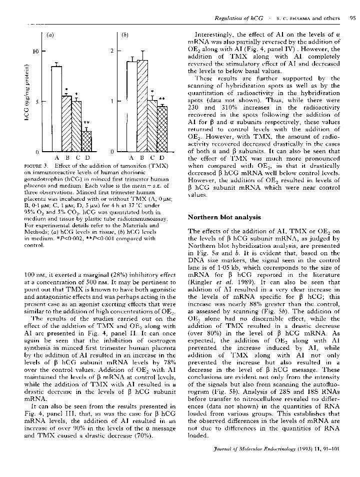
figure 4. Effect of the addition of oestradiol-17ß (OE2), aromatase inhibitor (AI), tamoxifen (TMX), AI±OE2 or
AI + TMX on ß (panels I and II) and a (panels III and IV) hCG subunit mRNA levels in first trimester humanplacenta (FTHP). (a) Dot blot hybridization: representative autofluorogram. Minced FTHP was incubated with OE2,AI or TMX for 4 h. Total RNA was isolated and a and ß hCG subunit specific mRNA was quantitated by dot blothybridization. Panel I: 1) control, 2) 10 nM OE2, 3) 100 nM OE2, 4) 500 nM OE2, 5) 15 pM AI, 6) 5 pM TMX. PanelII: 1) control, 2) 15 pM AI, 3) 15 pM AI + 10nM OE2, 4) 15 pM AI + 5 pM TMX. Panel III: 1) control, 2) 100 nM OE2,3) 500 nM OE2, 4) 15 pM AI, 5) control, 6) 5 pM TMX. Panel IV: 1) control, 2) 15 pM AI, 3) 15 pM AI + 10 nM OE2, 4)15 pM AI + 5 pM TMX. (b) Graphic representation of scans of the corresponding autofluorograms in (a). Each barrepresents the mean ± s.e. of the number of observations (n) indicated. The area of the control group is taken as 100%and values for treated groups are expressed as percentages of the control value.
200 r- (b)
100(3)
(3)
I
(3)JL
(3)JL
(3)
1
(2)
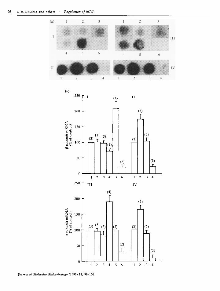
figure 5. Effect of the addition of aromatase inhibitor (AI), oestradiol-17ß (OE2) and tamoxifen (TMX) on ß humanchorionic gonadotrophin (hCG) subunit mRNA levels and OE2 and TMX on AI-stimulated ß hCG subunit mRNAlevels, (a) Northern analysis: representative autofluorogram. Minced first trimester human placental tissue was
incubated with OE2, TMX, AI, AI + OE2 or AI + TMX for 4 h. Total RNA was isolated, separated by denaturedformaldehyde agarose gel electrophoresis and Northern analysis was carried out. For experimental details refer to theMaterials and Methods. Lane 1, 1 kb ladder (lower band 1-01 kb, upper band 163 kb); lane 2, control; lane 3, AI(15 pM); lane 4, OE2 (10 nM); lane 5, TMX (5 um); lane 6, AI (15pM) + OE2 (10 nM); lane 7, AI (15 pM) + TMX (5 um).(b) Graphic representation of a scan of the corresponding autoradiogram in (a). Each bar represents the mean ± s.E. ofthe number of observations (n) indicated. The area of the control group is taken as 100% and the values of treatedgroups are expressed as percentages of the control value.
hCG would be adequate compared with theestimation of a and ß CG subunits separately.
The earlier report of Belleville et al. (1978) andthe recent work of Maruo et al. (1986) indicatingthat the addition of OE2 has no effect on hCGsynthesis or on the levels of its a and ß mRNAscould result from interference by endogenoussteroids. However, the addition of low concentra¬tions of OE2 was not very effective in decreasing thelevel of immunoreactive hCG. These experimentswere carried out with the assumption that if hCG isunder the negative control of endogenous OE2, thenthe addition of OE2 should be much more effectivein the inhibition of immunoreactive hCG levels.However, as can be seen from the results presentedin Fig. 1, while the effects of AI could be reversedby the simultaneous addition of OE2, OE2 was not
effective in suppressing hCG levels by itself,although at very high concentrations (3-7 and7-4 pM) it was partially effective (Fig. 2).
In contrast, TMX was effective in the presence orabsence of AI over a range of concentrations(01—5-0 pM, Fig. 3). In view of this, similar studieson the effect of addition of TMX and OE2 togetherwere not carried out as TMX was very effectiveindependently, even at a concentration as low as01 pM, and it was felt that TMX would override theeffects of OE2 and still act as an agonist. In fact,initially the purpose of using TMX was to block theaction of OE2 at the receptor level, and to examinewhether we could observe effects similar to thoseobserved following the addition of AI. However, we
consistently observed only an agonistic effect.Although traditionally TMX is used as an oestrogen

(*)
oo
200 r-
100 L
(3)X
100 r
(3)
50 h
II
1(2)(2)X
C AI C AIa hCG ß hCG
C TMX C TMXahCG ßhCG
ÜOX,
300 r-
3 200
100
(2)I
2 3 4 5ahCG
(2)
2 3 4 5
ßhCGIII
figure 6. Effect of the addition of aromatase inhibitor (AI) or tamoxifen (TMX) on the levels of a and ß humanchorionic gonadotrophin (hCG). (a) Panels I and II: representative autofluorograms are shown of SDS-PAGEanalysis of in-vitro translated and immunoprecipitated products (C is control). Poly(A)+ RNA was isolated from eachgroup and translated in vitro using rabbit reticulocyte lysate. The product was immunoprecipitated by a specificantiserum to whole hCG. Panel III: an autofluorogram of SDS-PAGE analysis of the in-vitro biosynthesis of hCGusing [ 5S]methionine, followed by immunoprecipitation of the in-vitro translated product by a specific antiserum towhole hCG. Minced first trimester human placenta was incubated with AI or TMX for 4 h. Lane 1, 125I-labelled ßsubunit; lane 2, control; lane 3, AI (15 pM); lane 4, oestradiol-17ß (10 pM); lane 5, TMX (5 pM). (b) Graphicrepresentation of the scans of the corresponding autofluorograms in (a). Each bar represents the mean ± s.E. of thenumber of observations (n) indicated. The area of the control group is taken as 100% and the values of treated groupsare expressed as percentages of the control value.
receptor antagonist it is known to behave both as an
agonist and an antagonist depending on the doseand the type of tissue used (Furr & Jordan, 1984). It
is possible that in the present situation TMX mayhave been acting as an agonist, thus exerting a
negative influence on the synthesis of hCG. In fact,

in an earlier study (Sharma et al. 1990), we havedemonstrated that TMX does act as an agonist inthe first trimester human placenta. However, itscomplete range of actions in the placenta is yet to beevaluated. Although we have not tested it at a
concentration lower than 01 pM, it is possible thatat even lower concentrations it may behave as an
antagonist, resulting in effects similar to those seenwith the AI. Our studies have also revealed (datanot shown) that TMX is not toxic to the mincedtissue at the concentration used, as judged by theincorporation of [ HJthymidine into DNA.
The results of the present study thus permit us to
suggest that the synthesis and secretion of hCGsubunits are under the negative control of OE2,although it is possible that the other major placentalsteroid, progesterone, may also have a role in theregulation of the synthesis of hCG. In fact, theinverse relationship between the declining levels ofhCG and the rising levels of placental steroidsduring human pregnancy provides additionalsupport for such a suggestion.
ACKNOWLEDGEMENTS
The authors wish to thank Dr I. Boime, WashingtonMedical University School, St Louis, MO, U.S.A.,for providing clones for the a and ß hCG subunitsand the Department of Science and Technology,New Delhi, India, for financial assistance. A. J. R. isgrateful to the Rockefeller Foundation for hisBiotechnology Career Fellowship and P. P. wishesto thank CSIR for awarding him the position ofPool Officer. The authors also wish to thank Prof.G. Padmanabhan, Department of Biochemistry,Indian Institute of Science, Bangalore, India, foradvice during the course of this work and theSuperintendent and Staff of the Family PlanningClinic of K. C. General Hospital, Bangalore, India,for permitting the collection of placentae.
REFERENCES
Aviv, H. & Leader, P. (1972). Purification of biologically activeglobin messenger RNA by chromatography on oligothymidicacid-cellulose. Proceedings of the National Academy ofSciences of the U.S.A. 69, 1408-1412.
Belleville, F., Lasbennes, A., Nabet, P. & Paysant, P. (1978).HCS, HCG regulation from cultured placenta. ActaEndocrinología 88, 169—181.
Birken, S. (1984). Chemistry of human chorionicgonadotrophin. Annales d'Endocrinologie 45, 297-305.
Boime, I., McWilliams, D., Szczesna, E. & Camel, M. (1976).Synthesis of human placental lactogen messenger RNA as a
function of gestation. Journal of Biological Chemistry 251,820-825.
Conn, P. M., McArdle, A. C, Andrews, W. V. & Huckle,W. R. (1987). The molecular basis of gonadotrophin-releasing hormone (GnRH) action in the pituitarygonadotrope. Biology of Reproduction 36, 17-35.
Counis, R., Corbani, M. & Jutisz, M. (1983). Estradiolregulates mRNAs encoding precursors to rat lutropin (LH)and follitropin (FSH) subunits. Biochemical and BiophysicalResearch Communications 114, 65-72.
Fabienne L'Horset, Perret, C, Brehier, A. & Thomasset, M.(1990). 17 B-Estradiol stimulates the calbindin-D9K(CaBP9K) gene expression at the transcriptional andpost-transcriptional levels in the rat uterus. Endocrinology127, 2891-2897.
Furr, B. J. A. & Jordan, V. C. (1984). The pharmacology andclinical uses of tamoxifen. Pharmacology and Therapeutics 25,127-205.
Gharib, S. D., Bowers, S. M., Need, L. R. & Chin, W. W.(1987). Regulation of rat luteinizing hormone (LH) subunitmRNAs by gonadal steroid hormones. Journal of ClinicalInvestigation 77, 582-587.
Gharib, S. D., Wierman, M. E., Shupnik, M. A. & Chin,W. W. (1990). Molecular biology of the pituitarygonadotrophins. Endocrine Reviews 11, 177—199.
Hsu, C.-Y. J. & Frankel, F. R. (1987). Effect of estrogen on theexpression of mRNAs of different actin isoforms inimmature rat uterus. Journal of Biological Chemistry 262,9594-9599.
Jagannadha Rao, A., Kotagi, S. G. & Moudgal, N. R. (1984).Serum concentrations of chorionic gonadotrophin oestradiol17 ß and progesterone during early pregnancy in the SouthIndian bonnet monkey (Macaca radiata). Journal ofReproduction and Fertility 70, 449-455.
Joel, P. B., Hagerman, D. D. & Ville, C. A. (1961). Effects ofestradiol added in vitro on the metabolism of humanplacenta. Journal of Biological Chemistry 236, 3151-3157.
Lowry, O. H., Rosebrough, N. J., Farr, A. L. & Randall, R. J.(1951). Protein measurement with the Folin phenol reagent.Journal of Biological Chemistry 193, 265-275.
McCann, S. M. (1974). Regulation of secretion of folliclestimulating hormone and luteinizing hormone. In Handbookof Physiology: Endocrinology, pp. 489-587. Eds R. O. Greep,E. B. Astwood, E. Knobil, W. H. Sawyer & S. R. Geiger.Washington: American Physiological Society.
McQueen, S. D., McWilliams, D., Birken, S., Carefield, R.,Landefeld, T. & Boime, I. (1978). Identification of mRNAsencoding the a and ß subunits of human chorionicgonadotrophin. Journal of Biological Chemistry 253,7109-7114.
Maniatis, T., Fritsch, E. F. & Sambrook, J. (1982). Extraction,purification and analysis of mRNA from eukaryotic cells. InMolecular Cloning. A Laboratory Manual, pp. 202-204. ColdSpring Harbor: Cold Spring Harbor Laboratory.
Maruo, T., Matsuo, H., Ohtani, Y., Hoshina, M. & Mochizuki,M. (1986). Differential modulation of chorionic gonadotropin(CG) subunit messenger ribonucleic acid levels and CGsecretion by progesterone in normal placenta andchoriocarcinoma cultured in vitro. Endocrinology 119,855-864.
Mathialagan, N. & Rao, A. J. (1986). Gonadotrophin releasinghormone (GnRH) stimulates both secretion and synthesis ofhuman chorionic gonadotrophin (hCG) by first trimesterhuman placental minces in vitro. Biochemistry International13, 757-765.
Murthy, G. S., Lakshmi, B. S. & Moudgal, N. R. (1989).Radioimmunoassay of polypeptide hormones usingimmunochemically coated plastic tubes. Journal of Biosciences14, 9-20.

Nilson, J. H., Nejedlik, T. M., Virgin, J. B., Crowder, M. E.& Nett, T. (1985). Expression of subunit and luteinizinghormone ß genes in the ovine anterior pituitary. Estradiolsuppresses accumulation of mRNAs for both a subunit andluteinizing hormone ß. Journal of Biological Chemistry 258,12087-12090.
Papavasiliou, S. S., Zmeili, S., Herbon, L., Duncan-weldon, J.,Marshall, J. C. & Landefeld, T. D. (1986). a and luteinizinghormone ß messenger ribonucleic acid (RNA) of male andfemale rats after castration: quantitation using an optimizedRNA dot blot hybridization assay. Endocrinology 119,691-698.
Ringler, G. E., Kallen, C. B. & Strauss, J. F., III. (1989).Regulation of human trophoblast function byglucocorticoids: dexamethasone promotes increasedsecretion of chorionic gonadotropin. Endocrinology 124,1625-1631.
Schwarzel, W. C, Krugell, W. G. & Brodie, H. J. (1973).Studies on the mechanism of estrogen biosynthesis VIII.The development of inhibitors of the enzyme system inhuman placenta. Endocrinology 92, 866—880.
Sharma, S. C, Usha Kumari, Dighe, R. R. & Rao, A. J.(1990). Regulation of protein synthesis in the firsttrimesterhuman placenta by 17 ß oestradiol andprogesterone. Placenta 11, 63—74.
Southern, E. (1975). Detection of specific sequence amongDNA fragments separated by gel electrophoresis. Journal ofMolecular Biology 98, 503-5Í 7.
Talamantes, F. & Ogren, L. (1988). The placenta as an
endocrine organ: polypeptides. In The Physiology ofReproduction, pp. 2093-2144. Eds E. Knobil, J. Neill et al.New York: Raven Press Ltd.
Thomas, P. S. (1980). Hybridization of denatured RNA andsmall DNA fragments transferred to nitrocellulose.Proceedings of the National Academy of Sciences of the U.S.A.77, 5201-5205.
Wunsch, D. M., Anderson, L. D., Pepe, G. J. & Albrecht, E.D. (1986). Regulation of progesterone formation by humanplacental cells in culture. Endocrinology 119, 998-1003.
revised manuscript received 23 October 1992