Regulation of tumorigenic Wnt signaling by cyclooxygenase...
23
See discussions, stats, and author profiles for this publication at: https://www.researchgate.net/publication/283268164 Regulation of tumorigenic Wnt signaling by cyclooxygenase-2, 5-lipoxygenase and their pharmacological... Article in Pharmacology [?] Therapeutics · October 2015 DOI: 10.1016/j.pharmthera.2015.11.001 CITATIONS 3 READS 103 17 authors, including: Some of the authors of this publication are also working on these related projects: Macrolides View project New therapeutic strategies for t(4;11)-leukemia View project Sabine Grösch Goethe-Universität Frankfurt am Main 79 PUBLICATIONS 3,561 CITATIONS SEE PROFILE Slava (née Chtarbova) Ziegler Max Planck Institute of Molecular Physiology 65 PUBLICATIONS 1,041 CITATIONS SEE PROFILE Dieter Steinhilber Goethe-Universität Frankfurt am Main 296 PUBLICATIONS 5,781 CITATIONS SEE PROFILE Michael Parnham Fraunhofer Institute for Molecular Biology an… 251 PUBLICATIONS 3,965 CITATIONS SEE PROFILE All content following this page was uploaded by Carmela Matrone on 20 January 2017. The user has requested enhancement of the downloaded file.
Transcript of Regulation of tumorigenic Wnt signaling by cyclooxygenase...

Seediscussions,stats,andauthorprofilesforthispublicationat:https://www.researchgate.net/publication/283268164
RegulationoftumorigenicWntsignalingbycyclooxygenase-2,5-lipoxygenaseandtheirpharmacological...
ArticleinPharmacology[?]Therapeutics·October2015
DOI:10.1016/j.pharmthera.2015.11.001
CITATIONS
3
READS
103
17authors,including:
Someoftheauthorsofthispublicationarealsoworkingontheserelatedprojects:
MacrolidesViewproject
Newtherapeuticstrategiesfort(4;11)-leukemiaViewproject
SabineGrösch
Goethe-UniversitätFrankfurtamMain
79PUBLICATIONS3,561CITATIONS
SEEPROFILE
Slava(néeChtarbova)Ziegler
MaxPlanckInstituteofMolecularPhysiology
65PUBLICATIONS1,041CITATIONS
SEEPROFILE
DieterSteinhilber
Goethe-UniversitätFrankfurtamMain
296PUBLICATIONS5,781CITATIONS
SEEPROFILE
MichaelParnham
FraunhoferInstituteforMolecularBiologyan…
251PUBLICATIONS3,965CITATIONS
SEEPROFILE
AllcontentfollowingthispagewasuploadedbyCarmelaMatroneon20January2017.
Theuserhasrequestedenhancementofthedownloadedfile.

Pharmacology & Therapeutics 157 (2016) 43–64
Contents lists available at ScienceDirect
Pharmacology & Therapeutics
j ourna l homepage: www.e lsev ie r .com/ locate /pharmthera
Associate Editor: M. Belvisi
Regulation of tumorigenic Wnt signaling bycyclooxygenase-2, 5-lipoxygenase and their pharmacologicalinhibitors: A basis for novel drugs targeting cancer cells?
Jessica Roos a,⁎,1, Sabine Grösch b,1, Oliver Werz c,1, Peter Schröder d,1, Slava Ziegler d, Simone Fulda e,Patrick Paulus f, Anja Urbschat g, Benjamin Kühn a, Isabelle Maucher a, Jasmin Fettel a, Thomas Vorup-Jensen h,Matthias Piesche h, Carmela Matrone h, Dieter Steinhilber a, Michael J. Parnham i,1, Thorsten J. Maier a,h,⁎⁎,1a Institute of Pharmaceutical Chemistry, Goethe-University, Max-von-Laue-Str. 9, D-60438 Frankfurt am Main, Germanyb Pharmazentrum Frankfurt/ZAFES, Institute of Clinical Pharmacology, Goethe-University, 60590, Frankfurt am Main, Germanyc Department of Pharmaceutical/Medicinal Chemistry, Institute of Pharmacy, University of Jena, Philosophenweg 14, 07743 Jena, Germanyd Max Planck Institute of Molecular Physiology, Otto-Hahn-Str. 11, 44227 Dortmund, Germanye Institute for Experimental Cancer Research in Pediatrics, Goethe-University, Frankfurt, Germanyf Department of Anesthesiology and Operative Intensive Care Medicine, Kepler University Hospital, 4021 Linz, Austriag Department of Urology and Pediatric Urology, Philipps-University Hospital, 35043 Marburg, Germanyh Aarhus University, Department of Biomedicine & Center for Study and Prevention of Neurodegenerative Inflammation (NEURODIN), Bartholins Allé 6, 8000 Aarhus C, Denmarki Fraunhofer Institute for Molecular Biology and Applied Ecology IME, Project Group for Translational Medicine and Pharmacology TMP, 60596 Frankfurt am Main, Germany
Abbreviations: AML, acute myeloid leukemia; APC, admyeloid leukemia; COX, cyclooxygenase; CSC, cancer stemstem cell; LEF, lymphoid enhancer factor; LSC, leukemic santi-inflammatory drugs; NF-κB, nuclear factor-κB; PG, pTCF, T-cell factor.⁎ Correspondence to: J. Roos, Institute of Pharmaceutic⁎⁎ Correspondence to: T.J. Maier, Aarhus University, Dep
E-mail addresses: [email protected] (1 These authors contributed equally.
http://dx.doi.org/10.1016/j.pharmthera.2015.11.0010163-7258/© 2015 Elsevier Inc. All rights reserved.
a b s t r a c t
a r t i c l e i n f oAvailable online 6 November 2015
Keywords:Wnt inhibitorTumor cellsLeukemic stem cellsLeukotrienesNon-steroidal anti-inflammatory drugs
Canonical Wnt signaling is a highly conserved pathway with a prominent role in embryogenic development,adult tissue homeostasis, cell polarization, stem cell biology, cell differentiation, and proliferation. Furthermore,canonical Wnt signaling is of pivotal importance in the pathogenesis of a number of cancer types and cruciallyaffects tumor initiation, cancer cell proliferation, cancer cell apoptosis, and metastasis.Reports over the last decade have provided strong evidence for a pathophysiological role ofWnt signaling in non-malignant classical inflammatory and neurodegenerative diseases.Although, several agents suppressing theWnt pathway at different levels have been identified, the developmentof clinically relevant Wnt-inhibiting agents remains challenging due to selectivity and toxicity issues.Several studies have shown that long-term administration of non-steroidal anti-inflammatory drugs protectsagainst colon cancer and potentially other tumor types by interfering both with the COX and the Wnt pathway.Our own studies have shown that non-steroidal anti-inflammatory drugs suppress Wnt signaling by targetingthe pro-inflammatory enzyme5-lipoxygenasewhich is the key enzymepathophysiologically involved in the syn-thesis of leukotrienes.Furthermore, we found a direct link between the 5-lipoxygenase andWnt signaling pathways, which is essentialfor the maintenance of leukemic stem cells. Accordingly, genetic and pharmacological inhibition of 5-lipoxygenase led to an impairment of Wnt-dependent acute and chronic myeloid leukemic stem cells.We believe that 5-lipoxygenase inhibitors might represent a novel type of Wnt inhibitor activating a potentiallynaturally occurring novel mechanism of suppression of Wnt signaling that is non-toxic, at least in mice, and ispotentially well tolerated in patients.
© 2015 Elsevier Inc. All rights reserved.
enomatous polyposis coli; CBP, CREB-binding protein; CK1, casein kinase 1; CLL, chronic lymphatic leukemia; CML, chroniccell; Dvl, disheveled; FLAP, 5-LO-activating protein; Fzd, Frizzled; GSK3β, glycogen synthase kinase 3β; HSC, hematopoietic
tem cell; 5-LO, 5-lipoxygenase; LRP, low density lipoprotein receptor-related protein; LT, leukotriene; NSAIDs, non-steroidalrostaglandin; PI3K, phosphatidylinositol 3-kinase; PKB, protein kinase B; PPAR, peroxisome proliferator activated receptor;
al Chemistry/ZAFES, Goethe-University, Max-von-Laue-Str. 9, 60438 Frankfurt/Main, Germany. Tel.: +49 69 7982 9341.artment of Biomedicine, Bartholins Allé 6, 8000 Aarhus C, Denmark. Tel.: +45 87167681; fax: +49 69 798 29323.J. Roos), [email protected] (T.J. Maier).

44 J. Roos et al. / Pharmacology & Therapeutics 157 (2016) 43–64
Contents
Table 1Selected Wnt sign
Agent
OMP-18R5
OMP-54F28
LGK974IWP-L6SalinomycinNiclosamideNSC6680363289-8625FJ9XAV939JW55IWR-1PyrviniumPKF115-584, CGPNU-74654BC21iCRT3, iCRT5
iCRT14StAx peptidesResveratrolICG-001PRI-724
SAH-BCL9CWP232291
1. Introduction . . . . . . . . . . . . . . . . . . . . . . . . . . . . . . . . . . . . . . . . . . . . . . 442. The canonical Wnt pathway . . . . . . . . . . . . . . . . . . . . . . . . . . . . . . . . . . . . . . . 453. The role of Wnt signaling in tumor cell growth . . . . . . . . . . . . . . . . . . . . . . . . . . . . . . . 454. Role of Wnt signaling in inflammatory disorders . . . . . . . . . . . . . . . . . . . . . . . . . . . . . . 455. Components of the Wnt signaling pathway as potential biomarkers of disease . . . . . . . . . . . . . . 456. Inhibitors of Wnt signaling . . . . . . . . . . . . . . . . . . . . . . . . . . . . . . . . . . . . . . . . 457. Non-steroidal anti-inflammatory drugs as Wnt pathway inhibitors . . . . . . . . . . . . . . . . . . . . 468. Introduction to the pro-inflammatory enzyme 5-lipoxygenase and its inhibitors . . . . . . . . . . . . . . . 479. Modulation by 5-LO and 5-LO inhibitors of Wnt signaling in leukemic stem cells . . . . . . . . . . . . . 4710. Conclusion and future direction . . . . . . . . . . . . . . . . . . . . . . . . . . . . . . . . . . . . . 47Conflict of interest statement . . . . . . . . . . . . . . . . . . . . . . . . . . . . . . . . . . . . . . . . . 47Acknowledgment . . . . . . . . . . . . . . . . . . . . . . . . . . . . . . . . . . . . . . . . . . . . . . . 47References . . . . . . . . . . . . . . . . . . . . . . . . . . . . . . . . . . . . . . . . . . . . . . . . . . 48
44454547484951535557585858
1. Introduction
CanonicalWnt signaling is of pivotal importance in the pathogenesisof a number of cancer types and crucially affects tumor initiation, cancercell proliferation, cancer cell apoptosis, self-renewal of leukemic cellsand metastasis. We and others have shown that the Wnt/β-cateninsignaling pathway is the key signaling pathway inmany cancers, partic-ularly in those involving maintenance of cancer stem cells (CSC)(Muller-Tidow et al., 2004; Wang et al., 2010). CSCs represent a smallportion of the total tumormass in solid aswell as hematological cancers(Tirino et al., 2013). They are thought to persist as a distinct populationin the remaining tumor tissues after destruction of the bulk of tumormass by conventional chemotherapy. Theymight trigger disease relapseand metastasis by generating novel tumor cells (Tirino et al., 2013). Forthis reason, therapeutics specifically targeting cancer stem cells havethe potential for significant improvement of future cancer therapies.
Considerable efforts have been undertaken to identify novel thera-peutics that could interfere with oncogenic Wnt signaling (B. Chenet al., 2009). Decades have been spent by both the pharmaceutical in-dustry and academic researchers who have used high-throughput
aling inhibitors, their targets and current stage of development.
Target Current Stage
Fzd receptors Clinical trials (NCT01345201, NCNCT01957007, NCT01973309)
Wnt proteins Clinical trials (NCT01608867, NCNCT02092363, NCT02050178)
Porcupine Clinical trial (NCT01351103)Porcupine In vivo (zebrafish)LRP6 In vivo (xenograft model)LRP6 In vivo (xenograft model)Dvl In vivo (Xenopus embryos)Dvl In vivo (Xenopus embryos)Dvl In vivo (xenograft model)Tankyrase 1/2 In vitro (cancer cell lines)Tankyrase 1/2 In vivo (mice)Tankyrase 1/2, axin In vivo (zebrafish)CK1α In vivo (xenograft models)
P049090 β-Catenin/TCF In vivo (Xenopus embryos)β-Catenin/TCF In vivo (zebrafish)β-Catenin/TCF In vitro (HCT116 cell line)β-Catenin/TCF In vitro (colon cancer cell lines, p
colon cancer specimens)β-Catenin/TCF In vivo (xenograft models)β-Catenin/TCF In vitro (colorectal cancer cell linβ-Catenin/TCF antioxidant Clinical trial (NCT00256334, NCTCBP In vivo (xenograft model)CBP Clinical trials (NCT01606579, NC
NCT02195440, NCT01302405)β-Catenin/Bcl9 In vivo (xenograft model)Sam68 Clinical trial (NCT01398462)
screens to identify small-molecule inhibitors targeted to different Wntsignaling pathway constituents. Regrettably, only one compound,namely PRI-724 (Prism Pharma Co, Ltd/Eisai, Table 1 and Supplementa-ry Table 1), which directly disrupts the transcriptional co-factor func-tion of β-catenin has been developed and entered phase I clinical trials(Safety and Efficacy Study of PRI-724 in Subjects with Advanced SolidTumors; ClinicalTrials.gov Identifier: NCT01302405). Other Wnt-suppressive drug candidates interfere with Wnt signaling at the levelof the Wnt ligand binding or ligand-induced receptor activation. Someof these compounds were/are also included in recent clinical trials(see Section 6). However, the potential side-effects and selectivity ofthe suppressive effects of this class of agents on the canonicalWnt path-way still remain unclear.
Non-steroidal anti-inflammatory drugs (NSAIDs), such as sulindac,indomethacin, and celecoxib, are able to inhibit Wnt signaling at highconcentrations and several mechanisms have been proposed to explainthese effects (see Section 7). We recently found that these drugs, atsomewhat higher concentrations than those required to inhibit cycloox-ygenase (COX) enzymes, also suppress 5-LOproduct formation suggest-ing a novel mechanistic linkage between the 5-LO pathway and
References
T02005315, (Gurney et al., 2012)
T02069145, (Smith et al., 2013; Yeung et al., 2014)
(Liu et al., 2013)(X. Wang et al., 2013)(Mao et al., 2014; Wang et al., 2012)(W. Lu et al., 2011; Osada et al., 2011; Sack et al., 2011)(Shan et al., 2005)(Grandy et al., 2009)(Fujii et al., 2007)(Huang et al., 2009)(Waaler et al., 2012)(B. Chen et al., 2009)(Thorne et al., 2010; Wiegering et al., 2014)(Lepourcelet et al., 2004)(Trosset et al., 2006; Buikema et al., 2013; Pradhan & Olsson, 2014)(Tian et al., 2012)
rimary human (Gonsalves et al., 2011)
(Gonsalves et al., 2011)es) (Grossmann et al., 2012)00433576) (Chen et al., 2012)
(Emami et al., 2004; Arensman et al., 2014)T01764477, (El-Khoueiry et al., 2013)
(Takada et al., 2012)JW Pharmaceuticals

45J. Roos et al. / Pharmacology & Therapeutics 157 (2016) 43–64
oncogenic Wnt signaling (Roos et al., 2014). Furthermore, we andothers showed that pharmacological or genetic targeting of 5-LO effi-ciently interferes with Wnt signaling and maintenance of leukemicstem cells (LSCs) in chronicmyeloid leukemia (CML) and acutemyeloidleukemia (AML) (Roos et al., 2014).
In the present review, we aim to simplify this recent but rather com-plex subject in order to appeal to a larger number of readerships andmaximize the interest in the topic. We provide a brief introduction toWnt signaling and its role in tumorigenesis, as well as an overview ofavailableWnt inhibitors and theirmolecularmechanisms ofWnt signal-ing suppressionwith focus on drugs interferingwith eicosanoid biosyn-thesis. Emphasis is placed on the recently discovered role of 5-LO inregulating the canonical Wnt pathway. Finally, we discuss the possibleclinical implications of these findings for new stem-cell specific thera-peutics in oncology.
2. The canonical Wnt pathway
The Wnt signaling pathway is a highly conserved pathway with aprominent role in embryogenic development, adult tissue homeostasis,cell polarization, stem cell biology, cell differentiation, and proliferation(Nusse & Varmus, 1992; Cadigan & Nusse, 1997; van deWetering et al.,2002; Clevers, 2006; Clevers & Nusse, 2012). Traditionally, theWnt sig-naling pathway is separated into two routes; the non-canonical and thecanonical pathways (see Section 2.1). They differ in their dependencyon the protein β-catenin. The non-canonical pathways, such as the pla-nar cell polarity (PCP) and Ca2+-pathway, regulate processes such ascell movement, migration (Kuhl et al., 2001; Mikels & Nusse, 2006;Angers & Moon, 2009), cell orientation (Seifert & Mlodzik, 2007;Angers &Moon, 2009), and function through an β-catenin independentmechanism.
2.1. Overview of the canonical Wnt/β-catenin pathway
In the canonical pathway (Fig. 1),β-catenin is the key effector,whichis responsible for the transduction of the signal to the nucleus. At thissite, β-catenin triggers the transcription ofWnt-specific genes responsi-ble for the control of cell fate decisions in many cells and tissues(Valenta et al., 2012). In addition to its role in the Wnt signalingpathway, β-catenin is associated with cadherins such as E-cadherin. β-Catenin connects cadherins with the actin cytoskeleton (through α-catenin), forming adherents junctions. It is important for cell stability,cell migration and adhesion (Harris & Peifer, 2005; Wilusz & Majka,2008).
2.2. Regulation of the Wnt/β-catenin pathway
If not in complex with E-cadherin, β-catenin is targeted for rapiddegradation by the proteasome, a protein complex involved in the elim-ination of mainly dysfunctional cellular proteins (Fig. 1A) (Aberle et al.,1997). This breakdown is mediated by a multiprotein destruction com-plex which includes the tumor suppressor proteins, adenomatouspolyposis coli (APC) and axin, glycogen synthase kinase 3 (GSK3β)and casein kinase 1α (CK1α) (McDonald & Silver, 2009; Clevers &Nusse, 2012), together with the serine/threonine phosphatase A2
(PPA2) (Luo et al., 2007; Su et al., 2008). APC and axin function as a scaf-fold, facilitating GSK3β- and CK1α-mediated phosphorylation of criticalresidues within β-catenin (Kimelman & Xu, 2006; Roberts et al., 2011).In particular, CK1α phosphorylates β-catenin at Ser45, priming the se-quential phosphorylation of Thr41, Ser37, and Ser33 by GSK3β (Liuet al., 2002; Xing et al., 2003; Valenta et al., 2012; Stamos & Weis,2013). Subsequently, β-catenin interacts with the ubiquitin machineryvia its phosphorylated Ser33 and Ser37 residues. As a consequence, β-catenin is ubiquitinated and subsequently degraded by the 26S protea-some (Aberle et al., 1997; Hart et al., 1999). The phosphatase PP2A, alsoassociated with axin and/or APC, counteracts the phosphorylation-
induced degradation of β-catenin by directly dephosphorylating β-catenin at the critical residues important for proteasomal degradation.Furthermore, PP2A binds APC and is suggested to dephosphorylatethe APC protein leading to impaired affinity of APC to β-catenin(Seshacharyulu et al., 2013). Recently, Huang et al. discovered that thetwo poly-ADP-ribosylating enzymes, tankyrase 1 and tankyrase 2,bind to a highly conserved domain of axin and trigger its degradationthrough the ubiquitin–proteasome pathway, thereby, releasing β-catenin (Huang et al., 2009).
The Wnt/β-catenin pathway is activated when a secreted Wnt gly-coprotein binds to the N-terminal extracellular cysteine-rich domainof a Frizzled (Fzd) receptor (Fig. 1B). Overall, there are currently 19Wnt ligands and 10 Fzd receptors that have been identified in humans(Silva-Garcia et al., 2014). Upon binding of the Wnt ligand to Fzd, theco-receptor, low density lipoprotein receptor-related protein 5/6(LRP5/6), interactswith Fzd and initiates a cascade of events that resultsin disruption of the β-catenin destruction complex. As consequence, β-catenin is stabilized and is facilitated in its nuclear translocation. Activa-tion of the Fzd receptor leads to phosphorylation of disheveled (Dvl)which causes direct binding of Dvl to the receptor complex (Cruciat,2014). Dvl subsequently multimerizes and induces the formation ofthe so-called LRP-associated Wnt signalosome (Bilic et al., 2007). Thisprocess triggers LRP6 phosphorylation by CK1 (Davidson et al., 2005;Bilic et al., 2007). Phosphorylated LRP6 serves as a docking site foraxin (Mao et al., 2001; Tamai et al., 2004; Davidson et al., 2005; Zenget al., 2005) which recruits the β-catenin destruction complex tosignalosomes (Niehrs, 2012). Furthermore, phosphorylation of LRP5/6leads to suppression of GSK3β activity by direct binding of the enzymeto the co-receptor (Piao et al., 2008). Both events protect β-cateninfrom GSK3β-mediated phosphorylation, which allows β-catenin to ac-cumulate in the cytoplasm and translocate into the nucleus (Taelmanet al., 2010).
Nuclear β-catenin forms a complex with the members of the T-cellfactor/lymphoid enhancer factor (TCF/LEF) transcription factors. Thiscomplexed β-catenin acts as a transcriptional co-activator by displacingco-repressors like Groucho and histone deacetylases (HDACs) (Brantjeset al., 2001; Daniels & Weis, 2005; Archbold et al., 2012; Cadigan,2012) from TCF/LEF. Furthermore, complexed β-catenin recruits otherco-activators such as Bcl9/Legless (Lgs), Pygopus (Pyg) (Kramps et al.,2002; Parker et al., 2002; Thompson et al., 2002), and CREB-binding pro-tein (CBP)/p300 (Hecht et al., 2000; Takemaru&Moon, 2000) to the TCF/LEF transcription factor. These events trigger DNA binding and transcrip-tion of the target genes, including c-myc, cyclin D1 and axin 2 (Lustiget al., 2002; Davidson et al., 2005; Bilic et al., 2007). For further informa-tion, the following website offers an extensive list of Wnt target genes:http://www.stanford.edu/group/nusselab/cgi-bin/wnt/target_genes.
3. The role of Wnt signaling in tumor cell growth
Wnt signaling plays a pivotal role in tumor cell growth by regulatinga number of genes involved in programmed cell death (Pecina-Slaus,2010), cell cycle control, and angiogenesis (Clifford et al., 2008).
Gain or loss-of-function mutations of several members of the Wnt/β-catenin signaling pathway that lead to constitutive activation of β-catenin have been shown to have oncogenic effects in solid tumors(Peifer & Polakis, 2000; Giles et al., 2003). Mutations in β-cateninwere first uncovered in colorectal cancer (Rubinfeld et al., 1993; Suet al., 1993) and later – with a lower frequency – also in other humantumor entities (Peifer & Polakis, 2000; Giles et al., 2003; Luu et al.,2004). Furthermore, Wnt signaling plays a crucial role in the processof leukemogenesis (see Section 3.4).
3.1. Cell survival
Programmed cell death constitutes a fundamental cellular event in-volved in the regulation of various physiological processes (Lockshin &

Fig. 1. A simplified overview of theWnt/β-catenin signaling pathway. In the absence of a Wnt ligand (A), β-catenin is either bound to E-cadherins connecting them to the actin cytoskel-eton or bound in a multiprotein destruction complex, consisting of axin, APC, GSK3β, CK1α, PP1, and PP2A. In this complex, β-catenin is sequentially phosphorylated (P) at different Thrand Ser residues by CK1α and GSK3β. This phosphorylation creates a binding site for E3 ubiquitin ligase β-TrCP, leading to β-catenin ubiquitination and therefore, proteasomal degrada-tion. The transcription of theWnt target genes is repressed by binding of co-repressors (i.e. Groucho and HDACs) to the TCF/LEF transcription factor. The binding of aWnt ligand (B) to itsreceptors, Fzd and LRP5/6, results in the activation of Dvl. Dvl is then recruited to the receptor complexwhich leads to LRP6 phosphorylation (P) and recruitment of theβ-catenin destruc-tion complex to the receptors (currently, there is only evidence for binding of axin, APC, GSK3β, and CK1α to the receptor complex). Thereby, the phosphorylation of β-catenin is inhibited,allowing the free protein to accumulate in the cytosol and translocate to the nucleus where it can act as a co-activator for TCF/LEF-mediated transcription. For this, β-catenin replaces theco-repressors while recruiting various cofactors, including Bcl9/Lgs and Pygopus. APC: adenomatous polyposis coli proteins and; the kinases, GSK3β: glycogen synthase kinase 3β; CBP,CREB-binding protein; CK1α: casein kinase α; Dvl, disheveled; GRG, Groucho; HDAC, histone deacetylase; Fzd, Frizzled; LRP5/6, low density lipoprotein receptor-related protein 5/6;PP2A, phosphatase A2; Pyg, Pygopus; TCF/LEF, T-cell factor/lymphoid enhancer factor.
46 J. Roos et al. / Pharmacology & Therapeutics 157 (2016) 43–64
Zakeri, 2007). Among the various forms of programmed cell death, ap-optosis represents one of the best characterized and is evolutionaryhighly conserved (Galluzzi et al., 2015). Since tissue homeostasis is theresult of a tight balance between cell proliferation and cell death, evensmall alterations in the rate of apoptosis may significantly impact theoverall cell number (Fulda, 2009).
Apoptosis can be viewed as a safeguard mechanism to limit abnor-mal tissue growth and tumor formation. Consequently, cellular abilityto evade apoptosis is one of the hallmarks of cancers (Hanahan &Weinberg, 2011). A number of components of the Wnt signaling path-way such as Dvl, β-catenin, and GSK3β have been shown to modulateapoptosis (Li et al., 2006). Furthermore, β-catenin silencing experi-ments using siRNA approaches identified several Wnt target genes re-lated to apoptosis, including MYBL2, BAG2, BAG3, PTEN, HIF1A, andDAP3 as well as the anti-apoptotic protein Bcl-XL (Huang et al., 2006).Thus, in the majority of cancer cell types, Wnt signaling is consideredto promote cell survival and to inhibit apoptosis, though pro-apoptoticeffects due to activated Wnt signaling have also been reported(Benchabane & Ahmed, 2009). In this context, it should be noted that,in cancer, not only is apoptosis dysfunctional, but there is also an in-crease in the activity of survival pathways that promote proliferationand cell growth.
3.2. Cell cycle regulation
Besides resistance to apoptosis, many cancer cells exhibit dysfunc-tion of the cell cyclemachinerywith regard to the complex series of pro-tein–protein interactions, protein degradation, and phosphorylationevents that regulate the transition to the respective four cell cyclephases (i.e., G1, S, G2, M). The major players at these checkpoints arecyclin-dependent kinases (CDK) which are activated by forming com-plexes with transiently expressed cyclins.
As described above, a key step in the activation of theWnt pathwayis the formation of a complex between nuclear β-catenin and membersof the TCF/LEF family of transcription factors (van de Wetering et al.,1997). The continuous presence of the nuclear TCF/β-catenin complexfosters cancer formation by increasing expression of a number ofproto-oncogenes regulating cell growth, inhibition of apoptosis, andgenes affecting cell shape and cellmigration. Consistentwith these find-ings, disruption of TCF/β-catenin complex formation in colon cancercells hindered target gene activation and inhibited tumor cell growthin vitro (Tetsu & McCormick, 1999; van de Wetering et al., 2002). In-creased stability of β-catenin leads to expression of a number ofdown-stream target genes, such as the G1 cell cycle control regulators,cellular myelocytomatosis (c-myc), and cyclin D1 (Reya & Clevers,

47J. Roos et al. / Pharmacology & Therapeutics 157 (2016) 43–64
2005). C-myc is a proto-oncogene overexpressed in a variety of humancancers including leukemia (Dang, 1999) and plays a role in cell cycleprogression, apoptosis, and cellular transformation (Hoffman et al.,2002). Cyclin D1 is an oncogenic cyclin, which is frequentlyoverexpressed or amplified in breast, prostate, and colon cancers(Ortega et al., 2002). Nevertheless, some components of theWnt sig-naling cascade also function directly to promote formation of thespindle apparatus during mitosis (Niehrs & Acebron, 2012).
3.3. Tumor vascularization
A crucial role forWnt signaling in tumor vascularization is supportedby the presence of Wnt target genes that encode for pro-angiogenicfactors. In particular, the gene encoding for vascular endothelial growthfactor A (VEGF-A), a potent andwell-characterized pro-angiogenic pro-tein, is directly regulated by the TCF/β-catenin complex (Clifford et al.,2008) and is clearly associated with boosted tumor angiogenesis and apoor prognosis (Gratzinger et al., 2007).
3.4. Leukemic stem cell self-renewal
Although the involvement of β-catenin in carcinogenesis was firstdiscovered in solid tumors,β-catenin overexpression is also highly asso-ciated with several leukemic subtypes (Chung et al., 2002). Further-more, the dysregulation of the Wnt//β-catenin signaling pathway wassuggested to play an important role in the self-renewal of LSC in atleast AML and CML patients (Lane et al., 2011; Mochmann et al., 2011;Siapati et al., 2011; Scheller et al., 2013).
LSCs are typically rare and possess properties that are distinct frommost other tumor cells (Lapidot et al., 1994). These cells can undergoself-renewal to maintain an undifferentiated state. They are alsomultipotent, highly proliferative (Jordan, 2007), and may originatefrom normal hematopoietic stem cells (HSC) (Krivtsov et al., 2006;Eppert et al., 2011). Although the role of Wnt/β-catenin signaling inthe self-renewal of normal HSCs is under debate (Cobas et al., 2004;Jeannet et al., 2008; Koch et al., 2008), several groups have shown thatthe Wnt/β-catenin signaling pathway is required for the self-renewalof LSCs derived from either HSC or more differentiated granulocytemacrophage progenitors (GMP) (Muller-Tidow et al., 2004; Malhotra& Kincade, 2009; Wang et al., 2010; Luis et al., 2012). Progression,disease relapse, and drug resistance of leukemic cells depend on LSCsthat resist treatment. Aberrant activation of the Wnt/β-catenin self-renewal pathway has been identified to drive human blast crisis LSCpropagation (Jamieson et al., 2004; Abrahamsson et al., 2009). Sup-pression of β-catenin reverts the LSC stage to a pre-LSC-like stageand significantly reduces the growth of human leukemic cells(Yeung et al., 2010). The chemotherapeutic agents that are usedtoday effectively eradicate the blast cells, which represent partially dif-ferentiated, usually unipotent precursor cells. However, these agentshave very little effect on LSCs. Since the canonical pathway is inactivein most normal cells, inhibition of the Wnt/β-catenin pathway holdspromise for developing novel, targeted therapies for LSCs.
Finally, Wnt/β-catenin signaling pathways have been shown to beable to control proliferation, survival, and differentiation of hematopoi-etic cells (Reya et al., 2003). Taken together, the functional versatility ofWnt/β-catenin signaling is remarkable through its influences, on manydevelopmental levels, including organ and cell specification and differ-entiation as well as maintenance of stem cell activity.
3.5. Tumor prognosis and outcome
In the last couple of years evidence has been accumulating suggest-ing that an aberrant regulated Wnt signaling pathway is correlatedwith the clinical outcome of various cancers such as colorectal cancer(CRC) (Ting et al., 2013; Bruun et al., 2014; Aguilera et al., 2015;Nazemalhosseini Mojarad et al., 2015), AML (Fu et al., 2014; Kuhnl
et al., 2015), gliomas (Rossi et al., 2011; Wu et al., 2013; Guo et al.,2015), nasopharyngeal cancer (NPC) (Xu et al., 2013; Ren et al., 2015)and breast cancer (Dey et al., 2013). In particular, it could be shownthat overexpression of canonical Wnt signaling components is highlyassociatedwith the risk of developingmetastases in patientswith an ag-gressive formof breast cancer (Dey et al., 2013). Similar resultswere ob-tained in a study of potential prognostic factors in glioblastomapatients.
In this case, the investigators demonstrated thatβ-catenin is a highlypredictive marker of malignant behavior and short survival (Rossi et al.,2011). Aberrant expression of β-catenin and other components and tar-get genes of theWnt pathway, such as sFRP1,Wnt-2, E-cadherin, c-mycand LGR5, have also been correlated with poor prognosis and tumor ag-gressiveness inNPC, brainstem glioma, AML and thyroid cancer patients(Wu et al., 2013; Xu et al., 2013; Fu et al., 2014; Michelotti et al., 2015;Ren et al., 2015). Yet not only the expression level, but also the mem-brane/cytosolic or nuclear localization of β-catenin – which is a sign ofactivated Wnt signaling – can be a negative prognostic factor, as wasshown in patients with CRC (Bruun et al., 2014; NazemalhosseiniMojarad et al., 2015). Additionally, nuclear DKK1, localized at specificchromatin sites of active transcription, is associated with decreasedoverall survival rate and progression-free survival after chemotherapyadministration (Aguilera et al., 2015). Furthermore, single nucleotidepolymorphism (SNPs) in APC and β-catenin (CTNNB1) genes, whichare known to alter consensus splicing sites sequences, transposableelements and transcription factor binding sites, are correlated withoverall survival rates (Ting et al., 2013). Based on the pathophysiologicalimportance of theWnt signaling pathway in relation to tumor prolif-eration, suppression of apoptosis, migration, resistance to chemothera-py and maintenance of cancer stem cells (Roarty & Rosen, 2010;Vermeulen et al., 2010; Alison et al., 2012; Debeb et al., 2012), it is notsurprising that an aberrantly activated Wnt signaling pathway is, inmany cases, associated with poor prognosis of cancer patients. Takentogether, all these findings support the concept ofWnt signaling inhibi-tion as a feasible therapeutic strategy.
4. Role of Wnt signaling in inflammatory disorders
4.1. Disease association
Reports over the last decade have provided strong evidence fora pathophysiological role of Wnt signaling in a number of non-malignant classical inflammatory diseases. In particular, several studieshave substantiated a crucial role of Wnt-mediated signaling in the acti-vation of fibroblast-like synoviocytes during the pathogenesis of rheu-matoid arthritis, triggering destruction of the articular cartilage andbone (Sen, 2005). Furthermore, Wnt signaling pathway and Wnt regu-latory proteins, like Dickkopf, may trigger bony fusion in ankylosingspondylitis and thus, may serve as potential biomarkers of this disease(Corr, 2014).
Changes in the expression of several genes of the Wnt signalingpathway were found to be associated with impaired lung function inchildrenwith asthma andmay contribute to the pathogenesis of the dis-ease (Sharma et al., 2010). Further mechanistic insight was recentlyprovided by Trischer et al. who showed that Wnt signaling via theWnt10b ligand is an important modulator of T cell activation duringasthma pathogenesis (Trischler et al., 2015). Guo et al. demonstratedthat intratracheal administration of the Wnt ligand, Wnt3a, or an anti-body raised against Dickkopf-1 activating Wnt signaling in alveolar ep-ithelial cells suppressed macrophage and neutrophil infiltration intolungs (Guo et al., 2015). Pathophysiological roles of Wnt signalinghave also been identified in chronic obstructive pulmonary disease(COPD) where increased β-catenin activity in fibroblasts was found tobe relevant for the diseases-associated remodeling of the extracellularmatrix (Baarsma et al., 2011). Interestingly, over-activity of Wnt signal-ing in primary bronchial epithelial cells sensitized individuals to

48 J. Roos et al. / Pharmacology & Therapeutics 157 (2016) 43–64
cigarette smoke-induced inflammation, potentially triggering COPD(Heijink et al., 2013).
A crucial role of Wnt signaling pathway components is also evidentin inflammatory bowel disease (IBD) and the transition to themalignantstage. Several studies found a stage-specific increased or decreasedexpression of a number of Wnt pathway-related genes, including Wntligands, Fzd and Dickkopf-4, in colonic biopsies of subjects with ulcera-tive colitis or in IBD-associated colorectal neoplasia as well as in cancers(You et al., 2007, 2008; Claessen et al., 2010).
The literature also supports a role of Wnt signaling in a number ofother diseases featuring an inflammatory component. Malgor et al. re-cently provided evidence for an increase in the protein levels of theWnt5a ligand in advanced arterial arteriosclerotic lesions, whencompared with less advanced arterial lesions. Serum levels of Wnt5awere also significantly higher in patients suffering from atherosclerosiscompared to healthy controls, suggesting that Wnt signaling enhancesthe process of atherosclerotic plaque formation at a late stage (Malgoret al., 2014). Polymorphism or reduced expression of the secretedFrizzled-related protein (sFRP)3 has also been observed in patientswith osteoarthritis (Loughlin et al., 2004) and Wnt4 ligand inductionhas been related to kidney damage or polycystic kidney disease(Rodova et al., 2002; Surendran & Simon, 2003; Terada et al., 2003).
Importantly, Wnt signaling appears to play a pathophysiologicallyrelevant role in neurodegenerative diseases, asWnt-mediated neuroin-flammation triggers infiltration of leukocytes and production of pro-inflammatory mediators in the central and peripheral nervous systems,contributing to neuronal cell damage or death as well as to generationand maintenance of chronic pain (Yuan et al., 2012; Ji et al., 2014).
Wnt is an important regulator of neuroinflammation in the braincontrolling the dialog between neurons and glia or glial and neuralstem/progenitor cells (NPCs) during the inflammatory processes(Marchetti & Pluchino, 2013). The expression of Wnt ligands and Wntsignaling components has only been identified in astrocyte and macro-phage/microglia (Halleskog et al., 2011; L'Episcopo et al., 2011) that canharbor Wnt receptors and respond to Wnt in either pro- or anti-inflammatorymanner (L'Episcopo et al., 2014a, 2014b). Indeed, deregu-lation of Wnt/β-catenin signaling pathway, dysregulation of Wnt/Fzdcascade and increase in inflammatory processes have been proposedas common determinants in some neurodegenerative events of schizo-phrenia (Miyaoka et al., 1999), Parkinson's (PD)/Alzheimer's Disease(AD) (De Ferrari & Inestrosa, 2000; Chong et al., 2007; De Ferrari et al.,2007; Varela-Nallar et al., 2009; Inestrosa & Arenas, 2010; Kim et al.,2011; Purro et al., 2012) and autistic syndrome (Cao et al., 2012).
Wnt1/β-catenin signaling is relevant in maintaining the integrity ofdopaminergic neurons by blocking GSK3β-induced phosphorylationand proteasome degradation of β-catenin. Stabilized β-catenin cantranslocate into the nucleus and associate with transcription factorsto regulate the expression of Wnt target genes (Rawal et al., 2009;Inestrosa & Arenas, 2010).
Furthermore, β-catenin levels are markedly reduced in AD pa-tients carrying presenilin-1-inherited mutations (Zhang et al.,1998) and it has been suggested that similar mutations may disturbβ-catenin translocation to the nucleus (Nishimura et al., 1999), likelyaffecting Wnt activity. In this context it is relevant that Aβ has beenreported to inhibit Wnt signaling by directly binding to the Frizzledreceptors (Magdesian et al., 2008) and to LRP-5 or LRP-6 leading tothe inhibition of GSK3β through a cascade of intracellular reactionsthat involve protein kinases and adaptor proteins (Toledo &Inestrosa, 2010). In addition, genetic studies revealed that LRP6polymorphisms are causally linked to AD (De Ferrari & Inestrosa,2000; De Ferrari et al., 2007). Collectively, the literature of the pastdecade suggests a pathophysiologically relevant modulatory role ofcomponents of the Wnt signaling pathway in a number of classicalinflammatory or inflammation-associated diseases including rheu-matoid arthritis, bronchial asthma, COPD; IBD, atherosclerosis, anky-losing spondylitis and neuroinflammatory diseases.
4.2. Connection with inflammatory signaling pathways
Themechanistic link betweenWnt signaling and classical inflamma-tory pathways, however, is only poorly understood. Yu et al. showedthat Wnt5a and Wnt3a ligands are capable of directly triggering pro-inflammatory cytokine production from murine macrophages in someway through the Toll-like receptor (TLR) 4 (C.H. Yu et al., 2014).Conversely, activation of TLR signaling in alveolar epithelial cells byMycobacterium bovis, Bacillus Calmette–Guerin or purified lipopolysac-charide led to suppression of Wnt/β-catenin signaling and decreasedtranscriptional activity of nuclear β-catenin complexes. Notably, Wnt4ligands were recently shown to prevent aging-related bone loss andosteoporosis in an animal model by inhibiting pro-inflammatorynuclear factor-κB (NF-κB) signaling (B. Yu et al., 2014). Very recentmechanistic studies support a role of the Wnt pathway in promotingpro-inflammatory IFN-γ signaling and T cell differentiation in the trig-gering of ulcerative colitis (Sato et al., 2015). Thus, taken together,these recent data suggest an important role of oncogenicWnt signalingin regulating inflammatory processes.
This mechanistic linkage sheds new light on the previously well-recognized, but poorly understood role of chronic inflammation as amajor risk factor in tumorigenesis. The recent findings indicate thatchronic inflammation may facilitate carcinogenesis by enhancing infil-tration of immune cells and promoting stromal remodeling and thiscould be maintained by activation of the Wnt signaling pathway(Hanahan&Coussens, 2012; Briso et al., 2013). Conversely, inflammato-ry processesmay trigger activation ofWnt signaling, thereby enhancingtumor cell proliferation. This may explain, at least partially, whycolorectal cancer tends to develop on the background of chronic inflam-matory bowel disease (Guina et al., 2015). Cutaneous squamous cellcarcinomas are also considered to develop on the basis of underlyinginflammatory skin diseases, such as lupus vulgaris (Baldursson et al.,1993; Motswaledi & Doman, 2007). Thus, therapeutics that efficientlytarget Wnt signaling may act to curtail the onset of a series ofinflammation-dependent disorders, including classical inflammatorydiseases, neurodegenerative diseases as well as malignancies.
5. Components of the Wnt signalingpathway as potential biomarkers of disease
An increasing number of studies document the use of components ofthe Wnt signaling pathways as possible biomarkers of the stage, sever-ity and progression of severalmalignant diseases. LEF-1may function asa potential marker of malignant transformation to aggressive gliomas(Pecina-Slaus et al., 2014). TheWnt5A ligand was found to be a predic-tive marker of survival in human cervical cancer (Lin et al., 2014), andhigh serum levels of Dickkopf-1 reliably indicated a poor prognosis inprostate cancer (Rachner et al., 2014). Furthermore, serum DKK1 levelswere significantly lower in patients with lung cancer but rapidly nor-malized after chemotherapy (Xu et al., 2014). The G-protein coupled re-ceptor LGR5 regulating Wnt signaling may also function as a potentialprognostic biomarker of colorectal cancer (He et al., 2014) and Fzd re-ceptors may represent novel surface markers for an aggressive type ofneuroblastoma cells with stem-cell like features (Cantilena et al., 2011).
Wnt components, such asβ-catenin, not onlymayprovide an indica-tion of stage and severity of the disease, but also may have potential aspredictive therapeutic markers of chemoresistance and radioresistancein cutaneous squamous cell carcinoma (CSCC) (Y. Zhang et al., 2014).Notably, Wnt components may be useful for monitoring stage andprogression of some non-malignant diseases, such as serum levels ofDickkopf-1 to assess inflammatory processes during ankylosing spon-dylitis (Yucong et al., 2014) or abnormalities of bone metabolisms inchronic kidney disease (Thambiah et al., 2012) and in patients withfluorine bone injury (W. Wang et al., 2013). Furthermore, LRP5 hasproven to be a crucial player in murine lung fibrosis and is a marker ofdisease progression and severity in subjects with idiopathic pulmonary

49J. Roos et al. / Pharmacology & Therapeutics 157 (2016) 43–64
fibrosis (Lam et al., 2014). It is reasonable to speculate that Wnt path-way biomarkers might well be useful for monitoring the efficacy ofWnt-targeting therapeutics (Serafino et al., 2014), as is true for urinaryLTE4 as amarker of anti-LT therapeutics (Chiu et al., 2014) or the urinaryPGE2 metabolite, PGE-M, to monitor pharmacological COX inhibition(Murphey et al., 2004). Future studies are needed to investigate theeffects of the different anti-Wnt therapeutics (see following section)on the diverseWnt pathway components and to identify possible candi-date biomarkers.
6. Inhibitors of Wnt signaling
Considering the enormous impact of canonical Wnt signaling oncancer development and other Wnt-related diseases, like fibrosis,neurological diseases, osteoporosis and regenerative medicine (seeSections 3 and 4), it is not surprising that intensive efforts have been un-dertaken to understand the fundamental processes and to discovermodulators of this pathway (Barker & Clevers, 2006; Verkaar &Zaman, 2011; Niehrs, 2012; Pattabiraman & Weinberg, 2014). Overthe last 30 years, a number of druggable components and protein–protein interactions of the Wnt pathway have been identified (targetsof selected Wnt pathway inhibitors are illustrated in Fig. 2). However,efficient targeting of Wnt signaling still remains in its infancy (Verkaar& Zaman, 2011; Anastas & Moon, 2013; Kahn, 2014).
In this section, we briefly review the most promising known targetsand Wnt pathway inhibitors, the latter consisting of small molecules,peptides, and antibodies (see Fig. 2, Table 1, and SupplementaryTable 1). The reader is also referred to several comprehensive reviews
A
Fig. 2.Mode of action of different inhibitors of Wnt signaling. Agents targeting different pathwadenomatous polyposis coli proteins and; GSK3β: glycogen synthase kinase 3β; CBP, CREB bdeacetylase; Fzd, Frizzled; LRP5/6, low density lipoprotein receptor-related protein 5/6; PP2Atankyrase.
on this topic for further thorough reading (Barker & Clevers, 2006;Verkaar & Zaman, 2011; Kahn, 2014; Le et al., 2015).
6.1. Inhibitors acting at the plasma membrane
The Wnt pathway (see Figs. 1 and 2) can be directly targeted at thecellmembrane, using antibodies against Fzd receptors andWntproteins(Rhee et al., 2002; He et al., 2004, 2005; You et al., 2004; Mikami et al.,2005; DeAlmeida et al., 2007; Gurney et al., 2012).
The monoclonal antibody, OMP-18R5, binds to five distinct Fzd re-ceptors (Fig. 2) through a conserved epitope and prevents binding ofWnt ligands (Gurney et al., 2012; Le et al., 2015). The therapeutic effectof this antibody was assessed in human xenograft models displayinggrowth inhibition of different tumor types (Gurney et al., 2012). OMP-18R5 is currently in phase I clinical trials as a single agent or for combi-nation therapy (see Table 1).
A very promising candidate is the antibody-based inhibitor OMP-54F28, a truncated Fzd8 receptor fused to the immunoglobulin Fc region(Le et al., 2015). OMP-54F28 inhibits Wnt signaling and tumor growthof a mouse mammary tumor virus (MMTV)-Wnt-1-induced tumorsand patient-derived cancer xenograft models (Smith et al., 2013;Yeung et al., 2014). OMP-54F28 is currently in a phase I clinical trialand further trials in combination with chemotherapeutics have beeninitiated (see Table 1).
Wnt maturation and secretion are specifically dependent on the O-acyltransferase, porcupine, which palmitoylates immature Wnt pro-teins. Inhibition of this enzyme by the selective and orally bioavailableinhibitor, LGK974 (Table 1 and Supplementary Table 1), prevented se-cretion of Wnt ligands and displayed good efficacy in murine and rat
B
ay components are displayed in purple boxes. For detailed description see Section 6. APC:inding protein; CK1α: casein kinase α; Dvl, disheveled; GRG, Groucho; HDAC, histone, phosphatase A2; Pyg, Pygopus; TCF/LEF, T-cell factor/lymphoid enhancer factor; TNKS,

50 J. Roos et al. / Pharmacology & Therapeutics 157 (2016) 43–64
cancer models at well-tolerated doses (Liu et al., 2013). LGK974 is cur-rently in phase I clinical trials for patients with Wnt-dependent malig-nancies. Inhibitors of Wnt production (IWP) represent a differentchemotype of potent porcupine inhibitors (B. Chen et al., 2009; Dodgeet al., 2012). IWP-L6 (Table 1 and Supplementary Table 1), which in-hibits porcupine at subnanomolar concentrations, prevented posterioraxis formation in zebrafish (X. Wang et al., 2013).
Several compounds have been described to act at the level of theWnt co-receptor, LRP6 (Gupta et al., 2009; M. Chen et al., 2009; D. Luet al., 2011;W. Lu et al., 2011; Lu et al., 2014). The potassium ionophoresalinomycin, a natural product from the fungus Streptomyces albus,blocks Wnt-induced phosphorylation of LRP6 and promotes its degra-dation. Along with reduced β-catenin levels, salinomycin decreasedthe expression ofWnt target genes. The activity of this agentwas furtherdemonstrated in different cancer cell lines and in in vivo studies (Maoet al., 2014; Wang et al., 2012). A similar mode of action was describedfor the FDA-approved antihelminthic drug, niclosamide. Niclosamideexhibited antiproliferative activity in several cancer cell lines at lowmi-cromolar concentrations and inhibited tumor growth and livermetasta-sis formation in mice (W. Lu et al., 2011; Osada et al., 2011; Sack et al.,2011). However, none of these LRP-targeting agents has entered clinicaltrials for cancer treatment so far.
A different approach to target the Wnt pathway at the cell mem-brane is to disrupt the Dvl-Fzd protein–protein interaction (PPI),which is essential for the transduction of the Wnt signal from Fzd toDvl. In-silico screenings revealed three compounds, NSC668036,3289–8625, and FJ9, that binddirectly to the PDZdomain ofDvl, therebydisabling the Dvl–Fzd interaction (Table 1 and Supplementary Table 1).These compounds inhibited Wnt-dependent signaling in Xenopus ormouse xenograft models (Shan et al., 2005; Fujii et al., 2007; Grandyet al., 2009).
6.2. Inhibitors acting at the level of axin
Another promising strategy to inhibitWnt signaling is the regulationof axin stability. The poly-ADP-ribosyltransferases (PARPs), tankyrases1/2, were shown to destabilize axin through PARsylation leading to sub-sequent proteasomal degradation.
Thus, tankyrase inhibitors stabilize axin, consequently enhance deg-radation of β-catenin, and suppress the growth of colon cancer cell lines(Huang et al., 2009; Lehtio et al., 2013).
Several chemotypes were described as tankyrase inhibitors, includ-ing XAV939, IWR-1, and JW55 (B. Chen et al., 2009; Huang et al.,2009; Waaler et al., 2012) (Table 1 and Supplementary Table 1). IWR-1 was active in zebrafish and JW55 reduced the growth of adenomasin mice (B. Chen et al., 2009; Waaler et al., 2012). Despite severalattempts to develop promising tankyrase inhibitors, none of them hasentered clinical trials so far.
The FDA-approved antihelminthic drug, pyrvinium pamoate(Table 1 and Supplementary Table 1), was described as a Wnt pathwayinhibitor in Xenopus laevis egg extracts (Thorne et al., 2010). Pyrviniumallosterically activates CK1α, leading to stabilization of axin and degra-dation of β-catenin, Pyg, and TCF/LEF. It showed a growth suppressiveeffect on various colon cancer cell lines and delayed tumor growthin vivo (Wiegering et al., 2014).
6.3. Inhibitors of β-catenin interactions
Interrupting the PPI of β-catenin in the nucleus has been a temptingbut demanding task (Hahne & Grossmann, 2013; Milroy et al., 2014).
A number of in vitro and in silico high-throughput screens revealeddifferent chemical scaffolds as disrupters of the TCF/β-catenin interac-tion, among them the natural fungal products, PKF115-584 andCGP049090 (Lepourcelet et al., 2004; Trosset et al., 2006; Z. Chenet al., 2009;Gonsalves et al., 2011; Tian et al., 2012) (Table 1 and Supple-mentary Table 1). Their activitywas demonstrated in colon and prostate
cancer cell lines, as well as in Xenopus embryos. These compounds werealso reported to disturb the β-catenin/APC interaction (Lepourceletet al., 2004).
An in-silico screen yielded PNU-74654 (Table 1 and SupplementaryTable 1) that selectively binds to β-catenin when applied in nanomolarconcentrations (Trosset et al., 2006). The biological activity of this com-pound has been described in zebrafish, during cardiomyocyte differen-tiation, and in adrenocortical tumors (Durand et al., 2011; Buikemaet al., 2013; Pradhan & Olsson, 2014).
Similarly, BC21 (Table 1 and Supplementary Table 1), a copper-containing agent, was identified in a virtual screen, and binds to β-catenin thus disrupting its interaction with TCF4. BC21 decreased theviability and colony formation of HCT-116 cells at low micromolarconcentrations (Tian et al., 2012). However, at higher concentrations,BC21 also targets protein phosphatase 2C and the proteasome (Danielet al., 2004; Rogers et al., 2006).
A high-throughput screen, employing an RNAi-based luciferase re-porter gene assay in Drosophila cells, identified the oxazole- andthiazole-based compounds, iCRT3, iCRT5, and iCRT14 (Table 1 and Sup-plementary Table 1)whichwere shown to disrupt theβ-catenin/TCF in-teraction in vitro and decrease Wnt target gene expression (Gonsalveset al., 2011). iCRT14 demonstrated modest activity in xenograft models(Gonsalves et al., 2011). Moreover, hydrocarbon-stapled peptides (StAxpeptides) have been reported to bind directly to β-catenin and specifi-cally disable its interaction with TCF4. These peptides selectivelyinhibited the Wnt pathway without influencing other developmentalpathways, such as Notch, Hedgehog, BMP, and transforming growthfactor-β (TGF-β) (Grossmann et al., 2012). Another stapled peptide,SAH-BCL9, selectively targets the β-catenin/Bcl9 interaction. SAH-BCL9blockedWnt-dependent gene transcription anddecreased proliferation,angiogenesis, and cellular migration in cell culture assays. Moreover,SAH-BCL9 demonstrated promising efficacy in colorectal carcinomaand multiple myeloma mouse xenograft models (Takada et al., 2012).
Some polyphenols, like quercetin and resveratrol (Table 1 and Sup-plementary Table 1), which are known for their polypharmacology,have also been reported to disrupt the β-catenin/TCF complex, to re-duceWnt target gene expression, and to show activity against differentcancer cells (Park et al., 2005; Chen et al., 2012). Resveratrol has alreadybeen studied in phase I clinical trials in patients with colon cancer (re-sults are pending).
A different approach to address canonical Wnt signaling at the levelof nuclear gene transcription is to prevent β-catenin interaction withthe transcriptional coactivator CBP in the nucleus. Emami et al. screeneda secondary structure-templated compound library for inhibition of theTOPFLASH reporter. The most promising hit, ICG-001 (Table 1 andSupplementary Table 1), selectively binds to CBP but not to the relatedp300 and competes with β-catenin for binding to CBP (Emami et al.,2004; Arensman et al., 2014). ICG-001 decreased Wnt target gene ex-pression and selectively inhibited the growth of colon cancer cell lines.In addition, a water-soluble form of ICG-001 reduced tumor volumesin mouse xenografts (Emami et al., 2004).The most promising hit, ICG-001 (Table 1 and Supplementary Table 1), selectively binds to CBP butnot to the related p300 and competes with β-catenin for binding toCBP (Emami et al., 2004; Arensman et al., 2014). ICG-001 decreasedWnt target gene expression and selectively inhibited the growth ofcolon cancer cell lines. In addition, a water-soluble form of ICG-001 re-duced tumor volumes in mouse xenografts (Emami et al., 2004).
Further structural development of β-catenin/CBP interaction inhibi-tors led to PRI-724, which is currently being assessed in phase I/II clini-cal trials for different types of cancers and liver cirrhosis (El-Khoueiryet al., 2013; Lenz & Kahn, 2014).
Interestingly, the activation of the nuclear receptors for retinoic acidand vitamin D by the respective ligands inhibits Wnt signaling. Thisinfluence was attributed to interaction of the nuclear receptors withβ-catenin, which sequester β-catenin away from TCF or coactivatorslike p300 and CBP (Takada et al., 2012).

51J. Roos et al. / Pharmacology & Therapeutics 157 (2016) 43–64
6.4. Additional targets and agents
Further small molecules and natural products have been describedto modulate Wnt signaling (Jaiswal et al., 2002; Kim et al., 2006; Z.Chen et al., 2009; Albring et al., 2013; Zeller et al., 2013). JWPharmaceu-ticals is developing a small molecule, CWP232291 (Table 1 and Supple-mentary Table 1), that targets Sam68 (Src-associated in mitosis), aprotein that forms a complex with APC and regulates alternative splic-ing of TCF1, thereby affecting Wnt signaling (Morishita et al., 2011;Sebio et al., 2014). This agent also modulates NF-κB signaling and alter-native splicing of the anti-apoptotic protein Bcl2 (Kahn, 2014).CWP232291 is currently undergoing a phase I clinical trial in patientswith acute myeloid leukemia (Pattabiraman & Weinberg, 2014).
In addition, NSAIDs such as celecoxib, aspirin, and sulindac exhibitchemopreventive properties owing to their reduction of the synthesisof prostaglandin E2 (PGE2) which has an effect on β-catenin stability(Dihlmann et al., 2001; Grosch et al., 2001; Maier et al., 2005). Potentialmolecular mechanisms of interference with Wnt signaling by NSAIDsare discussed in more detail in the following section.
Although druggable components of the Wnt pathway exist andpotent agents have been developed, only a few drug candidates havereached the clinic so far and no convincing results are available as yet.Apart from off-target effects and poor pharmacokinetic properties, thesluggish progress in the field of Wnt-targeted therapies can be attribut-ed to a variety of issues (Anastas &Moon, 2013; Kahn, 2014). Firstly, theWnt signaling network is very complex and crosstalk occurs with vari-ous pathways. Thus, Wnt signaling components may have redundantfunctions and/or may be regulated by other pathways. Secondly, theWnt pathway is essential for embryonic development as well as fortissue homeostasis and repair in adults. Therefore, it is not surprisingthat the systemic application of Wnt inhibitors is limited by on-targettoxicity which might affect hematopoiesis and normal bone and intes-tine functions. Thus, specificity of action and determination of theappropriate therapeutic window are of major importance to achieve atolerable balance between anti-tumorigenic effects and possible ad-verse side effects. Increased understanding of Wnt pathway-relatedmutations and their effects in patients or the introduction of predictivebiomarkers for patient selectionwill facilitate the choice of an agent thatacts upstream or downstream of the destruction complex and mayprove crucial to successful targeting of the Wnt pathway. Thirdly,aberrantWnt signaling does not necessarily correlate with poor patientsurvival in all types of cancer (Kahn, 2014). Therefore, Wnt-inhibitingdrugs must be carefully chosen based on the origin and genesis of thetargeted disease. Moreover, Wnt signaling has been linked to cancerchemoresistance and combination of Wnt pathway inhibitors andconventional chemotherapeutics is considered a promising approachto cancer treatment (Anastas & Moon, 2013; Kahn, 2014). Despitethese hurdles that still have to be addressed in the future, and theneed for improved insight into the clinical applicability of Wnt inhibi-tors, pharmacologicalmodulation ofWnt signaling remains a promisingstrategy for the treatment of various diseases, especially cancer. In thisregard, already approved drugs, like 5-LO and COX inhibitors, thatinfluence Wnt signaling and thus, provide an acceptable therapeuticwindow, are likely to be of great value for future research on Wnt-targeted therapies.
7. Non-steroidal anti-inflammatorydrugs as Wnt pathway inhibitors
As far back as the early 1980s, evidence was generated showing thatlong term treatment of cancer patients with NSAIDs causes regressionor stagnation of tumor progression (Waddell & Loughry, 1983;Waddell et al., 1983). Large retrospective and prospective studies haveconfirmed these findings for different NSAIDs, such as aspirin (Thunet al., 1991; Burn et al., 2011; Ng et al., 2015), celecoxib (Steinbachet al., 2000; Phillips et al., 2002), sulindac (Labayle et al., 1991;
Giardiello et al., 1993), and exisulind (Arber et al., 2006) in colon cancerpatients, mainly in familial adenomatous polyposis (FAP) patients. Fur-thermore, animal studies using ApcMin mice (a mouse model forhuman FAP) also indicate that these NSAIDs provide protective effectsagainst cancer (Boolbol et al., 1996; Chiu et al., 1997; Wechter et al.,1997; Mahmoud et al., 1998a, 1998b).
NSAIDs are classified as non-selective or selective inhibitors of COX-1 and/or -2 (Fig. 4B). COX-1, on the one hand, is constitutively expressedin a wide range of tissues and considered to maintain tissue homeosta-sis, platelet aggregation, and cell signaling in health. The immediateearly gene COX-2 is predominantly induced by a number of inflamma-tory cytokines in inflamed tissues and during cellular stress. COXs arekey enzymes in the biosynthesis of a series of (patho)physiologically ac-tive prostanoids, such as PGE2, PGD2, prostacyclin, and thromboxane A2.COX-2-derived PGE2 triggers induction of fever, inflammation-relatedvasodilatation, and hyperalgesia but also has well-documentedmitogenic effects on different cancer cells. However, COX-1-derivedPGE2 maintains mucosal integrity of the gastro-intestinal system byimpairing gastric acid secretion and increasing gastric mucus secretion(Simmons et al., 2004).
In numerous cancer types, the mechanisms controlling COX-2 ex-pression are abrogated, leading to overexpression of COX-2 proteinand enhanced PG production (Eberhart et al., 1994; Hida et al., 1998;Achiwa et al., 1999; Chan et al., 1999; Mohammed et al., 1999; Molinaet al., 1999; Soslow et al., 2000; Khan et al., 2001; Kulkarni et al.,2001). Especially in colon cancer, upregulation of COX-2 has beenshown to occur already at the early adenoma stage and is, therefore,one of the earliest mechanisms deregulated during tumor development(Shiff & Rigas, 1999; Dixon, 2003). Thus, enhanced COX-2 expressionhas been attributed a key role at early stages of cancer developmentby promoting cell division (Chinery et al., 1999; Fosslien, 2000),inhibiting apoptosis (Sheng et al., 1998; Lin et al., 2001; Nzeako et al.,2002; Tang et al., 2002), altering cell adhesion, and enhancingmetasta-sis (Tsujii et al., 1997; Tomozawa et al., 2000; Chen et al., 2001; Kakiuchiet al., 2002; Li et al., 2002) and neovascularization (Liu et al., 1999;Leahy et al., 2000).
As mentioned previously, several clinical studies have shown thatlong term administration of NSAIDs protects against colon cancer, espe-cially in FAP patients and ApcMin mice. FAP patients are characterizedbymutations in the APC gene, which is one of themain binding partnersfor β-catenin (see Section 2), and is required for the degradation of β-catenin by the ubiquitin–proteasome pathway (see Fig. 1). Therefore,clinical data suggest that the cancer-protective effect of NSAIDS isboth due to their COX-inhibiting potential as well as to interferencewith the Wnt/β-catenin pathway, independently of COX. Additionally,it has been reported that interdependency also exists between thePGE2- and Wnt-signaling pathways, which is not only a characteristicof colon cancer but is also conserved in mammalian hematopoieticstem cells and is important for tissue regeneration (Goessling et al.,2009).
7.1. Crosstalk between prostaglandinE2 and the Wnt/β-catenin signaling pathway
One possibility for NSAIDs to interfere with the Wnt/β-cateninpathway is by inhibiting COX-2 activity. As Fig. 3 illustrates, crosstalkexists between PGE2 and β-catenin via the binding of PGE2 to itsheterotrimeric guanine nucleotide-binding protein (G protein)-coupledreceptors, EP2 and EP4, at the surface of the cell (Fujino et al., 2002).Sonoshita et al. demonstrated that homozygous deletion of the geneencoding EP2 causes a decrease in the number and size of intestinalpolyps in Apc (Delta 716) min mice, whereas homozygous deletion ofEP1 or EP3 had no effect on polyp formation in these mice (Sonoshitaet al., 2001). Binding of PGE2 to EP2/4 provokes the release of activatedGαs subunit which binds to axin through its regulator of G-proteinsignaling (RGS)-domain. The binding of Gαs to axin causes the release

52 J. Roos et al. / Pharmacology & Therapeutics 157 (2016) 43–64
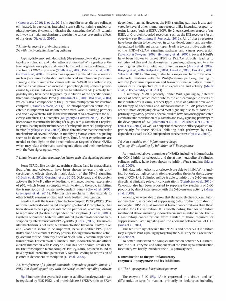
of GSK3β from the β-catenin complex. Furthermore, Gαs activatesadenylate cyclase, leading to the formation of cyclic adenosinemonophosphate (cAMP) and activation of protein kinase A (PKA)(Fujino et al., 2002). PKA, in turn, phosphorylates β-catenin at Ser675,thereby preventing its degradation by the proteasome pathway (Hinoet al., 2005). Additionally, recruited Gβ/γ activates phosphatidylinositol3-kinase (PI3K), which subsequently stimulates and phosphorylatesprotein kinase B (PKB/Akt) through protein dependent kinase 1(PDK1). PKB/Akt causes the phosphorylation and thereby, inactivationof GSK3β, which leads to stabilization and nuclear translocation of β-catenin (Castellone et al., 2005). In the nucleus, β-catenin bindstogether with TCF/LEF to the promoter regions of several genes whichare involved in proliferation, invasion, angiogenesis, survival, or thedevelopment of CSC and other tumor cells (Fig. 3). Consequently,inhibition of COX-2 activity by NSAIDs thwarts these effects, whichmay account for the anti-carcinogenic influence of these substances(Kaur & Sanyal, 2010; Vaish & Sanyal, 2012; S. Zhang et al., 2014).
However, recent studies provided strong evidence suggesting thatin addition to COX-2 inhibition, COX-2-independent mechanismscontribute to the anti-tumorigenic effects of these drugs. Apparently,
Fig. 3.Modes of action of NSAIDs on the Wnt signaling pathway. In the absence of a Wnt stimcomplex” that includes the tumor suppressors, axin and adenomatous polyposis coli (APC), the Sβ-TrCP.WhenWnt binds to the surface receptors, Frizzled and LRP5/6 (low-density lipoproteincatenin degradation complex, consisting of adenomatous polyposis coli (APC), axin, and glycoB) which phosphorylates and thereby inactivates GSK3βb (Cohen & Frame, 2001). Other proteiPKG inhibits β-catenin expression on the transcriptional level and blocks its transcriptional trCrosstalk between EP2 receptors and the β-catenin pathway: Binding of PGE2 to EP2 receptconcomitantly binds axin through its regulator of G-protein signaling (RGS)-domain. The bininactivated by PKB, which leads to stabilization and nuclear translocation of β-catenin. Gαs aPKA. PKA stabilizes β-catenin by phosphorylation. In the nucleus, β-catenin binds, togetherinvasion, angiogenesis, survival or the development of cancer stemcells (CRC). Transcriptional alike NF-κB and PPARγ/RXRα leading to a repression of β-catenin/TCF-dependent genes. A numproteins at different levels of theWnt signal transduction cascade. APC, adenomatous polyposiscyclooxygenase; Dvl, disheveled; FOXO4, Forkhead-Box-Protein 4; Fz, Frizzled; GRG, Grouchlipoprotein receptor-related protein 5/6; mPGES1, microsomal prostaglandin E2 synthase 1; PDPK A/B/G, protein kinase A/B/G; PP2A, protein phosphatase 2A; PPARγ, Peroxisome proliferatretinoid X receptor.
each NSAID influences a more or less separate COX-independent target(Grosch et al., 2001; Grösch et al., 2003; Soh &Weinstein, 2003; Gröschet al., 2005, 2006). NSAIDs also influence the Wnt/β-catenin pathwaythrough a number of COX-independent targets. Some of these targetsand related molecular mechanisms leading to interference with theWnt/β-catenin pathway are described below and illustrated in Fig. 3.The mechanisms of suppression of canonical Wnt signaling by 5-LO in-hibitors are described in Section 9.
7.2. Interference of protein kinaseGwith the Wnt/β-catenin signaling pathway
In human colon cancer cells, sulindac and its metabolite, sulindac sul-fone (exisulind), block the activity of guanosine 3′,5′-monophosphate(cGMP)-specific phosphodiesterases (PDE) 2 and 5, thus, leading tosustained increases in cellular levels of cGMP and activation of cGMP-dependent protein kinase (PKG) (W.J. Thompson et al., 2000).
Active PKG inhibitsWnt/β-catenin signaling, on the onehand, by de-creasing β-cateninmRNA expression, and on the other hand, by depriv-ing β-catenin of the TCF/LEF complex, attaching it to active FOXO4
ulus, the transcriptional coactivator, β-catenin, is degraded by a multiprotein “destructioner/Thr kinases GSK3β and CK1, protein phosphatase 2A (PP2A), and the E3-ubiquitin ligase(LDL)-receptor related proteins 5 and 6), disheveled (Dvl) is activated. This inhibits the β-gen synthetase kinase 3b (GSK3β) (Moon et al., 2004), by activating PKB (Protein kinasen kinases, like activated PKG (Protein kinase G), also interfere with the β-catenin pathway.ansactivation activity, in combination with TCF, by sequestration of β-catenin by FOXO4.ors provokes the release of Gβ/γ subunits, which stimulate PKB through PI3K, and Gαs
ding of Gαs to axin releases GSK3β from the complex and GSK3β is phosphorylated andlso activates adenylate cyclase (AC) leading to the formation of cAMP and activation ofwith TCF/LEF, to the promoter region of several genes that are involved in proliferation,ctivity ofβ-catenin/TCF/LEF complex can be inhibitedby several other transcription factors,ber of NSAIDs (highlighted in red) suppress Wnt signaling by targeting several regulatorycoli protein; cAMP, cyclic adenosinemonophosphate; GSK3β; CK1α, casein kinaseα; COX,o; GSK3β, glycogen synthase kinase 3β; HDAC, histone deacetylase; LRP5/6, low densityK-1,3-phosphoinositide dependent protein kinase-1; PI3K, phosphatidylinositol 3-kinase;or-activated receptor gamma; TCF/Lef, T-cell factor/lymphoid enhancer factor; LEF, RXR,
53J. Roos et al. / Pharmacology & Therapeutics 157 (2016) 43–64
(Kwon et al., 2010; Li et al., 2013). In ApcMin mice, dietary sulindaceliminated, in particular, intestinal stem cells containing nuclear orphosphorylated β-catenin, indicating that targeting the Wnt/β-cateninpathway is a major mechanism to explain the cancer-preventing effectsof this drug (Qiu et al., 2010).
7.3. Interference of protein phosphatase2A with the β-catenin signaling pathway
Aspirin, diclofenac, sulindac sulfide (the pharmacologically activeme-tabolite of sulindac), and indomethacin diminished Wnt signaling at thelevel of gene transcription in different human colon cancer cell lines, irre-spective of COX-2 expression (Smith et al., 2000; Dihlmann et al., 2003;Gardner et al., 2004). This effect was apparently related to a decrease innuclear β-catenin localization and enhanced membranous β-cateninstaining in the human colon cancer cell line, SW480. In another study,Dihlmann et al. showed an increase in phosphorylated β-catenin proteincaused by aspirin that was not only due to enhanced GSK3β activity, butpossibly may have been triggered by inhibition of the specific serine/threonine-phosphatase, PP2A (Dihlmann et al., 2003; Bos et al., 2006),which is also a component of the β-catenin multiprotein “destructioncomplex” (Stamos & Weis, 2013). The phosphorylation status of β-catenin is important for its interaction with various binding partnersand also plays a pivotal role in the recruitment of co-activators to the nu-clear β-catenin/TCF/LEF complex (Daugherty & Gottardi, 2007). PP2A hasbeen shown to control the binding of CBP/p300 to β-catenin/TCF regulat-ed genes, leading to themaintenance of embryonic stem cell pluripotencyin mice (Miyabayashi et al., 2007). These data indicate that themolecularmechanisms of several NSAIDs in modifying Wnt/β-catenin signalingseem to be dependent on the cell type. Thus, further experiments areneeded to shed light on the direct molecular targets of these NSAIDswhich may relate to their anti-carcinogenic effects and their interferencewith the Wnt signaling pathway.
7.4. Interference of other transcription factors with Wnt signaling pathway
SomeNSAIDs, like diclofenac, aspirin, sulindac (and its metabolites),ibuprofen, and celecoxib, have been shown to exert their anti-carcinogenic effects through manipulation of the NF-κB signaling(Grösch et al., 2006; Gurpinar et al., 2013). Diclofenac and ibuprofenactivate the NF-κB pathway, leading to enhanced nuclear expressionof p65, which forms a complex with β-catenin, thereby, inhibitingthe transcription of β-catenin-dependent genes (Cho et al., 2005;Greenspan et al., 2011). Whether this mechanism also extends toother NSAIDS remains unclear and needs further investigation.
Besides NF-κB, the transcription factor-complex, PPARγ/RXRα (Per-oxisome Proliferator-Activated Receptor γ/Retinoid X receptor α), hasbeen shown to be a physical interaction partner of β-catenin, leadingto repression of β-catenin-dependent transcription (Lu et al., 2005).Eighteen of nineteen tested NSAIDs inhibit β-catenin-dependent tran-scription by interferencewith PPARγ/RXRα (Lu et al., 2005). Themolec-ular mechanism is not clear, but transactivation between PPARγ/RXRαand β-catenin seems to be important, because neither PPARγ norRXRα alone nor a mutant PPARγ protein, lacking transactivation activi-ty, account for the inhibitory effect of NSAIDs on β-catenin-dependenttranscription. For celecoxib, sulindac sulfide, indomethacin and others,a direct interaction with PPARγ or RXRα has been shown. Besides NF-κB, the transcription factor complex, PPARγ/RXRα, has been found tobe a physical interaction partner of β-catenin, leading to repression ofβ-catenin-dependent transcription (Lu et al., 2005).
7.5. Interference of 3-phosphoinositide-dependent protein kinase-1/PDK1/Akt signaling pathwaywith theWnt/β-catenin signaling pathway
Fig. 3 indicates that cytosolicβ-catenin stabilization/degradation canbe regulated by PI3K, PDK1, and protein kinase B (PKB/Akt) in an EP2/4
dependent manner. However, the PI3K signaling pathway is also acti-vated by several other membrane receptors, like integrins, receptor ty-rosine kinases (such as EGFR, VEGFR, Her2neu), cytokine receptors (e.g.IL2R), or G-protein coupled receptors, such as the EP2 receptor (for anoverview see Hemmings & Restuccia, 2012). All of these receptorshave been shown to be involved in cancer development and are oftenderegulated in different cancer types, leading to constitutive activationof the PI3K→PKB/Akt signaling pathway and cancer progression(Vivanco & Sawyers, 2002; Hennessy et al., 2005). Several NSAIDshave been shown to target PDK1 or PKB/Akt directly, leading toinhibition of this and the downstream signaling pathway and to anti-carcinogenic effects in vitro and in vivo in mice (Arico et al., 2002;Baoping et al., 2004; Kulp et al., 2004; Lee et al., 2005; Fan et al., 2006;Setia et al., 2014). This might also be a major mechanism by whichcelecoxib interferes with the Wnt/β-catenin pathway, leading toreduced β-catenin expression and transcriptional activity in humancancer cells, irrespective of COX-2 expression and activity (Maieret al., 2005; Sareddy et al., 2013).
In summary, NSAIDs potently inhibit Wnt signaling by differentmodes of action, which contribute to the anti-carcinogenic effects ofthese substances in various cancer types. This is of particular relevancefor therapy of adenomas and adenocarcinomas in FAP patients andother tumors displaying elevated Wnt signaling due to mutations inpathway-regulatory proteins. Several studies have convincingly indicateda concomitant contribution of β-catenin and PGE2 signaling pathways tothe development of CSC (Ishimoto et al., 2010; Al-Kharusi et al., 2013;Femia et al., 2013) as well as a superior therapeutic efficacy against CRC,particularly for those NSAIDs inhibiting both pathways by COX-dependent as well as COX-independent mechanisms (Qiu et al., 2010).
7.6. Non-steroidal anti-inflammatory drugsaffecting Wnt signaling by inhibition of 5-lipoxygenase
As mentioned above, a number of NSAIDs including indomethacin,the COX-2 inhibitor celecoxib, and the active metabolite of sulindac,sulindac sulfide, have been shown to inhibit Wnt signaling (Maieret al., 2005).
Sulindac, indomethacin, or celecoxib are able to inhibit Wnt signal-ing, but only at high concentrations, exceeding those for the suppres-sion of COX-1/-2. Sulindac sulfide is able to inhibit the 5-LO enzymedirectly at clinically relevant concentrations (Steinbrink et al., 2010).Celecoxib also has been reported to suppress the synthesis of 5-LOproducts by direct interference with the 5-LO enzyme activity (Maieret al., 2008).
Recently, wewere able to show that the non-selective COX inhibitor,indomethacin, is capable of suppressing 5-LO product formation inmonocytic THP-1 cells at somewhat higher concentrations than thoseneeded for COX inhibition. It is worth noting that for inhibitorsmentioned above, including indomethacin and sulindac sulfide, the 5-LO-inhibitory concentrations were similar to those required forsuppression of Wnt signaling and the aberrant self-renewal of LSCs(Roos et al., 2014).
This led us to hypothesize that NSAIDs and other 5-LO inhibitorsmay suppressWnt signaling by targeting the 5-LO enzyme, as describedin Section 9.
To better understand the complex interaction between 5-LO inhibi-tors, the 5-LO enzyme, and components of the Wnt signal transductionmachinery we briefly introduce the 5-LO pathway here.
8. Introduction to the pro-inflammatoryenzyme 5-lipoxygenase and its inhibitors
8.1. The 5-lipoxygenase biosynthetic pathway
The enzyme 5-LO (Fig. 4A) is expressed in a tissue- and celldifferentiation-specific manner, primarily in leukocytes including

54 J. Roos et al. / Pharmacology & Therapeutics 157 (2016) 43–64
neutrophils, basophils, eosinophils, monocytes/macrophages, dendriticcells, mast cells, and B-lymphocytes (Radmark et al., 2007). The expres-sion level of 5-LO is regulated by promoter methylation and differentia-tion inducers [dimethyl sulfoxide (DMSO), retinoic acid, 1α, 2,5-dihydroxyvitamin D3 and TGF-β] upregulate 5-LO in immature myeloidcells (Radmark et al., 2007). Mechanistically, 5-LO catalyzes the inser-tion of molecular oxygen into polyunsaturated fatty acids, such as ara-chidonic acid (AA). Upon activation by Ca2+ and/or phosphorylationevents, 5-LO translocates from the cytosol or the nucleoplasm to the nu-clear envelope and (I) catalyzes the insertion ofmolecular oxygen at C-5
Fig. 4. Regulation of cellular 5-LO and COX activity. (A) Depending on the cell type, 5-LO reside2002). Though nuclear localization sequences are present in the primary sequence of 5-LO, thefactors such as testosterone (Pergola et al., 2008) or cell adherence (Brock, 2005) may affect tperinuclear regionwhere 5-LO product synthesis may occur (Werz, 2002; Brock, 2005; Newcomonists like platelet-activating factor (PAF), LTB4, formyl-methionyl-leucyl-phenylalanine (fMLP)translocates to the nuclear membrane (Werz, 2002) where it is anchored to phosphatidylcholiphorylation of 5-LO at the C-terminal catalytic domain can modulate its subcellular localizationprotein kinase (MK)-2/3 (Werz et al., 2000) and ERK1/2 (Werz et al., 2002a, 2002b) phosphorunsaturated fatty acids (i.e. AA) (Werz et al., 2002b). Interestingly, phosphorylation of 5-LO isWerz et al., 2001; Fredman et al., 2014) where it is anchored to phosphatidylcholine (PC) in aof 5-LO at the C-terminal catalytic domain can modulate its subcellular localization as well as5-LO (at Ser523), thus impairing the catalytic activity of 5-LO (Luo et al., 2004). Moreover, mand monocytes (Pergola et al., 2008; Pergola et al., 2011). Novel phosphorylation sites were renases Fgr, hematopoietic cell kinase (HCK) and Yes (Markoutsa et al., 2014). In parallel to 5-LOand releases AA from phospholipids (Leslie, 2004). The free AA is then transferred by the 5-LO-tabolism to LTA4 (Evans et al., 2008). FLAP is essential for cellular 5-LO product synthesis, and tfunction as a scaffold for 5-LO at the nuclear membrane (Mandal et al., 2008; Bair et al., 2012). Isequent transformation of LTA4 to LTB4 and LTC4, respectively (Newcomer & Gilbert, 2010). (B)membrane and releases AA from phospholipids. Two isoforms of cyclooxygenase, COX-1 and COther converted by a number of synthases to various eicosanoids, including PGE2, PGI2, and TXA2
different physiological processes including gastrointestinal integrity. COX-2 is detected negligibsites of inflammation. AA, arachidonic acid, CLP, coactosine-like protein; cPLA2, cytosolic phospkinase; LT, leukotriene;MK-2/3,MAPK-activatedprotein kinase; PKA, protein kinase A; PGE2, prtaglandin E2, TXA2 Thromboxane A2, TXAS, Thromboxane A2 synthase.
of AA that is provided by the5-LO-activating protein (FLAP), yielding theintermediate 5(S)-hydroperoxy-6-trans-8,11,14-cis-eicosatetraenoicacid (5(S)-HpETE). Furthermore, it catalyzes (II) the dehydration of5(S)-HpETE to the epoxide leukotriene (LT)A4 (Radmark et al., 2015).Alternatively, 5(S)-HpETE can be reduced by glutathione peroxidases(GPx) to the corresponding alcohol, 5(S)-HETE,which can be further ox-idized to 5-oxo-ETE (Powell & Rokach, 2015).
Dependingon the cell type and enzymes expressed, LTA4 can be con-verted by soluble LTA4 hydrolase to LTB4 and/or conjugated with gluta-thione by LTC4 synthase to yield LTC4 (Bair et al., 2012).
s as a soluble protein either in the cytosol or in the nucleoplasm of resting cells (O. Werz,way in which the distinct subcellular localization is organized is unknown, but systemic
he subcellular redistribution. In activated cells, however, 5-LO is consistently found at theer & Gilbert, 2010; Bair et al., 2012). Upon cell activation by Ca2+-ionophores, soluble ag-and C5a, aswell as phagocytic particles such as zymosan, urate or phosphate crystals, 5-LOne (PC) in a Ca2+-dependent manner via the C2-like domain (Kulkarni et al., 2002). Phos-as well as its activation for product synthesis. The p38 MAPK-regulated MAPK-activatedylate 5-LO at Ser-271 and Ser-663, respectively, an action which is strongly promoted byassociated with increased accumulation of 5-LO at the nuclear membrane (Werz, 2002;Ca2+-dependent manner via the C2-like domain (Kulkarni et al., 2002). Phosphorylationits activation for product synthesis. Protein kinase A was also reported to phosphorylateoderate stimulation of ERK by androgens reduced 5-LO product formation in neutrophilscently identified at Tyr42, Tyr53 and either Tyr94 or Tyr445 phosphorylated by the Src ki-, the Ca2+-dependent cytosolic phospholipase A2 (cPLA2) moves to the nuclear membraneactivating protein (FLAP), an integral nuclear membrane protein, to 5-LO for efficient me-ogether with coactosine-like protein (CLP) that interacts with 5-LO as well, FLAP seems ton addition, LTC4S and LTA4Hmay participate in this multi-protein assembly to enable sub-Cyclooxygenase pathway. Cytosolic phospholipase A2 (cPLA2) translocates to the nuclearX-2, catalyze the conversion of arachidonic acid to prostaglandin H2 (PGH2), which is fur-
. COX-1 is constitutively expressed inmany tissues and is important for themaintenance ofly inmost tissues but can be rapidly and strongly induced by cytokines and cellular stress atholipase A2, FLAP, 5-LO activating protein; COX, cyclooxygenase; HCK, hematopoietic cellostaglandin E2, PGES, prostaglandin E2 synthase, PGIS, prostaglandin I2 synthase, PGI2 pros-

55J. Roos et al. / Pharmacology & Therapeutics 157 (2016) 43–64
Release of LTC4 into the extracellular environment and successiveamino acid (γ-glutamyl residue and glycine) cleavage yields LTD4 andthen LTE4. LTB4 is a potent chemotactic and chemokinetic mediator, ac-tivates granulocytes for degranulation, superoxide and nitrogen oxideproduction, enhances the phagocytic activity of neutrophils andmacro-phages, and stimulates secretion of immunoglobulins by lymphocytes(Haeggstrom & Funk, 2011). The cys-LTs, in particular LTD4, causesmooth muscle contraction, mucus secretion, vasoconstriction, plasmaextravasation, recruitment of eosinophils, and fibrocyte proliferation(Peters-Golden & Henderson, 2007; Haeggstrom & Funk, 2011).
Thus, LTs play roles in the immune response and in the pathogenesisof inflammatory diseases, such as arthritis, asthma, allergic rhinitis, andother chronic inflammatory disorders (Peters-Golden & Henderson,2007; Haeggstrom & Funk, 2011). Furthermore, 5-LO products contrib-ute to atherosclerosis (Poeckel & Funk, 2010) andmay regulate prolifer-ation and survival of cancer cells (Greene et al., 2011). Finally, 5-oxo-ETE may play important roles in asthma and allergic diseases, cancer,and cardiovascular disease as well (Powell & Rokach, 2015). The pro-inflammatory and immunomodulatory actions of LTs are mediatedby specific G protein-coupled receptors (GPCRs), the BLT1 and BLT2receptors for LTB4 and the CysLT1 and CysLT2 receptors for cys-LTs(Back et al., 2011).
8.2. 5-LO inhibitors
In order to intervene pharmacologically with 5-LO product synthe-sis, inhibition of cPLA2, antagonism of FLAP, and inhibition of 5-LO areall conceivable strategies (see (Pergola & Werz, 2010) for review). Di-rect 5-LO inhibitors can be categorized into four classes, according totheir inhibitorymode of action: (1) redox-active 5-LO inhibitors that re-duce the active site iron or scavenge activating hydroperoxides andthereby uncouple the catalytic cycle of the 5-LO enzyme; (2) iron-chelating agents, containing hydroxamic acid or N-hydroxyurea groupsthat form complexes with the active site iron; (3) non-redox-type in-hibitors that compete with AA or lipid hydroperoxides for binding to5-LOdevoid of redox properties; and (4) allosteric inhibitors that essen-tially act at the regulatory C2-like domain, e.g., by antagonizing thestimulatory effect of ATP or by interference with stimulatory effects ofphospholipids or diglycerides. In particular, for the latter group, severalstructurally different compounds with apparently distinct binding sites[e.g. indirubins at the ATP-binding site (Pergola et al., 2014), hyperforinat the PC-binding site (Feisst et al., 2009)] were recently reported withpromising molecular mechanisms and favorable pharmacodynamics.Nevertheless, despite intensive efforts to develop 5-LO inhibitors andthe resulting plethora of reported compounds, only the iron-ligand che-lator, zileuton, has become amarketed drug. In fact, all other 5-LO inhib-itors so far investigated failed in preclinical or clinical studies due toside-effects (e.g. methemoglobin formation, liver toxicity), pharmacoki-netic issues, or lack of efficacy (Pergola & Werz, 2010). Especially withnon-redox-type 5-LO inhibitors, phosphorylation of 5-LO and an elevat-ed oxidative tone in the cell can decrease their potency (O. Werz &Steinhilber, 2005).
9. Modulation by 5-LO and 5-LOinhibitors of Wnt signaling in leukemic stem cells
Over the last couple of years, evidence has been increasing for a keyrole of deregulated canonicalWnt signaling in the pathogenesis of hema-tological malignancies (HMs), though the exact underlying mechanismsremain poorly understood (Seke Etet et al., 2013). In AML and CML, therole of Wnt-signal activation for the pathogenesis and the maintenanceof the LSCs is well recognized (Zhao et al., 2007; Wang et al., 2010;Heidel et al., 2012). Deregulated activation ofWnt signaling leads to aber-rant self-renewal of the LSC in AML and CML and is fundamental to LSCmaintenance (Zhao et al., 2000; Passegue et al., 2003; Jamieson et al.,2004). Furthermore, it has been shown that not only Wnt signaling, but
also 5-LO is important for the maintenance of LSCs and the pathogenesisof AML and CML (Y. Chen et al., 2009; Roos et al., 2014). Although only asmall number of publications have addressed the role of 5-LO in the path-ogenesis of HMs, there is increasing evidence for an important role of 5-LO in the development of these diseases.
9.1. Disturbed 5-LO expression in leukemic cells
Differential 5-LO expression has been found in several types of pri-mary leukemic cells from patients with pre-B-cell ALL (Feltenmarket al., 1995; Vincent et al., 2008), CML (Radich et al., 2006; Grahamet al., 2007), AML (Valk et al., 2004; Vincent et al., 2008), B-cell chroniclymphocytic leukemia (B-CLL) (Rosenwald et al., 2001; Stratowa et al.,2001; Runarsson et al., 2005; Guriec et al., 2014), and mantle cell lym-phoma (MCL) (Boyd et al., 2009; Mahshid et al., 2009). Intriguingly,all these types of HM exhibit a deregulated Wnt/β-catenin signaling(Roman-Gomez et al., 2007; Zhao et al., 2007; Gelebart et al., 2008;Wu et al., 2009; Chung et al., 2010; Trowbridge et al., 2010; Wanget al., 2010; Kuhnl et al., 2011; Perez-Galan et al., 2011; Tandon et al.,2011; Heidel et al., 2012), suggesting an interrelation between 5-LOfunction and the Wnt/β-catenin signaling pathway.
One of the first studies to indicate involvement of 5-LO in HMs wasperformedby Tskuada et al. in 1986. In this study, treatment of leukemiccells with the competitive 5-LO inhibitor, AA-861, led to reduced cellproliferation which was accompanied by a reduction in cellular DNA,RNA, and protein synthesis. One of the conclusions of this study wasthat LTs play an important role in cancer cell viability (Tsukada et al.,1986). Similar results were obtained by Anderson et al., who observedthat primary human CML blast cells in culture, as well as leukemic celllines, showed reduced cell proliferation after treatment with different5-LO inhibitors (Anderson et al., 1995). Additionally, 5-LO inhibitionby AA-861 increased apoptosis in eicosapentaenoic acid (EPA)-treatedleukemic HL-60 cells (Gillis et al., 2007).
In a subsequent study, inhibition of 5-LO and FLAP was found to in-duce apoptosis inMCL cell lines and primary CLL cells, suggesting an im-portant role for 5-LO and/or its products in MCL and other B cellmalignancies. This was supported by the fact that 5-LO was up-regulated ~7-fold in MCL cells compared to normal B cells (Boyd et al.,2009). In addition, Runarsson et al. observed that treatment of B-CLLcells with the iron-ligand 5-LO inhibitor BWA4C and FLAP inhibitorMK886 counteracted CD40-dependent activation of these cells byinhibiting CD40-induced DNA synthesis and CD40-induced expressionof CD23, CD54, and CD150. This finding was likely LT-dependent, sinceaddition of exogenous LTB4 almost completely reversed the observedeffects (Runarsson et al., 2005).
It was also shown that the highly progressing disease in B-CLL pa-tients is associated with overexpression of 5-LO and other componentsof the 5-LO pathway, such as cPLA2 and BLT2 (Guriec et al., 2014). Theexpression levels of BLT1 and LTA4 hydroxylase were also significantlyraised in B-CLL cells compared to healthy B-cells. However, no statisti-cally significant correlation was found with the stage of the disease.Increased cPLA2α and LTC4 synthase expression was also found inleukemic granulocytes from AML patients. However, the formation ofLTB4 was also suppressed in the same patient samples (Stenke et al.,1998; Runarsson et al., 2007).
Taken together, it has been shown that in many different types ofleukemia there is an altered expression level of 5-LO protein and otherprotein components involved in LT biosynthesis suggesting a role ofthe 5-LO pathway in the pathogenesis of leukemia. Additionally, inhib-itor studies revealed a function of 5-LO products in proliferation andapoptosis of leukemic cells with particular relevance to B-CLL cells.
9.2. 5-LO and acute myeloid leukemia
Recently, it was shown that 5-LO is strongly upregulated in AML1/ETO-positive AML (DeKelver et al., 2013). The fusion protein AML1/

56 J. Roos et al. / Pharmacology & Therapeutics 157 (2016) 43–64
ETO is the result of the chromosomal t(8;21) translocation (transfer ofgenetic material between chromosome 8 and chromosome 21), whichtogether with t(15;17) (PML/RARα) is one of the most common trans-locations associated with AML. These leukemia-associated fusion pro-teins (LAFP) are also known to induce aberrant activation of canonicalWnt signaling which, as a result, leads to an increased self-renewal ca-pacity of LSCs (Tenen, 2003; Muller-Tidow et al., 2004; Zheng et al.,2004; Zhao et al., 2007;Wang et al., 2010). DeKelver et al. analyzed pub-lically available microarray data from AML M2 patients with and with-out the t(8;21) translocation (Valk et al., 2004). They found that 5-LOexpression was ~2.3-fold higher in t(8;21) patients than in patientswithout the translocation. Notably, both patient groups had elevated5-LO levels relative to normal CD34+ control samples (DeKelver et al.,2013). Additionally, it could be shown, by gene expression microarrayand chip analysis, that 5-LO is a potential disease-related target geneof AML1/ETO (Lo et al., 2012). AML1/ETO, as well as its C-terminallytruncated leukemic isoform, AML1/ETOex9, is able to up-regulate 5-LOvia the transcription factor, KLF6 (Krppl-like factor 6), a protein whichis also known to be a cell-specific positive regulator of LTC4 synthase(Zhao et al., 2000). Furthermore, studies with KLF6−/− mice revealedthat 5-LO is also critically required for embryonic stem cell differentia-tion and early hematopoiesis (Matsumoto et al., 2006).
Thus, 5-LO is a direct disease-related and pathophysiologicallyrelevant target gene upregulated by the oncogenic leukemia associatedfusion protein, AML1/ETO, which is crucially involved in the inductionand maintenance of AML.
9.3. 5-LO and self-renewal capacity of leukemic cells
To analyze the effect of 5-LO on the in vitro self-renewal capacityof the LAFPs AML1/ETOex9, PML/RARα, and MLL-AF9 (translocationt(9;11)), DeKelver et al. performed serial replating assays using wildtype and 5-LO−/− murine total bone marrow cells that were trans-duced with either control vector or LAFPs. 5-LO deficiency led to de-creased in-vitro self-renewal capacity with all LAFPs. Remarkably, lossof 5-LO abolished completely the aberrant replating capacity of AML1/ETOex9-positive cells. Nevertheless, AML1/ETOex9-expressing 5-LO−/− cells were still capable of leukemia induction. These results ledthe authors to hypothesize that in the murine AML1/ETOex9 leukemiainduction model, other factors may exist that allow AML1/ETOex9to overcome these defects. It is also possible that 5-LO expressionmay be required to a greater extent by AML1/ETO than by AML1/ETOex9 cells in leukemia development, especially since AML1/ETO up-regulates KLF6 more strongly than AML1/ETOex9. Another reasoncould be that in the in-vitro and in-vivo cell culture experiments, theauthors used different cell populations, containing different ratios ofHSCs (total bonemarrow versus fetal liver cells), suggesting that differ-ent subtypes of HSCs respond differently to the introduction of AML1/ETOex9 and 5-LO deficiency (DeKelver et al., 2013).
Taken together, these experiments demonstrated the impact of 5-LOon the self-renewal capacity of LSC. Thus, 5-LO seems to be involved inthe maintenance of the undifferentiated state of these leukemic cellswhich is a characteristic feature of LSCs.
9.4. 5-LO and human leukemic stem cell function
5-LO also seems to be involved in human CML stem cell function, asrevealed by microarray studies. In these studies, 5-LO was differentiallyexpressed in human CD34+ CML cells, compared to CD34+ controlcells (Radich et al., 2006; Graham et al., 2007). In a recent publication,the expression levels of 5-LO and BLT1 were decreased both in bloodsamples and CD34+ stem cells from t(9;22)-positive CML patients har-boring the BCR/ABL fusion protein, compared to samples from healthydonors (Lucas et al., 2014). On the other hand, it was shown that 5-LOis up-regulated in BCR/ABL-positive GFP+Lin−ckit+Sca1+ LSCs in com-parison to non-BCR/ABL expressing GFP+Lin−ckit+Sca1+ cells and that
5-LO is indispensable for the aberrant self-renewal capacity of theseLSCs and the development of CML in mice (Y. Chen et al., 2009). Thiswas revealed using amurine BCR/ABL leukemia inductionmodel. Recip-ients of BCR/ABL transduced bone marrow cells from 5-LO−/− donormice failed to develop CML, whereas recipients of BCR/ABL transducedbone marrow cells from wild type donor mice developed the diseaseand died within 4 weeks. In the absence of 5-LO, myeloid leukemiacells gradually disappeared in the CML mice. A functional defect in nor-mal HSCs could not be detected. 5-LO deficiency led only to an impair-ment of LT-LSCs, preventing these cells from developing CML. Thisdefective function of LSCs in the absence of 5-LO was correlated with areduction ofβ-catenin expression levels, suggesting a further regulatorylink between 5-LO and Wnt signaling.
The effect of 5-LO deficiency on BCR/ABL-positive LSCs was furtherinvestigated in studies using the approved iron-chelating 5-LO inhibitorzileuton. Zileuton led to impairment of BCR/ABL positive LSCs in a sim-ilar manner to that observed with 5-LO knock-out. Pharmacologicalsuppression of 5-LO by zileuton in mice with BCR/ABL-induced CMLprolonged their survival compared to control mice. An even better ther-apeutic effect could be achieved with imatinib, a first generation BCR/ABL tyrosine kinase inhibitor, in combination with zileuton. However,BCR/ABL-induced lymphoid leukemia, which originates from morecommitted lymphoid progenitors than BCR/ABL-induced CML, was notaffected by 5-LO deficiency (S. Li et al., 1999), suggesting that 5-LOis specifically required by CML stem cells and not by ALL stem cells (Y.Chen et al., 2009). These results were followed by a clinical phase Istudy in which zileuton was tested in combination with imatinib forthe treatment of CML (Safety of Zileuton (Zyflo) in CombinationWith I-matinib Mesylate (Gleevec) in CML; http://clinicaltrials.gov trial No.NCT01130688). This study has been completed but the results still re-main to be published. Another phase I study for treatment of BCR/ABL-positive CMLwith zileuton in combinationwith dasatinib, a secondgeneration BCR/ABL tyrosine kinase inhibitor, is currently ongoing(Evaluating the Safety of Zileuton (Zyflo®) in Combination WithDasatinib (Sprycel®) in Chronic Myelogenous Leukemia; http://clinicaltrials.gov trial No. NCT02047149).
Recently, we demonstrated that 5-LO is also crucial for the mainte-nance of LSC in a PML/RARα-positive stem cell model of AML (Rooset al., 2014). First, we performed inhibitor studies on Sca1+Lin−murine hematopoietic stem and progenitor cells (HSPC). The HSPCwere either transducedwith PML/RARα or an empty vector. As 5-LO in-hibitors, we used the non-redox type inhibitor CJ-13,610 and zileuton.These inhibitors were both able to abolish the aberrant replating effi-ciency of PML/RARα-expressing HSPCs in a concentration dependentmanner. The same inhibitory effect could be observed on the LT- andST-stem cell capacity. No cytotoxic effect could be observed after treat-ment of the non-PML/RARα-positive control cells, which is consistentwith the work of Y. Chen et al. (2009). Furthermore, the occurrence ofstem cell suppression coincided with inhibition of Wnt signaling. Thiswas shownby immunohistochemical staining ofmurine spleen coloniesinduced by PML/RARα against β-catenin. Treatment with CJ-13,610 ledto reversal of the PML/RARα-activation of Wnt signaling.
Collectively, these experiments confirmadirect link between the 5-LOandWnt signaling pathways which is indispensable for the maintenanceof LSCs. Accordingly, genetic andpharmacological inhibition of 5-LO led toan impairment of the LSC in AML andCML cells supporting the hypothesisof an regulatory interaction of both pathways. Additionally, pharmacolog-ical inhibition of 5-LO significantly increased the therapeutic effect of ima-tinib, a classical therapeutic agent used in the standard therapy of CML.Thus, a combination of standard therapeutics and 5-LO inhibitors couldbe beneficial for the clinical outcome of these patients.
9.5. Potential mechanism of 5-LO interaction with Wnt signaling
To confirm and investigate more extensively the involvement of 5-LO in the regulation of the Wnt signaling pathway, we performed co-

57J. Roos et al. / Pharmacology & Therapeutics 157 (2016) 43–64
immunoprecipitation experiments as well as reporter gene assays andimmunofluorescence microscopy in different cancer cell lines. In thesestudies, we demonstrated that enzymatically inactive 5-LO traps β-catenin at the nuclear envelope (Fig. 5), prevents the translocation ofβ-catenin to the nucleus, and therefore, suppresses the PML/RARα-induced activation of Wnt target genes. Additionally, we observed that5-LO directly interacts with β-catenin (Roos et al., 2014), suggesting aconnection between Wnt signaling and the 5-LO pathway representedby a 5-LO/β-catenin association. However, an interaction of 5-LO withadditional components of the Wnt signaling pathway could not beexcluded. Furthermore, the exact molecular mechanism(s) by whichinactive 5-LO inhibits the translocation of β-catenin to the nucleus andinteracts with β-catenin and possibly other Wnt pathway componentsstill needs detailed experimental investigation.
9.6. 5-LO and Wnt signaling in solid tumors
Increasing evidence in the literature implicates 5-LO as a player inthe growth of several solid tumor types, including pancreatic, colorectal,prostate and breast cancers (Kort et al., 1992; Ding et al., 1999a, 1999b;Gupta et al., 2001; Hennig et al., 2002; Jiang et al., 2003; Soumaoro et al.,2006; Ihara et al., 2007; Bishayee & Khuda-Bukhsh, 2013) and the Wntsignaling pathway contributes to the pathogenesis of these cancers (Liuet al., 2000; Milovanovic et al., 2004; Yardy & Brewster, 2005;Yokoyama et al., 2014). However, even though a correlation between5-LO and Wnt signaling in solid tumors is obvious, at the moment nostudy has yet been published that directly addresses the relation be-tween 5-LO or the effects of 5-LO inhibitors and theWnt signaling path-way in these tumors types.
Fig. 5. Proposedmechanism ofWnt signaling suppression by 5-LO. Binding to β-catenin of enzysignaling pathway by preventing the translocation of β-catenin to the nucleus and subsequent gdeacetylase; TCF/LEF, T-cell factor/lymphoid enhancer factor.
10. Conclusion and future direction
Dysfunctional Wnt signaling is emerging as having pathophysiolog-ical significance in a number of diseases, including cancer, classicalinflammatory diseases, and neurodegenerative diseases triggered byneuroinflammatory processes.
Numerous high-throughput screens have been employed to obtainsmall-molecule targeted drugs that would interfere with componentsof Wnt signaling, mainly directed towards disruption of the binding ofβ-catenin to its nuclear target, TCF/LEF.
Many compounds with promising activity in cell culture have beenidentified and characterized, but translation into clinically applicabledrugs is still an apparently insurmountable obstacle due to lack ofselectivity and a number of pleiotropic off-target effects. This relatesparticularly to drugs that interfere with the proteasomal degradationof β-catenin, which results in severe side effects in normal tissue asnon-nuclearβ-catenin contributes to the stabilization of physiologicallyimportant intercellular adherens junctions.
The clinical value of silencingWnt signaling further upstream, i.e., atthe level of the Frizzled receptor, remains unclear as a number of tissuesdepend on a certain basic Wnt activity which may be associated withunknown adverse side effects.
Thus, a complex mixture of problems is encountered in developingWnt inhibitors using in silico methods or high-throughput screensusing compound libraries.
Several inhibitors of eicosanoid signaling, such as NSAIDs, have beenshown to interfere in a clinically relevant manner with Wnt signaling,by COX/PGE2 aswell as COX/PGE2-independentmechanisms and there-by, induce Wnt-dependent anti-tumorigenic effects (see Section 7).
matically inactive 5-LO, after treatmentwith the 5-LO inhibitor CJ-13610, inhibits theWntene transcriptional activity. For further details see Section 9. GRG, Groucho; HDAC, histone

58 J. Roos et al. / Pharmacology & Therapeutics 157 (2016) 43–64
Mechanistically, NSAIDs differ widely from classical Wnt inhibitors intheir actions on the Wnt signal transduction cascade and follow novelmechanistic routes of suppression ofWnt signaling. Furthermore, sever-al very recent studies presented here provide valid and comprehensiveevidence for an essential role of 5-LO in and interconnection with Wntsignaling in the pathogenesis of leukemia and the maintenance ofLSCs in certain sub-types of leukemia.
As a consequence, pharmacological 5-LO inhibition,with concomitantsuppression of Wnt signaling, is a potential novel approach to adjuvantstem cell therapy in AML and CML as well as other Wnt-dependent dis-eases, such as breast cancer, human head and neck squamous cell carci-nomas, and some inflammatory diseases (see Section 4) (Liu et al., 2013).
Further insight into the value of anti-5-LO therapy is likely to begained from two clinical trials — the recently finished study withzileuton and imatinib as combined treatment for CML, as well as thenew study with zileuton and dasatinib. Yet to fully understand therole of 5-LO in the Wnt signaling pathway (and possible otherleukemia-related pathways), more detailed and comprehensive studiesare necessary.
A number of approved NSAIDs, administered at high doses, suppressWnt signaling in patients but without causing severe concomitantsystemic toxicity in the organism. The majority of side effects ofthese drugs arewell documented and derives from suppression of pros-taglandin biosynthesis. This includes mainly cardiovascular and throm-boembolic events and gastrointestinal ulceration (Bhala et al., 2013).However, there are no reports of adverse effects or toxicity directly aris-ing from the Wnt-inhibitory activity of NSAIDs.
Thus, suppression of Wnt signaling by NSAIDs may follow advanta-geous mechanistic strategies and NSAIDs as well as 5-LO inhibitors,therefore, should be reassessed as a basis for the design of a new classof Wnt inhibitors.
Following the clarification of the exact molecular mechanism bywhich these drugs inhibitWnt signaling, it seems reasonable that chem-ical structures responsible for Wnt inhibition could be separated fromthe structural elements causing suppression of COX enzymes. The factthat a number of drugs targeting eicosanoid synthesis concomitantlysuppress Wnt signaling independently of COX could further indicate apossible common, but still unidentified key target for the regulation ofWnt activity.
In this respect, 5-LO inhibitors, such as CJ-13610, activate a poten-tially naturally occurring novel 5-LO-dependentmechanism of suppres-sion of Wnt signaling that is non-toxic, at least in mice, and might bewell tolerated in patients.
Future careful studies are needed to clarify the regulatory mecha-nism linking eicosanoid synthesis pathways to oncogenicWnt signalingas a potential basis for novel anti-inflammatory drugs that concomitant-ly suppress Wnt signaling.
Supplementary data to this article can be found online at http://dx.doi.org/10.1016/j.pharmthera.2015.11.001.
Conflict of interest statement
MJP is a consultant to Leo Pharma a/s and Xellia PharmaceuticalsApS. The other authors declare that there are no conflicts of interest.
Acknowledgment
Thisworkwas funded by theGermanResearch Foundation (DFGpro-jects MA-5825/1-1, GR2011/3-2) and the DFG SonderforschungsbereichSFB 1039/1, the Graduate School TRIP (Translational Research Innova-tion — Pharma), the European Research Council under the EuropeanUnion's Seventh Framework Programme (FP7/2007–2013; ERC grant268309) and the LOEWE Lipid Signaling Forschungszentrum Frankfurt(LIFF). MJP is supported by LOEWE Research Centre for TranslationalMedicine and Pharmacology, financed by the State of Hessen. T.J.M. is
recipient of a Heisenberg fellowship of the German Research Foundation(DFG-MA-5825-2-1).
References
Aberle, H., Bauer, A., Stappert, J., Kispert, A., & Kemler, R. (1997). Beta-catenin is a targetfor the ubiquitin–proteasome pathway. EMBO J 16, 3797–3804.
Abrahamsson, A. E., Geron, I., Gotlib, J., Dao, K. H., Barroga, C. F., Newton, I. G., et al. (2009).Glycogen synthase kinase 3beta missplicing contributes to leukemia stem cell gener-ation. Proc Natl Acad Sci U S A 106, 3925–3929.
Achiwa, H., Yatabe, Y., Hida, T., Kuroishi, T., Kozaki, K., Nakamura, S., et al. (1999). Prog-nostic significance of elevated cyclooxygenase 2 expression in primary, resectedlung adenocarcinomas. Clin Cancer Res 5, 1001–1005.
Aguilera, O., Gonzalez-Sancho, J. M., Zazo, S., Rincon, R., Fernandez, A. F., Tapia, O., et al.(2015). Nuclear DICKKOPF-1 as a biomarker of chemoresistance and poor clinicaloutcome in colorectal cancer. Oncotarget 6, 5903–5917.
Albring, K. F., Weidemuller, J., Mittag, S., Weiske, J., Friedrich, K., Geroni, M. C., et al.(2013). Berberine acts as a natural inhibitor of Wnt/beta-cateninsignaling—identification of more active 13-arylalkyl derivatives. Biofactors 39,652–662.
Alison,M. R., Lin,W. R., Lim, S. M., & Nicholson, L. J. (2012). Cancer stem cells: in the line offire. Cancer Treat Rev 38, 589–598.
Al-Kharusi, M. R., Smartt, H. J., Greenhough, A., Collard, T. J., Emery, E. D., Williams, A. C.,et al. (2013). LGR5 promotes survival in human colorectal adenoma cells and is up-regulated by PGE2: implications for targeting adenoma stem cells with NSAIDs.Carcinogenesis 34, 1150–1157.
Anastas, J. N., & Moon, R. T. (2013). WNT signalling pathways as therapeutic targets incancer. Nat Rev Cancer 13, 11–26.
Anderson, K. M., Seed, T., Plate, J. M., Jajeh, A., Meng, J., & Harris, J. E. (1995). Selective in-hibitors of 5-lipoxygenase reduce CML blast cell proliferation and induce limited dif-ferentiation and apoptosis. Leuk Res 19, 789–801.
Angers, S., & Moon, R. T. (2009). Proximal events in Wnt signal transduction. Nat Rev MolCell Biol 10, 468–477.
Arber, N., Kuwada, S., Leshno, M., Sjodahl, R., Hultcrantz, R., & Rex, D. (2006). Sporadic ad-enomatous polyp regression with exisulind is effective but toxic: a randomised, dou-ble blind, placebo controlled, dose–response study. Gut 55, 367–373.
Archbold, H. C., Yang, Y. X., Chen, L., & Cadigan, K. M. (2012). How do they do Wnt theydo?: regulation of transcription by the Wnt/beta-catenin pathway. Acta Physiol(Oxf) 204, 74–109.
Arensman, M. D., Telesca, D., Lay, A. R., Kershaw, K. M., Wu, N., Donahue, T. R., et al.(2014). The CREB-binding protein inhibitor ICG-001 suppresses pancreatic cancergrowth. Mol Cancer Ther 13, 2303–2314.
Arico, S., Pattingre, S., Bauvy, C., Gane, P., Barbat, A., Codogno, P., et al. (2002). Celecoxibinduces apoptosis by inhibiting 3-phosphoinositide-dependent protein kinase-1 ac-tivity in the human colon cancer HT-29 cell line. J Biol Chem 277, 27613–27621.
Baarsma, H. A., Spanjer, A. I., Haitsma, G., Engelbertink, L. H., Meurs, H., Jonker, M. R., et al.(2011). Activation of WNT/beta-catenin signaling in pulmonary fibroblasts by TGF-beta(1) is increased in chronic obstructive pulmonary disease. PLoS One 6, e25450.
Back, M., Dahlen, S. E., Drazen, J. M., Evans, J. F., Serhan, C. N., Shimizu, T., et al. (2011). In-ternational Union of Basic and Clinical Pharmacology. LXXXIV: leukotriene receptornomenclature, distribution, and pathophysiological functions. Pharmacol Rev 63,539–584.
Bair, A. M., Turman, M. V., Vaine, C. A., Panettieri, R. A., Jr., & Soberman, R. J. (2012). Thenuclear membrane leukotriene synthetic complex is a signal integrator and transduc-er. Mol Biol Cell 23, 4456–4464.
Baldursson, B., Sigurgeirsson, B., & Lindelof, B. (1993). Leg ulcers and squamous cell carci-noma. An epidemiological study and a review of the literature. Acta Derm Venereol 73,171–174.
Baoping, Y., Guoyong, H., Jieping, Y., Zongxue, R., & Hesheng, L. (2004). Cyclooxygenase-2inhibitor nimesulide suppresses telomerase activity by blocking Akt/PKB activation ingastric cancer cell line. Dig Dis Sci 49, 948–953.
Barker, N., & Clevers, H. (2006). Mining theWnt pathway for cancer therapeutics. Nat RevDrug Discov 5, 997–1014.
Benchabane, H., & Ahmed, Y. (2009). The adenomatous polyposis coli tumor suppressorand Wnt signaling in the regulation of apoptosis. Adv Exp Med Biol 656, 75–84.
Bhala, N., Emberson, J., Merhi, A., Abramson, S., Arber, N., Baron, J. A., et al. (2013). Vascu-lar and upper gastrointestinal effects of non-steroidal anti-inflammatory drugs:meta-analyses of individual participant data from randomised trials. Lancet 382,769–779.
Bilic, J., Huang, Y. L., Davidson, G., Zimmermann, T., Cruciat, C. M., Bienz, M., et al. (2007).Wnt induces LRP6 signalosomes and promotes dishevelled-dependent LRP6 phos-phorylation. Science 316, 1619–1622.
Bishayee, K., & Khuda-Bukhsh, A. R. (2013). 5-Lipoxygenase antagonist therapy: a newapproach towards targeted cancer chemotherapy. Acta Biochim Biophys Sin(Shanghai) 45, 709–719.
Boolbol, S. K., Dannenberg, A. J., Chadburn, A., Martucci, C., Guo, X. J., Ramonetti, J. T., et al.(1996). Cyclooxygenase-2 overexpression and tumor formation are blocked bysulindac in a murine model of familial adenomatous polyposis. Cancer Res 56,2556–2560.
Bos, C. L., Kodach, L. L., van den Brink, G. R., Diks, S. H., van Santen, M. M., Richel, D. J., et al.(2006). Effect of aspirin on the Wnt/beta-catenin pathway is mediated via proteinphosphatase 2A. Oncogene 25, 6447–6456.
Boyd, R. S., Jukes-Jones, R., Walewska, R., Brown, D., Dyer, M. J., & Cain, K. (2009). Proteinprofiling of plasma membranes defines aberrant signaling pathways in mantle celllymphoma. Mol Cell Proteomics 8, 1501–1515.

59J. Roos et al. / Pharmacology & Therapeutics 157 (2016) 43–64
Brantjes, H., Roose, J., van De Wetering, M., & Clevers, H. (2001). All Tcf HMG box tran-scription factors interact with Groucho-related co-repressors. Nucleic Acids Res 29,1410–1419.
Briso, E. M., Guinea-Viniegra, J., Bakiri, L., Rogon, Z., Petzelbauer, P., Eils, R., et al. (2013).Inflammation-mediated skin tumorigenesis induced by epidermal c-Fos. Genes Dev27, 1959–1973.
Brock, T. G. (2005). Regulating leukotriene synthesis: the role of nuclear 5-lipoxygenase. JCell Biochem 96, 1203–1211.
Bruun, J., Kolberg, M., Nesland, J. M., Svindland, A., Nesbakken, A., & Lothe, R. A. (2014).Prognostic significance of beta-catenin, E-cadherin, and SOX9 in colorectal cancer: re-sults from a large population-representative series. Front Oncol 4, 118.
Buikema, J. W., Mady, A. S., Mittal, N. V., Atmanli, A., Caron, L., Doevendans, P. A., et al.(2013). Wnt/beta-catenin signaling directs the regional expansion of first and secondheart field-derived ventricular cardiomyocytes. Development 140, 4165–4176.
Burn, J., Gerdes, A. M., Macrae, F., Mecklin, J. P., Moeslein, G., Olschwang, S., et al. (2011).Long-term effect of aspirin on cancer risk in carriers of hereditary colorectal cancer:an analysis from the CAPP2 randomised controlled trial. Lancet 378, 2081–2087.
Cadigan, K. M. (2012). TCFs andWnt/beta-catenin signaling:more than oneway to throwthe switch. Curr Top Dev Biol 98, 1–34.
Cadigan, K. M., & Nusse, R. (1997). Wnt signaling: a common theme in animal develop-ment. Genes Dev 11, 3286–3305.
Cantilena, S., Pastorino, F., Pezzolo, A., Chayka, O., Pistoia, V., Ponzoni, M., et al. (2011).Frizzled receptor 6 marks rare, highly tumourigenic stem-like cells in mouse andhuman neuroblastomas. Oncotarget 2, 976–983.
Cao, F., Yin, A., Wen, G., Sheikh, A. M., Tauqeer, Z., Malik, M., et al. (2012). Alteration of as-trocytes and Wnt/beta-catenin signaling in the frontal cortex of autistic subjects. JNeuroinflammation 9, 223.
Castellone, M. D., Teramoto, H., Williams, B. O., Druey, K. M., & Gutkind, J. S. (2005). Pros-taglandin E2 promotes colon cancer cell growth through a Gs-axin-beta-catenin sig-naling axis. Science 310, 1504–1510.
Chan, G., Boyle, J. O., Yang, E. K., Zhang, F., Sacks, P. G., Shah, J. P., et al. (1999).Cyclooxygenase-2 expression is up-regulated in squamous cell carcinoma of thehead and neck. Cancer Res 59, 991–994.
Chen, B., Dodge, M. E., Tang, W., Lu, J., Ma, Z., Fan, C. W., et al. (2009a). Small molecule-mediated disruption of Wnt-dependent signaling in tissue regeneration and cancer.Nat Chem Biol 5, 100–107.
Chen, H. J., Hsu, L. S., Shia, Y. T., Lin, M. W., & Lin, C. M. (2012). The beta-catenin/TCF com-plex as a novel target of resveratrol in the Wnt/beta-catenin signaling pathway.Biochem Pharmacol 84, 1143–1153.
Chen, Y., Hu, Y., Zhang, H., Peng, C., & Li, S. (2009b). Loss of the Alox5 gene impairs leuke-mia stem cells and prevents chronic myeloid leukemia. Nat Genet 41, 783–792.
Chen, Z., Venkatesan, A. M., Dehnhardt, C. M., Dos Santos, O., Delos Santos, E., Ayral-Kaloustian, S., et al. (2009c). 2,4-Diamino-quinazolines as inhibitors of beta-catenin/Tcf-4 pathway: Potential treatment for colorectal cancer. Bioorg Med ChemLett 19, 4980–4983.
Chen, M., Wang, J., Lu, J., Bond, M. C., Ren, X. R., Lyerly, H. K., et al. (2009d). The anti-helminthic niclosamide inhibits Wnt/Frizzled1 signaling. Biochemistry 48,10267–10274.
Chen, W. S., Wei, S. J., Liu, J. M., Hsiao, M., Kou-Lin, J., & Yang, W. K. (2001). Tumor inva-siveness and liver metastasis of colon cancer cells correlated with cyclooxygenase-2(COX-2) expression and inhibited by a COX-2-selective inhibitor, etodolac. Int J Can-cer 91, 894–899.
Chinery, R., Coffey, R. J., Graves-Deal, R., Kirkland, S. C., Sanchez, S. C., Zackert, W. E., et al.(1999). Prostaglandin J2 and 15-deoxy-delta12,14-prostaglandin J2 induce prolifera-tion of cyclooxygenase-depleted colorectal cancer cells. Cancer Res 59, 2739–2746.
Chiu, C. H., McEntee, M. F., & Whelan, J. (1997). Sulindac causes rapid regression ofpreexisting tumors in Min/+ mice independent of prostaglandin biosynthesis.Cancer Res 57, 4267–4273.
Chiu, C. Y., Tsai, M. H., Yao, T. C., Tu, Y. L., Hua, M. C., Yeh, K.W., et al. (2014). Urinary LTE4levels as a diagnostic marker for IgE-mediated asthma in preschool children: a birthcohort study. PLoS One 9, e115216.
Cho, M., Gwak, J., Park, S., Won, J., Kim, D. E., Yea, S. S., et al. (2005). Diclofenac attenuatesWnt/beta-catenin signaling in colon cancer cells by activation of NF-kappaB. FEBS Lett579, 4213–4218.
Chong, Z. Z., Li, F., & Maiese, K. (2007). Cellular demise and inflammatory microglial acti-vation during beta-amyloid toxicity are governed by Wnt1 and canonical signalingpathways. Cell Signal 19, 1150–1162.
Chung, E. J., Hwang, S. G., Nguyen, P., Lee, S., Kim, J. S., Kim, J. W., et al. (2002). Regulationof leukemic cell adhesion, proliferation, and survival by beta-catenin. Blood 100,982–990.
Chung, R., Peters, A. C., Armanious, H., Anand, M., Gelebart, P., & Lai, R. (2010). Biologicaland clinical significance of GSK-3beta in mantle cell lymphoma–an immunohisto-chemical study. Int J Clin Exp Pathol 3, 244–253.
Claessen, M. M., Schipper, M. E., Oldenburg, B., Siersema, P. D., Offerhaus, G. J., & Vleggaar,F. P. (2010). WNT-pathway activation in IBD-associated colorectal carcinogenesis:potential biomarkers for colonic surveillance. Cell Oncol 32, 303–310.
Clevers, H. (2006). Wnt/beta-catenin signaling in development and disease. Cell 127,469–480.
Clevers, H., & Nusse, R. (2012). Wnt/beta-catenin signaling and disease. Cell 149,1192–1205.
Clifford, R. L., Deacon, K., & Knox, A. J. (2008). Novel regulation of vascular endothelialgrowth factor-A (VEGF-A) by transforming growth factor (beta)1: requirement forSmads, (beta)-CATENIN, AND GSK3(beta). J Biol Chem 283, 35337–35353.
Cobas, M., Wilson, A., Ernst, B., Mancini, S. J., MacDonald, H. R., Kemler, R., et al. (2004).Beta-catenin is dispensable for hematopoiesis and lymphopoiesis. J Exp Med 199,221–229.
Cohen, P., & Frame, S. (2001). The renaissance of GSK3. Nat Rev Mol Cell Biol 2, 769–776.Corr, M. (2014). Wnt signaling in ankylosing spondylitis. Clin Rheumatol 33, 759–762.Cruciat, C. M. (2014). Casein kinase 1 and Wnt/beta-catenin signaling. Curr Opin Cell Biol
31, 46–55.Dang, C. V. (1999). c-Myc target genes involved in cell growth, apoptosis, and metabo-
lism. Mol Cell Biol 19, 1–11.Daniel, K. G., Gupta, P., Harbach, R. H., Guida, W. C., & Dou, Q. P. (2004). Organic copper
complexes as a new class of proteasome inhibitors and apoptosis inducers inhuman cancer cells. Biochem Pharmacol 67, 1139–1151.
Daniels, D. L., &Weis,W. I. (2005). Beta-catenin directly displaces Groucho/TLE repressorsfrom Tcf/Lef in Wnt-mediated transcription activation. Nat Struct Mol Biol 12,364–371.
Daugherty, R. L., & Gottardi, C. J. (2007). Phospho-regulation of beta-catenin adhesion andsignaling functions. Physiology (Bethesda) 22, 303–309.
Davidson, G., Wu, W., Shen, J., Bilic, J., Fenger, U., Stannek, P., et al. (2005). Casein kinase 1gamma couples Wnt receptor activation to cytoplasmic signal transduction. Nature438, 867–872.
De Ferrari, G. V., & Inestrosa, N. C. (2000). Wnt signaling function in Alzheimer's disease.Brain Res Brain Res Rev 33, 1–12.
De Ferrari, G. V., Papassotiropoulos, A., Biechele, T., Wavrant De-Vrieze, F., Avila, M. E.,Major, M. B., et al. (2007). Common genetic variation within the low-density lipopro-tein receptor-related protein 6 and late-onset Alzheimer's disease. Proc Natl Acad SciU S A 104, 9434–9439.
DeAlmeida, V. I., Miao, L., Ernst, J. A., Koeppen, H., Polakis, P., & Rubinfeld, B. (2007). Thesoluble wnt receptor Frizzled8CRD-hFc inhibits the growth of teratocarcinomasin vivo. Cancer Res 67, 5371–5379.
Debeb, B. G., Lacerda, L., Xu, W., Larson, R., Solley, T., Atkinson, R., et al. (2012). Histonedeacetylase inhibitors stimulate dedifferentiation of human breast cancer cellsthrough WNT/beta-catenin signaling. Stem Cells 30, 2366–2377.
DeKelver, R. C., Lewin, B., Lam, K., Komeno, Y., Yan, M., Rundle, C., et al. (2013). Coopera-tion between RUNX1-ETO9a and novel transcriptional partner KLF6 in upregulationof Alox5 in acute myeloid leukemia. PLoS Genet 9, e1003765.
Dey, N., Barwick, B. G., Moreno, C. S., Ordanic-Kodani, M., Chen, Z., Oprea-Ilies, G., et al.(2013). Wnt signaling in triple negative breast cancer is associated with metastasis.BMC Cancer 13, 537.
Dihlmann, S., Klein, S., & Doeberitz Mv, M. (2003). Reduction of beta-catenin/T-cell tran-scription factor signaling by aspirin and indomethacin is caused by an increased sta-bilization of phosphorylated beta-catenin. Mol Cancer Ther 2, 509–516.
Dihlmann, S., Siermann, A., & von Knebel Doeberitz, M. (2001). The nonsteroidal anti-inflammatory drugs aspirin and indomethacin attenuate beta-catenin/TCF-4 signal-ing. Oncogene 20, 645–653.
Ding, X. Z., Iversen, P., Cluck, M. W., Knezetic, J. A., & Adrian, T. E. (1999a). Lipoxygenaseinhibitors abolish proliferation of human pancreatic cancer cells. Biochem BiophysRes Commun 261, 218–223.
Ding, X. Z., Kuszynski, C. A., El-Metwally, T. H., & Adrian, T. E. (1999b). Lipoxygenase inhi-bition induced apoptosis, morphological changes, and carbonic anhydrase expressionin human pancreatic cancer cells. Biochem Biophys Res Commun 266, 392–399.
Dixon, D. A. (2003). Regulation of COX-2 expression in human cancers. Prog Exp TumorRes 37, 52–71.
Dodge, M. E., Moon, J., Tuladhar, R., Lu, J., Jacob, L. S., Zhang, L. S., et al. (2012). Diversechemical scaffolds support direct inhibition of the membrane-bound O-acyltransferase porcupine. J Biol Chem 287, 23246–23254.
Durand, J., Lampron, A., Mazzuco, T. L., Chapman, A., & Bourdeau, I. (2011). Characteriza-tion of differential gene expression in adrenocortical tumors harboring beta-catenin(CTNNB1) mutations. J Clin Endocrinol Metab 96, E1206–E1211.
Eberhart, C. E., Coffey, R. J., Radhika, A., Giardiello, F. M., Ferrenbach, S., & DuBois, R. N.(1994). Up-regulation of cyclooxygenase 2 gene expression in human colorectal ad-enomas and adenocarcinomas. Gastroenterology 107, 1183–1188.
El-Khoueiry, A. B., Ning, Y., Yang, D. Y., Cole, S., Kahn, M., Zoghbi, M., et al. (2013). A phaseI first-in-human study of PRI-724 in patients (pts) with advanced solid tumors. J ClinOncol 31.
Emami, K. H., Nguyen, C., Ma, H., Kim, D. H., Jeong, K. W., Eguchi, M., et al. (2004). A smallmolecule inhibitor of beta-catenin/CREB-binding protein transcription [corrected].Proc Natl Acad Sci U S A 101, 12682–12687.
Eppert, K., Takenaka, K., Lechman, E. R., Waldron, L., Nilsson, B., van Galen, P., et al. (2011).Stem cell gene expression programs influence clinical outcome in human leukemia.Nat Med 17, 1086–1093.
Evans, J. F., Ferguson, A. D., Mosley, R. T., & Hutchinson, J. H. (2008). What's all the FLAPabout?: 5-lipoxygenase-activating protein inhibitors for inflammatory diseases.Trends Pharmacol Sci 29, 72–78.
Fan, X. M., Jiang, X. H., Gu, Q., Ching, Y. P., He, H., Xia, H. H., et al. (2006). Inhibition of Akt/PKB by a COX-2 inhibitor induces apoptosis in gastric cancer cells. Digestion 73,75–83.
Feisst, C., Pergola, C., Rakonjac, M., Rossi, A., Koeberle, A., Dodt, G., et al. (2009). Hyperforinis a novel type of 5-lipoxygenase inhibitor with high efficacy in vivo. Cell Mol Life Sci66, 2759–2771.
Feltenmark, S., Runarsson, G., Larsson, P., Jakobsson, P. J., Bjorkholm, M., & Claesson, H. E.(1995). Diverse expression of cytosolic phospholipase A2, 5-lipoxygenase and prosta-glandin H synthase 2 in acute pre-B-lymphocytic leukaemia cells. Br J Haematol 90,585–594.
Femia, A. P., Dolara, P., Salvadori, M., & Caderni, G. (2013). Expression of LGR-5, MSI-1 andDCAMKL-1, putative stem cell markers, in the early phases of 1,2-dimethylhydrazine-induced rat colon carcinogenesis: correlation with nuclear beta-catenin. BMC Cancer13, 48.
Fosslien, E. (2000). Molecular pathology of cyclooxygenase-2 in neoplasia. Ann Clin Lab Sci30, 3–21.

60 J. Roos et al. / Pharmacology & Therapeutics 157 (2016) 43–64
Fredman, G., Ozcan, L., Spolitu, S., Hellmann, J., Spite, M., Backs, J., et al. (2014). ResolvinD1 limits 5-lipoxygenase nuclear localization and leukotriene B4 synthesis byinhibiting a calcium-activated kinase pathway. Proc Natl Acad Sci U S A 111,14530–14535.
Fu, Y., Zhu, H.,Wu,W., Xu, J., Chen, T., Xu, B., et al. (2014). Clinical significance of lymphoidenhancer-binding factor 1 expression in acute myeloid leukemia. Leuk Lymphoma 55,371–377.
Fujii, N., You, L., Xu, Z., Uematsu, K., Shan, J., He, B., et al. (2007). An antagonist of dishev-elled protein–protein interaction suppresses beta-catenin-dependent tumor cellgrowth. Cancer Res 67, 573–579.
Fujino, H., West, K. A., & Regan, J. W. (2002). Phosphorylation of glycogen synthasekinase-3 and stimulation of T-cell factor signaling following activation of EP2 andEP4 prostanoid receptors by prostaglandin E2. J Biol Chem 277, 2614–2619.
Fulda, S. (2009). Tumor resistance to apoptosis. Int J Cancer 124, 511–515.Galluzzi, L., Bravo-San Pedro, J. M., Vitale, I., Aaronson, S. A., Abrams, J. M., Adam, D., et al.
(2015). Essential versus accessory aspects of cell death: recommendations of theNCCD 2015. Cell Death Differ 22, 58–73.
Gardner, S. H., Hawcroft, G., & Hull, M. A. (2004). Effect of nonsteroidal anti-inflammatorydrugs on beta-catenin protein levels and catenin-related transcription in human co-lorectal cancer cells. Br J Cancer 91, 153–163.
Gelebart, P., Anand, M., Armanious, H., Peters, A. C., Dien Bard, J., Amin, H. M., et al. (2008).Constitutive activation of theWnt canonical pathway inmantle cell lymphoma. Blood112, 5171–5179.
Giardiello, F. M., Hamilton, S. R., Krush, A. J., Piantadosi, S., Hylind, L. M., Celano, P., et al.(1993). Treatment of colonic and rectal adenomaswith sulindac in familial adenoma-tous polyposis. N Engl J Med 328, 1313–1316.
Giles, R. H., van Es, J. H., & Clevers, H. (2003). Caught up in a Wnt storm: Wnt signaling incancer. Biochim Biophys Acta 1653, 1–24.
Gillis, R. C., Daley, B. J., Enderson, B. L., Kestler, D. P., & Karlstad, M. D. (2007). Regulation ofapoptosis in eicosapentaenoic acid-treated HL-60 cells. J Surg Res 137, 141–150.
Goessling, W., North, T. E., Loewer, S., Lord, A. M., Lee, S., Stoick-Cooper, C. L., et al. (2009).Genetic interaction of PGE2 andWnt signaling regulates developmental specificationof stem cells and regeneration. Cell 136, 1136–1147.
Gonsalves, F. C., Klein, K., Carson, B. B., Katz, S., Ekas, L. A., Evans, S., et al. (2011). An RNAi-based chemical genetic screen identifies three small-molecule inhibitors of the Wnt/wingless signaling pathway. Proc Natl Acad Sci U S A 108, 5954–5963.
Graham, S.M., Vass, J. K., Holyoake, T. L., & Graham, G. J. (2007). Transcriptional analysis ofquiescent and proliferating CD34+ human hemopoietic cells from normal andchronic myeloid leukemia sources. Stem Cells 25, 3111–3120.
Grandy, D., Shan, J., Zhang, X., Rao, S., Akunuru, S., Li, H., et al. (2009). Discovery and char-acterization of a small molecule inhibitor of the PDZ domain of dishevelled. J BiolChem 284, 16256–16263.
Gratzinger, D., Zhao, S., Marinelli, R. J., Kapp, A. V., Tibshirani, R. J., Hammer, A. S., et al.(2007). Microvessel density and expression of vascular endothelial growth factorand its receptors in diffuse large B-cell lymphoma subtypes. Am J Pathol 170,1362–1369.
Greene, E. R., Huang, S., Serhan, C. N., & Panigrahy, D. (2011). Regulation of inflammationin cancer by eicosanoids. Prostaglandins Other Lipid Mediat 96, 27–36.
Greenspan, E. J., Madigan, J. P., Boardman, L. A., & Rosenberg, D. W. (2011). Ibuprofen in-hibits activation of nuclear {beta}-catenin in human colon adenomas and induces thephosphorylation of GSK-3{beta}. Cancer Prev Res (Phila) 4, 161–171.
Grösch, S., Maier, T. J., Schiffmann, S., & Geisslinger, G. (2006). Cyclooxygenase-2 (COX-2)-independent anticarcinogenic effects of selective COX-2 inhibitors. J Natl Cancer Inst98, 736–747.
Grösch, S., Schilling, K., Janssen, A., Maier, T. J., Niederberger, E., & Geisslinger, G. (2005).Induction of apoptosis by R-flurbiprofen in human colon carcinoma cells: involve-ment of p53. Biochem Pharmacol 69, 831–839.
Grosch, S., Tegeder, I., Niederberger, E., Brautigam, L., & Geisslinger, G. (2001). COX-2 in-dependent induction of cell cycle arrest and apoptosis in colon cancer cells by the se-lective COX-2 inhibitor celecoxib. FASEB J 15, 2742–2744.
Grösch, S., Tegeder, I., Schilling, K., Maier, T. J., Niederberger, E., & Geisslinger, G. (2003).Activation of c-Jun-N-terminal-kinase is crucial for the induction of a cell cycle arrestin human colon carcinoma cells caused by flurbiprofen enantiomers. FASEB J 17,1316–1318.
Grossmann, T. N., Yeh, J. T., Bowman, B. R., Chu, Q., Moellering, R. E., & Verdine, G. L.(2012). Inhibition of oncogenic Wnt signaling through direct targeting of beta-catenin. Proc Natl Acad Sci U S A 109, 17942–17947.
Guina, T., Biasi, F., Calfapietra, S., Nano, M., & Poli, G. (2015). Inflammatory and redox re-actions in colorectal carcinogenesis. Ann N Y Acad Sci 1340, 95–103.
Guo, Y., Mishra, A., Howland, E., Zhao, C., Shukla, D., Weng, T., et al. (2015). Platelet-derived Wnt antagonist Dickkopf-1 is implicated in ICAM-1/VCAM-1-mediated neu-trophilic acute lung inflammation. Blood 126, 2220–2229.
Gupta, P. B., Onder, T. T., Jiang, G., Tao, K., Kuperwasser, C., Weinberg, R. A., et al. (2009).Identification of selective inhibitors of cancer stem cells by high-throughput screen-ing. Cell 138, 645–659.
Gupta, S., Srivastava, M., Ahmad, N., Sakamoto, K., Bostwick, D. G., & Mukhtar, H. (2001).Lipoxygenase-5 is overexpressed in prostate adenocarcinoma. Cancer 91, 737–743.
Guriec, N., Le Jossic-Corcos, C., Simon, B., Ianotto, J. C., Tempescul, A., Dreano, Y., et al.(2014). The arachidonic acid–LTB4–BLT2 pathway enhances human B-CLL aggres-siveness. Biochim Biophys Acta 1842, 2096–2105.
Gurney, A., Axelrod, F., Bond, C. J., Cain, J., Chartier, C., Donigan, L., et al. (2012). Wntpathway inhibition via the targeting of Frizzled receptors results in decreasedgrowth and tumorigenicity of human tumors. Proc Natl Acad Sci U S A 109,11717–11722.
Gurpinar, E., Grizzle, W. E., & Piazza, G. A. (2013). COX-independent mechanisms of can-cer chemoprevention by anti-inflammatory drugs. Front Oncol 3, 181.
Haeggstrom, J. Z., & Funk, C. D. (2011). Lipoxygenase and leukotriene pathways: biochem-istry, biology, and roles in disease. Chem Rev 111, 5866–5898.
Hahne, G., & Grossmann, T. N. (2013). Direct targeting of beta-catenin: inhibition of pro-tein–protein interactions for the inactivation of Wnt signaling. Bioorg Med Chem 21,4020–4026.
Halleskog, C., Mulder, J., Dahlstrom, J., Mackie, K., Hortobagyi, T., Tanila, H., et al.(2011). WNT signaling in activated microglia is proinflammatory. Glia 59,119–131.
Hanahan, D., & Coussens, L. M. (2012). Accessories to the crime: functions of cells recruit-ed to the tumor microenvironment. Cancer Cell 21, 309–322.
Hanahan, D., &Weinberg, R. A. (2011). Hallmarks of cancer: the next generation. Cell 144,646–674.
Harris, T. J., & Peifer, M. (2005). Decisions, decisions: beta-catenin chooses between adhe-sion and transcription. Trends Cell Biol 15, 234–237.
Hart, M., Concordet, J. P., Lassot, I., Albert, I., del los Santos, R., Durand, H., et al. (1999). TheF-box protein beta-TrCP associates with phosphorylated beta-catenin and regulatesits activity in the cell. Curr Biol 9, 207–210.
He, B., Reguart, N., You, L., Mazieres, J., Xu, Z., Lee, A. Y., et al. (2005). Blockade of Wnt-1signaling induces apoptosis in human colorectal cancer cells containing downstreammutations. Oncogene 24, 3054–3058.
He, B., You, L., Uematsu, K., Xu, Z., Lee, A. Y., Matsangou, M., et al. (2004). A monoclonalantibody against Wnt-1 induces apoptosis in human cancer cells. Neoplasia 6, 7–14.
He, S., Zhou, H., Zhu, X., Hu, S., Fei, M., Wan, D., et al. (2014). Expression of Lgr5, a markerof intestinal stem cells, in colorectal cancer and its clinicopathological significance.Biomed Pharmacother 68, 507–513.
Hecht, A., Vleminckx, K., Stemmler, M. P., van Roy, F., & Kemler, R. (2000). The p300/CBPacetyltransferases function as transcriptional coactivators of beta-catenin in verte-brates. EMBO J 19, 1839–1850.
Heidel, F. H., Bullinger, L., Feng, Z., Wang, Z., Neff, T. A., Stein, L., et al. (2012). Genetic andpharmacologic inhibition of beta-catenin targets imatinib-resistant leukemia stemcells in CML. Cell Stem Cell 10, 412–424.
Heijink, I. H., de Bruin, H. G., van den Berge, M., Bennink, L. J., Brandenburg, S. M.,Gosens, R., et al. (2013). Role of aberrant WNT signalling in the airway epithelialresponse to cigarette smoke in chronic obstructive pulmonary disease. Thorax68, 709–716.
Hemmings, B. A., & Restuccia, D. F. (2012). PI3K–PKB/Akt pathway. Cold Spring HarbPerspect Biol 4, a011189.
Hennessy, B. T., Smith, D. L., Ram, P. T., Lu, Y., &Mills, G. B. (2005). Exploiting the PI3K/AKTpathway for cancer drug discovery. Nat Rev Drug Discov 4, 988–1004.
Hennig, R., Ding, X. Z., Tong, W. G., Schneider, M. B., Standop, J., Friess, H., et al. (2002). 5-Lipoxygenase and leukotriene B(4) receptor are expressed in human pancreatic can-cers but not in pancreatic ducts in normal tissue. Am J Pathol 161, 421–428.
Hida, T., Yatabe, Y., Achiwa, H., Muramatsu, H., Kozaki, K., Nakamura, S., et al. (1998). In-creased expression of cyclooxygenase 2 occurs frequently in human lung cancers,specifically in adenocarcinomas. Cancer Res 58, 3761–3764.
Hino, S., Tanji, C., Nakayama, K. I., & Kikuchi, A. (2005). Phosphorylation of beta-catenin bycyclic AMP-dependent protein kinase stabilizes beta-catenin through inhibition of itsubiquitination. Mol Cell Biol 25, 9063–9072.
Hoffman, B., Amanullah, A., Shafarenko, M., & Liebermann, D. A. (2002). The proto-oncogene c-myc in hematopoietic development and leukemogenesis. Oncogene 21,3414–3421.
Huang, S. M., Mishina, Y. M., Liu, S., Cheung, A., Stegmeier, F., Michaud, G. A., et al. (2009).Tankyrase inhibition stabilizes axin and antagonizes Wnt signalling. Nature 461,614–620.
Huang, M., Wang, Y., Sun, D., Zhu, H., Yin, Y., Zhang, W., et al. (2006). Identification ofgenes regulated by Wnt/beta-catenin pathway and involved in apoptosis via micro-array analysis. BMC Cancer 6, 221.
Ihara, A., Wada, K., Yoneda, M., Fujisawa, N., Takahashi, H., & Nakajima, A. (2007). Block-ade of leukotriene B4 signaling pathway induces apoptosis and suppresses cell prolif-eration in colon cancer. J Pharmacol Sci 103, 24–32.
Inestrosa, N. C., & Arenas, E. (2010). Emerging roles of Wnts in the adult nervous system.Nat Rev Neurosci 11, 77–86.
Ishimoto, T., Oshima, H., Oshima, M., Kai, K., Torii, R., Masuko, T., et al. (2010). CD44+slow-cycling tumor cell expansion is triggered by cooperative actions of Wnt andprostaglandin E2 in gastric tumorigenesis. Cancer Sci 101, 673–678.
Jaiswal, A. S., Marlow, B. P., Gupta, N., & Narayan, S. (2002). Beta-catenin-mediatedtransactivation and cell–cell adhesion pathways are important in curcumin(diferuylmethane)-induced growth arrest and apoptosis in colon cancer cells.Oncogene 21, 8414–8427.
Jamieson, C. H., Ailles, L. E., Dylla, S. J., Muijtjens, M., Jones, C., Zehnder, J. L., et al. (2004).Granulocyte-macrophage progenitors as candidate leukemic stem cells in blast-crisisCML. N Engl J Med 351, 657–667.
Jeannet, G., Scheller, M., Scarpellino, L., Duboux, S., Gardiol, N., Back, J., et al. (2008). Long-term, multilineage hematopoiesis occurs in the combined absence of beta-cateninand gamma-catenin. Blood 111, 142–149.
Ji, R. R., Xu, Z. Z., & Gao, Y. J. (2014). Emerging targets in neuroinflammation-driven chron-ic pain. Nat Rev Drug Discov 13, 533–548.
Jiang, W. G., Douglas-Jones, A., & Mansel, R. E. (2003). Levels of expression oflipoxygenases and cyclooxygenase-2 in human breast cancer. Prostaglandins LeukotEssent Fatty Acids 69, 275–281.
Jordan, C. T. (2007). The leukemic stem cell. Best Pract Res Clin Haematol 20, 13–18.Kahn, M. (2014). Can we safely target the WNT pathway? Nat Rev Drug Discov 13,
513–532.Kakiuchi, Y., Tsuji, S., Tsujii, M., Murata, H., Kawai, N., Yasumaru, M., et al. (2002).
Cyclooxygenase-2 activity altered the cell-surface carbohydrate antigens on coloncancer cells and enhanced liver metastasis. Cancer Res 62, 1567–1572.

61J. Roos et al. / Pharmacology & Therapeutics 157 (2016) 43–64
Kaur, J., & Sanyal, S. N. (2010). PI3-kinase/Wnt association mediates COX-2/PGE(2) pathway to inhibit apoptosis in early stages of colon carcinogenesis: chemo-prevention by diclofenac. Tumour Biol 31, 623–631.
Khan, K. N., Masferrer, J. L., Woerner, B. M., Soslow, R., & Koki, A. T. (2001). Enhancedcyclooxygenase-2 expression in sporadic and familial adenomatous polyposis of thehuman colon. Scand J Gastroenterol 36, 865–869.
Kim, H., Won, S., Hwang, D. Y., Lee, J. S., Kim, M., Kim, R., et al. (2011). Downregulation ofWnt/beta-catenin signaling causes degeneration of hippocampal neurons in vivo.Neurobiol Aging 32(2316), e2311–e2315.
Kim, J., Zhang, X., Rieger-Christ, K. M., Summerhayes, I. C., Wazer, D. E., Paulson, K. E., et al.(2006). Suppression of Wnt signaling by the green tea compound (−)-epigallocate-chin 3-gallate (EGCG) in invasive breast cancer cells. Requirement of the transcrip-tional repressor HBP1. J Biol Chem 281, 10865–10875.
Kimelman, D., & Xu, W. (2006). Beta-catenin destruction complex: insights and questionsfrom a structural perspective. Oncogene 25, 7482–7491.
Koch, U., Wilson, A., Cobas, M., Kemler, R., Macdonald, H. R., & Radtke, F. (2008). Simulta-neous loss of beta- and gamma-catenin does not perturb hematopoiesis orlymphopoiesis. Blood 111, 160–164.
Kort, W. J., Bijma, A. M., van Dam, J. J., van der Ham, A. C., Hekking, J. M., van der Ingh, H. F.,et al. (1992). Eicosanoids in breast cancer patients before and after mastectomy.Prostaglandins Leukot Essent Fatty Acids 45, 319–327.
Kramps, T., Peter, O., Brunner, E., Nellen, D., Froesch, B., Chatterjee, S., et al. (2002). Wnt/wingless signaling requires BCL9/legless-mediated recruitment of pygopus to the nu-clear beta-catenin-TCF complex. Cell 109, 47–60.
Krivtsov, A. V., Twomey, D., Feng, Z., Stubbs, M. C., Wang, Y., Faber, J., et al. (2006). Trans-formation from committed progenitor to leukaemia stem cell initiated by MLL-AF9.Nature 442, 818–822.
Kuhl, M., Geis, K., Sheldahl, L. C., Pukrop, T., Moon, R. T., &Wedlich, D. (2001). Antagonisticregulation of convergent extension movements in Xenopus by Wnt/beta-catenin andWnt/Ca2+ signaling. Mech Dev 106, 61–76.
Kuhnl, A., Gokbuget, N., Kaiser, M., Schlee, C., Stroux, A., Burmeister, T., et al. (2011). Over-expression of LEF1 predicts unfavorable outcome in adult patients with B-precursoracute lymphoblastic leukemia. Blood 118, 6362–6367.
Kuhnl, A., Valk, P. J., Sanders, M. A., Ivey, A., Hills, R. K., Mills, K. I., et al. (2015). Downreg-ulation of the Wnt inhibitor CXXC5 predicts a better prognosis in acute myeloid leu-kemia. Blood 125, 2985–2994.
Kulkarni, S., Das, S., Funk, C. D., Murray, D., & Cho, W. (2002). A molecular basis of specificsubcellular localization of the C2-like domain of 5-lipoxygenase. J Biol Chem 277,13167–13174.
Kulkarni, S., Rader, J. S., Zhang, F., Liapis, H., Koki, A. T., Masferrer, J. L., et al. (2001).Cyclooxygenase-2 is overexpressed in human cervical cancer. Clin Cancer Res 7,429–434.
Kulp, S. K., Yang, Y. T., Hung, C. C., Chen, K. F., Lai, J. P., Tseng, P. H., et al. (2004). 3-Phosphoinositide-dependent protein kinase-1/Akt signaling represents a majorcyclooxygenase-2-independent target for celecoxib in prostate cancer cells. CancerRes 64, 1444–1451.
Kwon, I. K., Wang, R., Thangaraju, M., Shuang, H., Liu, K., Dashwood, R., et al. (2010). PKGinhibits TCF signaling in colon cancer cells by blocking beta-catenin expression andactivating FOXO4. Oncogene 29, 3423–3434.
Labayle, D., Fischer, D., Vielh, P., Drouhin, F., Pariente, A., Bories, C., et al. (1991). Sulindaccauses regression of rectal polyps in familial adenomatous polyposis.Gastroenterology 101, 635–639.
Lam, A. P., Herazo-Maya, J. D., Sennello, J. A., Flozak, A. S., Russell, S., Mutlu, G. M., et al.(2014). Wnt coreceptor Lrp5 is a driver of idiopathic pulmonary fibrosis. Am J RespirCrit Care Med 190, 185–195.
Lane, S. W., Wang, Y. J., Lo Celso, C., Ragu, C., Bullinger, L., Sykes, S. M., et al. (2011). Differ-ential niche and Wnt requirements during acute myeloid leukemia progression.Blood 118, 2849–2856.
Lapidot, T., Sirard, C., Vormoor, J., Murdoch, B., Hoang, T., Caceres-Cortes, J., et al. (1994). Acell initiating human acute myeloid leukaemia after transplantation into SCID mice.Nature 367, 645–648.
Le, P. N., McDermott, J. D., & Jimeno, A. (2015). Targeting theWnt pathway in human can-cers: therapeutic targeting with a focus on OMP-54F28. Pharmacol Ther 146, 1–11.
Leahy, K. M., Koki, A. T., & Masferrer, J. L. (2000). Role of cyclooxygenases in angiogenesis.Curr Med Chem 7, 1163–1170.
Lee, H. C., Park, I. C., Park, M. J., An, S., Woo, S. H., Jin, H. O., et al. (2005). Sulindac and itsmetabolites inhibit invasion of glioblastoma cells via down-regulation of Akt/PKB andMMP-2. J Cell Biochem 94, 597–610.
Lehtio, L., Chi, N. W., & Krauss, S. (2013). Tankyrases as drug targets. FEBS J 280,3576–3593.
Lenz, H. J., & Kahn, M. (2014). Safely targeting cancer stem cells via selective catenin co-activator antagonism. Cancer Sci 105, 1087–1092.
L'Episcopo, F., Tirolo, C., Caniglia, S., Testa, N., Morale, M. C., Serapide, M. F., et al. (2014a).Targeting Wnt signaling at the neuroimmune interface for dopaminergic neuropro-tection/repair in Parkinson's disease. J Mol Cell Biol 6, 13–26.
L'Episcopo, F., Tirolo, C., Testa, N., Caniglia, S., Morale, M. C., Cossetti, C., et al. (2011). Re-active astrocytes and Wnt/beta-catenin signaling link nigrostriatal injury to repair in1-methyl-4-phenyl-1,2,3,6-tetrahydropyridine model of Parkinson's disease.Neurobiol Dis 41, 508–527.
L'Episcopo, F., Tirolo, C., Testa, N., Caniglia, S., Morale, M. C., Serapide, M. F., et al. (2014b).Wnt/beta-catenin signaling is required to rescue midbrain dopaminergic progenitorsand promote neurorepair in ageing mouse model of Parkinson's disease. Stem Cells32, 2147–2163.
Lepourcelet, M., Chen, Y. N., France, D. S., Wang, H., Crews, P., Petersen, F., et al. (2004).Small-molecule antagonists of the oncogenic Tcf/beta-catenin protein complex.Cancer Cell 5, 91–102.
Leslie, C. C. (2004). Regulation of the specific release of arachidonic acid by cytosolicphospholipase A2. Prostaglandins Leukot Essent Fatty Acids 70, 373–376.
Li, F., Chong, Z. Z., &Maiese, K. (2006).Winding through theWNT pathway during cellulardevelopment and demise. Histol Histopathol 21, 103–124.
Li, S., Ilaria, R. L., Jr., Million, R. P., Daley, G. Q., & Van Etten, R. A. (1999). The P190, P210,and P230 forms of the BCR/ABL oncogene induce a similar chronic myeloid leukemia-like syndrome in mice but have different lymphoid leukemogenic activity. J Exp Med189, 1399–1412.
Li, N., Xi, Y., Tinsley, H. N., Gurpinar, E., Gary, B. D., Zhu, B., et al. (2013). Sulindac selective-ly inhibits colon tumor cell growth by activating the cGMP/PKG pathway to suppressWnt/beta-catenin signaling. Mol Cancer Ther 12, 1848–1859.
Li, G., Yang, T., & Yan, J. (2002). Cyclooxygenase-2 increased the angiogenic and metasta-tic potential of tumor cells. Biochem Biophys Res Commun 299, 886–890.
Lin, M. T., Lee, R. C., Yang, P. C., Ho, F. M., & Kuo, M. L. (2001). Cyclooxygenase-2 inducingMcl-1-dependent survivalmechanism in human lung adenocarcinoma CL1.0 cells. In-volvement of phosphatidylinositol 3-kinase/Akt pathway. J Biol Chem 276,48997–49002.
Lin, L., Liu, Y., Zhao, W., Sun, B., & Chen, Q. (2014). Wnt5A expression is associated withthe tumor metastasis and clinical survival in cervical cancer. Int J Clin Exp Pathol 7,6072–6078.
Liu, W., Dong, X., Mai, M., Seelan, R. S., Taniguchi, K., Krishnadath, K. K., et al. (2000). Mu-tations in AXIN2 cause colorectal cancer with defective mismatch repair by activatingbeta-catenin/TCF signalling. Nat Genet 26, 146–147.
Liu, X. H., Kirschenbaum, A., Yao, S., Stearns, M. E., Holland, J. F., Claffey, K., et al. (1999).Upregulation of vascular endothelial growth factor by cobalt chloride-simulated hyp-oxia is mediated by persistent induction of cyclooxygenase-2 in a metastatic humanprostate cancer cell line. Clin Exp Metastasis 17, 687–694.
Liu, C., Li, Y., Semenov, M., Han, C., Baeg, G. H., Tan, Y., et al. (2002). Control of beta-cateninphosphorylation/degradation by a dual-kinase mechanism. Cell 108, 837–847.
Liu, J., Pan, S., Hsieh, M. H., Ng, N., Sun, F., Wang, T., et al. (2013). Targeting Wnt-drivencancer through the inhibition of Porcupine by LGK974. Proc Natl Acad Sci U S A 110,20224–20229.
Lo, M. C., Peterson, L. F., Yan, M., Cong, X., Jin, F., Shia, W. J., et al. (2012). Combined geneexpression and DNA occupancy profiling identifies potential therapeutic targets oft(8;21) AML. Blood 120, 1473–1484.
Lockshin, R. A., & Zakeri, Z. (2007). Cell death in health and disease. J Cell Mol Med 11,1214–1224.
Loughlin, J., Dowling, B., Chapman, K., Marcelline, L., Mustafa, Z., Southam, L., et al. (2004).Functional variants within the secreted frizzled-related protein 3 gene are associatedwith hip osteoarthritis in females. Proc Natl Acad Sci U S A 101, 9757–9762.
Lu, D., Choi, M. Y., Yu, J., Castro, J. E., Kipps, T. J., & Carson, D. A. (2011a). Salinomycin in-hibits Wnt signaling and selectively induces apoptosis in chronic lymphocytic leuke-mia cells. Proc Natl Acad Sci U S A 108, 13253–13257.
Lu, D., Cottam, H. B., Corr, M., & Carson, D. A. (2005). Repression of beta-catenin functionin malignant cells by nonsteroidal antiinflammatory drugs. Proc Natl Acad Sci U S A102, 18567–18571.
Lu, W., Lin, C., & Li, Y. (2014). Rottlerin induces Wnt co-receptor LRP6 degradation andsuppresses bothWnt/beta-catenin and mTORC1 signaling in prostate and breast can-cer cells. Cell Signal 26, 1303–1309.
Lu, W., Lin, C., Roberts, M. J., Waud, W. R., Piazza, G. A., & Li, Y. (2011b). Niclosamide sup-presses cancer cell growth by inducing Wnt co-receptor LRP6 degradation andinhibiting the Wnt/beta-catenin pathway. PLoS One 6, e29290.
Lucas, C. M., Harris, R. J., Giannoudis, A., McDonald, E., & Clark, R. E. (2014). Low leukotri-ene B4 receptor 1 (LTB4R1) leads to ALOX5 down-regulation at diagnosis of chronicmyeloid leukemia. Haematologica 99, 1710–1715.
Luis, T. C., Killmann, N. M., & Staal, F. J. (2012). Signal transduction pathways regulatinghematopoietic stem cell biology: introduction to a series of spotlight reviews.Leukemia 26, 86–90.
Luo, M., Jones, S. M., Phare, S. M., Coffey, M. J., Peters-Golden, M., & Brock, T. G. (2004).Protein kinase A inhibits leukotriene synthesis by phosphorylation of 5-lipoxygenase on serine 523. J Biol Chem 279, 41512–41520.
Luo, W., Peterson, A., Garcia, B. A., Coombs, G., Kofahl, B., Heinrich, R., et al. (2007). Proteinphosphatase 1 regulates assembly and function of the beta-catenin degradation com-plex. EMBO J 26, 1511–1521.
Lustig, B., Jerchow, B., Sachs, M., Weiler, S., Pietsch, T., Karsten, U., et al. (2002). Negativefeedback loop ofWnt signaling through upregulation of conductin/axin2 in colorectaland liver tumors. Mol Cell Biol 22, 1184–1193.
Luu, H. H., Zhang, R., Haydon, R. C., Rayburn, E., Kang, Q., Si, W., et al. (2004). Wnt/beta-catenin signaling pathway as a novel cancer drug target. Curr Cancer Drug Targets 4,653–671.
Magdesian, M. H., Carvalho, M. M., Mendes, F. A., Saraiva, L. M., Juliano, M. A.,Juliano, L., et al. (2008). Amyloid-beta binds to the extracellular cysteine-richdomain of Frizzled and inhibits Wnt/beta-catenin signaling. J Biol Chem 283,9359–9368.
Mahmoud, N. N., Boolbol, S. K., Dannenberg, A. J., Mestre, J. R., Bilinski, R. T., Martucci, C.,et al. (1998a). The sulfide metabolite of sulindac prevents tumors and restoresenterocyte apoptosis in a murine model of familial adenomatous polyposis.Carcinogenesis 19, 87–91.
Mahmoud, N. N., Dannenberg, A. J., Mestre, J., Bilinski, R. T., Churchill, M. R., Martucci, C.,et al. (1998b). Aspirin prevents tumors in a murine model of familial adenomatouspolyposis. Surgery 124, 225–231.
Mahshid, Y., Lisy, M. R., Wang, X., Spanbroek, R., Flygare, J., Christensson, B., et al. (2009).High expression of 5-lipoxygenase in normal and malignant mantle zone B lympho-cytes. BMC Immunol 10, 2.
Maier, T. J., Janssen, A., Schmidt, R., Geisslinger, G., & Grosch, S. (2005). Targeting the beta-catenin/APC pathway: a novel mechanism to explain the cyclooxygenase-2-

62 J. Roos et al. / Pharmacology & Therapeutics 157 (2016) 43–64
independent anticarcinogenic effects of celecoxib in human colon carcinoma cells.FASEB J 19, 1353–1355.
Maier, T. J., Tausch, L., Hoernig, M., Coste, O., Schmidt, R., Angioni, C., et al. (2008).Celecoxib inhibits 5-lipoxygenase. Biochem Pharmacol 76, 862–872.
Malgor, R., Bhatt, P. M., Connolly, B. A., Jacoby, D. L., Feldmann, K. J., Silver, M. J., et al.(2014). Wnt5a, TLR2 and TLR4 are elevated in advanced human atherosclerotic le-sions. Inflamm Res 63, 277–285.
Malhotra, S., & Kincade, P.W. (2009).Wnt-relatedmolecules and signaling pathway equi-librium in hematopoiesis. Cell Stem Cell 4, 27–36.
Mandal, A. K., Jones, P. B., Bair, A.M., Christmas, P., Miller, D., Yamin, T. T., et al. (2008). Thenuclear membrane organization of leukotriene synthesis. Proc Natl Acad Sci U S A 105,20434–20439.
Mao, J., Fan, S., Ma, W., Fan, P., Wang, B., Zhang, J., et al. (2014). Roles of Wnt/beta-cateninsignaling in the gastric cancer stem cells proliferation and salinomycin treatment. CellDeath Dis 5, e1039.
Mao, J., Wang, J., Liu, B., Pan, W., Farr, G. H., III, Flynn, C., et al. (2001). Low-density lipopro-tein receptor-related protein-5 binds to Axin and regulates the canonical Wnt signal-ing pathway. Mol Cell 7, 801–809.
Marchetti, B., & Pluchino, S. (2013). Wnt your brain be inflamed? Yes, it Wnt! Trends MolMed 19, 144–156.
Markoutsa, S., Surun, D., Karas, M., Hofmann, B., Steinhilber, D., & Sorg, B. L. (2014). Anal-ysis of 5-lipoxygenase phosphorylation on molecular level by MALDI-MS. FEBS J 281,1931–1947.
Matsumoto, N., Kubo, A., Liu, H., Akita, K., Laub, F., Ramirez, F., et al. (2006). Developmen-tal regulation of yolk sac hematopoiesis by Kruppel-like factor 6. Blood 107,1357–1365.
McDonald, S. L., & Silver, A. (2009). The opposing roles of Wnt-5a in cancer. Br J Cancer101, 209–214.
Michelotti, G., Jiang, X., Sosa, J. A., Diehl, A. M., & Henderson, B. B. (2015). LGR5 is associ-ated with tumor aggressiveness in papillary thyroid cancer. Oncotarget 6,34549–34560.
Mikami, I., You, L., He, B., Xu, Z., Batra, S., Lee, A. Y., et al. (2005). Efficacy of Wnt-1 mono-clonal antibody in sarcoma cells. BMC Cancer 5, 53.
Mikels, A. J., & Nusse, R. (2006). Wnts as ligands: processing, secretion and reception.Oncogene 25, 7461–7468.
Milovanovic, T., Planutis, K., Nguyen, A., Marsh, J. L., Lin, F., Hope, C., et al. (2004). Expres-sion of Wnt genes and frizzled 1 and 2 receptors in normal breast epithelium and in-filtrating breast carcinoma. Int J Oncol 25, 1337–1342.
Milroy, L. G., Grossmann, T. N., Hennig, S., Brunsveld, L., & Ottmann, C. (2014). Modulatorsof protein–protein interactions. Chem Rev 114, 4695–4748.
Miyabayashi, T., Teo, J. L., Yamamoto, M., McMillan, M., Nguyen, C., & Kahn, M. (2007).Wnt/beta-catenin/CBP signaling maintains long-term murine embryonic stem cellpluripotency. Proc Natl Acad Sci U S A 104, 5668–5673.
Miyaoka, T., Seno, H., & Ishino, H. (1999). Increased expression of Wnt-1 in schizophrenicbrains. Schizophr Res 38, 1–6.
Mochmann, L. H., Bock, J., Ortiz-Tanchez, J., Schlee, C., Bohne, A., Neumann, K., et al.(2011). Genome-wide screen reveals WNT11, a non-canonical WNT gene, as a directtarget of ETS transcription factor ERG. Oncogene 30, 2044–2056.
Mohammed, S. I., Knapp, D. W., Bostwick, D. G., Foster, R. S., Khan, K. N., Masferrer, J. L.,et al. (1999). Expression of cyclooxygenase-2 (COX-2) in human invasive transitionalcell carcinoma (TCC) of the urinary bladder. Cancer Res 59, 5647–5650.
Molina, M. A., Sitja-Arnau, M., Lemoine, M. G., Frazier, M. L., & Sinicrope, F. A. (1999). In-creased cyclooxygenase-2 expression in human pancreatic carcinomas and cell lines:growth inhibition by nonsteroidal anti-inflammatory drugs. Cancer Res 59,4356–4362.
Moon, R. T., Kohn, A. D., De Ferrari, G. V., & Kaykas, A. (2004). WNT and beta-catenin sig-nalling: diseases and therapies. Nat Rev Genet 5, 691–701.
Morishita, E. C., Murayama, K., Kato-Murayama, M., Ishizuka-Katsura, Y., Tomabechi, Y.,Hayashi, T., et al. (2011). Crystal structures of the armadillo repeat domain of adeno-matous polyposis coli and its complex with the tyrosine-rich domain of Sam68.Structure 19, 1496–1508.
Motswaledi, M. H., & Doman, C. (2007). Lupus vulgaris with squamous cell carcinoma. JCutan Pathol 34, 939–941.
Muller-Tidow, C., Steffen, B., Cauvet, T., Tickenbrock, L., Ji, P., Diederichs, S., et al. (2004).Translocation products in acute myeloid leukemia activate the Wnt signaling path-way in hematopoietic cells. Mol Cell Biol 24, 2890–2904.
Murphey, L. J., Williams, M. K., Sanchez, S. C., Byrne, L. M., Csiki, I., Oates, J. A., et al.(2004). Quantification of the major urinary metabolite of PGE2 by a liquid chro-matographic/mass spectrometric assay: determination of cyclooxygenase-specific PGE2 synthesis in healthy humans and those with lung cancer. AnalBiochem 334, 266–275.
Nazemalhosseini Mojarad, E., Kashfi, S. M., Mirtalebi, H., Almasi, S., Chaleshi, V., KishaniFarahani, R., et al. (2015). Prognostic significance of nuclear beta-catenin expressionin patients with colorectal cancer from Iran. Iran Red Crescent Med J 17, e22324.
Newcomer, M. E., & Gilbert, N. C. (2010). Location, location, location: compartmentaliza-tion of early events in leukotriene biosynthesis. J Biol Chem 285, 25109–25114.
Ng, K., Meyerhardt, J. A., Chan, A. T., Sato, K., Chan, J. A., Niedzwiecki, D., et al. (2015). As-pirin and COX-2 inhibitor use in patients with stage III colon cancer. J Natl Cancer Inst107, 345.
Niehrs, C. (2012). The complex world of WNT receptor signalling.Nat Rev Mol Cell Biol 13,767–779.
Niehrs, C., & Acebron, S. P. (2012). Mitotic and mitogenic Wnt signalling. EMBO J 31,2705–2713.
Nishimura, M., Yu, G., & St George-Hyslop, P. H. (1999). Biology of presenilins as causativemolecules for Alzheimer disease. Clin Genet 55, 219–225.
Nusse, R., & Varmus, H. E. (1992). Wnt genes. Cell 69, 1073–1087.
Nzeako, U. C., Guicciardi, M. E., Yoon, J. H., Bronk, S. F., & Gores, G. J. (2002). COX-2 inhibitsFas-mediated apoptosis in cholangiocarcinoma cells. Hepatology 35, 552–559.
Ortega, S., Malumbres, M., & Barbacid,M. (2002). Cyclin D-dependent kinases, INK4 inhib-itors and cancer. Biochim Biophys Acta 1602, 73–87.
Osada, T., Chen, M., Yang, X. Y., Spasojevic, I., Vandeusen, J. B., Hsu, D., et al. (2011).Antihelminth compound niclosamide downregulates Wnt signaling and elicits anti-tumor responses in tumors with activating APCmutations. Cancer Res 71, 4172–4182.
Park, C. H., Chang, J. Y., Hahm, E. R., Park, S., Kim, H. K., & Yang, C. H. (2005). Quercetin, apotent inhibitor against beta-catenin/Tcf signaling in SW480 colon cancer cells.Biochem Biophys Res Commun 328, 227–234.
Parker, D. S., Jemison, J., & Cadigan, K. M. (2002). Pygopus, a nuclear PHD-finger proteinrequired for Wingless signaling in Drosophila. Development 129, 2565–2576.
Passegue, E., Jamieson, C. H., Ailles, L. E., & Weissman, I. L. (2003). Normal and leukemichematopoiesis: are leukemias a stem cell disorder or a reacquisition of stem cell char-acteristics? Proc Natl Acad Sci U S A 100(Suppl. 1), 11842–11849.
Pattabiraman, D. R., &Weinberg, R. A. (2014). Tackling the cancer stem cells—what chal-lenges do they pose? Nat Rev Drug Discov 13, 497–512.
Pecina-Slaus, N. (2010). Wnt signal transduction pathway and apoptosis: a review. CancerCell Int 10, 22.
Pecina-Slaus, N., Kafka, A., Tomas, D., Markovic, L., Okstajner, P. K., Sukser, V., et al. (2014).Wnt signaling transcription factors TCF-1 and LEF-1 are upregulated in malignant as-trocytic brain tumors. Histol Histopathol 29, 1557–1564.
Peifer, M., & Polakis, P. (2000). Wnt signaling in oncogenesis and embryogenesis—a lookoutside the nucleus. Science 287, 1606–1609.
Perez-Galan, P., Dreyling, M., &Wiestner, A. (2011). Mantle cell lymphoma: biology, path-ogenesis, and the molecular basis of treatment in the genomic era. Blood 117, 26–38.
Pergola, C., Dodt, G., Rossi, A., Neunhoeffer, E., Lawrenz, B., Northoff, H., et al. (2008). ERK-mediated regulation of leukotriene biosynthesis by androgens: a molecular basis forgender differences in inflammation and asthma. Proc Natl Acad Sci U S A 105,19881–19886.
Pergola, C., Gaboriaud-Kolar, N., Jestadt, N., Konig, S., Kritsanida, M., Schaible, A. M., et al.(2014). Indirubin core structure of glycogen synthase kinase-3 inhibitors as novelchemotype for intervention with 5-lipoxygenase. J Med Chem 57, 3715–3723.
Pergola, C., Rogge, A., Dodt, G., Northoff, H., Weinigel, C., Barz, D., et al. (2011). Testoster-one suppresses phospholipase D, causing sex differences in leukotriene biosynthesisin human monocytes. FASEB J 25, 3377–3387.
Pergola, C., & Werz, O. (2010). 5-Lipoxygenase inhibitors: a review of recent develop-ments and patents. Expert Opin Ther Pat 20, 355–375.
Peters-Golden, M., & Henderson, W. R., Jr. (2007). Leukotrienes. N Engl J Med 357,1841–1854.
Phillips, R. K., Wallace, M. H., Lynch, P. M., Hawk, E., Gordon, G. B., Saunders, B. P., et al.(2002). A randomised, double blind, placebo controlled study of celecoxib, a selectivecyclooxygenase 2 inhibitor, on duodenal polyposis in familial adenomatouspolyposis. Gut 50, 857–860.
Piao, S., Lee, S. H., Kim, H., Yum, S., Stamos, J. L., Xu, Y., et al. (2008). Direct inhibition ofGSK3beta by the phosphorylated cytoplasmic domain of LRP6 in Wnt/beta-cateninsignaling. PLoS One 3, e4046.
Poeckel, D., & Funk, C. D. (2010). The 5-lipoxygenase/leukotriene pathway in preclinicalmodels of cardiovascular disease. Cardiovasc Res 86, 243–253.
Powell, W. S., & Rokach, J. (2015). Biosynthesis, biological effects, and receptors ofhydroxyeicosatetraenoic acids (HETEs) and oxoeicosatetraenoic acids (oxo-ETEs) de-rived from arachidonic acid. Biochim Biophys Acta 1851, 340–355.
Pradhan, A., & Olsson, P. E. (2014). Juvenile ovary to testis transition in zebrafish involvesinhibition of ptges. Biol Reprod 91, 33.
Purro, S. A., Dickins, E. M., & Salinas, P. C. (2012). The secretedWnt antagonist Dickkopf-1is required for amyloid beta-mediated synaptic loss. J Neurosci 32, 3492–3498.
Qiu, W., Wang, X., Leibowitz, B., Liu, H., Barker, N., Okada, H., et al. (2010). Chemopreven-tion by nonsteroidal anti-inflammatory drugs eliminates oncogenic intestinal stemcells via SMAC-dependent apoptosis. Proc Natl Acad Sci U S A 107, 20027–20032.
Rachner, T. D., Thiele, S., Gobel, A., Browne, A., Fuessel, S., Erdmann, K., et al. (2014). Highserum levels of Dickkopf-1 are associated with a poor prognosis in prostate cancerpatients. BMC Cancer 14, 649.
Radich, J. P., Dai, H., Mao, M., Oehler, V., Schelter, J., Druker, B., et al. (2006). Gene expres-sion changes associated with progression and response in chronic myeloid leukemia.Proc Natl Acad Sci U S A 103, 2794–2799.
Radmark, O., Werz, O., Steinhilber, D., & Samuelsson, B. (2007). 5-Lipoxygenase: regula-tion of expression and enzyme activity. Trends Biochem Sci 32, 332–341.
Radmark, O., Werz, O., Steinhilber, D., & Samuelsson, B. (2015). 5-Lipoxygenase, a key en-zyme for leukotriene biosynthesis in health and disease. Biochim Biophys Acta 1851,331–339.
Rawal, N., Corti, O., Sacchetti, P., Ardilla-Osorio, H., Sehat, B., Brice, A., et al. (2009). Parkinprotects dopaminergic neurons from excessive Wnt/beta-catenin signaling. BiochemBiophys Res Commun 388, 473–478.
Ren, X. Y., Zhou, G. Q., Jiang, W., Sun, Y., Xu, Y. F., Li, Y. Q., et al. (2015). Low SFRP1expression correlates with poor prognosis and promotes cell invasion by activat-ing the Wnt/beta-catenin signaling pathway in NPC. Cancer Prev Res (Phila) 8,968–977.
Reya, T., & Clevers, H. (2005). Wnt signalling in stem cells and cancer. Nature 434,843–850.
Reya, T., Duncan, A. W., Ailles, L., Domen, J., Scherer, D. C., Willert, K., et al. (2003). A rolefor Wnt signalling in self-renewal of haematopoietic stem cells. Nature 423, 409–414.
Rhee, C. S., Sen, M., Lu, D., Wu, C., Leoni, L., Rubin, J., et al. (2002). Wnt and frizzled recep-tors as potential targets for immunotherapy in head and neck squamous cell carcino-mas. Oncogene 21, 6598–6605.
Roarty, K., & Rosen, J. M. (2010). Wnt and mammary stem cells: hormones cannot flywingless. Curr Opin Pharmacol 10, 643–649.

63J. Roos et al. / Pharmacology & Therapeutics 157 (2016) 43–64
Roberts, D. M., Pronobis, M. I., Poulton, J. S., Waldmann, J. D., Stephenson, E. M., Hanna, S.,et al. (2011). Deconstructing theβscatenin destruction complex: mechanistic roles forthe tumor suppressor APC in regulating Wnt signaling. Mol Biol Cell 22, 1845–1863.
Rodova, M., Islam, M. R., Maser, R. L., & Calvet, J. P. (2002). The polycystic kidney disease-1promoter is a target of the beta-catenin/T-cell factor pathway. J Biol Chem 277,29577–29583.
Rogers, J. P., Beuscher, A. E. t., Flajolet, M., McAvoy, T., Nairn, A. C., Olson, A. J., et al. (2006).Discovery of protein phosphatase 2C inhibitors by virtual screening. J Med Chem 49,1658–1667.
Roman-Gomez, J., Cordeu, L., Agirre, X., Jimenez-Velasco, A., San Jose-Eneriz, E., Garate, L.,et al. (2007). Epigenetic regulation of Wnt-signaling pathway in acute lymphoblasticleukemia. Blood 109, 3462–3469.
Roos, J., Oancea, C., Heinssmann, M., Khan, D., Held, H., Kahnt, A. S., et al. (2014). 5-Lipoxygenase is a candidate target for therapeutic management of stem cell-likecells in acute myeloid leukemia. Cancer Res 74, 5244–5255.
Rosenwald, A., Alizadeh, A. A., Widhopf, G., Simon, R., Davis, R. E., Yu, X., et al. (2001). Re-lation of gene expression phenotype to immunoglobulin mutation genotype in B cellchronic lymphocytic leukemia. J Exp Med 194, 1639–1647.
Rossi, M., Magnoni, L., Miracco, C., Mori, E., Tosi, P., Pirtoli, L., et al. (2011). Beta-cateninand Gli1 are prognostic markers in glioblastoma. Cancer Biol Ther 11, 753–761.
Rubinfeld, B., Souza, B., Albert, I., Muller, O., Chamberlain, S. H., Masiarz, F. R., et al. (1993).Association of the APC gene product with beta-catenin. Science 262, 1731–1734.
Runarsson, G., Feltenmark, S., Forsell, P. K., Sjoberg, J., Bjorkholm, M., & Claesson, H. E.(2007). The expression of cytosolic phospholipase A2 and biosynthesis of leukotrieneB4 in acute myeloid leukemia cells. Eur J Haematol 79, 468–476.
Runarsson, G., Liu, A., Mahshid, Y., Feltenmark, S., Pettersson, A., Klein, E., et al. (2005).Leukotriene B4 plays a pivotal role in CD40-dependent activation of chronic B lym-phocytic leukemia cells. Blood 105, 1274–1279.
Sack, U., Walther, W., Scudiero, D., Selby, M., Kobelt, D., Lemm, M., et al. (2011). Novel ef-fect of antihelminthic Niclosamide on S100A4-mediated metastatic progression incolon cancer. J Natl Cancer Inst 103, 1018–1036.
Sareddy, G. R., Kesanakurti, D., Kirti, P. B., & Babu, P. P. (2013). Nonsteroidal anti-inflammatory drugs diclofenac and celecoxib attenuatesWnt/beta-catenin/Tcf signal-ing pathway in human glioblastoma cells. Neurochem Res 38, 2313–2322.
Sato, A., Kayama, H., Shojima, K., Matsumoto, S., Koyama, H., Minami, Y., et al. (2015). TheWnt5a–Ror2 axis promotes the signaling circuit between interleukin-12 andinterferon-gamma in colitis. Sci Rep 5, 10536.
Scheller, M., Schonheit, J., Zimmermann, K., Leser, U., Rosenbauer, F., & Leutz, A. (2013).Cross talk between Wnt/beta-catenin and Irf8 in leukemia progression and drug re-sistance. J Exp Med 210, 2239–2256.
Sebio, A., Kahn, M., & Lenz, H. J. (2014). The potential of targeting Wnt/beta-catenin incolon cancer. Expert Opin Ther Targets 18, 611–615.
Seifert, J. R., & Mlodzik, M. (2007). Frizzled/PCP signalling: a conserved mechanism regu-lating cell polarity and directed motility. Nat Rev Genet 8, 126–138.
Seke Etet, P. F., Vecchio, L., Bogne Kamga, P., Nchiwan Nukenine, E., Krampera, M., &Nwabo Kamdje, A. H. (2013). Normal hematopoiesis and hematologic malignancies:role of canonical Wnt signaling pathway and stromal microenvironment. BiochimBiophys Acta 1835, 1–10.
Sen, M. (2005). Wnt signalling in rheumatoid arthritis. Rheumatology (Oxford) 44,708–713.
Serafino, A., Moroni, N., Zonfrillo, M., Andreola, F., Mercuri, L., Nicotera, G., et al. (2014).WNT-pathway components as predictive markers useful for diagnosis, preventionand therapy in inflammatory bowel disease and sporadic colorectal cancer.Oncotarget 5, 978–992.
Seshacharyulu, P., Pandey, P., Datta, K., & Batra, S. K. (2013). Phosphatase: PP2Astructural importance, regulation and its aberrant expression in cancer. CancerLett 335, 9–18.
Setia, S., Nehru, B., & Sanyal, S. N. (2014). The PI3K/Akt pathway in colitis associated coloncancer and its chemoprevention with celecoxib, a Cox-2 selective inhibitor. BiomedPharmacother 68, 721–727.
Shan, J., Shi, D. L., Wang, J., & Zheng, J. (2005). Identification of a specific inhibitor of thedishevelled PDZ domain. Biochemistry 44, 15495–15503.
Sharma, S., Tantisira, K., Carey, V., Murphy, A. J., Lasky-Su, J., Celedon, J. C., et al. (2010). Arole forWnt signaling genes in the pathogenesis of impaired lung function in asthma.Am J Respir Crit Care Med 181, 328–336.
Sheng, H., Shao, J., Morrow, J. D., Beauchamp, R. D., & DuBois, R. N. (1998). Modulation ofapoptosis and Bcl-2 expression by prostaglandin E2 in human colon cancer cells.Cancer Res 58, 362–366.
Shiff, S. J., & Rigas, B. (1999). The role of cyclooxygenase inhibition in theantineoplasic effects of nonsteroidal antiinflammatory drugs (NSAIDs). J ExpMed 190, 445–450.
Siapati, E. K., Papadaki, M., Kozaou, Z., Rouka, E., Michali, E., Savvidou, I., et al. (2011). Pro-liferation and bone marrow engraftment of AML blasts is dependent on beta-cateninsignalling. Br J Haematol 152, 164–174.
Silva-Garcia, O., Valdez-Alarcon, J. J., & Baizabal-Aguirre, V. M. (2014). The Wnt/beta-catenin signaling pathway controls the inflammatory response in infections causedby pathogenic bacteria. Mediators Inflamm 2014, 310183.
Simmons, D. L., Botting, R. M., & Hla, T. (2004). Cyclooxygenase isozymes: the biology ofprostaglandin synthesis and inhibition. Pharmacol Rev 56, 387–437.
Smith, D. C., Gordon, M., Messersmith, W., Chugh, R., Mendelson, D., Dupont, J., et al.(2013). Abstract B79: a first-in-human Phase 1 study of anti-cancer stem cell (CSC)agent OMP-54F28 (FZD8-Fc) targeting the WNT pathway in patients with advancedsolid tumors. Mol Cancer Ther 12, B79.
Smith, M. L., Hawcroft, G., & Hull, M. A. (2000). The effect of non-steroidal anti-inflammatory drugs on human colorectal cancer cells: evidence of different mecha-nisms of action. Eur J Cancer 36, 664–674.
Soh, J. W., &Weinstein, I. B. (2003). Role of COX-independent targets of NSAIDs and relat-ed compounds in cancer prevention and treatment. Prog Exp Tumor Res 37, 261–285.
Sonoshita, M., Takaku, K., Sasaki, N., Sugimoto, Y., Ushikubi, F., Narumiya, S., et al. (2001).Acceleration of intestinal polyposis through prostaglandin receptor EP2 in Apc(Delta716) knockout mice. Nat Med 7, 1048–1051.
Soslow, R. A., Dannenberg, A. J., Rush, D., Woerner, B. M., Khan, K. N., Masferrer, J., et al.(2000). COX-2 is expressed in human pulmonary, colonic, and mammary tumors.Cancer 89, 2637–2645.
Soumaoro, L. T., Iida, S., Uetake, H., Ishiguro, M., Takagi, Y., Higuchi, T., et al. (2006). Ex-pression of 5-lipoxygenase in human colorectal cancer. World J Gastroenterol 12,6355–6360.
Stamos, J. L., & Weis, W. I. (2013). The beta-catenin destruction complex. Cold Spring HarbPerspect Biol 5, a007898.
Steinbach, G., Lynch, P. M., Phillips, R. K., Wallace, M. H., Hawk, E., Gordon, G. B., et al.(2000). The effect of celecoxib, a cyclooxygenase-2 inhibitor, in familial adenomatouspolyposis. N Engl J Med 342, 1946–1952.
Steinbrink, S. D., Pergola, C., Buhring, U., George, S., Metzner, J., Fischer, A. S., et al. (2010).Sulindac sulfide suppresses 5-lipoxygenase at clinically relevant concentrations. CellMol Life Sci 67, 797–806.
Stenke, L., Sjolinder, M., Miale, T. D., & Lindgren, J. A. (1998). Novel enzymatic abnormal-ities in AML and CML in blast crisis: elevated leucocyte leukotriene C4 synthase activ-ity paralleled by deficient leukotriene biosynthesis from endogenous substrate. Br JHaematol 101, 728–736.
Stratowa, C., Loffler, G., Lichter, P., Stilgenbauer, S., Haberl, P., Schweifer, N., et al. (2001).CDNA microarray gene expression analysis of B-cell chronic lymphocytic leukemiaproposes potential new prognostic markers involved in lymphocyte trafficking. Int JCancer 91, 474–480.
Su, Y., Fu, C., Ishikawa, S., Stella, A., Kojima, M., Shitoh, K., et al. (2008). APC is essential fortargeting phosphorylated beta-catenin to the SCFbeta-TrCP ubiquitin ligase. Mol Cell32, 652–661.
Su, L. K., Vogelstein, B., & Kinzler, K. W. (1993). Association of the APC tumor suppressorprotein with catenins. Science 262, 1734–1737.
Surendran, K., & Simon, T. C. (2003). CNP gene expression is activated by Wnt signalingand correlates with Wnt4 expression during renal injury. Am J Physiol Renal Physiol284, F653–F662.
Taelman, V. F., Dobrowolski, R., Plouhinec, J. L., Fuentealba, L. C., Vorwald, P. P., Gumper, I.,et al. (2010). Wnt signaling requires sequestration of glycogen synthase kinase 3 in-side multivesicular endosomes. Cell 143, 1136–1148.
Takada, K., Zhu, D., Bird, G. H., Sukhdeo, K., Zhao, J. J., Mani, M., et al. (2012). Targeted dis-ruption of the BCL9/beta-catenin complex inhibits oncogenicWnt signaling. Sci TranslMed 4, 148ra117.
Takemaru, K. I., & Moon, R. T. (2000). The transcriptional coactivator CBP interacts withbeta-catenin to activate gene expression. J Cell Biol 149, 249–254.
Tamai, K., Zeng, X., Liu, C., Zhang, X., Harada, Y., Chang, Z., et al. (2004). A mechanism forWnt coreceptor activation. Mol Cell 13, 149–156.
Tandon, B., Peterson, L., Gao, J., Nelson, B., Ma, S., Rosen, S., et al. (2011). Nuclear overex-pression of lymphoid-enhancer-binding factor 1 identifies chronic lymphocytic leu-kemia/small lymphocytic lymphoma in small B-cell lymphomas. Mod Pathol 24,1433–1443.
Tang, X., Sun, Y. J., Half, E., Kuo, M. T., & Sinicrope, F. (2002). Cyclooxygenase-2 overex-pression inhibits death receptor 5 expression and confers resistance to tumor necro-sis factor-related apoptosis-inducing ligand-induced apoptosis in human coloncancer cells. Cancer Res 62, 4903–4908.
Tenen, D. G. (2003). Disruption of differentiation in human cancer: AML shows the way.Nat Rev Cancer 3, 89–101.
Terada, Y., Tanaka, H., Okado, T., Shimamura, H., Inoshita, S., Kuwahara, M., et al. (2003).Expression and function of the developmental gene Wnt-4 during experimentalacute renal failure in rats. J. Am. Soc. Nephrol. 14, 1223–1233.
Tetsu, O., & McCormick, F. (1999). Beta-catenin regulates expression of cyclin D1 in coloncarcinoma cells. Nature 398, 422–426.
Thambiah, S., Roplekar, R., Manghat, P., Fogelman, I., Fraser, W. D., Goldsmith, D., et al.(2012). Circulating sclerostin and Dickkopf-1 (DKK1) in predialysis chronic kidneydisease (CKD): relationship with bone density and arterial stiffness. Calcif Tissue Int90, 473–480.
Thompson, W. J., Piazza, G. A., Li, H., Liu, L., Fetter, J., Zhu, B., et al. (2000). Exisulind induc-tion of apoptosis involves guanosine 3′,5′-cyclic monophosphate phosphodiesteraseinhibition, protein kinase G activation, and attenuated beta-catenin. Cancer Res 60,3338–3342.
Thompson, B., Townsley, F., Rosin-Arbesfeld, R., Musisi, H., & Bienz, M. (2002). A new nu-clear component of the Wnt signalling pathway. Nat Cell Biol 4, 367–373.
Thorne, C. A., Hanson, A. J., Schneider, J., Tahinci, E., Orton, D., Cselenyi, C. S., et al. (2010).Small-molecule inhibition of Wnt signaling through activation of casein kinase1alpha. Nat Chem Biol 6, 829–836.
Thun, M. J., Namboodiri, M. M., & Heath, C. W., Jr. (1991). Aspirin use and reduced risk offatal colon cancer. N Engl J Med 325, 1593–1596.
Tian, W., Han, X., Yan, M., Xu, Y., Duggineni, S., Lin, N., et al. (2012). Structure-based dis-covery of a novel inhibitor targeting the beta-catenin/Tcf4 interaction. Biochemistry51, 724–731.
Ting, W. C., Chen, L. M., Pao, J. B., Yang, Y. P., You, B. J., Chang, T. Y., et al. (2013). Commongenetic variants in Wnt signaling pathway genes as potential prognostic biomarkersfor colorectal cancer. PLoS One 8, e56196.
Tirino, V., Desiderio, V., Paino, F., De Rosa, A., Papaccio, F., La Noce, M., et al. (2013). Cancerstem cells in solid tumors: an overview and new approaches for their isolation andcharacterization. FASEB J 27, 13–24.
Toledo, E. M., & Inestrosa, N. C. (2010). Activation of Wnt signaling by lithium androsiglitazone reduced spatial memory impairment and neurodegeneration in brains

64 J. Roos et al. / Pharmacology & Therapeutics 157 (2016) 43–64
of an APPswe/PSEN1DeltaE9 mouse model of Alzheimer's disease. Mol Psychiatry 15,272–285 (228).
Tomozawa, S., Tsuno, N. H., Sunami, E., Hatano, K., Kitayama, J., Osada, T., et al. (2000).Cyclooxygenase-2 overexpression correlates with tumour recurrence, especiallyhaematogenous metastasis, of colorectal cancer. Br J Cancer 83, 324–328.
Trischler, J., Shiomi, T., Turner, D. L., Sklepkiewicz, P. L., Goldklang, M. P., Tanaka, K. F., et al.(2015). Immune modulation of the T cell response in asthma through Wnt10b. Am JRespir Cell Mol Biol (Epub ahead of print).
Trosset, J. Y., Dalvit, C., Knapp, S., Fasolini, M., Veronesi, M., Mantegani, S., et al. (2006). In-hibition of protein–protein interactions: the discovery of druglike beta-catenin inhib-itors by combining virtual and biophysical screening. Proteins 64, 60–67.
Trowbridge, J. J., Guezguez, B., Moon, R. T., & Bhatia, M. (2010). Wnt3a activates dormantc-Kit(−) bone marrow-derived cells with short-term multilineage hematopoietic re-constitution capacity. Stem Cells 28, 1379–1389.
Tsujii, M., Kawano, S., & DuBois, R. N. (1997). Cyclooxygenase-2 expression in humancolon cancer cells increases metastatic potential. Proc Natl Acad Sci U S A 94,3336–3340.
Tsukada, T., Nakashima, K., & Shirakawa, S. (1986). Arachidonate 5-lipoxygenase inhibi-tors show potent antiproliferative effects on human leukemia cell lines. BiochemBiophys Res Commun 140, 832–836.
Vaish, V., & Sanyal, S. N. (2012). Role of Sulindac and Celecoxib in the regulation of angio-genesis during the early neoplasm of colon: exploring PI3-K/PTEN/Akt pathway tothe canonical Wnt/beta-catenin signaling. Biomed Pharmacother 66, 354–367.
Valenta, T., Hausmann, G., & Basler, K. (2012). The many faces and functions of beta-catenin. EMBO J 31, 2714–2736.
Valk, P. J., Verhaak, R. G., Beijen, M. A., Erpelinck, C. A., Barjesteh vanWaalwijk van Doorn-Khosrovani, S., Boer, J. M., et al. (2004). Prognostically useful gene-expression profilesin acute myeloid leukemia. N Engl J Med 350, 1617–1628.
van de Wetering, M., Cavallo, R., Dooijes, D., van Beest, M., van Es, J., Loureiro, J., et al.(1997). Armadillo coactivates transcription driven by the product of the Drosophilasegment polarity gene dTCF. Cell 88, 789–799.
van de Wetering, M., Sancho, E., Verweij, C., de Lau, W., Oving, I., Hurlstone, A., et al.(2002). The beta-catenin/TCF-4 complex imposes a crypt progenitor phenotype oncolorectal cancer cells. Cell 111, 241–250.
Varela-Nallar, L., Grabowski, C. P., Alfaro, I. E., Alvarez, A. R., & Inestrosa, N. C. (2009). Roleof theWnt receptor Frizzled-1 in presynaptic differentiation and function.Neural Dev4, 41.
Verkaar, F., & Zaman, G. J. (2011). New avenues to target Wnt/beta-catenin signaling.Drug Discov Today 16, 35–41.
Vermeulen, L., De Sousa, E. M. F., van der Heijden, M., Cameron, K., de Jong, J. H., Borovski,T., et al. (2010). Wnt activity defines colon cancer stem cells and is regulated by themicroenvironment. Nat Cell Biol 12, 468–476.
Vincent, C., Fiancette, R., Donnard, M., Bordessoule, D., Turlure, P., Trimoreau, F., et al.(2008). 5-LOX, 12-LOX and 15-LOX in immature forms of human leukemic blasts.Leuk Res 32, 1756–1762.
Vivanco, I., & Sawyers, C. L. (2002). The phosphatidylinositol 3-kinase AKT pathway inhuman cancer. Nat Rev Cancer 2, 489–501.
Waaler, J., Machon, O., Tumova, L., Dinh, H., Korinek, V., Wilson, S. R., et al. (2012). A noveltankyrase inhibitor decreases canonical Wnt signaling in colon carcinoma cells andreduces tumor growth in conditional APC mutant mice. Cancer Res 72, 2822–2832.
Waddell, W. R., Gerner, R. E., & Reich, M. P. (1983). Nonsteroid antiinflammatory drugsand tamoxifen for desmoid tumors and carcinoma of the stomach. J Surg Oncol 22,197–211.
Waddell, W. R., & Loughry, R. W. (1983). Sulindac for polyposis of the colon. J Surg Oncol24, 83–87.
Wang, F., He, L., Dai, W. Q., Xu, Y. P., Wu, D., Lin, C. L., et al. (2012). Salinomycin inhibitsproliferation and induces apoptosis of human hepatocellular carcinoma cellsin vitro and in vivo. PLoS One 7, e50638.
Wang, Y., Krivtsov, A. V., Sinha, A. U., North, T. E., Goessling, W., Feng, Z., et al. (2010). TheWnt/beta-catenin pathway is required for the development of leukemia stem cells inAML. Science 327, 1650–1653.
Wang, X., Moon, J., Dodge, M. E., Pan, X., Zhang, L., Hanson, J. M., et al. (2013a). The devel-opment of highly potent inhibitors for porcupine. J Med Chem 56, 2700–2704.
Wang, W., Xu, J., Liu, K., Liu, X., Li, C., Cui, C., et al. (2013b). Suppression of Sclerostin andDickkopf-1 levels in patients with fluorine bone injury. Environ Toxicol Pharmacol 35,402–407.
Wechter, W. J., Kantoci, D., Murray, E. D., Jr., Quiggle, D. D., Leipold, D. D., Gibson, K. M.,et al. (1997). R-flurbiprofen chemoprevention and treatment of intestinal adenomasin the APC(Min)/+ mouse model: implications for prophylaxis and treatment ofcolon cancer. Cancer Res 57, 4316–4324.
Werz, O. (2002). 5-Lipoxygenase: cellular biology and molecular pharmacology. CurrDrug Targets Inflamm Allergy 1, 23–44.
Werz, O., Burkert, E., Fischer, L., Szellas, D., Dishart, D., Samuelsson, B., et al. (2002a). Ex-tracellular signal-regulated kinases phosphorylate 5-lipoxygenase and stimulate 5-lipoxygenase product formation in leukocytes. FASEB J 16, 1441–1443.
Werz, O., Klemm, J., Samuelsson, B., & Radmark, O. (2000). 5-Lipoxygenase is phos-phorylated by p38 kinase dependent MAPKAP kinases. Proc Natl Acad Sci 97,5261–5266.
Werz, O., Klemm, J., Samuelsson, B., & Radmark, O. (2001). Phorbol ester up-regulates ca-pacities for nuclear translocation and phosphorylation of 5-lipoxygenase in MonoMac 6 cells and human polymorphonuclear leukocytes. Blood 97, 2487–2495.
View publication statsView publication stats
Werz, O., & Steinhilber, D. (2005). Development of 5-lipoxygenase inhibitors—lessonsfrom cellular enzyme regulation. Biochem Pharmacol 70, 327–333.
Werz, O., Szellas, D., Steinhilber, D., & Radmark, O. (2002b). Arachidonic acid promotesphosphorylation of 5-lipoxygenase at Ser-271 by MAPKAP kinase-2. J Biol Chem277, 14793–14800.
Wiegering, A., Uthe, F. W., Huttenrauch, M., Muhling, B., Linnebacher, M., Krummenast, F.,et al. (2014). The impact of pyrvinium pamoate on colon cancer cell viability. Int J Co-lorectal Dis 29, 1189–1198.
Wilusz, M., & Majka, M. (2008). Role of the Wnt/beta-catenin network in regulating he-matopoiesis. Arch Immunol Ther Exp (Warsz) 56, 257–266.
Wu, W., Tian, Y., Wan, H., Song, Y., Li, J., & Zhang, L. (2013). The expressions of Wnt/beta-catenin pathway-related components in brainstem gliomas. Can J Neurol Sci 40,355–360.
Wu, Q. L., Zierold, C., & Ranheim, E. A. (2009). Dysregulation of Frizzled 6 is a critical com-ponent of B-cell leukemogenesis in a mouse model of chronic lymphocytic leukemia.Blood 113, 3031–3039.
Xing, Y., Clements, W. K., Kimelman, D., & Xu, W. (2003). Crystal structure of a beta-catenin/axin complex suggests a mechanism for the beta-catenin destruction com-plex. Genes Dev 17, 2753–2764.
Xu, L., Jiang, Y., Zheng, J., Xie, G., Li, J., Shi, L., et al. (2013). Aberrant expression of beta-catenin and E-cadherin is correlated with poor prognosis of nasopharyngeal cancer.Hum Pathol 44, 1357–1364.
Xu, H., Wu, J., Chen, B., Li, M., Tian, Y., He, M., et al. (2014). Serum Dickkopf-1 (DKK1) issignificantly lower in patients with lung cancer but is rapidly normalized after treat-ment. Am J Transl Res 6, 850–856.
Yardy, G. W., & Brewster, S. F. (2005). Wnt signalling and prostate cancer. Prostate CancerProstatic Dis 8, 119–126.
Yeung, P., Beviglia, L., Cancilla, B., Dee-Hoskins, C., Evans, J.W., Fischer, M.M., et al. (2014).Abstract 1907: Wnt pathway antagonist OMP-54F28 (FZD8-Fc) inhibits tumorgrowth and reduces tumor-initiating cell frequency in patient-derived hepatocellularcarcinoma and ovarian cancer xenograft models. Cancer Res 74, 1907.
Yeung, J., Esposito, M. T., Gandillet, A., Zeisig, B. B., Griessinger, E., Bonnet, D., et al. (2010).Beta-catenin mediates the establishment and drug resistance of MLL leukemic stemcells. Cancer Cell 18, 606–618.
Yokoyama, N. N., Shao, S., Hoang, B. H., Mercola, D., & Zi, X. (2014). Wnt signaling incastration-resistant prostate cancer: implications for therapy. Am J Clin Exp Urol 2,27–44.
You, X. J., Bryant, P. J., Jurnak, F., & Holcombe, R. F. (2007). Expression of Wnt pathwaycomponents frizzled and disheveled in colon cancer arising in patients with inflam-matory bowel disease. Oncol Rep 18, 691–694.
You, L., He, B., Xu, Z., Uematsu, K., Mazieres, J., Mikami, I., et al. (2004). Inhibition of Wnt-2-mediated signaling induces programmed cell death in non-small-cell lung cancercells. Oncogene 23, 6170–6174.
You, J., Nguyen, A. V., Albers, C. G., Lin, F., & Holcombe, R. F. (2008). Wnt pathway-relatedgene expression in inflammatory bowel disease. Dig Dis Sci 53, 1013–1019.
Yu, B., Chang, J., Liu, Y., Li, J., Kevork, K., Al-Hezaimi, K., et al. (2014a). Wnt4 signaling pre-vents skeletal aging and inflammation by inhibiting nuclear factor-kappaB. Nat Med20, 1009–1017.
Yu, C. H., Nguyen, T. T., Irvine, K. M., Sweet, M. J., Frazer, I. H., & Blumenthal, A. (2014b).Recombinant Wnt3a and Wnt5a elicit macrophage cytokine production andtolerization to microbial stimulation via Toll-like receptor 4. Eur J Immunol 44,1480–1490.
Yuan, S., Shi, Y., & Tang, S. J. (2012). Wnt signaling in the pathogenesis of multiplesclerosis-associated chronic pain. J Neuroimmune Pharmacol 7, 904–913.
Yucong, Z., Lu, L., Shengfa, L., Yongliang, Y., Ruguo, S., & Yikai, L. (2014). Serum functionalDickkopf-1 levels are inversely correlated with radiographic severity of ankylosingspondylitis. Clin Lab 60, 1527–1531.
Zeller, J., Turbiak, A. J., Powelson, I. A., Lee, S., Sun, D., Showalter, H. D., et al. (2013). Inves-tigation of 3-aryl-pyrimido[5,4-e][1,2,4]triazine-5,7-diones as small molecule antago-nists of beta-catenin/TCF transcription. Bioorg Med Chem Lett 23, 5814–5820.
Zeng, X., Tamai, K., Doble, B., Li, S., Huang, H., Habas, R., et al. (2005). A dual-kinase mech-anism for Wnt co-receptor phosphorylation and activation. Nature 438, 873–877.
Zhang, S., Da, L., Yang, X., Feng, D., Yin, R., Li, M., et al. (2014a). Celecoxib potentially in-hibits metastasis of lung cancer promoted by surgery in mice, via suppression ofthe PGE2-modulated beta-catenin pathway. Toxicol Lett 225, 201–207.
Zhang, Z., Hartmann, H., Do, V. M., Abramowski, D., Sturchler-Pierrat, C., Staufenbiel, M.,et al. (1998). Destabilization of beta-catenin by mutations in presenilin-1 potentiatesneuronal apoptosis. Nature 395, 698–702.
Zhang, Y., Liu, B., Zhao, Q., Hou, T., & Huang, X. (2014b). Nuclear localization of beta-catenin is associated with poor survival and chemo-/radioresistance in human cervi-cal squamous cell cancer. Int J Clin Exp Pathol 7, 3908–3917.
Zhao, J. L., Austen, K. F., & Lam, B. K. (2000). Cell-specific transcription of leukotrieneC(4) synthase involves a Kruppel-like transcription factor and Sp1. J Biol Chem 275,8903–8910.
Zhao, C., Blum, J., Chen, A., Kwon, H. Y., Jung, S. H., Cook, J. M., et al. (2007). Loss of beta-catenin impairs the renewal of normal and CML stem cells in vivo. Cancer Cell 12,528–541.
Zheng, X., Beissert, T., Kukoc-Zivojnov, N., Puccetti, E., Altschmied, J., Strolz, C., et al.(2004). Gamma-catenin contributes to leukemogenesis induced by AML-associatedtranslocation products by increasing the self-renewal of very primitive progenitorcells. Blood 103, 3535–3543.