Regulation of interleukin-2 signaling by fatty acids in ... · PDF fileRegulation of...
12
Regulation of interleukin-2 signaling by fatty acids in human lymphocytes Renata Gorja ˜o, 1, * Sandro Massao Hirabara, * ,† Thaı ´s Martins de Lima, * Maria Fernanda Cury-Boaventura, * ,† and Rui Curi * Department of Physiology and Biophysics,* Institute of Biomedical Sciences, University of Sa ˜o Paulo, Sa ˜o Paulo, Brazil; and Program of Post-Graduation Studies in Physical Education, † Center of Biological Sciences and Health, Cruzeiro do Sul University, Sa ˜o Paulo, Brazil Abstract Docosahexaenoic (DHA; C22:6 n-3), eicosapen- taenoic (EPA; C20:5 n-3), palmitic (PA; C16:0), and stearic (SA; C18:0) acids decrease lymphocyte proliferation in con- centrations of .50 mM, as observed in our previous study. However, oleic acid (OA; C18:1 n-9) and linoleic acid (LA; C18:2 n-6) increase lymphocyte proliferation at 25 mM. In this study, the effect of these FAs on the interleukin-2 (IL-2) signaling pathway in human lymphocytes was investi- gated. Cells were isolated from heparinized venous blood of healthy human donors by density-gradient sedimenta- tion. Cells were stimulated with 5 mg/ml concanavalin A and treated with FAs in the absence or presence of IL-2 for 1 hour. CD25-a externalization was analyzed by flow cy- tometry, and Janus kinase 1 ( JAK1), JAK3, signal transducer and activator of transcription (STAT) 5, extracellular signal- regulated kinases (ERKs) 1 and 2, Akt, and protein kinase C (PKC)-Z phosphorylation were analyzed by Western blotting. The expression of CD25-a at the cell surface was increased by DHA, SA, and PA but was unaffected by EPA, OA, and LA. PA, SA, DHA, and EPA decreased JAK1, JAK3, STAT5, and Akt phosphorylation induced by IL-2, but OA and LA did not cause any effect. OA and LA increased ERK1/2 phos- phorylation, whereas the other FAs caused a marked de- crease. PKC-Z phosphorylation was decreased by OA and LA and was not altered by the remaining FAs. In conclu- sion, the inhibitory effect of PA, SA, DHA, and EPA on lym- phocyte proliferation observed in our previous study was attributable to a decrease in JAK/STAT, ERK, and Akt pathways activated by IL-2. Probably, OA and LA stimulated lymphocyte proliferation by increasing ERK1/2 phosphory- lation through PKC-Z activation. The inhibition of JAK1, JAK3, STAT5, ERK1/2, and Akt phosphorylation caused by DHA, SA, and PA is associated with an alteration of CD25 expression at the cell surface.—Gorja ˜o, R., S. M. Hirabara, T. M. de Lima, M. F. Cury-Boaventura, and R. Curi. Regulation of interleukin-2 signaling by fatty acids in human lymphocytes. J. Lipid Res. 2007. 48: 2009–2019. Supplementary key words oleic acid & linoleic acid & palmitic acid & stearic acid & eicosapentaenoic acid & docosahexaenoic acid & Janus kinase-signal transducer and activator of transcription & mitogen- activated protein kinase & protein kinase B Lymphocyte proliferation and growth is a tightly reg- ulated process: the T-cell antigen receptor controls the G 0 /G 1 cell cycle transition and stimulates T-cells to pro- duce interleukin-2 (IL-2) and express interleukin-2 recep- tors (IL-2Rs). The IL-2-IL-2R interaction drives G 1 -S phase progression and T-cell clonal expansion (1). The high- affinity receptor IL-2R consists of three subunits, the a, b, and g chains. The IL-2-induced heterodimerization of b and g chains results in intermolecular transphosphoryla- tion of their corresponding receptor associated Janus kinase 1 (JAK1) and JAK3 (2, 3). Members of the STAT (signal transducer and activator of transcription) family of transcription factors, downstream effectors of the JAK, are also regulated by IL-2 (4). IL-2-induced tyrosine phosphory- lation of STAT5 allows src homology 2 domain-mediated homodimerization or heterodimerization, with a resultant induction of nuclear migration and sequence-specific DNA binding by the STATs (5). The STAT5 serine phosphoryla- tion is rapidly induced by IL-2, and tyrosine phosphorylation is required for the transcriptional activity of this factor. The serine/threonine kinase inhibitor H7 clearly blocks IL-2- mediated signals by a mechanism not yet elucidated (6). IL-2 signal transduction also involves the activation of other distinct signaling pathways: the Shc/Ras/Raf/mitogen- activated protein kinase (MAPK) cascade and the phos- phatidylinositol 3-kinase (PI3K) signaling pathway. The activation of these two pathways is necessary for cell pro- liferation and differentiation. Fatty acids can control leukocyte functions such as cyto- kine production (7), macrophage phagocytosis (8), and lymphocyte proliferation (9). Some FAs (e.g., n-3) are gen- Manuscript received 9 April 2007 and in revised form 7 June 2007. Published, JLR Papers in Press, June 25, 2007. DOI 10.1194/jlr.M700175-JLR200 Abbreviations: ConA, concanavalin A; DHA, docosahexaenoic acid; EPA, eicosapentaenoic acid; ERK, extracellular signal-regulated kinase; IL-2, interleukin-2; IL-2R, interleukin-2 receptor; JAK, Janus kinase; LA, linoleic acid; MAPK, mitogen-activated protein kinase; NF-kB, nuclear factor kB; OA, oleic acid; PA, palmitic acid; PI3K, phosphatidylinositol 3-kinase; PKC, protein kinase C; PMA, phorbol myristate acetate; SA, stearic acid; STAT, signal transducer and activa- tor of transcription. 1 To whom correspondence should be addressed. e-mail: [email protected] Copyright D 2007 by the American Society for Biochemistry and Molecular Biology, Inc. This article is available online at http://www.jlr.org Journal of Lipid Research Volume 48, 2007 2009 by guest, on April 23, 2018 www.jlr.org Downloaded from by guest, on April 23, 2018 www.jlr.org Downloaded from by guest, on April 23, 2018 www.jlr.org Downloaded from
Transcript of Regulation of interleukin-2 signaling by fatty acids in ... · PDF fileRegulation of...

Regulation of interleukin-2 signaling by fatty acids in
human lymphocytes
Renata Gorjao,1,* Sandro Massao Hirabara,*,† Thaıs Martins de Lima,*Maria Fernanda Cury-Boaventura,*,† and Rui Curi*
Department of Physiology and Biophysics,* Institute of Biomedical Sciences, University of Sao Paulo,Sao Paulo, Brazil; and Program of Post-Graduation Studies in Physical Education,† Center of BiologicalSciences and Health, Cruzeiro do Sul University, Sao Paulo, Brazil
Abstract Docosahexaenoic (DHA; C22:6 n-3), eicosapen-taenoic (EPA; C20:5 n-3), palmitic (PA; C16:0), and stearic(SA; C18:0) acids decrease lymphocyte proliferation in con-centrations of .50 mM, as observed in our previous study.However, oleic acid (OA; C18:1 n-9) and linoleic acid (LA;C18:2 n-6) increase lymphocyte proliferation at 25 mM.In this study, the effect of these FAs on the interleukin-2(IL-2) signaling pathway in human lymphocytes was investi-gated. Cells were isolated from heparinized venous bloodof healthy human donors by density-gradient sedimenta-tion. Cells were stimulated with 5 mg/ml concanavalin Aand treated with FAs in the absence or presence of IL-2 for1 hour. CD25-a externalization was analyzed by flow cy-tometry, and Janus kinase 1 ( JAK1), JAK3, signal transducerand activator of transcription (STAT) 5, extracellular signal-regulated kinases (ERKs) 1 and 2, Akt, and protein kinase C(PKC)-Z phosphorylation were analyzed by Western blotting.The expression of CD25-a at the cell surface was increasedby DHA, SA, and PA but was unaffected by EPA, OA, andLA. PA, SA, DHA, and EPA decreased JAK1, JAK3, STAT5,and Akt phosphorylation induced by IL-2, but OA and LAdid not cause any effect. OA and LA increased ERK1/2 phos-phorylation, whereas the other FAs caused a marked de-crease. PKC-Z phosphorylation was decreased by OA andLA and was not altered by the remaining FAs. In conclu-sion, the inhibitory effect of PA, SA, DHA, and EPA on lym-phocyte proliferation observed in our previous study wasattributable to a decrease in JAK/STAT, ERK, and Aktpathways activated by IL-2. Probably, OA and LA stimulatedlymphocyte proliferation by increasing ERK1/2 phosphory-lation through PKC-Z activation. The inhibition of JAK1,JAK3, STAT5, ERK1/2, and Akt phosphorylation causedby DHA, SA, and PA is associated with an alteration ofCD25 expression at the cell surface.—Gorjao, R., S. M.Hirabara, T. M. de Lima, M. F. Cury-Boaventura, and R. Curi.Regulation of interleukin-2 signaling by fatty acids in humanlymphocytes. J. Lipid Res. 2007. 48: 2009–2019.
Supplementary key words oleic acid & linoleic acid & palmitic acid &stearic acid & eicosapentaenoic acid & docosahexaenoic acid & Januskinase-signal transducer and activator of transcription & mitogen-activated protein kinase & protein kinase B
Lymphocyte proliferation and growth is a tightly reg-ulated process: the T-cell antigen receptor controls theG0/G1 cell cycle transition and stimulates T-cells to pro-duce interleukin-2 (IL-2) and express interleukin-2 recep-tors (IL-2Rs). The IL-2-IL-2R interaction drives G1-S phaseprogression and T-cell clonal expansion (1). The high-affinity receptor IL-2R consists of three subunits, the a, b,and g chains. The IL-2-induced heterodimerization of b
and g chains results in intermolecular transphosphoryla-tion of their corresponding receptor associated Januskinase 1 (JAK1) and JAK3 (2, 3). Members of the STAT(signal transducer and activator of transcription) family oftranscription factors, downstream effectors of the JAK, arealso regulated by IL-2 (4). IL-2-induced tyrosine phosphory-lation of STAT5 allows src homology 2 domain-mediatedhomodimerization or heterodimerization, with a resultantinduction of nuclear migration and sequence-specific DNAbinding by the STATs (5). The STAT5 serine phosphoryla-tion is rapidly induced by IL-2, and tyrosine phosphorylationis required for the transcriptional activity of this factor. Theserine/threonine kinase inhibitor H7 clearly blocks IL-2-mediated signals by a mechanism not yet elucidated (6).IL-2 signal transduction also involves the activation of otherdistinct signaling pathways: the Shc/Ras/Raf/mitogen-activated protein kinase (MAPK) cascade and the phos-phatidylinositol 3-kinase (PI3K) signaling pathway. Theactivation of these two pathways is necessary for cell pro-liferation and differentiation.
Fatty acids can control leukocyte functions such as cyto-kine production (7), macrophage phagocytosis (8), andlymphocyte proliferation (9). Some FAs (e.g., n-3) are gen-
Manuscript received 9 April 2007 and in revised form 7 June 2007.
Published, JLR Papers in Press, June 25, 2007.DOI 10.1194/jlr.M700175-JLR200
Abbreviations: ConA, concanavalin A; DHA, docosahexaenoicacid; EPA, eicosapentaenoic acid; ERK, extracellular signal-regulatedkinase; IL-2, interleukin-2; IL-2R, interleukin-2 receptor; JAK, Januskinase; LA, linoleic acid; MAPK, mitogen-activated protein kinase;NF-kB, nuclear factor kB; OA, oleic acid; PA, palmitic acid; PI3K,phosphatidylinositol 3-kinase; PKC, protein kinase C; PMA, phorbolmyristate acetate; SA, stearic acid; STAT, signal transducer and activa-tor of transcription.
1 To whom correspondence should be addressed.e-mail: [email protected]
Copyright D 2007 by the American Society for Biochemistry and Molecular Biology, Inc.
This article is available online at http://www.jlr.org Journal of Lipid Research Volume 48, 2007 2009
by guest, on April 23, 2018
ww
w.jlr.org
Dow
nloaded from
by guest, on April 23, 2018
ww
w.jlr.org
Dow
nloaded from
by guest, on April 23, 2018
ww
w.jlr.org
Dow
nloaded from

erally considered beneficial for human health becauseof their immunosuppressive effects in some autoimmuneand inflammatory diseases (10–12). The n-3 FAs eicosa-pentaenoic acid (EPA; 20:5 n-3) and docosahexaenoicacid (DHA; 22:6 n-3) cause a marked decrease in lym-phocyte proliferation at 50 mM and promote an inhibi-tion of cell cycle progression induced by IL-2 (9). On theother hand, n-6 FAs have proinflammatory properties byincreasing intercellular adhesion molecule-1 expression(13), natural killer cell activity (14), and IgE production(15). Linoleic acid (LA; C18:2 n-6) promotes an increaseof lymphocyte proliferation at low concentration (25 mM)and increases the stimulatory effect of IL-2 on lymphocyteproliferation (9).
The effect of FAs on lymphocyte function has been stud-ied extensively, but the mechanisms involved are still un-clear. EPA and DHA have been shown to inhibit the MAPKpathway in Jurkat T-cells during an antigenic challenge.Denys and colleagues (16–18) showed that EPA and DHAdecreased the phosphorylation of extracellular signal-regulated kinases 1 and 2 (ERK1/2) induced by phorbolmyristate acetate (PMA) and anti-CD3 antibodies in JurkatT-cells. PMA is known to activate MAPKs via the proteinkinase C (PKC) pathway (19). These results suggest thatthese FAs may exert their inhibitory action on ERK1/2activation by reducing PKC activity. Denys, Hichami, andKhan (19) showed that DHA and EPA inhibit membranerecruitment of PKC-a and PKC-e. These PKC isoforms arecoupled to MAPK activation upstream of ERK1/2, whichleads to a decrease in nuclear translocation of nuclearfactor kB (NF-kB), resulting in an inhibition of IL-2 geneexpression and cell proliferation. Other studies have dem-onstrated that these FAs may exert suppressive effectson the phosphorylation of other MAPKs. Zeyda et al. (20)studied the effect of PUFAs on MAPK phosphorylationand the transcriptional activity of AP-1, NF-AT, and NF-kBin Jurkat and primary T-cells after 2 days of treatment.The authors found that PUFAs promote a highly selec-tive inhibition of c-jun N-terminal kinase phosphorylationand activation, whereas phosphorylation of other MAPKs,such as p38 MAPK, remained essentially unaltered byPUFA treatment in Jurkat and peripheral blood T-cells.These studies suggest that PUFAs have a selective action onsome intracellular proteins involved in T-cell activation.
Studies involving the effects of FAs on signaling path-ways activated by T-cell receptor stimulation have beenperformed (17, 19–21). However, the effect of FAs oncytokine signaling pathways, such as IL-2 signaling, isstill unclear. Proteins related to IL-2R-activated pathwaysmay be modulated by FAs, which could explain some oftheir immunomodulatory effects. We have shown previ-ously that DHA, EPA, palmitic acid (PA), and stearic acid(SA) decreased the stimulatory effect of IL-2 on lym-phocyte proliferation, increasing the percentage of cellsin G1 phase and decreasing the proportion of cells inS and G2/M phases after 48 h of treatment, whereasLA and oleic acid (OA) at low concentration (25 mM)caused an additive effect to IL-2, increasing cell prolifer-ation (9).
In this study, we investigated whether the effects of FAson human lymphocyte proliferation stimulated by IL-2 re-ported previously (9) are related to alterations in the IL-2signaling pathway. The effects of 25 mM OA (C18:1 n-9)and LA (C18:2 n-6) and 50 mM PA (C16:0), SA (C18:0),DHA (C22:6 n-3), and EPA (C20:5 n-3) on the IL-2 signal-ing pathway in human lymphocytes after 1 h of treatmentwere examined. These FA concentrations were chosenbased on a previous study in which we observed that OAand LA at 25 mM increased lymphocyte proliferation. Thisfinding is in opposition to that caused by the other FAs at50 mM, leading to a decrease of cell proliferation that didnot promote cell death (9).
MATERIALS AND METHODS
Chemicals
RPMI-1640 medium, HEPES, fetal bovine serum, penicillin, andstreptomycin were obtained from Invitrogen (Carlsbad, CA). PA,SA, OA, LA, DHA, EPA, concanavalin A (ConA), Histopaque-1077,glutamine, and recombinant IL-2 were purchased from SigmaChemical Co. (St. Louis, MO). Anti-pJAK1 (Tyr 1022/1023), anti-pJAK3 (Tyr 980), anti-pSTAT5 (Tyr 694), anti-pSTAT5 (Ser 726),anti-pERK1/2 (Tyr 204), anti-pAkt (Ser 473), anti-JAK1, anti-JAK3,anti-STAT5, anti-ERK1/2, anti-Akt, anti-rabbit and anti-mouse anti-bodies were obtained from Santa Cruz Biotechnology (Santa Cruz,CA). Anti-pPKC-z, anti-PKC-z, and anti-CD25 were obtained fromCell Signaling Technology (Beverly, MA). FITC-conjugated anti-CD25 antibody was purchased from Pharmingen-BD Biosciences(San Diego, CA). Salts for buffer preparation were from Labsynth(Diadema, Brazil).
Study design
The study was approved by the Ethical Committee of theInstitute of Biomedical Sciences, University of Sao Paulo. Thehuman blood for lymphocyte isolation was obtained fromthe Blood Bank of the Federal University of Sao Paulo. Theblood was considered healthy after a routine laboratory analysis.
Isolation of peripheral blood lymphocytes
Peripheral blood lymphocytes were isolated as described pre-viously (9). Blood was diluted in PBS (1:1), and this suspensionwas layered on Histopaque-1077 and centrifuged for 30 min at400 g at room temperature. Peripheral blood mononuclear cells(a mixture of monocytes and lymphocytes) were collected fromthe interphase, and erythrocytes were lysed with 150 mM NH4Cl,10 mM NaHCO3, and 0.1 mM EDTA, pH 7.4, and washed oncewith PBS.
The peripheral blood mononuclear cells were maintainedin RPMI-1640 medium to allow the adherence of monocytes tothe plates to obtain a pure lymphocyte suspension (?98%).
Culture conditions
The cells were grown in culture flasks containing RPMI-1640medium with 10% fetal bovine serum. This medium was sup-plemented with glutamine (2 mM), HEPES (20 mM), strepto-mycin (10,000 mg/ml), penicillin (10,000 IU/ml), and sodiumbicarbonate (24 mM). Cells were grown in 25 ml flasks containing1 3 106 cells/ml. The cells were kept in a humidified atmosphereat 37jC containing 5% CO2.
2010 Journal of Lipid Research Volume 48, 2007
by guest, on April 23, 2018
ww
w.jlr.org
Dow
nloaded from

Fatty acid treatment
The assays to evaluate the effects of FAs on IL-2-activated intra-cellular signaling were performed by incubating the cells with5 mg/ml ConA for 24 h to stimulate the expression of thea subunit of the IL-2R (CD25). Afterward, lymphocytes werewashed with PBS and then cultured with different FAs in thepresence and absence of IL-2 (30 ng/ml) for 1 h. This con-centration of IL-2 was chosen because it promoted the highestresponse of lymphocyte proliferation (data not shown).
The FAs were previously diluted in ethanol. The concentra-tion of ethanol was always ,0.5%. This concentration was nottoxic to the cells (data not shown), as also reported by Siddiquiet al. (22). The concentrations of the FAs used were not toxic
but caused significant effects on the proliferation of human lym-phocytes: 50 mM for EPA, DHA, PA, and SA and 25 mM for OAand LA (9).
Western blotting
Lymphocytes were incubated for 1 h as described above in theabsence or presence of 30 ng/ml IL-2 and 50 mM DHA, EPA, SA,and PA or 25 mM OA and LA. After the incubation period, IL-2stimulation was stopped by the addition of ice-cold PBS, andcells were pelleted by short centrifugation. Lymphocytes (1 3 107
cells) were homogenized in 120 ml of extraction buffer (100 mMTrizma, pH 7.5, 10 mM EDTA, 10% sodium dodecyl sulfate,100 mM NaF, 10 mM sodium pyrophosphate, and 10 mM sodiumorthovanadate) and immediately sonicated at 4jC for 60 s. Sam-ples were boiled for 5 min and centrifuged at 13,000 g for 40 minat 4jC. Aliquots of the supernatants were used for the mea-surement of total protein content, as described by Bradford (23).Equal amounts of proteins of each sample (40 mg) were dilutedin Laemmli buffer containing DL-dithiothreitol (1 M), heated in aboiling-water bath for 3 min, and separated using a 12% SDS-polyacrylamide gel (24). Western blotting was carried out accord-ing to the method described by Towbin, Staehelin, and Gordon(25). The proteins on the gel were transferred to a nitrocellulosemembrane at 120 V for 1 h. Nonspecific bonds were blocked byincubating the membranes with 5% defatted milk in basal solu-tion (10 mM Trizma, pH 7.5, 150 mM NaCl, and 0.05% Tween20) at room temperature for 2 h. Membranes were washed threetimes for 10 min each in basal solution and then incubated in
Fig. 1. Time course of Janus kinase 1 (JAK1) and JAK3 phos-phorylation in human lymphocytes stimulated with interleukin-2(IL-2). Lymphocytes were stimulated with concanavalin A (ConA)for 24 h and then washed and incubated with 30 ng/ml IL-2 for10, 20, 30, 60, and 90 min.
Fig. 2. Effects of 50 mM docosahexaenoic (DHA), eicosapentaenoic (EPA), stearic (SA), and palmitic (PA) acids and 25 mM oleic (OA)and linoleic (LA) acids on JAK1 phosphorylation induced by IL-2. After densitometry analysis, data were normalized to the respectivecontrols, which received a value of 100% in each experiment. The values are presented as means 6 SEM of four experiments. * P, 0.05, forcomparison between the treatments with fatty acids versus control (in the absence of FAs and IL-2); & P, 0.05, for comparison between thetreatments with fatty acids versus control treated with IL-2.
Regulation of lymphocyte signaling by fatty acids 2011
by guest, on April 23, 2018
ww
w.jlr.org
Dow
nloaded from
basal solution containing 3% defatted milk at room temperaturefor 3 h with the following antibodies: anti-pJAK1 (1:1,000dilution, Tyr 1022/1023), anti-JAK1 (1:1,000), anti-pJAK3(1:1,000, Tyr 980), anti-JAK3 (1:1,000), anti-pSTAT5 (1:500, Tyr694) or anti-pSTAT5 (1:400, Ser 726), anti-STAT5 (1:1,000), anti-pERK1/2 (1:500, Tyr 204), anti-pAkt (1:500, Ser 473), anti-pPKC-z(1:1,000), and anti-PKC-z (1:1,000) or anti-CD25 (1:1,000). Mem-branes were washed again (three times for 10 min each) andincubated with the corresponding secondary antibody (1:10,000dilution) linked to horseradish peroxidase in basal solutioncontaining 1% defatted milk at room temperature for 1 h. Afterthe washings, membranes were incubated with substrate for per-oxidase and chemiluminescence enhancer (ECL Western BlottingSystem Kit) for 1 min and immediately exposed to X-ray film.Films were then developed in the conventional manner (25, 26).Quantitative analysis of blots was performed using Scion Imagesoftware (Scion Corp., Frederick, MD).
Flow cytometric analysis
After 1 h of treatment with FA and IL-2, lymphocytes (1 3 106
cells) were resuspended in PBS and labeled with FITC-conjugated anti-CD25 antibody (1:50) (Pharmingen-BD Biosci-ences), and the cell suspension was incubated for 1 h at 4jCin the dark. Negative control cells were incubated with isotype-matched nonreactive IgG1 antibody. Afterward, these cells werewashed with PBS and analyzed using a FACSCalibur flow cytom-eter (Becton Dickinson, San Juan, CA). Fluorescence was mea-
sured using FL1-H channel (green fluorescence 5 530/30 nm).Ten thousand events were analyzed per experiment. Cells withFITC fluorescence were evaluated using Cell Quest software(Becton Dickinson).
RESULTS
JAK1, JAK3, and STAT5 phosphorylation
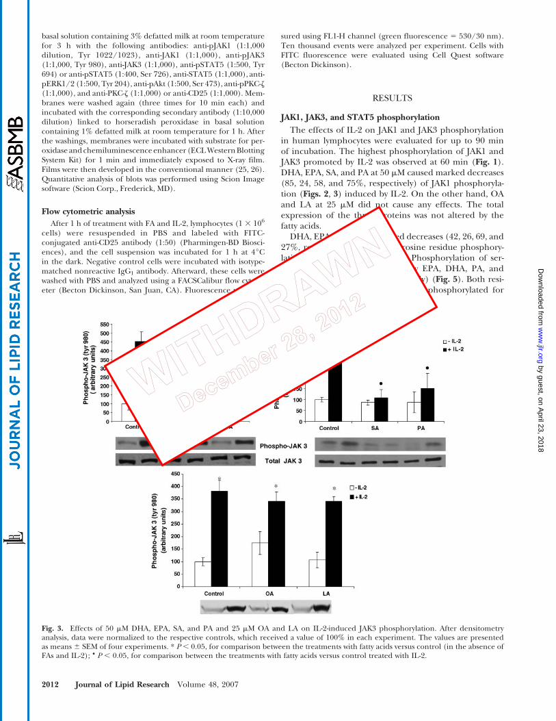
The effects of IL-2 on JAK1 and JAK3 phosphorylationin human lymphocytes were evaluated for up to 90 minof incubation. The highest phosphorylation of JAK1 andJAK3 promoted by IL-2 was observed at 60 min (Fig. 1).DHA, EPA, SA, and PA at 50 mM caused marked decreases(85, 24, 58, and 75%, respectively) of JAK1 phosphoryla-tion (Figs. 2, 3) induced by IL-2. On the other hand, OAand LA at 25 mM did not cause any effects. The totalexpression of the three proteins was not altered by thefatty acids.
DHA, EPA, PA, and SA caused decreases (42, 26, 69, and27%, respectively) of STAT5 tyrosine residue phosphory-lation induced by IL-2 (Fig. 4). Phosphorylation of ser-ine residue was also decreased by EPA, DHA, PA, andSA (45, 52, 24, and 20%, respectively) (Fig. 5). Both resi-dues (tyrosine and serine) must be phosphorylated for
Fig. 3. Effects of 50 mM DHA, EPA, SA, and PA and 25 mM OA and LA on IL-2-induced JAK3 phosphorylation. After densitometryanalysis, data were normalized to the respective controls, which received a value of 100% in each experiment. The values are presentedas means 6 SEM of four experiments. * P , 0.05, for comparison between the treatments with fatty acids versus control (in the absence ofFAs and IL-2); & P , 0.05, for comparison between the treatments with fatty acids versus control treated with IL-2.
2012 Journal of Lipid Research Volume 48, 2007
by guest, on April 23, 2018
ww
w.jlr.org
Dow
nloaded from

the highest transcriptional activity of STAT5 induced byIL-2 (6). OA and LA did not cause significant effects (datanot shown).
ERK1/2 phosphorylation
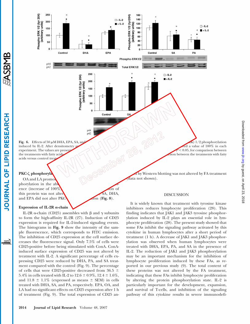
The phosphorylation of ERK1/2 induced by IL-2 wasdecreased (by 83% and 27%) by DHA and EPA treatment,respectively (Fig. 6). DHA also caused a decrease of 62% incells not stimulated with IL-2. PA and SA promoted de-creases of 64% and 38% in the phosphorylation of theseproteins (Fig. 6). The total content of ERK1/2 was notchanged by these FAs. On the other hand, OA and LAcaused increases of ?25% in the ERK1/2 phosphoryla-
tion induced by IL-2 (Fig. 6). LA also increased (by 62%)ERK1/2 phosphorylation in unstimulated cells.
Akt phosphorylation
DHA and EPA exerted inhibitory effects on Akt phos-phorylation induced by IL-2 (decreases of 86% and 31%,respectively) (Fig. 7). PA and SA also decreased Akt phos-phorylation in cells stimulated (decreases of 78% and72%, respectively) or not stimulated (decreases of 62%and 75%, respectively) with IL-2. OA and LA did not alterAkt phosphorylation after 1 h of treatment (Fig. 7). Thisobservation suggests that these FAs probably exert theirstimulatory effect through the Ras/ERK1/2 pathway.
Fig. 4. Effects of 50 mM DHA, EPA, SA, and PA and 25 mM OA and LA on signal transducer and activator of transcription (STAT) 5 tyrosine694 phosphorylation induced by IL-2. After densitometry analysis, data were normalized to the respective controls, which received a valueof 100% in each experiment. The values are presented as means 6 SEM of four experiments. * P , 0.05, for comparison between thetreatments with fatty acids versus control (in the absence of FAs and IL-2); & P , 0.05, for comparison between the treatments with fattyacids versus control treated with IL-2.
Fig. 5. Effects of 50 mM DHA, EPA, SA, and PA and 25 mM OA and LA on STAT5 serine 726 phosphorylation induced by IL-2. Afterdensitometry analysis, data were normalized to the respective controls, which received a value of 100% in each experiment. The valuesare presented as means 6 SEM of four experiments. * P , 0.05, for comparison between the treatments with fatty acids versus control(in the absence of FAs and IL-2); & P , 0.05, for comparison between the treatments with fatty acids versus control treated with IL-2.
Regulation of lymphocyte signaling by fatty acids 2013
by guest, on April 23, 2018
ww
w.jlr.org
Dow
nloaded from
PKC-Z phosphorylation
OA and LA promoted stimulatory effects on PKC-z phos-phorylation in the absence (increase of 50%) and pres-ence (increase of 100%) of IL-2. The total expression ofthis protein was not altered by these FAs. PA, SA, DHA,and EPA did not alter PKC-z phosphorylation (Fig. 8).
Expression of IL-2R a-chain
IL-2R a-chain (CD25) assembles with b and g subunitsto form the high-affinity IL-2R (27). Induction of CD25expression is required for IL-2-induced signaling events.The histograms in Fig. 9 show the intensity of the sam-ple fluorescence, which corresponds to FITC emission.The inhibition of CD25 expression at the cell surface de-creases the fluorescence signal. Only 7.5% of cells wereCD25-positive before being stimulated with ConA. ConA-induced surface expression of CD25 was not altered bytreatment with IL-2. A significant percentage of cells ex-pressing CD25 were reduced by DHA, PA, and SA treat-ment compared with the control (Fig. 9). The percentageof cells that were CD25-positive decreased from 36.5 6
5.4% in cells treated with IL-2 to 12.6 6 0.9%, 12.4 6 1.6%,and 11.8 6 1.1% (expressed as means 6 SEM) in cellstreated with DHA, SA, and PA, respectively. EPA, OA, andLA had no significant effects on CD25 expression after 1 hof treatment (Fig. 9). The total expression of CD25 an-
alyzed by Western blotting was not altered by FA treatment(data not shown).
DISCUSSION
It is widely known that treatment with tyrosine kinaseinhibitors reduces lymphocyte proliferation (28). Thisfinding indicates that JAK1 and JAK3 tyrosine phosphor-ylation induced by IL-2 plays an essential role in lym-phocyte proliferation (28). The present study showed thatsome FAs inhibit the signaling pathway activated by thiscytokine in human lymphocytes after a short period oftreatment (1 h). A decrease of JAK1 and JAK3 phosphor-ylation was observed when human lymphocytes weretreated with DHA, EPA, PA, and SA in the presence ofIL-2. The reduction of JAK1 and JAK3 phosphorylationmay be an important mechanism for the inhibition oflymphocyte proliferation induced by these FAs, as re-ported in our previous study (9). The total content ofthese proteins was not altered by the FA treatment,indicating that these FAs inhibit lymphocyte proliferationby altering the protein phosphorylation state. IL-2 isparticularly important for the development, expansion,and survival of T-cells, and inhibition of the signalingpathway of this cytokine results in severe immunodefi-
Fig. 6. Effects of 50 mM DHA, EPA, SA, and PA and 25 mM OA and LA on extracellular signal-regulated kinase (ERK) 1/2 phosphorylationinduced by IL-2. After densitometry analysis, data were normalized to the respective controls, which received a value of 100% in eachexperiment. The values are presented as means 6 SEM of three determinations from four experiments. * P, 0.05, for comparison betweenthe treatments with fatty acids versus control (in the absence of FAs and IL-2); & P, 0.05, for comparison between the treatments with fattyacids versus control treated with IL-2.
2014 Journal of Lipid Research Volume 48, 2007
by guest, on April 23, 2018
ww
w.jlr.org
Dow
nloaded from

ciency (29). Thus, inhibition of both IL-2 production (30)and IL-2 action in lymphocytes markedly contributes tothe potent suppressive effects of DHA and EPA on thefunction of these cells.
Inhibition of IL-2-induced STAT5 phosphorylation byDHA, EPA, PA, and SA occurred at the level of tyrosineand serine residues (Figs. 3, 4). JAK3 activates JAK1, whichis then able to phosphorylate the target tyrosine residuesin STAT5 molecules docked with the b subunit of theIL-2R. Therefore, STAT5 activation occurs when JAK1 andJAK3 are phosphorylated. Once JAK phosphorylation wasdecreased by PA, SA, EPA, and DHA treatment, STAT5phosphorylation was also inhibited (Fig. 4, 5). JAK-STATsignaling by IL-2 is critically important for immune andinflammatory responses and for T-cell expansion and dif-ferentiation. Inhibition of the signaling of this cytokine,as occurs in genetic deficiency of the g subunit of JAK3,results in severe immunodeficiency. The STAT5 mole-cule does not appear to be the only protein involved inthis process. In fact, deletion and mutational experimentsof STAT5 DNA binding activity or the prevention ofSTAT5 activation did not block IL-2R mitogenic signaling(29, 31). Although STAT5 does not seem to be essentialfor mitogenesis, it is involved in the induction of genesrequired for cell proliferation and survival (32, 33).
JAK1 and JAK3 activation promotes the phosphoryla-tion of tyrosine residues in the cytoplasmic tail of b and g
subunits of IL-2R. Ras is activated through the binding of
the adaptor Shc to the tyrosine-phosphorylated receptor,which recruits the Grb2-Sos complex and activates theRaf-MEK-ERK pathway. EPA and DHA promoted a de-crease of ERK1/2 phosphorylation stimulated by IL-2.This inhibitory effect is attributable to a reduction ofJAK1 and JAK3 phosphorylation promoted by these FAsthat possibly led to a decrease in the activity of ERK1/2(or p42/p44 MAPK). Activated ERK1/2 phosphorylatesvarious substrates in the cell compartments. ERK activa-tion plays an essential role in cell growth and providesan integrated response: increases nucleotide synthesis,activates the transcription of many genes acting via trans-cription factors (e.g., Elk-1, Fos, AP-1, NF-AT, and c-myc)and chromatin phosphorylation, stimulates protein syn-thesis, and finally facilitates the formation of an activecyclin D-CDK4 complex, which is a rate-limiting step forcell growth (34). Khan et al. (35) also showed that DHAarrested the progression from the late G1 to the S phaseof the FM3A mouse mammary cancer cell cycle by de-creasing ERK1/2 phosphorylation.
Denys and colleagues (16, 17) showed that EPA andDHA inhibited ERK1/2 phosphorylation, but this oc-curred by decreasing PKC-b activation in PMA-stimulatedJurkat cells. In fact, PKC is the main pathway activated byPMA stimulation in these cells. In our study, these two FAsdecreased JAK1 and JAK3 phosphorylation, a signalingpathway stimulated by IL-2 that also results in ERK1/2activation. The saturated FA, PA, and SA decreased ERK
Fig. 7. Effects of 50 mM DHA, EPA, SA, and PA and 25 mM OA and LA on Akt phosphorylation induced by IL-2. After densitometryanalysis, data were normalized to the respective controls, which received a value of 100% in each experiment. The values are presentedas means 6 SEM of four experiments. * P , 0.05, for comparison between the treatments with fatty acids versus control (in the absence ofFAs and IL-2); & P , 0.05, for comparison between the treatments with fatty acids versus control treated with IL-2.
Regulation of lymphocyte signaling by fatty acids 2015
by guest, on April 23, 2018
ww
w.jlr.org
Dow
nloaded from

phosphorylation stimulated by IL-2. Similarly, Hirabaraet al. (2003) showed that PA decreased ERK1/2 phos-phorylation induced by insulin in 1 h incubated ratsoleus muscle.
OA and LA enhanced the effect of IL-2 on ERK1/2phosphorylation, suggesting that this pathway may beinvolved in the stimulation of lymphocyte proliferationpromoted by these FAs, as demonstrated previously (9).Cury-Boaventura et al. (36) observed a decrease in the pro-liferation of lymphocytes from volunteers who receiveda lipid emulsion rich in LA. This effect was possiblyattributable to the toxicity of the LA, whose plasma levelwas markedly increased. Thanasak et al. (37) also showedthat the treatment of bovine lymphocytes with low con-centrations of LA (,25 mM) increased lymphocyte pro-liferation, whereas higher concentrations of this fatty aciddecreased it. The results presented here suggest that LAat low concentration acts as a proinflammatory agentby stimulating the MAPK cascade, leading to lymphocyteproliferation. Activation of PKC is a key signaling event forcell growth and proliferation in response to several mito-gens.Cis-unsaturated fatty acids (e.g., OA) can activate PKCand, in turn, ERKs, as observed by others (38). OA morepotently and completely activates the Ca21-independentand atypical PKC isoforms (39, 40) in platelets. PKC-z hasbeen shown to activate ERK, being associated with mito-genesis in various cell lines (41). Other cis-fatty acids, suchas LA, also fully activate PKC in the same manner (39). In
contrast, PKC-a, a Ca21-dependent isoform, is less potentlystimulated by OA (40) and has been associated with thedifferentiation rather than the proliferation of vascularsmooth muscle cells (42). In the present study, OA andLA increased PKC-z phosphorylation after 1 h of treat-ment. Once OA and LA did not alter JAK/STAT phos-phorylation, activation of the novel and/or atypical PKCisoforms may explain the stimulatory effect of OA onERK1/2 phosphorylation induced by IL-2.
Akt phosphorylation was decreased by DHA, EPA, PA,and SA. The effect of DHA was more pronounced thanthat of the other FAs, whereas OA and LA did not affectthis protein phosphorylation (Fig. 7). The activation ofthe PI3K pathway can potentiate proliferative signalingassociated with STAT5 (43). PI3K may activate transcrip-tion factors that mediate events downstream of the G1
cell cycle checkpoint. Several Akt substrates are transcrip-tion factors or are involved directly in the regulation ofgene transcription (44). Probably, this inhibitory effect onAkt phosphorylation and the JAK-STAT and ERK1/2 path-ways induced by DHA, EPA, PA, and SA are responsible forthe decreased lymphocyte proliferation observed in ourprevious study (9).
The a subunit of IL-2R (CD25-a) is undetectable in rest-ing T-cells. The expression of this protein is triggered byantigens (45), a stimulus that can be mimicked by lectinssuch as ConA (46). If a decrease in CD25-a externaliza-tion occurs, IL-2R-stimulated pathways are suppressed and
Fig. 8. Effects of 50 mM DHA, EPA, SA, and PA and 25 mM OA and LA on protein kinase C (PKC)-z phosphorylation induced by IL-2.After densitometry analysis, data were normalized to the respective controls, which received a value of 100% in each experiment. The valuesare presented as means 6 SEM of three experiments. * P , 0.05, for comparison between the treatments with fatty acids versus control(in the absence of FAs and IL-2); & P , 0.05, for comparison between the treatments with fatty acids versus control treated with IL-2.
2016 Journal of Lipid Research Volume 48, 2007
by guest, on April 23, 2018
ww
w.jlr.org
Dow
nloaded from

lymphocyte proliferation is inhibited. DHA, PA, and SAdecreased CD25-a content at the plasma membrane sur-face in lymphocytes after 1 h of treatment (Fig. 9), withno alteration in the total expression of CD25 (data notshown). The CD25 surface membrane expression is neces-sary to form the high-affinity IL-2R that actually activatesthe intracellular signal pathways (JAK-STAT, Ras-ERK1/2,
and PI3K-Akt) leading to lymphocyte proliferation.Probably, the inhibition of JAK1 and JAK3, ERK1/2, andAkt phosphorylation promoted by DHA, PA, and SA isattributable to the decrease in CD25-a expression and con-tent in the plasma membrane. Other studies showed thatPUFAs can alter receptor localization on the cell surfaceafter 24 h of treatment (21, 47). However, a change in the
Fig. 9. Effects of 50 mM DHA, EPA, SA, and PA and 25 mM OA and LA on CD25-a lymphocyte surfaceexpression. Lymphocytes were stimulated with ConA for 24 h, washed, and incubated with 30 ng/ml IL-2for 1 h. Cells were pelleted and labeled with FITC-conjugated anti-CD25 antibody and analyzed by flowcytometry. Negative control cells were incubated with a marked nonreactive control antibody. Histogramsfrom 10,000 events are shown. Fluorescence was measured in channel FL1-H (green fluorescence 5 530/30 nm). A: Representative histograms obtained by flow cytometry. B: Percentage of cells expressing CD25-a.The values are presented as means 6 SEM of three determinations from four experiments. * P , 0.05,for comparison between the treatments with fatty acids versus control (in the absence of FAs and IL-2);& P , 0.05, for comparison between the treatments with fatty acids versus control treated with IL-2.
Regulation of lymphocyte signaling by fatty acids 2017
by guest, on April 23, 2018
ww
w.jlr.org
Dow
nloaded from

content of this receptor subunit in the plasma membranedoes not fully explain the effect of all FAs on IL-2 sig-naling. It is noteworthy that OA and LA stimulated ERK1/2 phosphorylation but did not change CD25 expression.Measurements using red-fluorescent Alexa Fluor 594 con-jugate of cholera toxin subunit B showed that, except forDHA, none of the FAs tested caused marked changes inlipid raft distribution under the conditions of this study(data not shown). Therefore, the FAs tested hardly affectedintracellular signaling through changes in lipid rafts.
Although DHA and EPA have promoted an inhibi-tory effect on lymphocyte proliferation (9), DHA caused amore pronounced decrease of ERK1/2 and JAK1 phos-phorylation in relation to EPA (Figs. 2, 6). Other studieshave shown that different n-3 FAs can exert divergent ef-fects on immune response. DHA-rich fish oil caused anincrease in neutrophil and monocyte phagocytosis (48),whereas others demonstrated that EPA-rich fish oil didnot alter this cell function (49). Verlengia et al. (50)showed that the effect of EPA on gene expression in ahuman B-lymphocyte cell line (Raji) was more pronounced(25.9% of genes investigated were altered) compared withthat of DHA (8.4% of genes investigated were altered).We observed here that an alteration in lipid raft organiza-tion was induced by DHA, but it did not occur whenlymphocytes were treated with EPA. The differences ob-served in the effects of these two fatty acids may occurbecause the spatial conformation of DHA is different fromthat of EPA as a result of its carbon length and degree ofunsaturation. Therefore, we cannot generalize the effectsof FAs from the same family on lymphocytes.
In conclusion, the findings reported here are importantfor understanding the mechanisms by which different FAsalter lymphocyte proliferation. The inhibitory effect of PA,SA, and DHA on lymphocyte proliferation was associatedwith a reduction in the IL-2-induced activation of the JAK/STAT, ERK, and Akt pathways and a decrease in CD25-aexpression. EPA decreased the phosphorylation of theIL-2R signaling proteins by a mechanism that did notinvolve CD25. Probably, OA and LA stimulated lympho-cyte proliferation by increasing ERK1/2 phosphorylationthrough PKC-z activation.
The authors are indebted to J. R. Mendonc$a, G. de Souza, E. P.Portiolli, Dr. T. C. Alba-Loureiro, and A. S. Alves for technicalassistance. The authors are grateful to Dr. Jose Augusto Barretoof the Blood Bank of the Federal University of Sao Paulo.This study was supported by Fundac$ao de Amparo a Pesquisado Estado de Sao Paulo, Coordenac$ao de Aperfeic$oamentode Pessoal de Nıvel Superior, and Conselho Nacional deDesenvolvimento Cientıfico e Tecnologico.
REFERENCES
1. Smith, K. A. 1988. The interleukin 2 receptor. Adv. Immunol. 42:165–179.
2. Kirken, R. A., H. Rui, M. G. Malabarba, O. M. Howard, M.Kawamura, J. J. O’Shea, and W. L. Farrar. 1995. Activation of JAK3,but not JAK1, is critical for IL-2-induced proliferation and STAT5
recruitment by a COOH-terminal region of the IL-2 receptor beta-chain. Cytokine. 7: 689–700.
3. Russell, S. M., J. A. Johnston, M. Noguchi, M. Kawamura, C. M.Bacon, M. Friedmann, M. Berg, D. W. McVicar, B. A. Witthuhn,O. Silvennoinen, et al. 1994. Interaction of IL-2R beta and gammac chains with Jak1 and Jak3: implications for XSCID and XCID.Science. 266: 1042–1045.
4. Beadling, C., D. Guschin, B. A. Witthuhn, A. Ziemiecki, J. N. Ihle,I. M. Kerr, and D. A. Cantrell. 1994. Activation of JAK kinases andSTAT proteins by interleukin-2 and interferon alpha, but not theT cell antigen receptor, in human T lymphocytes. EMBO J. 13:5605–5615.
5. Darnell, J. E., Jr., I. M. Kerr, and G. R. Stark. 1994. Jak-STATpathways and transcriptional activation in response to IFNs andother extracellular signaling proteins. Science. 264: 1415–1421.
6. Beadling, C., J. Ng, J. W. Babbage, and D. A. Cantrell. 1996.Interleukin-2 activation of STAT5 requires the convergent actionof tyrosine kinases and a serine/threonine kinase pathway distinctfrom the Raf1/ERK2 MAPK pathway. EMBO J. 15: 1902–1913.
7. Costabile, M., C. S. Hii, M. Melino, C. Easton, and A. Ferrante.2005. The immunomodulatory effects of novel beta-oxa, beta-thia,and gamma-thia polyunsaturated fatty acids on human T lympho-cyte proliferation, cytokine production, and activation of proteinkinase C and MAPKs. J. Immunol. 174: 233–243.
8. Kew, S., E. S. Gibbons, F. Thies, G. P. McNeill, P. T. Quinlan, andP. C. Calder. 2003. The effect of feeding structured triacylglycerolsenriched in eicosapentaenoic or docosahexaenoic acids on murinesplenocyte fatty acid composition and leucocyte phagocytosis.Br. J. Nutr. 90: 1071–1080.
9. Gorjao, R., M. F. Cury-Boaventura, T. M. de Lima, and R. Curi.2007. Regulation of human lymphocyte proliferation by fatty acids.Cell Biochem. Funct. 25: 305–315.
10. Camuesco, D., J. Galvez, A. Nieto, M. Comalada, M. E. Rodriguez-Cabezas, A. Concha, J. Xaus, and A. Zarzuelo. 2005. Dietary oliveoil supplemented with fish oil, rich in EPA and DHA (n-3) poly-unsaturated fatty acids, attenuates colonic inflammation in ratswith DSS-induced colitis. J. Nutr. 135: 687–694.
11. Mills, S. C., A. C. Windsor, and S. C. Knight. 2005. The potentialinteractions between polyunsaturated fatty acids and colonicinflammatory processes. Clin. Exp. Immunol. 142: 216–228.
12. Robinson, D. R., L. L. Xu, S. Tateno, M. Guo, and R. B. Colvin.1993. Suppression of autoimmune disease by dietary n-3 fatty acids.J. Lipid Res. 34: 1435–1444.
13. Young, V. M., M. Toborek, F. Yang, C. J. McClain, and B. Hennig.1998. Effect of linoleic acid on endothelial cell inflammatorymediators. Metabolism. 47: 566–572.
14. Pompeia, C., L. R. Lopes, C. K. Miyasaka, J. Procopio, P. Sannomiya,and R. Curi. 2000. Effect of fatty acids on leukocyte function. Braz.J. Med. Biol. Res. 33: 1255–1268.
15. Harbige, L. S. 1998. Dietary n-6 and n-3 fatty acids in immunity andautoimmune disease. Proc. Nutr. Soc. 57: 555–562.
16. Denys, A., A. Hichami, and N. A. Khan. 2002. Eicosapentaenoicacid and docosahexaenoic acid modulate MAPK enzyme activity inhuman T-cells. Mol. Cell. Biochem. 232: 143–148.
17. Denys, A., A. Hichami, B. Maume, and N. A. Khan. 2001.Docosahexaenoic acid modulates phorbol ester-induced activationof extracellular signal-regulated kinases 1 and 2 in NIH/3T3 cells.Lipids. 36: 813–818.
18. Denys, A., A. Hichami, and N. A. Khan. 2001. Eicosapentaenoicacid and docosahexaenoic acid modulate MAPK (ERK1/ERK2)signaling in human T cells. J. Lipid Res. 42: 2015–2020.
19. Denys, A., A. Hichami, and N. A. Khan. 2005. n-3 PUFAs modulateT-cell activation via protein kinase C-alpha and -epsilon and theNF-kappaB signaling pathway. J. Lipid Res. 46: 752–758.
20. Zeyda, M., A. B. Szekeres, M. D. Saemann, R. Geyeregger, H.Stockinger, G. J. Zlabinger, W. Waldhausl, and T. M. Stulnig. 2003.Suppression of T cell signaling by polyunsaturated fatty acids:selectivity in inhibition of mitogen-activated protein kinase andnuclear factor activation. J. Immunol. 170: 6033–6039.
21. Li, Q., M. Wang, L. Tan, C. Wang, J. Ma, N. Li, Y. Li, G. Xu,and J. Li. 2005. Docosahexaenoic acid changes lipid compositionand interleukin-2 receptor signaling in membrane rafts. J. Lipid Res.46: 1904–1913.
22. Siddiqui, R. A., L. J. Jenski, K. Neff, K. Harvey, R. J. Kovacs, andW. Stillwell. 2001. Docosahexaenoic acid induces apoptosis inJurkat cells by a protein phosphatase-mediated process. Biochim.Biophys. Acta. 1499: 265–275.
2018 Journal of Lipid Research Volume 48, 2007
by guest, on April 23, 2018
ww
w.jlr.org
Dow
nloaded from

23. Bradford, M. M. 1976. A rapid and sensitive method for thequantitation of microgram quantities of protein utilizing the prin-ciple of protein-dye binding. Anal. Biochem. 72: 248–254.
24. Shapiro, A. L., E. Vinuela, and J. V. Maizel, Jr. 1967. Molecularweight estimation of polypeptide chains by electrophoresis in SDS-polyacrylamide gels. Biochem. Biophys. Res. Commun. 28: 815–820.
25. Towbin, H., T. Staehelin, and J. Gordon. 1979. Electrophoretictransfer of proteins from polyacrylamide gels to nitrocellulosesheets: procedure and some applications. Proc. Natl. Acad. Sci. USA.76: 4350–4354.
26. Hirabara, S. M., C. R. de Oliveira Carvalho, J. R. Mendonca,E. Piltcher Haber, L. C. Fernandes, and R. Curi. 2003. Palmitateacutely raises glycogen synthesis in rat soleus muscle by a mech-anism that requires its metabolization (Randle cycle). FEBS letters.541: 109–114.
27. Taniguchi, T., and Y. Minami. 1993. The IL-2/IL-2 receptor system:a current overview. Cell. 73: 5–8.
28. Wang, H., X. Xie, W. G. Lu, D. F. Ye, H. Z. Chen, X. Li, andQ. Cheng. 2004. Ovarian carcinoma cells inhibit T cell prolifera-tion: suppression of IL-2 receptor beta and gamma expression andtheir JAK-STAT signaling pathway. Life Sci. 74: 1739–1749.
29. Gaffen, S. L., S. Y. Lai, W. Xu, F. Gouilleux, B. Groner, M. A.Goldsmith, and W. C. Greene. 1995. Signaling through the inter-leukin 2 receptor beta chain activates a STAT-5-like DNA-bindingactivity. Proc. Natl. Acad. Sci. USA. 92: 7192–7196.
30. Terada, S., M. Takizawa, S. Yamamoto, O. Ezaki, H. Itakura, andK. S. Akagawa. 2001. Suppressive mechanisms of EPA on humanT cell proliferation. Microbiol. Immunol. 45: 473–481.
31. Fujii, H., Y. Nakagawa, U. Schindler, A. Kawahara, H. Mori,F. Gouilleux, B. Groner, J. N. Ihle, Y. Minami, T. Miyazaki, et al.1995. Activation of Stat5 by interleukin 2 requires a carboxyl-terminal region of the interleukin 2 receptor beta chain but is notessential for the proliferative signal transmission. Proc. Natl. Acad.Sci. USA. 92: 5482–5486.
32. Lord, J. D., B. C. McIntosh, P. D. Greenberg, and B. H. Nelson.2000. The IL-2 receptor promotes lymphocyte proliferation andinduction of the c-myc, bcl-2, and bcl-x genes through the transac-tivation domain of Stat5. J. Immunol. 164: 2533–2541.
33. Yu, C. R., J. R. Ortaldo, R. E. Curiel, H. A. Young, S. K. Anderson,and P. Gosselin. 1999. Role of a STAT binding site in the regulationof the human perforin promoter. J. Immunol. 162: 2785–2790.
34. Whitmarsh, A. J., and R. J. Davis. 2000. Regulation of transcrip-tion factor function by phosphorylation. Cell. Mol. Life Sci. 57:1172–1183.
35. Khan, N. A., K. Nishimura, V. Aires, T. Yamashita, D. Oaxaca-Castillo, K. Kashiwagi, and K. Igarashi. 2006. Docosahexaenoic acidinhibits cancer cell growth via p27Kip1, CDK2, ERK1/ERK2, andretinoblastoma phosphorylation. J. Lipid Res. 47: 2306–2313.
36. Cury-Boaventura, M. F., R. Gorjao, T. M. de Lima, T. M. Piva, C. M.Peres, F. G. Soriano, and R. Curi. 2006. Toxicity of a soybean oilemulsion on human lymphocytes and neutrophils. JPEN J. Parenter.Enteral Nutr. 30: 115–123.
37. Thanasak, J., K. E. Muller, S. J. Dieleman, A. Hoek, J. P.Noordhuizen, and V. P. Rutten. 2005. Effects of polyunsaturatedfatty acids on the proliferation of mitogen stimulated bovineperipheral blood mononuclear cells. Vet. Immunol. Immunopathol.104: 289–295.
38. Lu, G., T. A. Morinelli, K. E. Meier, S. A. Rosenzweig, andB. M. Egan. 1996. Oleic acid-induced mitogenic signaling in vas-cular smooth muscle cells. A role for protein kinase C. Circ. Res. 79:611–618.
39. Murakami, K., S. Y. Chan, and A. Routtenberg. 1986. Protein kinaseC activation by cis-fatty acid in the absence of Ca21 and phos-pholipids. J. Biol. Chem. 261: 15424–15429.
40. Khan, W. A., G. Blobe, A. Halpern, W. Taylor, W. C. Wetsel,D. Burns, C. Loomis, and Y. A. Hannun. 1993. Selective regula-tion of protein kinase C isoenzymes by oleic acid in humanplatelets. J. Biol. Chem. 268: 5063–5068.
41. Berra, E., M. T. Diaz-Meco, I. Dominguez, M. M. Municio, L. Sanz,J. Lozano, R. S. Chapkin, and J. Moscat. 1993. Protein kinase Czeta isoform is critical for mitogenic signal transduction. Cell. 74:555–563.
42. Haller, H., C. Lindschau, P. Quass, A. Distler, and F. C. Luft. 1995.Differentiation of vascular smooth muscle cells and the regulationof protein kinase C-alpha. Circ. Res. 76: 21–29.
43. Moon, J. J., and B. H. Nelson. 2001. Phosphatidylinositol 3-kinasepotentiates, but does not trigger, T cell proliferation mediated bythe IL-2 receptor. J. Immunol. 167: 2714–2723.
44. Datta, S. R., A. Brunet, and M. E. Greenberg. 1999. Cellular sur-vival: a play in three Akts. Genes Dev. 13: 2905–2927.
45. Robb, R. J., and K. A. Smith. 1981. Heterogeneity of human T-cellgrowth factor(s) due to variable glycosylation. Mol. Immunol. 18:1087–1094.
46. Kimura, G. M., M. E. Miller, R. D. Leake, R. Raghunathan, and A. T.Cheung. 1981. Reduced concanavalin A capping of neonatal poly-morphonuclear leukocytes (PMNs). Pediatr. Res. 15: 1271–1273.
47. Schley, P. D., D. N. Brindley, and C. J. Field. 2007. (n-3) PUFAalter raft lipid composition and decrease epidermal growth factorreceptor levels in lipid rafts of human breast cancer cells. J. Nutr.137: 548–553.
48. Gorjao, R., R. Verlengia, T. M. Lima, F. G. Soriano, M. F.Boaventura, C. C. Kanunfre, C. M. Peres, S. C. Sampaio,R. Otton, A. Folador, et al. 2006. Effect of docosahexaenoic acid-rich fish oil supplementation on human leukocyte function. Clin.Nutr. 25: 923–938.
49. Miles, E. A., T. Banerjee, M. M. Dooper, L. M’Rabet, Y. M.Graus, and P. C. Calder. 2004. The influence of different com-binations of gamma-linolenic acid, stearidonic acid and EPA onimmune function in healthy young male subjects. Br. J. Nutr. 91:893–903.
50. Verlengia, R., R. Gorjao, C. C. Kanunfre, S. Bordin, T. M. de Lima,E. F. Martins, P. Newsholme, and R. Curi. 2004. Effects of EPA andDHA on proliferation, cytokine production, and gene expressionin Raji cells. Lipids. 39: 857–864.
Regulation of lymphocyte signaling by fatty acids 2019
by guest, on April 23, 2018
ww
w.jlr.org
Dow
nloaded from

Journal of Lipid Research Volume 54, 2013 869
The authors of “A common variant highly associated with plasma VEGFA levels also contributes to the variation of both LDL-C and HDL-C” (J. Lipid Res. 2013. 54: 535–541 ) have informed the Journal that the fi rst three authors (Maria G. Stathopoulou, Amélie Bonnefond, and Ndeye Coumba Ndiaye) contributed equally to this work and this should be noted on the manuscript. This notation was omitted in the fi nal publication of the manuscript and it has since been corrected online. The Journal sincerely regrets this error.
DOI 10.1194/jlr.P030551ERR
ERRATUM
This article has been withdrawn by the authors: Regulation of interleukin-2 signaling by fatty acids in human lympho-cytes. J. Lipid Res. 2007. 48: 2009 – 2019 .
DOI 10.1194/jlr.M700175ERR
ERRATUM