Regulation of human EP4 prostanoid receptor expression by ...
50
1 Regulation of human EP4 prostanoid receptor expression by HIF-1α (HIF-1α によるヒト EP4 プロスタノイド受容体発現の制御) 2018 Naofumi SEIRA Laboratory of Chemical Pharmacology, Molecular Pharmacotherapeutics Graduate School of Pharmaceutical Sciences, Chiba University
Transcript of Regulation of human EP4 prostanoid receptor expression by ...

1
Regulation of human EP4 prostanoid receptor expression by HIF-1α
(HIF-1α によるヒト EP4プロスタノイド受容体発現の制御)
2018
Naofumi SEIRA
Laboratory of Chemical Pharmacology,
Molecular Pharmacotherapeutics
Graduate School of Pharmaceutical Sciences, Chiba University

2
CONTENTS
1. INTRODUCTION 3
2. ABBREVIATIONS 6
3. MATERIAL AND METHODS 7
4. RESULTS 15
5. DISCUSSION 21
6. CONCLUSIONS 25
7. REFERENCE 26
8. EXAMINERS 31
9. ACKNOWLEDGEMENTS 32
10. FIGURES AND LEGENDS 33
11. LIST OF PUBLICATIONS 50
3
1. INTRODUCTION
Up-regulation of the levels of cyclooxygenase-2 (COX-2) and prostaglandin E2 (PGE2) are
known to be biomarkers for the early stage of colorectal cancer (Eiginger et al., 2007; Woodward
et al., 2011). The increase of COX-2 expression is related with the activation of EP4 prostanoid
receptors (Yoshida et al., 2013), and the EP4 receptor signaling pathway is also related with
increases in the cellular motility and proliferation (Sheng et al., 2001) in human colon cancer
cells. Moreover, it has reported that, during the progression of colorectal cancer, the EP4
receptors (Chell et al., 2006; Hawcroft et al., 2007), and PGE2 synthase (Yoshimatsu et al., 2001)
are up-regulated. It has also shown that the activation of EP4 receptors mediates increased PGE2
synthase expression (Fujino et al., 2003). Thus, the activated EP4 receptors develop a positive
feedback loop that may induce the expression of COX-2, PGE2 synthase, and the synthesis of
PGE2. From the above, it believes that EP4 receptors play essential roles in the malignancy of
colorectal cancer (Fujino, 2016).
EP4 receptors are generally involved in colorectal carcinogenesis. However EP4 receptors
may also be implicated in maintaining intestinal homeostasis (Yokoyama et al., 2013). It has
shown that EP4 receptors are expressed in intact colorectal epithelial cells (Yokoyama et al.,
2013) and lateral crypt epithelia (Takafuji et al., 2000), and epithelial tissues have a turnover
cycle of 3- to 5-day (Potten and Loeffler., 1990). Thus, epithelial stem cells at the crypt bottom
produce epithelial progenitor cells. These cells grow, differentiate, and migrate to the intestinal
lumen. Then differentiated cells undergo apoptosis on the extrusion of lumen over 3- to 5-day
(van de Wetering et al., 2002). It has reported that β-catenin accumulates new epithelial
progenitor cells and β-catenin-mediated transcriptional activity is stimulated, which induces the
cellular proliferation and migration until epithelial progenitor cells reach the mid-crypt region.
At the mid-crypt region, epithelial cells inhibit β-catenin-mediated signaling, which leads to cell
cycle arrest and differentiation. Then apoptosis and/or extrusion are induced when cells reach the

4
tip of the lumen (van de Wetering et al., 2002). Therefore, β-catenin mediated signaling may
work as the significant switch between the proliferation and differentiation of colorectal epithelial
cells.
Although it has demonstrated the EP4 receptors are over-expressed during colorectal
development, another study reported that the mRNA expression levels of EP4 receptors increased
in normal colorectal tissues compared with cancer tissues (Gustafsson et al., 2007). The previous
report showed specificity protein 1 (Sp1) which is a zinc finger transcription factor regulating the
protein levels of EP4 receptors by binding to two Sp1-binding sites in the human EP4 receptor
promoter region from -197 to -160 bp (Kambe et al., 2008). However, hypoxia-inducible factor
(HIF)-1α which is, a transcription factor induced in hypoxic conditions, also down-regulated the
EP4 receptor expression when human colon adenoma HCA-7 cells are cultured at a high cellular
density (Otake et al., 2015). The high cellular density is thought to be equivalent to excessive
proliferated conditions. Thus, the high cellular density induced HIF-1α protein expression
reduces EP4 receptors expression in HCA-7 cells (Otake et al., 2015). Since the HIF-1α
expression level is known to be up-regulated at the malignant tumor and correlates with cancer
malignancy (Semenza, 2003; Mabjeesh and Amir, 2007; Rohwer et al., 2013), severe
proliferating cancer cells may exhibit the reduction in the expression of EP4 receptors.
The previous report showed the activated β-catenin mediated signaling caused stimulation
of EP4 receptors with PGE2 (Fujino et al., 2002). It has also suggested that c-Myc mediates the
growth of colorectal cancer epithelial cells induced by β-catenin mediated signaling (van de
Wetering et al., 2002). Therefore, the c-Myc induced the cellular proliferation may be modulated
via EP4 receptor activation. It has known that HIF-1α binds to Sp1 by displacing c-Myc from
Sp1 binding sites to decrease the DNA mismatch repair system in colon cancer cells (Koshiji et
al., 2005; To et al., 2006). Thus, in the light of the modulation of EP4 receptor expression, c-Myc
has been involved in the increase of EP4 receptor expression. However, HIF-1α works the
adverse effects, with the involvement of Sp1.

5
We herein reveal that HIF-1α strictly regulate the EP4 receptor expression by competing
Sp1 and c-Myc as the cellular density-dependently in HCA-7 cells. This strict modulation of EP4
receptor expression by c-Myc and HIF-1α may be a critical system for maintaining homeostasis
in the intestinal epithelial cells. However, once this system is changed, it may cause excessive
cellular growth, a transformation from ordinary to malignant phenotypes, which represents the
initiation for the early stage of intestinal carcinogenesis.

6
2. ABBREVIATIONS
EP, E-type prostanoid; COX-2, cyclooxygenase-2; PGE2, prostaglandin E2; HIF, hypoxia-
inducible factor; DMEM, Dulbecco's modified Eagle's medium, FBS, fetal bovine serum; HRE,
hypoxia response element; WT, wild-type; PCR, polymerase chain reaction; ChIP, chromatin
immunoprecipitation; BSA, bovine serum albumin; ANOVA, analysis of variance; UCSC, The
University of California Santa Cruz; VEGF, vascular endothelial growth factor; VEGFR, VEGF
receptor; GLUT, glucose transporter; VHL, von Hippel-Lindau; PHD, prolyl hydroxylase
domain-containing enzyme; EGLN, Egl-9 family hypoxia-inducible factor; FIH1, factor-
inhibiting HIF-1α; HIF1AN, HIF-1α inhibitor; COMMD, copper metabolism domain containing;
HSP, heat shock protein; CBP, cAMP response element-binding protein-binding protein; CITED,
CBP/p300-interacting transactivator.
7
3. MATERIALS AND METHODS
Cell culture
Human colon adenocarcinoma, HCA-7 cells were maintained in culture medium,
Dulbecco’s modified Eagle’s medium (DMEM; Nacalai Tesque, Kyoto, Japan) containing 10%
fetal bovine serum (FBS) (Thermo Fisher Scientific, Waltham MA), 100 UI/ml penicillin (Meiji
Seika, Tokyo, Japan), and 0.1 mg/ml streptomycin (Meiji Seika, Tokyo, Japan) at 37oC in a
humidified atmosphere (5% CO2). We obtained all materials from Wako Pure Chemical (Osaka,
Japan) unless otherwise stated.
Construction of deletion and mutated EP4 receptor promoter luciferase reporter plasmids
Deletion and point mutations introduced into the hypoxia response element (HRE) region,
of human EP4 receptor promoter-conjugated luciferase reporter plasmids were constructed using
the human EP4 receptor promoter-conjugated luciferase reporter plasmid, wild-type (WT) (-
1238/+1) (Otake et al., 2015), as a template. In order to construct the deletion mutants of human
EP4 receptor promoter luciferase plasmids, primers with the following sequences were used for
polymerase chain reaction (PCR) amplification: deletion 1 (-789/+1): 5'-
GGGGCTAGCCCAAGGCTCCACCTCTCTCCAAAGCCGCAA-3' (sense), deletion 2 (-
473/+1): 5'-GGGGCTAGCTCGGCCAACCCTAGGTAGAATCCTAAAAC-3' (sense),
deletion 3 (-197/+1); 5'-GGGGCTAGCGCCCAGCCCCGCCCCAGCCCAGACACCGCCC-3',
deletion 4 (-160/+1); 5'-GGGGCTAGCAGTCTTCCCTGCGGC-3' (sense). The sequence of the
antisense primer for all deleted plasmids was as follows: 5’-
GGAAGCTTTGGAGCTCGCGTGCTGCGGCCTTTC-3’. The products of the deleted EP4
receptor promoter by PCR were digested with Nhe I (Takara Bio, Shiga, Japan) and Hind III
(Takara Bio) restriction enzymes and then purified and ligated into pGL3-basic vectors (Promega,
Madison, WI). The point mutations introduced in HRE containing the human EP4 receptor
8
promoter luciferase plasmid were amplified by PCR using the primers 5’-
TCCGCACCCCCGAGGGAATGAAAACCACGGGAGCC-3' (sense) and 5’-
GGCTCCCGTGGTTTTCATTCCCTCGGGGGTGCGGA-3'. The deletion 3 (-197/+1)
luciferase plasmids, in which point mutations were introduced at each or both Sp1-binding sites,
were constructed by PCR using the following primers: mut-A-del 3: 5’-
GCCCAGCCCTTCCCCAGCCCA-3' (sense) and 5’-TGGGCTGGGGAAGGGCTGGGC-3’
(antisense), mut-B-del 3: 5’-GCCCAGACACTTCCCCCCGCCA-3’ (sense) and 5’-
TGGCGGGGGGAAGTGTCTGGGC-3' (antisense) and both primer sets for mut-A,B-del 3 [15].
The PCR products of each or both Sp1 site point mutations introduced into del 3 plasmids were
digested with Dpn I (Takara Bio) and self-ligated. Each construct was sequenced and verified.
Luciferase assay
Cells were cultured at low (2 × 10⁵ cells/each well) and high (2 ×10⁶ cells/each well)
cellular density-conditions in 6-well plates, and culture medium was replaced with Opti-MEM I
(Thermo Scientific, Waltham MA, USA) containing 100 UI/mL penicillin and 100 μg/mL
streptomycin. Cells were transiently transfected with 10 μg/each well of firefly WT (-1238/+1),
deleted or mutated reporter luciferase plasmids, and with 10 ng/each well of renilla luciferase
control plasmids, pRL-CMV (Promega) using Polyetherimide MAX (MW 40,000) (Polysciences,
Warrington, PA) reagent. After approximately 6 hr, the transfection reagent was removed by a
medium change using DMEM containing 10% FBS, and cells were incubated for a further 16 hr.
To measure the effects of HIF-1α and c-Myc on EP4 receptor promoter activity, cells were
cultured at low cellular density-conditions, and culture medium was replaced with Opti-MEM I
containing 100 UI/mL penicillin and 100 μg/mL streptomycin. In HIF-1α and c-Myc
overexpression experiments, cells were cultured at low cellular density-conditions in 12-well
plates. Then, cells were transiently transfected with 2 μg/each well of firefly WT (-1238/+1) EP4
receptor promoter luciferase plasmids, 3 ng/each well of pRL-CMV renilla luciferase control

9
plasmids, and either the pHA-N1 vector, which was created by replacing EGFP with HA in the
pEGFP-N1 vector (Clontech Laboratories, Mountain View, CA), HA-HIF1alpha-pcDNA3 or the
pCMFlag_hsc-Myc (RDB06671, RIKEN BRC, Saitama, Japan) expression vector, using the
same reagents described above. Cells were then incubated for a further 42 hr. pHA-N1 vector
was created by replacing EGFP with HA in the pEGFP-N1 vector (Clontech Laboratories,
Mountain View, CA). HA-HIF1alpha-pcDNA3 was a gift from William Kaelin (Addgene
plasmid #18949) (Kondo et al., 2002). The pCMFlag_hsc-Myc was purchased from RIKEN BRC
(RDB06671, RIKEN BRC, Saitama, Japan). In competition assays, HA-tagged HIF-1α
expression plasmids alone (0.5 μg/each well), or HA-tagged HIF-1α expression plasmids (0.5
μg/each well) plus various amounts of FLAG-tagged c-Myc expression plasmids (0.05, 0.15, and
0.5 μg/each well, respectively); or FLAG-tagged c-Myc expression plasmids alone (0.5 μg/each
well), or FLAG-tagged c-Myc expression plasmids plus various amounts of HA-tagged HIF-1α
expression plasmids (0.05, 0.15, and 0.5 μg/each well, respectively), and the total amounts of
transfected plasmids were adjusted by adding pHA-N1 control plasmids to 1 μg/each well.
Reporter plasmid-transfected cells were then lysed and assayed using the Dual-luciferase reporter
assay system (Promega) according to the manufacturer’s instructions with the GL-200
luminometer (Microtech Nichon, Chiba, Japan). Data were normalized by calculating the ratios
of firefly luciferase scores to the corresponding renilla luciferase values.
Chromatin immunoprecipitation (ChIP) assay
HCA-7 cells were cultured at low (5.5×10⁵ cells/dish) and/or high (5.5 × 10⁶ cells/dish)
density-conditions in a 10-cm dish, and culture medium was replaced with Opti-MEM I
containing 100 UI/mL penicillin and 100 μg/mL streptomycin. Cells were transiently transfected
with 25 μg/dish of WT (-1238/+1) or point mutations-introduced mut-HRE (-1238/+1) of EP4
receptor promoter reporter luciferase plasmids using Polyethylenimine MAX (MW 40,000)
reagent. After approximately 6 hr, transfection reagents were removed by medium changes and

10
cells were incubated for a further 18 hr. A ChIP analysis was performed using a ChIP assay kit
according to the manufacturer’s instructions (Merck, Kenilworth, NJ) or Dynabeads Protein G
(1004D: Invitrogen, CA, USA). Briefly, cells were fixed with 1% formaldehyde at room
temperature for 10 min. Fixed cells were scraped into microcentrifuge tubes and lysed in SDS
lysis buffer. DNA was sheared to fragments by sonication. Sonicated lysates were immobilized
with anti-Sp1 antibody (GTX110593: GeneTex, CA, USA), anti-c-Myc antibody (GTX103436:
GeneTex), and/or an anti-HIF-1α antibody (H1alpha67) (NB100-105; Novus Biologicals,
Littleton, CO). Crosslinked DNA by formaldehyde was released from the antibody-captured
protein-DNA complex and purified. Captured DNA was then eluted and detected by PCR
amplification (C1000 Thermal Cycler, Bio-Rad, USA) or real-time PCR amplification (Eco Real-
Time PCR systems, illumine, USA). The region between -1238 to +1 of the human EP4 receptor
promoter was amplified using the following primers: 5’-
GGGCTAGCCTGCAGATGGGAAGAGGTTTTTCCAGGAATTTAAA-3’ (sense), 5’-
GGAAGCTTTGGAGCTCGCGTGCTGCGGCCTTTC-3’ (antisense) by PCR. The relative
recruitment of Sp1, c-Myc, and HIF-1α to Sp1 binding consensus motifs were detected by using
PowerUp SYBR Green Master Mix (Thermo Fisher Scientific) and the following primers: 5'-
GGGGCTAGCGCCCAGCCCCGCCCCAGCCCAGACACCGCCC-3' (sense) 5’-
GTCCACCTCGATATGTGCATC-3' (antisense) from -197 of human the EP4 receptor promoter
to +104 of the firefly gene body by real-time PCR.
Western blotting
Regarding the detection of Sp1, HIF-1α, and c-Myc, HCA-7 cells were cultured at low (2
x 105 cells/each well), middle (6 x 10⁵ cells/each well), and high (2 x10⁶ cells/each well) density-
conditions in 6-well plates, and culture medium was replaced with Opti-MEM I containing 100
UI/mL penicillin and 100 μg/mL streptomycin for 16 hrs. In the detection of EP4 receptor

11
expression, low or high cellular density-cultured HCA-7 cells were treated with 100 nM
mithramycin A (Wako, Osaka, Japan) for 16 hr. Cells were then scraped with lysis buffer
consisting of 150 nM NaCl, 50 mM Tris-HCl (pH 8.0), 5 nM EDTA (pH 8.0), 1% Igepal CA-
630 (MP Biomedicals, Aurora, OH), 0.5% sodium deoxycholate, 10 mM sodium fluoride, 10
mM disodium pyrophosphate, 0.1% SDS, 0.1 mM phenylmethylsulfonyl fluoride, 1 mM sodium
orthovanadate, 10 μg/mL leupeptin (Sigma, St Louis, MO), and 10 μg/mL aprotinin, and then
transferred to microcentrifuge tubes. Samples were rotated at 4oC for 30 min and centrifuged at
16,000 g for 15 min. Aliquots of samples containing 20 - 40 μg of protein were electrophoresed
on 10% SDS-polyacrylamide gels and transferred to nitrocellulose membranes as described
previously (Otake et al., 2015). Membranes were incubated at room temperature for 1 hr in 5%
non-fat milk. Incubations were conducted at 4oC for 16 hr in 5% bovine serum albumin (BSA)
(Sigma) containing a 1:1,000 dilution of an anti-human EP4 receptor antibody (101775; Cayman
Chemical, Ann Arbor, MI); a 1:1,000 dilution of an anti-Sp1 antibody (sc-420; Santa Cruz
Biotechnology, Santa Cruz, CA); a 1:1,000 dilution of an anti-c-Myc antibody (sc-40; Santa Cruz
Biotechnology); a 1:1,000 dilution of an anti-HIF-1α antibody (H1alpha67); or a 1:5,000 dilution
of an anti-β-tubulin-antibody (014-25041; Wako). After being incubated with primary antibodies,
membranes were washed three times and then incubated at room temperature for 2 hr with a
1:10,000 dilution of appropriate secondary antibodies conjugated with horseradish peroxidase at
the same blocking conditions as those for the primary antibodies (Otake et al., 2015). After
washing three times, immunoreactivity was detected and visualized with ChemiDoc XRS Plus
Image Lab (Bio-Rad Laboratories, Hercules, CA). To ensure the equal loading of proteins,
membranes were stripped and re-probed with the anti-β-tubulin antibody at the conditions
described above. The intensity of chemiluminescence was measured with ImageJ software
(National Institutes of Health, Bethesda, MD).
Cell imaging of a hypoxia probe

12
Cells were cultured at low and high cell density conditions in 6-well plates, and culture
medium was replaced with Opti-MEM I containing 100 UI/mL penicillin and 100 μg/mL
streptomycin. The hypoxia probe solution LOX-1 (Medical & Biological Laboratories, Aichi,
Japan) was then added to the medium at a final concentration of 2 μM for 24 hr for visualization
with the all-in-one fluorescence microscope BZ-X710 (Keyence, Osaka, Japan). The intensity of
fluorescence was measured with ImageJ software.
Statistical analysis
Data are expressed as the mean ± S.D., and statistical analyses were performed using
Prism 7 for windows or Prism 5 for Mac OS X software (GraphPad Software, La Jolla, CA). The
t-test or multiple comparison tests in the analysis of variance (ANOVA) were used to evaluate
three or more independent experiments. Significance was assumed at p< 0.05.
Bioinformatic analysis
The University of California Santa Cruz (UCSC) Xena browser (http://xena.ucsc.edu) was
used to obtain Cancer Genome Atlas Colon and Rectal Cancer data. In order to evaluate the
mRNA expression of human EP4 receptors and each HIF-1α-related gene in cancer tissues and
non-cancer tissues, the following paired cancer tissue samples and non-cancer tissue samples (32
each) from whole samples based on “patient_id” of clinical data were extracted: 2675, 2682,
2684, 2685, 2686, 3489, 3496, 3511, 3655, 3660, 3662, 3663, 3697, 3712, 3713, 3725, 3731,
3732, 3742, 5654, 5659, 5662, 5665, 5667, 6598, 6599, 6600, 6601, 6603, 6605, 6643, and 6704.
Statistical analyses were performed with R (version 3.4.1, http://www.R-project.org). The
relationships for the expression of each gene between cancer tissues and non-cancer tissues were
analyzed using the Mann-Whitney U-test. Significance was assumed at p< 0.05.
13
4. RESULTS
HIF-1α mediates the cellular density-dependent reduction in EP4 receptor promoter activity.
The previous report showed human EP4 receptor expression was decreased in a cellular
density-dependent manner in HCA-7 human colon adenocarcinoma cells, and inversely related
with HIF-1α expression levels (Otake et al., 2015; Fujino, 2016). One primary HIF-1α-binding
sequence is GCGTG (Pullamsetti et al., 2013), namely HRE, which is located between -230 and
-226 bp of the human EP4 receptor promoter region (Figures 1A). To confirm HIF-1α and the
cellular density-dependent decreases in EP4 receptor expression, deleted human EP4 receptor
promoter mutants induced into luciferase reporter plasmids were constructed and their
translational activities were assessed (Figure 1A). According to our previous study (Otake et al.,
2015), high cellular density-condition HCA-7 cells transfected with WT (-1238/+1) luciferase
reporter genes decreased in activities compared to low cellular density-condition HCA-7 cells
(Figure 1A, WT). In cells transfected with the del 1 (-789/+1) and del 2 (-473/+1) reporter gene
plasmids, EP4 receptor promoter activity also decreased. Moreover, when HCA-7 cells were
transfected with the del 3 (-197/+1) luciferase reporter gene plasmids, which deleted the HRE
region, the total EP4 receptor promoter activity decreased at low cellular density-condition.
However, the promoter activity significantly reduced in a cellular density-dependent manner.
When HCA-7 cells were transfected with the del 4 (-160/+1) luciferase reporter genes, the
cellular density-dependent reduction in the promoter activity was inhibited. Therefore, similar
promoter activities were exhibited at low and high cellular density-condition.
The cellular density-dependent reduction in EP4 receptor promoter activity was suggested
to be induced by the increases in HIF-1α protein expression (Otake et al. 2015). To confirm this,
HA-tagged HIF-1α expression plasmids were transfected into low cellular density-condition
HCA-7 cells with the WT (-1238/+1), del 3 (-197/+1), or del 4 (-160/+1) luciferase reporter gene
plasmids. When transfected with HA-tagged HIF-1α, WT (-1238/+1) luciferase reporter gene

14
plasmids transfected at low cellular density-condition cells significantly reduced EP4 receptor
promoter activity compared to those of HA-empty vector transfected control cells (Figure 1B).
Similar EP4 receptor promoter activity reduction was obtained for HA-tagged HIF-1α with the
del 3 (-197/+1) luciferase reporter genes at low cellular density-condition cells (Figure 1C).
Contrastingly, when the del 4 (-160/+1) luciferase reporter genes were transfected with the HA-
tagged HIF-1α expression plasmids, as shown in Figure 1D, EP4 receptor promoter activity did
not change. Therefore, increased HIF-1α expression appear to affect the EP4 receptor promoter
region between -197 and -160 bp.
HRE may not be associated with the cellular density-dependent reduction in EP4 receptor
promoter activity.
The cellular density-dependency reduction was also detected in the del 3 (-197/+1)
luciferase reporter genes, which delete HRE sequence. To examine whether HRE is associated
with the reduction of EP4 receptor promoter activity, point mutated luciferase reporter genes
(GAATG: mut-HRE) (Pullamesetti et al., 2013) were constructed by using the WT (-1238/+1)
luciferase reporter genes (Figure 2A). Before investigating the effect of HRE on EP4 receptor
promoter activity, the binding activity of mut-HRE to HIF-1α was assessed. WT, but not mut-
HRE, detected the HIF-1α-bound DNA sequence, indicating that mut-HRE lost its interaction
with HIF-1α (Figure 2B). EP4 receptor promoter activity was then determined using the mut-
HRE (-1238/+1) luciferase reporter gene plasmids. In mut-HRE (-1238/+1) reporter gene-
transfected cells, EP4 receptor promoter activity was not inhibited in a cellular density-dependent
manner, similar to WT (-1238/+1) reporter gene-transfected cells (Figure 2C). Therefore, the
effects of overexpressing HA-tagged HIF-1α on EP4 receptor promoter activity were then
examined. Similar results were obtained in WT (-1238/+1) or mut-HRE (-1238/+1) luciferase
reporter gene plasmids transfected cells at low cellular density-conditions (Figure 2D). Thus,
when HA-tagged HIF-1α protein is over-expressed, in both WT (-1238/+1) reporter gene

15
plasmids and mut-HRE (-1238/+1) luciferase reporter gene plasmids, cellular density-dependent
reduction in EP4 receptor promoter activity was detected. These results indicate that HIF-1α
protein, which directly binds to HRE, may not be associated with the cellular density-dependent
reduction in EP4 receptor promoter activity.
Sp1-binding consensus motifs are necessary for cellular density-dependent EP4 receptor
promoter activity.
A crucial sequence for the HIF-1α-mediated cellular density-dependent reduction in EP4
receptor promoter activity should locate from -197 and -160 bp on EP4 receptor promoter region.
Two Sp1-binding consensus motifs, CCGCCC, were existed between -197 to -160 bp on the EP4
receptor promoter region (Figure 3A). Therefore, mutations were introduced into each Sp1-
binding consensus motif or in both consensus motifs on EP4 receptor promoter region. The WT
and mutated two Sp1-binding consensus motifs of the del 3 (-197/+1) luciferase reporter genes
and their corresponding EP4 receptor promoter activities were assessed (Figure 3B). Although
the EP4 receptor promoter activities of low density-cultured cells transfected with luciferase
reporter genes significantly decreased when mutations were introduced into one of the two Sp1
binding motifs, mut-A-del 3 and mut-B-del 3, the cellular density-dependent decrease of EP4
receptor promoter activities were retained, similar to the del 3 (-197/+1) luciferase reporter gene-
transfected cells. However, when mutations were introduced into both Sp1-binding consensus
motifs, mut-A,B-del 3, the cellular density-dependent reduction in EP4 receptor promoter activity
was not detected, similar to the del 4 (-160/+1) luciferase reporter gene plasmids. These results
indicate that both of the Sp1-binding consensus motifs are necessary for cellular density-
dependent reduction in EP4 receptor promoter activity.
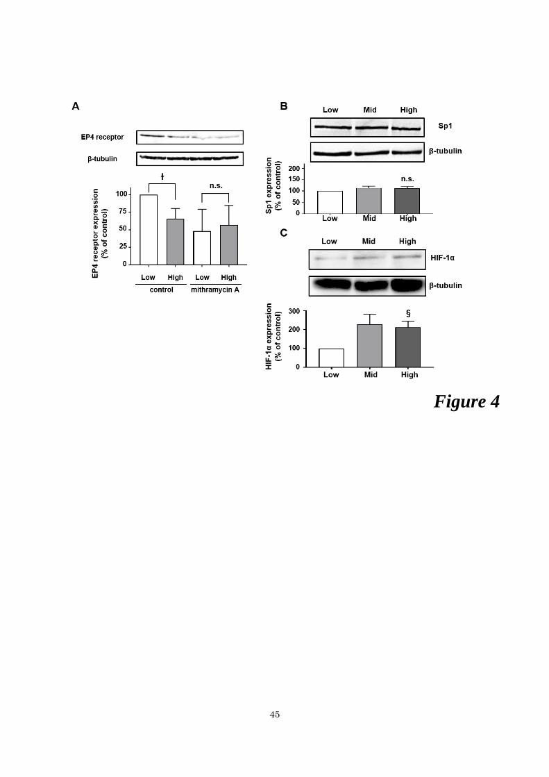
Sp1 regulates the expression level of the EP4 receptor protein.
Since Sp1 binding motifs are associated with the regulation of EP4 receptor expression,

16
we then used mithramycin A, an antibiotic that binds to the Sp1-binding site to displace Sp1 and
inhibit its activity, for HCA-7 cells in low and high cellular density-condition cells. EP4 receptor
protein expression levels were significantly decreased by approximately 30 to 40%, in high
density-conditions cells compared to low density-condition cells (Figure 4A). However, EP4
receptor protein expression levels decreased to similar levels in high and low cellular density-
condition cells treated with mithramycin A, and the cellular density-dependent reduction in EP4
receptor protein expression levels was canceled (Figure 4A). These results indicate that the
interaction of Sp1 with Sp1-binding consensus motifs in the EP4 receptor promoter region is
directly associated with the cellular density-dependent decrease of EP4 receptor expression.
We previously reported that HIF-1α protein expression levels were up-regulated in a
cellular density-dependent manner with a negative relation to EP4 receptor protein expression
levels in HCA-7 cells (Otake et al., 2015). Thus, because Sp1 proteins appeared to bind directly
to the EP4 receptor promoter region, the cellular density-dependent Sp1 protein expression levels
in HCA-7 cells were examined. Sp1 protein expression levels were not changed among the
different cellular density-condition cells tested (Figure 4B). On the other hand, the HIF-1α
protein expression levels were significantly up-regulated in a cellular density-dependent manner,
as shown in previous our report (Otake et al., 2015).
The transcriptional activity of EP4 receptors is oppositely regulated by HIF-1 and c-Myc on
the promoter at Sp-1-binding sites
The previous studies reported that HIF-1α interacts with Sp1, which binds to the promoter
lacking the HRE region (Koshiji et al., 2005; To et al., 2006). Moreover, HIF-1α has been shown
to displace c-Myc from the promoter binding Sp1, resulting in the repression of its promoter
activity, i.e., the MSH2 promoter of sporadic human colon cancer cells (Koshiji et al., 2005; To
et al., 2006). Thus, to examine whether HIF-1α and/or c-Myc regulates the cellular density-
dependent reduction in EP4 receptor promoter activity through the interaction with Sp1, HIF-1α

17
and c-Myc proteins were over-expressed in low cellular density-condition HCA-7 cells. When
transfected with WT (-1238/+1) luciferase reporter gene plasmids, HIF-1α-overexpressing cells
exhibited the significant reduction in EP4 receptor promoter activity. In contrast, EP4 receptor
promoter activity was significantly increased in c-Myc overexpressing cells compared to vector
control-transfected cells (Figure 5A). Moreover, when transfected with the del 4 (-160/+1) or
mut-A,B-del 3 luciferase reporter gene plasmids, which lack or introduce the mutation into two
Sp1-binding consensus motifs respectively, these inhibitory by HIF-1α and stimulatory effects
by c-Myc were canceled (Figure 5A). These results indicate that the EP4 receptor transcriptional
activity is reversely regulated by HIF-1α and c-Myc on Sp1-binding consensus motifs of the EP4
receptor promoter region. In contrast, when the c-Myc protein was expressed in HCA-7 cells,
cellular density-dependent differences of the c-Myc protein were not observed at any cellular
density-conditions tested (Figure 5B).
Because of the adverse effects exerted by HIF-1α and c-Myc, competition assays were
examined to reveal whether up-regulation of HIF-1α or c-Myc reduced the effects of its
counterpart. HIF-1α-overexpressing cells exhibited the significant reduction in WT (-1238/+1)
luciferase reporter gene promoter activity (Figure 5C). However, in HCA-7 cells co-transfected
with c-Myc and HIF-1α expression plasmids, the suppression of EP4 receptor promoter activity
was recovered at the same levels as the control cells in the amount of the c-Myc expression
plasmid-dependent manner. In contrast, in HCA-7 cells co-transfected with HIF-1α expression
plasmids, the increased EP4 receptor promoter activity declined at the same levels as the control
cells in the amount of the HIF-1α plasmid-dependent manner (Figure 5D).
Finally, to confirm whether Sp1, HIF-1a, and c-Myc bind to the Sp1 binding consensus
motifs on EP4 receptor promoter region, the relative recruitment were analyzed by ChIP assay.
As shown in Figure 5E, the significant cellular density-dependent reduction in the binding ability
of Sp1 and c-Myc to mut-HRE (-1238/+1), but not of HIF-1α at high cellular density-condition.
Therefore these data indicated that the promoter region of the EP4 receptor lost its binding ability
18
to Sp1 and/or c-Myc when cells were cultured at high cellular density-condition.
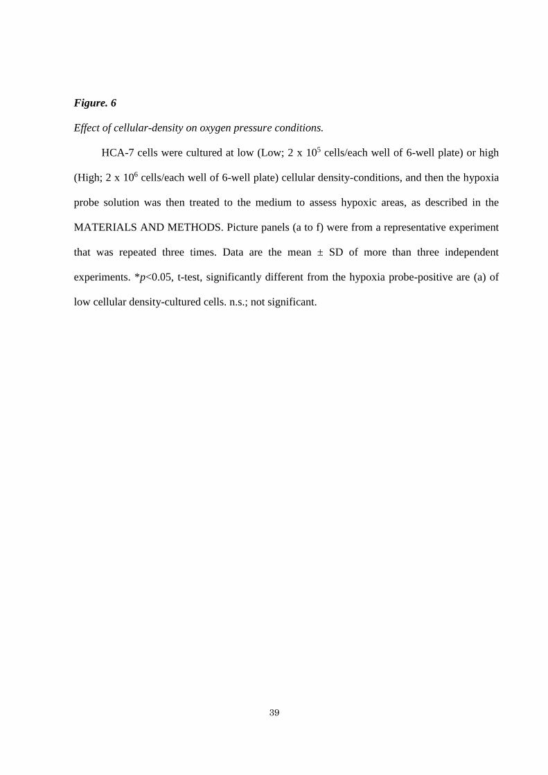
High cellular density-cultured HCA-7 cells showed significantly larger hypoxia-positive are than
in low cellular density-cultured cells
As shown in Figures 4B, 4C, and 5B, the protein expression levels of Sp1 and c-Myc were
not altered in a cellular density-dependent manner, in contrast to HIF-1. Hypoxia is one of the
critical factors inducing HIF-1, which is frequently overexpressed in many cancers including
colon cancer (Semenza, 2003; Mabjeesh and Amir, 2007; Rohwer et al., 2013). Thus, in order to
confirm whether the cellular density-dependent induction of HIF-1 was due to hypoxic cellular
conditions, hypoxic areas were measured using the hypoxia probe. As shown in Figure 6, the
hypoxia-positive area in low cellular density-cultured HCA-7 cells was approximately 0.087%
whereas that in high cellular density-cultured cells was approximately 1.076%, an area that was
approximately 10-fold more significant than that in low cellular density-cultured cells. Thus,
elevations in hypoxia under high cellular density conditions are a plausible reason for the up-
regulated expression of HIF-1. Although the hypoxia-positive area was approximately 1% of
the total area, the high density-cultured HCA-7 cells were in sphere phase so that there was a
possibility that the hypoxia probe might not penetrate all the way down to the underlying and/or
bottom cells. Therefore, even the majority of the cells did not appear to be hypoxia positive, it
was difficult to conclude that the bulk of the HCA-7 cells were not in hypoxia.
HIF-1α protein levels may increase by the inhibition of the activity of its degradation pathways.
To confirm the relationship of these results, an in silico analysis was examined using the
Cancer Genome Atlas database; http://xena.ucsc.edu (Weinstein et al., 2013). The EP4 receptor
mRNA expression levels were significantly decreased in colorectal cancer tissues compared to
the normal colorectal tissues (Figure 7A). On the other hand, the c-Myc mRNA expression levels
were significantly increased in colorectal cancer tissues. The expression levels of Sp1 mRNAs

19
were similar to the HIF-1α mRNAs.
Previously we reported that the cellular density-dependent reductions in EP4 receptors by
the induction of HIF-1α caused the change of the dominant EP receptor subtypes from EP4
receptor to the EP3 receptor (Otake et al., 2015; Fujino, 2016). We also showed that the activation
of EP3 receptor up-regulated vascular endothelial growth factor (VEGF)-A165 (VEGF-A) and
VEGF receptor-1 (VEGFR-1, also known as FLT-1) expressions in HEK-293 cells stably
expressing human EP3 receptors and HCA-7 cells (Taniguchi et al., 2008; Fujino et al., 2011).
The expression levels of VEGF-A and FLT-1 mRNAs were significantly up-regulated in
colorectal cancer tissues (Figure 7B). Although we have not yet shown whether EP3 receptor
induces the up-regulation of VEGF-A and FLT-1 mRNAs in colorectal cancer tissues, the
expression of these angiogenic-correlated genes is known to be regulated by HIF-1α (Majmundar
et al., 2010). Moreover, the expression of glucose transporter (GLUT)-1 (also known as SLC2A1)
mRNA is regulated by HIF-1α, because the glucose transporter has essential roles in the
development of embryos in the relatively hypoxic environment at the placenta (Hayashi et al.,
2004). The expression levels of SLC2A1 mRNA were also significantly increased in colorectal
cancer tissues. Most of the HCA-7 cells may not be in hypoxia, resulting in the expression of
HIF-1α mRNA not being induced. However, the expression levels of HIF-1α stability gene are
also regulated by degradation rates through the ubiquitin-proteasome degradation system in
normoxia (Majmundar et al., 2010). Therefore, the expression levels of HIF-1α protein may be
up-regulated despite mRNA levels by inhibiting the degradation pathways. Several factors are
associated with the stabilization of HIF-1α such as the von Hippel-Lindau (VHL) protein, prolyl
hydroxylase domain-containing enzymes (PHDs; also known as Egl-9 family hypoxia-inducible
factors (EGLNs)), copper metabolism domain containing 1 (COMMD1), factor-inhibiting HIF-
1α (FIH1; also known as HIF-1α inhibitor (HIF1AN)), heat shock protein (HSP) 70 proteins,
such as HSPA1A, and cAMP response element-binding protein-binding protein (CBP)/p300-
interacting transactivator 2 (CITED2) (Majmundar et al., 2010). Comparisons to mRNAs

20
associated with HIF-1α stabilization between colorectal cancer and normal colorectal tissues
(Figure 7C). Except for VHL and HSPA1A, 6 out of 8 gene expression were significantly reduced
in colorectal cancer tissues, indicating that HIF-1α stability increases because of the reduction in
the HIF-1α degradation pathways. Therefore, even if significant differences were not observed
in HIF-1α mRNA expression levels between colorectal cancer and normal colorectal tissues,
HIF-1α protein levels may be up-regulated because of the decreases in the activity of HIF-1α
degradation pathways.
21
5. DISCUSSION
The regulation mechanism of EP4 receptor expression in HCA-7 cells and colon cancer
We previousresearchly reported that EP4 receptor expression was down-regulated by HIF-
1α in a cellular density-dependent manner in HCA-7 cells (Otake et al., 2015). However, it was
unclear how HIF-1α down-regulated EP4 receptor expression. In this research, we investigate the
new regulation mechanism of EP4 receptor expression. Some research groups previously
reported the regulation of Sp1 target genes expression on their promoters by HIF-1α and c-Myc
in sporadic human colon cancer (Koshiji et al., 2005; To et al., 2006). Indeed, our results showed
that HIF-1α and c-Myc also regulate EP4 receptor expression via Sp1 (Figure 5C and 5D). The
recruitment of Sp1 in the human EP4 receptor promoter region was significantly inhibited in high
cellular density-condition (Figure 5E). Combined with these results, I proposed a novel
mechanism of the EP4 expression through the competition between HIF-1α and c-Myc on SP1
(Figure 8). In a low cellular density condition, the HIF-1α levels are low therefore both Sp1 and
c-Myc can form complex each other as previously reported (Koshiji et al., 2005; To et al., 2006)
and up-regulate the EP4 receptor expression (Figure 8; Left panel). When cells reach high cellular
density, increased levels of HIF-1α proteins can inhibit not only the interaction of Sp1 with c-
Myc but also the recruitment of Sp1 on EP4 receptor promoter region, which leads to decrease
the expression levels of the EP4 receptor (Figure 8; right panel). Although we could not show
the direct interaction among Sp1, HIF-1α, and c-Myc, our data clearly show that the balance of
HIF-1α is critical for the regulation of EP4 receptor expression.
c-Myc and HIF-1α may be important factors for maintenance of the homeostasis in colorectal
epithelial cells by modulating EP4 receptor expression.
While EP4 receptors are well-known to get involved in malignancy of colorectal cancer,
EP4 receptors have also had a role in modulating gastrointestinal homeostasis (Yokoyama et al.,

22
2013). Therefore, during the 3- to 5-day turnover of colorectal epithelial cells, the β-catenin
signaling pathway is activated for cellular proliferation and migration in the first half. Then
inactivation of β-catenin signaling pathway caused cellular differentiation and apoptosis in the
last half. Since β-catenin signaling pathway is known to be activated by EP4 receptors (Fujino et
al., 2002), the cellular proliferation and migration of colorectal epithelial cells appear to be
induced in the activated EP4 receptor cells in the first of the cycle. However, in the last half of
the cycle, the β-catenin signaling pathway is inhibited toward cellular differentiation and
apoptosis (van de Wetering et al., 2002). Therefore, because of the up-regulation of cellular
proliferation by EP4 receptor inducing the β-catenin signaling pathway, HIF-1α protein
expression levels may also be up-regulated. However, when cells come up the mid-crypt region,
the increased HIF-1α may inhibit β-catenin signaling pathway. Then EP4 receptor protein
expression is down-regulated by displacing c-Myc for Sp1 binding at the EP4 receptor promoter
region and followed by pulling Sp1 out from Sp1 binding consensus motifs (Figure 8).
Therefore, HIF-1α may be an essential factor for modulating the homeostasis of colorectal
epithelial cells by inducing a negative feedback loop of EP4 receptor expression. Another key
factor for regulating the cell fates by controlling EP4 receptor expression levels may be c-Myc.
The increased c-Myc up-regulated the transcriptional activities of the EP4 receptor. The
expression of the c-Myc protein is known to be up-regulated by β-catenin signaling pathway
(Dang, 1999). Since the activation of EP4 receptors modulates β-catenin signaling pathway, c-
Myc may induce a positive feedback loop of EP4 receptor expression.
The 3- to the 5-day cycle of turnover of normal colorectal epithelial cells appears to be
increased by the expression levels of the EP4 receptor via c-Myc in the first half. Then the
turnover may be negatively regulated via HIF-1α in the last half. However, the regulation of this
regular homeostasis cycle is variable. Therefore, once the balance between c-Myc- and HIF-1α
changes, colorectal epithelial cells may easily become cancerous because of extraordinary
cellular proliferation, migration, and differentiation. When colorectal epithelial cells come up the

23
mid-crypt region, normal colorectal cells inhibit β-catenin signaling pathway for cellular
differentiation and apoptosis. However, if the c-Myc protein is over-expressed, β-catenin
signaling pathway remains activated in progenitor cells in the first half, which leads to
extraordinary cellular proliferation and a cancerous phenotype (van de Wetering et al., 2002).
The expression of c-Myc mRNA was significantly up-regulated in colorectal cancer tissues, and
the up-regulation of c-Myc may be one of the first steps in tumorigenesis, which may be due to
the up-regulation of EP4 receptors (Chell et al., 2006; Hawcroft et al., 2007). Moreover, the up-
regulation of c-Myc expression was not detected in HCA-7 cells. These reports indicate that
HCA-7 cells have some features like normal colon epithelial cells (Kirkland, 1985).
Modulation mechanisms of EP4 receptor expression in homeostasis to the malignancy of
colorectal cells.
It remains unclear if EP4 receptors are involved in colorectal cancer malignancy, how the
down-regulation of EP4 receptor expression is concomitant with increases in cancer cellular
proliferation. Our results indicate that EP4 receptors are a crucial factor in the initiation of
tumorigenesis, but the only function at the very early stage of colorectal cancer. Therefore, if the
expression levels of the EP4 receptor increase for some reason in normal colorectal epithelial
cells, the expression levels of c-Myc will also be up-regulated through the activation of the β-
catenin signaling pathway. However, if the cellular proliferation is rapidly increased and
colorectal epithelial cells subsequently become cancerous, the expression levels of the EP4
receptor are down-regulated via extraordinary increased HIF-1α protein levels, although up-
regulated HIF-1α may promote the reductions in EP4 receptor expression levels. Therefore, at
the stage of HIF-1α accumulation in cells, EP4 receptors do not appear to be associated with
malignancy because the increased HIF-1α also up-regulates VEGF-A165 and VEGFR-1 gene
expression, to absorb nutrients for rapidly growing cancer cells by inducing angiogenesis and/or
cellular migration/metastasis via the activation of EP3 receptor signaling pathway (Taniguchi et

24
al., 2008; Fujino et al., 2011). Therefore, at the stage of HIF-1α accumulation in colorectal
epithelial cells, cells appear to get malignant phenotypes and homeostasis.

25
6. CONCLUSION
When the EP4 receptor is over-expressed, the cells continue to grow extraordinary and
have malignant phenotypes. Although EP4 receptors appear to be necessary for the first step of
tumorigenesis, EP4 receptors no longer have a role, once cells have extraordinary proliferated.
To elucidate the dual and adverse involvement of HIF-1α in EP4 receptor expression, we need
further study. However, our present results indicate one possible reason for why conflicting
findings exist for the roles of the expression levels of the EP4 receptors in tumorigenesis.

26
7. REFERENCES
Chell SD, Witherden IR, Dobson RR, Moorghen M, Herman AA, Qualtrough D et al. (2006).
Increased EP4 receptor expression in colorectal cancer progression promotes cell growth and
anchorage independence. Cancer Res. 66:3106-13.
Dang CV (1999). c-Myc target genes involved in cell growth, apoptosis, and metabolism. Mol.
Cell. Biol. 19:1-11.
Eiginger AL, Prescott SM, Jones DA, Stafforini DM (2007). The role of cyclooxygenase-2 and
prostaglandins in colon cancer. Prostaglandins Other Lipid Mediat. 82:147-54.
Fujino H (2016). The roles of EP4 prostanoid receptors in cancer malignancy signaling. Biol.
Pharm. Bull. 39:149-55.
Fujino H, Toyomura K, Chen X, Regan JW, Murayama T (2011). Prostaglandin E2 regulates
cellular migration via induction of vascular endothelial growth factor receptor-1 in HCA-7
human colon cancer cells. Biochem. Pharmacol. 81:379-87.
Fujino H, West KA, Regan JW (2002). Phosphorylation of glycogen synthase kinase-3 and
stimulation of T-cell factor signaling following activation of EP2 and EP4 prostanoid receptors
by prostaglandin E2. J. Biol. Chem. 277:2614-9.
Fujino H, Xu W, Regan JW (2003). Prostaglandin E2 induced functional expression of early
growth response factor-1 by EP4, but not EP2, prostanoid receptors via the phosphatidylinositol
3-kinase and extracellular signal-regulated kinases. J. Biol. Chem. 278:12151-6.

27
Gustafsson A, Hansson E, Kressner U, Nordgren S, Andersson M, Wang W et al. (2007). EP1-4
subtype, COX and PPARγ receptor expression in colorectal cancer in prediction of disease-
specific mortality. Int. J. Cancer. 121:232-40.
Hawcroft G, Ko CWS, Hull MA (2007). Prostaglandin E2-EP4 receptor signaling promotes
tumorigenic behavior of HT-29 human colorectal cancer cells. Oncogene 26:3006-19.
Hayashi M, Sakata M, Takeda T, Yamamoto T, Okamoto Y, Sawada K et al. (2004). Induction
of glucose transporter 1 expression through hypoxia-inducible factor 1α under hypoxic
conditions in trophoblast-derived cells. J. Endocrinol. 183:145-54.
Kambe A, Iguchi G, Moon Y, Kamitani H, Watanabe T, Eling TE (2008). Regulation of EP4
expression via the Sp-1 transcription factor: inhibition of expression by anti-cancer agents.
Biochim. Biophys. Acta. 1783:1211-9.
Kirkland SC (1985). Dome formation by a human colonic adenocarcinoma cell line (HCA-7).
Cancer Res. 45: 3790-5.
Kondo K, Kico J, Nakamura E, Lechpammer M, Kaelin WG (2002). Inhibition of HIF is
necessary for tumor suppression by the von Hippel-Lindau protein. Cancer Cell. 1:237-46.
Koshiji M, To KK, Hammer S, Kumamoto K, Harris AL, Modrich P et al. (2005). HIF-1α induces
genetic instability by transcriptional downregulating MutSα expression. Mol. Cell. 17:793-803.
Mabjeesh NJ, Amir S (2007). Hypoxia-inducible factor (HIF) in human tumorigenesis. Histol.

28
Histopathol. 22:559-72.
Majmundar AJ, Wong WJ, Simon MC (2010). Hypoxia-inducible factors and the response to
hypoxic stress. Mol. Cell. 40:294-309.
Otake S, Yoshida k, Seira N, Sanchez CM, Regan JW, Fujino H et al. (2015). Cellular density-
dependent down-regulation of EP4 prostanoid receptors via the up-regulation of hypoxia-
inducible factor-1α in HCA-7 human colon cancer cells. Pharmacol. Res. Perspect. 3:e00083.
Potten CS, Loeffler M (1990). Stem cells: attributes, cycles, spirals, pitfalls and uncertainties.
Lessons for and from the crypt. Development. 110:1001-20.
Pullamsetti SS, Banat GA, Schmall A, Szibor M, Pomagruk D, Kolosionek E et al. (2013).
Phosphodiesterase-4 promotes proliferation and angiogenesis on lung cancer by crosstalk with
HIF. Oncogene 32:1121-34.
Rohwer N, Zasada C, Kempa S, Cramer T (2013). The growing complexity of HIF-1α’s role on
tumorigenesis: DNA repair and beyond. Oncogene 32:3569-76.
Semenza GL (2003). Targeting HIF-1 for cancer therapy. Nat. Rev. Cancer 3:721-32.
Sheng H, Shao J, Washington MK, DuBois RN (2001). Prostaglandin E2 increases growth and
motility of colorectal carcinoma cells. J. Biol. Chem. 276:18075-81.
Takafuji V, Cosme R, Lubin D, Lynch K, Roche JK (2000). Prostanoid receptors in intestinal
epithelium; selective expression, function, and change with inflammation. Prostaglandins Leukot.

29
Essent. Fatty Acids 63:223-35.
Taniguchi T, Fujino H, Israel DD, Regan JW, Murayama T (2008). Human EP3I prostanoid
receptor induces VEGF and VEGF receptor-1 mRNA expression. Biochem. Biophys. Res.
Commun. 377:1173-8.
To KK, Sedelinikova OA, Samons M, Bonner WM, Huang LE (2006). The phosphorylation
status of PAS-B distinguishes HIF-1α from HIF-2α in NBS1 repression. EMBO J. 25:4784-94.
Weinstein JN, Collisson EA, Mills GB, Shaw KM, Ozenberger BA, Ellrott K et al. (2013). The
cancer genome atlas pan-cancer analysis project. Nat. Genet. 45:1113-20.
van de Wetering M, Sancho E, Verweij C, de Lau W, Oving I, Hurlstone A et al. (2002). The β-
catenin/TCF-4 complex imposes a crypt progenitor phenotypes on colorectal cancer cells. Cell
111:241-50.
Woodward DF, Jones RL, Narumiya S (2011). International Union of basic and clinical
pharmacology. LXXXIII: classification of prostanoid receptors, updating 15 years of progress.
Pharmacol. Rev. 63:471-538.
Yokoyama U, Iwatsubo K, Umemura M, Fujita T, Ishikawa Y (2013). The prostanoid EP4
receptor and its signaling pathway. Pharmacol. Rev. 65:1010-52.
Yoshida K, Fujino H, Otake S, Seira N, Regan JW, Murayama T (2013). Induction of
cyclooxygenase-2 expression by prostaglandin E2 stimulation of the prostanoid EP4 receptor via
coupling to Gαi and transactivation of the epidermal growth factor receptor in HCA-7 human

30
colon cancer cells. Eur. J. Pharmacol. 718: 408-17.
Yoshimatsu K, Golijanin D, Paty PB, Soslow RA, Jakobsson PJ, DeLellis RA et al. (2001).
Inducible microsomal prostaglandin E synthase is overexpressed in colorectal adenomas and
cancer. Clin. Cancer Res. 7:3971-6.

31
8. EXAMINERS
This thesis was revised by the following examiners authorized by the Graduate School of
Pharmaceutical Sciences, Chiba University
Dr. Naoto Yamaguchi, Ph.D., Professor of Chiba University
(Graduate School of Pharmaceutical Sciences) Chief Examiner
Dr. Hiroyuki Takano, M.D. and Ph.D., Professor of Chiba University
(Graduate School of Pharmaceutical Sciences)
Dr. Takashi Miki, M.D. and Ph.D., Professor of Chiba University
(Graduate School of Pharmaceutical Sciences)
Dr. Mark Bix, Ph.D., Visiting Professor at Chiba University
(Graduate School of Pharmaceutical Sciences)

32
9. ACKNOWLEDGEMENTS
I would like to express my grateful thanks to Professor Toshihiko Murayama of the Laboratory
of Chemical Pharmacology, Graduate School of Pharmaceutical Sciences, Chiba University for
my invaluable guidance, kindness and continuous encouragement.
I sincerely thank Professor Hiromichi Fujino and Assistant Professor Kazuyuki Yamagata for
continuous and patient support, advice and guidance for my research.
I am grateful to Professor Naoto Yamaguchi, Professor Hiroyuki Takano, Professor Takashi Miki,
and visiting Professor Mark Bix for reviewing this thesis and grateful suggestion.
I sincerely thank to Associate Professor Hiroyuki Nakamura and Assistant professor Takuya
Honda for valuable suggestion and advice, and kind support for my laboratory life.
This work was partially supported by Grants from Leading Graduate School, Chiba University.
I greatly appreciate it.
I would like to thank friends for the same Ph.D. course for their kindness and friendship. Also, I
would like to appreciate to the members of the laboratory of Chemical Pharmacology for their
kindness, assistance, and meaningful discussions.
Finally, I would like to thank my parents and family for their understanding and unconditional
support throughout my life.
33
10. FIGURES AND LEGENDS
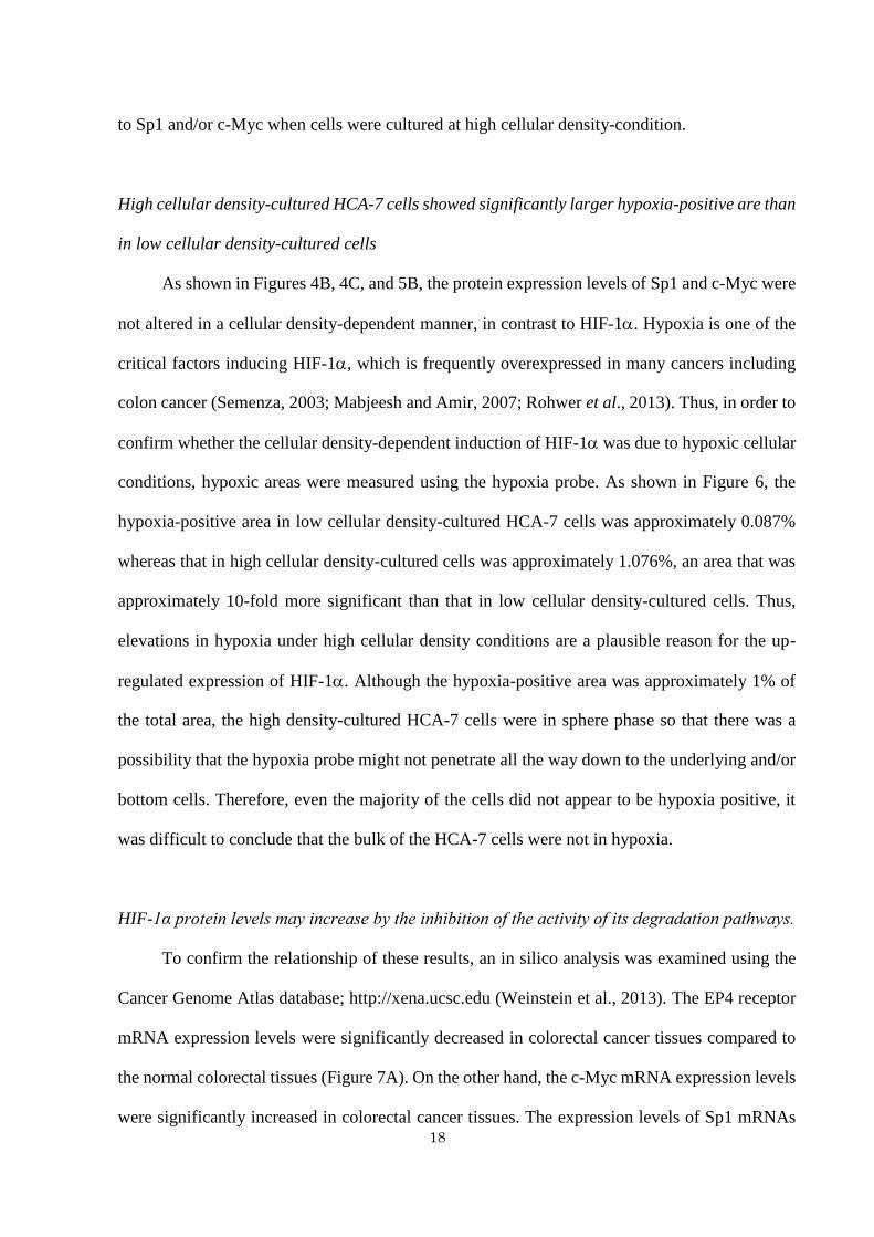
Figure. 1
EP4 receptor transcriptional activities of 5’ deletion mutants in HCA-7 cells.
HCA-7 cells were cultured at low (Low; 2 x 10⁵ cells/each well of 6-well plate) or high
(High; 2 × 10⁶ cells/each well of 6-well plate) cellular density-conditions. Then HCA-7 cells
were transfected with luciferase reporter gene plasmids containing WT or each promoter-mutated
plasmid and subjected to the luciferase reporter gene assay, as described in the MATERIALS &
METHODS. A; The promoter deletion maps and corresponding luciferase activities of each WT
(-1238/+1) or the 5’ deletion of EP4 receptor promoter-contained reporter gene plasmids (del 1
to del 4) obtained from either HCA-7 cells cultured at low or high cellular density-conditions. B,
C, D; The luciferase activities of HCA-7 cells cultured at low cellular density-conditions
transfected with WT (-1238/+1) (B), del 3 (-197/+1) (C), or del 4 (-160/+1) (D) of each human
EP4 receptor promoter plasmid concomitantly with either HA-control vector plasmids or HA-
tagged HIF-1α expression plasmids. Data are the mean ± SD of more than three independent
experiments. *p<0.05, t-test, significantly different from the low cellular density-cultured cells
transfected with WT or mutated human EP4 receptor promoter plasmids. n.s.; not significant.

34
Figure. 2
EP4 receptor transcriptional activities of mutated HRE in HCA-7 cells.
HCA-7 cells were cultured at low (Low; 2 x 10⁵ cells/each well of 6-well plate) or high
(High; 2 × 10⁶ cells/each well of 6-well plate) cellular density-conditions. Then HCA-7 cells
were transfected with luciferase reporter gene plasmids containing WT or HRE point mutated
WT plasmids and subjected to the luciferase reporter gene assay, as described in the
MATERIALS AND METHODS. A; Schematic maps of WT (-1238/+1) or HRE point mutated
WT (mut-HRE (-1238/+1)) of the human EP4 receptor promoter. B; ChIP assay with anti-HIF-
1α antibodies performed to detect the interaction of HIF-1α with WT (-1238/+1) or HRE point
mutated WT (mut-HRE (-1238/+1)) of the human EP4 receptor promoter in HCA-7 cells cultured
at high cellular density-conditions. C; The human EP4 receptor promoter luciferase activities of
each WT (-1238/+1) or HRE point mutated WT (mut-HRE (-1238/+1)) luciferase reporter gene
plasmids obtained from HCA-7 cells cultured at low or high cellular density-conditions. D; The
human EP4 receptor promoter luciferase activities of HCA-7 cells cultured at low cellular
density-conditions transfected either with WT (-1238/+1) or HRE point mutated WT (mut-HRE
(-1238/+1)) luciferase reporter gene plasmids concomitantly with either HA-control vector
plasmids or HA-tagged HIF-1α expression plasmids. Luciferase activity was assessed, as
described in the MATERIALS & METHODS. Data are normalized at the low cellular density-
cultured cells transfected with WT, or HA control vector plasmid-transfected cells at low cellular
density-conditions as 100%. Data are the mean ± SD of more than three independent experiments.
*p<0.05, t-test, significantly different from the low cellular density-cultured cells transfected
with WT or mutated human EP4 receptor promoter plasmids. †p<0.05, t-test, significantly
different from the HA control vector plasmid-transfected cells at low cellular density-conditions.
n.s.; not significant.

35
Figure. 3
Sp1 binding consensus motifs are responsible for the regulation of the human EP4 receptor
promoter activity.
HCA-7 cells were cultured at low (Low; 2 x 10⁵ cells/each well of 6-well plate), and high
(High; 2 x 10⁶ cells/each well of 6-well plate) cellular density-conditions. A; Two Sp1 binding
consensus motifs located between -197 and -160 of the human EP4 receptor promoter region. B;
HCA-7 cells were transfected with luciferase reporter gene plasmids containing del 3 (-197/+1)
or each or both Sp1 binding consensus motifs-mutated del 3 plasmids and then subjected to the
luciferase reporter gene assay, as described in the MATERIALS & METHODS. Sp1-binding
consensus motifs mutation maps and the corresponding luciferase activities of del 3 (-197/+1) or
each or both Sp1-binding consensus motifs-mutated del 3 plasmids (mut-A-del 3, mut-B-del 3,
and mut-A,B-del 3) obtained from HCA-7 cells cultured at low or high cellular density-
conditions. Data are normalized to low cellular density-cultured cells transfected with del 3 (B)
from low cellular density-cultured control HCA-7 cells as 100%. *p<0.05, t-test, significantly
different from low cellular density-cultured cells transfected with WT (B). n.s.; not significant.

36
Figure. 4
The regulation of human EP4 receptor protein expression by Sp1.
HCA-7 cells were cultured at low (Low; 2 x 10⁵ cells/each well of 6-well plate), middle
(Mid; 6 x 10⁵ cells/each well of 6-well plate), and high (High; 2 x 10⁶ cells/each well of 6-well
plate) cellular density-conditions in HCA-7 cells. A; An immunoblot analysis with an antibody
against EP4 receptor (upper panel) or β-tubulin (lower panel), and a histogram represent the ratio
of EP4 receptor to β-tubulin as assessed with pooled densitometric data (mean ± SD) from more
than three independent experiments on HCA-7 cells cultured at low or high cellular density-
conditions and treated with either vehicle or 100 nM mithramycin A. B, C; An immunoblot
analysis with an antibody against Sp1 (B), HIF-1α (C) (upper panels), or β-tubulin (lower panels),
and a histogram representing the ratio of Sp1 (B) or HIF-1α (C) to b-tubulin as assessed with
pooled densitometric data (mean ± SD) from more than three independent experiments in HCA-
7 cells cultured at low, middle or high cellular density-conditions. Data are normalized to
immunoblot ratios of the EP4 receptor (A), Sp1 (B), or HIF-1α (C) to β-tubulin detected from
low cellular density-cultured HCA-7 cells as 100%. †p<0.05 and §p<0.05, analysis of variance,
versus each immunoblot ratios obtained at low cellular density-cultured cells (A, B, C). n.s.; not
significant.

37
Figure. 5
Effects of HIF-1α and c-Myc on human EP4 receptor promoter activity in the low cellular
density-dependent condition in HCA-7 cells.
HCA-7 cells were cultured at low (Low; 2 x 10⁵ cells/each well of 6-well plate), middle
(Mid; 6 x 10⁵ cells/each well of 6-well plate), and high (High; 2 x 10⁶ cells/each well of 6-well
plate) cellular density-conditions. A; The luciferase activities of HCA-7 cells cultured at low
cellular density-conditions and transfected either with WT (-1238/+1), del 4 (-160/+1), or mut-
A,B-del 3 of human EP4 receptor promoter contained luciferase reporter gene plasmids
concomitantly with either HA-control vector plasmids, HA-tagged HIF-1α expression plasmids,
or FLAG-tagged c-Myc expression plasmids. B; An immunoblot analysis with an antibody
against c-Myc (upper panel) or β-tubulin (lower panel), and a histogram representing the ratio of
c-Myc to β-tubulin as assessed with pooled densitometric data (mean ± SD) from more than three
independent experiments in HCA-7 cells cultured at low, middle or high cellular density-
conditions. C, D; The luciferase activities of HCA-7 cells cultured at low cellular density-
conditions transfected with WT (-1238/+1) of EP4 receptor promoter containing luciferase
reporter gene plasmids concomitantly with either HA-control vector plasmids or HA-tagged HIF-
1α expression plasmids alone, or HA-tagged HIF-1α expression plasmids plus various amounts
of FLAG-tagged c-Myc expression plasmids (C); or with either HA-control vector plasmids or
FLAG-tagged c-Myc expression plasmids, or FLAG-tagged c-Myc expression plasmids plus
various amounts of HA-tagged HIF-1α expression plasmids (D). E; ChIP assay with anti-Sp1,
anti-HIF-1α, and anti-c-Myc antibodies performed to detect the binding of Sp1, HIF-1α and c-
Myc to HRE point mutated WT (mut-HRE (-1238/+1)) of the EP4 receptor promoter in HCA-7
cells cultured at low and high cellular density-conditions. Data are normalized to the low cellular
density-cultured HCA-7 cells transfected with the HA-control vector plasmids-transfected cells
(A, C, D), the low cellular density-cultured HCA-7 cells transfected with HRE point mutated WT

38
(mut-HRE (-1238/+1)) of the EP4 receptor promoter (E) or immunoblot ratios of low cellular
density-cultured cells (B) as 100%. †p<0.05, analysis of variance, versus HA control vector
plasmids-transfected cells or HRE point mutated WT (mut-HRE (-1238/+1)) of the EP4 receptor
promoter luciferase reporter gene plasmids-transfected cells at low cellular density-conditions.
§p<0.05, analysis of variance, versus the HA control vector plasmids-transfected cells at low
cellular density-conditions. ¶p<0.05, analysis of variance, versus the results of HA-tagged HIF-
1α expression plasmids-transfected cells at low cellular density-conditions. &p<0.05, analysis of
variance, versus the results of FLAG-tagged c-Myc expression plasmids-transfected cells at low
cellular density-conditions. n.s.; not significant.
39
Figure. 6
Effect of cellular-density on oxygen pressure conditions.
HCA-7 cells were cultured at low (Low; 2 x 10⁵ cells/each well of 6-well plate) or high
(High; 2 x 10⁶ cells/each well of 6-well plate) cellular density-conditions, and then the hypoxia
probe solution was then treated to the medium to assess hypoxic areas, as described in the
MATERIALS AND METHODS. Picture panels (a to f) were from a representative experiment
that was repeated three times. Data are the mean ± SD of more than three independent
experiments. *p<0.05, t-test, significantly different from the hypoxia probe-positive are (a) of
low cellular density-cultured cells. n.s.; not significant.

40
Figure. 7
EP4 receptors and HIF1α-related gene expressions in human colon and rectal cancer
tissues analyzed using the TCGA database.
A, B, C; Comparison of the mRNA expression of EP4 receptors (PTGER4), c-Myc (MYC),
Sp1 (SP1), and HIF-1α (HIF1A) (A), HIF-1α target mRNAs such as VEGF-A165 (VEGFA),
VEGFR-1 (FLT1) as well as GLUT1 (SLC2A1) (B), and the factors involved in the stabilization
of HIF-1α such as von Hippel-Lindau (VHL), prolyl hydroxylase domain-containing enzymes
(PHDs; also known as Egl-9 family hypoxia-inducible factors (EGLNs)), factor-inhibiting HIF-
1α (FIH1; also known as HIF-1α inhibitor (HIF1AN)), copper metabolism domain containing 1
(COMMD1), heat shock protein (HSP) 70 proteins, such as HSPA1A, and cAMP response
element-binding protein-binding protein (CBP)/p300-interacting transactivator 2 (CITED2) (C),
between the cancer tissues (gray boxes) and paired normal cancer tissues (white boxes). ‡p<0.05,
the Mann-Whitney U-test, significantly lower than non-cancer tissues. #p<0.05, the
Mann=Whitney U-test, significantly higher than non-cancer tissues. n.s.; not significant.
41
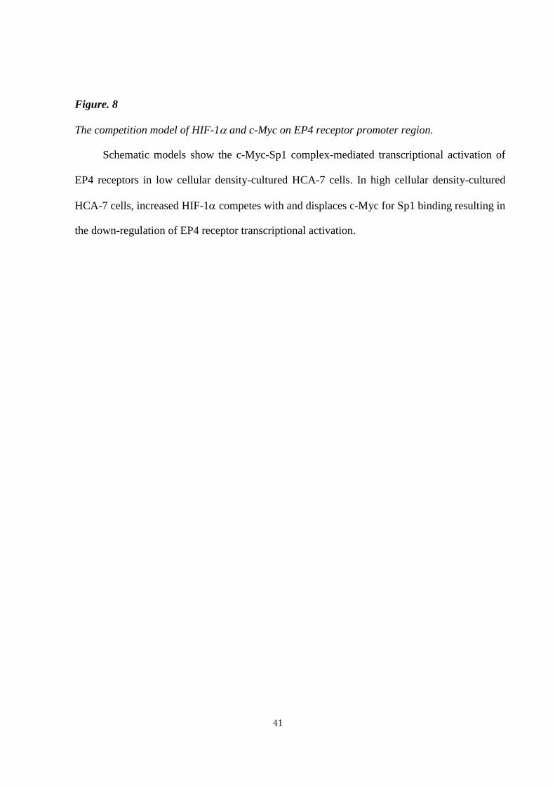
Figure. 8
The competition model of HIF-1 and c-Myc on EP4 receptor promoter region.
Schematic models show the c-Myc-Sp1 complex-mediated transcriptional activation of
EP4 receptors in low cellular density-cultured HCA-7 cells. In high cellular density-cultured
HCA-7 cells, increased HIF-1 competes with and displaces c-Myc for Sp1 binding resulting in
the down-regulation of EP4 receptor transcriptional activation.

42
Figure 1

43
Figure 2

44
Figure 3
45
Figure 4

46
Figure 5

47
Figure 6

48
Figure 7

49
Figure 8

50
LIST OF PUBLICATIONS
[Main thesis publication]
Seira N, Yamagata K, Fukushima K, Araki Y, Kurata N, Yanagisawa N, Mashimo M, Nakamura H
Regan JW, Murayama T, Fujino H: Cellular density-dependent increases in HIF-1α compete with c-
Myc to down-regulate human EP4 receptor promoter activity through Sp-1-binding region. Pharmacol.
Res. Perspect., 2018 6:e00441. doi: 10.1002/prp2.441.