Regulation of Bone by Wnt Pathway · Wnt signaling pathway schematic Wntproteins are...
21
Slide 1 Regulation of Bone by Wnt Pathway Anna Georgieva Ruben Bibas, Gabriel Helmlinger, Brian Stoll Modeling and Simulation, Novartis Pharma
Transcript of Regulation of Bone by Wnt Pathway · Wnt signaling pathway schematic Wntproteins are...
Slide 1
Regulation of Bone by Wnt Pathway
Anna Georgieva
Ruben Bibas, Gabriel Helmlinger, Brian StollModeling and Simulation, Novartis Pharma
Slide 2
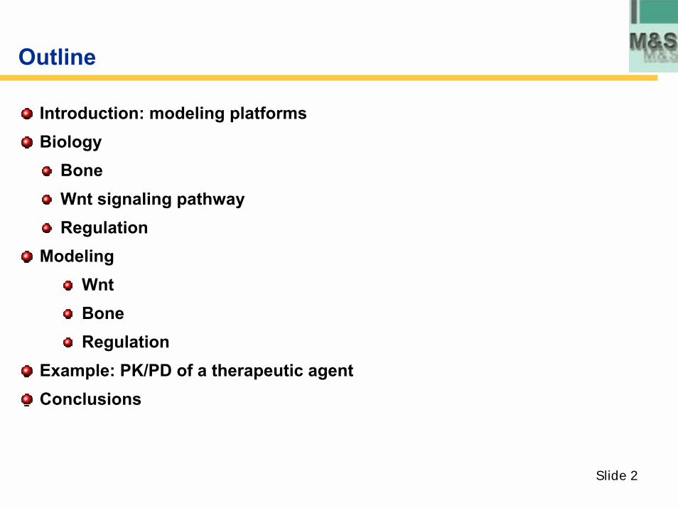
Outline
Introduction: modeling platformsBiology
Bone Wnt signaling pathwayRegulation
ModelingWntBoneRegulation
Example: PK/PD of a therapeutic agentConclusions
Slide 3
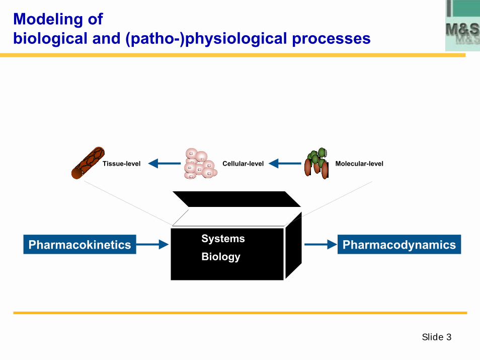
Modeling ofbiological and (patho-)physiological processes
SystemsBiology
Pharmacokinetics Pharmacodynamics
Cellular-levelTissue-level VEGF Molecular-level
Slide 4
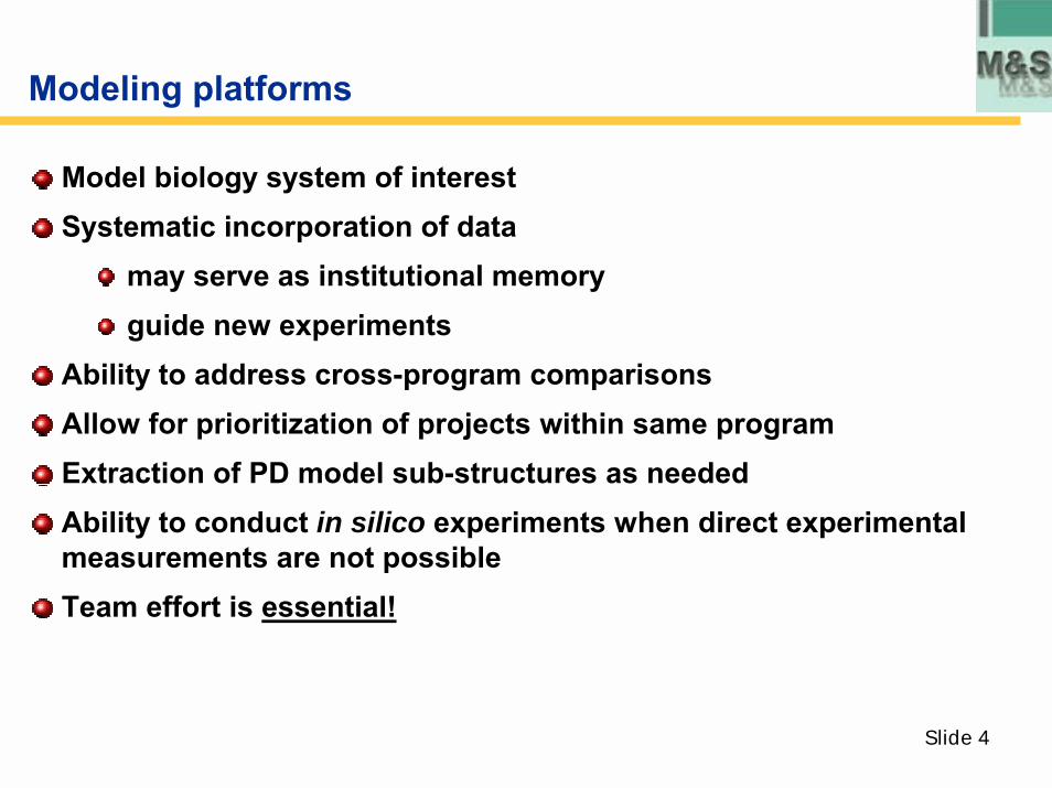
Modeling platforms
Model biology system of interestSystematic incorporation of data
may serve as institutional memoryguide new experiments
Ability to address cross-program comparisons Allow for prioritization of projects within same programExtraction of PD model sub-structures as neededAbility to conduct in silico experiments when direct experimental measurements are not possibleTeam effort is essential!
Slide 5
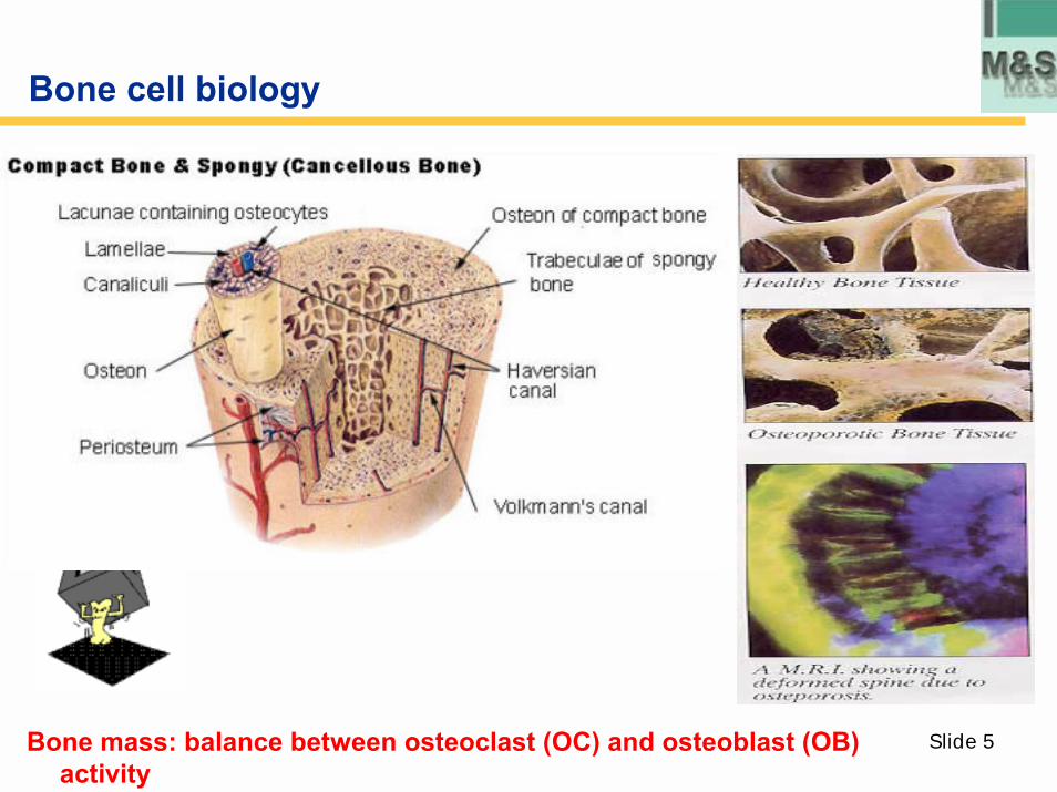
Bone cell biology
Bone mass: balance between osteoclast (OC) and osteoblast (OB) activity
Slide 6
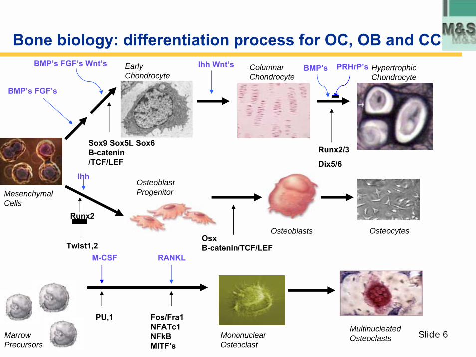
Bone biology: differentiation process for OC, OB and CC
Osteoblasts
Early Chondrocyte
Runx2
lhh
OsxB-catenin/TCF/LEF
Sox9 Sox5L Sox6 B-catenin /TCF/LEF
BMP�s FGF�s Wnt�s
BMP�s FGF�s
lhh Wnt�s PRHrP�sColumnar Chondrocyte
Hypertrophic Chondrocyte
BMP�s
Runx2/3
Dix5/6
Osteoblast Progenitor
Osteocytes
Mesenchymal Cells
Twist1,2M-CSF RANKL
Marrow Precursors
PU,1 Fos/Fra1NFATc1NFkBMITF�s
Multinucleated OsteoclastsMononuclear
Osteoclast
Slide 7
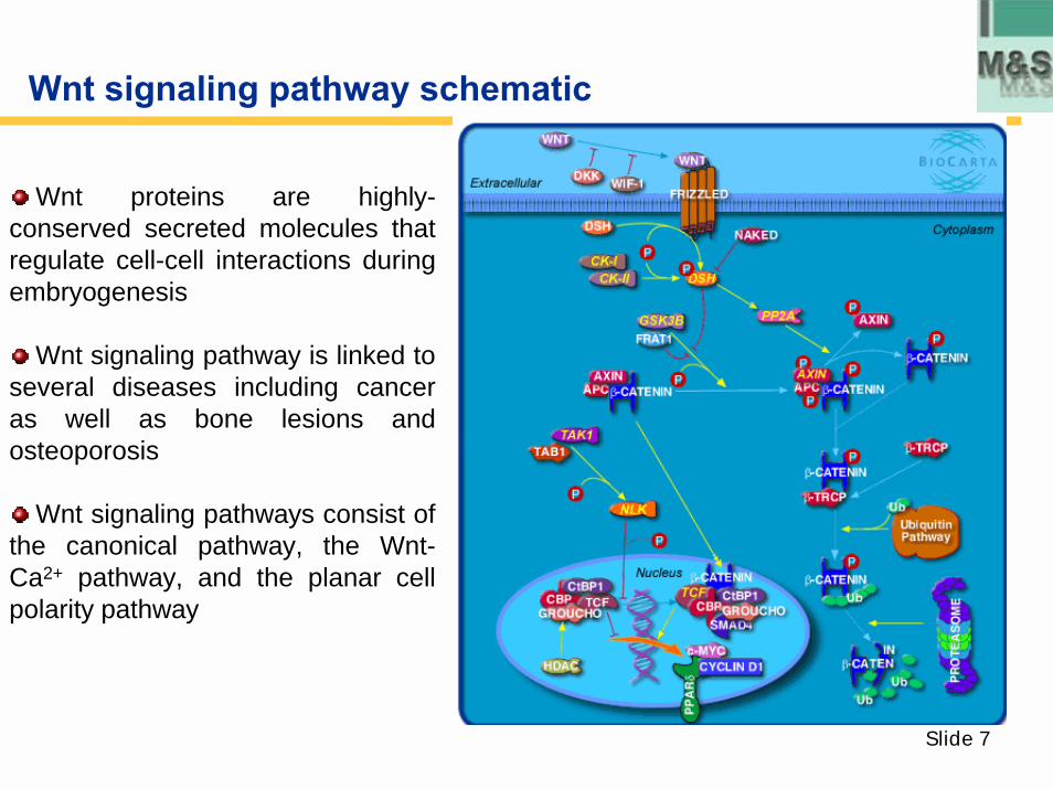
Wnt signaling pathway schematic
Wnt proteins are highly-conserved secreted molecules that regulate cell-cell interactions during embryogenesis
Wnt signaling pathway is linked to several diseases including cancer as well as bone lesions and osteoporosis
Wnt signaling pathways consist of the canonical pathway, the Wnt-Ca2+ pathway, and the planar cell polarity pathway
Slide 8
Key steps in pathway
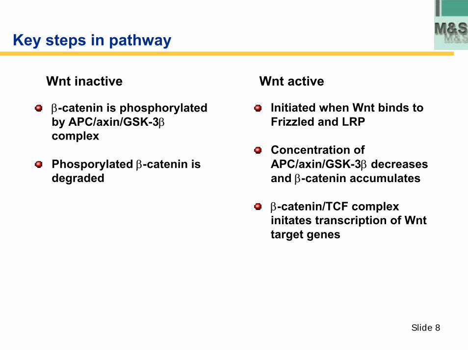
Wnt inactive Wnt active
β-catenin is phosphorylatedby APC/axin/GSK-3βcomplex
Phosporylated β-catenin is degraded
Initiated when Wnt binds to Frizzled and LRP
Concentration of APC/axin/GSK-3β decreases and β-catenin accumulates
β-catenin/TCF complex initates transcription of Wnttarget genes
Slide 9
Wnt signaling pathway regulation of bone
Wnt proteins involvement in bone (development and disease)
DKK-1 in MM with bone lesions, osteoporosis
LRP5 for osteoporosis (HBM phenotype)
Mechanism elucidation
Helps determine fate of mesenchymal stem cells
Increase of OPG/RANKL ratio leads to osteoclastogenesissuppression
Increases rate of proliferation and differentiation of osteoblasts
Inhibits rate of apoptosis of osteoblasts
Slide 10
Overview of pathway model
Model componentsDKK1, Wnt, other ligands as neededLRP5/6, Frizzled, Kremen2DisheveledAxin, GSK3β, APCTCF/ LEFphosphorylated forms and intermediate complexes
Data used to construct model: only published data usedProtein expression data to establish initial concentrations; directly measured or derivedKinetic experiments to determine KD or kon and koff; derive using experimental data and mathematical optimizationUncertainty and variability in parameters owing to different cell and tissue types addressed through sensitivity analyses
Slide 11
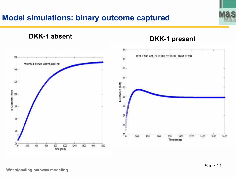
Model simulations: binary outcome captured
DKK-1 absent DKK-1 present
Wnt signaling pathway modeling
Slide 12
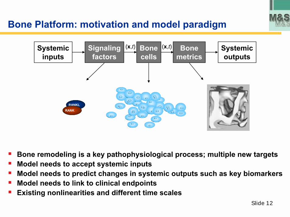
Bone Platform: motivation and model paradigm
Systemicoutputs
Bonemetrics
Bonecells
Signalingfactors
Systemicinputs
RANKLRANK
(x,t) (x,t)
! Bone remodeling is a key pathophysiological process; multiple new targets! Model needs to accept systemic inputs ! Model needs to predict changes in systemic outputs such as key biomarkers! Model needs to link to clinical endpoints! Existing nonlinearities and different time scales
Slide 13
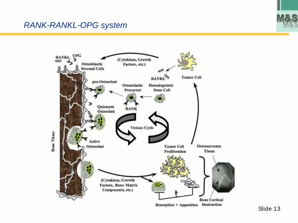
RANK-RANKL-OPG system
Slide 14
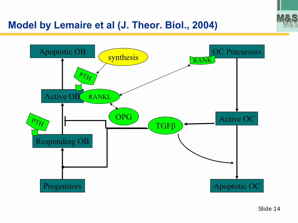
Model by Lemaire et al (J. Theor. Biol., 2004)
Responding OB
Active OC
OC Precursors
OPG
RANKApoptotic OB
synthesis
PTH
Active OB RANKL
PTH TGFβ
Progenitors Apoptotic OC
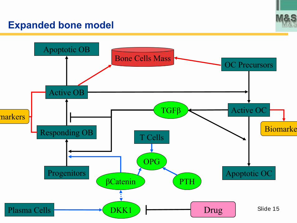
Slide 15
Expanded bone model
Apoptotic OB
Responding OB
Active OC
T CellsBiomarker
TGFβmarkers
Active OB
Progenitors
Plasma Cells
Apoptotic OC
OC Precursors
OPG
DKK1
PTHβCatenin
Bone Cells Mass
Drug
Slide 16
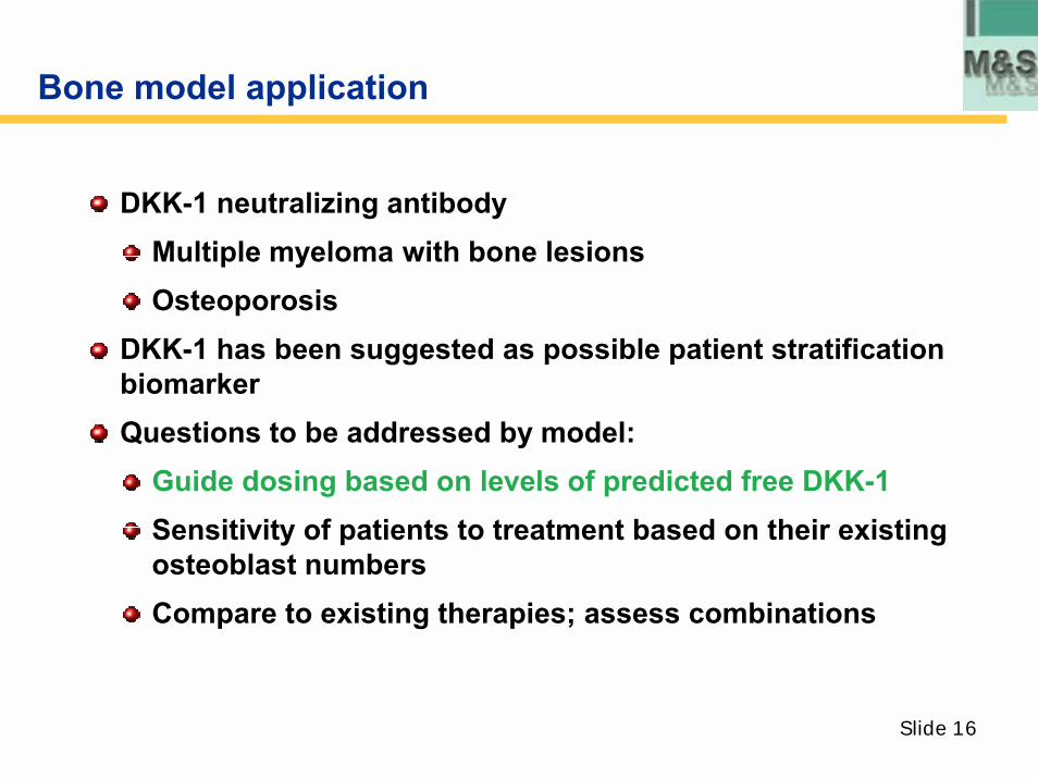
Bone model application
DKK-1 neutralizing antibodyMultiple myeloma with bone lesionsOsteoporosis
DKK-1 has been suggested as possible patient stratification biomarkerQuestions to be addressed by model:
Guide dosing based on levels of predicted free DKK-1Sensitivity of patients to treatment based on their existing osteoblast numbersCompare to existing therapies; assess combinations
Slide 17
Clinical applications
Dose will be given when free DKK-1 level goes above a certain level
No assay measuring free DKK-1 is available
Assay measuring total DKK-1 is available
Calibrate the model based on free level of DKK-1 at baseline (for a range of subjects) and then use model to predict time courses of bound, free and total levels of DKK-1 for different dose levels of antibody
Slide 18
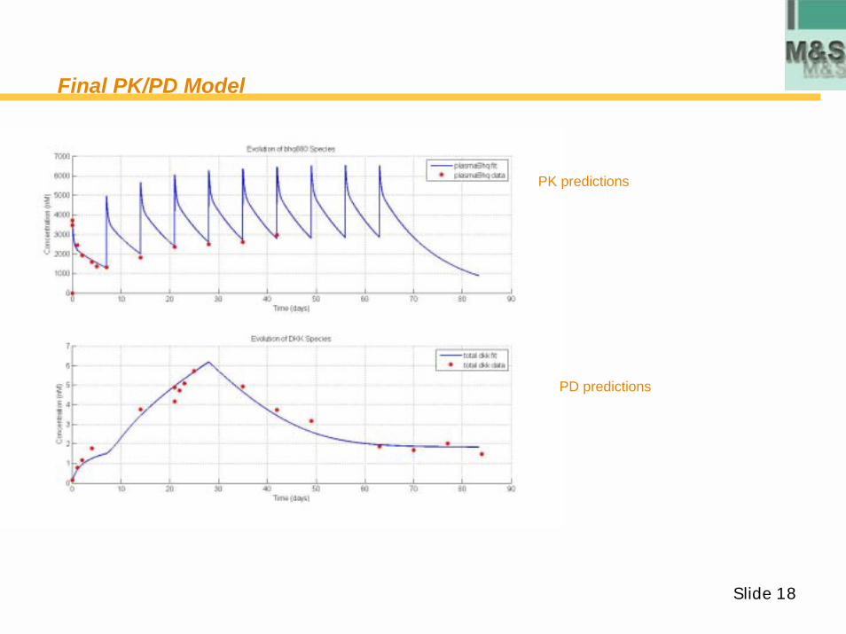
Final PK/PD Model
PD predictions
PK predictions
Slide 19
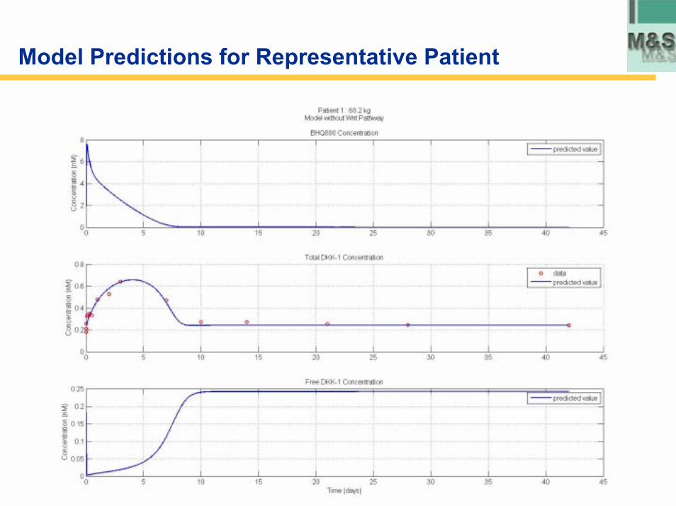
Model Predictions for Representative Patient
Slide 20
Summary of results
Model can guide clinical therapy by predicting levels of free DKK-1Model suggests that hypothesized presence of a DKK-1 negative
feedback loop, i.e. Wnt-signaling and β-catenin translocation leads to DKK-1 up-regulation by osteoblasts (Gonzalez-Sancho et al., Nida et al) is true.
Model can quantify impact of disease severityModel output ultimately consists of BM
Effectiveness of therapyComparison with existing and planned therapies

Slide 21
Thank you!
Questions?