Reducing Hemorrhagic Complication by Dabigatran...
32
1 Reducing Hemorrhagic Complication by Dabigatran via Neurovascular Protection after Recanalization with tPA in Ischemic Stroke of Rat Syoichiro Kono, MD; Kentaro Deguchi, MD; Yoshio Omote, MD; Taijun Yunoki, MD; Toru Yamashita, MD, PhD; Tomoko Kurata, MD; Yoshio Ikeda, MD, PhD; and Koji Abe, MD, PhD Department of Neurology, Graduate School of Medicine, Dentistry and Pharmaceutical Sciences, Okayama University, Okayama, Japan Correspondence to: Dr. Koji Abe, Department of Neurology, Graduate School of Medicine, Dentistry and Pharmaceutical Sciences, Okayama University, 2-5-1 Shikatacho Kitaku, Okayama 700-8558, Japan. Tel.: 81-86-235-7365 Fax: 81-86-235-7368 E-mail: [email protected] Acknowledgement: This work was partly supported by a Grant-in-Aid for Scientific Research (B) 21390267, (C) 24591263 and Challenging Research 24659651, and by Grants-in-Aid from the Research Committees (Mizusawa H, Nakano I, Nishizawa M, Sasaki H, and Aoki M) from the Ministry of Health, Labour and Welfare of Japan. Running title: Reducing Hemorrhage by Dabigatran
-
Upload
phungkhuong -
Category
Documents
-
view
214 -
download
0
Transcript of Reducing Hemorrhagic Complication by Dabigatran...

1
Reducing Hemorrhagic Complication by Dabigatran
via Neurovascular Protection after Recanalization
with tPA in Ischemic Stroke of Rat
Syoichiro Kono, MD; Kentaro Deguchi, MD; Yoshio Omote, MD;
Taijun Yunoki, MD; Toru Yamashita, MD, PhD; Tomoko Kurata, MD;
Yoshio Ikeda, MD, PhD; and Koji Abe, MD, PhD
Department of Neurology, Graduate School of Medicine, Dentistry and Pharmaceutical Sciences,
Okayama University, Okayama, Japan
Correspondence to: Dr. Koji Abe, Department of Neurology, Graduate School of Medicine,
Dentistry and Pharmaceutical Sciences, Okayama University, 2-5-1 Shikatacho Kitaku, Okayama
700-8558, Japan.
Tel.: 81-86-235-7365
Fax: 81-86-235-7368
E-mail: [email protected]
Acknowledgement: This work was partly supported by a Grant-in-Aid for Scientific Research (B)
21390267, (C) 24591263 and Challenging Research 24659651, and by Grants-in-Aid from the
Research Committees (Mizusawa H, Nakano I, Nishizawa M, Sasaki H, and Aoki M) from the
Ministry of Health, Labour and Welfare of Japan.
Running title: Reducing Hemorrhage by Dabigatran

2
Abstract
This study aimed to assess the risk and benefit of tPA treatment under oral anticoagulation with
dabigatran compared to warfarin or vehicle control in transient middle cerebral artery occlusion
(tMCAO). After pretreatment with warfarin (0.2 mg/kg/day), dabigatran (20 mg/kg/day), or vehicle
(0.5% carboxymethyl cellulose sodium salt) for 7 days, tMCAO was induced for 120 min followed by
reperfusion and tPA (10 mg/kg/10 ml). Clinical parameters, including cerebral infarction volume,
hemorrhagic volume, and blood coagulation, were examined. At 24 h after reperfusion, markers for
the neurovascular unit at the periischemic lesion were immunohistochemically examined in brain
sections, and MMP-9 activity was measured by zymography. Paraparesis and intracerebral
hemorrhage volume were significantly improved in the dabigatran-pretreated group than in the
warfarin-pretreated group. A marked dissociation between astrocyte foot processes and the basal
lamina or pericyte was observed in the warfarin-pretreated group, which was greatly improved in the
dabigatran-pretreated group. Furthermore, a remarkable activation of MMP-9 in the ipsilateral
warfarin-pretreated rat brain was greatly reduced in dabigatran-pretreated rats. The present study
reveals that the mechanism of intracerebral hemorrhage with warfarin-pretreatment plus tPA in
ischemic stroke rats is the dissociation of the neurovascular unit, including the pericyte. Neurovascular
protection by dabigatran, which was first shown in this study, could partly explain the reduction in
hemorrhagic complication by dabigatran reported in the clinical study.
Key Words: dabigatran; hemorrhagic complication; neurovascular unit; pericyte; thrombolysis; tPA

3
Introduction
As the world’s population is progressively aging in most countries, so too is the
number of patients who are suffering from stroke also rapidly increasing. Half of all
strokes occur in people who are over 70 years old, and a quarter occurs in patients who
are > 85 years of age (Bamford et al., 1988; Brown et al., 1996). Since atrial
fibrillation (AF) is an age-dependent incident that is more common in the elderly,
cardiogenic cerebral embolic stroke is the major cause of increasing strokes among the
elderly.
The new oral anticoagulant (NOAC) dabigatran is a direct thrombin inhibitor that
was approved by the US Food and Drug Administration in October 2010 based upon
data from the Randomized Evaluation of Longfrom Term Anticoagulant Therapy With
Dabigatran Etexilate (RE-LY) study which demonstrated it to be as safe as, safer than
or at least as effective as warfarin (Connolly et al., 2009). A notable benefit of
dabigatran, unlike warfarin, is that it does not need the international normalized ratio
(INR) to be monitored nor the diet to be restricted. Therefore, the number of
dabigatran-treated patients having an ischemic stroke is increasing around the world.
Upon ischemic stroke, a patient that was pretreated with warfarin can still receive
tissue plasminogen activator (tPA) if their INR is 1.7, which could increase the risk

4
of hemorrhagic complication. The National Institute of Neurological Disorders and
Stroke (NINDS) tPA study (The National Institute of Neurological Disorders and
Stroke rtPA Stroke Study Group, 1995), the European Cooperative Acute Stroke Study
trials (ECASS I-III) (Hacke et al., 1995; Hacke et al., 1998; Hacke et al., 2008), and
the Safe Implementation of Thrombolysis in Stroke Monitoring Study (SITS-MOST)
(Wahlgren et al., 2007) excluded patients receiving oral anticoagulant treatment,
regardless of the INR. In the Japan post-Marketing Alteplase Registration Study
(J-MARS) (Nakagawara et al., 2010), 3.5% of patients had symptomatic intracranial
hemorrhage, but there is limited data on the safety of tPA in warfarin-pretreated
patients. In addition, there is no guideline whether dabigatran-pretreated patients
within 4.5 h with acute ischemic stroke can be considered eligible for tPA treatment or
not. The aim of this study was to assess the risk and benefit of tPA treatment under oral
anticoagulation with dabigatran compared to warfarin or a placebo control.
Materials and Methods
Experimental Model
Male Wistar rats (SLC, Shizuoka, Japan) 11 weeks old (body weight 240–270 g)
were divided into 3 groups: vehicle-treated (0.5% carboxymethyl cellulose sodium salt;

5
V+tissue plasminogen activator (tPA)) group, warfarin-treated (0.2 mg/kg/day; W+tPA)
group, and dabigatran-treated (20 mg/kg/day; D+tPA) group, with n=9 in each group.
For each drug, the dose and interval between the last intake of drug and the induction of
cerebral ischemia were determined so as to inhibit clot formation by 70% in the rat
venous thromboembolism model (Toomey et al., 2006; Wienen et al., 2007). Each drug
was administered orally for 7 days starting from when rats were 11 weeks old. Warfarin
was administered once a day and dabigatran twice a day. Thrombus formation was
reduced by 91% with 10 mg/kg of dabigatran after 30 min of administration and
reduced by 70% after 1 h of administration according to Wienen et al. Thrombus
formation was also reduced by 70% with 0.2 mg/kg of warfarin after 1 h of
administration according to Toomey et al. Thus, the last intake of both drugs was 1 h
before the induction of cerebral ischemia. Body weight and blood pressure were
measured twice before the first and last administration. Blood was drawn from the left
femoral vein prior to and 1 h after the last administration of each drug, and prothrombin
time (PT), activated prothrombin time (aPTT), and thrombin-antithrombin complex
(TAT) were measured.
At 7 days of daily administration of the vehicle (12 w of age), warfarin or
dabigatran, the rats were anesthetized with a mixture of nitrous oxide/oxygen/isoflurane

6
(69: 30: 1) during surgical preparation with an inhalation mask. Body temperature was
monitored and maintained at 37 ± 0.3 °C using a heating pad during the surgical
procedure. The right middle cerebral artery (MCA) was occluded by inserting a 4–0
surgical nylon thread with silicon coating through the common carotid artery as
described previously (Abe et al., 1992). After 120 min of transient MCA occlusion
(tMCAO), the nylon thread was gently removed to restore blood flow in the MCA
territory and was treated with tissue plasminogen activator (tPA; Grtpa, Mitsubishi
Tanabe Pharma Corporation, Osaka, Japan, intravenous bolus, 10 mg/kg/10 ml). 24 h
after reperfusion, blood pressure was measured and behavior was analyzed.
For histological examinations, the rats (n=9 each) were transcardially perfused
with heparinized saline followed by 4% paraformaldehyde in phosphate buffered saline
(PBS, pH 7.2). The whole brain was removed and immersed in the same fixation for 12
h at 4°C. After washing with PBS (pH 7.2), the tissues were transferred into a 10%,
20%, and 30% (w/v) sucrose gradient and then embedded in powdered dry ice and
stored a -80°C. Coronal brain sections 20 µm thick were prepared using a cryostat at
-18°C and mounted on a silane-coated glass slide.
For gelatin zymography, a different set of rats (each group, n=5) were treated as
above. 24 h after reperfusion, rats were anesthetized by intraperitoneal injection of

7
pentobarbital (40 mg/kg) and transcardially perfused with chilled heparin (5 U/ml in
PBS; pH 7.2). Brains were removed quickly and divided into ipsilateral-periischemic
and contralateral-nonischemic hemispheres. Each hemispheric brain was frozen
immediately in dry ice and stored at -80°C until use.
All experimental procedures were approved by the Animals Committee of the
Graduate School of Medicine and Dentistry, Okayama University.
Behavioral Analysis
Before cerebral ischemia and 24 h after reperfusion, the rats were tested for
behavioral activities and scored according to the Zhang et al. (Zhang et al., 2002) corner
test with a minor modification by calculating the difference in the numbers of turning
right (the paraparesis score).
Histology and immunohistochemistry
To determine the area of ischemic lesions, sections were stained with
hematoxylin-eosin (HE) and examined under a light microscope (Olympus SZX-12;
Olympus Optical Co.). The sections were made at 2, 0, −2, −4, and −6 mm from the
Bregma. The infarct area was measured at these five sections by counting pixels using

8
Photoshop CS5 and infarct volume was calculated by multiplying the infarct area by 2
mm thickness (Kawai et al., 2011). To analyze brain hemorrhage, iron staining was
performed using an enhanced Perl’s reaction. Brain sections were incubated with Perl’s
solution (5% potassium ferrocyanide and 5% HCl, 1:1) for 45 min, washed in distilled
water, and incubated again in 0.5% diamine benzidine tetrahydrochloride with nickel
for 60 min, as described by Wu et al. (Wu et al., 2003).
For immunohistochemistry, the following primary antibodies were used: rabbit
anti-MMP-9 antibody (1:200; Abcam); rabbit anti-collagenIV antibody (1:200;
Novotec); mouse anti-glial fibrillary acidic protein (GFAP) antibody (1:1000;
Chemicon); and rabbit anti-platelet-derived growth factor (PDGF) receptor beta
antibody (1:500; Abcam). To detect vascular endothelial cells, N-acetylglucosamine
oligomer (NAGO) was used as the specific endothelial cell marker (Augustin et al.,
1995). Brain sections were washed with PBS (pH 7.4) and then incubated in 0.3%
hydrogen peroxidase/methanol for 10 min to block endogenous peroxidase activity and
incubated with bovine serum albumin for 1 h. Then they were incubated overnight at
4 °C with mouse anti-GFAP antibody and rabbit anti-collagenIV antibody or rabbit
anti-PDGF receptor beta antibody, and with biotinylated Lycopersicon esculentum
lectin (1:500; Vector Laboratories), which binds NAGO and rabbit anti-PDGF receptor

9
beta antibody. On the next day, the slices were washed in PBS (pH 7.4) and incubated
for 2 h at room temperature with fluorochrome-coupled secondary antibody (1:500;
Alexa FluorTM, Molecular Probes, A21424, A21429, and A11034). The sections were
then rinsed 3 times in PBS (pH 7.4) and mounted with VECTASHIELD Mounting
Medium with DAPI (Vector Laboratories, H1200). A confocal microscope equipped
with argon and HeNe1 lasers (Zeiss, LSM 510) was used to capture fluorescent images.
Vascular dissociation index
To assess the detachment of astrocyte endfeet from the basement membrane in the
GFAP/collagenIV double-labeled sections or from the pericyte in the GFAP/PDGF
receptor beta double-labeled sections, and to assess the detachment of the pericyte from
vascular endothelial cells in the PDGF receptor beta/NAGO, 3 levels of the caudate
putamen (1.2, 0.7, and 0.2 mm rostral to the bregma) (Paxinos and Watson, 1982) of
each animal, and 4 areas in the ipsilateral peri-infarcted cortex in each section were
chosen randomly and captured at ×100 magnification with a confocal laser microscope.
We confirmed the border between the ischemic core and peri-infarct lesion through
cresyl violet staining of adjacent sections according to a previous method (Omori et al.,
2002), and measured the area between astrocyte endfeet and the basement membrane of

10
each blood vessel, as well as the length of each blood vessel. Then, the area to length
ratio was calculated as the ‘vascular dissociation index’ (Yamashita et al., 2009). In the
same way, the area between astrocyte endfeet and pericyte, as well as the area between
pericyte and vascular endothelial cells were measured, and the area to length ratio was
calculated in each blood vessel.
Gelatin Zymography
Gelatin zymography was performed using frozen brain tissue from the cerebral
cortex. Frozen brain samples were homogenized in 10× volume lysis buffer (150 mM
NaCl, 1% SDS, 0.1% deoxycholic acid and 50 mM Tris-HCl, pH 7.4) containing
protease inhibitors. After centrifugation at 9,000 × g for 15 min at 4°C, the supernatant
was collected. Total protein concentration of each supernatant was
spectrophotometrically determined using the Bradford assay (Ultrospec 3100 Pro; GE
Healthcare, Tokyo, Japan). The activity of MMP-9 in each sample was measured using
a gelatin-zymography kit (Primary Cell, Sapporo, Japan) according to the
manufacturer’s instructions. In brief, each sample containing 20 µg protein was diluted
with the homogenizing buffer in the kit, mixed with an equal volume of sample buffer,

11
and loaded for electrophoresis for 2 h. The gels were washed and incubated for 24 h in
incubation buffer at 37°C, then stained with Coomassie blue and scanned. Quantitative
densitometric analysis was performed in Image J software.
Statistical Analysis
All data are presented as the mean ± SD. Statistical analyses were performed
using 1-factor analysis of variance followed by Tukey–Kramer’s postcomparison test.
Differences with a probability value of p < 0.05 were considered statistically significant.
Results
Mean body weight and systolic and diastolic blood pressure were not significantly
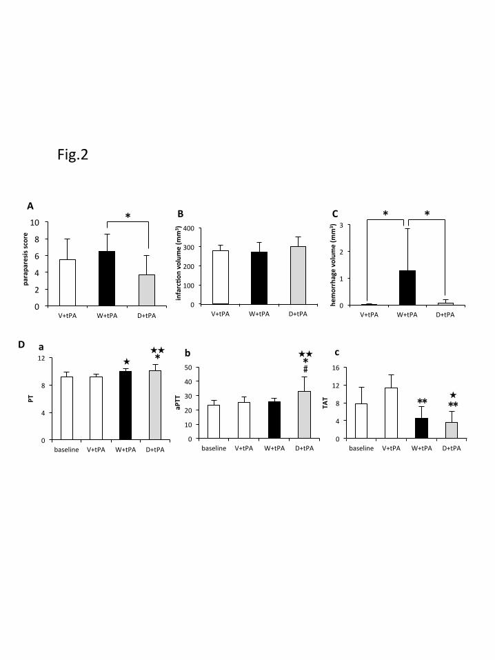
different among the three groups (Table 1). The paraparesis score was significantly
improved in the D+tPA group (3.7 ± 2.3, *p < 0.05) than in the W+tPA group (6.5 ±
2.1) (Fig. 2A). Infarction volume was not different among the three groups (Fig. 2B).
Intracerebral hemorrhage volume was significantly larger in the W+tPA group than in
V+tPA or D+tPA groups (Fig. 2C, *p < 0.05). Significant PT prolongation was
observed in the W+tPA group compared to the baseline (Fig. 2Da, ★p < 0.05), in
D+tPA groups compared to the baseline (Fig. 2Da, p < 0.01), and in the V+tPA

12
group (Fig. 2Da, *p < 0.05). Significant aPTT prolongation was found only in the
D+tPA group compared to the baseline (Fig. 2Db, p < 0.01), in the V+tPA group
(Fig. 2Db, *p < 0.05), and in the W+tPA group (Fig. 2Db, #p < 0.05). Although TAT
was significantly reduced in both W+tPA and D+tPA groups compared to the V+tPA
group (Fig. 2Dc, **p < 0.01), and in the D+tPA group compared to the baseline (Fig.
2Dc, p < 0.05), there was no difference between W+tPA and D+tPA groups,
indicating that the antithrombotic effect was almost the same in both groups.
Intracerebral hemorrhage was sometimes observed on the surface (Fig. 3A,
arrowheads) and in the coronal section (Fig. 3B, arrowheads) of the brain in W+tPA
and D+tPA groups, but was more evident in the W+tPA group.
In the V+tPA group, little dissociation of the neurovascular unit was found in the
periischemic lesion (Fig. 4Aa, left panels). In contrast, a marked dissociation of the
basal lamina (collagen IV) and astrocyte foot processes (GFAP) was observed in the
periischemic lesion of the W+tPA group (Fig. 4Aa, middle panels, arrowheads, Fig.
4Ab, **p < 0.01), which was dramatically improved in the D+tPA group (Fig. 4Aa,
right panels, Fig. 4Ab, **p < 0.01). Dissociation of pericyte (PDGFRβ) and astrocyte
foot processes (GFAP) was significantly larger in the W+tPA group (Fig. 4Ba, middle
panels, arrowheads) than in the V+tPA or D+tPA groups. The vascular dissociation

13
index revealed a larger dissociation in the W+tPA group than in V+tPA or D+tPA
groups (Fig. 4Bb, **p < 0.01). On the other hand, there was no difference among the
three groups in terms of dissociation between pericyte (PDGFRβ) and vascular
endothelial cells (NAGO) (Fig. 4Ca), with no quantitative vascular dissociation index
among the three groups (Fig. 4Cb).
Gelatin zymography indicated that there was no activation and no difference in
contralateral MMP-9 activities among the three groups (Fig. 5A, 4B). In contrast, the
ipsilateral brain showed considerable activation of MMP-9 in the W+tPA group
compared to contralateral V+tPA (Fig. 5B, **p < 0.01), and contralateral W+tPA (Fig.
5B, ##p < 0.01). This ipsilateral activation of the W+tPA group was greatly reduced in
the D+tPA group (Fig. 5A, 5B, p < 0.05).
Discussion
In the present study, pretreatment with dabigatran greatly improved the clinical
score (Fig. 2A) and intracerebral hemorrhage (Fig. 2C, 2E, 2F) than
warfarin-pretreated rats after thrombolytic therapy with tPA. In the Randomized
Evaluation of Longfrom Term Anticoagulant Therapy With Dabigatran Etexilate
(RE-LY) study, both 110 and 150 mg doses of dabigatran lowered intracranial

14
bleeding with a similar or lower rate of stroke than warfarin (Connolly et al., 2009).
Among six previous reports on the dabigatran+tPA combination in acute stroke
patients, there was only one case of fatal intracerebral hemorrhage, and one case of
asymptomatic arm ecchymosis (Smedt et al., 2010; Matute et al., 2011; Naranjo et al.,
2011; Marrone and Marrone, 2012; Lee et al., 2012; Sangha et al., 2012). Although the
above fatal intracerebral hemorrhage patient carried other risks of intracerebral
hemorrhage such as large infact volume (>2/3 of the MCA area) and diabetes mellitus,
the last intake of dabigatran was the shortest of the six cases (6 h before tPA),
suggesting that a shorter time interval from dabigatran to tPA is an important risk
factor of intracerebral hemorrhage. However, the interval between the last dabigatran
intake and tPA treatment is different between these clinical reports and the present
study. Thus, we may not be able to directly compare our findings with these clinical
reports.
In the present study, macroscopic intracerebral hemorrhage was observed in
66.7% of the warfarin-pretreated rats, 44.4% of the dabigatran-pretreated rats, and
44.4% of the vehicle rats (Table 1, p=0.55), which suggests a lower risk of occurrence
of macroscopic intracerebral hemorrhage with dabigatran+tPA. Hemorrhagic volume
was much lower in the D+tPA group (Fig. 2C). Thus, dabigatran reduced not only

15
hemorrhagic incidence but reduced hemorrhagic volume even more (Table 1, Fig. 2C).
At a higher plasma level of dabigatran, the risk of severe intracerebral
hemorrhage may still rise (Pfeilschifter et al., 2012), although there is no common
marker for anticoagulant activity by dabigatran at emergency. In the present study,
significant prolongation of PT was observed in W+tPA and D+tPA groups (Fig. 2Da),
and significant aPTT prolongation was found only in the D+tPA group (Fig. 2Db). The
TAT complex was significantly reduced in both W+tPA and D+tPA groups compared
to the vehicle group (Fig. 2Dc, **p < 0.01), indicating a similar antithrombotic effect
in both groups with each dose for obtaining 70% inhibition of clot formation in the rat
venous thromboembolism model (Toomey et al., 2006; Wienen et al., 2007).
Various proinflammatory mediators (MMPs, thrombin, vascular endothelial
growth factor, and bradykinin) increase in the ischemic brain (Aschner et al., 1997;
Kamiya et al., 1993; Rosenberg GA, 2002; Suarez and Ballmer-Hofer, 2001),
accompanied by brain edema, endothelial cell death (Maier et al., 2006), disruption of
tight junctions, and loss of the basal lamina/extracellular matrix (collagenIV, laminin-1,
and fibronectin). Any of these changes could promote intracerebral hemorrhage
associated with tPA treatment (Zoppo and Mabuchi, 2003). We previously reported
that MMP-9 activation after tMCAO induced the dissociation between the vascular

16
basal lamina and the astrocyte endfeet in the ischemic rat brain treated with tPA
(Yamashita et al., 2009), which was confirmed in the present study (Fig. 4A, 4). A
marked dissociation between astrocyte foot processes and the basal lamina was
observed on the periischemic lesion of warfarin-pretreated rats in association with
MMP-9 activation (Fig. 4A, 4), which improved dramatically in the
dabigatran-pretreated group (Fig. 4A, 4).
The mechanism in which warfarin activates MMP-9 while dabigatran inhibits the
activation, remains obscure, but several inferences can be drawn from other reports.
Factor VII (FVII) forms a complex with the cell surface co-factor, tissue factor (TF),
which appears after vascular injury, and the FVII and TF complex initiates the
coagulation cascade (Vadivel and Bajaj, 2012). Warfarin inhibits FVII (Sakata et al.,
1995) and the coagulation cascade does not start, even after endothelial cells are
injured. Therefore, the inability to repair endothelial cells and asymptomatic
microbleeding may occur recurrently. tPA causes disruption of the neurovascular
matrix through MMP-9 upregulation when it leaks into the parenchyma (Goto et al.,
2007). We speculate that recurrent injury and the inability to repair endothelial cells by
warfarin-pretreatment may facilitate the leakage of tPA outside the vessel, leading to
the upregulation of MMP-9 activity after ischemia and tPA treatment.

17
In the present study, we focused on the role of pericytes which encircle capillary
vessels, and are important for the maturation and stabilization of the capillary vessels
during angiogenesis. After cerebral ischemia, pericytes increased neurotrophin-3
production, which potentiated the secretion of nerve growth factor (NGF) from
astrocytes (Ishitsuka et al., 2012). Ischemia and reperfusion-induced injury to pericytes
may impair microcirculatory reflow and negatively affect survival (Yemisci et al.,
2009). Our present study showed a marked dissociation between astrocyte foot
processes and pericytes in the periischemic lesion of warfarin-pretreated rats, which
was dramatically improved in the dabigatran-pretreated group (Fig. 4B). The
dissociation between astrocyte foot processes and pericytes could also allow the
development of a neurovascular unit for intracerebral hemorrhage (Fig. 2-4).
In summary, the present data suggests a lower risk of intracerebral hemorrhage
after tPA in ischemic stroke rats with pretreated dabigatran compared to pretreated
warfarin. A remarkable activation of MMP-9 with warfarin caused a marked
dissociation of the neurovascular unit raising the risk of intracerebral hemorrhage after
tPA, which was greatly ameliorated by replacing with dabigatran. Thus, this study is
the first evidence of neurovascular protection by dabigatran which could partly explain
the mechanism of reducing hemorrhagic complications by dabigatran reported in the

18
RE-LY clinical study.
Acknowledgement
This work was partly supported by a Grant-in-Aid for Scientific Research (B)
21390267, (C) 24591263 and Challenging Research 24659651, and by Grants-in-Aid
from the Research Committees (Mizusawa H, Nakano I, Nishizawa M, Sasaki H, and
Aoki M) from the Ministry of Health, Labour and Welfare of Japan.
References
Abe K, Kawagoe J, Araki T, Aoki M, Kogure K. 1992. Differential expression of heat
shock protein 70 gene between the cortex and caudate after transient focal
cerebral ischaemia in rats. Neurol Res 14: 381-385.
Aschner JL, Lum H, Fletcher PW, Malik AB. 1997. Bradykinin- and thrombin-induced
increases in endothelial permeability occur independently of phospholipase C but
require protein kinase C activation. J Cell Physiol 173: 387-396.
Augustin HG, Braun K, Telemenakis I, Modlich U, Kuhn W. 1995. Ovarian
angiogenesis. Phenotypic characterization of endothelial cells in a physiological
model of blood vessel growth and regression. Am J Pathol 147: 339-351.

19
Bamford J, Sandercock P, Dennis M, Warlow C, Jones L, McPherson K, Vessey M,
Fowler G, Molyneux A, Hughes T, et al. 1988. A prospective study of acute
cerebrovascular disease in the community: the Oxfordshire Community Stroke
Project 1981-86. 1. Methodology, demography and incident cases of first-ever
stroke. J Neurol Neurosurg Psychiatry 51: 1373-1380.
Brown RD, Whisnant JP, Sicks JD, O'Fallon WM, Wiebers DO. 1996. Stroke incidence,
prevalence, and survival: secular trends in Rochester, Minnesota, through 1989.
Stroke 27: 373–380.
Casado Naranjo I, Portilla-Cuenca JC, Jiménez Caballero PE, Calle Escobar ML,
Romero Sevilla RM. 2011. Fatal intracerebral hemorrhage associated with
administration of recombinant tissue plasminogen activator in a stroke patient on
treatment with dabigatran. Cerebrovasc Dis 32: 614-615.
Connolly SJ, Ezekowitz MD, Yusuf S, Eikelboom J, Oldgren J, Parekh A, Pogue J,
Reilly PA, Themeles E, Varrone J, et al; RE-LY Steering Committee and
Investigators. 2009. Dabigatran versus warfarin in patients with atrial fibrillation.
N Engl J Med 361: 1139-1151.
De Smedt A, De Raedt S, Nieboer K, De Keyser J, Brouns R. 2010. Intravenous
thrombolysis with recombinant tissue plasminogen activator in a stroke patient

20
treated with dabigatran. Cerebrovasc Dis 30: 533-534.
del Zoppo GJ, Mabuchi T. 2003. Cerebral microvessel responses to focal ischemia. J
Cereb Blood Flow Metab 23: 879-894.
Goto H, Fujisawa H, Oka F, Nomura S, Kajiwara K, Kato S, Fujii M, Maekawa T,
Suzuki M. 2007. Neurotoxic effects of exogenous recombinant tissue-type
plasminogen activator on the normal rat brain. J Neurotrauma 24: 745-752.
Hacke W, Kaste M, Bluhmki E, Brozman M, Dávalos A, Guidetti D, Larrue V, Lees KR,
Medeghri Z, Machnig T, et al; ECASS Investigators. 2008. Thrombolysis with
alteplase 3 to 4.5 hours after acute ischemic stroke. N Engl J Med 359:
1317-1329.
Hacke W, Kaste M, Fieschi C, Toni D, Lesaffre E, von Kummer R, Boysen G, Bluhmki
E, Höxter G, Mahagne MH, et al. 1995. The European Cooperative Acute Stroke
Study (ECASS). Intravenous thrombolysis with recombinant tissue plasminogen
activator for acute hemispheric stroke. JAMA 274: 1017-1025.
Hacke W, Kaste M, Fieschi C, von Kummer R, Davalos A, Meier D, Larrue V, Bluhmki
E, Davis S, Donnan G, Schneider D, et al. 1998. Second European-Australasian
Acute Stroke Study Investigators. Randomised double-blind placebo-controlled
trial of thrombolytic therapy with intravenous alteplase in acute ischaemic stroke

21
(ECASS II). Lancet 352: 1245-1251.
Ishitsuka K, Ago T, Arimura K, Nakamura K, Tokami H, Makihara N Kuroda J,
Kamouchi M, Kitazono T. 2012. Neurotrophin production in brain pericytes
during hypoxia: a role of pericytes for neuroprotection. Microvasc Res 83:
352-359.
Kamiya T, Katayama Y, Kashiwagi F, Terashi A. 1993. The role of bradykinin in
mediating ischemic brain edema in rats. Stroke 24: 571-575; discussion 575-576.
Kawai H, Deguchi S, Deguchi K, Yamashita T, Ohta Y, Shang J, Tian F, Zhang X, Liu N,
Liu W, et al. 2011. Synergistic benefit of combined amlodipine plus atorvastatin
on neuronal damage after stroke in Zucker metabolic rat. Brain Res 1368:
317-323.
Lee VH, Conners JJ, Prabhakaran S. 2012. Intravenous thrombolysis in a stroke patient
taking dabigatran. J Stroke Cerebrovasc Dis 21: 916. e11-12.
Maier CM, Hsieh L, Crandall T, Narasimhan P, Chan PH. 2006. Evaluating therapeutic
targets for reperfusion-related brain hemorrhage. Ann Neurol 59: 929-938.
Marrone LC, Marrone AC. 2012. Thrombolysis in an ischemic stroke patient on
dabigatran anticoagulation: a case report. Cerebrovasc Dis 34: 246-247.
Matute MC, Guillán M, García-Caldentey J, Buisan J, Aparicio M, Masjuan J, Alonso
22
de Leciñana M. 2011. Thrombolysis treatment for acute ischaemic stroke in a
patient on treatment with dabigatran. Thromb Haemost 106: 178-179.
Nakagawara J, Minematsu K, Okada Y, Tanahashi N, Nagahiro S, Mori E, Shinohara Y,
Yamaguchi T; J-MARS Investigators. 2010. Thrombolysis with 0.6 mg/kg
intravenous alteplase for acute ischemic stroke in routine clinical practice. The
Japan post-Marketing Alteplase Registration Study (J-MARS). Stroke 41:
1984–1989.
Omori N, Jin G, Li F, Zhang WR, Wang SJ, Hamakawa Y, Nagano I, Manabe Y, Shoji M,
Abe K. 2002. Enhanced phosphorylation of PTEN in rat brain after transient
middle cerebral artery occlusion. Brain Res 954: 317-322.
Paxinos G, Watson C. 1982. The rat brain in stereotaxic coordinates. New York, NY:
Academic Press.
Pfeilschifter W, Bohmann F, Baumgarten P, Mittelbronn M, Pfeilschifter J,
Lindhoff-Last E, Steinmetz H, Foerch C. 2012. Thrombolysis with recombinant
tissue plasminogen activator under dabigatran anticoagulation in experimental
stroke. Ann Neurol 71: 624-633.
Rosenberg GA. 2002. Matrix metalloproteinases in neuroinflammation. Glia 39:
279-291.

23
Sakata T, Kario K, Matsuo T, Katayama Y, Matsuyama T, Kato H, Miyata T. 1995.
Suppression of plasma-activated factor VII levels by warfarin therapy.
Arterioscler Thromb Vasc Biol 15: 241-246.
Sangha N, El Khoury R, Misra V, Lopez G. 2012. Acute ischemic stroke treated with
intravenous tissue plasminogen activator in a patient taking dabigatran with
radiographic evidence of recanalization. J Stroke Cerebrovasc Dis 21: 917. e5-8.
Suarez S, Ballmer-Hofer K. 2001. VEGF transiently disrupts gap junctional
communication in endothelial cells. J Cell Sci 114: 1229-1235.
The National Institute of Neurological Disorders and Stroke rtPA Stroke Study Group.
1995. Tissue plasminogen activator for acute ischemic stroke. N Engl J Med 333:
1581–1587.
Toomey JR, Abboud MA, Valocik RE, Koster PF, Burns-Kurtis CL, Pillarisetti K,
Danoff TM, Erhardt JA. 2006. A comparison of the beta-D-xyloside, odiparcil, to
warfarin in a rat model of venous thrombosis. J Thromb Haemost 4: 1989-1996.
Vadivel K, Bajaj SP. 2012. Structural biology of factor VIIa/tissue factor initiated
coagulation. Front Biosci (Landmark Ed) 17: 2476-2494.
Wahlgren N, Ahmed N, Dávalos A, Ford GA, Grond M, Hacke W, Hennerici MG, Kaste
M, Kuelkens S, Larrue V, et al; SITS-MOST investigators. 2007. Thrombolysis

24
with alteplase for acute ischaemic stroke in the Safe Implementation of
Thrombolysis in Stroke-Monitoring Study (SITS-MOST): an observational study.
Lancet 369: 275–282.
Wienen W, Stassen JM, Priepke H, Ries UJ, Hauel N. 2007. Effects of the direct
thrombin inhibitor dabigatran and its orally active prodrug, dabigatran etexilate,
on thrombus formation and bleeding time in rats. Thromb Haemost 98: 333-338.
Wu J, Hua Y, Keep RF, Nakamura T, Hoff JT, Xi G. 2003. Iron and iron-handling
proteins in the brain after intracerebral hemorrhage. Stroke 34: 2964-2969.
Yamashita T, Kamiya T, Deguchi K, Inaba T, Zhang H, Shang J, Miyazaki K, Ohtsuka A,
Katayama Y, Abe K. 2009. Dissociation and protection of the neurovascular unit
after thrombolysis and reperfusion in ischemic rat brain. J Cereb Blood Flow
Metab 29: 715-725.
Yemisci M, Gursoy-Ozdemir Y, Vural A, Can A, Topalkara K, Dalkara T. 2009. Pericyte
contraction induced by oxidative-nitrative stress impairs capillary reflow despite
successful opening of an occluded cerebral artery. Nat Med. 15: 1031-1037.
Zhang L, Schallert T, Zhang ZG, Jiang Q, Arniego P, Li Q, Lu M, Chopp M. 2002. A test
for detecting long-term sensorimotor dysfunction in the mouse after focal cerebral
ischemia. J Neurosci Methods 117: 207-214.

25
Figure legends
Fig. 1) Experimental groups including V (vehicle) +tPA group, W (warfarin) +tPA
group, and D (dabigatran) +tPA group. tMCAO, transient middle cerebral artery
occlusion; tPA, tissue plasminogen activator. At 24 h after 2 h tMCAO, rats were
sacrificed.
Fig. 2) Clinical, serum chemical and hemorrhagic complications in animal groups. (A)
The paraparesis score was significantly improved in the D+tPA group (*p < 0.05) than
in the W+tPA group. (B) Infarction volume was not different among the three groups.
(C) Intracerebral hemorrhage volume was significantly larger in the W+tPA group than
in V+tPA or D+tPA groups (*p < 0.05). (D) Significant PT prolongation was observed
in W+tPA and D+tPA groups compared to baseline (Da), and significant aPTT
prolongation was found only in the D+tPA group compared to the baseline (Db).
Although TAT was significantly reduced in both W+tPA and D+tPA groups compared
to the V+tPA group, there was no difference between W+tPA and D+tPA groups (Dc).
p < 0.05, p < 0.01 versus baseline, *p < 0.05, **p < 0.01 versus V+tPA, #p <
0.05 versus W+tPA.

26
Fig. 3) Intracerebral hemorrhage was sometimes observed on the surface (A) and
coronal section (B) of the brain in W+tPA and D+tPA groups (arrowheads), but was
more evident in the W+tPA group.
Fig. 4) Double immunofluorescent analysis of collagen IV+GFAP (A),
PDGFRβ+GFAP (B), and NAGO+PDGFRβ (C). A marked dissociation between
astrocyte foot processes and basal lamina (A) or pericyte (B) was observed on the
periischemic lesion of warfarin-pretreated rats (Bar = 20 µm) but there was no
dissociation between pericyte and vascular endothelial cells among the three groups
(bar = 100 µm) (C). The vascular dissociation index estimates the space between each
construction (**p < 0.01) (Ab to Cb).
Fig. 5) Gelatin zymography (A) showing considerable activation of MMP-9 in the
ipsilateral warfarin-pretreated rat brain but which was greatly reduced in
dabigatran-pretreated rats (A). (B) Densitometric analysis of zymography. **p < 0.01
versus contralateral hemisphere of V+tPA, ##p < 0.01 versus contralateral hemisphere
of W+tPA, p < 0.05 versus ipsiralateral hemisphere of W+tPA.

Table 1. Physiological Parameter in 3 experimental groups
V+tPA W+tPA D+tPA
(n=9) (n=9) (n=9)
Body weight (g) 282.6 ± 15.4 282.6 ± 11.5 284.3 ± 13.0
Before tMCAO Systolic BP (mmHg) 145.4 ± 36.0 152.6 ± 29.7 132.9 ± 28.3 Diastolic BP (mmHg) 88.4 ± 8.1 93.3 ± 12.5 88.7 ± 5.9
24 h after tMCAO Systolic BP (mmHg) 165.5 ± 33.2 166.6 ± 29.1 152.0 ± 32.4 Diastolic BP (mmHg) 106.4 ± 23.1 95.3 ± 15.9 95.2 ± 13.2
Intracerebral hemorrhage 44.4 66.7 44.4occurrence (%) (p=0.55)

tPA
tPA
tPA
24h
24h
24h
2h tMCAO
2h tMCAO
2h tMCAO
V + tPA
W + tPA
D + tPA
1h
1h
Vehicle
Warfarin
Dabigatran
7d
Fig.1
1h
0
2
4
6
8
10
V+tPA W+tPA D+tPA
parapa
resis s
core
0
4
8
12
16
baseline V+tPA W+tPA D+tPA
TAT
** ** ★
0
4
8
12
baseline V+tPA W+tPA D+tPA
PT
★ ★★
*
0
100
200
300
400
V+tPA W+tPA D+tPA
infarc/o
n volume (m
m3 )
0
1
2
3
V+tPA W+tPA D+tPA
hemorrhage volume (m
m3 )
* * A
B C
D b c a
Fig.2
#
★★ *
0
10
20
30
40
50
baseline V+tPA W+tPA D+tPA
aPTT
*

A
B
V+tPA W+tPA D+tPA
Fig.3

V+tPA W+tPA D+tPA
collagen IV
GFAP
merge 0
1
2
V+tPA W+tPA D+tPA
vas
cula
r d
isso
ciat
ion
ind
ex
(μm
2 /μm
)
GFAP
PDGFRβ
merge
V+tPA W+tPA D+tPA
0
0.2
0.4
V+tPA W+tPA D+tPA
vas
cula
r d
isso
ciat
ion
ind
ex
(μ
m2 /μ
m)
NAGO
PDGFRβ
merge
V+tPA W+tPA D+tPA
** **
A a
b
B a
b
C
** **
0
0.01
0.02
V+tPA W+tPA D+tPA
vas
cula
r d
isso
ciat
ion
ind
ex
(μ
m2 /μ
m)
a
b
Fig.4

MMP-‐9
op'cal den
sity
(rela'
ve to
con
trol of
con
tralateral hem
isph
ere)
contralateral ipsilateral
MMP marker
V+tPA
W+tPA
D+tPA
V+tPA
W+tPA
D+tPA
contralateral ipsilateral
0
1
2
3
V+tPA W+tPA D+tPA V+tPA W+tPA D+tPA
** ##
◆
A
B
Fig.5