Reducing Contrast Induced Renal Insufficiency · Reducing Contrast Induced Renal Insufficiency...
32
Reducing Contrast Induced Renal Insufficiency Daniela Branzan, MD, Department of Vascular Surgery University Hospital Leipzig
Transcript of Reducing Contrast Induced Renal Insufficiency · Reducing Contrast Induced Renal Insufficiency...
Reducing Contrast Induced Renal Insufficiency
Daniela Branzan, MD,
Department of Vascular Surgery
University Hospital Leipzig

Disclosure
I do not have any potential conflict of interest.

How to Define CIRI?
Tepel M et al. Circulation. 2006
• Absolute rise in serum creatinine (SCr) of 0.5 mg/dL
• A 25% increase from the baseline value assessed within 48 hours after the procedure without an alternative etiology

Incidence of CIRI
Prasad A et al. Catheter and Cardiovasc Interv. 2016

Morbidity and Mortality Associated With CIRI
Grossman PM et al. J Interv Cardiol. 2017

Prevention of CIRI
• Recognize risk factors
• Withdrawal of all nephrotoxic medications
• Hydration
• Pharmacological strategies
• Minimizing volume of contrast media

Withdrawal of AllNephrotoxic Medications
Drug Class Examples
NSAIDs Ibuprofen, Diclofenac, Celecoxib
Antibiotics Aminoglycosides
Antifungals Amphotericin B
Antivirals Acyclovir, Ganciclovir, Tenofovir
Immunosuppressants Cyclosporine, Tacrolimus
Antineoplastic Cisplatin, Ifosfamide
Angiotensin-converting enzyme inhibitors/ angiotensin receptor blockers • CAPTAIN study demonstrated no nephrotoxicity
Bainey KR et al. Am Heart J. 2015
Chi Hong Chau et al. Circulation: Cardiovascular Interventions. 2016

Hydration • The most effective prophylactic intervention
• Guidelines:
– AHA: isotonic saline infusion:1 to 1.5 mL/kg/h for 3 to 12 hours before the procedure and 6 to 24 hours after the procedure
– ESC: saline infusion:1 to 1.5 mL/kg/ for 12 hours before and up to 24 hours after the procedure

Statins
CIRI – 3.6% (105/2889) in high-
dose statin group
– 8.3% (245/2936) in control group
NNT of high-dose statin: 16
Lee JM et al. PLoS One. 2014
• High-dose statin pretreatment significantly reduced overall incidence of CIRI in patients undergoing CAG
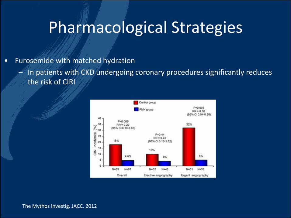
Pharmacological Strategies
• Furosemide with matched hydration
– In patients with CKD undergoing coronary procedures significantly reduces the risk of CIRI
The Mythos Investig. JACC. 2012

Choice of Contrast Media
• No differences were found in CIRI risk among types of LOCM
• Nonionic low-osmolar and iso-osmolar CM are safer than high-osmolar CM

Volume of Contrast Media
• Maximum safe volume of CM has been proposed by Laskey, et al:– A ratio of the CM-volume: creatinine clearance < 3.7
Laskey WK, et al. J Am Coll Cardiol. 2007
• When contrast volume exceeds 3.7× of creatinine clearance:– Staged procedure at least 3 days later

• Remove contrast from the sheath/guide catheter by back bleeding prior to exchange of devices
Minimizing Volume of CM
• Display previous angiographic images to use as guidance during guide wire passage
• Use automated contrast injectors
• IVUS
• CO2 angiography
• Percutaneous US guidance for interventions
• Use small diameter catheters without side-holes
• Perform simultaneous a cine angiogram when you inject CM
• Using a small syringe

CO2 Angiography
• CO2 is an gaseous contrast medium, pioneered by Hawkins in the 1970s
• The technological solutions available up to today for injecting CO2

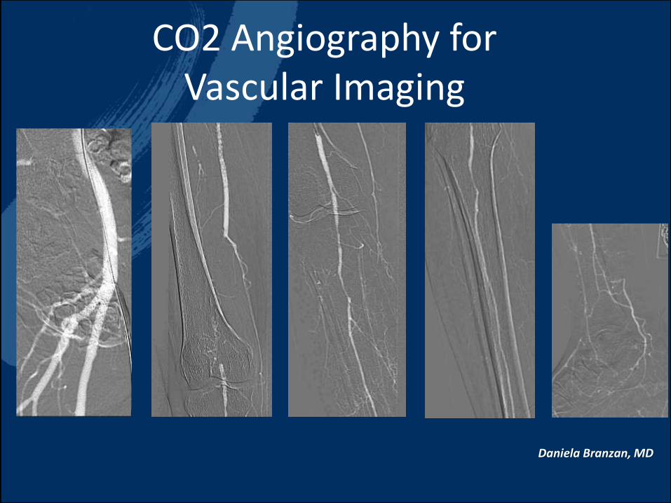
CO2 Angiography for Vascular Imaging
Visceral Angiography Aortography
outflow assessment
Daniela Branzan, MD
CO2 Angiography for Vascular Imaging
Daniela Branzan, MD

CO2 for EVT of Aorto-iliac Disease
Daniela Branzan, MD

CO2 for RAS
Kawasaki D et al. Angiology. 2015

CO2 for EVAR
Daniela Branzan, MD

Incidence of CIRI With CO2
• Possible mechanisms of CIRI after CO2:– The vapor lock phenomena
– Atheroembolization
– The adjunctive use of CM with CO2
Ghumman SS et al. Catheter Cardiovasc Interv . 2017

Challenges With CO2 Angiography
• Non-renal adverse events up to 17%:Fujihara et al. Catheter Cardiovasc Interv. 2014
– Limb and abdominal pain
– Nausea and vomiting
– NOMI and death
• Image quality with CO2:
– Aortoiliac: artifacts due to the presence of bowel gas
– Infrapopliteal: motion artifacts

What Is IVUS?
• Introduced originally in interventional cardiology in the late 1980s
• 2 clinical roles:
– Assess and measure the severity of the disease before treatment
– Demonstrate the complement of the treatment after intervention
• IVUS is now used in a variety of endovascular procedures

IVUS for Type-B Aortic Dissection
• Identifies the true and the false lumen
Koschyk DH et al. Circulation. 2005
• Identifies the size of the graft to be implanted
• Visualizes:– The supra-aortic vessels
– The visceral vessels
– Retrograde dissection
– Expansion of the true lumen after stent placement

IVUS for EVAR

IVUS for EVAR

IVUS for Endovascular Treatment of PAD
Pretreatment

IVUS for Endovascular Treatment of PAD

IVUS for Endovascular Treatment of PAD
Post-treatment

IVUS to Reduce CIRI
Standard EVT IVUS-Guided EVT p
N 21 31
Iliac Artery 86 100
Fem-pop Artery 79 96
ABI after 0.89±0.22 1.00±0.14 <0.01
CM 201±100 104±0.14 <0.01
Radiation exposure time (min) 65±55 60±35
Kawasaki et al. Catheter Cardiovasc Interv. 2008
IVUS-Guided Endovascular Therapy for CTO
• EVT of iliofemoral disease WITHOUT CM in pts with CKD• 36 pts, 51 lesions
• Technical success: 100%
• SCr did not change after treatment and at 3 months
Kawasaki et al. Circulation J. 2010

ConclusionHow to Prevent CIRI?
• Identify the high-risk patient
• Stop all nephrotoxic drugs
• Hydrate prior to and after the procedure
• Preload the patient with statins
• Minimize/eliminate the contrast media by liberal use of IVUS and/or CO2

Reducing Contrast Induced Renal Insufficiency
Daniela Branzan, MD,
Department of Vascular Surgery
University Hospital Leipzig