redescription of some species of chone kroyer and euchone malmgren, and three new species
37
REDESCRIPTION OF SOME SPECIES OF CHONE KROYER AND EUCHONE MALMGREN, AND THREE NEW SPECIES (SABELLIDAE, POLYCHAETA) 1 KARL BANSE" ABSTRACT Generic diagnoses are given for Chane (syn. Megachone) and Euchone. Redescriptions and additions to the descriptions, in part based on study of the types, are provided for some species, Chane aurantiaca (new combination), C. duneri (syn. C. bimaculata) , C. ecaudata (syn. C. minuta), C. eniwetolcensis (new combination), C. gracilis, C. in- fundibuliformis (syn. C. teres), C. magna, C. mollis, C. paucibranchiata, Euchone alicau- data, E. analis, E. capensis, E. elegans, E. papillosa, and E. rubrocincta. Three new species, Chone alboeincta, Chone veleronis, and Euchone velifera are described. The number of lobes of the collar is rejected for the Sabellidae as a character on the generic level; the diagnostic value of the palmate membrane is discussed. The sabellid polychaetes are filter feeders which USually live in permanent tubes. Two genera, Chone Kroyer and Euchone Malmgren, which occur worldwide, are treated here taxonomically. The length of species of Chane ranges from 2.5 mm to almost 10 em, that of species of Eu- chone from a few millimeters to a few centi- lheters. Some species are quite common on Sandy or muddy bottoms (cf. McIntosh, 1916; Bartman, 1944b; Ktihlmorgen-Hille, 1963); at least some species are eaten by fishes (McIntosh, 1916; Berkeley and Berkeley, 1954). The study was initiated because of difficulties in identifying large species of Chane in the Syn- optic Collection of the Friday Harbor Labora- tories, University of Washington. This led to reexaminations of some types and other speci- lhens primarily from the North Pacific and North Atlantic Oceans. The results are an emenda- tion of the generic diagnosis of Chane, including the placement of Me{}achone Johnson in Chone, the redescription or improvement of the knowl- edge of some previously known species, and the 1 Contribution No. 644 from the Department of Ocean- ography, University of Washington, Seattle, Wash. i Department of Oceanography, University of Wash- ngton, Seattle, WA 98195. ---- accepted November t 971. SI-IERY BULLETIN: VOL. 70, NO.2, 1972. erection of three new species. Because material of several large species of Euchone was at hand, additions to their descriptions are also included, the generic diagnosis is emended, and a new spe- cies is described. Of general taxonomic interest is the discovery of a new species of Euchone, E. velifera, which has a ventrally deeply incised collar with an ad- ditional pair of lateral notches (Figure 12a). A collar that is ventrally moderately incised is known also for E. papiUosa (Sars) and E. ca- pensis Day (Figure Un). The species otherwise agree fully with the characters of Euchone. Sim- ilarly, Chone trilobata Gallardo has deep lateral notches in its collar but otherwise is typical. Especially the occurrence of bilobed collars in species of Euchone, a genus uniquely character- ized by the anal depression, provides further evidence in support ofJohansson's (1927) strong criticism of the attempt by Bush (1904) to make the collar a character of major importance in distinguishing genera among the Sabellidae. Consequently, genera like Pseudopotam'illa Bush, separated from Potamilla Malmgren only by dif- ferences in the collar, should not be maintained. In view of the fact that not all Euchone species have a palmate membrane, the question is dis- 459
Transcript of redescription of some species of chone kroyer and euchone malmgren, and three new species

REDESCRIPTION OF SOME SPECIES OF CHONE KROYER
AND EUCHONE MALMGREN, AND THREE NEW SPECIES
(SABELLIDAE, POLYCHAETA) 1
KARL BANSE"
ABSTRACT
Generic diagnoses are given for Chane (syn. Megachone) and Euchone. Redescriptionsand additions to the descriptions, in part based on study of the types, are provided forsome species, Chane aurantiaca (new combination), C. duneri (syn. C. bimaculata) ,C. ecaudata (syn. C. minuta), C. eniwetolcensis (new combination), C. gracilis, C. infundibuliformis (syn. C. teres), C. magna, C. mollis, C. paucibranchiata, Euchone alicaudata, E. analis, E. capensis, E. elegans, E. papillosa, and E. rubrocincta. Three newspecies, Chone alboeincta, Chone veleronis, and Euchone velifera are described. Thenumber of lobes of the collar is rejected for the Sabellidae as a character on the genericlevel; the diagnostic value of the palmate membrane is discussed.
The sabellid polychaetes are filter feeders whichUSually live in permanent tubes. Two genera,Chone Kroyer and Euchone Malmgren, whichoccur worldwide, are treated here taxonomically.The length of species of Chane ranges from2.5 mm to almost 10 em, that of species of Euchone from a few millimeters to a few centilheters. Some species are quite common onSandy or muddy bottoms (cf. McIntosh, 1916;Bartman, 1944b; Ktihlmorgen-Hille, 1963); atleast some species are eaten by fishes (McIntosh,1916; Berkeley and Berkeley, 1954).
The study was initiated because of difficultiesin identifying large species of Chane in the Synoptic Collection of the Friday Harbor Laboratories, University of Washington. This led toreexaminations of some types and other specilhens primarily from the North Pacific and NorthAtlantic Oceans. The results are an emendation of the generic diagnosis of Chane, includingthe placement of Me{}achone Johnson in Chone,the redescription or improvement of the knowledge of some previously known species, and the
1 Contribution No. 644 from the Department of Oceanography, University of Washington, Seattle, Wash.i Department of Oceanography, University of Washngton, Seattle, WA 98195.----~anuscript accepted November t 971.
SI-IERY BULLETIN: VOL. 70, NO.2, 1972.
erection of three new species. Because materialof several large species of Euchone was at hand,additions to their descriptions are also included,the generic diagnosis is emended, and a new species is described.
Of general taxonomic interest is the discoveryof a new species of Euchone, E. velifera, whichhas a ventrally deeply incised collar with an additional pair of lateral notches (Figure 12a).A collar that is ventrally moderately incised isknown also for E. papiUosa (Sars) and E. capensis Day (Figure Un). The species otherwiseagree fully with the characters of Euchone. Similarly, Chone trilobata Gallardo has deep lateralnotches in its collar but otherwise is typical.Especially the occurrence of bilobed collars inspecies of Euchone, a genus uniquely characterized by the anal depression, provides furtherevidence in support of Johansson's (1927) strongcriticism of the attempt by Bush (1904) to makethe collar a character of major importance indistinguishing genera among the Sabellidae.Consequently, genera like Pseudopotam'illa Bush,separated from Potamilla Malmgren only by differences in the collar, should not be maintained.In view of the fact that not all Euchone specieshave a palmate membrane, the question is dis-
459

cussed (p. 461) whether the character shouldbe used in the generic diagnosis for the closelyrelated Chone.
The observations on some primitive characters in Sabellidae by Banse (1970), especiallyin regard to the abdominal uncini, are supportedby the observations on the species discussed below. To those characters may be added theglandular girdle on the second setiger which iscommon to all species of Chone and Euchone andis found also in the sabellid genera JasmineiraLangerhans and Myxicola Koch. It is consideredto be a phylogenetically primitive character notonly because of the position of Chone and Euchone within the family but also because similargirdles occur in other families (e.g., Southern,1914, for Terebellidae).
METHODS
Because the form of the uncini can vary considerably within the abdomen, the shape of thesetae in the anterior abdominal setigers isstressed in the following descriptions. The accessory teeth above the main fang of an uncinusare arranged in several vertical "columns" appearing in side view as horizontal (not strictlyso) "rows." The nomenclature for the abdominal segments in Euchone follows Banse (1970).The procedure for staining with methyl green introduced by Hofsommer (1913) is described inthe same article.
The following abbreviations are used: AHFfor Allan Hancock Foundation, University ofSouthern California, Los Angeles, Calif.; FHLfor Friday Harbor Laboratories, University ofWashington, Friday Harbor, Wash.; MCZ forMuseum of Comparative Zoology, Harvard University, Cambridge, Mass.; NMI for NationalMuseumof Ireland, Dublin; PMNH for PeabodyMuseum of Natural History, New Haven, Conn.;SMNH for Swedish State Museum of NaturalHistory, Stockholm; SEP for Systematics-Ecology Program, Marine Biological Laboratory,Woods Hole, Mass.; UCT for University of CapeTown; USNM for U.S. National Museum ofNatural History, Washington, D.C.; ZMC forZoological Museum of the University of Copen-
460
FISHERY BULLETIN: VOL. 70. NO.2
hagen; and ZMO for Zoological Museum, University of Oslo. When not noted otherwise, thespecimens are in my collection.
SPECIAL PART
CHONE KROYER, 1856 EMENDED
CHONE Kroyer, 1856, p. 13.- Sars, 1862, p. 119.Malmgren, 1866, p. 404.- Langerhans, 1881,p. 111.- Saint-Joseph, 1894, p. 250.- Bush,1904, p. 189.- Hofsommer, 1913, p. 332.- McIntosh, 1923, p. 287.- Fauvel, 1927, p. 334.Berkeley and Berkeley, 1952, p. 122.- Ushakov,1955, p. 417.- Day, 1967, p. 776.
PARACHONIA Kinberg, 1867, p. 355.MEGACHONE Johnson, 1901, p. 430.METACHONE Bush, 1904, p. 190,216.
Type species: Chone infundibuliformis Kroyer (cf. Bush, 1904).
Diagnosis: Sabellidae with semicircular branchial lobes united by palmate membrane. Collar conspicuous. Postsetal girdle of glands onsecond setiger. Ventral shields sometimespresent. Ends of abdomen of typical sabellidform. Three types of thoracic notosetae: upper, anterior ones limbate, sometimes in twoseries of different length; lower, anterior onesnarrowly limbate (bayonet-type); and lower,posterior ones spatulate or subspatulate. Thoracic neuropodial uncini long-handled, acicular.Abdominal notopodial uncini avicular, withsquare or rounded bases; abdominal neurosetaelimbate.
Remarlcs: The emended diagnosis comprisesthe same species as previously (cf. Hartman,1959, 1965, and below), although it contains asnew characters the postsetal girdle of glands,the ventral shields, the bayonet-like setae, andthe qualification "or subspatulate setae." Theusual occurrence of ventral nude filaments inthe branchial crown, the occasional presence ofotocysts, and the diagnostic characters pertaining to the family are omitted. There are usuallyeight thoracic setigers; very rarely a specimendeviates from this number.

BANSE: REDESCRIPTIONS OF SOME SPECIES OF CHONE AND EUCHONE
Parachonia has been included in Chone byJohannson (1927), Metachone by Fauvel (1927;see also Hartman, 1942b).
The type species of Megachone Johnson, M.aurantiaca, is redescribed below, based in parton the holotype. Contrary to the statement byJohnson (1901), the species does have ordinaryspatulate setae in the thoracic notopodia and thusagrees in every respect with Chone.
The diagnosis as given has one weakness: Theoriginal reason of Kroyer (1856) in creatingChone was the funnel-like appearance of thelophophores, or branchial crown, caused by thepalmate membrane. This membrane is not ofSo unique value as a generic character as originally thought. Sars (1862) was the first topoint out that the membrane occurs also in othergenera. More important is that it may be present or absent in the closely related Euchone(Banse, 1970). If the palmate membrane wereconsidered also in Chone as a specific character,Dialychone ClaparMe would become a synonymof Chone (cf. Fauvel, 1927). For the practicalneed of identification, however, considerable difficUlties would result. For example, a large species of Oriopsis Caullery and Mesnil (cf. O. rivularis Annenkova, with bayonet-type thoracicnotosetae) would externally be distinguishablefrom Chone only by the absence of the postsetalglandular girdle on the second setiger. Anatomically, Oriopsis would, of course, be quite different because it belongs to the thoracogoneateFabriciinae (Zenkevitch, 1925) with theirunique gonoducts. Because eggs may occur inthoracic segments also in abdominogoneate sabellids (cf. Euchone hancocki Banse), identification without anatomical study may sometimesnot be possible. In view of these problems itSeems wise to define Chone as above until intensive anatomical studies of representatives ofthe various subgroups of Fabriciinae have beenlUade. These may yield a more natural andhence more convincing system than is availablenow.
Among most species of Ch9ne, the collar has aVery narrow dorsal gap. C. trilobata has a collar with deep lateral notches. The bayonet-typesetae (also called basal or geniculate setae) weredescribed in some species by previous authors
(cf. Fauvel, 1927; Eliason, 1962) but seem tobe present in all members of the genus. Thelower posterior thoracic notosetae are sometimessubspatulate, as in C. filicaudata Southern andC. eniwetokensis (Reish).
The abdominal uncini with square bases andfew teeth, thought to be typical for Chone (cf.Figure 1k) are found in many species only inanterior abdominal segments, as is the case alsoin Euchone (Banse, 1970). The uncini of posterior setigers can have instead small roundedbases and a high crown of accessory teeth (cf.Figure 6i). Such uncini occur similarly in othergenera of Sabellidae and in related families andare thus phylogenetically more primitive thanthe typical hooks of Chone. Further, they canbe present in the same tori together with typicalones, as in C. ecaudata Moore and C. albocinctanew species (cf. Euchone elegans). Their position shows that ontogenetically they have beenformed first. Also this points to the primitiveness of the posterior uncini. Likewise Okuda(1946) has shown in a developmental study of?C. ecaudata (see below) that the abdominaluncini first formed are of the primitive type andsimilar to those found, for example, in Oriopsis;initially they occur even in the thorax.
In spite of the clarification of diagnostic characters, very little can yet be said about the relations among species of Chone. Certainly, species with ventral shields are more primitive thanthose with fully glandularized epidermes becausereasonably primitive Sabellidae should haveshields. Significantly, the typical abdominaluncini with square bases seem to be presentthroughout the abdomen only among Chone species having a fully glandularized epidermis (possibly also in C. eniwetokensis, according to Reish,1968) .
CHONE INFUNDIBULIFORMIS KROYER
Figure 1
CHONE INFUNDIBULIFORMIS Kroyer,1856,p. 33.- Malmgren, 1866, p. 404; 1867, p. 214.Hofsommer, 1913, p. 332.- McIntosh, 1916,P. 35, 39.- Imajima, 1961, p. 98.- HartmannSchroder, 1971, p. 520. See also p. 465.
461

CHONE TERES Bush, 1904, p. 215.- Hartman,1942b, p. 87; 1959, p. 539.
not CHONE TERES.- Okuda, 1934, p. 236;1946, p. 171.- Imajima and Hartman, 1964, p.365 (for these three references see C. ecaudata).- Chlebovich, 1961, p. 231 (see Remarks).
a
FISHERY BULLETIN: YOLo 70, NO.2
Diagnosis:' A large Chane species withoutventral shields. Branchial basis as long as levelcollar. About 20 pairs of radioles, connectedfor two-thirds to three-fourths of length by palmate membrane, with free ends very short andbroad. First bundle of setae of normal size,inserted more dorsally than following notosetae.
FIGURE I.-Chane infu.ndibu.lifarmis; a from Godhavn; b-g from Egedesminde; h-lfrom holotype of C. teres: a, end of dorso-median radiole; b, c, dorsal and ventral staining pattern of anterior end of trunk; d, anterior view of third thoracic notopodium. Setaeschematic, most upper limbate setae omitted from this segment; f, g, uncini from thirdand approximately 45th abdominal setigers; h, slightly schematic side view of anteriorend, branchial crown omitted; i, end of fully developed radiole; j, spatulate seta fromsixth setiger; k, I, abdominal uncini from third and approximately 70th abdominalsetigers. .
462

BANSE: REDESCRJPTIONS OF SOME SPECIES OF CHONE AND EUCHONE
Broad notopodiallips in thorax. Spatulate setaewith pointed tips. Abdominal uncini uniform,with straight crests; small crowns with somesmall teeth in several columns; rostra not extending beyond bases.
Material studied: Egedesminde and Godhavn,west coast of Greenland (SMNH 6861; 6862),and Safehavn and Hornsund, Spitsbergen(SMNH 6863; 6864 in part). The material ofMalmgren (1866), 8 specimens.
Greenland, locality unknown. Collected by C.Liitken in the last century, USNM 372, 2 specimens.
Dutch Harbor, Alaska, holotype of Choneteres. PMNH 2761 (previous number: 5-2).
Additions to the Description: Since the typematerial from "the Greenlandic sea" is lost (letter by Dr. J. B. Kirkegaard, ZMC, 27 Feb. 1970),excellently preserved Arctic specimens of Malmgren (1866) are used for additions to the description of the species. The features of theespecially suitable specimen from Egedesmindeare stressed on the assumption that Kroyer's material was collected also off the west coast ofGreenland where most of the traffic went during that time.
The trunks of the smallest specimens are alittle less than 2 em long, are about 1 mm wide,and have about 50 abdominal segments. Largespecimens have 70 to 80 abdominal segmentsand are, when fixed in their tubes, up to 6 or7 cm long (total) and about 3 mm wide. About1% em of the length of the large animals is contributed by the branchial crown. The radiolesnumber up to 15 or 16 pairs (up to 22 accordingto Kroyer, 1856). In addition to the radioles,about half a dozen pairs of inconspicuous nudeVentral filaments are present; the longest onesare almost a fifth of the length of the branchialcrown. The palmate membrane extends to twothirds or three-quarters of the length of the radioles and continues as bread flanges on theradioles. The ends of the radioles are broadand short (Figure la); however, the mostventral pinnated radioles have relatively longernUde tips which are twice as long as shown in
the figure and sometimes have an additionalshort filiform end.
The collar extends dorsally to the end of thesecond setiger (Figure Ib). Thus the furrowseparating the first and second setigers does notreach the fecal groove. This character is easilymade out. On the ventral side (Figure lc), thefirst (buccal) segment is not distinctly separated from the collar in unstained animals. Thebody is essentially rounded and entirely coveredby glandular tissue, Le., no ventral shields arepresent. The glandularization is conspicuouseven in unstained specimens. The segments appear as biannulate in the thorax and through the40th to 50th abdominal setiger which is most ofthe abdomen, by length. Dorsally in the thoraxafter the second setiger, the intrasegmental furrows bend posteriorly before joining the fecalgroove. This seems characteristic for the species; Hofsommer (1913), however, stated thatthe furrows bend forward. The postsetal glandular ring on the second setiger, usually appearing in C. infundibuliforrnis as a sunken fine line,is present (contrary to Ushak<w, 1955).
The first bundle of thoracic setae originatesslightly dorsad to the level of the notosetae of thefollowing segments. In these segments notopodial lips are conspicuous (Figure Id) when notwithdrawn. Limbate and spatulate thoracicsetae were figured by Malmgren (1866, Plate 28,Figure 87B, Bt, B2). Whereas Malmgren depicted spatulate setae with and without pointed tips,I have found only setae with pointed tips (Figure Ie; animals from the four localities of Malmgren's material); these, of course, are oftenbroken off. In the juvenile specimens, the tipsare relatively twice as long as shown here. Bayonet-type setae have already been observed byFauvel (1927) for the species. The thoracicuncini are arranged in irregular double rows except in juveniles; therefore, the oldest (Le., thedorsal) upper parts of the tori of the adults haveonly single rows. The abdominal uncini havebeaks which do not extend beyond the bases ofthe uncini (Figures 1f and 19; checked also at80th setiger, about 10 segments before the pygidium). The tips of the accessory teeth lie onthe continuation of the rostra. In the anteriorsegments, these teeth seem to be of uniform size,
463

whereas posteriorly one larger accessory toothcan sometimes be distinguished. Several columns of teeth are present both anteriorly andposteriorly. The long and short limbate abdominal neurosetae are much finer posteriorly thananteriorly.
When treated with methyl green, the thoraxand the anterior part of the abdomen stain uniformly. This holds also for the first segmentwhich is visible only ventrally (Figure lc). Thecollar shows a variable pattern, not related tolocality or size of specimens. It can be either almost fully stained-nearly as intensive as thetrunk-or be almost devoid of stain-acceptingcells so that the anterior border of the first setiger is indicated only by a midventral white line.The intrasegmental furrow on the first setigercan be weakly indicated or not visible. Stainaccepting cells (and hence, presumably, theglandularization of the body surface) are notfully developed in juveniles. The cells are present in conspicuous concentrations only alongthe inter- and intrasegmental furrows leavingfree areas between them, especially dorsally inthe thorax.
The holotype of Chone teres was inspected,and the following additions to the descriptionmay be made; slight differences from typicalC. infundibuliformis will be pointed out. Thecommon base of the branchiae is hidden belowin the collar which is relatively large (FigureIh). The collar ends dorsally at the posteriorborder of the second setiger. The radioles areof unequal length, as if some were regenerating.The longest ones have very broad ends (FigureIi). Segments are clearly biannulate throughapproximately the 35th abdominal segment;posterior to this region, staining helps to recognize the intrasegmental furrows. The anterior border of the first setiger is visible onlyventrally and laterally (Figure Ih). The intrasegmental furrow of this segment is indicatedmerely by yellowish color after staining of theanimal with methyl green. The anterior borderand the intrasegmental furrow of the secondsetiger do not reach the fecal groove. The glandular girdle is irregularly broadened laterally.On the following thoracic segments, the intrasegmental furrows bend rearward dorsad of the
464
FISHERY BULLETIN: YOLo 70, NO.2
notosetae and the intersegmental furrows bendforward before joining the fecal groove.
The setal bundle of the first setiger is onlyslightly smaller than those of the following segments but is situated in a slightly more dorsalposition than that of the second setiger. Asalready stated by Bush (1904), geniculate setaeare present in the remaining segments; the spatulate setae have pointed tips (Figure Ij). Thoracic uncini are arranged in irregular doublerows in some parapodia. Abdominal uncini arenearly identical anteriorly and posteriorly (Figures Ik and 11). Their crests are almost straight(less so posteriorly). Several rows of small accessory teeth are arranged in several columnS.The rostra do not surpass the bases of the uncini;the cavities below the rostra are not rounded inside view as in Greenlandic, C. infundibuliformis(Figure If).
Upon staining, the thorax does not accept thedye dorsally as readily as laterally and ventrallY;similarly, the ventral epidermis of the abdomenretains the dye longer than that of the dorsalside. In the abdomen the tissues along the interand intrasegmental furrows stain more stronglythan the fields between them so that most segments appear to bear three whitish rings ratherthan one, the intrasegmental furrow. Yet, Ihave the impression that the epidermis is fullYglandularized.
Remarks: Chone teres is considered a synonym of C. infundibuliformis although the abdominal uncini are slightly different and theventral side in C. te1'es accepts stain relativelymore than the dorsal side. Noteworthy is theshortness of the branchial crown which contributes approximately one-fifth of total length inGreenlandic specimens preserved in their tubes,but one-eighth in C. teres. All other specific characters agree. When more material from Alaskabecomes available, special attention should bepaid to the mentioned deviating characters tocheck the justification of placing C. teres in synonymy.
The record of C. teres by Chlebovich (1961)is not included among the synonyms of C. injund1:buliformis because he lists C. cincta Zachs,as described by Ushakov (1955), among his

BANSE: REDESCRIPTIONS OF SOME SPECIES OF CHONE AND EUCHONE
Synonyms. The figure of the tips of the radiolesof C. cincta by Ushakov (1955, Figure 159 K)clearly shows a feature very different from C.infundibuliformis; it may be noted that Zachs(1933), while describing C. cincta, observed thatthe gills are like those of C. infundibuliformis.
The number of records for the long-knownC. infundibuliformis is very large. Lists by McIntosh (1923), Pettibone (1954), and Hartman(1969) may be consulted, but it must be notedthat most of the records cannot be evaluatedunder the viewpoints presented here without thespecimens of the various authors. Therefore,the list of synonyms on p. 461 has been keptshort.
Regarding the distribution and synonymy ofC. infundibuliformis in the North Atlantic, McIntosh (1916) was the first to emphasize thedifference between the abdominal uncini ofGreenlandic specimens (very small accessoryteeth, forming a straight line with the rostrum,cf. Figure 1£) and those of southern forms (teethcoarse, their tips lying on a convex line abovethe continuation of the rostrum similar to Figure 3g), thus implying that different forms areinvolved. Most European records of C. infundibuliformis will not be identifiable without theauthentic material of the several species; confUSion is possible with C. fauveli, C. normani, andC. reayi, all described by McIntosh, and C. ungavana Chamberlin. Fauvel (1927, p. 346) hasalready suggested that C. reayi may belong toJasmineira Langerhans.
From the description alone, it is certain thatlUost, if not all, of the material of Fauvel (1913,1914) is not C. infundibuliformis. McIntosh(1916) even believed that Fauvel had before himC. duneri, which is very diffierent from any ofthe species listed in the previous paragraph.All of Fauvel's drawings of abdominal uncinishow coarse accessory teeth not found in C. infundibulifo1'mis from Greenland. Also, Fauvel(1913) stated that the teeth in some specimenswere arranged in one row only. Fauvel emphasized the variability within specimens of the ab~
dOminal uncini (as well as of the tips of theSPatulate setae) for his material. The shape ofabdominal uncini, however, does not vary in C.infundibuliformis. Similarly, Lukasch (1911,
Plate 2, Figures 7 and 8) shows a conspicuousdivergence of the form of the anterior and posterior abdominal uncini in material from theKola Fjord; as shown, this does not occur inGreenlandic material. Also, drawings in Fauvel(1927) do not refer to typical C. infundibuliformis. Further, Hartman's (1944b, Plate 23,Figure 46) figure of an uncinus of C. infundibuliformis from the Swedish west coast resemblesthat of C. fauveli; according to Eliason (1962,see his remarks under C. duneri) , true C. infundibuliformis is not found on that coast. Finally,uncini vaguely similar to those of C. fauveli have
. been drawn by Rioja and Lo Bianco (1931) forIberic material identified as C. infundibuliformis,so that this record too is uncertain.
CHONE PAUCIBRANCHIATA (KROYER)
SABELLA PAUCIBRANCHIATA Kroyer,1856, p. 22.
MatM'ial studied: Holotype, from Finmark,northern Norway (from label). ZMC.
The original description was based on a singlespecimen. The species has been regarded sinceMalmgren (1866) as a junior synonym of Choneinfundibuliforntis Kroyer. However, the holotype has ventral shields whereas C. infundibuliformis does not. The shields are especially visibleafter using methyl green; the thorax laterallydoes not stain. The synonymy of this old speciescannot be given without a thorough redescriptionof other European forms: possibly, the speciesis indeterminable.
The mature specimen, with polygonal eggs ofabout 50 f-A. diameter, is small, having a trunklength of about 10 mm to which the branchialcrown adds about 3.5 mm. The greatest widthis slightly less than 1 mm. Approximately 45abdominal setigers are present. As observed byKroyer (1856), the animal has three pairs ofbranchia and some short nude filaments. Thelength of the palmate membrane, which is nowtorn, extended at least to one-third of that ofthe branchial crown but not more than twothirds. The thoracic uncini are long-handled.The first bundle of setae originates slightly moredorsally than the notosetae of the second setiger,
465

but all Iimbate setae are broken off so that acomplete redescription of C. paucibmnchiata isnot possible.
CRONE DUNERI MALMGREN
Figure 2a, b
CHONE DUNERI Malmgren, 1867, p. 225.~ Hofsommer, 1913, p. 336.- Pettibone, 1954, p. 339.Hartmann-Schroder, 1971, p. 517.
CHONE sp. I Banse, Hobson, and Nichols, 1968,p. 547.
CHONE BIMACULATA Banse and Nichols,1968, ,po 227.~ Nichols, 1968, p. 61.
not CHONE DUNERI.- Pettibone, 1956, p. 577(a Euchone sp., see Remarks).
Diagnosis: A Chone of intermediate size without ventral shields. Branchial basis somewhathigher than oblique collar. Up to about 10 pairsof radioles, connected for about two-thirds oflength by palmate membrane, with narrow, verylong free ends. First bundle of setae very small.Conspicuous notopodial lips in thorax. Spatulatesetae with pointed tips. Abdominal uncini withbroad bases and small accessory teeth.
Material studied: Point Barrow, northernAlaska, 5 miles offshore, 89 m, 6 Oct. 1949. Collected by G. E. McGinitie, identified by M. H.Pettibone (Pettibone, 1954). USNM 23633, 2specimens.
Paratypes of C. bimaculata, from station 5 ofLie (1968,3 May 1963). USNM 36280, 4 specimens.
West Sound, Orcas Island, Wash., approx. lat48°36'N, long 112°57'W, 40 m, mud, July 1967.Collected by H. L. Sanders. 1 specimen. Unpublished record.
Additions to the Description: The longestcomplete specimens at hand, from Point Barrow,are 18 and 26 mm long. In all animals examined,the free ends of the radioles are one-quarter toone-third as long as the entire radioles, tendingto be relatively longest on the dorsal side of thebranchial crown. Ventrally, about four pairs ofnude filaments are found; two pairs are almostas long as the branchial crown and at least some
466
FISHERY BULLETIN: VOL. 70, NO. 2
have cartilaginous axes. As described by MalIU~
gren (1867, Plate 14, Figure 75), the collarbarely conceals the peristomium so that thebranchial base is fully visible. The first (buccal)segment is not clearly distinguishable from thecollar in unstained animals. A postsetal girdleof glands is present on the second setiger. Ven~
tral shields are absent. The first fascicle of setaeis very small in comparison with the followingones. From the second setiger on, notopodiallips are conspicuous. The thoracic neurosetaeare winged hooks in Arctic and Puget Soundspecimens (see Banse and Nichols, 1968. Figure2g). An anterior abdominal uncinus is shownin Figure 2a.
The staining pattern of the specimens froIUPoint Barrow (Figure 2b) is similar to that described by Hofsommer (1913) for an animalfrom the North Sea. In contrast to the appearance in species with ventral shields, the intensityand appearance of stained tissue in C. duneri isuniform around the entire circumference of thethorax and anterior abdomen. Ventrally, thesmall, weakly dyed fields in the presetal andpostsetal rings are present also in the first abdominal setigers but disappear gradually byabout the tenth abdominal segment. The dorsalpattern is characterized as follows: The medianfolds of the collar remain white. The unstainedrings in the plane of the parapodia bend rearward on the dorsal surface similarly to but lesSpronounced than in C. infundibuliformis (Figure Ib). Weakly stained fields are present within the presetal stained rings of the thorax (d.Hofsommer, 1913, text figure h); they extendlaterally on the abdomen to the level of the neuropodia. Consequently the presetal rings of theanterior abdomen are nearly split in two. Occasionally, the postsetal rings are equally splitbut this is a condition which may occur also inother species without it having been noted herein. In most of the specimens from Washingtonwhich are smaller and in part not as well preserved, the pattern agrees but in some the darklongitudinal midventral strip is less conspicuoUSthan in Figure 2b.
Remarks: Earlier authors have apparentlynot noted the glandular ring on the second tho-

BANSE: REDESCRIPTIONS OF SOME SPECIES OF CHONE AND EUCHONE
d
h
30ft: a, 45ft: h, k; 90fll C, i, j
FIGURE 2.-Chone duneri, from Point Barrow: a, middle uncinus from fourth abdominalsetiger; b, ventral view of anterior end of thorax after staining; C. aurantiaca; c fromholotype, others from USNM 43637: c, uncinus from second abdominal setiger; d, anterior end from the right side; e, end of median radiole, somewhat schematic; f, g, anterior end in dorsal and ventral views after staining. Slightly schematic, branchialcrown omitted; h, i, bayonet and spatulate setae from sixth setiger; j, uncinus fromfourth setiger; k, uncinus from approximately 60th (12th to last) abdominal setiger.
racic setiger; Ushakov (1955) has specificallystated that it is absent. This character was oneof the main reasons for describing C. bimaculata;at that time it was not realized that all Chanespecies h.ave such a ring. Contrary to the original description, the intersegmental furrows ofthe paratype do not stain and the thoracic intrasegmental furrows, in the plane of the parapodia,are ventrally interrupted by the strongly stainedlongitudinal midventralline (cf. Banse and Nichols, 1968, Figure 2j). Thus, there is no justification for maintaining C. bimaculata.
The record of C. duneri from Labrador (Pettibone, 1956, USNM 23635,2 specimens) is aEuchone sp. with conspicuous ventral shields.
CHONE AURANTIACA (JOHNSON)
Figure 2c-k
MEGACHONE AURANTIACA Johnson, 1901,p. 430.- Hartman, 1938, p. 19; 1959, p. 549.
Diagnosis: A large Chane species withoutventral shields. Branchal basis shorter than theslightly oblique collar. About 25 pairs of radiolesconnected for four-fifths of length by palmatemembrane, with free ends short and tapered.First bundle of setae small, inserted at samelevel as following notosetae. Spatulate setaewithout pointed tips. Abdominal uncini uniform, with a few columns of three to four coarse
467

accessory teeth each; rostra extending beyondbases. With caudal appendix?
Material studied: Holotype, from Port Orchard, Wash. MCZ 1933.
Near Friday Harbor, Wash., lat 48°31.7'N,long 122°58.0'W, at the O-ft level, in shell gravel,6 July 1953. Collected and identified as C. infundibuliformis by R. I. Smith. FHL 1700, 1specimen. Unpublished record.
From same site, presumably intertidal, in finegravel, 10 July 1968. Collected by S. Heller.USNM 43637, 1 specimen. Unpublished record.
Redescription: The holotype is now verypoorly preserved; it apparently had been allowedto become dry. The diagnostic characters, i.e.,the position of the setae on the first setiger, theform of the spatulate setae (in one notopodium) ,and the abdominal uncini (Figure 2c) can stillbe recognized; the collar has been depicted byJohnson (1901, Plate 18, Figure 186). According to Johnson, the trunk had 75 setigers and was87 mm long.
The following description is based on the twoanimals from near Friday Harbor which arecomplete, fixed outside their tubes, and well preserved. One specimen (USNM) has 8 thoracicand almost 70 abdominal setigers. The lengthof the branchial crown is approximately 10 mm,that of the trunk roughly 60 mm, and the greatestwidth is about 3 mm. The other animal (FHL)has approximately 60 abdominal setigers, itstrunk length is about 60 mm, the greatest widthalmost 5 mm, and the branchial crown approaches 15 mm in length.
In the preserved material the branchial crownforms a funnel with reflexed upper margin, ascommon in the genus. The basis of the crownis hidden by the collar (Figure 2d). The number of radioles is 20 pairs (USNM) and 26 pairs(FHL). They are united by a palmate membrane for about four-fifths of their length. Thetips of the radioles extend beyond the origin ofthe distal pinnules; they are short and broad,tapering at the ends (Figure 2e). Ventrally thetips are slimmer, dorsally a little wider thanshown. Many pinnules reach almost to the endsof the radioles. As nude filaments were not
468
FISHERY BULLETIN: VOL. 70, NO. Z
found, they must be very short. The palps aresmall triangular processes.
The collar is slightly oblique, being a littlelonger ventrally than dorsally. It extends dorsally to the intrasegmental furrow of the secondsetiger (Figure 2f). Consequently, the posteriorborder of the first setiger does not end in thefecal groove. Ventrally, the first (buccal) segment is not clearly set off from the collar. Ventral shields are not present, and the entire epidermis is glandularized. Setigers in the thoraxand abdomen are biannulate. The intrasegmental division of the first setiger is poorly discernible and has not been drawn in Figure 2g. Thedorsal pattern of the inter- and intrasegmentalborders in the thorax is shown in Figure 2f.The pygidium is pointed. A damaged filiforIllappendage was seen in one specimen (USNM).It appears to be similar to the caudal appendageof C. filicaudata Southern. It is absent in theother animal (FHL).
The first setiger has bundles of a few shortsetae which insert at right angles to the notosetae of the following segments but on the samelevel (Figure 2d). Each of the remaining thoracic notopodia has almost 2 doz of upper limbatesetae in two sizes and below an anterior seriesof bayonet-type setae (Figure 2h) with shaftsalmost as thick as the posterior series of spatulate setae; the bayonet-type setae taper abruptly at a:bout the level of the body surface. Thespatulate setae (Figure 2i), 1% to 2 doz perparapodium, are without pointed tips but somehave very tiny hairs instead. The distal end ofsuch a seta can appear as having a dimple. Thesespatulate setae are present also in the holotype,but were overlooked by Johnson (1901).
About 3 doz of neuropodial hooks (Figure 2j)are arranged in irregular double rows in mostthoracic neuropodia. The uppermost half dozenor so are in single rows. The abdominal uncini(Figure 2k) are uniform in shape anteriorlyand posteriorly. When seen from the side, theyhave three or four large coarse accessory teetharranged in three or four columns. The rostraextend beyond the bases. Two dozen uncini arepresent even in the 50th to 60th setigers.
Some irregular orange-red speckles remain 011the collar and anterior segments of the hoJotype

BANSE: REDESCRIPTIONS OF SOME SPECIES OF CHONE AND EUCHONE
which was preserved in Formalin and stored inalcohol. The second specimen was orange-redin life as was the holotype while freshly preserved (Johnson, 1901). Upon staining withmethyl green the entire body surface turns uniformly green except for the collar and the interand intrasegmental furrows.
Remarks: Because of the presence of spatulate setae in the type species, Megachone is asYnonym of Chone (cf. p. 461).
Megachone aurantiaca may be distinguishedfrom the similar species, C. infundibuliformis,by the insertion of the first thoracic bundle ofsetae and the form of the a;bdominal uncini.Chone magna, when deprived of the branchialcrown, will also be difficult to distinguish from
C. aurantiaca but can be recognized by the samediagnostic characters. Chone mollis, the thirdfairly large species of Chone on the North American west coast, has a different arrangement ofintersegmental furrows anteriorly (Figure 3c),large notopodiallips and somewhat different abdominal uncini, especially posteriorly (Figure3h).
CHaNE MaLLIS (BUSH)
Figure 3
METACHONE MOLLIS Bush, 1904, p. 216.CHONE MOLLIS.- Hartman, 1942b, p. 87;
1944b, p. 279; 1969, p. 673.
d
e
f
h
FIGURE 3.-Chane mallis; AHF 003258, setae not drawn to scale: a, end of median radiole, pinnules schemafically drawn for one side; b, ventral view of anterior end. Somewhat schematic, branchial crown omitted; c, anterior end from the right side, branchialcrown omitted; d, anterior view of thoracic notopodium. In dotted outline the extendedcirrus; e, bayonet-type seta; f, spatulate seta; g, uncinus from second abdominal setiger.Detail: top viewed en face; h, uncinus from 30th abdominal setiger (of about 40 totalabdominal segments).
469

Diagnosis: A large Chone species withoutventral shields. Branchial basis hidden by levelcollar. Approximately 15 pairs of radioles connected for two-thirds of length by palmate membrane, with free filiform ends of intermediatelength. First bundle of setae small, inserted ina furrow (continuation of anterior border offirst setiger) on same level as following notosetae. Conspicuous notocirri and notopodiallipsin thorax. Spatulate setae without pointed tips.Anterior abdominal uncini with rostra about aslong as bases, few coarse teeth in two columns;crowns somewhat enlarged in posterior segments.
Material studied: Tomales Bay, Calif., nearthe low tide line. Summer 1941. Collected byF. A. Pitelka; identified by O. Hartman. AHFn 424, 2 specimens.
Tomales Bay, Calif., clam flats. 8 June 1941.Collected by S. F. Light; identified by O. Hartman. AHF 003258, 2 specimens.
Off Long Beach, Calif., Velero station 4886,lat 32°42'34"N, long 118°08'15"W, 22 Feb. 1957.AHF 003218, 6 specimens.
Inner Tomales Bay, Calif., across from thevillage of Marconi. Coarse intertidal sand. Collected and identified by J. S. Tucker. 6 specimens.
Additions to the Description: The largestanimals (AHF 003258) are up to 5 em long (total) and 3 mm wide. The ends of their radioleshave flanges (Figure 3a) that taper fairlyabruptly at the level of the last pinnules. Inspecimens from Tomales Bay of 3-cm length and2-mm width, the free ends may be almost twiceas long as shown in the figure. The largest ofthe several ventral nude filaments recognizablein the latter specimens reach two-thirds of thelength of the branchial crown.
The anterior end is distinguished ventrally bya very prominent first (buccal) segment (Figure 3b) that is clearly set off from the collarand tends to be split longitudinally. The anterior border of the first setiger is complete; laterally, it converges with the anterior border ofthe second setiger where the first bundle of setaeinserts (Figure 3c). The furrow between the
470
FISHERY BULLETIN: VOL. 70. NO. Z
first and the second setiger extends to the fecalgroove. The segments may appear as biannulateat least through the 25th abdominal setiger.
The thoracic notopodia (Figure 3d) have massive lips. The posterior lips bear more or lesScontracted notocirri. The whole structure maybe almost completely withdrawn into the surrounding epidermal pocket so that only the contracted cirrus, resembling a papilla, is visibleunder favorable illumination. The bayonet-typesetae (Figure 3e) arise from the anterior lowerlip shown in Figure 3d. The diameter of theirshafts is three-fourths the width of the spatulatesetae. They taper abruptly at about the levelof the body surface. Spatulate setae (describedby Hartman, 1944b, as having a dimple) sometimes have very minute tips (Figure 3f) whichpresumably are usually broken off. Thoracicuncini mayor may not be arranged in irregulardouble rows. The anterior abdominal uncini(Figure 3g; cf. Bush, 1904, Plate 35, Figure 28;Hartman, 1942b, Figure 141; 1944b, Plate 23,Figure 49) have small and few teeth, in two columns. Posteriorly, however, the number ofrows increases (Figure 3h) as well as the number of columns.
In stained specimens, the entire epidermis isuniformly green, i.e., ventral shields are absent.A white crescent-shaped area is found ventrallYon the collar anterior to the first segment. Theintrasegmental furrows, dorsally in the thorax,are inclined rearwards as in C. magna (cf. Figure 5e).
CHONE GRACILIS MOORE
Figure 4
CHaNE GRACILIS Moore, 1906, p. 257.- ?Hartman, 1961, p. 42; 1969, p. 665 (same record).
not CHaNE GRACILIS Moore.- Berkeley andBerkeley, 1932, p. 315; 1942, p. 206; 1952,p. 123, in part (for all, see C. ecaudata).
not CHaNE GRACILIS Hofsommer, 1913,p. 342 (homonym).
CHaNE INFUNDIBULIFORMIS.- Pettibone,1954, p. 378, in part.
Diagnosis: A Chane species of intermediatesize without ventral shields. Ten pairs of ra-

BANSE: REDESCRIPTIONS OF SOME SPECIES OF CHONE AND EUCHONE
b c~
d,ed e
FIGURE 4.-Chone gracilis, from holotype: a, end of median radiole. Origin of pinnulesindicated schematically; b, dorsal view of anterior end, branchial crown omitted; c,ventral staining pattern of anterior end of trunk; d, spatulate seta from sixth setiger;e, uncinus from third abdominal setiger.
dioles connected for three-fifths of length byPalmate membrane, with free ends graduallytapering to pointed tips. Branchial basis as highas collar; collar almost level, with distinct ventral indentation. First bundle of setae of normalsize, inserted at same level as following notosetae. Rounded notopodiallips in thorax. SpatUlate setae with long pointed tips. Abdominaluncini uniform, with small accessory teeth inseveral columns; rostra not extended beyondbases.
Material studied: Holotype, from KodiakIsland (originally spelled Radiak), southwestern.Alaska. USNM 5513.
Additions to the Desc'ription: The originaldescription of the specimen, a mature female,Was rather complete but was accompanied onlyby a few figures so that there has been someconfusion. with other species. The branchialbasis is as high as the collar. A tip of a median(lateral) radiole is shown in Figure 4a. Thecollar (Figures 4b and 4c) inserts dorsally onthe anterior part of the second setiger. The freedorsal edge of the collar is folded inwards asstated by Moore (1906). Th~ edge rises ventrolaterally and ventrally slightly over the heightattained dorsally. A midventral indentation ofthe collar is very conspicuous. The first (buccal)~egment is distinct ventrally even without stainIng. The intrasegmental furrow on the second
setiger reaches the fecal furrow. A postsetalgirdle of glands is present on this setiger. Thefirst bundle of setae, now preserved on only oneside, is of ordinary relative size and inserts at thesame level as the notopodia of the following segments. Moore (1906, Plate 12, Figure 62) depicted bayonet-type setae without designatingthem as such. The spatulate setae (Figure 4d)have unusually long tips as shown also by Moore(1906, Plate 12, Figure 64). The abdominaluncini (Figure 4e) are uniform throughout andhave about six rows of approximately six smallteeth which are difficult to count. Upon stainingwith methyl green, thorax and most of the abdomen turn uniformly green excepting the intersegmental and intrasegmental furrows; also,the presetal parts of the segments dorsal in thethorax are paler than the postsetal parts. Ventrally, the incomplete anterior and posterior borders of the first segment are distinct. The intrasegmental furrow of the first setigerous segmentis poorly visible.
Remarks: I concur with Moore (1906) thatthis is a species of its own; it is distinct fromC. in[undibuli[ormis (contrary to Pettibone1954). Based on his description and the observations provided here, C. gracilis is distinguishedamong the species with glandularized epidermisby its ventrally indented collar; another diagnostic character is the combination of a relativelylong branchial crown which contributes more
471

FISHERY BULLETIN, VOL. 70, NO.2
than one-third of the total length, with the formof the tips of the radioles and spatulate setae,the insertion of the collar dorsal on the secondsetiger, and the form of the abdominal uncini.
The species is definitely known from southwestern Alaska. Hartman (1961,1969) recorded it from California but referred expressly tothe description by Berkeley and Berkeley(1952). As this largely represents C. ecaudata(see p. 473), the record needs confirmation.
CHONE MAGNA (MOORE)
Figure 5
EUCHONE MAGNA Moore, 1923, p. 245.- Hart-
man, 1961, p. 42.CHONE MAGNA.- Hartman, 1969, p. 669.
Diagnosis: A large Chone species withoutventral shields. Branchial basis shorter thanoblique collar. About 30 pairs of radioles, connected for two-thirds of their length by palmatemembrane, with free ends narrow and very long.First bundle of thoracic setae small, insertedslightly ventrad to following notsetae. Spatulateseta without pointed tips. Abdominal unciniuniform, with three of four coarse accessoryteeth in two columns; rostra extending beyondbases.
f
\.~
"~
.... I·U"·..,.."'
---- .......
d
b
50f<: g-i
FIGURE 5.-Chane magna; e, g, and h from holotype, the others from animals from Washington: a, end of radiole; b, c, ventral and side views of anterior end, branchial crownomitted, setae schematic; d, ventral view of anterior end of stained specimen, body outlineschematic; e, dorsal view of posterior thoracic segment of stained specimen; f, spatulateseta; g, h, uncini from 30th and approximately 70th (last of fragment) abdominal segments; i, uncinus from 70th (10th before last) abdominal segments from other animal.
472

BANSE: RElDESCRIPTIONS OF SOME SPECIES OF CHONE AND EUCIIONE
Material studied: Holotype, from almost500-m depth off California. USNM 17281.
North of Santa Catalina Island, Calif., Velerostation 1178, between lat 33°30'45"N, long 118°30'40"W, andlat33°27'40"N, long 118°30'00"W,72 to 78 m, sand, 10 Sept. 1940. Identified byO. Hartman. AHF 003259, 1 specimen.
Off Upright Head, Lopez Island, Wash., approx. lat 48°35.5'N, long 122°53'W, 2 Aug. 1960.Collected by R. P. Dales and G. John. FHL 29~,
1 specimen. New record.South of Orcas Island, Wash., lat 48°34.9'N,
long 122°50.8'W, 40 to 54 m, 13 Aug. 1964. Collected by S. van Niel. USNM 43636,1 specimen.New record.
Additions to the Description: The two largecomplete animals from the San Juan Islands,Wash., were compared with the holotype. Theslightly larger one (FHL) has about 65 abdominal setigers and is approximately 7.5 cm long(total) and 0.5 cm wide. The trunk of the Californian specimen (AHF) is slightly longer than2.5 em.
The branchial crown, previously unknown,contributes about one-fourth of the total length.The larger specimen has 30 pairs of radioles; inaddition, about six pairs of nude filaments maximally one-fifth as long as the branchial crownare present. The palmate membrane connectsthree-fifths of the length of the radioles. Theirfree ends (Figure 5a) are 3 to 5 mm long andsometimes coiled up like a watch spring. TheVery numerous pinnules are about 1.5 mm long.
The collar (Figures 5b and 5c), well recognizable also in the holotype, is large for a speciesof Chone, markedly higher ventrally than dorsally and often opened and folded backward.When closed, it probably conceals fully the common basis of the radioles. The dorsal slit extends to the middle of the second setiger (FigUre 5b). The first setiger is indistinctly set offfrom the first segment; the border is morphologically visible but is not indicated by a whiteline after staining (Figures 5c and 5d). Thesetae on the first setiger arise in a more ventralPosition than the notosetae on the following segments (Figure 5c).
Additions to Moore's description of the trunk
are the following: A glandular postsetal girdleis present on the second setiger. The intrasegmental furrows in the middle and posterior thorax are not parallel to the intersegmental furrows(Figure 5e). Ventral shields are absent, i.e., theepidermis is completely glandularized and stainsuniformly in the thorax and most of the abdomen, except for the inter- and intrasegmentalfurrows (Figure 5d).
Bayonet-type setae are present. The spatulatesetae of the holotype, (stated by Moore, 1923,to be without a mucron) are lost and those of theCalifornian specimen are damaged. The spatulate setae of the material from Washington havehairs on their tips (Figure 5f). The thoracicuncini tend to be arranged in irregular doublerows in the large animals. Anterior and posterior abdominal uncini (Figures 5g-i) havebeaks longer than the bases, with three or fourcoarse accessory teeth above them. In frontalview, the teeth appear to be in a single row inmost cases; occasionally I have seen two sideby side. The difference in outline between Figures 5h and 5i is probably more a question oforientation than real.
Remarks: Euchone magna was transferredto Chone by Hartman (1969). It seems likelythat Moore (1923) placed the species in Euchonedue to a mistake while reconstructing his lostmanuscript of 1911: His text states clearly theabsence of a caudal membrane. Also, the vialcontains a provisional label, Chone magna, whichis written on stationery of the PhiladelphiaAcademy and, according to Dr. M. H. Pettibone,is in Moore's handwriting.
The species has been known previously onlyfrom California.
CHONE ECAUDATA (MOORE)
Figure 6
JASMINIERA ECAUDATA Moore, 1923, p.246.CHONE ECAUDATA.- Hartman, 1942a, p. 135;
1969, p. 663.- Berkeley and Berkeley, 1950,p. 67; 1952, p. 124.
CHONE GRACILIS.- Berkeley and Berkeley,1932, p. 315; 1942, p. 206; 1952, p. 123, inpart.
473

FISHERY BULLETIN: VOL. 70, NO.2
CHONE MINUTA Hartman, 1944b, p. 280;1969, p. 671.- Rioja, 1963, p. 218.
?CHONE TERES.- Okuda, 1934, p. 236; 1946,p. 171.- Imajima and Hartman, 1964, p. 365.
Diagnosis: A small Chone species with ventralshields. Six to eight radioles connected for approximately one-half of length by palmate membrane, with free ends of variable length. Collarlow, almost level, barely covering the branchialbasis, with overlapping dorsal ends. Firstbundle of setae large, on same level as followingnotosetae. Conspicuous rounded notopodial lipsin thorax. Spatulate setae with long pointedtips. Anterior abdominal uncini with rostra
longer than bases, with several rows of teeth inseveral columns; crowns posteriorly much higher, bases rounded.
Material studied: Two syntypes from offSanta Cruz, Calif. Dr. M. H. Pettibone (personal communication) suggested that followingMoore's handwritten label, the smaller, completespecimen should be designated the holotype.USNM 17319.
Dillon Beach, Calif., July 1933. Collected byO. L. Williams; originally identified as C. minuta.AHF 003229, 11 specimens of a larger sample.
Fort Bragg, Calif., abundant in tunicate andLissodendoryx colonies and eel grass, 29 June
25f<
f-h
9
25f<
€)h
a
FIGURE 6.-Chone ecaudata j a-c, f from types d, e, and k from USNM 40304, h, i fromAHF 2720, g, j from AHF 2721, I, m from USNM 45267: a, b, ends of dorso-Iateraland ventralmost radiolesj c, dorsal view of anterior end, branchial crown omitted (paratype); d, same view, with opened collar; e, ventral view of anterior end of specimenin Figure d; f, spatulate seta from third setiger; g, bayonet-type setae j h, i, largestand smallest uncini from 18th abdominal setiger (out of 25 total). Figure i is enlargedtwice over Figure h j j, dorsal staining pattern in midthorax; k, end of lateral radiole;I, uncinus from third abdominal setigerj m, uncinus from 28th abdominal setiger, 20 p.high.
474

BANSE: REDESCRIPTIONS OF SOME SPECIES OF CHaNE AND EUCHONE
1934. Originally identified as C. minuta. AHF003230, 5 specimens of a larger sample.
Holotype of C. minuta from Dillon Beach,Calif., in holdfasts of algae and among compoundascidians, 14 June 1941. AHF n 417; type AHF67, Poly. 0182.
Wreck Bay, British Columbia, 1921 (see Berkeley and Berkeley, 1932, as C. gracilis). USNM40722, 16 specimens.
Wreck Bay, west coast of Vancouver Island,1925. Collected and identified by E. and C.Berkeley (see Berkeley and Berkeley, 1950).USNM 40303, 2 specimens.
Horsewell Point, British Columbia, in seaWeed, 25 Apr. 1934. Collected by E. and C.Berkeley and identified as C. gracilis. USNM40721, 2 specimens. Unpublished record.
Dodd's Narrows, British Columbia, 19 June1957. Collected and identified by E. and C.Berkeley. USNM 40304, 1 specimen. Unpublished record.
Pavlov Bay, Alaska (see Berkeley and Berkeley, 1942, as C. gracilis). USNM 40723,3 spechnens.
Samami, Hokkaido, Japan, Aug. 1970. Collected by M. Imajima and identified as C. teres.DSNM 45267, 3 specimens out of a much largeroriginal sample.
Additions to the Description: The syntypesof C. ecaudata are mature females. The complete holotype has 18 abdominal setigers and isabout 9 mm long (total); the paratype is a largeranterior fragment which is almost twice as wide.
A palmate membrane that "extends well overhalf the length of the radioles" (cf. Hartman,1942a, p. 136) connects seven pairs of radiolesbearing pinnules. A few pairs of nude filamentsare present ventrally, the longest being almosthalf as long as the radioles proper. The endsof the radioles beyond the origin of the distalpinules are rather short dorsally (cf. Figure 6a).Laterally and ventrally, the ends can be "coiledlike a watch spring" (cf. Moore, 1923, p. 246),having long filiform tips and peing about a quarter as long as the entire radioles. A ventralradiole may be present, as long as the others butWith an especially long terminal portion, the pinnUles not extending as far as on the other radioles
(Figure 6b; in one of the types, and USNM40304) .
The low collar barely conceals the bases of thebranchial crown. When closed, it overlaps dorsally (Figure 6c); when open as in the holotype,it is formed as shown in Figure 6d (from anotherspecimen; the outer heavy lines in this figurecorrespond to the edges of the collar in Figure6c; the anterior incision extends almost to thelevel of the first bundle of setae in the holotypeof C. minuta).
Ventral shields and intrasegmental furrowsare more or less visible depending on the contraction of the animals (Figure 6e) . After weakstaining, however, the ventral shields and the biannulated character of the setigers are very distinct as far as the middle of the abdomen in allanimals.
The first (buccal) segment (Figure 6e) is unusually large and distinct ventrally. The anterior border of the first setiger approaches the posterior border dorsad to the setae (Figures 6cand 6d). This bundle of setae is as large as thoseof the following notopodia and emerges on thesame level. Rounded anterior and posterior notopodiallips are conspicuous. The limbate setaeoriginate above the lips. All specimens havespatulate setae with long tips (Figure 6f). Bayonet-type setae, with shafts of the same diameter as the spatulate setae, are also present(Figure 6g). A thoracic uncinus of the holotypeof C. ecaudata was figured by Hartman (1942a,Figure 15g). Other Californian material seemsto have uncjni with slight wings (Hartman,1944b, Plate 23, Figure 51) as I have observedfor a specimen from British Columbia (USNM40304). Hartman (1942a, Figure 15f) depictedtwo abdominal uncini from anterior segments.No satisfactory view of posterior abdominal uncini was obtained from the type material. Anterior and posterior abdominal uncini differin other California specimens. Anteriorly,four or five rows of accessory teeth in sixto eight columns are situated above the rostrum; the rostrum extends beyond the basis (cf.Figure 6h; also Hartman, 1944b, Plate 23, Figure 50). Posteriorly, some uncini having thesame shape occur in the same tori with smaller
475

ones that have higher crowns and more roundedbases (Figure 6i).
After staining of the types of C. ecaudata, theventral shields in the thorax and abdomen aredarkest, except for a small area on the anteriorborder of the first segment (cf. Figure 6e). Mostparts of the collar, except for the anterior edge,are stained in the types but may be whitish inother specimens. Dark areas around the thoracic parapodia are separated from the shieldsby a spottily stained longitudinal band. This isespecially obvious in specimens from BritishColumbia. In some of the other Californian animals (AHF 003230), the thorax seems to stainsimilarly laterally and ventrally under low power, but when viewed under 100 X, the lateralareas appear in a granulated pattern, in contrastto the uniformly stained ventral region.
The dorsal pattern, beginning in the middlethoracic segments (Figure 6j), is present in allanimals and seems to be very characteristic.It results from the widening of the nonstainingepidermis of the intrasegmental furrow. Thispattern continues on the anterior abdominal setigers, but the unstained areas become progressively narrower until, in the middle part of theabdomen, the pale intrasegmental lines are asnarrow as the intersegmental rings. In someCalifornian specimens (AHF, originally identified as C. minuta) , a conspicuous subcircular spotof heavily stained cells straddles each intersegmental furrow at the level of the parapodia inthe posterior thoracic and anterior abdominalsegments. The staining pattern was not studiedin the holotype of C. minuta.
The variability of the ends of the radioles isnoteworthy; their shape is not related to sizeor the region where collected. In material fromBritish Columbia (USNM 40303, 40304), a complete mature female, 11 mm long and 0.9 mmwide, has six pairs of radioles connected for twothirds of their length by the palmate membrane.The free ends of the radioles scarcely extend beyond the ends of the pinnules (Figure 6k). Alsowithin the samples from California, the lengthof the free ends of the lateral radioles is variable, sometimes being intermediate between thatshown in Figures 6a and 6b, or; is even shorter(cf. Hartman, 1944b, Plate 24, Figure 59). The
476
FISHERY BULLETIN: VOL. 70. NO. Z
Californian specimens (AHF), some of whichare mature, are up to 15 mm long, and have UPto 29 abdominal setigers and seven or eight pairsof radioles (usually connected for half of theirlength). Although in other species of Chane thewidth of the flanges (i.e., the extension of thepalmate membrane) varies between the dorsaland ventralmost radioles such that the ventralones appear slimmer, the relative length of thefree part is usually fairly consistent (see, hoWever, C. infundibul-iformis, p. 462).
Remarks: Chane minuta agrees with C. ecaudata by its size, the shape of the collar, the fairlydistinct ventral shields, the form of the spatulate setae and anterior abdominal uncini, and thedorsal staining pattern in the thorax (the lastcharacter was not checked in the holotype ofC. minuta). The variability in the length of thefree ends of the radioles of the specimens originally described as C. minuta is notable.
Among the material identified by the Berkeleys as C. gracilis, the lot from Horsewell point(USNM 40721) allowed staining; the pattern,in addition to the form of the branchial crown,the collar, the ventral shields, and the form ofthe spatulate setae make identification certain.The lot from Wreck Bay (USNM 40722) is referred here with some hesitation because of thebad preservation. The branchial crown, ventralshields, and the form of spatulate setae, hoWever, agree with C. ecaudata. Berkeley andBerkeley had recorded C. ecaudata from WreckBay in 1950.
With considerable hesitation, Japanese material from Hokkaido, originally identified by Dr.M. Imajima as C. teres, is included here. Thesespecimens correspond closely to the descriptionof C. teres by Okuda (1934), which, however,does not refer to C. infundibuliformis (cf. p.461). The collars are of even height ventrallYand laterally but are lowered dorsally, which isnot the case in typical C. ecaudata (cf. Okuda,1934, Figure 3). The radioles end as shown inFigures 6a and 6k. The uncini from anteriorsegments differ considerably from those in posterior segments (Figures 61 and 6m). The staining pattern dorsally in the thorax is similar tothat of C. ecaudata (Figure 6j) except that the

BANSE: REDESCRIPTIONS OF SOME SPECIES OF CHONE AND EUCHONE
pigment-free areas in the middle of the segmentsare narrower. The main difference between thespecimens from Hokkaido and typical C. ecaudata is the apparent absence of ventral shields,both morphologically and in fully stained specimens; the distribution of stain-absorbing cellsseems to be uniform ventrally of the setae. Inanimals where the dye has almost been differentiated, however, one does see stronger stainingventrally, and the indication of a whitish lineventral to the parapodia. The Japanese animalsare included in C. ecaudata, in spite of the factthat the ventral shields seem to be almost absent,because of the general agreement with the typical form and because of the transitional character of specimens from Alaska (USNM 40723).Here, the collar is formed as in Japanese material, but morphologically distinct shields are present. Staining was not possible. Evidently,additional collections are needed for the studyof the Alaskan and northwest Pacific forms.The record of C. teres by Uchida (1968) cannotbe identified from the brief description.
CHONE ENIWETOKENSIS (REISH)NEW COMBINATION
EUCHONE ENIWETOKENSIS Reish, 1968,p. 225.
Material studied: Holotype (USNM 38406)and paratype (USNM 38407), from the Marshall Islands.
Diagnosis: A very small Chone species withweakly developed ventral shields and 13 to 14abdominal setigers. Three to four pairs of radioles connected by palmate membrane for onethird of length. Collar oblique. Subspatulatesetae with very long pointed tips. Abdominaluncini uniform, with a few teeth; rostra largerthan bases.
Additions to the Descripf,ion: The collar ishigher ventrally than dorsally and has a broad,midventral lowering of the anterior edge. ThePostsetal girdle of glands is present on the second thoracic setiger. The holotype, but not the
paratype, has a similar but somewhat irregularring also on the fifth setiger. The fifth setigerof the paratype does not appear to be damaged,although otherwise the paratype is somewhatmacerated. The ventrum of the thorax stainsstrongly; however, ventral shields could not bedistinguished morphologically. The posteriorend is strongly compressed dorso-ventrally suggesting, in unstained material, an anal depression. Treatment with methyl green showed uninterrupted segmental rings of staining cellsaround the posterior ends.
Remarks: This 8pecies is referred to Chonebecause the posterior end lacks the anal depression typical of Euchone. The species is not yetwell characterized; moreover, it is not knownwhether the specimens were fully grown. Ifthe fifth thoracic setiger indeed carries a glandular ring, the species would be uniquely distinguished.
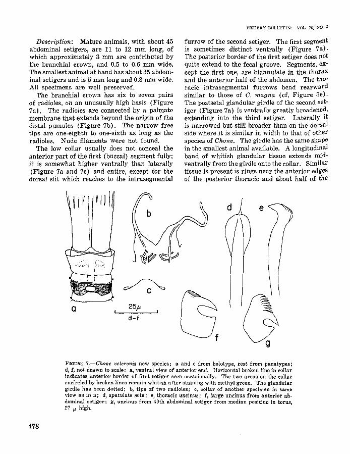
CHONE VELERONIS NEW SPECIES
Figure 7
Holotype: From Velero station 6104, lat33°39'45"N, long 118°06'40"W, 26 m, dark graysilty fine sand, 19 Feb. 1959. AHF Poly. 0459.
Paratypes: Same station and date. AHFPoly. 0460 (>10 specimens).
Diagnosis: A small species of Chane withoutventral shields, with a greatly broadened postsetal girdle of glands on second setiger. Branchial crown long, with very high basis and sixto seven pairs of radioles with filiform free ends.Collar low. Palmate membrane reaching beyonddistal pinnules. Spatulate setae with pointedtips. Anterior abdominal uncini markedly different within tori, largest with small teeth, rostra as long as bases; small uncini with greatlyenlarged crown. Posterior uncini only of thelatter kind.
The species is named after the present shipof the Allan Hancock Foundation, the Velero IV,which collected the specimens.
Material studied: About 10 specimens of thetype series.
477
FISHERY BULLETIN: VOL. 70, NO.2
Description: Mature animals, with about 45abdominal setigers, are 11 to 12 mm long, ofwhich approximately 3 mm are contributed bythe branchial crown, and 0.5 to 0.6 mm wide.The smallest animal at hand has about 35 abdominal setigers and is 5 mm long and 0.3 mm wide.All specimens are well preserved.
The branchial crown has six to seven pairsof radioles, on an unusually high basis (Figure7a). The radioles are connected by a palmatemembrane that extends beyond the origin of thedistal pinnules (Figure 7b). The narrow freetips are one-eighth to one-sixth as long as theradioles. Nude filaments were not found.
The low collar usually does not conceal theanterior part of the first (buccal) segment fully;it is somewhat higher ventrally than laterally(Figure 7a and 7c) and entire, except for thedorsal slit which reaches to the intrasegmental
furrow of the second setiger. The first segmentis sometimes distinct ventrally (Figure 7a).The posterior border of the first setiger does notquite extend to the fecal groove. Segments, except the first one, are biannulate in the thoraxand the anterior half of the abdomen. The thoracic intrasegmental furrows bend rearwardsimilar to those of C. magna (cf. Figure 5e).The postsetal glandular girdle of the second setiger (Figure 7a) is ventrally greatly broadened,extending into the third setiger. Laterally itis narrowed but still broader than on the dorsalside where it is similar in width to that of otherspecies of Chane. The girdle has the same shapein the smallest animal available. A longitudinalband of whitish glandular tissue extends midventrally from the girdle onto the collar. Similartissue is present is rings near the anterior edgesof the posterior thoracic and about half of the
25~
d-fa
FIGURE 7.-Chone veleronis new species; a and c from holotype, rest from paratypes;d, f, not drawn to scale: a, ventral view of anterior end. Horizontal broken line in collarindicates anterior border of first setiger seen occasionally. The two areas on the collarencircled by broken lines remain whitish after staining with methyl green. The glandulargirdle has been dotted; b, tips of two radioles; c, collar of another specimen in sameview as in a; d, spatulate seta; e, thoracic uncinus; f, large uncinus from anterior abdominal setiger; g, uncinus from 40th abdominal setiger from median position in torus,17 p. high.
478

BANSE: REDESCRIPTIONS OF SOME SPECIES OF CHONE AND EUCHONE
abdominal setigers. The pygidium is triangularand pointed. Eggs occur from the sixth thoracicsetiger.
The inconspicuous first bundle of setae originates at the level of the following notopodia.These have four to six of each upper limbate,very tiny bayonet, and pointed spatulate (Figure 7d) setae. Notopodial lips were not seen.Thoracic neuropodia contain slightly more thanhalf a dozen long-handled uncini (Figure 7e).Abdominal setigers carry approximately half adozen finely limbate neurosetae and, at leastthrough the 40th setiger, 12 to 15 uncini. Theuncini from the anterior abdomen, in the mostrecently formed sections of the tori, are of theusual form of Chone (Figure 7f) with severalcolumns of five to six accessory teeth each. Thesmallest, i.e., oldest, uncini of these tori, and alluncini in posterior segments (Figure 7g), havesmaller bases and appear similar to the unciniof Oriopsis. The posterior uncini have a,boutsix columns of a:bout seven teeth each.
Staining with methyl green shows the absenceof ventral shields. The whole surface in the anterior half of the body accepts the dye uniformlyexcept for the anterior edge and ventral partsof the collar (cf. Figure 7a), the inter- and intrasegmental furrows, and the glandular girdle onthe second setiger. Ventrally, the anterior border of the first setiger, which is often invisible inunstained material, is marked as a whitish line.The presetal rings of glandular tissue stain lesswell than the rest of the epidermis. On theposterior half of the abdomen only few epidermalcells accept the stain.
Remarks: Hartman (1959, 1965) has listedthe species of Chone, to which C. rosea Hartmann-Schroder, C. striata Hartmann-Schroder,C. trilobata Gallardo, and C. albocincta describedbelow should be added. Among these, C. veleronis is distinguished by the ventral enlargementof the postsetal glandular girdle on the secondsetiger, the very long branchial bases, and thelength of the palmate membrane. Only veryfew species (e.g., C. arenicola Langerhans) havea palmate membrane extending beyond the originof the distal pinnules.
CHONE ALBOCINCTA NEW SPECIES
Figure 8
Holotype: From Velero station 6104, lat33°39'45"N, long 118°06'40"W, 26 m, dark graysilty fine sand, 19 Feb. 1959. AHF Poly. 0454.
Paratypes: From same station and date.AHF Poly. 0455, three adults or near-adults, ninejuveniles.
Diagnosis: A Chone species of intermediatesize without ventral shields. Branchial basisslightly longer than the slightly oblique collar.Eight to ten pairs of radioles, connected by palmate membrane up to origins of distal pinnules,with abruptly tapered filiform free ends. Presetal whitish rings of tissue in thorax; presetaland postsetal whitish rings in anterior and median abdomen. First bundle of setae small, onsame level as following notosetae. Spatulatesetae with pointed tips. Anterior abdominaluncini markedly different within tori, smallestwith rounded bases and high crowns. Posteriorabdominal uncini predominantly of latter type.
The name of the species refers to the whitishrings contrasting in unstained animals with thered-brown color of the remaining epidermis.
Material studied: Type series.
Description: The holotype is a mature femalewith 8 thoracic and approximately 50 abdominalsetigers. The total length is about 18 mm, thegreatest width is 1 mm. The branchial crownmeasures about 6 mm. Two other adult animalsof 51 and 52 abdominal setigers are slightlylarger (total 20 mm; branchial crown, 7 mm)and somewhat shorter than the holotype. Thejuveniles have trunks about 5 mm long and thesame body proportions. All specimens are wellpreserved.
The branchial crown has a base slightly longerthan the collar and 9 to 10 pairs of radioles whichare united by the palmate membrane up to theinsertion of the distal pinnules. A 0.2-mm broadflange extends for a short distance beyond thispoint but tapers abruptly to a filiform free end(Figure 8a). The free ends of the radioles are
479

FISHERY BULLETIN: VOL. 70, NO.2
from one-fifth to one-sixth the length of the totalradioles. The ends of the radioles surpass theends of the distal pinnules. Ventral nude filaments were not seen.
The collar (Figures 8b and 8c) is oblique, withentire margin. The first (buccal) segment isinvisible and the collar is not clearly set off fromthe first setiger; the border can be recognizedonly in stained material. The thoracic and 15to 20 anterior abdominal setigers are clearly biannulate; after staining also the following ones
appear divided. Ventral shields are absent. Athick glandular postsetal girdle is present on thesecond setiger. Dorsally in the thorax, the intrasegmental borders bend rearward similar toC. magna (cf. Figure 5e). Rings of elevatedwhitish tissue, presumably glandular, are present in the presetal annuli of the thoracic setigersand in the presetal and postsetal annuli of theanterior 15 to 20 abdominal setigers. The ringon the second thoracic setiger is ventrally expanded (Figure 8b). Except on the second se-
))1\\\\\
a
25p
e-g
h
FIGURE 8.-Chone albocincta new species, from large types; abdominal uncini not toscale: a, end of median radiole; b, ventral view of anterior end, showing staining pattern; c, dorsal view of anterior end, branchial crown omitted; d, anterior view of fourthnotopodium; e, spatulate seta; f, thoracic uncinus; g, large uncinus from fourth abdominal setiger (about 30 p. high); h, small uncinus from ninth abdominal setiger; i, j,small and large uncini from approximately 40th abdominal setiger.
480

8ANSE: REDESCRIPTIONS OF SOME SPECIES OF CHaNE AND EUCHONE
tiger, the rings are invisible after using methylgreen, which stains the setigers in question uniformly except for the inter- and intrasegmentalfurrows. Posterior to the 15th to 20th abdominal setiger, the stain is accepted by numerous,distinct cells arranged in presetal and postsetalbands. These bands coalesce in the posteriorPart of the abdomen.
The first bundle of setae, slightly smaller thanthe following ones, inserts in the collar at thesame level as the following notopodia (Figure8c). The notopodia have small lips (Figure 8d)and long limbate, bayonet-type and narrow spatUlate setae; the latter have pointed tips (Figure8e). The thoracic uncini, with small wings (FigUre 8f) are arranged in single rows.
Anterior abdominal parapodia have about 30uncini. The large (ontogenetically most recent)Uncini (Figure 8g) have squarish bases andthree to four rows of four or five coarse teeth.The uncini change gradually within the torusinto smaller hooks (Figure 8h) with roundedbases and somewhat more teeth (five on thefourth setiger, seven on the ninth). In the posterior abdomen, the smallest uncini of a torusare similar (Figure 8i; about 10 columns ofteeth); the largest ones (Figure 8j), however,have very broad posterior portions and crownsWith more numerous teeth in approximately halfas many rows.
Juveniles of about 5-mm trunk length and 004to 0.5-mm width, with about 35 abdominal setigers, show the white rings clearly. StainingWith methyl green, however, indicates that theglandularization of the epidermis is not complete.The dye-absorbing cells are distributed princiPally along the edges of the stained areas so thatthe centers of the areas are light.
Remarks: Chane albocincta seems to be distingUished from all other species of the genus(cf. p. 479) by the whitish segmental rings oftissue. Somewhat similar presetal rings, howeVer, also occur in C. veleronis described above.Neglecting differences in size, C. albocincta maybe separated from the northeast Pacific speciesWithout ventral shields (C. aurantiaca, C. duneri,C. infundibuliformis, C. magna, C. mallis, andC. veleronis) also by the following: the post-
setal glandular girdle on the second segment isnarrow ventrally (from C. veleronis); the firstsegment is indistinguishable in the first segmentin unstained animals (from C. mollis); the radioles have long free ends (from C. aurantiacaand C. injundibulijormis); the abdominal uncinihave coarse teeth (from C. duneri). Chonemagna has spatulate setae without pointed tipsand only one form of abdominal uncini.
EUCHONE MALMGREN, 1866 EMENDED
EUCHONE Malmgren, 1866, p. 405.- Langerhans, 1881, p. 111.- Saint-Joseph, 1894, p. 250.Bush, 1904, p. 190.- Hofsommer, 1913, p. 327.McIntosh, 1923, p. 281.- Fauvel, 1927, p. 339.Berkeley and Berkeley, 1952, p. 121.- Ushakov,1955, p. 418.- Day, 1967, p. 774.- Banse, 1970,p. 389.
Type species: Euchone analis (Kroyer) (cf.Bush, 1904).
Diagnosis: SabeIIidae with semicircularbranchial lobes sometimes united by palmatemembrane. Collar conspicuous, sometimes bilobed. Postsetal girdle of glands on second setiger. Ventral shields sometimes present. Analdepression with lateral wings formed by variable number of caudal segments. Three typesof thoracic notosetae: upper, or upper-anteriorones limbate; lower, anterior ones narrowly limbate (bayonet-type); and lower, posterior onesspatulate or usually subspatulate. Thoracicneuropodial uncini long-handled, acicular. Abdominal notopodial uncini avicular, with roundedor square bases; abdominal neurosetae limbate.
Remarks: The genus continues to compriseall Fabriciinae with an anal depression. Addedto previous diagnoses are presence or absence ofthe palmate membrane, the note about the collar,the postsetal girdle of glands, the bayonet-typesetae, and the ventral shields. The last threecharacters had previously been mentioned byBanse (1970). It must be noted that E. alicaudata Moore, known so far only from one specimen, possibly lacks bayonet-type setae (cf. p.489). The number of thoracic setigers (almost
481

always eight) is not considered as a diagnosticcharacter for this genus.
Of great consequence for the systematics ofthe Sabellidae is the opinion that the number oflobes in the collar and the presence or absenceof a palmate membrane are specific charactersin Euchone. Therefore, they cannot be used fordividing genera (cf. p. 459 and 461). The reason is my opinion that the anal depression is ofmonophyletic origin and therefore a primarycharacter, sufficient to characterize Euchone.
EUCHONE ANALIS (KROYER)
Figure 9a-c
SABELLA ANALIS Kroyer, 1856, p.17.EUCHONE ANALIS.- Malmgren, 1866, p. 406;
1867, p. 223.- Berkeley and Berkeley, 1952, p.121.- Pettibone, 1954, p. 339 (synonymy).-
FISHERY BULLETIN: VOL. 70, NO.2
Hartmann-Schroder, 1971, p. 521.not EUCHONE ANALIS Imajima, 19G3, p. 367.
Diagnosis: A Euchone species with ventralshields, with 16 to 22 anterior abdominal setigersand 9 to 12 setigers in anal depression. Nine to17 pairs of radioles united by palmate membranefor half their length, with free ends tapered, intermediate in length; 5 to 10 pairs of ventralnude filaments. Collar level, with slight ventralincision, almost concealing the branchial basis.Lower thoracic notosetae subspatulate. Uncinifrom anterior and posterior abdomen similar,with squarish bases.
Material studied: Point Barrow, northernAlaska, about 65 m. Identified by M. H. Pettibone (cf. Pettibone, 1954). USNM 23651,1 specimen.
a
c50p
a-c, e-gg
e f
{jh
FIGURE 9.-Euchone analis: a, subspatulate notoseta; b, median uncinus from thirdabdominal setiger; c, same, from 21st. Accessory teeth schematic; E. papillosa: d, anterior end of thorax, ventral view, showing staining pattern; e, bayonet-type seta; f,spatulate seta; g, median uncinus from third abdominal segment; h, uncinus from anterior depression segment, approximately one and a half times enlarged over Figure g.
482

BANSE: REDESCRIPTIONS OF SOME SPECIES OF CHONE AND EUCllONE
Addition to the Description: The specimen iscomplete. The length of the trunk is about 15mm, that of the branchial crown approximately4 mm. The branchial crown consists of someventral nude filaments (5 to 10 pairs accordingto Kroyer, 1856, and Malmgren, 1866), and 10pairs of radioles bearing pinnules and connectedby a palmate membrane. The ends of the radioles are like those of E. elegans (cf. Figure l1a).The collar has a small ventral incision. The second setiger is ringed by a postsetal girdle ofglands. Segments are biannulate up to the analdepression. There are 17 anterior abdominalsegments and 9 setigers in the anal depression.Ventral shields are present in thorax and abdomen. The shields of the first segment and firstsetiger are divided by intrasegmental furrowswhich do not stand out as whitish lines afterstaining. In the abdomen the shields have theform depicted by Malmgren (1866, Plate 28,Figure 88).
The notopodia, including the large first one,have conspicuous lips of the same form as E.rubrocincta (cf. Figure 10d). The bayonet-typesetae have shafts almost as thick as the subsPatulate ones (Figure 9a) behind them and areterminally thin, as usual. Anterior abdominaluncini (Figure 9b) are all alike within the tori;each has a squarish basis and a crown of about10 rows of accessory teeth in several columns.The bases of the posterior uncini (Figure 9c)are similar but the crowns have about 15 rowsof teeth in approximately a dozen columns.
After staining, the ventral shields and, separate from them, areas before and behind theparapodia are conspicuous in thorax and abdomen. The first setiger does not seem to be separated from the preceding and following segments by nonstaining lines, nor is the intrasegmental furrow distinct. The intrasegmentalfurrow on the second setiger resembles that ofE. rub1'()cincta (Figure 10c). Dorsally, the thorax and the anterior segments of the abdomenare essentially free of stain-accepting cells. Inmiddle and posterior abdomiI)al setigers, presetalbroad rings are complete; . they are visible asWhitish tissue also in the unstained animal.
Remarks: This specimen has 17 anterior ab-
dominal setigers, fewer than stated by Malmgren(1866, about 19) and Berkeley and Berkeley(1952, 20 to 22) but similar to Kroyer (1856,about 16). More material must be studied inorder to see whether the number of anterior abdominal setigers overlaps those of the similar E.elegans and E. rubrocincta. If so, a probablyreliable difference of E. analis from the otherswill be the form of the anterior abdominal uncini.
The record by Imajima (1963) from the westcoast of Kamchatka is not this species becauseof the low number of anterior abdominal setigersand the high number of segments in the analdepression.
EUCHONE PAPILLOSA (SARS)
Figure 9d-h
SABELLA PAPILLOSA Sal's, 1851, p. 203.EUCHONE PAPILLOSA.- Malmgren 1866, p.
407.- Hofsommer, 1913, p. 327.- Augener,1928, p. 805 (synonymy).- Evenkamp, 1931,p. 412.- Pettibone, 1956, p. 578 (synonymy).Ktihlmorgen-HilIe, 1963, p. 51.- HartmannSchroder, 1971, p. 521.
SABELLA TUBERCULOSA Kroyer, 1856, p.18.
EUCHONE TUBERCULOSA.- Malmgren, 1866,p. 407.Diagnosis: A large Euckone species with
ventral shields, with at least 21 abdominal setigers; 8 to 10 form anal depression. Ten to 15pairs of radioles connected for approximatelyone-half their length by palmate membrane, withlong filiform free ends. Two to five pairs ofventral nude filaments. Collar oblique, with midventral incision, scarcely concealing the branchial basis. Very conspicuous round fields of glandsventrally in abdomen. Spatulate setae with verylong tips. Uncini of anterior abdomen with smallcrowns of teeth and bases with posterior extension; markedly higher crowns in posterior abdomen.
Material studied: Kiel Bight, western Baltic(unknown locality). Collected and identified byG. Kiihlmorgen-Hille (cf. Kiihlmorgen-Hille,1963). Part of sample, USNM 45390, 6 specimens.
483

Additions to description: Because the speciesis well known, only some characters have beenstudied, mostly for a comparison with E. capensisDay and E. velifera new species, both of whichalso have collars with ventral incisions. Theanimals studied were fixed in their tubes, are upto 2.5 cm long, and are well preserved.
The radioles have free filiform ends as shownby Malmgren (1866, Plate 29, Figure 92G butnot 94G). The collar (Figure 9d) is markedlylonger ventrally than dorsally. The number ofabdominal segments in six animals is 21, 21, 22,23, 23, and 24, of which 8 to 10 form the analdepression. Similarly Hofsommer (1913) foundspecimens from the western Baltic, which he believed to be juveniles, with a total of 29 segments (= setigers, from the context) and statedthat in adults the number is constantly 35, similar to Malmgren (1866). Evenkamp (1931)in material from Riel Fjord, however, observedup to 62 segments of which, again, 8 were thoracic.
Thoracic notopodia contain broad bayonettype setae (Figure ge) and spatulate setae (Figure 9f). The spatulate setae in the presentmaterial are broader than those depicted byMalmgren (1866, Plat 29, Figures 92Bl and94Bl). The uncini on anterior abdominal segments (Figure 9g) have broad bases with smallposterior appendages and crowns with approximately 10 columns with 10 to 13 teeth each, depending on the size of the uncini. Posteriorlyin the abdomen, the bases of the uncini becomemore rounded, and there are about 15 rows ofteeth in much enlarged crowns (Figure 9h).
The thoracic and abdominal ventral shields arevery conspicuous even in unstained material(cf. Malmgren, 1866, Plate 29, Figures 92 and94). After using methyl green, the ventral pattern in the anterior thorax is as shown in Figure9d. Laterally in the thorax, crescentic areas ofstained tissue occur posterior to the parapodiaand to a lesser degree anterior to them. Thedorsal side of the thorax is unstained except fora pair of conspicuous rounded areas near the anterior border of each segment from the 4th setiger onwards. In the anterior and middle regions of the abdomen, each setiger showsventrally two pairs of rounded patches, one near
484
FISHERY BULLETIN: VOL. 70, NO. Z
the anterior and one near the posterior border.These have been figured first by Malmgren(1866, Plate 29, Figures 92 and 94). Their sizevaries among indiViduals; they sometimes occupy almost half of the ventral area of the segments. Laterally in the abdomen, the stainedtissue around the parapodia, observed in thethorax, becomes gradually larger and extendsonto the dorsal side so that from the middle ofthe abdomen, a pigmented band crosses the posterior part of the segments (the parapodia insert here posteriorly on the segments). In thesame region, the anterior dorsal patches coalesce.Shortly before the depression segments, the intrasegmental furrows disappear and each segment is uniformly stained both dorsally andventrally.
EUCHONE RUBROCINCTA (SARS)
Figure 10
CHONE RUBROCINCTA Sal's, 1862, p. 128.EUCHONE RUBROCINCTA.- Malmgren, 1866,
p. 406.- Southern, 1914, p. 144.- McIntosh,1916, p. 33; 1923, p. 282.- Southward, 1956,p. 278.
Diagnosis: A Euchone species with conspicuous ventral shields, with 11 to 15 anterior abdominal setigers and up to 12 setigers in analdepression. Up to 16 pairs of radioles unitedby palmate membrane for about half their length,with long free ends. Up to eight pairs of nudefilaments. Collar level, with a small ventralnotch. Lower thoracic notosetae subspatulate.Uncini of anterior abdomen with small crowns ofteeth, slim necks, and narrow bases with markedposterior extensions. Uncini of posterior abdomen with large crowns, without necks.
Material studied: Syntypes, from Flom, nearBergen, Norway, approximately lat 61°40'N,long 5°00'E, stony bottom (Sal's, 1862). ZMO,3 specimens.
Ballynakill Harbour, Northwest Ireland, stations L 245, L 310, and ML L 11a (cf. Southern,

BANSE: REDESCRIPTIONS OF SOME SPECIES OF CHONE AND EVCHONE
FIGURE lO.-Euchone rubrocincta; b-h from syntypes, a and i from NMI, j from Southward's material: a, end of representative radiole. Broken line indicates extent of flangein syntypes (see also text); b, dorsal view of anterior end, branchial crown omitted;c, ventral view of anterior end, branchial crown omitted, after staining. Slightly schematic; x indicates the extent of the incision of the ventral shield on the first setiger ofE. elegans; d, part of lateral body wall of second setiger, seen slightly from below andbehind, with setae and glandular girdle; e, f, bayonet-type and two subspatulate setaefrom fifth thoracic setiger; g, anterior abdominal uncini (seventh abdominal setiger).Accessory teeth schematic; h, uncinusJrom depression segment (l7th abdominal setiger) ;i, subspatulate and limbate setae from sixth thoracic setiger; j, uncinus from seventhabdominal setiger.
1914). NMI 77.1908, 1 specimen from each station.
Irish Sea, station 22 of Southward (1956,1957). Dr. E. C. Southward's private collection.3 specimens.
Additions to the Description: The largest ofthe wen-preserved syntypes, a fairly relaxedspecimen, is approximately 2.5 cm long of whichabout one-fourth is contributed by the branchialcrown. The width is about 2 mm. The othersyntypes are approximately two-thirds as long;in one of them, a mature female, the branchialcrown contributes about one-third of the totallength, in the other one it is lost.
The branchial crowns of the two completespecimens consist of 10 and 13 pairs of radioleswith pinnules and up to 8 pairs of slender nakedventral filaments. Two to three pairs of thelatter are very short, and one or two pairs areas long as the radioles. The radioles are unitedfor. about half their length by a conspicuouspalmate membrane. They end in bare tips, proximally broad owing to the continuation of thepalmate membrane, but narrow in the terminalsections. The latter tend to be shorter ventrally than dorsally and are, on the average,about half as long in the syntypes as in the material from northwestern Ireland (Figure lOa).The length of the sections of the radioles freeof pinnules is very approximately one-eighth of
485

the total length in the syntypes and in Southern'smaterial.
In the syntypes, the collar is of equal heightdorsally and ventrally and has only a small ventral notch (Figures lOb and c). Setigers arebiannulate, which is especially obvious in thethorax. In the three syntypes, there are 11 anterior abdominal, 1 transitional, and 11 depression setigers; 15, 1, 9; and 12, 0, 10 setigers,respectively. Ventral shields are distinct in thethorax and abdomen as depicted by Malmgren(1866, Plate 29, Figure 91).
The thoracic notopodia (Figure 10d) are veryconspicuous; each contains about a dozen eachof long limbate, short bayonet-type (Figure 10e),and subspatulate (Figure 10f) setae. The longlimbate setae originate above the semicircularlip shown in Figure 10d which is about 0.2 mmlong; the other two kinds below it. Each neuropodium contains approximately 11;2 doz of longhandled hooks of the usual form (cf. McIntosh,1923, Plate 131, Figure 2a). The uncini of theanterior abdomen (Figure 109; cf. Figure 10j)do not have the squarish basis usually found inEuchone species but have a slim neck and a largeposterior extension making them almost Sshaped. Even the dorsalmost ones in the toristudied have essentially this outline. Only a fewcolumns of teeth are found in these uncini. Theuncini of the depression segments are formed asshown in Figure 10h; they have approximatelyeight columns of teeth, and the number of rowsis 12 to 14. The number of uncini is about 2 dozper torus anteriorly in the abdomen and in median segments of the anal depression. The abdominal capillary setae are arranged in two series per neuropodium.
After using methyl green, the collar (Figure10c) is lightly stained except for a ventral unstained area and, in some animals, a diffuse bandanterior to it. The thoracic ventral shields areclearly marked. Laterally strongly stainingareas are found posterior to and, to a lesser degree, anterior to the thoracic parapodia. In thelast thoracic segments, these areas touch theventral shields but do not continue onto the dorsal side, except as traces. In the abdomen, however, the stained cells form two girdles aroundeach segment, interrupted only by the fecal
486
FISHERY BULLETIN: VOL. 70, NO.2
groove; the presetal ring, which may appeardorsally as whitish glandular tissue even in unstained material, stains more strongly than thepostsetal one. The ventral shields are not markedly darker after staining than the lateral anddorsal parts. The intersegmental unstained furrows disappear gradually in the depression segments.
In the material from northwestern Ireland,the largest animal, a mature male, is almost 2.5cm long, of which about one-third is contributedby the branchial crown; the smallest specimenis 1.5 cm long. The latter has 11, the former,15 pairs of radioles, with about 4 doz pairs ofpinnules each. In all specimens, there are 12anterior abdominal setigers, the twelfth sometimes appearing to be a transitional segment,and 10 to 12 depression segments. The notopodia (cf. Figure 10d) appear to be divided intoanterior and posterior lips, rather than form onesemicircular fold. Limbate and subspatulatenotosetae are shown in Figure 10i; the totalnumber of notopodial setae is 2 doz. The othercharacters agree with the syntypes.
No color is left in the syntypes of the Irish material, but Southern (1914) stated that the brightred bands, which gave the species its name, werepresent in Formalin-preserved material until thetransfer into alcohol. Sars (1862) observed onepurple ring on each segment.
McIntosh (1916) reported on animals with15 to 16 pairs of radioles. It must be noted thatMcIntosh's description (1916, also 1923) of otherfeatures of the branchial crown contains qUOtations of Southern's (1914) text for E. rosea.
Southward's specimens are small, up to 7 mmlong. Two have 11 anterior abdominal setigers,and one transitional setiger; the third animalhas 13 or 14 anterior abdominal setigers. Anuncinus from this region (Figure 10j) has abroader neck than the material treated above butotherwise has the same characteristic form. Thestaining pattern is as described by the syntypesas far as this can be recognized in the previouslydried specimens.
Remarks: Euchone 1'ubrocincta is close tothe smaller E. elegans Verrill from which it is

BANSE: REDESCRIPTIONS OF SOME SPECIES OF CHONE AND EUCHONE
distinguished by the larger number of radioleswith long slender ends, and especially the formof the anterior abdominal uncini. Further, theincision of the ventral shield at the level of thefirst parapodium is very short in E.rubrocincta(see also E. analis, p. 483). E. rosea Langerhans, as described by Banse (1970) for specimens from the Ivory Coast, is very similar inregard to the tips of the radioles, the collar, thesubspatulate notosetae, and especially the abdominal uncini. However, the ventral shield onthe first setiger is not divided, and the numberof anterior abdominal setigers is only 10 or 11,and that of the anal depression only 6. Thesmaller number of radioles in the African material may be due to their smaller size.
The distribution of the species is not wellknown. Material of Southern (1914) and Southward (1956) collected around Ireland has beentreated above. The specimens of Malmgren(1866) from the Skagerak and McIntosh (1916,1923) from the Shetlands are certainly E. rubrocincta. The species is also reported from theMediterranean Sea (cf. Fauvel, 1927) , but I havenot searched the literature for Lusitanian andmore southern records. Also, the reports of E.rubrocincta from the northwest Atlantic byMoore (1909) and Baillie (1946) should bechecked because of the possible confusion withE. elegans treated below. The former authordid not give any reference for identification; thelatter referred to Malmgren's description.
EUCHONE ELEGANS VERRILL
Figure lla-e
EUCHONE ELEGANS Verrill, 1873, p. 618.Hartman, 1944a, p. 334 (with other references).
Diagnosis: A Euchone species with ventralshields, with 12 to 15 anterior abdominal setigersand 8 to 10 setigers in anal depression. Six toeight pairs of radioles, united by palmate membrane for about half their length, and ratherbroad and free ends. Three to four pairs of nudefilaments. Collar level, with small ventral notch.Lower thoracic notosetae subspatulate. Markeddivergence of uncini within tori of anterior ab-
domen; ontogenetically recent uncini with broadbases and small posterior extension; old unciniwith rounded bases and high crowns, similarto those in depression.
Material studied: Syntypes, collected fromBlock Island Sound, Mass., in 1871. USNM 7513,5 specimens.
Cape Cod Bay, Mass., SEP station 1412 E5,lat 41°55.0'N, long 70 0 08.5'W, 33 m, muddy sand,11 June 1968. SEP, 4 specimens. Unpublishedrecord.
Cape Cod Bay, Mass., SEP station 2318 Ep, lat41°45.0'N, long 70 0 06.5'W, 16 m, sand, 13 Oct.1966. SEP. 22 specimens. Unpublished record.
Additions to the Description: The availablespecimens are up to 1 cm long; about one-thirdof the length is contributed by the branchialcrown. Verrill (1873) observed animals upto 2 cm long. Most of the branchial crownshave seven pairs of radioles with up to3 doz pinnules each and three to four pairs ofventral nude filaments; one pair of these is aboutas long as the radioles. The free ends (Figurella) contribute about one-sixth of the totallength of the radioles. The collar is of equalheight dorsally and ventrally and has only a smallventral notch. Segments are biannulate, particularly in the thorax. A postsetal girdle of glandsis present on the second setiger. Ventral shieldsare moderately visible in unstained material inthe thorax and anterior abdomen but stand outclearly upon staining. In the syntypes, the number of abdominal setigers is 13 (12 in one) followed by 1 transitional and 8 depression setigers.In the 10 SEP specimens, up to 9 mm long, 15setigers are present prior to the beginning ofthe depression in 7 specimens; 14 setigers in 2specimens; and 14 or 15 in 1 specimen. Theanal depression extends through 10 setigers inthe largest animals.
The thoracic notopodia are almost as conspicuous as in E. rubrocincta (cf. Figure 10d). Eachcontains long limbate, short bayonet-type, andsubspatulate (Figure lIb) setae, totaling lessthan a dozen. Each neuropodium has about 10long-handled hooks. The uncini of the anteriorabdominal segments (Figure 11c) have a broad
487

FISHERY BULLETIN: VOL. 70, NO. Z
?
c
!
9 t- -,•. ~
IIII 111!l )II /Iii!!.J
.:....{
U~~-t11_
'\-II!- 0
n
25~
? c,d,L,m~ .
12.5ft: e; 25~: h, i
FIGURE 11.-Euchone elegans; a, c, d from syntypes, h, e from SEP stn. 2318Ep; a, endof medio-ventral radiole; h, subspatulate notoseta; c, d, middle and dorsalmost uncinifrom second abdominal setiger; e, uncinus from 17th abdominal setiger (depression);E. alicaudata, from holotype: f, end of median radiole; g, ventral view of anteriorend of thorax after staining; h, i, uncini from second and 18th abdominal setigers; j,ventral view of middle abdominal segment after staining; k, dorsal view of middle thoracic segment; g, j, and k are oriented in the same way; E. capensis, from paratypes:1, m, uncini from second and 19th abdominal setigers, the latter slightly enlarged; n, 0,
ventral views of anterior end of thorax and median abdominal segment after staining,slightly schematic.
488

BANSE: REDESCRIPTIONS OF SOME SPECIES OF CHONE AND EUCHONE
square base similar to those of many other Choneand Euchone species. The dorsalmost and ontogenetically oldest ones (Figure lId), have amuch higher crown with about 10 rows of teethand are similar to the uncini in the depressionsegments (Figure lIe).
After staining with methyl green, the patternon the first setiger agrees with that of E. rubrocincta (cf. Figure 10c) where the incompletedivision of the ventral shield reaches somewhatcloser to the midline than that of the second one(Figure IDe, marked by x).
Remarks: E1whone elegans is close to E.rubrocincta from which it is distinguished bythe much smaller number of radioles and theirshort free ends and the form of the uncini ofthe anterior abdominal segments. The divisionof the ventral shield at the level of the first parapodium is marked. The species is also similar toE. analis as described by Malmgren (1866,1867;cf. p. 482) but has slightly fewer anterior abdominal setigers. The posterior abdominal uncini of E. analis are similar to the anterior onesbut are markedly different in E. elegans.
EUCHONE ALICAUDATA MOORE AND BUSH
Figure llf-k
EUCHONE ALICAUDATA Moore and Bush,1904, p. 165.
Diagnosis: A large Euchone species withventral shields, with 22 abdominal setigers; 8form anal depression with broad wings. Fifteento 20 pairs of radioles connected by palmatemembrane for one-quarter of their length, withbroad flanges extending beyond distal pinnules,and tapering to slender tips. Several pairs ofventral nude filaments. Collar level, with ventro-Iateral incisions or folds, probably ventrallyincised, concealing the branchial basis. Lowerthoracic notosetae subspatulate. Uncini of anterior abdomen with strong necks and bases withposterior extensions; uncini of the posterior ab-domen with high crowns. '
Material studied: Holotype from 280 m nearTokyo. USNM 5496.
Additions to the Description: The radiolesnumber somewhat more than 15 as stated in theoriginal description; they end in tips as shownin Figure 11f. The collar (Figure 11g) appearsto have ventro-Iateral incisions rather than folds,as originally stated; owing to injury the presence or absence of a marked midventral incision,implied by Moore and Bush (1904), cannot beverified. A postsetal girdle of glands is presenton the second setiger. Bayonet-type setae werenot found on the two dissected thoracic notopodia. In this respect, E. alicaudata is uniqueamong the 25 species of Chone and Euchone Ihave studied; an investigation of more specimens is VE\ry desirable. The subspatulate setaewere correctly depicted by Moore and Bush(1904, Plate 11, Figure 15). The uncini of theanterior abdomen (Figure llh) have a posteriorextension which is not so distinct in depressionsegments (Figure 11i). The latter, however,have high crowns with 13 to 15 rows of teeth.
The ventral shields are distinct in thorax andabdomen, becoming even more marked afterstaining (Figures llg and llj). The shieldsof the first and second setigers are not completely divided by the intrasegmental furrows.The dorsal thoracic staining pattern is shownin Figure 11k. In the abdomen, simple broadbands extend from the level of the parapodiaacross the dorsum except in the segments of theanal depression where the intrasegmental furrows disappear.
Remark: Euchone capensis Day bears someresemblance to E. ctlicaudata, but the species arewell distinguished by the form of the ventralshields on the first (buccal) segment (cf. Figures 11g and lin).
EUCHONE CAPENSIS DAY
Figure 111-0
EUCHONE CAPENSIS Day, 1961, p. 540; 1967,p. 776.
Diagnosis: A Euchone species with ventralshields, with 25 to 32 abdominal setigers; 7 or 8form anal depression. Nine pairs of radioles,
489

united by palmate membrane for two-thirds oflength, with very long filiform tips. Two to threepairs of ventral nude filaments. Collar approximately as long as branchial basis, with deepventral incision. Lower thoracic notosetae subspatulate. Vncini of anterior abdomen withstrong necks and few rows of teeth, bases withsmall posterior elongation; uncini of posteriorabdomen with greatly enlarged crowns.
Material studied: Paratypes from Stn. TRA.73M, off South Africa, Ecological Survey, VCT.311 m. Material at VCT, 3 specimens.
Additions to the Description: The radiolesare flanged some distance beyond the palmatemembrane. Many pinnules of the middle sections of the radioles extend as far as the end ofthe distal pinnules. The very long and filiformtips of the radioles extend beyond this level.Whereas Day (1961, Figure 14t; 1967, Figure37.6j) depicted the branchial base as not concealed by the collar; the branchial base in atleast one of the paratypes is hidden. A postsetal girdle of glands is present on the secondthoracic setiger. The ventral shields are divided within each thoracic segment. While Day(1961) reported 32 abdominal setigers for theholotype, 2 complete specimens have 25 and 25or 26 abdominal setigers; 7 or 8 form the analdepression.
An uncinus from the anterior abdominal region is shown in Figure 111 (cf. Day, 1961, Figure 14n). The uncini of the posterior regionseem to be similar except that the number ofrows of teeth above the main fang is about 10(Figure 11m). The number of uncini is anteriorly just below, posteriorly slightly above, adozen per torus.
After using methyl green, the ventral shieldsare bordered laterally by unstained epidermis(Figure lIn), except in the first thoracic setigers. In the abdomen, intra- and intersegmentalborders appear broader than in the thorax, andthe outline of the shields becomes rounded (Figure llq); they are almost circular large patchesanterior to the anal depression. Broad, lateralareas of staining cells are present on the firstand second thoracic setiger, and a small anterior
490
FISHERY BULLETIN: VOL. 70. NO.2
and a large posterior patch are found aroundeach parapodium on the subsequent segments.These patches seem to be characteristic for thespecies. The posterior patches tend to formcrescents around the parapodia in the abdominalsegments. From about the middle part of theabdomen, bands of scattered staining cells croSSthe dorsal side forming two bands per setigerand becoming most distinct on the depressionsegments.
Remarks: Euchone capensis has abdominaluncini that are somewhat similar to those of Eoalicaudata, E. rosea, and E. rubrocincta. Theventral incision in the collar and the distinctabdominal ventral shields separate the SouthAfrican form clearly from the two latter species.A clear distinction between E. capensis and E.alicaudata is provided by the form of the ventralshields of the first (buccal) segment (cf. FigureSllg and lIn).
EUCHONE VELIFERA NEW SPECIES
Figure 12
EUCHONE sp. Hartman, 1955, p. 114.
Holotype: Off Catalina Island, Calif., Velerostation 2412, lat 33°30'02"N, long 118°12'02"W,593 m, fine mud, 17 Sept. 1953 (cf. Hartman,1955). AHF, Poly. 0256.
Pamtypes: Off Point Vincente Light, Calif.,Velero station 4854, long 33°47'30"W, lat118°30'OO"W, 179 m, 8 Feb. 1957. AHF Poly.0453, 2 specimens.
Diagnosis: A large Euchone species with indistinct ventral shields, with 27 to 32 abdominalsetigers; 7 to 9 form anal depression with largewings. Nine to 10 pairs of radioles united bYpalmate membrane for half of their length atleast in juveniles, with short filiform free ends.Collar with lateral notches, ventrally deeply incised and folded in between the two halves of thebranchial crown, concealing branchial basis.Lower thoracic notosetae subspatulate. Unciniof anterior abdomen with small crowns andsquare bases; uncini of posterior abdomen with

BANSE: REDESCRIPTIONS OF SOME SPECIES OF CHONE AND EUCllONE
9
II
25~
e, fd
a
FIGURE 12.-Euchone velifera new species; c-e from holotype, a, b from paratypes;a-c, g not drawn to scale: a, b, ventral and left side views of anterior end of juvenilespecimen; c, posterior end from the left side; d, subspatulate thoracic seta; e, f, uncinifrom fifth and 27th (sixth to last) abdominal segments. Accessory teeth schematic; g,anterior end in ventral view showing staining pattern.
high crowns and rounded, posteriorly enlargedbases.
The name refers to the sail-like wings of theanal depression.
Material studied: Type series.
Description: The holotype is a complete, wellpreserved mature female of almost 3-cm totallength (trunk length 17 mm) and lo5-mm width.The paratypes are juveniles. In the holotypethe branchial crown contributes a little morethan one-third of the total length. The 9 or 10pairs of pinnulated slender radioles are in theholotype free from each other through their entire length and have only rudimentary flanges.The juveniles have a palmate membrane unitingthe radioles for approximately half of theirlength. The pinnules, which are of a uniform,relatively short length, are present almost to theends of the radioles so that there are only rel-
atively short filiform free tips. A few thin nakedfilaments are also present, the longest one beingabout half as long as the radioles. Triangularlips occur on both sides of the mouth.
The collar (Figures 12a and 12b) is slightlylonger ventrally than dorsally. The dorsal gapis narrow; laterally there is a small notch. Ventrally, a deep incision is formed by the collar being folded-inward between the two halves of thebranchial crown. A postsetal girdle of glandsencircles the second setiger. The ventral shieldsof unstained specimens are more distinct in thejuveniles than in the adult animal. They areindicated more by the appearance of the ventraland lateral epidermis in the thorax, as contrastedto that of the dorsal side, than by morphologicalelevation (pads) on the ventrum. This is borneout by staining. In the adult, very conspicuous,subepidermal tissue of a creamy color is presentdorsally from the third setiger, having the
491

appearance of ventral shields. The intrasegmental division of this tissue into anterior andposterior sections is particularly obvious in theabdomen. In contrast to shields, this material isnot stained by methyl green. The very largewings of the anal depression are shown in Figure 12c.
Each thoracic notopodium after the first setiger contains three series of setae: about a dozenIimbate, about half a dozen bayonet-type setaewith very small wings, and 2 doz subspatulatesetae (Figure 12d). The thoracic hooks are ofthe ordinary kind. An uncinus from the anteriorabdominal region is shown in Figure 12e; thereare about 8 teeth above the main fang in thesmaller, and about 10 teeth in the larger, hooksof a torus. Uncini from the. posterior regionsdiffer markedly (Figure 12f), not only becauseof an increase of the number of teeth above themain fang but also by a marked posterior enlargement. There are about half a dozen ventralIimbate setae in each abdominal neuropodiumwith very narrow wings.
The color of the preserved holotype is palered-brown, except for the dorsal creamy tissue.Very numerous polygonal eggs have diametersup to 150 ft. The tube is clearly constructed bythe successive addition of rings of mucus andmud.
After using methyl green, the collar is dorsallyfully stained except at the anterior margin; theventral pattern is indicated in Figure 12g; thepatterns on the fourth and following setigers arethe same as those on the third. In the abdomen,the intrasegmental divisions appear broader inthe thorax so that, owing to the fecal groove, fourrectangular fields of pigment per segment areseen. Laterally in the thorax and abdomen, thestained epidermis extends from the shieldsaround the parapodia, uniting immediately dorssal to the notopodia. Staining cells are absentdorsally in the thorax but appear as faint linesin the abdomen.
The two young specimens (paratypes) areabout 10 mm long and 0.5 to 0.6 mm wide. Theapproximately six pairs of radioles are almostas long as the trunk and united by the palmatemembrane for about half their length. The collar (Figure 12b) is not as level as in the holo-
492
FISHERY BULLETIN: VOL. 70, NO.2
type. The number of abdominal setigers is difficult to ascertain but seems to be 27 or 28 in one,with 7 setigers in the anai depression, and 29 inthe other specimen, with 9 setigers in the analdepression.
The smallest uncini in the third and fourth abdominal setiger of one of the specimens werevery similar to Figure 12f, that of an uncinus ofthe adult animal from the posterior segments.The staining pattern is similar to that of theholotype except for a lateral tongue of nonstaining tissue which extends latero-ventrally between the posterior border of the collar, at thelevel of the lateral notch, and the first parapodium.
Remarks: Euchone velifera is unique in regard to its very deeply bilobed collar. Distinctmidventral incisions are also known for E. papillosa and E. capensis. The latter two specieslack laterally notched collars and very largewings in the anal depression.
ACKNOWLEDGMENTS
It is a pleasure to acknowledge the assistanceand financial support by Dr. R. L. Fernald, Director, Friday Harbor Laboratories, and theu.s. Atomic Energy Commission, Contract No.AT(45-1)-1725 (ref: RLO-1725-196). Material was kindly lent by the 11 museums and laboratories named on page 460. I am especiallyindebted to the colleagues at these institutions.Also, Dr. O. Hartman, Los Angeles, allowed meto describe the three new species included in thispaper. Dr. M. Imajima, Tokyo, gave some specimens. Dr. M. H. Pettibone, Washington, D.C.,provided study space in the U.S. National Museum. Dr. H. L. Sanders, Woods Hole, put hiscollection made near Friday Harbor, Wash., atmy disposal, and Dr. E. C. Southward, Plymouth,England, lent specimens from her private collection. Finally, Dr. D. P. Henry gave veryvaluable advice on the preparation of the manuscript.

BANSE: REDESCRIPTIONS OF SOME SPECIES OF CHaNE AND EUCHONE
LITERATURE CITED
AUGENER, H.1928. Die Polychaten von Spitsbergen. In F.
Romer and F. Schaudinn (editors), Fauna Arctica 5:647-834.
BAILLIE, W. H. T.1946. Polychaeta from the Bay of Fundy, 1911
1912. J. Fish. Res. Board Can. 6:472-475.BANSE, K.
1970. The small species of Euchone Malmgren (SabeIIidae, Polychaeta). Proc. Bio!. Soc. Wash. 83:387-408.
BANSE, K., AND F. H. NICHOLS.1968. Two new species and three new records of
benthic polychaetes from Puget Sound (Washington). Proc. BioI. Soc. Wash. 81 :223-230.
BANSE, K., K. D. HOBSON, AND F. H. NICHOLS.1968. Annotated list of polychaetes. Fiskeridir.
Skr. Ser. Havunders. 14:521-548.BERKELEY, E., AND C. BERKELEY.
1932. On a collection of littoral Polychaeta from thewest coast of Vancouver Island. Contrib. Can.Bio!. Fish. New Ser. 7 :309-318.
1942. North Pacific Polychaeta, chiefly from thewest coast of Vancouver Island, Alaska and Bering Sea. Can. J. Res., Sect. D, 20 :183-208.
1950. Notes on Polychaeta from the coast of western Canada. IV. Polychaeta Sedentaria. Ann.Mag. Nat. Hist., Ser. 12, 3 :50-69.
1952. Annelida, Polychaeta Sedentaria. CanadianPacific Fauna, 9b(2), 1-139 p. Fish. Res. BoardCan.
1954. Additions to the polychaete fauna of Canada,with comments on some older records. J. Fish.Res. Board Can., 11 :454-471.
BUSH, K. J.1904. Tubicolous annelids of the tribes Sabellides
and Serpulides from the Pacific Ocean. HarrimanAlaska Exped., Smithsonian Inst., Wash., D.C.,12:169-355.
CHLEBOVICH, V. V.1961. [Littoral polychaetes from the Kurile
Islands.] [In Russian.] Akad. Nauk SSSR Is-sled. Dal'nevost. Morei SSSR 7:151-260. '
DAY, J. H.1961. The polychaet fauna of South Africa. Part 6.
Sedentary species dredged off Cape coasts with afew new records from the shore. J. Linn. Soc.Lond. Zoo!' 44 :463-560.
1967. A monograph on the Polychaeta of SouthernAfrica. Part 2. Sedentaria. Br. Mus. (Nat.Hist.), Pub!. 656 :459-878.,
ELIASON, A.1962. Weitere Untersuchungen tiber die Polychaeten
fauna des l)resundes. Lunds Univ. Arsskr., N.F.,Avd. 2, 58 (9) :1-98.
EVENKAMP, H.
1931. Morphologie, Histologie und Biologie der Sabelliden species Laonome kroyeri Malmgr. undEuchone papillosa M. Sars. Zoo!' Jahrb. (Anat.)53: 406-534.
FAUVEL, P.1913. Campagne du Pourquoi-Pas? (Islande et Jan
Mayen, 1912). Annelides Polychetes. Bull. Mus.Hist. Nat. Paris 19 (2) :80-93.
1914. Annelides polychetes non pelagiques provenant des campagnes de I'Hirondelie et de Ill. Princesse-Alice (1885-1910). Result. Campagnes Sci.Albert Ier, Prince de Monaco 46, 432 p.
1927. PolychHes sedentaires. Faune Fr. 16:1-494.HARTMAN, O.
1938. Annotated list of the types of polychaetousannelids in the Museum of Comparative Zoology.Bull. Mus. Compo Zoo!. Harvard ColI. 85: 1-31.
1942a. The identity of some marine annelid wormsin the United States National Museum. Proc. U.S.Nat!. Mus. 92:101-140.
1942b. A review of the types of polychaetous annelids at the Peabody Museum of Natural History,Yale University. Bull. Bingham Oceanogr. Coil.,Yale Univ. 8 (1), 98 p.
1944a. New England Annelida. Part 2, includingthe unpublished plates by Verrill with reconstructed captions. Bull. Am. Mus. Nat. Hist. 82:327-344.
1944b. Polychaetous annelids from California, including the description of two new genera andnine new species. Allan Hancock Pac. Exped.10 :239-308.
1955. Quantitative survey of the benthos of SanPedro Basin, southern California. Part 1. Preliminary results. Allan Hancock Pac. Exped. 19:1-185.
1959. Catalogue of the polychaetous annelids of theworld. Part II. Allan Hancock Found. Publ.,Occas. Pap. 23 :355-628.
1961. Polychaetous annelids from California. Allan Hancock Pac. Exped. 25:1-226.
1965. Catalogue of the polychaetous annelids ofthe world: Supplement 1960-1965 and index. Allan Hancock Found. Pub!., Occas. Pap. 23:1-197.
1969. Atlas of the sedentariate polychaetous annelids from California. Allan Hancock Found.(Univ. Southern California), Los Angeles, 812 p.
HARTMANN-SCHRODER, G.1971. Annelida, Borstenwiirmer, Polychaeta. In
F. Dahl (editor), Tierwelt Deutschlands 58:1-594.HOFSOMMER, A.
1913. Die Sabelliden-Ausbeute der Poseidon-Fahrten und die Sabelliden der Kieler Bucht. Wiss.Meeresunters. Abt. Kiel, N.F., 15:305-364.
IMAJIMA, M.1961. Polychaetous annelids collected off the west
coast of Kamchatka. 1. Notes on species found in
493

the collection of 1957-58. Publ. Seto Mar. BioI.Lab. 9 :81-102.
1963. Polychaetous annelids collected off the westcoast of Kamchatka. II. Notes on species foundin the collection of 1959. Pub!. Seto Mar. BioI.Lab. 11 :345-372.
IMAJIMA, M., AND O. HARTMAN.
1964. The polychaetous annelids of Japan. Part II.Allan Hancock Found. Pub!., Occas. Pap. 26:239452.
JOHANSSON, K. E.1927. Beitrage zur Kenntnis del' Polychaeten-Fa
milien Hermellidae, Sabellidae und Serpulidae.Zool. Bidr. Upps. 11 :1-184.
JOHNSON, H. P.1901. The Polychaeta of the Puget Sound region.
Proc. Boston Soc. Nat. Hist. 29 :381-437.KINBERG, J. G. H.
1867. Annulata nova. Oefv. K. Vetensk. Akad.Stokh., Forh. 23: 337-357.
KROYER, K.1856. Bidrag til kundskab af Sabellerne. Defv. K.
Dan. Vidensk. Selsk. Forh. 1856, p. 1-36.KUHLMORGEN-HILLE,G.
1963. Quantitative Untersuchungen del' Bodenfauna in del' Kieler Bucht und ihre jahreszeitlichen Veranderungen. Kiel. Meeresforsch. 19 :42-66.
LANGERHANS, P.1880. Die Wurmfauna von Madeira. III. Z. wiss.
Zool. 34:87-143.LIE, U.
1968. A quantitative study of benthic infauna inPuget Sound, Washington, USA, in 1963-1965.Fiskeridir. Skr., Ser. Havunders. 14 :237-556.
LUKASH, B. S.1911. . (Annelida Polychaeta des Kola-Fjords (Mur
man-Kiiste). Family SabelJidae.] [In Russian.]Tr. Obsch. Estestvoispyt. Leningr., Sect. Zool.Physio!. 41 (2) :1-78.
MALMGREN, A. J.
1866. Nordiska Hafs-annulater. Defv. K. Vetensk.Akad. Forh. 22 :355-410.
1867. Annulata polychaeta Spetsbergiae, Groenlandiae, Islandiae et Scandinaviae hactenus cognita. Oefv. K. Vetensk. Akad. Forh. 24:127-235.
McINTOSH, W. C.1916. Notes from the Gatty Marine Laboratory,
St. Andrews. No. XXXVIII.-1. On the BritishSabellidae. Ann. Mag. Nat. Rist., Ser. 8, 17:1-52.
1923. A monograph of the British marine annelids,4(2) :251-538. Ray Society, Land.
MOORE, J. P.1906. Additional new species of Polychaeta from
the North Pacific. Proc. Acad. Nat. Sci. Phila.58:217-260.
1909. The polychaetous annelids dredged in 1908by Mr. Owen Bryant off the coasts of Labrador,Newfoundland, and Nova Scotia. Proc. U.S. Nat!.Mus. 37:133-146.
494
FISHERY BULLETIN: VOL. 70, NO.2
1923. The polychaetous annelids dredged by theU. S. S. "Albatross" off the coast of southern California in 1904. IV. Spionidae to Sabellariidae.Proc. Acad. Nat. ScL Phila. 75:179-259.
MOORE, J. P., AND K. J. BUSH.
1904. Sabellidae and Serpulidae from Japan, withdescriptions of new species of Spirorbis. Proc.Acad. Nat. Sci. Phila. 56:157-179.
NICHOLS, F. H.1968. A quantitative study of benthic polychaete
assemblages in Port Madison, Washington. M.S.Thesis, Univ. Washington, Seattle, 78 p.
OKUDA, S.1934. Some tubicolous annelids from Hokkaido.
J. Fac. Sci. Hokkaido Univ., Ser. VI, 3 :233-246.1946. Studies on the development of Annelida Poly
chaeta. I. J. i Fac. ScL Hokkaido Univ., Ser. VI,9 :115-219.
PETTIBONE, M. H.1954. Marine polychaete worms from Point Bar
row, Alaska, with additional records from theNorth Atlantic and North Pacific. Proc. U.S.Natl. Mus. 103:203-356.
1956. Marine polychaete worms from Labrador.Proc. U.S. Nat!. Mus. 105 :531-584.
REISCH, D. J.1968. The polychaetous annelids of the Marshall
Islands. Pac. ScL 22 :208-231.RIOJA, E.
1963. Estudios anelidolOgicos. XXVI. Algunospoliquetos de las costas del Pacifico de Mexico.An. Inst. BioI. Mex. 33: 131-229.
RIOJA, E., AND E. Lo BIANCO.
1931. Estudio de los poliquetos de la PeninsulaIberica. Mem. Cienc. exact., fis. nat. Madrid, Ser.Cienc. Nat. 2:1-471.
SAINT-JOSEPH, A. DE.
1894. Les Annelides polychetes des cotes de Dinard.Troisieme Partie. Ann. Sci. Nat. Paris, Sel'. 7,17 :1-395.
SARS, M.1851. Beretning om en i Sommeren 1849 foretagen
zoologisk Reise i Lofoten og Finmarken. NyttMag. Naturvidensk. 6:121-211.
1862. Foredrag om de ved Norges Kyster forkommende Arter af den Linneiske Annelideslaegt Sabella. Forh. Vidensk. Selsk. Christiania for 1861,p. 116-133.
SOUTHERN, R.1914. Archiannelida and Polychaeta (Clare Island
Survey). Proc. R. Irish Acad., Sect. B 31 (47) :1-160.
SOUTHWARD, E. C.1956. On some Polychaeta of the Isle of Man. Ann.
Mag. Nat. Hist., Ser. 12, 9 :257-279.1957. The distribution of Polychaeta in offshore de
posits in the Irish Sea. J. Mar. BioI. Assoc. U.K.36:49-75.

BANSE: REDESCRIPTIONS OF SOME SPECIES OF CHONE AND EUCHONE
UCHIDA, H.1968. Polychaetous annelids from Shakotan (Hok
kaido). I. The collection in 1967. J. Fac. Sci.Hokkaido Univ., Ser. VI, 16 :595-612.
USHAKOV, P. V.1955. [Polychaeta of the Far Eastern Seas of the
USSR.] [In Russian.] Akad. Nauk SSSR, Zoo!.Inst., Opred. Faune SSSR 56, 445 p. (Trans!.1965, 419 p., Isr. Program Sci. Trans!., U.S. Dep.Commer., Washington, D.C., TT 665-50020.)
VERRILL, A. E.1873. Report upon the invertebrate animals of
Vineyard Sound and the adjacent waters, with
an account of the physical characters of the region.Rep. U.S. Comm. Fish Fish. 1871-1872, Part 1,295-747.
ZACHS, J.1933. [The Annelidean fauna of the northern part
of the Sea of Japan.] [In Russian.] Issled.Morei SSSR 19: 125-137.
ZENKEVITSCH, L. A.1925. Biologie, Anatomie und Systematik der SU
swasser-Polychaten des Baikalsees. Zoo!. Jahrb.(Syst.) 50:1-60.
495

![[Theatre, Drama, Acting] Vladimir Mirodan - The Way of Transformation - The Laban-Malmgren System of Dramatic Character Analysis (1997)](https://static.fdocuments.net/doc/165x107/55720691497959fc0b8b9490/theatre-drama-acting-vladimir-mirodan-the-way-of-transformation-the-laban-malmgren-system-of-dramatic-character-analysis-1997.jpg)