Recurrent Proliferating Trichilemmal Tumor with Malignant … · 2016-06-01 · 390 Recurrent...
5
Copyrights © 2016 The Korean Society of Radiology 389 Case Report pISSN 1738-2637 / eISSN 2288-2928 J Korean Soc Radiol 2016;74(6):389-393 http://dx.doi.org/10.3348/jksr.2016.74.6.389 INTRODUCTION Proliferating trichilemmal tumor (PTT), also referred to as proliferating trichilemmal cyst or pilar tumor, is an uncommon neoplasm that originates from the outer root sheath of a hair follicle. It occurs commonly on the scalp in elderly women (1). PTT is usually considered as a benign skin appendageal tumor, biologically; however, malignant PTT with recurrence/metasta- sis has been reported recently (2-5). e principle of F-18 fluorodeoxyglucose positron emission tomography (F-18 FDG PET) scan is based on the modified glu- cose metabolism of the tumor cell. e proliferative tumor cell has enhanced activity of glycolytic enzyme (e.g., hexokinase) and increased glucose transporter on cell membrane; thus, the glucose utilization of proliferative tumor cell is higher than that of normal cell. FDG, glucose analogue, is rapidly transported into the tumor cell and phosphorylated into FDG-6-phosphate by hexokinase. In contrast to normal glucose, metabolism of FDG is terminated in the production of FDG-6-phosphate that remains trapped within the cell (6, 7). To date, the diagnostic usefulness of F-18 FDG PET in PTT has not been assessed due to its rarity. Herein, we reported our experience of a rare case of recurrent malignant PTT with in- creased FDG uptake on PET/computed tomography (CT). CASE REPORT A 49-year-old man was admitted to our hospital with a pal- pable scalp mass on the right occipital area. Physical examination revealed an approximately 3 cm-sized, hard, and fixed mass Recurrent Proliferating Trichilemmal Tumor with Malignant Change on the F-18 Fluorodeoxyglucose Positron Emission Tomography/ Computed Tomography 악성변화를 보인 재발성 증식성 모낭종양의 F-18 Fluorodeoxyglucose Positron Emission Tomography/Computed Tomography 스캔 소견 Eun Ha Moon, MD 1 , Min-Woo Kim, MD 1 * , Young Jun Kim, MD 2 , Seol Bong Yoo, MD 3 , Kyung-Hwa Nam, MD 4 Departments of 1 Nuclear Medicine, 2 Radiology, 3 Pathology, 4 Dermatology, Presbyterian Medical Center, Seonam University College of Medicine, Jeonju, Korea F-18 fluorodeoxyglucose (FDG) positron emission tomography/computed tomography scan has been used for the diagnosis, assessment of treatment response, and follow- up of various neoplasms. Proliferating trichilemmal cyst or tumor (PTT) is a rare neo- plasm, originated from the outer root sheath of a hair follicle. Because this tumor has unpredictable biological and clinical behavior, the long-term clinical follow-up is necessary to detect metastasis or recurrence. We reported a case of recurrent malig- nant PTT on scalp that showed increased FDG uptake. Index terms Fluorodeoxyglucose F18 Positron Emission Tomography Trichilemmal Cyst Received April 21, 2015 Revised November 3, 2015 Accepted November 26, 2015 *Corresponding author: Min-Woo Kim, MD Department of Nuclear Medicine, Presbyterian Medical Center, Seonam University College of Medicine, 365 Seowon-ro, Wansan-gu, Jeonju 54987, Korea. Tel. 82-63-230-8780 Fax. 82-63-230-8789 E-mail: [email protected] This is an Open Access article distributed under the terms of the Creative Commons Attribution Non-Commercial License (http://creativecommons.org/licenses/by-nc/3.0) which permits unrestricted non-commercial use, distri- bution, and reproduction in any medium, provided the original work is properly cited.
Transcript of Recurrent Proliferating Trichilemmal Tumor with Malignant … · 2016-06-01 · 390 Recurrent...

Copyrights © 2016 The Korean Society of Radiology 389
Case ReportpISSN 1738-2637 / eISSN 2288-2928J Korean Soc Radiol 2016;74(6):389-393http://dx.doi.org/10.3348/jksr.2016.74.6.389
INTRODUCTION
Proliferating trichilemmal tumor (PTT), also referred to as proliferating trichilemmal cyst or pilar tumor, is an uncommon neoplasm that originates from the outer root sheath of a hair follicle. It occurs commonly on the scalp in elderly women (1). PTT is usually considered as a benign skin appendageal tumor, biologically; however, malignant PTT with recurrence/metasta-sis has been reported recently (2-5).
The principle of F-18 fluorodeoxyglucose positron emission tomography (F-18 FDG PET) scan is based on the modified glu-cose metabolism of the tumor cell. The proliferative tumor cell has enhanced activity of glycolytic enzyme (e.g., hexokinase) and increased glucose transporter on cell membrane; thus, the glucose utilization of proliferative tumor cell is higher than that
of normal cell. FDG, glucose analogue, is rapidly transported into the tumor cell and phosphorylated into FDG-6-phosphate by hexokinase. In contrast to normal glucose, metabolism of FDG is terminated in the production of FDG-6-phosphate that remains trapped within the cell (6, 7).
To date, the diagnostic usefulness of F-18 FDG PET in PTT has not been assessed due to its rarity. Herein, we reported our experience of a rare case of recurrent malignant PTT with in-creased FDG uptake on PET/computed tomography (CT).
CASE REPORT
A 49-year-old man was admitted to our hospital with a pal-pable scalp mass on the right occipital area. Physical examination revealed an approximately 3 cm-sized, hard, and fixed mass
Recurrent Proliferating Trichilemmal Tumor with Malignant Change on the F-18 Fluorodeoxyglucose Positron Emission Tomography/Computed Tomography악성변화를 보인 재발성 증식성 모낭종양의 F-18 Fluorodeoxyglucose Positron Emission Tomography/Computed Tomography 스캔 소견
Eun Ha Moon, MD1, Min-Woo Kim, MD1*, Young Jun Kim, MD2, Seol Bong Yoo, MD3, Kyung-Hwa Nam, MD4
Departments of 1Nuclear Medicine, 2Radiology, 3Pathology, 4Dermatology, Presbyterian Medical Center, Seonam University College of Medicine, Jeonju, Korea
F-18 fluorodeoxyglucose (FDG) positron emission tomography/computed tomography scan has been used for the diagnosis, assessment of treatment response, and follow-up of various neoplasms. Proliferating trichilemmal cyst or tumor (PTT) is a rare neo-plasm, originated from the outer root sheath of a hair follicle. Because this tumor has unpredictable biological and clinical behavior, the long-term clinical follow-up is necessary to detect metastasis or recurrence. We reported a case of recurrent malig-nant PTT on scalp that showed increased FDG uptake.
Index termsFluorodeoxyglucose F18Positron Emission TomographyTrichilemmal Cyst
Received April 21, 2015Revised November 3, 2015Accepted November 26, 2015*Corresponding author: Min-Woo Kim, MDDepartment of Nuclear Medicine, Presbyterian Medical Center, Seonam University College of Medicine, 365 Seowon-ro, Wansan-gu, Jeonju 54987, Korea.Tel. 82-63-230-8780 Fax. 82-63-230-8789E-mail: [email protected]
This is an Open Access article distributed under the terms of the Creative Commons Attribution Non-Commercial License (http://creativecommons.org/licenses/by-nc/3.0) which permits unrestricted non-commercial use, distri-bution, and reproduction in any medium, provided the original work is properly cited.

390
Recurrent Proliferating Trichilemmal Tumor with Malignant Change on the F-18 FDG PET/CT
jksronline.orgJ Korean Soc Radiol 2016;74(6):389-393
(Fig. 1). His medical history indicated that the mass was first noted on the right occipital area in 2003 and subsequently, the mass recurred twice on the identical site of scalp.
These masses were confirmed as PTT on excisional biopsies. However, although the current mass was highly suggestive of recurrent PTT, the clinical physicians conducted MRI and F-18 FDG PET/CT to exclude the possibility of malignant change of PTT and evaluate metastasis due to frequent tumor recurrence.
MRI showed an approximately 3 cm-sized, well-defined soft tissue mass on the right occipital scalp with infiltration of the cutaneous and subcutaneous layers. The mass showed hetero-
Fig. 1. A 3 cm-sized fixed mass with discrete margin (arrows) on the scalp of right occipital area.
A B
C DFig. 2. MRI of a 49-year-old male with scalp mass.A, B. Axial T1-weighted and T2-weighted MR image shows an approximately 3 cm-sized, well-defined soft tissue mass with low signal intensity on the right occipital scalp. C, D. Axial (C) and sagittal (D) enhanced T1-weighted MR image shows heterogeneously enhanced, soft tissue mass (arrow) on the right occipi-tal scalp.
391
Eun Ha Moon, et al
jksronline.org J Korean Soc Radiol 2016;74(6):389-393
geneous enhancement on the enhanced T1-weighted images (Fig. 2).
On the PET images, focal FDG uptake (maximum standard-ized uptake value = 8.18) was noted in the mass of right occipital area and there was no abnormal FDG uptake representing me-tastasis to the other site (Fig. 3).
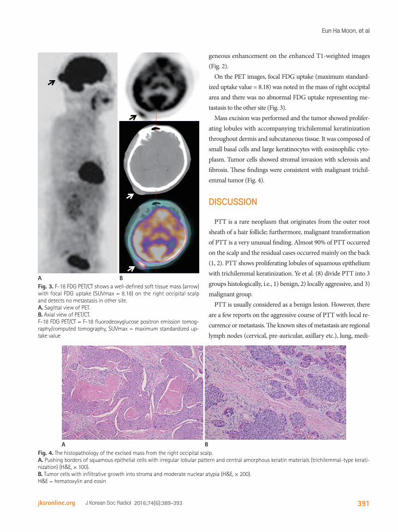
Mass excision was performed and the tumor showed prolifer-ating lobules with accompanying trichilemmal keratinization throughout dermis and subcutaneous tissue. It was composed of small basal cells and large keratinocytes with eosinophilic cyto-plasm. Tumor cells showed stromal invasion with sclerosis and fibrosis. These findings were consistent with malignant trichil-emmal tumor (Fig. 4).
DISCUSSION
PTT is a rare neoplasm that originates from the outer root sheath of a hair follicle; furthermore, malignant transformation of PTT is a very unusual finding. Almost 90% of PTT occurred on the scalp and the residual cases occurred mainly on the back (1, 2). PTT shows proliferating lobules of squamous epithelium with trichilemmal keratinization. Ye et al. (8) divide PTT into 3 groups histologically, i.e., 1) benign, 2) locally aggressive, and 3) malignant group.
PTT is usually considered as a benign lesion. However, there are a few reports on the aggressive course of PTT with local re-currence or metastasis. The known sites of metastasis are regional lymph nodes (cervical, pre-auricular, axillary etc.), lung, medi-
AFig. 3. F-18 FDG PET/CT shows a well-defined soft tissue mass (arrow) with focal FDG uptake (SUVmax = 8.18) on the right occipital scalp and detects no metastasis in other site. A. Sagittal view of PET. B. Axial view of PET/CT.F-18 FDG PET/CT = F-18 fluorodeoxyglucose positron emission tomog-raphy/computed tomography, SUVmax = maximum standardized up-take value
B
Fig. 4. The histopathology of the excised mass from the right occipital scalp. A. Pushing borders of squamous epithelial cells with irregular lobular pattern and central amorphous keratin materials (trichilemmal-type kerati-nization) (H&E, × 100). B. Tumor cells with infiltrative growth into stroma and moderate nuclear atypia (H&E, × 200).H&E = hematoxylin and eosin
A B

392
Recurrent Proliferating Trichilemmal Tumor with Malignant Change on the F-18 FDG PET/CT
jksronline.orgJ Korean Soc Radiol 2016;74(6):389-393
astinum and liver. Some authors also reported cases of the ma-lignant PTT with metastasis, which developed after recurrence (2-5). Therefore, malignant potential and possibility of metasta-sis cannot be excluded in patients with PTT, especially recurrent tumor, and appropriate evaluation is necessary.
F-18 FDG PET/CT plays an increasingly important role in the initial diagnosis, staging, and monitoring of treatment response in cancer patients. The PET/CT can acquire whole-body images and hence, is useful for the detection of regional and distant me-tastasis, as well as the primary tumor. Jung et al. (9) and Leyen-decker et al. (10) reported that malignant PTT and its metastatic lesion showed increased FDG uptake. However, there are few case reports on F-18 FDG PET finding of PTT.
We reported a case of recurrent PTT, which developed to ma-lignant change with increased FDG uptake. Although more cas-es of this tumor are required for assessing the usefulness of F-18 FDG PET/CT, we suggest that F-18 FDG PET/CT might be a helpful diagnostic tool for evaluation of PTT, especially recurrent tumor, because of its unpredictable tumor behavior that can pres-ent malignant change and metastasis with FDG avidity.
REFERENCES
1. Markal N, Kurtay A, Velidedeoglu H, Hücümenoglu S. Ma-
lignant transformation of a giant proliferating trichilemmal
tumor of the scalp: patient report and literature review.
Ann Plast Surg 1998;41:314-316
2. Park BS, Yang SG, Cho KH. Malignant proliferating trichil-
emmal tumor showing distant metastases. Am J Dermato-
pathol 1997;19:536-539
3. Weiss J, Heine M, Grimmel M, Jung EG. Malignant prolif-
erating trichilemmal cyst. J Am Acad Dermatol 1995;32(5
Pt 2):870-873
4. Amaral AL, Nascimento AG, Goellner JR. Proliferating pilar
(trichilemmal) cyst. Report of two cases, one with carcino-
matous transformation and one with distant metastases.
Arch Pathol Lab Med 1984;108:808-810
5. Noto G, Pravatà G, Aricò M. Proliferating tricholemmal cyst
should always be considered as a low-grade carcinoma.
Dermatology 1997;194:374-375
6. Lindholm P, Minn H, Leskinen-Kallio S, Bergman J, Ruots-
alainen U, Joensuu H. Influence of the blood glucose con-
centration on FDG uptake in cancer--a PET study. J Nucl
Med 1993;34:1-6
7. Smith TA. FDG uptake, tumour characteristics and response
to therapy: a review. Nucl Med Commun 1998;19:97-105
8. Ye J, Nappi O, Swanson PE, Patterson JW, Wick MR. Prolif-
erating pilar tumors: a clinicopathologic study of 76 cases
with a proposal for definition of benign and malignant
variants. Am J Clin Pathol 2004;122:566-574
9. Jung J, Cho SB, Yun M, Lee KH, Chung KY. Metastatic malig-
nant proliferating trichilemmal tumor detected by positron
emission tomography. Dermatol Surg 2003;29:872-874
10. Leyendecker P, de Cambourg G, Mahé A, Imperiale A,
Blondet C. 18F-FDG PET/CT findings in a patient with a pro-
liferating trichilemmal cyst. Clin Nucl Med 2015;40:598-599

393
Eun Ha Moon, et al
jksronline.org J Korean Soc Radiol 2016;74(6):389-393
악성변화를 보인 재발성 증식성 모낭종양의 F-18 Fluorodeoxyglucose Positron Emission Tomography/
Computed Tomography 스캔 소견
문은하1 · 김민우1* · 김영준2 · 유설봉3 · 남경화4
F-18 fluorodeoxyglucose positron emission tomography/computed tomography (이하 F-18 FDG PET/CT) 스캔은 현
재까지 다양한 종류의 암종의 진단, 치료반응평가 및 추적관찰에 이용되어 왔다. 증식성 모낭종양(proliferating trichilem-mal tumor)은 모낭 말단부위의 외근모초에서 기원하는 드문 종양으로, 이 종양은 예측하기 어려운 생물학적 또는 임상적
양상을 보이기 때문에 전이 및 재발에 대한 장기간의 추적관찰을 시행하는 것이 필요하겠다. 저자들은 F-18 FDG PET/
CT 스캔에서 FDG 섭취증가를 보인, 재발성 악성 증식성 모낭종양에 대한 드문 예가 있어 이를 보고하고자 한다.
서남대학교 의과대학 예수병원 1핵의학과, 2영상의학과, 3병리과, 4피부과