Recurrent intrahepatic cholestasis of pregnancy ...jgld.ro/2008/4/20.pdf · Recurrent intrahepatic...
9
J Gastrointestin Liver Dis December 2008 Vol.17 No 4, 479-487 LETTERS TO THE EDITOR Recurrent intrahepatic cholestasis of pregnancy: ursodeoxycholic acid should be the first line of treatment To the Editor, We read with interest the case report “Recurrent intrahepatic cholestasis of pregnancy’’ published in the September 2008 issue of JGLD [1]. We also treated a 28 year old female who had intrahepatic cholestasis of pregnancy (ICP) in two successive pregnancies at an interval of four years, with ursodeoxycholic acid (UDCA) with close monitoring of fetus and mother. She presented at 30 weeks and 32 weeks of gestation in the first and second pregnancy with generalized pruritus disturbing her night sleep. On examination, she had scratch marks on lower limbs, abdomen, palm and sole. Her blood pressure was normal with no pedal edema. Her serum bilirubin, aspartate aminotransferase, alanine aminotransferase, alkaline phosphatase were 1.2mg/dl, 64U/L, 56U/L, 430U/ L respectively in the first pregnancy. Her total bile acid level was 22 µmol/L and 15 µmol/L in the first and second pregnancy, respectively. Ultrasound of the abdomen was normal. The viral markers (HBsAg, IgM anti-HEV), and antinuclear antibodies were negative. None of her family members had suffered from a similar illness. A diagnosis of ICP was made and UDCA 15mg/kg/day in two divided doses was started. The pruritus score decreased from 4+ to 2+ on a visual analog scale in four weeks. Aspartate aminotransferase and alanine aminotransferase were 46U/L and 40U/L after four weeks in first pregnancy. The pregnancies were closely monitored and she delivered at term her first and second child. In the puerperium, the pruritus disappeared after four days. The differential diagnosis of ICP should also include skin disease, renal pruritis, Hodgkin’s disease, polycythemia rubra vera, hyperemesis gravidarum. Kondrackiene et al [2] have shown that early onset pruritus and serum bile acid level were independent predictors of preterm delivery in ICP. Our patient had late onset pruritus and serum bile acid less than 40µmol/L with the delivery of the fetus at term. Davies et al [3] also showed that UDCA was a safe and effective therapy in causing rapid clinical improvement and resolution of deranged biochemistry. At a dose of 600-1,000 mg per day, significant improvement in pruritus, bilirubin and transaminase levels was observed. This was confirmed in a randomized double blind placebo- controlled trial in 16 patients with ICP [4]. UDCA normalizes the increased cholic acid/chenodeoxycholic acid ratio, reduces plasma concentration and urinary excretion rates of sulfated steroid metabolites [5], restores impaired bile acid transport across the trophoblast in ICP and decreases the delivery of bile acids to the fetus. Kondrackiene et al [6] showed in an open, randomized parallel group study of 84 patients that UDCA induced more pronounced relief of pruritus, endogenous serum bile acid levels reduction than cholestyramine. Glantz et al [7] in a double blind placebo- controlled trial comparing UDCA and dexamethasone showed improvement in serum alanine aminotransferase and bilirubin levels irrespective of disease severity in the UDCA group. Dexamethasone did not alleviate pruritus. Therefore, we agree that the first line treatment should be UDCA. Pankaj Jain SMS Medical College, Jaipur, India References 1. Muresan D, Ona D, Cruciat G, Rotar I, Stamatian F. Recurrent intrahepatic cholestasis of pregnancy. A case report. J Gastrointestin Liver Dis 2008; 17: 323-325. 2. Kondrackiene J, Beuers U, Zalinkevicius R, Tauschel HD, Gintautas V, Kupcinskas L. Predictors of premature delivery in patients with intrahepatic cholestasis of pregnancy. World J Gastroentrol 2007; 13: 6226-6230. 3. Davies MH, da Silva RC, Jones SR, Weavers JB, Elias E. Fetal mortality associated with cholestasis of pregnancy and the potential benefit of therapy with ursodeoxycholic acid. Gut 1995; 37: 580- 584. 4. Diaferia A, Nicastri PL, Tartagni M, Loizzi P, Iacovizzi C, Di Leo A.
-
Upload
phungkhanh -
Category
Documents
-
view
229 -
download
0
Transcript of Recurrent intrahepatic cholestasis of pregnancy ...jgld.ro/2008/4/20.pdf · Recurrent intrahepatic...
J Gastrointestin Liver DisDecember 2008 Vol.17 No 4, 479-487
LETTERS TO THE EDITOR
Recurrent intrahepatic cholestasis of pregnancy: ursodeoxycholic acid should be the first line of treatmentTo the Editor,We read with interest the case report “Recurrent
intrahepatic cholestasis of pregnancy’’ published in the September 2008 issue of JGLD [1].
We also treated a 28 year old female who had intrahepatic cholestasis of pregnancy (ICP) in two successive pregnancies at an interval of four years, with ursodeoxycholic acid (UDCA) with close monitoring of fetus and mother. She presented at 30 weeks and 32 weeks of gestation in the firstand second pregnancy with generalized pruritus disturbing her night sleep. On examination, she had scratch marks on lower limbs, abdomen, palm and sole. Her blood pressure was normal with no pedal edema. Her serum bilirubin, aspartate aminotransferase, alanine aminotransferase, alkaline phosphatase were 1.2mg/dl, 64U/L, 56U/L, 430U/L respectively in the first pregnancy. Her total bile acidlevel was 22 µmol/L and 15 µmol/L in the first and secondpregnancy, respectively. Ultrasound of the abdomen was normal. The viral markers (HBsAg, IgM anti-HEV), and antinuclear antibodies were negative. None of her family members had suffered from a similar illness. A diagnosis of ICP was made and UDCA 15mg/kg/day in two divided doses was started. The pruritus score decreased from 4+ to 2+ on a visual analog scale in four weeks. Aspartate aminotransferase and alanine aminotransferase were 46U/L and 40U/L after four weeks in first pregnancy. The pregnancies were closelymonitored and she delivered at term her first and secondchild. In the puerperium, the pruritus disappeared after four days.
The differential diagnosis of ICP should also include skin disease, renal pruritis, Hodgkin’s disease, polycythemia rubra vera, hyperemesis gravidarum.
Kondrackiene et al [2] have shown that early onset
pruritus and serum bile acid level were independent predictors of preterm delivery in ICP. Our patient had late onset pruritus and serum bile acid less than 40µmol/L with the delivery of the fetus at term. Davies et al [3] also showed that UDCA was a safe and effective therapy in causing rapid clinical improvement and resolution of deranged biochemistry. At a dose of 600-1,000 mg per day, significant improvementin pruritus, bilirubin and transaminase levels was observed. This was confirmed in a randomized double blind placebo-controlled trial in 16 patients with ICP [4]. UDCA normalizes the increased cholic acid/chenodeoxycholic acid ratio, reduces plasma concentration and urinary excretion rates of sulfated steroid metabolites [5], restores impaired bile acid transport across the trophoblast in ICP and decreases the delivery of bile acids to the fetus. Kondrackiene et al [6] showed in an open, randomized parallel group study of 84 patients that UDCA induced more pronounced relief of pruritus, endogenous serum bile acid levels reduction than cholestyramine. Glantz et al [7] in a double blind placebo-controlled trial comparing UDCA and dexamethasone showed improvement in serum alanine aminotransferase and bilirubin levels irrespective of disease severity in the UDCA group. Dexamethasone did not alleviate pruritus. Therefore, we agree that the first line treatment should be UDCA.
Pankaj Jain SMS Medical College, Jaipur, India
References 1. Muresan D, Ona D, Cruciat G, Rotar I, Stamatian F. Recurrent
intrahepatic cholestasis of pregnancy. A case report. J Gastrointestin Liver Dis 2008; 17: 323-325.
2. Kondrackiene J, Beuers U, Zalinkevicius R, Tauschel HD, Gintautas V, Kupcinskas L. Predictors of premature delivery in patients with intrahepatic cholestasis of pregnancy. World J Gastroentrol 2007; 13: 6226-6230.
3. Davies MH, da Silva RC, Jones SR, Weavers JB, Elias E. Fetal mortality associated with cholestasis of pregnancy and the potential benefit of therapy with ursodeoxycholic acid. Gut 1995; 37: 580-584.
4. Diaferia A, Nicastri PL, Tartagni M, Loizzi P, Iacovizzi C, Di Leo A.
480 Letters to the editor
Ursodeoxycholic acid therapy in pregnant women with cholestasis. Int J Gynaecol Obstet 1996; 52: 133-140.
5. Meng LJ, Reyes H, Palma J, Hernandez I, Ribalta J, Sjovall J. Effects of ursodexycholic acid on conjugated bile acids and progesterone metabolites in serum and urine of patients with intrahepatic cholestasis of pregnancy. J Hepatol 1997; 27: 1029-1040.
6. Kondrackiene J, Beuers U, Kupcinskas L. Efficacy and safetyof ursodeoxycholic acid versus cholestyramine in intrahepatic cholestasis of pregnancy. Gastroenterology 2005; 129: 894-901.
7. Glantz A, Marschall HU, Lammert F, Mattsson LA. Intrahepatic cholestasis of pregnancy: a randomized controlled trial comparing dexamethasone and ursodeoxycholic acid. Hepatology 2005; 42: 1399-1405.
Factors associated with gastrointestinal symptom overlap and clustering in a predominant Malay Asian population
To the Editor,Gastrointestinal (GI) symptoms are common and most
are self-limiting [1]. Presence of GI symptom overlap or clustering is not uncommon and needs to be managed appropriately for symptom resolution. However, reports have mainly come from the West [2-4]. Reports on GI symptoms from the Asian region have mainly concentrated on predominantly Chinese populations. To date, there is no data on GI symptom overlap or clustering from this region.
We assessed the factors associated with GI symptom overlap and clustering in our local setting with predominant Malay Southeast Asian population. Relatives (n=607, male - 40.2%, ethnic - Malay, 85%, mean age 40.5±15.1 years) visiting the wards of a referral hospital were interviewed regarding GI symptoms experienced in the past 12 months. Explanations were given and consents obtained prior to the interview. Gastrointestinal symptom overlap was considered as presence of both upper and lower GI symptoms and clustering as the presence of three or more symptoms experienced at one time period (3 months). Demographic
data, background medical conditions, medications use, self-medication, GI symptoms (dyspepsia, heartburn, dysphagia, odynophagia, nausea, vomiting, loss of appetite, early satiety, non-epigastric abdominal pain, bloating, altered bowel habit and bleeding per rectum) and previous experience with endoscopy were collected. Psychosomatic symptoms of depression (anxiety, backache, depression, headache and insomnia) were also sought. Analysis was performed using the SPSS (Version 10.0, Chicago, Il USA) for analysis.
Overall, upper GI symptoms were reported by 33.4% (n=203) and lower GI symptoms by 20.1% (n=120) of subjects. GI symptom overlap and clustering were reported by 13.5% (mean of 3.2±1.3 symptoms) and 10.0% (mean of 3.9±1.1 symptoms) respectively. On univariate analysis, female gender, presence of co-morbid conditions, use of prescribed medications, previous endoscopies and presence of any psychosomatic symptoms of depression were predictive of both GI symptom overlap and clustering (all p <0.05). Ethnic group (Malays vs. others), age group (<40 yrs vs. ≥40 yrs), smoking status and use of self-prescribed medication were not significant factors (Table I).
In the multivariate analysis, comorbid conditions, previous endoscopies and presence of any psychosomatic symptoms of depression remained significant for GIsymptom overlap whereas female gender, co-morbid conditions, previous endoscopies and presence of any psychosomatic symptoms of depression remained significantfor GI symptom clustering. In total, 6.3% (n=38) and 1.8% (n=11) of subjects had previously undergone upper GI and lower GI endoscopy, respectively. None had any significantfindings.
This study showed that GI symptom overlap and clustering are not uncommon in our local setting. Overlap has been shown to be common particularly in functional GI disorders [2-4]. Management of overlapping upper and lower GI symptoms are different and require different strategies. Untreated, patients remain affected and this can lead to patients’ dissatisfaction, further consultations, increased health care utilizations and impaired quality of life. Factors
Table I. Factors associated with gastrointestinal symptoms overlap and clustering (Univariate analysis)Overlap (n=82) Clustering (n=61)
Factors n (%) OR 95% CI p value n (%) OR 95% CI p value
Age group (<40 vs. ≥40 yrs) 37(12.2):45(14.8) 0.801 0.502-1.278 0.350 25(8.3):36(11.8) 0.672 0.393-1.150 0.145
Gender (Male vs. Female) 22(9.0):60(16.5) 0.500 0.298-0.840 0.008 12(4.9):49(13.5) 0.333 0.173-0.640 0.001*
Ethnic groups (Malays vs. Others)
71(13.8):11(12.1) 1.160 0.589-2.286 0.667 56(10.9):5(5.5) 2.908 0.817-5.390 0.116
Comorbid conditions (Yes: No) 50(22.6):32(8.3) 3.235 2.002-5.226 <0.001* 40(18.1):21(5.5) 3.831 2.194-6.689 <0.001*
Medications (Yes: No) 41(21.6):41(9.8) 2.523 1.573-4.048 <0.001 34(17.9):27(6.5) 3.140 1.833-5.379 <0.001
Smoking (Yes: No) 14(14.6):68(13.3) 1.112 0.597-2.071 0.737 10(10.4):51(10.0) 1.047 0.511-2.141 0.901
Self prescribed medications (Yes: No)
24(16.3):58(12.6) 1.352 0.807-2.267 0.251 19(12.9):42(9.2) 1.474 0.828-2.624 0.186
Previous endoscopies (Yes: No) 18(40):64(11.4) 5.188 2.706-9.944 <0.001* 14(31.1):47(8.4) 3.140 1.833-5.379 <0.001*
Psychosomatic symptoms (Yes: No)
69(22.7):13(4.3) 6.550 3.534-12.140 <0.001* 54(17.8):7(2.3) 9.170 4.099-20.515 <0.001*
* Factors that remained significant on multivariate analysis
Letters to the editor 481
significantly associated with GI symptom overlap were thepresence of any co-morbid conditions, previous endoscopies and any psychosomatic symptom of depression. Numerous studies have shown increased prevalence of GI symptoms in patients with chronic non-psychiatric disorders [5-7]. Previous endoscopy experience is more an indication of the chronicity of GI complaints and is a consequence rather than a predictive factor. Psychosomatic symptoms of depression are common among patients with chronic GI symptoms, especially those with functional disorders [5]. Therefore, it is not an unexpected finding that they were predictive forsymptom overlap. Female gender was not significant in themultivariate analysis for symptom overlap. This may be due to the overall small sample size and the fewer lower GI symptoms examined in this study. Similarly, these factors were also significant for symptom clustering. However,female gender was also significant. Female gender hasbeen shown to be associated with a higher prevalence of GI symptoms and psychosomatic symptoms of depression [6]. Ethnic groups, age, smoking status and use of prescribed or self-prescribed medications were not significant factors.
Finally, only a minority of the subjects interviewed had any endoscopic investigations. This suggests that majority of the complaints were mild and self-limiting. The strong association with psychosomatic symptoms of depression also suggests strong functional elements.
In conclusion, GI symptom overlap and clustering is not uncommon in our local setting, a predominant Malay Southeast Asian population. Female gender, presence of co-morbidities, psychosomatic symptoms of depression and previous endoscopy were predictive factors.
Vui Heng Chong Raja Isteri Pengiran, Anak Saleha Hospital, Bandar
Seri Beagawan, Brunei Darussalam
References 1. Holtman G, Goebell H, Talley NJ. Dyspepsia in consulters and non-
consulters: prevalence, health seeking behaviour and risk factors. Eur J Gastroenterol Hepatol 1994; 5: 917-924.
2. Locke GR 3rd, Zinsmeister AR, Fett SL, Melton LJ 3rd, Talley NJ. Overlap of gastrointestinal symptom complexes in a US community. Neurogastroenterol Motil 2005; 17: 29-34.
3. Talley NJ, Dennis EH, Schettler-Duncan VA, Lacy BE, Olden KW, Crowell MD. Overlapping upper and lower gastrointestinal symptoms in irritable bowel syndrome patients with constipation or diarrhea. Am J Gastroenterol 2003; 98: 2454-2459.
4. Talley NJ, Boyce P, Jones M. Identification of distinct upper and lowergastrointestinal symptom groupings in an urban population. Gut. 1998; 42:690-5.
5. Stanghellini V. Relationship between upper gastrointestinal symptoms and lifestyle, psychosocial factors and comorbidity in the general population: results from the Domestic/International Gastroenterology Surveillance Study (DIGEST). Scand J Gastroenterol Suppl 1999; 231: 29-37.
6. Delgado-Aros S, Locke GR 3rd, Camilleri M, et al. Obesity is associated with increased risk of gastrointestinal symptoms: a population-based study.Am J Gastroenterol 2004; 99: 1801-1806.
7. Strid H, Simrén M, Johansson AC, Svedlund J, Samuelsson O, Björnsson ES. The prevalence of gastrointestinal symptoms in patients with chronic renal failure is increased and associated with impaired psychological general well-being. Nephrol Dial Transplant 2002; 17: 1434-1439
Concomitant colitis in a patient with Behcet’s disease
To the Editor,Behçet’s disease is a chronic, relapsing, occlusive
vasculitis of unknown etiology affecting multiple organ systems. The disease is named after a Turkish dermatologist Hulusi Behçet, who in 1937 recognized a clinical entity characterized by recurrent iridocyclitis with hypopyon, aphthous lesions in the mouth, and ulceration of the genitalia. Formal diagnostic criteria propose that recurrent oral ulceration (at least three times in one year) is an absolute criterion; any two of (a) recurrent genital ulceration, (b) uveitis or retinal vasculitis, (c) skin lesions, including erythema nodosum, or acneiform nodules, pseudofolliculitis or papulopustular lesions, or (d) a positive pathergy test (read at 24-48 hours) are also required to confirm the diagnosis[1].
Intestinal Behçet’s disease is a specific subtype of thisdisease characterized by gastrointestinal (GI) symptoms such as nausea, vomiting, diarrhea, and ulcerative colitis-like lesions. It has been reported to occur in 3–26% of patients with Behçet’s disease [2]. An overlap in clinical manifestations can occur between intestinal Behçet’s disease and other inflammatory bowel diseases including ulcerativecolitis and Crohn’s disease [3].
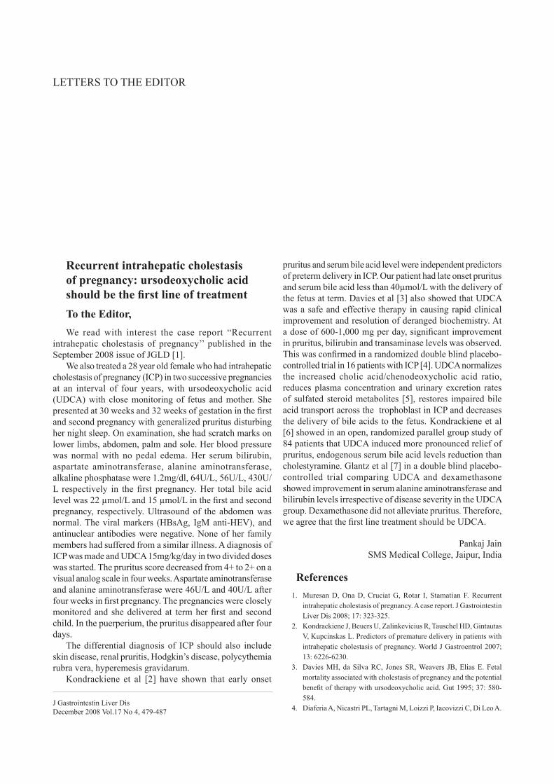
We describe here the case of a 27-year-old man, who was referred to our Ophtalmology Department with acute uveitis in March 2006. His past medical history revealed repeated episodes of uveitis in the last three years and a biopsy-proven diagnosis of ulcerative colitis one year ago. A regimen of 5-aminosalicylate (Salofalk, 2.0 g/day) was started for the treatment of colitis and bloody diarrhea recovered after 3 weeks of therapy. At the time of presentation, his chief complaint was blurred vision at distance and near, which had worsened over the past year. His best corrected visual acuity was 20/40 in the right eye and 20/70 in the left eye. Slit-lamp examination revealed +3 cells in the anterior chamber and vitreous cavity. Deep conjunctival injection, keratic precipitates, extensive posterior synechiae, iridotomy and posterior subcapsular cataract formation were also evident in both eyes (Fig.1). Intraocular pressures were within the normal limits. Funduscopic examination and fluorescein angiography were essentially normal, except thepresence of subtle retinal pigment epithelium changes in the macula of both eyes. The patient was started on subtenon injections of triamcinolone acetonide, topical dexamethasone and cycloplegics. The steroid drops were tapered and discontinued four months later. The visual acuity returned to 20/20 in the right and 20/40 in the left eye four months later.
482 Letters to the editor
His uveitis responded to local steroid therapy and the patient had no relapse at one year. At follow-up (April 2007), his best corrected visual acuity was recorded as 20/25 in the right and 20/200 in the left eye. Control fluorescein angiographyof the left eye was not possible. Posterior synechiotomy and cataract extraction with intraocular lens implantation procedures were offered to the patient for his left eye.
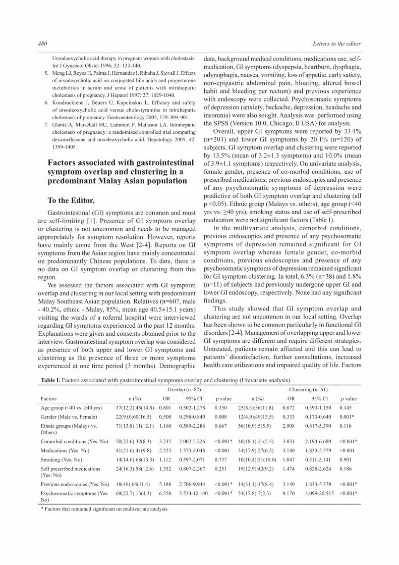
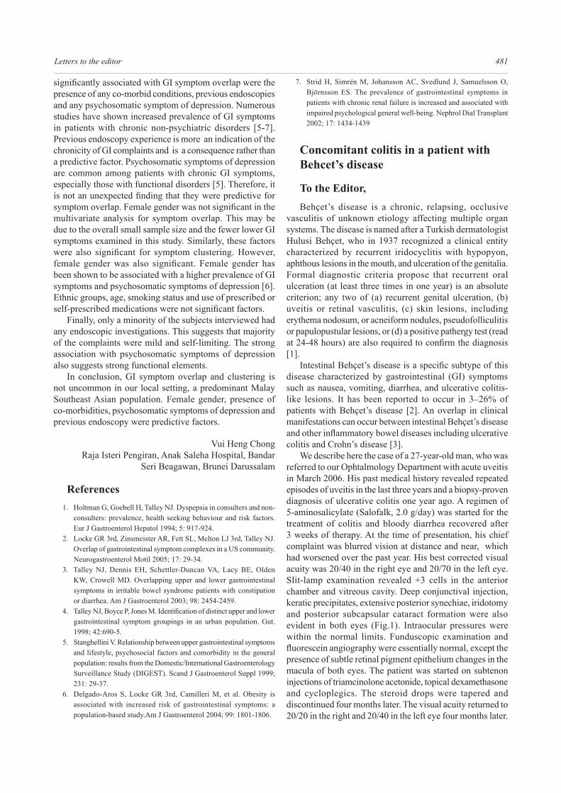
During his hospital stay (April 2007), the patient complained of backache and was referred to the Rheumatology Department for further investigation. Physical examination was unremarkable except for two minor aphthae in the oral mucosa. Schober’s test (lumbar anterior flexion) wasnormal, and joint mobility was preserved. MR imaging of the sacroiliac joint, however, disclosed destruction of adjacent articular tissues and subchondral irregularities compatible with sacroileitis (Fig.2). Laboratory findings includeda hemoglobin level of 13 g/dl and a C-reactive protein concentration of 2.1 mg/dl. The patient tested negative for HLA-B27 but was positive for HLA-B51. Skin pathergy test was positive. A punch biopsy (skin) was obtained which revealed the presence of inflammatory cells (mainlyneutrophils) up to the keratin layer. Leukocyte infiltrationaround hair follicles and fibrin extravasation were alsoevident (Fig.3).
The patient underwent repeated colonoscopy for re-evaluation of colitis. Endoscopy findings showed anulceration of 5 mm in size at 7-10 cm from the anal verge.
Fig 1. Slit-lamp photograph of the left eye showing extensive posterior synechiae and an iridotomy site (arrow head).
Fig 2. Contrast enhanced FS coronal T1-image showing subchondral bone destruction in both sacroiliac joints and contrast material staining of the joint space (arrow).
Fig 3. Punch biopsy (skin) reveals superficialand deep perivascular neutrophilic infiltrate.
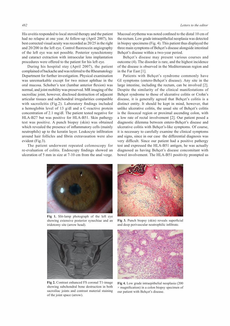
Fig 4. Low grade intraepithelial neoplasia (200 × magnification) in a colon biopsy specimen ofour patient with Behçet’s disease.
Mucosal erythema was noted confined to the distal 10 cm ofthe rectum. Low grade intraepithelial neoplasia was detected in biopsy specimens (Fig. 4). This patient thus displayed the three main symptoms of Behçet’s disease alongside intestinal Behçet’s disease within a two-year period.
Behçet’s disease may present various courses and outcome (4). The disorder is rare, and the highest incidence of the disease is observed in the Mediterranean region and in the Far East [1].
Patients with Behçet’s syndrome commonly have GI symptoms (entero-Behçet’s disease). Any site in the large intestine, including the rectum, can be involved [2]. Despite the similarity of the clinical manifestations of Behçet syndrome to those of ulcerative colitis or Crohn’s disease, it is generally agreed that Behçet’s colitis is a distinct entity. It should be kept in mind, however, that unlike ulcerative colitis, the usual site of Behçet’s colitis is the ileocecal region or proximal ascending colon, with a low rate of rectal involvement [2]. Our patient posed a diagnostic dilemma between entero-Behçet’s disease and ulcerative colitis with Behçet’s-like symptoms. Of course, it is necessary to carefully examine the clinical symptoms and signs, since in our case the differential diagnosis was very difficult. Since our patient had a positive pathergytest and expressed the HLA-B51 antigen, he was actually diagnosed as having Behçet’s disease concomitant with bowel involvement. The HLA-B51 positivity prompted us
Letters to the editor 483
the very unusual diagnosis of inflammatory bowel disease(IBD) associated with Behçet’s disease. Otherwise, HLA-B27 positivity would have suggested classification of ourcase as a HLAB-27 positive IBD patient [2]. Moreover, the skin pathergy reaction, consisting of an exaggerated non-specific inflammatory response to a minor trauma, is almostunique to Behçet’s disease. It occurs in 60% of patients and is more frequently observed in male patients or in individuals with active disease [1].
Notably, our case of entero-Behçet’s disease developed in his clinical course an early colon cancer as confirmed bypathologic examination. Although an association between Behçet’s disease and malignancy is not unexpected, to our knowledge only a few cases of colon cancer have been reported so far in this clinical entity [5, 6]. Moreover, the considerable overlap between the systemic features of Behçet’s disease and IBD needs further investigation to increase our understanding of pathogenesis and to improve therapy. Accordingly, the pathogenesis of bowel involvement in Behçet’s disease is not completely understood, but it is probably mediated by some factors including immune dysregulation, inflammatory mediators and infectious agents[2]. We conclude that bowel involvement may be present in Behçet’s disease. With a thorough clinical evaluation and the determination of the HLA-B51 antigen, the clinician should be able to make a differential diagnosis between Behçet’s disease and inflammatory bowel disease.
Yusuf Yilmaz1, Enver Dolar2
1) Department of Internal Medicine; 2) Department of Gastroenterology. Uludag University School of Medicine,
Uludag University Hospital, Bursa, Turkey.
References 1. Sakane T, Takeno M, Suzuki N, Inaba G. Behcet’s disease. N Engl
J Med 1999; 341: 1284-1291. 2. Lee CR, Kim WH, Cho YS, et al. Colonoscopic findings in intestinal
Behçet’s disease. Inflamm Bowel Dis 2001; 7: 243-249. 3. Kobashigawa T, Okamoto H, Kato J, et al. Ulcerative colitis followed
by the development of Behçet’s disease. Intern Med 2004; 43: 243-247.
4. Seyahi E, Melikoglu M, Yazici H. Clinical feautures and diagnosis of Behçet’s syndrome. Int J Adv Rheumatol 2007; 5: 8-13.
5. Kaklamani VG, Tzonou A, Kaklamanis PG. Behçet’s disease associated with malignancies. Report of two cases and review of the literature. Clin Exp Rheumatol 2005; 23: S35-41.
6. Cengiz M, Altundag MK, Zorlu AF, Güllü IH, Ozyar E, Atahan IL. Malignancy in Behçet’s disease: a report of 13 cases and a review of the literature. Clin Rheumatol 2001; 20: 239-244.
Is liver cirrhosis a risk factor for breast cancer in men?
To the Editor,Breast cancer is rare in men. It accounts for less than 1%
of all cases of cancer in men. It resembles breast cancer in women in that it is hormonally driven. A family history of
breast cancer, increasing age, exposure to radiation, history of benign breast lesions, patients with undescended testes, Kleinefelter’s syndrome and infertility are associated with an increased risk of breast cancer in men. We report a case of carcinoma of the right breast in an individual with liver cirrhosis.
A 65-year-old male, with known ethanol related cirrhosis of the liver, on therapy with spironolactone presented with a history of vague abdominal discomfort in the right upper quadrant for 6 months duration. On examination of the abdomen the liver was enlarged, firm and nodular with tenseascites. He had bilateral gynecomastia. The areola of the right breast felt hard, while the left was firm. Bony hard swellingwas felt over the fourth rib on the left side. There was no regional lymphadenopathy. Clinical diagnosis of carcinoma of the breast with rib secondaries was considered in a patient with cirrhosis of the liver.
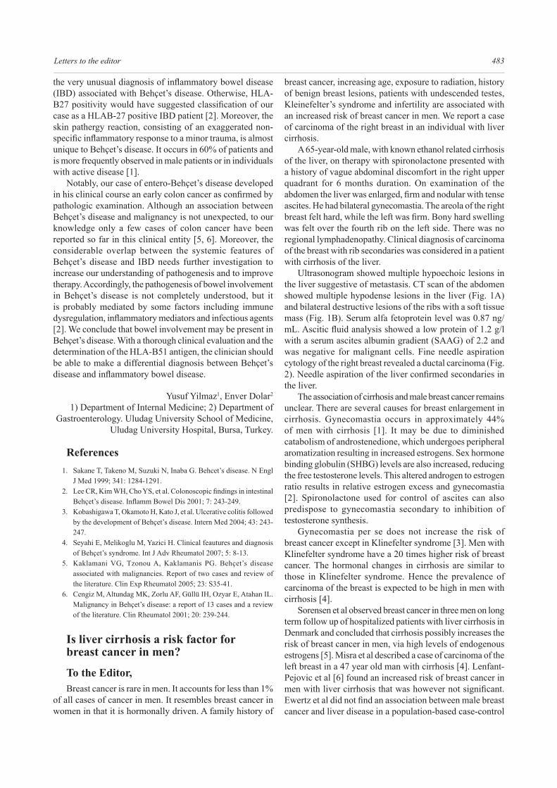
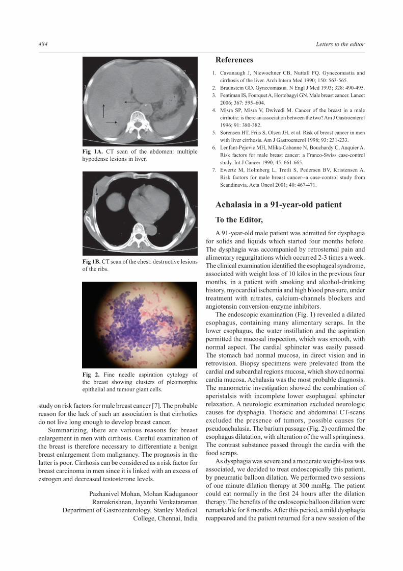
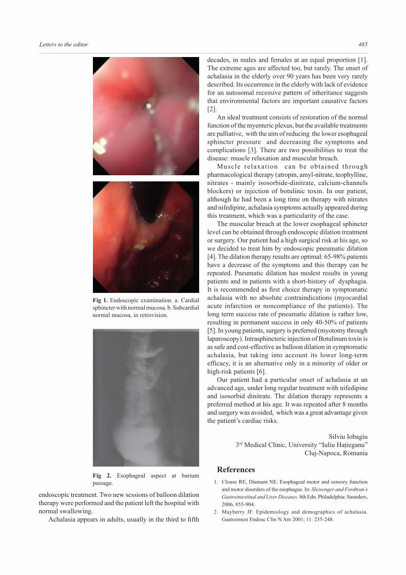
Ultrasonogram showed multiple hypoechoic lesions in the liver suggestive of metastasis. CT scan of the abdomen showed multiple hypodense lesions in the liver (Fig. 1A) and bilateral destructive lesions of the ribs with a soft tissue mass (Fig. 1B). Serum alfa fetoprotein level was 0.87 ng/mL. Ascitic fluid analysis showed a low protein of 1.2 g/lwith a serum ascites albumin gradient (SAAG) of 2.2 and was negative for malignant cells. Fine needle aspiration cytology of the right breast revealed a ductal carcinoma (Fig. 2). Needle aspiration of the liver confirmed secondaries inthe liver.
The association of cirrhosis and male breast cancer remains unclear. There are several causes for breast enlargement in cirrhosis. Gynecomastia occurs in approximately 44% of men with cirrhosis [1]. It may be due to diminished catabolism of androstenedione, which undergoes peripheral aromatization resulting in increased estrogens. Sex hormone binding globulin (SHBG) levels are also increased, reducing the free testosterone levels. This altered androgen to estrogen ratio results in relative estrogen excess and gynecomastia [2]. Spironolactone used for control of ascites can also predispose to gynecomastia secondary to inhibition of testosterone synthesis.
Gynecomastia per se does not increase the risk of breast cancer except in Klinefelter syndrome [3]. Men with Klinefelter syndrome have a 20 times higher risk of breast cancer. The hormonal changes in cirrhosis are similar to those in Klinefelter syndrome. Hence the prevalence of carcinoma of the breast is expected to be high in men with cirrhosis [4].
Sorensen et al observed breast cancer in three men on long term follow up of hospitalized patients with liver cirrhosis in Denmark and concluded that cirrhosis possibly increases the risk of breast cancer in men, via high levels of endogenous estrogens [5]. Misra et al described a case of carcinoma of the left breast in a 47 year old man with cirrhosis [4]. Lenfant-Pejovic et al [6] found an increased risk of breast cancer in men with liver cirrhosis that was however not significant.Ewertz et al did not find an association between male breastcancer and liver disease in a population-based case-control
484 Letters to the editor
study on risk factors for male breast cancer [7]. The probable reason for the lack of such an association is that cirrhotics do not live long enough to develop breast cancer.
Summarizing, there are various reasons for breast enlargement in men with cirrhosis. Careful examination of the breast is therefore necessary to differentiate a benign breast enlargement from malignancy. The prognosis in the latter is poor. Cirrhosis can be considered as a risk factor for breast carcinoma in men since it is linked with an excess of estrogen and decreased testosterone levels.
Pazhanivel Mohan, Mohan Kaduganoor Ramakrishnan, Jayanthi Venkataraman
Department of Gastroenterology, Stanley Medical College, Chennai, India
References 1. Cavanaugh J, Niewoehner CB, Nuttall FQ. Gynecomastia and
cirrhosis of the liver. Arch Intern Med 1990; 150: 563-565. 2. Braunstein GD. Gynecomastia. N Engl J Med 1993; 328: 490-495. 3. Fentiman IS, Fourquet A, Hortobagyi GN. Male breast cancer. Lancet
2006; 367: 595–604. 4. Misra SP, Misra V, Dwivedi M. Cancer of the breast in a male
cirrhotic: is there an association between the two? Am J Gastroenterol 1996; 91: 380-382.
5. Sorensen HT, Friis S, Olsen JH, et al. Risk of breast cancer in men with liver cirrhosis. Am J Gastroenterol 1998; 93: 231-233.
6. Lenfant-Pejovic MH, Mlika-Cabanne N, Bouchardy C, Auquier A. Risk factors for male breast cancer: a Franco-Swiss case-control study. Int J Cancer 1990; 45: 661-665.
7. Ewertz M, Holmberg L, Tretli S, Pedersen BV, Kristensen A. Risk factors for male breast cancer--a case-control study from Scandinavia. Acta Oncol 2001; 40: 467-471.
Achalasia in a 91-year-old patient
To the Editor,A 91-year-old male patient was admitted for dysphagia
for solids and liquids which started four months before. The dysphagia was accompanied by retrosternal pain and alimentary regurgitations which occurred 2-3 times a week. The clinical examination identified the esophageal syndrome,associated with weight loss of 10 kilos in the previous four months, in a patient with smoking and alcohol-drinking history, myocardial ischemia and high blood pressure, under treatment with nitrates, calcium-channels blockers and angiotensin conversion-enzyme inhibitors.
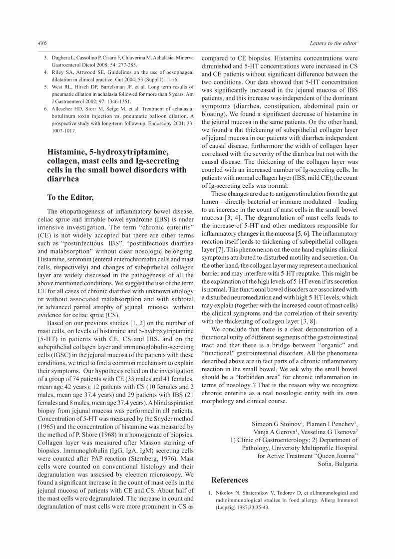
The endoscopic examination (Fig. 1) revealed a dilated esophagus, containing many alimentary scraps. In the lower esophagus, the water instillation and the aspiration permitted the mucosal inspection, which was smooth, with normal aspect. The cardial sphincter was easily passed. The stomach had normal mucosa, in direct vision and in retrovision. Biopsy specimens were prelevated from the cardial and subcardial regions mucosa, which showed normal cardia mucosa. Achalasia was the most probable diagnosis. The manometric investigation showed the combination of aperistalsis with incomplete lower esophageal sphincter relaxation. A neurologic examination excluded neurologic causes for dysphagia. Thoracic and abdominal CT-scans excluded the presence of tumors, possible causes for pseudoachalasia. The barium passage (Fig. 2) confirmed theesophagus dilatation, with alteration of the wall springiness. The contrast substance passed through the cardia with the food scraps.
As dysphagia was severe and a moderate weight-loss was associated, we decided to treat endoscopically this patient, by pneumatic balloon dilation. We performed two sessions of one minute dilation therapy at 300 mmHg. The patient could eat normally in the first 24 hours after the dilationtherapy. The benefits of the endoscopic balloon dilation wereremarkable for 8 months. After this period, a mild dysphagia reappeared and the patient returned for a new session of the
Fig 1A. CT scan of the abdomen: multiple hypodense lesions in liver.
Fig 1B. CT scan of the chest: destructive lesions of the ribs.
Fig 2. Fine needle aspiration cytology of the breast showing clusters of pleomorphic epithelial and tumour giant cells.
Letters to the editor 485
endoscopic treatment. Two new sessions of balloon dilation therapy were performed and the patient left the hospital with normal swallowing.
Achalasia appears in adults, usually in the third to fifth
decades, in males and females at an equal proportion [1]. The extreme ages are affected too, but rarely. The onset of achalasia in the elderly over 90 years has been very rarely described. Its occurrence in the elderly with lack of evidence for an autosomal recessive pattern of inheritance suggests that environmental factors are important causative factors [2].
An ideal treatment consists of restoration of the normal function of the myenteric plexus, but the available treatments are palliative, with the aim of reducing the lower esophageal sphincter pressure and decreasing the symptoms and complications [3]. There are two possibilities to treat the disease: muscle relaxation and muscular breach.
Muscle relaxat ion can be obtained through pharmacological therapy (atropin, amyl-nitrate, teophylline, nitrates - mainly isosorbide-dinitrate, calcium-channels blockers) or injection of botulinic toxin. In our patient, although he had been a long time on therapy with nitrates and nifedipine, achalasia symptoms actually appeared during this treatment, which was a particularity of the case.
The muscular breach at the lower esophageal sphincter level can be obtained through endoscopic dilation treatment or surgery. Our patient had a high surgical risk at his age, so we decided to treat him by endoscopic pneumatic dilation [4]. The dilation therapy results are optimal: 65-98% patients have a decrease of the symptoms and this therapy can be repeated. Pneumatic dilation has modest results in young patients and in patients with a short-history of dysphagia. It is recommended as first choice therapy in symptomaticachalasia with no absolute contraindications (myocardial acute infarction or noncompliance of the patients). The long term success rate of pneumatic dilation is rather low, resulting in permanent success in only 40-50% of patients [5]. In young patients, surgery is preferred (myotomy through laparoscopy). Intrasphincteric injection of Botulinum toxin is as safe and cost-effective as balloon dilation in symptomatic achalasia, but taking into account its lower long-term efficacy, it is an alternative only in a minority of older orhigh-risk patients [6].
Our patient had a particular onset of achalasia at an advanced age, under long regular treatment with nifedipine and isosorbid dinitrate. The dilation therapy represents a preferred method at his age. It was repeated after 8 months and surgery was avoided, which was a great advantage given the patient’s cardiac risks.
Silviu Iobagiu3rd Medical Clinic, University “Iuliu Haţieganu”
Cluj-Napoca, Romania
References 1. Clouse RE, Diamant NE. Esophageal motor and sensory function
and motor disorders of the esophagus. In: Sleisenger and Fordtran’s Gastrointestinal and Liver Diseases. 8th Edn. Philadelphia: Saunders, 2006, 855-904.
2. Mayberry JF. Epidemiology and demographics of achalasia. Gastrointest Endosc Clin N Am 2001; 11: 235-248.
Fig 1. Endoscopic examination. a. Cardial sphincter with normal mucosa. b. Subcardial normal mucosa, in retrovision.
Fig 2. Esophageal aspect at barium passage.
486 Letters to the editor
3. Dughera L, Cassolino P, Cisarò F, Chiaverina M. Achalasia. Minerva Gastroenterol Dietol 2008; 54: 277-285.
4. Riley SA, Attwood SE. Guidelines on the use of oesophageal dilatation in clinical practice. Gut 2004; 53 (Suppl I): i1–i6.
5. West RL, Hirsch DP, Bartelsman JF, et al. Long term results of pneumatic dilation in achalasia followed for more than 5 years. Am J Gastroenterol 2002; 97: 1346-1351.
6. Allescher HD, Storr M, Seige M, et al. Treatment of achalasia: botulinum toxin injection vs. pneumatic balloon dilation. A prospective study with long-term follow-up. Endoscopy 2001; 33: 1007-1017.
Histamine, 5-hydroxytriptamine, collagen, mast cells and Ig-secreting cells in the small bowel disorders with diarrhea
To the Editor,
The etiopathogenesis of inflammatory bowel disease,celiac sprue and irritable bowel syndrome (IBS) is under intensive investigation. The term “chronic enteritis” (CE) is not widely accepted but there are other terms such as “postinfectious IBS”, “postinfectious diarrhea and malabsorption” without clear nosologic belonging. Histamine, serotonin (enteral enterochromafin cells and mastcells, respectively) and changes of subepithelial collagen layer are widely discussed in the pathogenesis of all the above mentioned conditions. We suggest the use of the term CE for all cases of chronic diarrhea with unknown etiology or without associated malabsorption and with subtotal or advanced partial atrophy of jejunal mucosa without evidence for celiac sprue (CS).
Based on our previous studies [1, 2] on the number of mast cells, on levels of histamine and 5-hydroxytriptamine (5-HT) in patients with CE, CS and IBS, and on the subepithelial collagen layer and immunoglobulin-secreting cells (IGSC) in the jejunal mucosa of the patients with these conditions, we tried to find a common mechanism to explaintheir symptoms. Our hypothesis relied on the investigation of a group of 74 patients with CE (33 males and 41 females, mean age 42 years); 12 patients with CS (10 females and 2 males, mean age 37.4 years) and 29 patients with IBS (21 females and 8 males, mean age 37.4 years). A blind aspiration biopsy from jejunal mucosa was performed in all patients. Concentration of 5-HT was measured by the Snyder method (1965) and the concentration of histamine was measured by the method of P. Shore (1968) in a homogenate of biopsies. Collagen layer was measured after Masson staining of biopsies. Immunoglobulin (IgG, IgA, IgM) secreting cells were counted after PAP reaction (Sternberg, 1976). Mast cells were counted on conventional histology and their degranulation was assessed by electron microscopy. We found a significant increase in the count of mast cells in thejejunal mucosa of patients with CE and CS. About half of the mast cells were degranulated. The increase in count and degranulation of mast cells were more prominent in CS as
compared to CE biopsies. Histamine concentrations were diminished and 5-HT concentrations were increased in CS and CE patients without significant difference between thetwo conditions. Our data showed that 5-HT concentration was significantly increased in the jejunal mucosa of IBSpatients, and this increase was independent of the dominant symptoms (diarrhea, constipation, abdominal pain or bloating). We found a significant decrease of histamine inthe jejunal mucosa in the same patients. On the other hand, we found a flat thickening of subepithelial collagen layerof jejunal mucosa in our patients with diarrhea independent of causal disease, furthermore the width of collagen layer correlated with the severity of the diarrhea but not with the causal disease. The thickening of the collagen layer was coupled with an increased number of Ig-secreting cells. In patients with normal collagen layer (IBS, mild CE), the count of Ig-secreting cells was normal.
These changes are due to antigen stimulation from the gut lumen – directly bacterial or immune modulated – leading to an increase in the count of mast cells in the small bowel mucosa [3, 4]. The degranulation of mast cells leads to the increase of 5-HT and other mediators responsible for inflammatory changes in the mucosa [5, 6]. The inflammatoryreaction itself leads to thickening of subepithelial collagen layer [7]. This phenomenon on the one hand explains clinical symptoms attributed to disturbed motility and secretion. On the other hand, the collagen layer may represent a mechanical barrier and may interfere with 5-HT reuptake. This might be the explanation of the high levels of 5-HT even if its secretion is normal. The functional bowel disorders are associated with a disturbed neuromediation and with high 5-HT levels, which may explain (together with the increased count of mast cells) the clinical symptoms and the correlation of their severity with the thickening of collagen layer [3, 8].
We conclude that there is a clear demonstration of a functional unity of different segments of the gastrointestinal tract and that there is a bridge between “organic” and “functional” gastrointestinal disorders. All the phenomena described above are in fact parts of a chronic inflammatoryreaction in the small bowel. We ask why the small bowel should be a “forbidden area” for chronic inflammation interms of nosology ? That is the reason why we recognize chronic enteritis as a real nosologic entity with its own morphology and clinical course.
Simeon G Stoinov1, Plamen I Penchev1, Vanja A Gerova1, Vesselina G Tsenova2
1) Clinic of Gastroenterology; 2) Department of Pathology, University Multiprofile Hospital
for Active Treatment “Queen Joanna” Sofia, Bulgaria
References 1. Nikolov N, Shaternikov V, Todorov D, et al.Immunological and
radioimmunological studies in food allergy. Allerg Immunol (Leipzig) 1987;33:35-43.

Letters to the editor 487
2. Drumcheva M, Todorov D, Stoinov S, Nikolov N, Boneva M. [Mastocytes in the human intestinal mucosa]. Vutr Boles 1986;25:14-17
3. Talley MJ, Butterfield JH. Mast cell infiltration and degranulation incolonic mucosa in the irritable bowel syndrome. Am J Gastroenterol 1996; 91: 1675-1676.
4. Kirsch R, Riddell RH. Histopathological alterations in irritable bowel syndrome. Mod Pathol 2006; 19: 1638-1645.
5. Bacci S, Faussone-Pellegrini S, Mayer B, Romagnoly P. Distribution
of mast cells in human ileocecal region. Dig Dis Sci 1995; 40: 357-365.
6. Von der Ohe MR, Hanson RB, Camilleri M. Serotonergic mediation of postprandial colonic tonic and phasic responces in humans. Gut 1994; 35: 536-541.
7. Hansen MB. Neurohumoral control of gastrointestinal motility. Physiol Res 2003; 52: 1-30.
8. Zins BJ, Sandborn WJ, Tremaine WJ. Collagenous and lymphocytic colitis: subject review and therapeutic alternatives. Am J Gastroenterol 1995; 90: 1394-1400.