Recurrence of Pancreatic Pseudocyst following Endoscopic · PDF file ·...
8
Recurrence of Pancreatic Pseudocyst following Endoscopic Ultrasonography-Guided Drainage Corresponding author: Nobuhiko Fukuba, MD, Ph.D Department of Internal Medicine, Izumo City General Medical Center, 613 Nadabun-cho, Izumo, Shimane 691-0003, Japan Tel: +81-853-63-5111 Fax: +81-853-63-4228 E-mail: [email protected] Ahmed SELIM 1,2) , Nobuhiko FUKUBA 1) , Ichiro MORIYAMA 3) , Hiroki SONOYAMA 1) , Chika FUKUYAMA 1) , Shohei SUMI 1) , Yoshiyuki MISHIMA 1) , Shunji ISHIHARA 1) , Yoshikazu KINOSHITA 1) 1) Department of Internal Medicine II, Shimane University Faculty of Medicine, Izumo, 693-8501, Japan 2) Department of Gastroenterology and hepatology, Tanta University, Faculty of Medicine, Egypt 3) Cancer Center, Shimane University Hospital, Izumo, 693-8501, Japan (Received May 22, 2017; Accepted October 13, 2017) INTRODUCTION According to the revised Atlanta criteria for in- flammatory pancreatic fluid collection, a pancreatic pseudocyst is defined as an encapsulated collection of fluid with a well-defined inflammatory wall usu- ally outside the pancreas, with minimal or no ne- crosis. Usually, more than 4 weeks after onset of interstitial edematous pancreatitis is required for this pseudocyst to mature [1] . The prevalence of pancreatic pseudocyst in acute pancreatitis cases has been reported to range from 6% to 18.5% [2, 3] , while that in cases of chronic pancreatitis ranges from 20% to 40% [4] . A pan- creatic pseudocyst most commonly arises in patients with alcoholic chronic pancreatitis (70% to 78%) [5] , while the second most common cause is idio- pathic chronic pancreatitis (6% to 16%) , followed by biliary pancreatitis (6% to 8%) . This type of pseudocyst may be asymptomatic or can present a variety of symptoms, such as abdominal pain, sati- ety, upper gastrointestinal bleeding, infection, nau- sea, and vomiting that necessitates drainage [6, 7] . Traditionally, a pancreatic pseudocyst is managed by open surgical internal drainage. However, with con- tinued medical technology development, less inva- sive options including percutaneous, endoscopic, and laparoscopic drainage are now possible [8] . Hereby we described a case of pancreatic pseudocyst recur- rence following endoscopic ultrasonography-guided drainage. CASE STUDY We present here a 71-year-old female who was A 71-year-old female came to our hospital with upper abdominal pain, fever, and jaundice, and was diagnosed with severe acute biliary pancreatitis. Upon admission, the patient underwent emergency endoscopic retrograde cholangiopancreatography and endoscopic sphincterotomy procedures, as well as endoscopic retrograde biliary drainage. Although pseudocyst formation was observed, no symptoms were presented and she was discharged. One month after, she was admitted again due to pain caused by pseudocyst enlargement and the symptoms persisted following conservative treatment. Although we per- formed endoscopic ultrasonography-guided drainage for the pancreatic pseudocyst, abdominal pain con- tinued and abdominal computed tomography revealed that the pseudocyst was again enlarged. Since insuf- ficient drainage seemed to be the cause, additionally balloon dilatation and placement of plastic stents for the fistulae were performed. There are no guidelines available for selection of drainage period or number of stents in pseudocyst cases, and additional stud- ies regarding management of affected patients are needed. Keywords: EUS, drainage, pancreatic pseudocyst, re- currence 73 Shimane J. Med. Sci., Vol.34 pp.73-80, 2017
Transcript of Recurrence of Pancreatic Pseudocyst following Endoscopic · PDF file ·...

Recurrence of Pancreatic Pseudocyst following Endoscopic Ultrasonography-Guided Drainage
Corresponding author: Nobuhiko Fukuba, MD, Ph.DDepartment of Internal Medicine, Izumo City General Medical Center, 613 Nadabun-cho, Izumo, Shimane 691-0003, JapanTel: +81-853-63-5111Fax: +81-853-63-4228E-mail: [email protected]
Ahmed SELIM1,2), Nobuhiko FUKUBA1), Ichiro MORIYAMA3), Hiroki SONOYAMA1), Chika FUKUYAMA1), Shohei SUMI1), Yoshiyuki MISHIMA1), Shunji ISHIHARA1), Yoshikazu KINOSHITA1)
1) Department of Internal Medicine II, Shimane University Faculty of Medicine, Izumo, 693-8501, Japan 2) Department of Gastroenterology and hepatology, Tanta University, Faculty of Medicine, Egypt3) Cancer Center, Shimane University Hospital, Izumo, 693-8501, Japan(Received May 22, 2017; Accepted October 13, 2017)
INTRODUCTION
According to the revised Atlanta criteria for in-flammatory pancreatic fluid collection, a pancreatic pseudocyst is defined as an encapsulated collection of fluid with a well-defined inflammatory wall usu-ally outside the pancreas, with minimal or no ne-crosis. Usually, more than 4 weeks after onset of interstitial edematous pancreatitis is required for this pseudocyst to mature [1].
The prevalence of pancreatic pseudocyst in acute pancreatitis cases has been reported to range from 6% to 18.5% [2, 3], while that in cases of chronic pancreatitis ranges from 20% to 40% [4]. A pan-creatic pseudocyst most commonly arises in patients with alcoholic chronic pancreatitis (70% to 78%) [5], while the second most common cause is idio-pathic chronic pancreatitis (6% to 16%), followed by biliary pancreatitis (6% to 8%). This type of pseudocyst may be asymptomatic or can present a variety of symptoms, such as abdominal pain, sati-ety, upper gastrointestinal bleeding, infection, nau-sea, and vomiting that necessitates drainage [6, 7]. Traditionally, a pancreatic pseudocyst is managed by open surgical internal drainage. However, with con-tinued medical technology development, less inva-sive options including percutaneous, endoscopic, and laparoscopic drainage are now possible [8]. Hereby we described a case of pancreatic pseudocyst recur-rence following endoscopic ultrasonography-guided drainage.
CASE STUDY
We present here a 71-year-old female who was
A 71-year-old female came to our hospital with upper abdominal pain, fever, and jaundice, and was diagnosed with severe acute biliary pancreatitis. Upon admission, the patient underwent emergency endoscopic retrograde cholangiopancreatography and endoscopic sphincterotomy procedures, as well as endoscopic retrograde biliary drainage. Although pseudocyst formation was observed, no symptoms were presented and she was discharged. One month after, she was admitted again due to pain caused by pseudocyst enlargement and the symptoms persisted following conservative treatment. Although we per-formed endoscopic ultrasonography-guided drainage for the pancreatic pseudocyst, abdominal pain con-tinued and abdominal computed tomography revealed that the pseudocyst was again enlarged. Since insuf-ficient drainage seemed to be the cause, additionally balloon dilatation and placement of plastic stents for the fistulae were performed. There are no guidelines available for selection of drainage period or number of stents in pseudocyst cases, and additional stud-ies regarding management of affected patients are needed.
Keywords: EUS, drainage, pancreatic pseudocyst, re-currence
73Shimane J. Med. Sci., Vol.34 pp.73-80, 2017
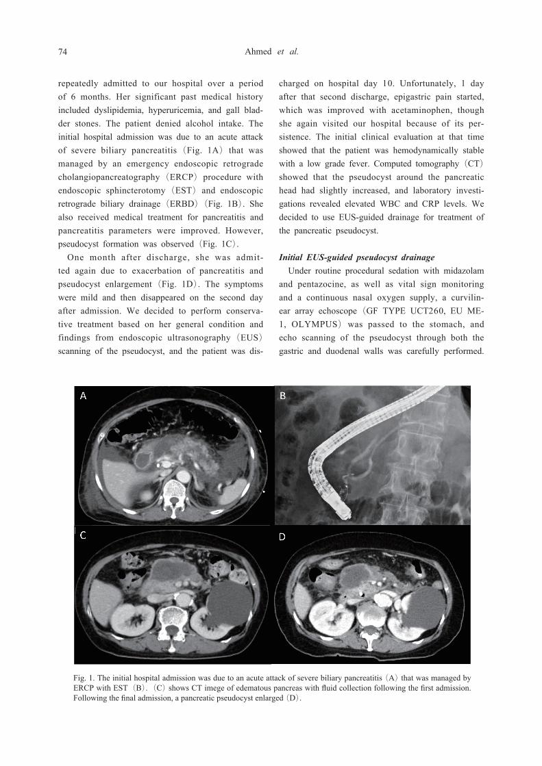
repeatedly admitted to our hospital over a period of 6 months. Her significant past medical history included dyslipidemia, hyperuricemia, and gall blad-der stones. The patient denied alcohol intake. The initial hospital admission was due to an acute attack of severe biliary pancreatitis (Fig. 1A) that was managed by an emergency endoscopic retrograde cholangiopancreatography (ERCP) procedure with endoscopic sphincterotomy (EST) and endoscopic retrograde biliary drainage (ERBD) (Fig. 1B). She also received medical treatment for pancreatitis and pancreatitis parameters were improved. However, pseudocyst formation was observed (Fig. 1C).
One month after discharge, she was admit-ted again due to exacerbation of pancreatitis and pseudocyst enlargement (Fig. 1D). The symptoms were mild and then disappeared on the second day after admission. We decided to perform conserva-tive treatment based on her general condition and findings from endoscopic ultrasonography (EUS) scanning of the pseudocyst, and the patient was dis-
charged on hospital day 10. Unfortunately, 1 day after that second discharge, epigastric pain started, which was improved with acetaminophen, though she again visited our hospital because of its per-sistence. The initial clinical evaluation at that time showed that the patient was hemodynamically stable with a low grade fever. Computed tomography (CT) showed that the pseudocyst around the pancreatic head had slightly increased, and laboratory investi-gations revealed elevated WBC and CRP levels. We decided to use EUS-guided drainage for treatment of the pancreatic pseudocyst.
Initial EUS-guided pseudocyst drainageUnder routine procedural sedation with midazolam
and pentazocine, as well as vital sign monitoring and a continuous nasal oxygen supply, a curvilin-ear array echoscope (GF TYPE UCT260, EU ME-1, OLYMPUS) was passed to the stomach, and echo scanning of the pseudocyst through both the gastric and duodenal walls was carefully performed.
Fig. 1. The initial hospital admission was due to an acute attack of severe biliary pancreatitis (A) that was managed by ERCP with EST(B). (C) shows CT imege of edematous pancreas with fluid collection following the first admission. Following the final admission, a pancreatic pseudocyst enlarged (D).
74 Ahmed et al.

We chose the gastric position for drainage because of the straight position of the scope, and small gap between the pseudocyst and gastric wall. Follow-ing application of color Doppler and determina-tion of the puncture site, a 19-G needle (Echo-Tip, ULTRA, Wilson-Cook Medical) was introduced through the working channel of the endoscope and used to puncture the cyst wall under EUS guidance (Fig. 2). Next, the stylet was removed and replaced by a guidewire (VisiGlide 2, 0.025” OLYMPUS), which was coiled inside the cyst cavity under cys-tography with fluoroscopic guidance (Fig. 3A). We then slowly removed the needle, leaving the wire
inside the cystography device. Dilation of the fistula was done in 3 steps. First, we performed diathermic dilation using a Cysto-Gastro-Set (ENDO-FLEX, DEU) passed above the already placed wire based on observations of endoscopic and fluoroscopic im-ages (Fig. 3B). The second step was mechanical dilation by use of a biliary balloon dilator (REN 8 Fr, KANEKA, JPN) based on observations of fluoroscopic images (Fig. 3C). Finally, mechanical dilation was performed using a Soehendra Dilation Catheter (6-9 fr, Cook Medical, NC). During this third step, an additional guidewire (the Soehendra dilator can accommodate 2 guidewires) was inserted
Fig. 2. EUS scanning and pseudocyst puncture.
Fig. 3. Fluoroscopic images of EUS-guided (A) guidewire placement, (B) biliary ballon dilation, and (C) placement of dilatation catheter.
75Pancreatic pseudocyst recurrence following EUS-guided drainage

into the cyst cavity under fluoroscopy guidance, which was repeated to finally have 3 guidewires placed inside the cyst. Thereafter, we placed 2 dou-ble pigtail plastic stents (Through Pass 7 Fr, 7 & 10 cm, GADELIUS, JPN) and a nasocystic tube us-ing a Nasal Biliary Drainage Set (NB-BRAID 6 Fr, PIOLAX, JPN ) (Fig. 4). The procedure was fin-ished uneventfully and antibiotic administration was prescribed. Four days after the procedure, enhanced abdominal CT showed that the pancreatic pseudocyst was reduced in size.
One week after drainage, cystography was per-formed with injection of a contrast agent through the nasocystic tube, which showed that the cyst cav-ity was shrunken. The pseudocyst content obtained by the nasocystic tube was small and submitted for a bacteriological examination, which revealed neutro-philia and candida. Furthermore cytological examina-tion had no findings of malignancy.
Second pseudocyst drainageAfter confirmation of pseudocyst shrinkage in
abdominal CT results and abdominal pain improve-ment (Fig. 5), we removed the nasocystic tube 10 days after first drainage and left the 2 double pigtail stents in place. However, on the planned day of dis-charge 6 day after removal of nasobiliary drainage, she suffered again from abdominal pain. Abdominal CT was performed, which showed that the pseudo-cyst had enlarged again with the 2 stents in place (Fig. 6). Following diagnosis of pseudocyst recur-rence, we decided to perform another drainage pro-cedure. An upper endoscope (GIF-260J, OLYMPUS, JPN) was passed to the stomach (Fig. 7A), the 1 of the double pigtail stents was removed by a snare, followed by insertion of a guidewire inside the pseudocyst cavity with widening of the fistula again by biliary balloon dilation(REN 10mm, KANEKA, JPN) (Fig.7B). We then changed the ultra-slim scope to a nasal type (GIF-XP260, OLYMPUS, JPN) and carefully passed over the wire inside the pseudocyst cavity under fluoroscopic guidance (Fig. 7C). Irrigation was performed with 500 ml of nor-mal saline, then we switched back to an upper en-doscopy with great care so as to not lose the wire position (Fig. 8). Then, additionally 2 double pigtail stents (Through Pass 7 Fr, 7 and 10 cm, GADE-
Fig. 4. Two double pigtail stents and nasocystic tube placed inside the pancreatic pseudocyst.
Fig. 5. Enhanced CT after EUS-guided drainage, showing that the pseudocyst size has been reduced.
Fig. 6. Enhanced abdominal CT showing recurrence of pseu-docyst with 2 stents inside.
76 Ahmed et al.

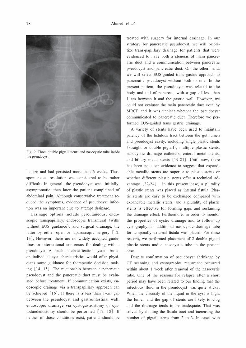
LIUS, JPN) and a nasocystic tube (NB-BRAID 6 Fr, PIOLAX, JPN) were placed. As a result, the patient had placed a total of 3 double pigtail stents and a nasocystic tube (Fig. 9). The pseudocyst con-dition improved following this additional therapy. She discharged 7 days after the final treatment.
DISCUSSION
A pseudopancreatic cyst is a known complication of both acute and chronic pancreatitis, and shows a wide variety of clinical presentations, ranging from completely asymptomatic lesions to multiple pseu-docysts with pancreatic and/or bile duct obstruction,
which may require immediate intervention to prevent secondary complications. Other indications for drain-age include infection, hemorrhage, and presence of symptoms [9]. The probability of spontaneous reso-lution of a pancreatic pseudocyst ranges widely from 8% to 85%, depending on etiology, localization, and predominantly size[10]. Warshaw and Rattner re-ported that a pseudocyst is unlikely to resolve spon-taneously if: a) it persists for more than 6 weeks, b) chronic pancreatitis is evident, c) there is a pancre-atic duct anomaly (except for communication with the pseudocyst), or d) the pseudocyst is surrounded by a thick wall [11]. The pseudocyst in our patient was a sequel of biliary pancreatitis, relatively large
Fig. 7. Images obtained during endoscopic-guided drainage. (A) Narrow fistula opening. (B) Dilation with biliary ballon. (C) Nasal-type scope above the wire inside the cyst cavity.
Fig. 8. (A) Image of pseudocyst lumen by ultra-slim scope, (B) Image of irrigation for pseudocystic lumen with saline by ultra-slim scope
77Pancreatic pseudocyst recurrence following EUS-guided drainage
in size and had persisted more than 6 weeks. Thus, spontaneous resolution was considered to be rather difficult. In general, the pseudocyst was, initially, asymptomatic, then later the patient complained of abdominal pain. Although conservative treatment re-duced the symptoms, evidence of pseudocyst infec-tion was an important clue to attempt drainage.
Drainage options include percutaneous, endo-scopic transpapillary, endoscopic transmural (with/without EUS guidance), and surgical drainage, the latter by either open or laparoscopic surgery [12, 13]. However, there are no widely accepted guide-lines or international consensus for dealing with a pseudocyst. As such, a classification system based on individual cyst characteristics would offer physi-cians some guidance for therapeutic decision mak-ing [14, 15]. The relationship between a pancreatic pseudocyst and the pancreatic duct must be evalu-ated before treatment. If communication exists, en-doscopic drainage via a transpapillary approach can be achieved [16]. If there is a less than 1-cm gap between the pseudocyst and gastrointestinal wall, endoscopic drainage via cystogastrostomy or cys-toduodenostomy should be performed [17, 18]. If neither of those conditions exist, patients should be
treated with surgery for internal drainage. In our strategy for pancreatic pseudocyst, we will priori-tize trans-papillary drainage for patients that were evidenced to have both a stenosis of main pancre-atic duct and a communication between pancreatic pseudocyst and pancreatic duct. On the other hand, we will select EUS-guided trans gastric approach to pancreatic pseudocyst without both or one. In the present patient, the pseudocyst was related to the body and tail of pancreas, with a gap of less than 1 cm between it and the gastric wall. However, we could not evaluate the main pancreatic duct even by MRCP and it was unclear whether the pseudocyst communicated to pancreatic duct. Therefore we per-formed EUS-guided trans gastric drainage.
A variety of stents have been used to maintain patency of the fistulous tract between the gut lumen and pseudocyst cavity, including single plastic stents (straight or double pigtail), multiple plastic stents, nasocystic drainage catheters, enteral metal stents, and biliary metal stents [19-21]. Until now, there has been no clear evidence to suggest that expand-able metallic stents are superior to plastic stents or whether different plastic stents offer a technical ad-vantage[22-24]. In this present case, a plurality of plastic stents was placed as internal fistula. Plas-tic stents are easy to be exchanged compared with expandable metallic stents, and a plurality of plastic stents is effective for forming gaps and sustaining the drainage effect. Furthermore, in order to monitor the properties of cystic drainage and to follow up cystography, an additional nasocystic drainage tube for temporally external fistula was placed. For these reasons, we performed placement of 2 double pigtail plastic stents and a nasocystic tube in the present case.
Despite confirmation of pseudocyst shrinkage by CT scanning and cystography, recurrence occurred within about 1 week after removal of the nasocystic tube. One of the reasons for relapse after a short period may have been related to our finding that the infectious fluid in the pseudocyst was quite sticky. When the viscosity of the liquid in the cyst is high, the lumen and the gap of stents are likely to clog and the drainage tends to be inadequate. That was solved by dilating the fistula tract and increasing the number of pigtail stents from 2 to 3. In cases with
Fig. 9. Three double pigtail stents and nasocystic tube inside the pseudocyst.
78 Ahmed et al.
high viscosity cystic fluid, a larger fistula expansion and placement of more stents might be required as compared to cases with cystic fluid of low viscosity.
In a systemic review of current guidelines for minimally invasive management of pancreatic pseu-docyst cases published in 2008 [25], the American Gastroenterological Association stated that an infect-ed pseudocyst should be termed an abscess and can be treated with percutaneous drainage [26]. Also, guidelines presented by the Society for Surgery of the Alimentary Tract (SSAT) [27] and Sociétié Nationale Francais de Gastro-Entérologie (SNFG) [28] recommend that infected pancreatic fluid be drained in a percutaneous manner, though they do not differentiate between a pancreatic abscess and infected pseudocyst. Japanese guidelines for man-agement of acute pancreatitis presented in 2015 state that during therapeutic intervention for infected pancreatic necrosis, percutaneous (retroperitoneal) drainage or endoscopic transluminal drainage should be given first, then if no improvement is achieved, a necrosectomy should be performed. In addition to a necrosectomy, an endoscopic or retroperitoneal ap-proach is recommended. However, there is no men-tion regarding selection of the period of drainage or number of stents in the current guidelines. Each guideline recommends drainage for infectious pancre-atic pseudocysts. In recent years endoscopic drainage has been developed and becoming the first choice treatment for it. However, there are no evidences re-garding selection of the period of drainage or num-ber of stents. Until evidence accumulates, we think that it is necessary to have as many stents as pos-sible and a long term stent placement period. Based on our experience, we consider that a key factor for success of endoscopic-guided drainage of a pseudo-cyst may be the patency of the draining tubes and fistula tract. Further investigations for management of patients with a pseudocyst are needed.
REFERENCES
1) Banks PA, Bollen TL, Dervenis C, et al. Clas-sification of acute pancreatitis 2012: revision of the Atlanta classification and definitions by inter-national consensus. Gut 2013;62:102-11.2) Imrie CW, Buist LJ, Shearer MG. Importance
of cause in the outcome of pancreatic pseudo-cysts. AM J SURG 1988;156:159-62.3) Maringhini A, Uomo G, Patti R, et al. Pseudo-
cysts in acute non alcoholic pancreatitis: incidence and natural history. Dig Dis Sci 1999;44:1669-73.4) Barthet M, Bugallo M, Moreira LS, Bastid C,
Sastre B, Sahel J. Management of cysts and pseu-docysts complicating chronic pancreatitis. A retro-spective study of 143 patients. Gastroenterol Clin Biol 1993;17:270-6.5) Ammann RW, Akovbiantz A, Largiader F,
Schueler G. Course and outcome of chronic pan-creatitis. Longitudinal study of a mixed medical-surgical series of 245 patients. Gastroenterology 1984;86(5 Pt 1):820-8.6) Bradley EL, Clements JL Jr, Gonzalez AC. The
natural history of pancreatic pseudocysts: a unified concept of management. Am J Surg 1979;137:135-41. 7) Pitchumoni CS, Agarwal N. Pancreatic pseu-
docysts: when and how should drainage be performed? Gastroenterol Cl in North Am 1999;28:615-39.8) Aghdassi AA, Mayerle J, Kraft M, Sielen-
kämper AW, Heidecke CD, Lerch MM. Pancre-atic pseudocysts--when and how to treat? HPB 2006;8:432-41.9) Bradley EL 3rd. A clinically based classifica-
tion system for acute pancreatitis. Summary of the International Symposium on Acute Pancreatitis, Atlanta, Ga, September 11 through 13,1992. Arch Surg 1993;128:586-90.10) Gouyon B, Levy P, Ruszneiwski P, Zins M,
Hammel P,Vilgrain V, et al. Predictive factors in the outcome of pseudocysts complicating alcoholic chronic pancreatitis. Gut 1997;41:821-5.11) Warshaw AL, Rattner DW. Timing of surgical
drainage for pancreatic pseudocyst. Clinical and chemical criteria. Ann Surg 1985;202:720-4.12) Loveday BP, Mittal A, Phillips A, Windsor
JA. Minimally invasive management of pancre-atic abscess, pseudocyst, and necrosis: a system-atic review of current guidelines. World J Surg 2008;32:2383-9413) Akshintala VS, Saxena P, Zaheer A, et al.
A comparative evaluation of outcomes of endo-
79Pancreatic pseudocyst recurrence following EUS-guided drainage

scopic versus percutaneous drainage for symptom-atic pancreatic pseudocysts. Gastrointest Endosc 2014;79:921-8.14) D’Eqidio A, Schein M. Pancreatic pseudocysts:
a proposed classification and its management im-plications. Br J Surg 1991;78:981-4.15) Vitas GJ, Sarr MG. Selected management of
pancreatic pseudocysts: operative versus expectant management. Surgery 1992;111:123.16) Nealon WH, Walser E. Main pancreatic ductal
anatomy can direct choice of modality for treating pancreatic pseudocysts (surgery versus percutane-ous drainage). Ann Surg 2002;235:751.17) Chak A. Endosonographic-guided therapy
of pancreatic pseudocysts. Gastrointest Endosc 2000;52:S23-7.18) Monkemuller KE, Kahl S, Malfertheiner P. En-
doscopic therapy of chronic pancreatitis. Dig Dis 2005;22:280-91.19) Bello B, Matthews JB. Minimally invasive
treatment of pancreatic necrosis. World J Gastro-enterol 2012;18:6829-35.20) Fabbri C, Luigiano C, Maimone A, Polifemo
AM, Tarantino I, Cennamo V. Endoscopic ultra-sound-guided drainage of pancreatic fluid collec-tions. World J Gastrointest Endosc 2012;4:479-88.21) Lin H, Zhan XB, Sun SY, et al. Stent se-
lection for endoscopic ultrasound-guided drain-age of pancreatic fluid collections: a multi-center study in china. Gastroenterol Res Pract
2014;2014:193562, doi: 10.1155/2014/193562. Epub 2014 Jun 11.22) ASGE Technology Committee, Desilets DJ, Ba-
nerjee S, et al. New devices and techniques for management of pancreatic fluid collections. Gas-trointest Endosc 2013;77:835-8.23) Bang JY, Wilcox CM, Trevino JM, et al. Re-
lationship between stent characteristics and treat-ment outcomes in endoscopic transmural drainage drainage of uncomplicated pancreatic pseudocysts. Surg Endosc 2014;28:2877-83. 24) Bang JY, Hawes R, Bartolucci A, Varadarajulu
S. Efficacy ofmetal and plastic stents for trans-mural drainage of pancreatic fluid collections: a systematic review. Dig Endosc 2015;27:486-98.25) Loveday BP, Mittal A, Phillips A, Windsor
JA. Minimally invasive management of pancre-atic abscess, pseudocyst, and necrosis: a system-atic review of current guidelines. World J Surg 2008;32:2383-94.26) Banks PA, Freeman ML. Practice guide-
lines in acute pancreatitis. Am J Gastroenterol 2006;101:2379-400.27) The Society for Surgery of the Alimentary
Tract Patient Care Committee. Treatment of acute pancreatitis. J Gastrointest Surg 1998;2:487-8.28) French consensus conference on acute pancre-
atitis: conclusions and recommendations. Paris, France, 25-26 January 2001. Eur J Gastroenterol Hepatol 13(Suppl 4):S1-13.
80 Ahmed et al.