Mink Practical 1 - Review 2015. Green = Pink = Orange = Latissimus Dorsi Masseter Clavotrapezius.
RECONSTRUCTIVE
Surgical Technique in Pedicled ThoracodorsalArtery Perforator Flaps: A Clinical Experiencewith 99 Patients
Moustapha Hamdi, M.D.,Ph.D.
Koenraad Van Landuyt,M.D., Ph.D.
John B. Hijjawi, M.D.Nathalie Roche, M.D.
Phillip Blondeel, M.D., Ph.D.Stan Monstrey, M.D., Ph.D.
Gent, Belgium
Background: The thoracodorsal artery perforator flap is considered a techni-cally difficult flap because of significant anatomical variations in perforatorlocation. The authors’ strategy to facilitate the harvest of these flaps includescareful preoperative mapping of perforators and a standardized planning andharvesting technique. The authors evaluated 99 pedicled thoracodorsal arteryperforator flaps, with an emphasis on preoperative planning, surgical technique,and analysis of complications.Methods: Between May of 2000 and October of 2006, 99 patients underwentpedicled thoracodorsal artery perforator flaps in the authors’ department. Theircharts and postoperative results were reviewed retrospectively.Results: A thoracodorsal artery perforator flap was harvested in 90 cases. Theperforators were unsuitable in the other 10 flaps; therefore, a muscle-sparing tech-nique was used (type I or type II). Unidirectional Doppler imaging was usedexclusively in 92 percent of cases to map the perforator preoperatively. The averageflap size was 20 � 8 cm. Average operative time for flap harvest was 80 minutes.Perforators were located at 8 to 13 cm from the axillary crease (average, 10.8 cm).Major flap necrosis occurred in one case (1 percent). Limited partial or palpablefat necrosis occurred in three cases (3 percent). Seroma formation was not en-countered in any of the thoracodorsal artery perforator flaps or muscle-sparingthoracodorsal artery perforator type I flaps.Conclusion: Careful preoperative perforator mapping and a standardized ap-proach to flap planning and harvest can significantly reduce the difficulty of exe-cuting pedicled thoracodorsal artery perforator flaps. (Plast. Reconstr. Surg. 121:1632, 2008.)
As the perforator flap concept has gainedacceptance in the reconstructive ladder,many previously described musculocutane-
ous flaps have been harvested as perforatorflaps.1–12 Although the thoracodorsal artery per-forator flap has been reportedly used in multiplereconstructive situations, it has not gained thepopularity of other perforator flaps such as thedeep inferior epigastric artery perforator flap.Reasons for this include inconsistent perforatorsize, quality, quantity, and location, all of whichcontribute to tedious dissection. The lack of data
demonstrating a clear advantage of thoracodorsalartery perforator flaps over the more straightfor-ward and time-tested latissimus dorsi flap or otherlocal flaps such as scapular/parascapular flaps has
From the Plastic Surgery Department, Gent University Hos-pital.Received for publication February 7, 2007; accepted March27, 2007.Portions of the video provided with this article were presentedat the American Society of Reconstructive Microsurgery 2007Meeting, in Rio Grande, Puerto Rico, January of 2007.Copyright ©2008 by the American Society of Plastic Surgeons
DOI: 10.1097/PRS.0b013e31816c3bfa
Disclosure: None of the authors has a financialinterest in any of the products, devices, or drugsmentioned in this article.
Supplemental digital content is available forthis article. Direct URL citations appear in theprinted text; simply type the URL address intoany web browser to access this content. Click-able links to the material are provided in theHTML text and PDF of this article on theJournal’s Web site (www.PRSJournal.com).
www.PRSJournal.com1632
also contributed to criticism of the thoracodorsalartery perforator flap.4,11,12 Thus, only small clin-ical series of thoracodorsal artery perforator flapshave been published.1–12 The thoracodorsal arteryperforator flap has been used widely in our de-partment as either a free9 or a pedicled flap.6
The purpose of this article is to report our clin-ical experience with a large series of pedicled tho-racodorsal artery perforator flaps used for a varietyof indications, with an emphasis on preoperativeplanning, safe and standardized dissection tech-nique, and an analysis of major and minor postop-erative complications. In addition, a detailed videoof the harvest technique is provided [see Video,Supplemental Digital Content 1, which demon-strates the harvest technique (described below),http://links. lww.com/A413].
The Harvest TechniqueFlap DesignThe anterior border of the latissimus dorsi
muscle is marked with the patient in the standingposition. Preoperative perforator mapping is per-formed with unidirectional Doppler imaging or byany of the other radiologic methods.
To anticipate possible septal cutaneous branchesof the thoracodorsal artery, the anterior border ofthe flap should lie in front of the anterior borderof the latissimus dorsi muscle. The vertically ori-ented flap can reach the distal third of the upperarm and elbow, and the neck, shoulder, and upperback area. When the thoracodorsal artery perfo-rator flap is oriented horizontally, the incisionsexploit the relaxed skin tension lines of the back.The flap is typically designed as in the classic la-tissimus dorsi musculocutaneous flap for breastreconstruction but is outlined more anteriorly toreach the lateral border of the inframammaryfold.
Patient PositionThe patient is in the lateral decubitus position
with 90 degrees of shoulder abduction and 90degrees of elbow flexion. This positioning placesthe skin on stretch, causing the perforators toenter the skin with a more perpendicular orien-tation, which makes their Doppler signal morediscrete. Flap harvesting starts once the recipientsite is ready. Dissection is performed under loupemagnification.
Skin IncisionIncision starts at the anteroinferior border of
the flap, which allows identification of the anteriorborder of the latissimus dorsi muscle and the pos-sibility of repositioning the anterior border of the
flap accordingly. Then, the distal border of theflap is incised up to the area where the perforatorsignal has been detected.
Suprafascial DissectionA caudal (posterior) approach is used in har-
vesting a pedicled thoracodorsal artery perforatorflap, developing a plane above the deep fascia ofthe latissimus dorsi and serratus anterior muscles,which is thick medially and becomes thinnerabove the anterior part of the latissimus dorsi mus-cle. Only visibly pulsatile perforators with a diam-eter greater than 0.5 mm are considered suitableand preserved.
Perforator DissectionOnce a suitable perforator has been identi-
fied, the cleavage plane of the latissimus dorsimuscle in which that perforator resides is devel-oped. Perforators originating from the descend-ing branch of the thoracodorsal vessels are chosenpreferentially over those originating from thetransverse branch. Muscle fibers are spread, main-taining their longitudinal integrity. The perfora-tor is dissected from the surrounding muscle fi-bers, where they tend to lie in a fibrofatty layer.Wide exposure is essential to perforator dissectionwithin the muscle. The dissection should be per-formed close to the pedicle, ligating muscularbranches with surgical clips or bipolar cautery.Nerve branches to the latissimus dorsi muscleshould be freed from the pedicle with atraumaticdissection technique.
Thoracodorsal Pedicle DissectionThe main thoracodorsal pedicle is dissected
free until the required pedicle length is obtained.Dissection of the main pedicle begins through thesplit muscle fibers. After the dissection of the mainpedicle is performed, the segment of the perfo-rator at the level where it enters the subcutaneoustissue is dissected from the latissimus dorsi musclewith the help of vessel loops.
Completing the Flap DissectionThe flap is then dissected from the serratus
fascia and from the latissimus dorsi muscle ante-riorly. Intercostal perforators, which may be en-countered in front of the anterior border of thelatissimus dorsi muscle, are clipped. The flap isthen based totally on the thoracodorsal perforatorand separated from the underlying muscles.
The flap is pulled through the muscle andtransposed into the defect. As such, its anteriorborder becomes the medial or inferior part ofthe reconstruction.
Volume 121, Number 5 • Thoracodorsal Artery Perforator Flap
1633
PATIENTS AND METHODSBetween May of 2000 and October of 2006, 99
patients underwent pedicled flaps based on thethoracodorsal vessels in our department. One pa-tient had bilateral flaps; thus, 100 flaps are in-cluded in this study. The medical charts were re-viewed retrospectively for information regardingpatient characteristics, flap dimensions, the num-ber and location of perforators, operative time,and complications. The average patient age was 44years (range, 17 to 69 years) and 89 percent werefemale patients. Active smoking history was foundin 11 patients (all presenting for immediate breastreconstruction for breast cancer). Surgical indi-cations are summarized in Table 1. The resultsshow data regarding flap type, number and loca-tions of perforators, and operative time. Postop-erative complications are presented with a com-parison among the different flap types.
Flap DesignThe anterior border of the latissimus dorsi
muscle is palpated through the skin and markedwith the patient in the standing position and bothhands grasping the waist. Preoperative perforatormapping is performed with a unidirectionalDoppler probe (8 Hz) with the patient in thelateral decubitus position to simulate operativepositioning. Based on previous anatomical stud-ies, the perforators are sought out in a region 8 cmbelow the axillary crease (i.e., an axillary wrinklefound at the junction of the upper arm with theshoulder/back region) and within 5 cm of theanterior border of the latissimus dorsi muscle.1 Apossible pitfall of using unidirectional Dopplerimaging to identify the thoracodorsal perforatorsis background signal from the main thoracodorsal
pedicle. To avoid this, the patient is positioned forperforator marking in the same position in whichthey will be placed during surgery: the lateral de-cubitus position with 90 degrees of shoulder ab-duction and 90 degrees of elbow flexion. Thispositioning places the skin on stretch, causingthe perforators to enter the skin with a moreperpendicular orientation, which makes theirDoppler signal more discrete. In this orienta-tion, the Doppler device detects the signal fromthe discrete end of the perforator rather thanalong the length of the perforator. This allowsone to distinguish the perforator signal from thelongitudinally oriented thoracodorsal pedicle.True perforator tones can be distinguished fromthe thoracodorsal artery by moving the Dopplerdevice proximally and distally. If the signal dis-appears, the signal belongs to a perforator, al-though signals from the thoracodorsal vesselswill be present continuously along the length ofthe latissimus dorsi muscle. In difficult cases, aduplex examination is performed. More re-cently, multidetector row computed tomogra-phy has been introduced for the preoperativelocalization of various perforators.13,14
To anticipate possible septal cutaneous branchesof the thoracodorsal artery, the anterior border ofthe flap should lie in front of the anterior borderof the latissimus dorsi muscle. Depending on theindication, the flap is outlined as a vertical ellipseor horizontally over the latissimus dorsi muscle.The vertically oriented flap can reach the distalthird of the upper arm and elbow, and the neck,shoulder, and upper back area. The thoracodorsalartery perforator flap can also be harvested as abilobed flap with a large longitudinal and asmaller transversely oriented skin island which,after a 90-degree rotation, allows for easier pri-mary closure. When oriented horizontally, the in-cisions exploit the relaxed skin tension lines of theback. The flap is typically designed as in theclassic latissimus dorsi musculocutaneous flapfor breast reconstruction but it is outlined moreanteriorly to reach the lateral border of theinframammary fold. The main indications are inbreast surgery, either in partial breast recon-struction after ablative surgery6 or in autologousaugmentation.7 The flap reaches the superolat-eral, inferolateral, and inferomedial quadrantsof the breast. Flaps 15 cm wide can be raisedsafely on a single perforator, depending on therecipient-site requirements and the ability toachieve primary closure of the donor site.
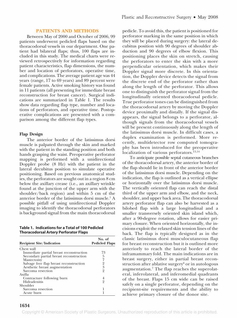
Table 1. Indications for a Total of 100 PedicledThoracodorsal Artery Perforator Flaps
Recipient Site/IndicationNo. of
Pedicled Flaps
Chest wallImmediate partial breast reconstruction 73Secondary partial breast reconstruction 5Mastectomy 7Salvage free flap breast reconstruction 2Aesthetic breast augmentation 3Sarcoma resection 2
AxillaContracture following burn 4Hidradenitis 2
ShoulderSarcoma resection 1Acute burn 1
Plastic and Reconstructive Surgery • May 2008
1634
Surgical TechniqueWhen a pedicled thoracodorsal artery perfo-
rator flap is harvested, the patient is positioned asdescribed above (see Video, Supplemental DigitalContent 1, which demonstrates the harvest tech-nique, http://links.lww.com/A413). Flap harvestingstarts once the recipient site is ready. Dissection isperformed under loupe magnification. Incisionstarts at the anterointerior border of the flap, whichallows identification of the anterior border of thelatissimus dorsi muscle and the possibility of repo-sitioning the anterior border of the flap accordingly.Then, the distal border of the flap is incised up to thearea where the perforator signal has been detected.A caudal (posterior) approach is used in harvestinga pedicled thoracodorsal artery perforator flap, de-veloping a plane above the deep fascia of the latis-simus dorsi and serratus anterior muscles, which isthick medially and becomes thinner above the an-terior part of the latissimus dorsi muscle. Only per-forators that are visibly pulsatile with a diametergreater than 0.5 mm are considered suitable andpreserved. The main perforators tend to lie in a rowoverlying the course of the descending branch of thethoracodorsal artery.
Once a suitable perforator has been identi-fied, the cleavage plane of the latissimus dorsimuscle in which that perforator resides is devel-oped. Perforators originating from the descend-ing branch of the thoracodorsal vessels are chosenpreferentially over those originating from thetransverse branch because they typically have ashorter intramuscular course with less involve-ment of nerve branches. More distal perforatorsresult in a longer pedicle than perforators enter-ing the flap more proximally or cranially. Thedisadvantage of more distally located perforatorsis that dissection requires more time because oftheir longer intramuscular course. Muscle fibersare spread, maintaining their longitudinal integ-rity. The perforator is dissected from the sur-rounding muscle fibers, where they tend to lie ina fibrofatty layer. Wide exposure is essential toperforator dissection within the muscle. The dis-section should be performed close to the pedicle,ligating muscular branches with surgical clips orbipolar cautery. Nerve branches to the latissimusdorsi muscle should be freed from the pedicle withatraumatic dissection technique.
The main thoracodorsal pedicle is dissectedfree until the required pedicle length is obtained.Dissection of the main pedicle begins through thesplit muscle fibers and is then completed underthe latissimus dorsi muscle by lifting its anterior
border. The flap is pulled through the muscle andtransposed into the defect. As such, its anteriorborder becomes the medial or inferior part of thereconstruction.
Conversion to Muscle-Sparing DesignsWhen the perforator is less than 0.5 mm in
diameter but pulsatile, there is a high risk forperforator rupture or avulsion. In this situation, amuscle-sparing technique (muscle-sparing thora-codorsal artery perforator type I) increases safetyby preserving a 2-cm cuff of latissimus dorsi musclearound the perforators. The perforators are stilldissected free of the latissimus dorsi muscle ante-riorly, but their posterior aspect remains attachedto a cuff of muscle, which is included in the flap.Including this small segment of latissimus dorsimuscle while maintaining direct visualization ofthe perforators reduces the potential for damag-ing them during repositioning of a pedicled tho-racodorsal artery perforator flap. Only nervebranches to this muscle segment need to be sac-rificed.
When the perforators are smaller than 0.5 mmand also nonpulsatile, the flap should be con-verted to a muscle-sparing thoracodorsal arteryperforator type II flap incorporating a maximumnumber of perforators. An approximately 5-cmsegment of latissimus dorsi muscle is included un-der the skin paddle between the cleavage planewhere the perforators are identified and the an-terior border of the latissimus dorsi muscle, witha length extending to the level of the main pedi-cle. The nerve supplying the remaining latissimusdorsi muscle is preserved.
Depending on the nature of the defect, theflap is partially or totally deepithelialized and canalso be folded to increase projection. However,tension-free flap inset is critical to avoid rupture ofthe perforator. All patients receive piracetam(Nootropil; UCB, Brussels, Belgium) 12 g/24hours intravenously for 5 days and as a 20% solu-tion orally, 25 cc four times per day for another 5days, to improve the viability of the distal flapthrough an increase of capillary blood flow.
RESULTSThe majority of patients underwent preoper-
ative perforator mapping with unidirectionalDoppler examination (92 percent). Both colorduplex and multidetector row computed tomo-graphic images were used in 4 percent of patientsfor preoperative perforator identification. The av-erage flap size was 20 � 8 cm (range of length, 16
Volume 121, Number 5 • Thoracodorsal Artery Perforator Flap
1635
to 25 cm; range of width, 6 to 10 cm). Ninetythoracodorsal artery perforator flaps were har-vested. Muscle-sparing thoracodorsal artery per-forator type I and type II flaps were dissected in sixand four cases, respectively. Thoracodorsal arteryperforator and muscle-sparing thoracodorsal ar-tery perforator type I flaps (96 flaps) were basedon a single perforator in 89 cases (92.7 percent)and on two perforators in seven cases (7.3 per-cent). In these flaps, most of the perforators orig-inated from the descending branch of the thora-codorsal vessels (93.7 percent), and only six flapswere harvested on a perforator from the transversebranch (6.3 percent). At least two perforators wereincluded in the flap (four flaps) when a muscle-sparing thoracodorsal artery perforator type IIflap was harvested. The thoracodorsal artery per-forator flap and its muscle-sparing variants aredemonstrated with clinical examples in Figures 1through 8.
Perforators were located between 8 and 13 cm(average, 10.8 cm) distal to the axillary crease andwithin 0 to 5 cm (average, 2.8 cm) of the anteriorborder of the latissimus dorsi muscle. In two cases,the thoracodorsal artery perforator flap was basedon a direct septal perforator that went around theanterior border of the latissimus dorsi musclerather than through the muscle. The average har-vesting time was 80 minutes (range, 25 to 120minutes).
Postoperative complications are summarizedin Table 2. Total flap survival was achieved in 96
cases (96.7 percent). Major flap necrosis occurredin one case (1 percent), which was an immediatepartial breast reconstruction. This required de-bridement and secondary flap surgery. Limitedpartial or palpable fat necrosis occurred in threecases (3 percent), requiring partial excision in onecase. None of these complications was related to
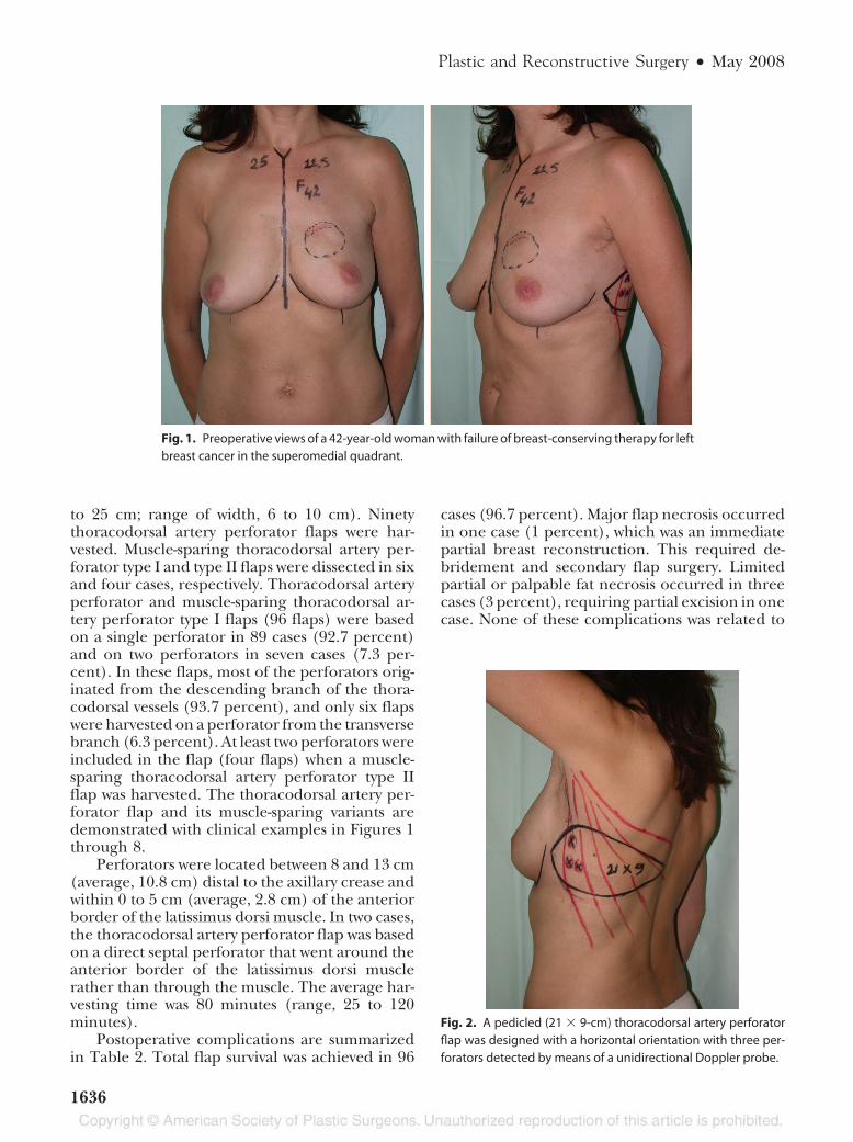
Fig. 1. Preoperative views of a 42-year-old woman with failure of breast-conserving therapy for leftbreast cancer in the superomedial quadrant.
Fig. 2. A pedicled (21 � 9-cm) thoracodorsal artery perforatorflap was designed with a horizontal orientation with three per-forators detected by means of a unidirectional Doppler probe.
Plastic and Reconstructive Surgery • May 2008
1636
smoking history. However, all three flaps that suf-fered partial necrosis were irradiated postopera-tively. Seroma formation at the donor site wasencountered in all cases of muscle-sparing thora-codorsal artery perforator type II flaps but in noneof the thoracodorsal artery perforator or muscle-sparing thoracodorsal artery perforator type Iflaps.
DISCUSSIONThe thoracodorsal artery perforator flap is a
versatile tool in reconstructive surgery, and flaps aslarge as 25 � 15 cm can be safely harvested.1,2,8
Several anatomical variations can make the har-vest of thoracodorsal artery perforator flaps tech-
nically challenging. These include a variable num-ber of large (�0.5 mm) perforators; difficultymapping these perforators preoperatively becauseof the proximity of the main thoracodorsal pedi-cle; and poorly described, sometimes tedious dis-section techniques. Lastly, there is a lack of con-vincing data demonstrating a clear advantage ofthe thoracodorsal artery perforator flap over otherpedicled flaps such as latissimus dorsi muscle orparascapular flaps.
The thoracodorsal vessels provide limitedlarge perforators to the overlying skin.3,7,15,16 Re-cent studies demonstrate only 5.5 � 1.8 perforatorswith a diameter greater than 0.5 mm supplying thisflap.15 However, only one or two perforators can beclinically used if a useful arc of rotation is to bemaintained. Multiple cadaveric dissection studieshave focused on determining the relationship be-tween topographic landmarks and the thoracodor-sal perforators.2,8,15 Since the original description of
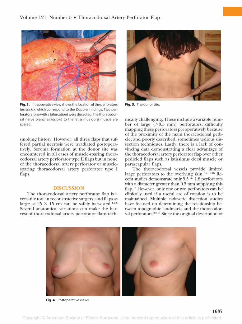
Fig. 3. Intraoperative view shows the location of the perforators(asterisks), which correspond to the Doppler findings. Two per-forators (one with a bifurcation) were dissected. The thoracodor-sal nerve branches (arrow) to the latissimus dorsi muscle arespared.
Fig. 4. Postoperative views.
Fig. 5. The donor site.
Volume 121, Number 5 • Thoracodorsal Artery Perforator Flap
1637
the thoracodorsal artery perforator flap by Angrigi-ani et al.,1 the location of the majority of perforators8 cm distal to the posterior axillary fold remains avalid and important anatomical landmark. At thispoint, the first perforator emerges from the descend-
ing branch of the thoracodorsal pedicle and usuallytravels above the deep fascia caudally and parallel tothe latissimus dorsi muscle fibers. The scapular tipwas used by Heitmann et al.5 to locate the bifurcationof the thoracodorsal vessel into its respective hori-
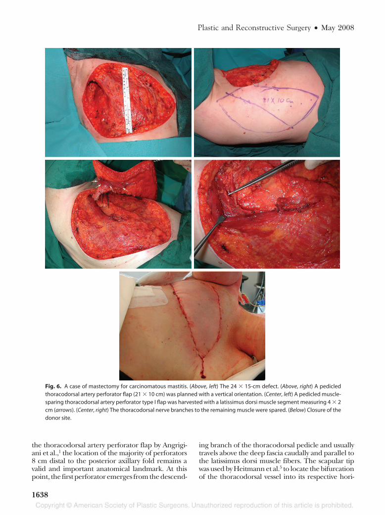
Fig. 6. A case of mastectomy for carcinomatous mastitis. (Above, left) The 24 � 15-cm defect. (Above, right) A pedicledthoracodorsal artery perforator flap (21 � 10 cm) was planned with a vertical orientation. (Center, left) A pedicled muscle-sparing thoracodorsal artery perforator type I flap was harvested with a latissimus dorsi muscle segment measuring 4 � 2cm (arrows). (Center, right) The thoracodorsal nerve branches to the remaining muscle were spared. (Below) Closure of thedonor site.
Plastic and Reconstructive Surgery • May 2008
1638

zontal and lateral branches. The neurovascular hiluswas found 3 to 6 cm inferior to the scapular tip and1 to 4 cm posterior to the anterior border of thelatissimus dorsi muscle. These landmarks help todetermine an area in which large perforators can befound consistently. However, this area depends onthe position of the patient and laxity of the skin andmay not correspond to intraoperative findings.16
Consequently, significant time can be spent search-ing for skin perforators and modifying the flapdesign.16
The ability to safely and expeditiously har-vest a thoracodorsal artery perforator flap is fa-cilitated by precise preoperative localization ofits perforators. Using one of the several availableradiologic assessment methods mentioned pro-vides information specific to an individual pa-
tient, making flap harvest safer and faster. Uni-directional Doppler examination is a simple andinexpensive method for mapping perforatorsbut is somewhat less accurate than color Dopp-ler flowmetry.17 Placing the patient in an oper-ative position is recommended, to increase theaccuracy of all perforator mapping techniques.We have used the Doppler probe in all of ourcases, with reliability as high as 95 percent. How-ever, color duplex flowmetry and multidetectorrow computed tomography have been shown tofacilitate dissection, particularly in inexperi-enced hands.13,14 These recent imaging devel-opments allow us to localize the “dominant per-forator” and obtain complete information aboutthe arterial tree of the thoracodorsal vessels andintercostal perforators, thus decreasing opera-
Fig. 7. A case of sarcoma on the left shoulder of a 56-year-old man. (Above, left) A compound (chimera) flap is designed. Theflap consists of a thoracodorsal artery perforator flap with a segment of latissimus dorsi muscle to reconstruct the deltoidmuscle because of tumor resection. (Above, right) The latissimus dorsi segment (10 � 5 cm) was dissected with a vascularpedicle independent of the pedicle of the thoracodorsal artery perforator flap (arrow). (Below, left) The muscle segment iselevated. The perforator arises from the horizontal branch and courses under the thoracodorsal nerve branch (two arrows)to enter the skin paddle (vessel loop and arrow). (Below, right) The thoracodorsal artery perforator flap is passed under thethoracodorsal nerve that is left intact to the rest of the latissimus dorsi muscle.
Volume 121, Number 5 • Thoracodorsal Artery Perforator Flap
1639
tive time and complications related to poor per-forator selection.
Another major advantage of the thoracodorsalartery perforator is pedicle length. With an averagelength of 14 � 1.4 cm, the flap can reach moremedial defects on the chest wall than the scapular/parascapular flap.8,15 When the thoracodorsal arteryperforator flap is based on a distal perforator, pedi-cle length can reach 25 cm,8 providing a broad arcof rotation extending over the anterior chest wall,clavicle, axilla, posterior arm, and lateral border ofthe sternum. Tension on the pedicle must beavoided, as this may lead to rupture of smaller per-forators. Alternative flaps such as those based on theintercostal or superior epigastric vessels are moresuitable for defects of the medial chest wall.18
Initial dissection begins with an anterior, cau-dal incision and identification of the anterior bor-
der of the latissimus dorsi muscle. Making theanterior, caudal incision first allows the remainingskin paddle to be adjusted to guarantee inclusionof perforators that course anterior to the latissi-mus dorsi edge. The caudal approach allows safevisualization of the perforator because the eleva-tion of the skin is performed parallel to the latis-simus dorsi fibers and the perforator can be seenthrough transparent fascia. The perforator lies inloose areolar tissue within a cleavage plane in thelatissimus dorsi muscle, facilitating dissection.Regular irrigation of the perforator with salineand delicate manipulation is recommended toavoid spasm. Spasm usually resolves spontaneouslyonce blood in the main pedicle is redirected en-tirely into the perforator after ligation of sidebranches. If spasm persists, topical papaverine ir-rigation can be used. Failure of spasm to resolvetypically indicates intraluminal damage of the per-forator intima because of excessive traction duringdissection and usually results in flap failure. Werecommend keeping the posterior aspect of theperforator attached to the latissimus dorsi musclewhere the perforator enters the skin until justbefore flap transfer.
Flap-related complications in this study werelimited. Partial flap necrosis occurs if the flap isextended beyond it vascular territory, estimated at255 cm by Thomas et al.15 In comparing our ex-perience with a vertical flap design (used mainlyin microvascular transfer) and a more horizontalflap orientation (used mainly in pedicled flaps),we have observed a slightly higher rate of partialnecrosis, typically in the distal tip, in the verticaldesign. This is likely related to the orientation ofthe angiosome territory of the thoracodorsal per-forators. According to Taylor’s findings,19 flapswould most reliably be designed parallel to thecourse of the ribs in this region. This favors ahorizontal, slightly oblique orientation of the flap,with its tip directed toward the scapular angle.This design was used in the majority of our pedi-cled thoracodorsal artery perforator flaps forbreast reconstruction, giving the additional ad-vantages of donor-site closure within the relaxedskin tension lines and scar placement within thebra area.
Although latissimus dorsi muscle preservationis an intuitive benefit that results in less contourdeformity of the donor site than latissimus dorsiharvest, prospective studies are still necessary toconfirm the objective functional benefits of latis-simus dorsi muscle sparing. Nevertheless, thisstudy does confirm that harvesting thoracodorsalartery perforator or muscle-sparing thoracodorsal
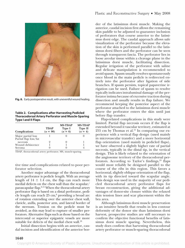
Fig. 8. Early postoperative result, with uneventful wound healing.
Table 2. Complications after Harvesting PedicledThoracodorsal Artery Perforator and Muscle-SparingType I and II Flaps
ComplicationsTDAP
(n � 90)
MS-TDAPType I(n � 6)
MS-TDAPType II(n � 4)
Major partial loss 1 0 0Partial flap loss/fat
necrosis 2 0 1Wound dehiscence 1 1 1Seroma 0 0 4TDAP, thoracodorsal artery perforator; MS, muscle-sparing.
Plastic and Reconstructive Surgery • May 2008
1640
artery perforator type I flaps dramatically reducesthe incidence of seroma formation in the donorsite, which has been reported to be as high as 60percent after harvesting a latissimus dorsi muscleflap.20 In addition, shorter postoperative recoveryand less pain at the donor site have been reportedafter harvesting other perforator flaps.21
CONCLUSIONSThis study demonstrates that pedicled thora-
codorsal artery perforator flaps can be harvestedsafely to cover a wide range of defects. Carefulsurgical planning by preoperative perforator map-ping is essential for successful harvesting of thethoracodorsal artery perforator flap. Using thesurgical technique described here provides asafe approach to avoid complications related toflap design or dissection. We feel that perforatorflaps can safely replace the classic latissimus dorsimuscle/musculocutaneous flap in many clinicalsituations, with an attendant reduction in mor-bidity to the patient. Muscle harvest can frequentlybe reserved for procedures requiring functionalor dynamic reconstruction. Reducing complica-tions at both the donor and recipient sites shouldremain a constant goal in reconstructive surgery.
Moustapha Hamdi, M.D.Plastic Surgery Department
Gent University HospitalDe Pintelaan 185
B-9000 Gent, [email protected]
REFERENCES1. Angrigiani, C., Grilli, D., and Siebert, J. Latissimus dorsi
musculocutaneous flap without muscle. Plast. Reconstr. Surg.96: 1608, 1995.
2. Spinelli, H. M., Fink, J. A., and Muzaffar, A. R. The latissimusdorsi perforator-based fasciocutaneous flap. Ann. Plast. Surg.37: 500, 1996.
3. Koshima, I., Saisho, H., Kawada, S., Hamanaka, T., Umeda,N., and Moriguchi, T. Flow-through thin latissimus dorsiperforator flap for repair of soft-tissue defects in the legs.Plast. Reconstr. Surg. 103: 1483, 1999.
4. Schwabegger, A. H., Bodner, G., Ninkovic, M., and Piza-Katzer, H. Thoracodorsal artery perforator (TAP) flap: Re-port of our experience and review of the literature. Br. J. Plast.Surg. 55: 390, 2002.
5. Heitmann, C., Guerra, A., Metzinger, S. W., Levin, L. S., andAllen, R. J. The thoracodorsal artery perforator flap: Anatomicbasis and clinical application. Ann. Plast. Surg. 51: 23, 2003.
6. Hamdi, M., Van Landuyt, K., Monstrey, S., and Blondeel, P.Pedicled perforator flaps in breast reconstruction: A newconcept. Br. J. Plast. Surg. 57: 531, 2004.
7. Van Landuyt, K., Hamdi, M., Blondeel, P., and Monstrey, S.Autologous breast augmentation by pedicled perforatorflaps. Ann. Plast. Surg. 53: 322, 2004.
8. Guerra, A. B., Metzinger, S. E., Lund, K. M., Cooper, M. M.,Allen, R. J., and Dupin, C. L. The thoracodorsal artery per-forator flap: Clinical experience and anatomic study withemphasis on harvest techniques. Plast. Reconstr. Surg. 114: 32,2004.
9. Van Landuyt, K., Hamdi, M., Blondeel, P., and Monstrey, S.The compound thoracodorsal perforator flap in the treat-ment of combined soft-tissue defects of sole and dorsum ofthe foot. Br. J. Plast. Surg. 58: 371, 2005.
10. Laredo-Ortiz, C., Salvador-Sanz, J., Marquez Mendoza, M.,et al. Pedicled thoracodorsal arterial perforator flap for re-construction of a large defect post-burn in posterior side ofthe arm. Burns 31: 108, 2005.
11. Er, E., and Ucar, C. Reconstruction of axillary contractureswith thoracodorsal perforator island flap. Burns 31: 726,2005.
12. Rehman, N., Kannan, R. Y., Hassan, S., and Hart, N. B.Thoracodorsal artery perforator (TAP) type I V-Y advance-ment flap in axillary hidradenitis suppurativa. Br. J. Plast.Surg. 58: 441, 2005.
13. Masia, J., Clavero, J. A., Larranaga, J. R., Alomar, X., Pons, G.,and Serret, P. Multidetector-row computed tomography inthe planning of abdominal perforator flaps. J. Plast. Reconstr.Aesthet. Surg. 59: 594, 2006.
14. Hamdi, M., Van Landuyt, K., Van Hedent, E., and Duyck, P.Advances in autogenous breast reconstruction: The role ofpreoperative perforator mapping. Ann. Plast. Surg. 58: 18,2007.
15. Thomas, B. P., Geddes, C. R., Tang, M., Williams, J., andMorris, S. F. The vascular basis of the thoracodorsal arteryperforator flap. Plast. Reconstr. Surg. 116: 818, 2005.
16. Lin, C. T., Huang, J. S., Yang, K. C., Hsu, K. C., Chen, J. S.,and Chen, L. W. Reliability of anatomical landmarks for skinperforators of the thoracodorsal artery perforator flap. Plast.Reconstr. Surg. 118: 1376, 2006.
17. Tsukino, A., Kurachi, K., Inamiya, T., and Tanigaki, T. Pre-operative color Doppler assessment in planning of antero-lateral thigh flaps. Plast. Reconstr. Surg. 113: 241, 2004.
18. Hamdi, M., Van Landuyt, K., de Frene, B., Roche, N.,Blondeel, P., and Monstrey, S. The versatility of the inter-costal artery perforator (ICAP) flaps. J. Plast. Reconstr. Aesthet.Surg. 59: 644, 2006.
19. Taylor, G. I. The angiosomes of the body and their supply toperforator flaps. Clin. Plast. Surg. 30: 331, 2003.
20. Randolph, L. C., Barone, J., Angelats, J., Dado, D. V., Vande-vender, D. K., and Shoup, M. Prediction of postoperativeseroma after latissimus dorsi breast reconstruction. Plast.Reconstr. Surg. 116: 1287, 2005.
21. Kroll, S. S., Sharma, S., Koutz, C., et al. Postoperative mor-phine requirements of free TRAM and DIEP flaps. Plast.Reconstr. Surg. 107: 338, 2001.
Volume 121, Number 5 • Thoracodorsal Artery Perforator Flap
1641