RECOMBINEERING IN MYCOBACTERIA USING MYCOBACTERIOPHAGE PROTEINS by Julia
286
RECOMBINEERING IN MYCOBACTERIA USING MYCOBACTERIOPHAGE PROTEINS by Julia Catherine van Kessel B.S. Biology, Utica College of Syracuse University, 2003 Submitted to the Graduate Faculty of Arts and Sciences in partial fulfillment of the requirements for the degree of Doctor of Philosophy University of Pittsburgh 2008
Transcript of RECOMBINEERING IN MYCOBACTERIA USING MYCOBACTERIOPHAGE PROTEINS by Julia

RECOMBINEERING IN MYCOBACTERIA USING MYCOBACTERIOPHAGE PROTEINS
by
Julia Catherine van Kessel
B.S. Biology, Utica College of Syracuse University, 2003
Submitted to the Graduate Faculty of
Arts and Sciences in partial fulfillment
of the requirements for the degree of
Doctor of Philosophy
University of Pittsburgh
2008

UNIVERSITY OF PITTSBURGH
SCHOOL OF ARTS AND SCIENCES
This dissertation was presented
by
Julia Catherine van Kessel
It was defended on
July 24, 2008
and approved by
Roger W. Hendrix, Ph.D., Biological Sciences, University of Pittsburgh
William R. Jacobs, Jr., Ph.D., Albert Einstein College of Medicine
Jeffrey G. Lawrence, Ph.D., Biological Sciences, University of Pittsburgh
Valerie Oke, Ph.D., Biological Sciences, University of Pittsburgh
Dissertation Advisor: Graham F. Hatfull, Ph.D., Biological Sciences, University of Pittsburgh
ii

Copyright © by Julia Catherine van Kessel
2008
iii

RECOMBINEERING IN MYCOBACTERIA USING
MYCOBACTERIOPHAGE PROTEINS
Julia Catherine van Kessel, Ph.D.
University of Pittsburgh, 2008
Genetic manipulations of Mycobacterium tuberculosis are complicated by its slow growth,
inefficient DNA uptake, and relatively high levels of illegitimate recombination. Most methods
for construction of gene replacement mutants are lengthy and complicated, and the lack of
generalized transducing phages that infect M. tuberculosis prevents simple construction of
isogenic mutant strains. Characterization and genomic analysis of mycobacteriophages has
provided numerous molecular and genetic tools for the mycobacteria. Recently, genes encoding
homologues of the Escherichia coli Rac prophage RecET proteins were revealed in the genome
of mycobacteriophage Chec9c. RecE and RecT are functional analogues of the phage λ Red
recombination proteins, Exo (exonuclease) and Beta (recombinase), respectively. These
recombination enzymes act coordinately to promote high levels of recombination in vivo in E.
coli and related bacteria using short regions of homology, facilitating the development of a
powerful genetic technique called ‘recombineering.’
Biochemical characterization of Che9c gp60 and gp61 demonstrated that they possess
exonuclease and DNA binding activities, respectively, similar to RecET and λ Exo/Beta.
Expression of gp60/gp61 in M. smegmatis and M. tuberculosis substantially increases
homologous recombination such that 90% of recovered colonies are the desired gene
replacement mutants. Further development of this system demonstrated that Che9c gp61
iv

facilitates introduction of selectable and non-selectable point mutations on mycobacterial
genomes at high frequencies using short (<50 nt) ssDNA substrates.
The mycobacterial recombineering system provides a simple and efficient method for
mutagenesis with minimal DNA manipulation. While it is clear that similar phage-encoded
recombinase homologues are rare, they can be readily identified by genomic studies and by in
vivo characterization. Several putative recombination systems have been identified in
mycobacteriophages Halo, BPs, and Giles, and recombineering of drug-resistance point
mutations provides an easy assay for recombinase activity. Analysis of recombinases from
various phages – including λ Beta and E. coli RecT – indicates that these proteins function best
in their native bacteria. The mycobacteriophage-encoded proteins exhibited varying levels of
activity, suggesting that analysis of multiple proteins is required to achieve optimal
recombination frequencies. The apparent species-specific nature of these recombinases suggests
the recombineering technology could likely be extended to any bacterial system through
characterization of host-specific bacteriophages.
v
TABLE OF CONTENTS
PREFACE.................................................................................................................................xviii
1.0 INTRODUCTION........................................................................................................ 1
1.1 GENETICS AND RECOMBINATION IN MYCOBACTERIA .................... 2
1.1.1 Barriers to genetics in M. tuberculosis ........................................................ 2
1.1.2 Genetics in other mycobacteria ................................................................... 5
1.1.3 Recombination in mycobacteria .................................................................. 7
1.1.3.1 Gene replacement by homologous recombination in M. smegmatis. 8
1.1.3.2 Evidence of illegitimate recombination in M. tuberculosis.............. 11
1.1.3.3 The recombination genes of M. tuberculosis..................................... 12
1.1.3.4 The debate over homologous and illegitimate recombination in
mycobacteria ...................................................................................................... 14
1.1.4 Mycobacteriophage-derived genetic tools................................................. 16
1.1.5 Genetic techniques for allelic replacement ............................................... 18
1.1.5.1 AES structural modifications ............................................................ 21
1.1.5.2 Treatment of the AES......................................................................... 22
1.1.5.3 Plasmid delivery of the AES .............................................................. 23
1.1.5.4 The counter-selection strategy........................................................... 24
1.1.5.5 Specialized transduction .................................................................... 27
vi

1.2 SINGLE STRAND ANNEALING PROTEINS.............................................. 31
1.2.1 Single strand annealing protein families .................................................. 34
1.2.2 The Red recombination proteins............................................................ 36
1.2.3 The Rac prophage RecET recombination proteins ................................. 38
1.2.4 The P22 Erf, Arf, and Abc recombination proteins ................................ 40
1.2.5 SSAP mechanisms of recombination in vivo: single strand annealing
versus strand exchange.............................................................................................. 41
1.3 RECOMBINEERING IN ESCHERICHIA COLI........................................... 42
1.3.1 Recombineering systems: λ Red and RecET ............................................ 43
1.3.2 The recombineering strategy for mutagenesis ......................................... 45
1.3.2.1 Recombineering with dsDNA substrates .......................................... 46
1.3.2.2 Recombineering with ssDNA substrates........................................... 48
1.4 SPECIFIC AIMS OF THIS STUDY................................................................ 55
1.4.1 Specific Aim 1: Bioinformatic and biochemical analysis of
mycobacteriophage Che9c-encoded RecET homologues. ...................................... 56
1.4.2 Specific Aim 2: Development of a mycobacterial recombineering system
using mycobacteriophage Che9c-encoded recombination proteins. ..................... 56
1.4.3 Specific Aim 3: Identification of additional mycobacteriophage-encoded
recombination systems............................................................................................... 57
2.0 MYCOBACTERIOPHAGE CHE9C ENCODES RECE AND RECT
HOMOLOGUES......................................................................................................................... 58
2.1 INTRODUCTION ............................................................................................. 58
vii

2.2 BIOINFORMATIC ANALYSES OF MYCOBACTERIOPHAGES
REVEALS A PUTATIVE RECOMBINATION SYSTEM............................................ 61
2.3 PURIFICATION OF CHE9C GP60 AND GP61 PROTEINS ...................... 65
2.4 CHE9C GP60 IS AN EXONUCLEASE .......................................................... 67
2.5 CHE9C GP61 BINDS SSDNA AND DSDNA ................................................. 69
2.6 CONCLUSIONS................................................................................................ 74
3.0 DEVELOPMENT OF THE MYCOBACTERIAL RECOMBINEERING
SYSTEM. ..................................................................................................................................... 76
3.1 INTRODUCTION ............................................................................................. 76
3.2 EXPRESSION OF CHE9C RECOMBINATION GENES IN VIVO ........... 78
3.3 ALLELIC REPLACEMENT MUTAGENESIS............................................. 85
3.3.1 Che9c gp60 and gp61 promote homologous recombination in vivo ....... 85
3.3.2 Recombineering requires both Che9c gp60 and gp61. ............................ 87
3.3.3 Recombineering of the M. smegmatis groEL1 gene.................................. 88
3.3.4 Recombineering frequencies are limited by DNA uptake efficiency...... 91
3.3.5 Recombineering of other M. smegmatis genes.......................................... 92
3.3.6 Recombineering of the M. tuberculosis groEL1 gene............................... 93
3.3.7 Recombineering efficiently targets replicating plasmids. ....................... 96
3.4 POINT MUTAGENESIS .................................................................................. 98
3.4.1 ssDNA recombineering of replicating plasmids requires only Che9c
gp61…… ..................................................................................................................... 98
3.4.2 Introducing point mutations in the M. smegmatis chromosome by ssDNA
recombineering......................................................................................................... 101
viii
3.4.3 Recombineering chromosomal mutations that confer antibiotic
resistance................................................................................................................... 103
3.4.4 Optimizing ssDNA recombineering conditions ...................................... 109
3.4.5 Development of a co-transformation strategy to select against non-
transformable cells ................................................................................................... 111
3.4.6 Point mutagenesis in the absence of selection......................................... 114
3.5 OTHER APPLICATIONS OF RECOMBINEERING................................ 118
3.6 CONCLUSIONS.............................................................................................. 121
3.6.1 Recombineering: a powerful technique for constructing gene
replacement mutants in the mycobacteria............................................................. 121
3.6.2 Recombineering of selectable and non-selectable point mutations ...... 124
3.6.3 Unique attributes of the mycobacterial recombineering system .......... 125
3.6.4 Other uses for mycobacterial recombineering ....................................... 127
3.6.5 Potential for optimizing the Che9c recombineering system ................. 128
4.0 IDENTIFICATION AND CHARACTERIZATION OF OTHER
BACTERIOPHAGE RECOMBINASES ............................................................................... 131
4.1 INTRODUCTION ........................................................................................... 131
4.2 BIOINFORMATIC ANALYSIS OF OTHER MYCOBACTERIOPHAGE
RECOMBINATION SYSTEMS..................................................................................... 134
4.3 COMPARISON OF SSAP ACTIVITY IN M. SMEGMATIS ..................... 137
4.4 CHARACTERIZATION OF A PUTATIVE RECOMBINATION SYSTEM
IN MYCOBACTERIOPHAGE TM4............................................................................. 141
4.5 CONCLUSIONS.............................................................................................. 149
ix

4.5.1 Mycobacteriophage-encoded recombination systems ........................... 149
4.5.2 SSAP species-specificity............................................................................ 150
4.5.3 The TM4 recombination system.............................................................. 152
5.0 DISCUSSION ........................................................................................................... 154
5.1 MYCOBACTERIAL RECOMBINEERING................................................ 154
5.1.1 Future applications of mycobacterial recombineering.......................... 155
5.2 MYCOBACTERIOPHAGE-ENCODED RECOMBINATION PROTEINS:
A MODEL FOR DEVELOPMENT OF A RECOMBINEERING SYSTEM ............ 157
6.0 MATERIALS AND METHODS ............................................................................ 160
6.1 REAGENTS AND BUFFERS ........................................................................ 160
6.1.1 Growth media............................................................................................ 160
6.1.2 Antibiotics and Supplements ................................................................... 162
6.1.3 Laboratory reagents and stock solutions................................................ 164
6.1.4 Gel electrophoresis.................................................................................... 166
6.1.4.1 Agarose gel electrophoresis.............................................................. 166
6.1.4.2 Polyacrylamide gel electrophoresis ................................................. 166
6.1.5 Assay buffers ............................................................................................. 167
6.2 PLASMID CLONING..................................................................................... 168
6.2.1 Plasmid maintenance in E. coli strains ................................................... 168
6.2.2 Plasmids ..................................................................................................... 169
6.2.3 Cloning procedures................................................................................... 180
6.2.3.1 Preparation of the insert and vector for plasmid constructions... 180
6.2.3.2 Ligations and transformations ........................................................ 181
x
6.3 PCR ................................................................................................................... 181
6.3.1 Colony PCR ............................................................................................... 182
6.3.2 MAMA-PCR.............................................................................................. 182
6.3.3 Reverse transcription-PCR...................................................................... 183
6.3.4 Sequencing................................................................................................. 184
6.3.5 Site-directed mutagenesis (SDM) ............................................................ 184
6.4 DNA SUBSTRATES........................................................................................ 184
6.5 PROTEIN PURIFICATION .......................................................................... 194
6.5.1.1 Antibody synthesis ............................................................................ 195
6.6 IN VITRO ASSAYS ........................................................................................ 196
6.6.1 Exonuclease assays.................................................................................... 196
6.6.2 DNA binding assays .................................................................................. 197
6.6.2.1 Double-filter binding assay .............................................................. 197
6.6.2.2 Gel shift assay.................................................................................... 199
6.6.3 Electron Microscopy................................................................................. 199
6.6.4 Gel filtration .............................................................................................. 200
6.7 WESTERN BLOT ANALYSIS...................................................................... 201
6.8 SOUTHERN BLOT ANALYSIS.................................................................... 202
6.8.1 Genomic DNA preparation from Mycobacterial cultures .................... 202
6.8.2 Southern blotting procedures .................................................................. 203
6.9 BACTERIAL STRAINS, GROWTH CONDITIONS, AND
MANIPULATIONS.......................................................................................................... 204
6.9.1 Escherichia coli.......................................................................................... 204
xi

6.9.1.1 Strains/Media .................................................................................... 204
6.9.1.2 Transformations ............................................................................... 205
6.9.2 Mycobacterium smegmatis mc2155........................................................... 205
6.9.2.1 Strains/Media .................................................................................... 205
6.9.2.2 Competent cell preparations............................................................ 207
6.9.2.3 Transformations ............................................................................... 207
6.9.2.4 Assay for UV sensitivity ................................................................... 208
6.9.3 Mycobacterium tuberculosis...................................................................... 208
6.9.3.1 Strains/Media .................................................................................... 208
6.9.3.2 Competent cell preparations............................................................ 209
6.9.3.3 Transformations ............................................................................... 210
6.10 RECOMBINEERING PROTOCOLS........................................................... 210
6.10.1 Strain growth and media.......................................................................... 210
6.10.1.1 M. smegmatis ................................................................................... 210
6.10.1.2 M. tuberculosis................................................................................. 211
6.10.2 Recombineering substrates: synthesis and preparation........................ 212
6.10.2.1 Gene replacements.......................................................................... 212
6.10.2.2 Point mutations ............................................................................... 213
6.10.2.3 Unmarked deletions........................................................................ 213
6.10.3 Construction of mutants........................................................................... 214
6.10.3.1 Gene replacements.......................................................................... 214
6.10.3.2 Point mutations ............................................................................... 215
6.10.3.3 Unmarked deletions........................................................................ 216
xii

6.10.4 Analysis of recombinant colonies ............................................................ 216
6.10.4.1 Gene replacements.......................................................................... 216
6.10.4.2 Point mutations ............................................................................... 217
6.10.4.3 Unmarked deletions........................................................................ 218
6.10.5 Strain unmarking...................................................................................... 218
6.10.5.1 Removing HygR by -resolvase .................................................... 218
6.10.5.2 Removing the recombineering plasmid ........................................ 219
6.11 MYCOBACTERIOPHAGE MANIPULATIONS........................................ 220
6.11.1 Mycobacteriophage lysate preparation................................................... 220
6.11.1.1 Large-scale preparation of mycobacteriophage CsCl stock ....... 220
6.11.1.2 Genomic DNA isolation from mycobacteriophage stock ............ 221
6.11.1.3 Small-scale genomic DNA isolation from lysates......................... 221
6.11.2 TM4 Cosmid library construction........................................................... 222
6.11.3 TM4 cosmid recombination assays.......................................................... 223
APPENDIX ............................................................................................................................. 224 BIBLIOGRAPHY..................................................................................................................... 244
xiii
LIST OF TABLES
Table 1. Isolation of illegitimate recombinants in M. tuberculosis and M. bovis BCG................ 12
Table 2. Size analysis of Che9c gp61 structures observed by electron microscopy..................... 71
Table 3. Recombineering requires both Che9c gp60 and gp61. ................................................... 87
Table 4. dsDNA recombineering dependence on host RecA. ...................................................... 91
Table 5. Recombineering of M. smegmatis loci. .......................................................................... 92
Table 6. Recombineering frequencies from targeted gene replacement of the M. tuberculosis
groEL1. ......................................................................................................................................... 94
Table 7. ssDNA recombineering of plasmids in M. smegmatis and M. tuberculosis. .................. 99
Table 8. ssDNA recombineering of a hygS gene in the M. smegmatis chromosome. ................. 102
Table 9. ssDNA recombineering frequencies of chromosomal mutations in strains expressing
Che9c gp61. ................................................................................................................................ 103
Table 10. Recombineering point mutations that confer drug-resistance in mycobacteria.......... 105
Table 11. ssDNA recombineering dependence on host RecA.................................................... 107
Table 12. Comparison of SSAP recombination activities in M. smegmatis. .............................. 139
Table 13. Recombination between TM4 cosmids as measured by plaque formation. ............... 145
Table 14: Plasmids constructed by others................................................................................... 169
Table 15: Plasmids constructed by JV........................................................................................ 171
xiv
Table 16: Oligonucleotides. ........................................................................................................ 185
Table 17. M. smegmatis strains................................................................................................... 206
Table 18. M. tuberculosis strains. ............................................................................................... 209
Table 19. Recombineering frequencies in recB, recD, and Gam-expressing M. smegmatis
strains .......................................................................................................................................... 238
xv
LIST OF FIGURES
Figure 1. Homologous recombination in M. smegmatis. .............................................................. 10
Figure 2. Development of allelic gene replacement techniques in the mycobacteria: 1990-present.
....................................................................................................................................................... 20
Figure 3. Gene replacement by counter-selection with sacB........................................................ 26
Figure 4. Construction of TM4 shuttle phasmids. ........................................................................ 28
Figure 5. Targeted gene replacement by specialized transduction. .............................................. 30
Figure 6. Single strand annealing pathways. ................................................................................ 33
Figure 7. Strategy for targeted gene replacement by recombineering. ......................................... 47
Figure 8. Models for the mechanism of ssDNA and dsDNA recombineering. ............................ 54
Figure 9. Che9c gp60 and gp61 are RecET homologues.............................................................. 62
Figure 10. SDS-PAGE analysis of purified Che9c gp60 and gp61 protein samples. ................... 66
Figure 11. In vitro assays demonstrate exonuclease activity of Che9c gp60. .............................. 68
Figure 12. Che9c gp61 binds ssDNA and dsDNA. ...................................................................... 70
Figure 13. Multimeric structures formed by gp61 in the presence and absence of DNA............. 73
Figure 14. Mycobacterial plasmids expressing Che9c genes. ...................................................... 80
Figure 15. Western blot analysis of mycobacterial strains expressing Che9c proteins. ............... 82
Figure 16. Growth curves and expression profiles of strains expressing Che9c gp60 and gp61. 84
xvi

Figure 17. Allelic gene replacement of the M. smegmatis leuD gene. ......................................... 86
Figure 18. Allelic gene replacement of the M. smegmatis groEL1 gene...................................... 89
Figure 19. dsDNA recombineering dependence on homology length.......................................... 90
Figure 20. Allelic replacement of the M. tuberculosis groEL1 gene by recombineering............. 95
Figure 21. Recombineering targets extrachromosomal plasmids efficiently................................ 97
Figure 22. ssDNA recombineering of plasmids in M. smegmatis and M. tuberculosiss. ........... 100
Figure 23. ssDNA recombineering of the M. smegmatis chromosome. ..................................... 106
Figure 24. ssDNA recombineering of the M. tuberculosis chromosome. .................................. 108
Figure 25. ssDNA recombineering dependence on oligonucleotide length. .............................. 110
Figure 26. Optimizing recovery of point mutations by co-transformation of a HygR substrate. 113
Figure 27. Construction of non-selectable point mutations. ....................................................... 117
Figure 28. Construction of an M. smegmatis leuD unmarked deletion by recombineering. ...... 120
Figure 29. Construction of a recombieering AES for allelic gene replacement mutagenesis. ... 123
Figure 30. Mycobacteriophage-encoded recombination systems............................................... 136
Figure 31. Comparison of SSAP recombination activities in M. smegmatis.............................. 140
Figure 32. Diagram of the TM4 cosmid library.......................................................................... 143
Figure 33. TM4 cosmids recombine in vivo to yield wild type TM4, independently of host RecA
and RecB..................................................................................................................................... 147
Figure 34. Multiple sequence alignments of putative mycobacterial and B. subtilis AddA
proteins........................................................................................................................................ 229
Figure 35. Recombineering frequencies in recB and recD M. smegmatis strains. ................ 237
Figure 36. UV phenotypes of recA, recB, recD, and Gam-expressing M. smegmatis strains
..................................................................................................................................................... 240
xvii

PREFACE
The contents of this dissertation, with some additions and alterations, were published previously
in references [227-229]. They are reprinted with permission following the guidelines of (1) the
Nature Publishing Group with license number 1935451191994, (2) the Journals Rights and
Permissions Controller of Blackwell Publishing, Ltd, and (3) with kind permission of Springer
Science and Business Media.
First and foremost I would like to thank my advisor and mentor, Graham Hatfull, for his
continued support and guidance throughout my graduate career. From the first day I started in the
lab as a rotation student, he has had the dual responsibility of being Chair of the Department and
a successful P.I. of a large and demanding lab. But no matter how busy he was, he has always
made time for his students. Whenever help was needed, at virtually any time of day, I could
always count on his advice for the smallest of science questions or at those times when I felt
completely overwhelmed and frantic (which was often). He has a great positive attitude about
science and life, and I aspire to be as enthusiastic and knowledgeable as he is. He has made me a
better scientist in countless ways, by giving guidance on seminar talks and writing papers,
support for scientific conferences, and by providing an encouraging, educational, and fun lab
xviii

environment in which to work. His unwavering support of my science career has given me more
opportunities than I could have imagined, and I will always be grateful. For all these things (and
the continuous beer supply in the breakroom refrigerator), thanks boss!
A number of people have generously given their time, advice, and materials to help me in
various ways throughout my project endeavors. I would like to thank Dr. Papavinasasundaram
for providing plasmid pKP134, Dr. Richard Kolodner for plasmid pRDK557, and Dr. William
Jacobs, Jr. and colleagues for plasmids p0004, p0004S, p0004s:leuB, and p0004S:leuD. I would
also like to thank Dr. Jacobs for kindly allowing us to use the avirulent strain of M. tuberculosis
(mc27000), which greatly simplified my recent work with M. tuberculosis. I would also like to
thank Drs. Lisa Sproul and Troy Krzysiak for their assistance with analytical gel filtration assays.
I am also appreciative of Dr. Tony Schwacha and Matthew Bochman for their advice on several
biochemical assays, and specifically Matthew Bochman for assistance with electron microscopy
and filter binding assays. In addition, I would like to acknowledge Drs. Jeffrey Lawrence and
Heather Hendrickson for identification of the mycobacterial dif sites and related discussions. Drs.
Don Ennis and Gareth Cromie identified the putative mycobacterial AddA proteins, and I would
like to thank Dr. Cromie specifically for providing multiple sequence alignments. Finally, Dr.
Joanne Flynn and members of her laboratory – in particular, Amy Myers – graciously allowed
me to spend a lot of time in their BSL3 lab and gave me excellent technical advice throughout
that project, which was all truly appreciated.
Many people have been incredibly supportive by way of discussion and advice. First, I
would like to thank Dr. William Jacobs, Jr. for his support and enthusiasm regarding the
recombineering project, and for the many discussions we have had over the years about the
wonderful things that phages have provided (and in particular, TM4). I would also like to thank
xix

Dr. Donald Court and his colleagues for answering questions related to their E. coli
recombineering technology, and also for his support at the Molecular Genetics of Bacteria and
Phages meeting in 2007. Also, Dr. Kenan Murphy has kindly given his time to answer many of
my questions pertaining to the various phage recombination systems. Dr. Keith Derbyshire has
also been generous in his gifts of the recA (rec42) and recB strains, as well as his thoughtful
insights throughout the development of the mycobacterial recombineering system.
My committee members have been incredibly supportive of me throughout grad school. I
have always enjoyed talking to Valerie about everything, from our shared love of sweets and
sewing to very helpful discussions about career goals. I hope someday to become as great a
teacher as she is, both in the lab and in the classroom. Jeffrey has also been a very helpful and
patient teacher, and I particularly appreciate the time he gave to me during comprehensive exams
and later on to help me with science questions. Roger is an inspiration, both for his love of
science and music, and I thoroughly enjoyed the annual BSO concerts as much as any other
science discussion we have had. Bill Jacobs was unbelievably generous in agreeing to share his
time and knowledge in the difficult task of being my outside committee member from so far
away. I also am grateful to my teaching mentors for all their help and support. Melanie Popa was
a joy to work with, always pleasant and enthusiastic, and I learned so much from her. Alison
Slinskey-Legg, and all the members of the Gene Team 2006, made me truly appreciate how fun
teaching can be. The Gene Team is a wonderful program, and I feel privileged to have been a
part of it. I cannot express my thanks enough for all my committee’s and mentors’ guidance and
support over the past five years.
I wish I could take the time to individually thank every person who has made my grad
school experience richer, but I fear that would take many pages. I would like to thank the
xx

members of the Hatfull lab, past and present, for all their help over the years. They have given
me countless science ideas and materials, listened patiently to my problems, and answered too
many questions to count. Each person has helped, no matter how big or small, to make me a
better scientist, and I have had a wonderful experience in this lab. I will miss the lab potlucks,
cookie exchanges, birthday celebrations, lab dinners at ASM, and even the messy breakroom.
Of all the experiences I have had in my 26 years, I have never had so much fun or met so
many amazing people. I want to spend a bit of time to mention some of these people, for they
have not only been great friends, but also have become great scientists. I will cherish so many of
the close friendships I have made with Becky Gonda, Shruthi Vembar, Maggie Braun, Alycia
Bittner, Grace Colletti, Heather Hendrickson, and Stephen Hancock. Many others I will
remember for the fun we have had at parties, BASHs, camping, and much more. In particular, I
must thank Lori Bibb, who taught me so many of the first things I learned in the Hatfull lab, but
also for her enthusiasm and crazy sense of humor that has made me so appreciative of our
friendship. Also, I could not have survived grad school without the help, support, and friendship
of Laura Marinelli. She has been a wonderful friend to me, both inside and outside the lab, and I
am so grateful for everything she has done for me.
Lastly, I would like to thank my family. My mom and dad have always been a constant
and steadfast source of support and love, and I feel so lucky to have them. I admire them both for
their achievements in their careers and family life, for no one could have had better parents than
me. My sisters, Christine and Katie, are both growing up into beautiful, intelligent women. I am
proud of them and thankful for their support. Many other family members are in my thoughts, as
well, for their love and support, and I thank them. I am also so glad to have become a small part
of Matt’s family, and I thank all of them for bringing me into their lives. And finally, the newest
xxi

xxii
member of my family, my best friend, Matthew “Marie” Bochman, deserves so many thanks. He
has been so generous with his help in science, but also in so many other countless ways, that this
experience would not have been complete without him. I love you all.

1.0 INTRODUCTION
Tuberculosis kills more than one million people each year, and it is estimated that one-third of
the global population is currently infected with the causative agent of this disease,
Mycobacterium tuberculosis [1]. The world struggles to control this epidemic, yet close to ten
million new cases are reported each year. Antibiotic resistance in pathogenic bacteria is a
continual concern, but it is even more devastating in M. tuberculosis when coupled with its
persistence. The recent emergence of multiple drug-resistant (MDR) and extensively drug-
resistant (XDR) strains of M. tuberculosis emphasizes the need for new treatments. Advances in
understanding the mechanisms of drug resistance and persistence are therefore critical to
improving drug treatments [194].
Scientific study of M. tuberculosis requires intricate genetic, molecular, and biochemical
approaches to determine what makes it such a successful pathogen. In particular, inactivation of
genes by allelic replacement is a crucial first step to understanding gene product function.
However, there are several road-blocks to basic genetics in this organism: slow growth,
inefficient DNA uptake, and relatively high rates of illegitimate recombination. Although
numerous genetic tools have been developed to overcome these limitations, none offer a stream-
lined method to manipulate the bacterial chromosome for multiple types of mutagenesis. Clearly
there is a need for efficient genetic techniques in M. tuberculosis, as well as the other
mycobacteria that are studied as model systems.
1

1.1 GENETICS AND RECOMBINATION IN MYCOBACTERIA
Traditional genetic techniques have been developed and extensively utilized in several
genetically tractable bacterial species, such as Escherichia coli and Bacillus subtilis. For
example, to study gene function, gene replacement mutants are often constructed in bacteria
using allelic exchange substrates (AESs) that contain homology to a target gene flanking a
selectable marker. Homologous recombination facilitates replacement of the endogenous gene
with the selectable marker, resulting in a mutant strain. A variety of strategies have been
developed for these types of genetic manipulations in several model bacterial systems such as E.
coli; however, this is not a simple task in most mycobacteria. While some species, such as the
non-pathogenic fast-growing species Mycobacterium smegmatis, are easier to use for traditional
genetics, others like M. tuberculosis present huge difficulties for even simple mutagenesis such
as allelic gene replacement. This section will examine the obstacles inherent to mycobacterial
genetics and the strategies developed to overcome these.
1.1.1 Barriers to genetics in M. tuberculosis
M. tuberculosis genetic studies are hindered by two factors related to its growth and cell biology.
First, the extremely slow growth rate (>24 hour doubling time) of this bacterium reduces the
speed with which any experiments can be performed. Second, the pathogenicity of the organism
requires working in a biosafety level three laboratory, which can also be cumbersome and time-
consuming. Researchers in this field often turn to other mycobacterial species that are easier to
manipulate, such as the fast-growing, non-pathogenic strain M. smegmatis, or the avirulent
vaccine strain Mycobacterium bovis BCG.
2
While both slow growth and pathogenicity limit the ease and speed with which
researchers can manipulate M. tuberculosis, there are more specific issues that complicate
genetic assays. Because of their waxy coats, mycobacterial cells have a tendency to grow in
aggregates or ‘clumps’ making isolation of single cells for genetic analyses difficult [41].
Additionally, generalized transducing bacteriophages that infect M. tuberculosis have not been
isolated, and therefore mutations cannot be simply moved to different strain backgrounds as can
be done in M. smegmatis [111,183].
In the past, the small cache of available antibiotic resistance markers was also a
limitation, but this is slowly being overcome [3,19]. Many mycobacterial species encode -
lactamases, which therefore eliminates ampicillin-resistance genes as usable markers, and the
instability of tetracycline over time in culture makes it impractical for use with the slow-growing
mycobacteria [19]. Other antibiotics, such as chloramphenicol, have been used, but high
background resistance make them less desirable [210]. The first demonstration of a selectable
marker for the mycobacteria was a kanamycin resistance gene (aph; kanR) used in E. coli-
mycobacterial shuttle phasmids and on replicating plasmids that enabled stable introduction of
foreign genes [62,87,107,210]. Later, hygromycin [59], apramycin [153], streptomycin [78], and
gentamicin [121,155] were also successfully utilized. Currently, the kanamycin-resistance (kanR)
and hygromycin-resistance (hygR) genes are still the selective markers of choice, although high
levels of spontaneous KanR colonies are reportedly a problem when using kanR in some assays
[139]. Another method for selection uses the mycobacteriophage L5 gene 71 that confers
superinfection immunity such that no antibiotic markers are required, and this is a huge benefit
for construction of recombinant vaccine strains [49]. Other selectable markers developed for the
mycobacteria include auxotrophic complementation [21] and mercury resistance [16].
3
Specifically, complementation of strains deleted for auxotrophic genes can be used as a form of
selection, which was recently demonstrated with a leuD M. bovis BCG strain [21]. Great
potential for other selective markers exists from sources, such as mycobacteriophages [70] and
mutant alleles isolated from drug-resistant strains.
A low rate of DNA uptake in the mycobacteria has also been troublesome; even the use
of electroporation [210] – an improved strategy over spheroplasting [86] – still yields relatively
low numbers of transformants of replicating or integrating plasmids in mycobacterial cells.
Although protocols for DNA transformation have been optimized repeatedly, typical
transformation rates average 105 – 106 transformed cells per microgram of DNA out of 109
viable cells [155,235], even though some have claimed up to 107 [139]. The most effective
strategy for improving transformation efficiency in M. tuberculosis is utilizing warmer
temperatures (up to 37°C) during incubations of cells prior to preparation for electroporation. In
contrast, lower temperatures (incubating on ice) are preferential for M. smegmatis [235]. Further,
in comparison to cells that are stored at -80°C prior to use, freshly prepared cells tend to have
higher transformation efficiencies [80]. Adding sub-lethal amounts of chemical agents that affect
cell wall integrity – such as glycine or ethionamide – can also moderately improve the efficiency
of transformation [3,235]. Others have treated the DNA substrates used for allelic replacements
with ultra-violet light (UV), alkali, or boiling to increase transformant recovery [80]. Overall,
while improvements can be made, transformation of mycobacterial cells will likely never reach
the high efficiencies of 10 % (transformants/viable cells) routinely seen in other bacteria such as
E. coli.
Despite the difficulties described above, the primary obstacle to simple genetics in M.
tuberculosis is the relatively high level of illegitimate recombination compared to homologous
4
recombination observed in these bacteria [3,91,125]. During attempts to make targeted gene
knockouts in M. tuberculosis and M. bovis BCG, it was seen that, instead of undergoing
homologous recombination with the target locus, linear AESs were incorporated into the genome
at seemingly random loci. This occurs at such high frequencies that it prevents simple isolation
of a colony that has undergone targeted gene replacement [3,91]. Clearly, illegitimate
recombination is a huge impediment to simple genetics in M. tuberculosis, and a variety of
techniques have been developed to overcome this (see section 1.1.5); the available information
on the molecular basis of illegitimate recombination will be examined in section 1.1.3.
1.1.2 Genetics in other mycobacteria
While M. tuberculosis is a central focus of research because of the health impact of the disease,
other mycobacteria are also commonly studied. There are over 130 species of mycobacteria that
have been classified, and these can be characterized broadly as either ‘fast-growing’ or ‘slow-
growing,’ the latter of which includes the pathogenic species. Many of these are grouped in two
classes: the M. tuberculosis complex and the Mycobacterium avium complex [226]. These are
the causative agents of tuberculosis and other diseases in animals and humans, especially in
AIDS patients. In addition, although most of the fast-growers are not generally pathogenic, some
can cause disease in immunocompromised individuals. Therefore, many mycobacteria are
studied as either model systems or as pathogens in their own right. There are inherent
characteristics of mycobacteria that make genetic manipulations of these organisms difficult,
such as the propensity for cell-clumping and inefficient DNA uptake discussed above. The
additional difficulties geneticists encounter with M. tuberculosis are also present in other slow-
growers: biosafety level three requirements and illegitimate recombination. While there are
5
innumerable specific differences in manipulations of mycobacterial species, some of the more
common model mycobacteria are briefly described below.
The vaccine strain M. bovis Bacille Calmette-Guerin (BCG; a member of the M.
tuberculosis complex) is often used to model M. tuberculosis because, even though it is a slow-
grower, it is relatively non-pathogenic and can be used in biosafety level two containment. M.
bovis was passaged 230 times, and the resulting strain has lost the ability to cause disease in
several animal models [26]. Experimental evidence has shown that deletion of the Region of
Difference 1 (RD1) largely contributes to its attenuation (reviewed in [24]). M. bovis BCG
exhibits many of the molecular characteristics of M. tuberculosis, including limited allelic
exchange due to illegitimate recombination [91].
Members of the M. avium complex are also frequently studied, including Mycobacterium
intracellulare and numerous subspecies of M. avium [121,226]. Unfortunately, DNA
transformation frequencies are particularly low in these organisms, compounded by relatively
high levels of inherent antibiotic resistance. However, gene replacement mutants are readily
obtained in M. intracellulare by homologous recombination, which is unique among the slow-
growers [121].
One of the most intractable mycobacterial species is Mycobacterium leprae, the causative
agent of leprosy, which has never been grown in artificial media and thus is not amenable to
classic genetics. However, growth in animal models such as the armadillo and in mouse footpads
facilitates metabolic and clinical study of this pathogen [202]. Also, recent sequencing of the M.
leprae genome has yielded new insights into its genomics and proteomics, enabling better
comparisons with more tractable mycobacterial species.
6
Arguably, M. smegmatis is one of the best model mycobacterial species: it grows
relatively fast (doubling time of approximately two hours), is non-pathogenic, and is amenable to
genetic manipulations [80,82]. Generalized transducing phages that infect M. smegmatis have
been isolated [111,183], as well as numerous plasmids – both replicating and integrating – and
promoter systems that can be used for cloning and gene expression [95,110,126,160,170]. A
significant advance to M. smegmatis genetics was the isolation of a transformation-proficient
strain, mc2155 [211], with which DNA transformation rates of up to 107 colonies per microgram
DNA are obtained [139]. The M. smegmatis mc2155 genome has also been sequenced, making
this widely-used strain a particularly ideal system.
1.1.3 Recombination in mycobacteria
Attempts at allelic gene replacement in M. tuberculosis were unsuccessful initially due to the
prevalence of ‘illegitimate recombination’: recombination between unrelated DNA sequences
with very short or no regions of homology [84,125,139]. This type of recombination is observed
broadly in prokaryotes and eukaryotes and causes genome rearrangements by two main
mechanisms, which are either dependent on, or occur independently, of short homology
[50,84,106]. Illegitimate recombination is thought be involved in repair of chromosomal breaks
as a mechanism of recombinational repair [106], is induced in response to DNA damage, and is
spontaneously induced at lower frequencies [205]. Although it is not surprising that illegitimate
recombination occurs in bacteria, the high levels of this found in some mycobacterial species
compared to homologous recombination is striking. Illegitimate recombination is troublesome
for mycobacterial researchers because it is a barrier to straight-forward genetics.
7

The relative frequencies of illegitimate and homologous recombination (as assayed by
allelic exchange) vary among the mycobacterial species. Although low levels of illegitimate
recombination have been reported in M. smegmatis [80], sufficient levels of homologous
recombination occur, such that gene knockouts are easily obtained [82,125]. In M. tuberculosis
and M. bovis BCG, illegitimate recombination rates are unusually high compared to homologous
recombination, making gene replacements difficult to isolate. However, this is not common to all
slow-growing mycobacteria, and single homologous recombinants are readily obtained in M.
intracellulare and Mycobacterium marinum. No double crossover events were observed in these
studies [80,121,184], indicating that while illegitimate recombination is not frequent in these
bacteria, homologous recombination occurs less frequently than in M. smegmatis [82]. Overall,
genetic manipulation is difficult in most slow-growing pathogenic mycobacteria; some of the
initial experiments illustrating this are discussed below.
1.1.3.1 Gene replacement by homologous recombination in M. smegmatis
The first report of successful targeted gene replacement in M. smegmatis was
accomplished by using a ‘suicide vector’ [82], which is a plasmid that replicates in E. coli for
propagation but lacks a mycobacterial origin of replication and relies on a homologous
recombination event to integrate into the mycobacterial chromosome (see section 1.1.5). The
plasmid was constructed with a kanR gene flanked by DNA segments with homology to the M.
smegmatis pyrF gene. This locus was chosen because strains with a wild type pyrF gene can
grow in media without uracil but are inviable in the presence of 5-fluroorotic acid (5-FOA),
while pyrF strains are uracil auxotrophs and 5-FOA resistant. These characteristics, therefore,
provide both positive and negative selection, and single versus double homologous
recombination events can be distinguished (Figure 1). Using this approach, single and double
8

crossovers occurred at similar frequencies in M. smegmatis (60% and 40%, respectively),
although these frequencies vary in other reports [168,196]. Gene replacement also occurs when a
second crossover event loops out the remaining vector sequence from the first single crossover.
These mutants can be identified by selection with 5-FOA and arise at a frequency of 10-4 (Figure
1B). Other groups have developed similar strategies for constructing gene replacements in M.
smegmatis, some of which include the use of other counter-selectable markers that make double
crossover allele identification easier [168,196]. Compiling data from multiple studies,
frequencies of homologous recombination resulting in plasmid integration (single crossovers)
average 10-3 – 10-4 cfu per microgram DNA, with respect to the number of colonies that arise
from transformation with a replicating control plasmid [168,196]. Gene replacement events
(double crossovers) are less frequent but still occur at a frequency of 10-4 – 10-6, which makes M.
smegmatis an ideal model system for mycobacterial genetics [125].
9
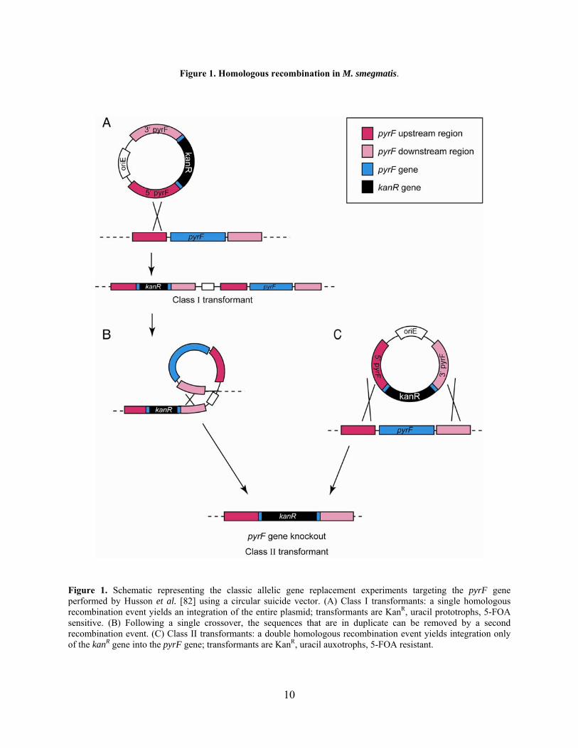
Figure 1. Homologous recombination in M. smegmatis.
Figure 1. Schematic representing the classic allelic gene replacement experiments targeting the pyrF gene performed by Husson et al. [82] using a circular suicide vector. (A) Class I transformants: a single homologous recombination event yields an integration of the entire plasmid; transformants are KanR, uracil prototrophs, 5-FOA sensitive. (B) Following a single crossover, the sequences that are in duplicate can be removed by a second recombination event. (C) Class II transformants: a double homologous recombination event yields integration only of the kanR gene into the pyrF gene; transformants are KanR, uracil auxotrophs, 5-FOA resistant.
10
1.1.3.2 Evidence of illegitimate recombination in M. tuberculosis
In the first report of illegitimate recombination in the mycobacteria, Kalpana et al. were
unable to replace either the M. bovis BCG or M. tuberculosis strain H37Rv met genes with a
kanR gene [91]. No correctly targeted gene replacements were identified out of more than 200
KanR colonies screened (Table 1). Linear double-stranded DNA (dsDNA) AESs were used in an
attempt to exclusively isolate mutants from double crossovers, and this resulted in an ~10-fold
increase in colonies. However, KanR recombinants were recovered irrespective of the presence of
homologous mycobacterial DNA sequences in the AES (pBR322::Tn5, Table 1), clearly
showing that integration of the kanR gene was not dependent on homologous DNA sequences.
The illegitimate recombinants were obtained at a relatively high frequency (i.e. 10-4 to 10-5
relative to plasmid transformants), such that they masked the presence of colonies (if any) arising
from correctly targeted recombination events. In the same study, M. smegmatis met mutants were
readily obtained using either linear or circular AESs, as expected from previous studies [82], and
recovery of recombinants was dependent on the presence of mycobacterial sequences in the AES
(Table 1). Subsequently, other groups successfully isolated mutant alleles that were generated
by homologous recombination in M. tuberculosis, but the frequencies of single crossovers were
low (<20%) and were lower for double crossovers (<5%) [3,8,147,168,188].
11
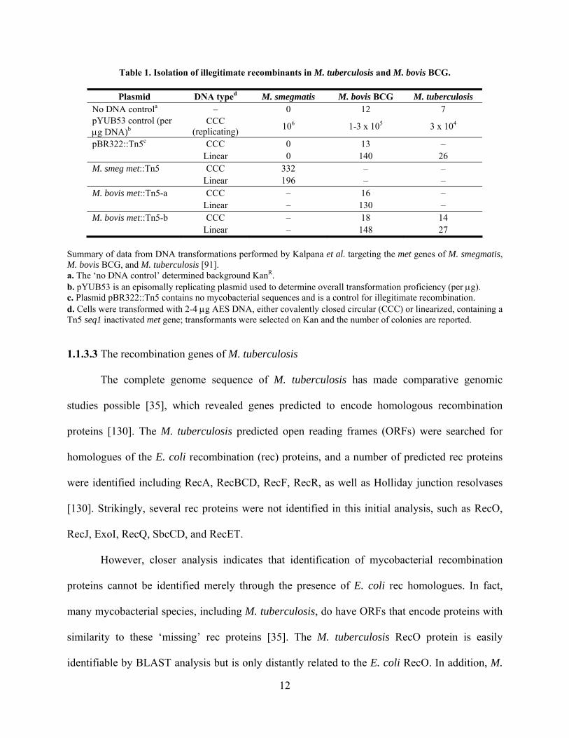
Table 1. Isolation of illegitimate recombinants in M. tuberculosis and M. bovis BCG.
Plasmid DNA typed M. smegmatis M. bovis BCG M. tuberculosis No DNA controla – 0 12 7 pYUB53 control (per g DNA)b
CCC (replicating)
106 1-3 x 10⁵ 3 x 10⁴
pBR322::Tn5c CCC 0 13 – Linear 0 140 26 M. smeg met::Tn5 CCC 332 – – Linear 196 – – M. bovis met::Tn5-a CCC – 16 – Linear – 130 – M. bovis met::Tn5-b CCC – 18 14 Linear – 148 27
Summary of data from DNA transformations performed by Kalpana et al. targeting the met genes of M. smegmatis, M. bovis BCG, and M. tuberculosis [91]. a. The ‘no DNA control’ determined background KanR. b. pYUB53 is an episomally replicating plasmid used to determine overall transformation proficiency (per g). c. Plasmid pBR322::Tn5 contains no mycobacterial sequences and is a control for illegitimate recombination. d. Cells were transformed with 2-4 g AES DNA, either covalently closed circular (CCC) or linearized, containing a Tn5 seq1 inactivated met gene; transformants were selected on Kan and the number of colonies are reported.
1.1.3.3 The recombination genes of M. tuberculosis
The complete genome sequence of M. tuberculosis has made comparative genomic
studies possible [35], which revealed genes predicted to encode homologous recombination
proteins [130]. The M. tuberculosis predicted open reading frames (ORFs) were searched for
homologues of the E. coli recombination (rec) proteins, and a number of predicted rec proteins
were identified including RecA, RecBCD, RecF, RecR, as well as Holliday junction resolvases
[130]. Strikingly, several rec proteins were not identified in this initial analysis, such as RecO,
RecJ, ExoI, RecQ, SbcCD, and RecET.
However, closer analysis indicates that identification of mycobacterial recombination
proteins cannot be identified merely through the presence of E. coli rec homologues. In fact,
many mycobacterial species, including M. tuberculosis, do have ORFs that encode proteins with
similarity to these ‘missing’ rec proteins [35]. The M. tuberculosis RecO protein is easily
identifiable by BLAST analysis but is only distantly related to the E. coli RecO. In addition, M.
12

tuberculosis Rv2837c is a member of the DHH protein family, which includes RecJ proteins
from several bacteria including E. coli. Another M. tuberculosis ORF, Rv3198c, is predicted to
encode a protein that has both a UvrD2 helicase domain and a fragment of the RecQ domain, and
is therefore described as a putative RecQ helicase. Finally, since the RecET proteins are encoded
by a cryptic prophage in E. coli, it is not surprising that these are absent in M. tuberculosis.
Therefore, this bacterium has a number of recognizable recombinational repair pathway
components.
Arguably, a comparison of the known recombination genes of more closely related
bacteria may provide better insights into the recombinational repair system of M. tuberculosis.
Comparative analysis of the B. subtilis and mycobacterial genomes revealed the presence of
multiple genes encoding B. subtilis AddA homologues (at least two) in several mycobacterial
species, including M. smegmatis and M. tuberculosis (D. Ennis and G. Cromie, personal
communication; see Appendix A and Figure 34). The AddAB proteins function similarly to
RecBCD for processing and repair of dsDNA lesions and are most commonly found in Gram-
positive bacteria, whereas RecBCD are typically encoded by Gram-negative bacteria [32,245].
The specific activities of RecBCD have not been fully characterized in mycobacteria for general
recombinational repair, and these have only been examined with regard to their role (or lack
thereof) in conjugation and non-homologous end-joining, respectively [120,234]. It is therefore
possible that both sets of recombination proteins – RecBCD and the two AddA homologues – are
active and perhaps redundant in mycobacteria. Alternatively, it may be that only one set of
proteins is expressed and/or active in vivo.
13

1.1.3.4 The debate over homologous and illegitimate recombination in mycobacteria
It was not clear from the initial studies discussed above if levels of homologous
recombination are actually decreased or if the levels of illegitimate recombination are merely
increased – or perhaps both – in slow-growing mycobacteria such as M. tuberculosis. One
hypothesis is that the presence of an intein in the M. tuberculosis recA gene reduces the activity
of this pivotal recombination enzyme, thereby decreasing overall levels of homologous
recombination (reviewed in McFadden, 1996).
In M. tuberculosis, the conserved RecA sequences are situated at the N- and C-termini of
the ORF and are interrupted by 440 amino acids that are not conserved in other RecA proteins
[45]. Splicing of the full-length protein is essential to remove this “spacer protein,” and the N-
and C-terminal regions are ligated to produce the mature active protein [46]. The recA gene of
M. leprae also includes an intein that is spliced in vivo [57], but the recA gene of M. smegmatis
does not [47], which further suggests that the abnormal gene structure of the M. tuberculosis
recA may correlate to low levels of homologous recombination. In vitro experiments with
purified M. tuberculosis RecA proteins – both full-length and mature – have shown that the
unspliced protein is defective in ATPase activity and strand exchange, whereas the mature
protein is active [103]. It is therefore possible that RecA activity in vivo is regulated by
conditional splicing of the full-length inactive protein.
In addition, expression of recA in M. tuberculosis is controlled by multiple transcriptional
regulatory elements, which adds to the complexity of regulation. Two promoters upstream of
recA are regulated in response to DNA damage, one by LexA and RecA in the classical
mechanism through an SOS box, while the other is independent of LexA and RecA (discussed
below) [48,65,127]. Additionally, RecA activity is negatively regulated by a co-transcribed
14
protein RecX in mycobacteria [154,155,230]. It is also intriguing that recA expression is much
more delayed in response to DNA damaging agents in M. tuberculosis as compared to M.
smegmatis [127,156]. It was suggested, therefore, that the genetic and biochemical
characteristics of M. tuberculosis RecA may result in reduced levels of homologous
recombination in this bacterium.
Subsequent experiments, however, suggested that the intein does not affect the function
of RecA in recombination or other activities. Expression of the M. tuberculosis RecA – with or
without the intein – in an M. smegmatis recA strain was sufficient to promote levels of
homologous recombination similar to wild type M. smegmatis, and no illegitimate recombination
was observed [56,155]. These data support two conclusions: 1) the M. tuberculosis RecA protein
inteins does not reduce the levels of homologous recombination in M. smegmatis, and 2) the
expression of M. tuberculosis RecA in M. smegmatis is not sufficient to introduce levels of
illegitimate recombination similar to those in M. tuberculosis. However, similar experiments
expressing the M. smegmatis recA in an M. tuberculosis recA strain would be required to
determine the specific role of RecA in illegitimate recombination. It is also possible that there are
factors regulating RecA splicing in M. tuberculosis that modulate its recombination activity
levels, and perhaps this does not occur in M. smegmatis.
There is evidence that suggests that the levels of homologous recombination are not
decreased in M. tuberculosis. Experiments by Pavelka et al. showed that similar numbers of
homologous transformants were obtained in M. smegmatis, M. tuberculosis, and M. bovis BCG
using circular suicide vectors, suggesting that illegitimate recombination likely occurs
predominantly with linear DNA substrates [163]. These data imply that homologous
15
recombination frequencies in mycobacteria are similar, and the increased level of illegitimate
recombination is likely what is different between the fast- and slow-growing mycobacteria.
It has also been speculated that the slow induction of recA expression in M. tuberculosis
may result in deficiencies in DNA repair and decreased SOS response, leading to high rates of
illegitimate recombination. Since recA expression is induced slowly (compared to M. smegmatis)
in response to DNA damage, this could result in reduced RecA-dependent autocatalytic cleavage
of LexA and decreased activation of downstream genes involved in the SOS response. In this
situation, it is conceivable that chromosomal breaks would be more prevalent, perhaps leading to
higher rates of illegitimate recombination for repair of these lesions. The LexA protein of M.
tuberculosis has been characterized and shown to bind an SOS box (as is typically seen with this
repressor [127,128]), and one SOS box is present in one of the promoter regions at recA.
However, it was found that two mechanisms for DNA damage response exist in M. tuberculosis,
one that is classically dependent on RecA and LexA and one that is independent of this process;
each mechanism controls a different set of genes [48,127,186]. Therefore it seems that even
though induction of recA expression is slow, other mechanisms for DNA repair and SOS
response are in place, perhaps negating the argument that recA expression kinetics play a role in
illegitimate recombination. Thus, the molecular basis of the relatively high frequencies of
illegitimate recombination in M. tuberculosis and other slow-growing mycobacteria remains an
open question.
1.1.4 Mycobacteriophage-derived genetic tools
Bacteriophages have long demonstrated their utility as sources for genetic tools in bacterial
model systems, especially those that are genetically intractable. Over fifty mycobacteriophages
16
have been isolated and sequenced to date ([73,165] and unpublished data), from which a plethora
of genetic information has been gathered, enabling the study of numerous phage genes [71,72].
For the mycobacteria, phage-derived vectors have proven extremely useful for expression of
foreign genes. Several integration-proficient vectors containing phage integration cassettes have
been developed and can be used simultaneously for stable introduction of multiple genetic
elements in a single cell [95,111,126,170]. Also of great use are shuttle phasmids, which are
chimeric cosmid molecules containing mycobacteriophage and E. coli plasmid DNA [86]. These
replicate as plasmids in E. coli and as phages in mycobacteria and are used as delivery vehicles;
their use for delivering AESs will be discussed in further detail in section 1.1.5.5 [14]. Shuttle
phasmids have also been used to deliver transposons for genetic assays [13] and as reporter
phages in clinical studies to assay for live mycobacterial cells and drug susceptibility
[10,27,88,164,189,197].
Phages have also been isolated that infect M. smegmatis and facilitate generalized
transduction, enabling transfer of mutations to other strains [111,183]. Generalized transduction
would be particularly useful for studying mutations conferring drug-resistance. However, no
generalized transducing phages that infect the slow-growing mycobacteria, such as M.
tuberculosis, have been isolated. Also of use in M. tuberculosis are phage-derived methods for
selection that can be used in place of antibiotic markers, which are not desireable in potential
vaccine strains. The mycobacteriophage L5 repressor gene product gp71 confers immunity to
superinfection. Thus, when gene 71 is expressed as a selective marker on plasmids, cells are
resistant to infection by a homo-immune phage [49]. Phage promoters have also been used for
gene expression in mycobacteria as an alternative to constitutive strong promoters such as the M.
bovis BCG hsp60 promoter [18,72]. It is clear that mycobacteriophages have contributed greatly
17
to the study of genetics in mycobacteria and will likely continue to do so as we learn more
through isolation and characterization [73].
1.1.5 Genetic techniques for allelic replacement
Characterization of isogenic mutants is a powerful method for the study of gene function, and
targeted gene replacement is a standard way to construct these defined mutants. Other techniques
such as transposon mutagenesis and random mutagenesis are extremely valuable but do not offer
the same precision or control over the type of mutations made. In many organisms, allelic gene
replacement is simple and fast, requiring little DNA manipulation and screening [38]; however,
this is not the case for the mycobacteria. Canonical substrates for targeted gene replacement
(AESs) contain a selectable genetic marker flanked by long (>1000 bp) regions of homology to
the gene locus being targeted. These substrates are introduced into the cell and homologous
recombination leads to single or double crossovers to yield a marked allelic replacement mutant.
While this strategy is successful in M. smegmatis, the prevalence of illegitimate recombination in
some of the slow-growing mycobacteria prevents this from being an efficient method for gene
replacement. Null mutations in genes resulting in an auxotrophic or otherwise identifiable
phenotype were the first constructed because they facilitated differentiation of double versus
single crossovers [3,8,9,80,158,188]. Clearly not all gene mutants would have screenable
phenotypes, and therefore even the limited success of these early methods suggested a need for
improvement.
A number of attempts have been made to improve the recovery of mutant alleles from
double homologous recombination events and reduce the need for screening. Figure 2
summarizes the multitude of techniques that were developed for the mycobacteria in a timeline
18
style and also shows the first gene replacements made in some of the more commonly studied
mycobacteria. The majority of mycobacterial genetic tools developed were aimed at modifying
the AES to make it more recombinogenic: altering the structure, treatments prior to
transformation, and delivery method. The preferred genetic techniques are successful because
they either utilize a selection for double crossovers or drastically reduce or eliminate illegitimate
recombination events. It is worth noting, however, that none of the strategies developed thus far
have successfully increased the levels of homologous recombination in M. tuberculosis. This
may be due to the complexity of recombination in the mycobacteria, or perhaps this was
attempted and never accomplished. Yet this still represents another potential method for
improving recovery of allelic replacement mutants.
19
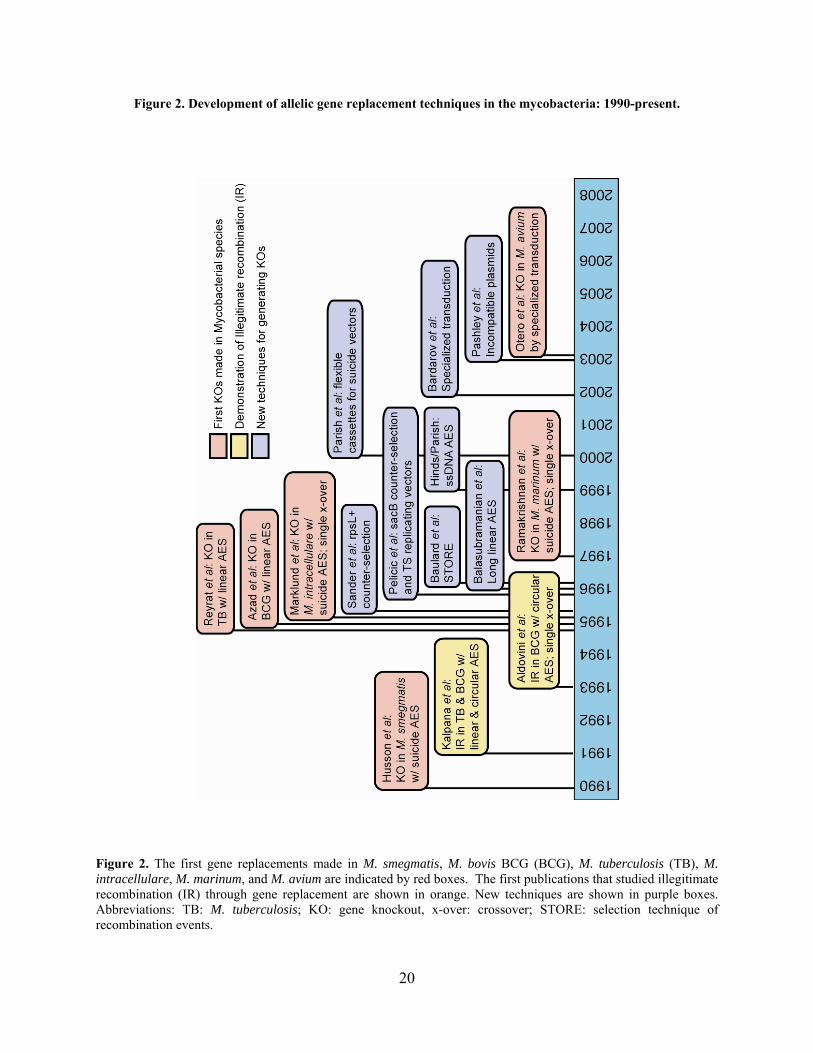
Figure 2. Development of allelic gene replacement techniques in the mycobacteria: 1990-present.
Figure 2. The first gene replacements made in M. smegmatis, M. bovis BCG (BCG), M. tuberculosis (TB), M. intracellulare, M. marinum, and M. avium are indicated by red boxes. The first publications that studied illegitimate recombination (IR) through gene replacement are shown in orange. New techniques are shown in purple boxes. Abbreviations: TB: M. tuberculosis; KO: gene knockout, x-over: crossover; STORE: selection technique of recombination events.
20

Arguably, there were two techniques that were most successful: (1) the use of suicide
vectors with counter-selectable markers, which aid in the selection of the desired double-
crossover events, and (2) the delivery of the AES by mycobacteriophages (referred to as
‘specialized transduction’). This section will discuss the numerous genetic tools developed for
the mycobacteria over the last 18 years.
1.1.5.1 AES structural modifications
Numerous AES designs were explored to optimize allelic exchange frequencies: linear
versus circular DNA substrates, the length of sequence identity, the presence of nonhomologous
DNA flanking the homologous regions, and the selectable marker. The initial experiments
performed by Kalpana et al. used both a linear and circular dsDNA AES [91], while Aldovini et
al. used a circular suicide vector as an AES [3]. Using a linearized AES yielded up to ten-fold
more colonies than the circular substrate and resulted in mostly illegitimate events in multiple
studies [91,163]. It therefore appears from these experiments that: (1) using a circular AES yields
lower numbers of recombinants compared to a linear AES, but these result from predominantly
illegitimate recombination and single crossover events in M. bovis BCG and M. tuberculosis
[3,91], (2) using linear AESs did not result in any identified homologous recombination events
(single or double crossovers), only illegitimate events [91] in M. tuberculosis and M. bovis BCG,
and (3) using circular AESs in M. smegmatis can facilitate both single and double homologous
recombination events [82] with low amounts of illegitimate recombination [80]. Later
experiments with linearized AESs were somewhat successful in M. tuberculosis and M. bovis
BCG for making double crossover mutants, although at low frequencies (~4%) [8,188].
Balasubramanian et al. succeeded in making gene replacements in leucine biosynthetic
genes using long (40-50 kbp) linear AESs [9]. Genomic cosmid libraries of M. tuberculosis
21
H3Rv and M. bovis BCG were constructed, and interplasmid recombination in E. coli was used
to make the kanR-marked disrupted leuD allele. In this case, transformants were obtained equally
with linear or circular cosmid AESs, but leucine auxotrophs were only found with the linear
AES; 6% double crossover mutants were identified. While this was a successful method, it was
time-consuming, and another group demonstrated similar frequencies (4%) of double crossover
using linear AESs with short (>1 kbp) homologies [188], albeit at a different locus.
Since low levels of spontaneous resistance to kanamycin occur in slow-growers [91],
others have used different antibiotic resistance genes such as hygR, gentamicin resistance (gentR),
streptomycin resistance (strR) and even mercury resistance as markers [14,15,82,147,159,161].
However, these methods did not generally improve the recovery of double crossover mutants. It
was also suggested that the presence of nonhomologous sequences flanking the homology
targeting the gene might increase the propensity for the AES to undergo illegitimate
recombination [3,91], although this has not been tested rigorously.
1.1.5.2 Treatment of the AES
Neil Stoker’s group has shown that treating the DNA substrate with agents that promote
the formation of single-stranded DNA (ssDNA) improves the frequency of homologous
recombination in M. smegmatis, M. intracellulare, and M. tuberculosis [80,158]. The most
effective experiments utilized treatments with alkali or by boiling to denature the DNA, or
merely used ssDNA derived from phagemids. In experiments with ssDNA AESs, not only were
transformant numbers typically increased, but also the proportion that had undergone double
crossovers. Importantly, the use of phagemid DNA eliminated the recovery of illegitimate
transformants.
22

1.1.5.3 Plasmid delivery of the AES
Numerous groups have also made allelic exchange mutants in mycobacteria using either a
circular or linearized suicide vector [3,8,121,159,184,188]. These are plasmids that rely on
integration via homologous recombination for maintenance in the mycobacteria, either through a
single crossover (in which the entire plasmid is integrated) or double crossover (in which the
targeted chromosomal gene is replaced by the disrupted gene) (see Figure 1). Despite the high
frequency of illegitimate recombination in the slow-growers, homologous recombination using
these substrates is still relatively successful. Further, although single crossovers occur at a higher
frequency than double crossovers, single crossover mutants can be propagated and screened for a
second recombination event between the duplicate sequences to loop out the excess vector
(Figure 1); however, this does not occur at a high frequency [91]. Plasmids with multiple cloning
sites flanking different antibiotic markers were constructed to simplify synthesis of the AES
suicide plasmid [159], but the screening was still labor-intensive. The development of a two-step
counter-selection strategy (discussed below) greatly improved this by reducing the number of
transformants screened.
Since the frequency of homologous recombination is lower than the transformation rate
in mycobacteria, large quantities of DNA are required for transformations (up to 4 g). The use
of a replicating vector for delivery of the AES could arguably work better than a suicide vector,
since extended survival of the plasmid would likely improve the frequency of recombination
with the target. A replicating plasmid was used in one study, but did not result in a stable mutant
allele of the targeted gene accBC in M. bovis BCG. However, the reason for this is unknown
since PCR and Southern blot analysis confirmed that homologous recombination with the AES
had occurred [147]. Another group developed a technique called STORE (selection technique of
23
recombination events) that uses a replicating plasmid with a promoter-less kanR gene targeted to
the M. bovis BCG hsp60 locus for replacement of the hsp60 gene [15]. Selection for KanR
therefore yielded recombinants that had undergone homologous recombination at the hsp60
locus, which placed the kanR gene under control of the constitutive hsp60 promoter. However,
extension of this technology for targeting other loci would require that the gene is expressed.
One concern with replicating vectors is removing the plasmid; temperature-sensitive
plasmids offer an advantage here, but for best results in the slow-growing mycobacteria these are
combined with SacB counter-selection (examined in more detail below) [169]. Pashley et al.
made use of incompatible plasmids to facilitate removal of the plasmid following gene
replacement [161]. This technique uses a pair of plasmids that replicate co-dependently and are
lost in the absence of selection. The plasmid carrying the AES can therefore undergo targeted
gene replacement. However, this method like many others requires multiple rounds of selection,
growth, and plating, making it less efficient than other techniques.
1.1.5.4 The counter-selection strategy
Husson et al. was the first to use counter-selection for allelic exchange in the
mycobacteria (discussed in section 1.1.3.1). In this study, the pyrF gene in M. smegmatis was
replaced with a kanR gene through a double crossover event. The mutant was selected by plating
on 5-FOA, since loss of wild type pyrF confers resistance [82]. This technique was extended
later by Knipfer et al. who used the pyrF gene as a selective marker in a pyrF strain for
unmarked introduction of genes [97]. Since this is therefore limited to the pyrF locus, broader
strategies were developed. Another useful counter-selection strategy is the introduction of the
wild type rpsL gene (rpsL+) in a strain with a specific rpsL mutation that confers streptomycin
resistance (StrR) [196]. Plating a strain that contains both wild type and mutant alleles on
24
streptomycin selects for loss of the wild type rpsL gene. Therefore when rpsL+ is placed on a
suicide AES, double selection on streptomycin and kanamycin (e.g., if kanR is the disrupting
genetic marker) results in generation of predominantly double crossover gene replacement
mutants in M. bovis BCG. However, this requires the use of a StrR resistant strain background,
which is not ideal for vaccine development.
The B. subtilits sacB gene has been extremely useful as a counter-selective marker in
mycobacterial genetics. The presence of the sacB gene causes sensitivity to sucrose, and
therefore plating on sucrose selects for loss or mutation of the gene (Figure 3) [166-168]. Allelic
exchange mutants that are the products of double homologous recombination events can be
obtained in a single step by dual positive and negative selection with antibiotics and sucrose at
100% efficiency. Alternatively, if this is unsuccessful, allelic exchange can be performed in two
steps, in which single crossover mutants are selected by antibiotic resistance, followed by
removal of the vector sequence by a second crossover event, selected by plating on sucrose (this
occurs in ~two-thirds of the colonies screened). This strategy can also be used to make unmarked
mutants; in this case, γδ resolvase sites are placed flanking the antibiotic marker and sacB, and
recombinants from expression of the resolvase can be selected by sucrose resistance (Figure 3).
The sacB gene has also been used for very effective gene replacement (100%) on replicating
temperature-sensitive plasmids as AES delivery vehicles: it ensures loss of the plasmid by
shifting to high temperature and plating on media with sucrose [169].
25

Figure 3. Gene replacement by counter-selection with sacB.
Figure 3. Genes (yfg: your favorite gene) targeted by using sacB on the vector DNA result first in a (A) single crossover and then loop out the vector, or a (B) double crossover in vectors which contain the sacB gene on the backbone. Simultaneous selection for antibiotic resistance (e.g. KanR) and sucrose resistance can yield either (C) removal of the vector containing sacB or (D) mutation of sacB. (E) Unmarked mutations can also be generated by using γδ resolvase: res sites are placed flanking the antibiotic resistance gene (e.g. kanR) and sacB (instead of it being on the vector backbone).
26
1.1.5.5 Specialized transduction
Delivery of the AES by phage infection, a method called ‘specialized transduction,’ has
proven to be a successful method for targeted gene replacement [14]. This was accomplished by
the development of shuttle phasmids, which are chimeric DNA molecules that replicate as
plasmids in E. coli and phages in mycobacteria [86]. Phasmids contain phage genomic DNA
with an E. coli plasmid inserted in a non-essential region of the genome (Figure 4). They can
therefore replicate as plasmids in E. coli and as phages in mycobacteria. This technology was
developed by Jacobs et al. using mycobacteriophage TM4, and later mycobacteriophages D29
and L1 [86,164,210]. The most commonly used shuttle phasmid is phAE87, which is a TM4
shuttle phasmid containing a temperature-sensitive mutation that allows phage propagation at
30°C but not at 37°C [13]. Shuttle phasmids have been used not only for delivery of transposons
and expression of reporter genes, but also for delivery of AES for targeted gene replacement in
both the fast- and slow-growing mycobacteria [13,14,88,164].
27
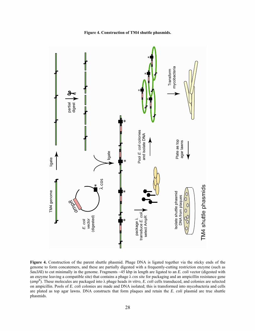
Figure 4. Construction of TM4 shuttle phasmids.
Figure 4. Construction of the parent shuttle phasmid. Phage DNA is ligated together via the sticky ends of the genome to form concatemers, and these are partially digested with a frequently-cutting restriction enzyme (such as Sau3AI) to cut minimally in the genome. Fragments ~45 kbp in length are ligated to an E. coli vector (digested with an enzyme leaving a compatible site) that contains a phage λ cos site for packaging and an ampicillin resistance gene (ampR). These molecules are packaged into λ phage heads in vitro, E. coli cells transduced, and colonies are selected on ampicillin. Pools of E. coli colonies are made and DNA isolated; this is transformed into mycobacteria and cells are plated as top agar lawns. DNA constructs that form plaques and retain the E. coli plasmid are true shuttle phasmids.
28

For gene replacements, a canonical AES is constructed by cloning ~1000 bp of upstream
and downstream homology to the target gene flanking an antibiotic marker (typically kanR or
hygR). This can be directly cloned into a parent shuttle phasmid such as phAE87 to replace the
existing E. coli plasmid sequences, and shuttle phasmid molecules containing the AES are
prepared. A mycobacterial culture is then infected with mycobacteriophage-packaged shuttle
phasmids at a non-permissive temperature for phasmid replication, and this facilitates delivery of
the AES and targeted gene replacement (Figure 5). This method has been used to make more
than 300 gene mutants in M. tuberculosis (W.R. Jacobs, Jr., personal communication).
Specialized transduction has also been used to construct a strain of M. tuberculosis containing a
single defined point mutation in the inhA gene [232]. This was the first experiment in which a
point mutation was placed in an endogenous gene in a wild type background, and is an example
of the power of specialized transduction.
29
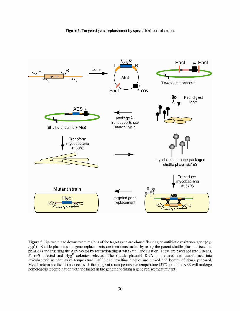
Figure 5. Targeted gene replacement by specialized transduction.
Figure 5. Upstream and downstream regions of the target gene are cloned flanking an antibiotic resistance gene (e.g. hygR). Shuttle phasmids for gene replacements are then constructed by using the parent shuttle phasmid (such as phAE87) and inserting the AES vector by restriction digest with Pac I and ligation. These are packaged into λ heads, E. coli infected and HygR colonies selected. The shuttle phasmid DNA is prepared and transformed into mycobacteria at permissive temperature (30°C) and resulting plaques are picked and lysates of phage prepared. Mycobacteria are then transduced with the phage at a non-permissive temperature (37°C) and the AES will undergo homologous recombination with the target in the genome yielding a gene replacement mutant.
30

In conclusion, a variety of techniques have been developed for gene replacement
mutagenesis of M. tuberculosis with varying success. Each method has drawbacks that include
time-consuming AES constructions or screening of large numbers of recombinant colonies. In
other organisms, technologies for mutagenesis have been greatly improved through the use of
phage-encoded recombination proteins. In particular, genetics in E. coli and related Gram-
negative bacteria have benefited enormously by exploiting these recombination proteins in a
genetic system called recombineering. The following sections will discuss the recombination
proteins of bacteriophages that promote single strand annealing homologous recombination and
their use for development of host genetic tools.
1.2 SINGLE STRAND ANNEALING PROTEINS
Homologous and non-homologous recombinational repair of DNA is an extremely well-studied
field that is exemplified by research in E. coli and bacteriophage λ [106]. Homologous
recombination – the pairing and exchange of complementary strands – can be divided into two
mechanisms: strand invasion and single strand annealing. The two classically defined
mechanisms of RecA-dependent strand invasion are: the ‘daughter strand gap repair pathway’
involving the RecF ‘machine,’ and the ‘double-strand end repair pathway’ mediated by the
RecBC complex (reviewed in Kuzminov 1999). Although alternative repair pathways exist that
involve different combinations of the Rec proteins, it is clear that RecA plays a central role in
recombinational repair of chromosomal lesions that occur during replication and DNA damage.
The second major recombination pathway that appears to be conserved through
eukaryotes is called the ‘single strand annealing pathway.’ As the name implies, single strand
31

annealing involves pairing of complementary single strands via a RecA-independent mechanism
that is initiated at double strand breaks (Figure 6) [220]. These recombination proteins, called
single strand annealing proteins (SSAPs), promote strand pairing, strand exchange, and strand
invasion [17,69,114,129,145,193]. SSAPs are found predominantly in bacteriophages and in
bacterial genomes in prophages, although they have also been identified in eukaryotes, including
yeast and humans. The SSAPs comprise three superfamilies based on sequence conservation: (1)
the Red /RecT family, (2) the Erf family, and (3) the Rad52 family [85]. It appears that these all
have bacteriophage origins and are typically found adjacent to other DNA recombination or
repair proteins, such as exonucleases. These groups of proteins and their biochemical
characteristics will be explored in this section.
32

Figure 6. Single strand annealing pathways.
Figure 6. SSAPs can catalyze recombination by three basic mechanisms: (A) strand pairing, (B) strand exchange, and (C) strand invasion. The partner exonuclease (RecE or Exo) degrades a dsDNA end 5-3 leaving behind a 3 ssDNA tail. This is bound by the SSAP (RecT or Beta) and recombined with its homologous target sequence.
33

1.2.1 Single strand annealing protein families
The founding members of the SSAP superfamilies – λ Beta, Rac RecT, P22 Erf, and yeast Rad52
– have been extensively characterized genetically, biochemically, and structurally, leading to the
general concept that these proteins are functional analogues and ‘structural homologues’ [162].
These ‘recombinases’ form ring structures, bind ssDNA and dsDNA, and catalyze pairing, strand
exchange, and strand invasion [162,174,204,224]. Although no sequence similarity was initially
observed between any of the founding members, they were shown to fall into three
evolutionarily defined superfamilies [85]. The ‘Red /RecT superfamily’ is comprised of the
bacteriophage λ Beta (Red ) and the E. coli Rac prophage RecT proteins. RecT and Beta have
no apparent sequence similarity but function analogously such that RecET can substitute for
Exo/Beta for phage λ recombination [66]. PSI-BLAST analysis with Beta homologues from
numerous other lambdoid phages retrieves the RecT protein and its homologues. Sequence
analyses further revealed several conserved residues as well as secondary structure predictions
that correlate well with some of their biochemical properties, such as Mg2+-dependent ssDNA-
pairing and dsDNA binding activities [96,145]. Further, λ Beta homologues are present in
numerous diverse bacteria and phages, while RecT-like proteins appear predominantly in low
G+C% Gram-positive bacteria and phages. Two proteins found in this superfamily – E. coli
EHAP1 and Borrelia hermsii PF161 – have an unusual domain structure; the N-terminal domain
is similar to the Beta/RecT family, while the C-terminus is similar to the Erf family.
The bacteriophage P22 Erf protein has also been described as a SSAP and defines another
superfamily [85,178]. Conserved motifs have been identified in these proteins, and much like the
34

Beta/RecT family, they seem to have originated in bacteriophages and subsequently appeared in
bacterial genomes as prophages. P22 Erf can also substitute functionally for λ Beta [175].
The third small superfamily of both eukaryotic and bacterial SSAPs was identified by
database searches with eukaryotic Rad52 proteins. Rad52 from yeast and humans has been
shown to act as a SSAP in conjunction with the RecA ortholog Rad51 [17]. Sequence alignments
and structural predictions detect a conservation of two large motifs and other structural elements
(including two putative helix-hairpin-helix folds) in both eukaryotic and bacterial Rad52s,
indicating that they all belong to a single superfamily [85]. Although these proteins have been
characterized biochemically, the following sections will focus on the bacteriophage systems of
phage λ, E. coli Rac prophage, and P22.
The genes adjacent to the SSAPs are commonly predicted to be DNA recombination or
repair proteins [85]. These include single-strand-binding protein (SSB), Holliday junction
resolvases, and nucleases, specifically exonucleases like λ Exo and RecE, which are found with λ
Beta and RecT, respectively. Most of the exonucleases fall in two families, the type II restriction
enzyme fold (e.g. λ Exo) and the type EndoVII fold. This suggests that SSAPs work in
conjunction with their partner proteins in recombination and recombinational DNA repair.
However, in some phages, the SSAPs and exonucleases are mixed, which is unexpected given
the apparent specificity of the exonuclease-SSAP protein interaction observed with the λ Red
and RecET systems [142]. For example, in several phages, a gene encoding a λ Exo-like protein
is located next to a RecT-like gene. Additionally, SbcC-like genes are adjacent to both RecT-
and λ Beta-like genes. In one unique case, a Beta-like gene was fused to a C-terminal fragment
of the P22 Erf gene (Borrelia hermsii circular plasmid pf161 gene). Also, the order of the genes
within the operon differs between phages such that either the SSAP or its partner gene may be
35

transcribed first [43,85]. Collectively, the organization of these phage-encoded recombination
genes reflects the modular structure that is characteristic of phage genomes.
1.2.2 The Red recombination proteins
The Red recombination system of bacteriophage λ was identified by the observation that
bacteriophage λ could replicate in the absence of RecA [23]. Red- mutants (recombination-
deficient) were found to map to genes encoding the Exo and Beta proteins [181,207], which were
shown to be required for the RecA-independent recombination observed in λ [206,207]. Red-
mediated recombination is stimulated by the presence of double-strand breaks that act as the
substrates for Exo. Exo is an ATP-dependent dsDNA exonuclease that degrades DNA in the 5
to 3 direction at approximately 1000 bases per second [29,63,116,124] and leaves behind long 3
ssDNA ends [79]. The enzyme requires a dsDNA end for activity and cannot degrade at nicks in
DNA [28,29]. The structure of the active enzyme is a trimer that forms a toroid through which
the dsDNA passes at one end and the resulting ssDNA substrate through the other [100].
The λ Beta protein is a SSAP that binds ssDNA substrates of lengths greater than 35
nucleotides [144] that protects ssDNA from nuclease attack prior to synapsis [92,114,129]. Beta
promotes renaturation of complementary ssDNAs [96,129], strand exchange (displacement)
[114], and strand invasion [193] all of which have been studied as recombination mechanisms of
the single strand annealing pathway. Following pairing of ssDNAs, Beta binds tightly to the
dsDNA complex [114]. Electron microscopic analyses show that Beta – like RecT and P22 Erf –
forms circular structures in the absence of DNA which increase in size and monomer
composition in the presence of ssDNA. Beta also forms helical filaments in the presence of
dsDNA [162]. The data from structural studies suggest that ssDNA molecules are actually
36

wrapped around the Beta toroid, perhaps to prevent ssDNA from forming secondary structure
and maintaining a conformation such that the bases are exposed for strand pairing. Beta also
interacts with other proteins as determined by co-purification which precipitate λ Exo [129,182],
host ribosomal protein S1 [129], and RNA polymerase subunit NusA [231]. Beta interacts
specifically with Exo [129], functioning to modulate its activity as it degrades linear dsDNA
substrates [225], and this interaction cannot be mimicked with other functionally analogous
exonucleases [142]. Another attractive idea is that Beta also functions to interact with
transcription and translation factors, perhaps to remove these complexes in front of the
exonuclease [106].
The third protein that acts with the Red system in λ, Gam, binds the RecB subunit of the
RecBCD nuclease in E. coli, preventing it from binding to dsDNA ends and thereby inhibiting
all known enzymatic activities of this complex [39,93,122,133,138,176]. It has also been shown
to interact genetically with the gene product of sbcC, though this is less-well characterized [102].
Although Gam is not required for recombination activities of Exo and Beta in phage λ [53], it
increases recombination by limiting host nuclease attack on linear dsDNA substrates
[38,106,142,240,241]. Alternatively, strains of E. coli that are recBC sbcBC or recD (which
are typically used for linear DNA transformation) show an increase in recombination of linear
AESs, though not as high as observed using a Gam-expressing strain (20- to 800-fold increase)
[135]. Numerous other bacteriophages are known to encode Gam functional analogues that
inactivate or block host nucleases [195], and examples include the phage T4 protein gp2
[6,115,208] and phage Mu Gam protein [2]. These proteins bind dsDNA ends and protect
injected linear DNA from degradation by RecBCD. In addition, the phage P22 Abc1 and Abc2
proteins work cooperatively to modulate RecBCD activity (discussed below). Therefore,
37
although the mechanisms of nuclease inhibition are different, the ultimate result is the protection
of linear DNA ends from degradation by host nuclease.
The Red genes exo and bet, along with gam, are expressed from the PL operon during
early infection or upon induction of lysis of the prophage [38]. The Exo and Beta proteins are
believed to play a role in phage λ infection during DNA replication by functioning to increase
DNA synthesis [106], although this is still not well understood. Phage λ DNA molecules are
replicated initially as circular molecules by theta replication, and this switches to rolling circle
(sigma) replication and forms concatemers of linear DNA. Since initiation of DNA replication
likely requires circular DNA (prior to concatemer formation), DNA synthesis could conceivably
be increased through generation of additional circular genomes by Exo/Beta recombination
[106]. Gam functions to inhibit the degradation of the linear concatemers of λ genomic DNA by
the RecBCD nuclease [53]. Therefore the Red and Gam proteins are not essential for λ
propagation but mutations in these genes result in fewer plaques [53]. The Red proteins also are
involved in generalized transduction of λ, although at low levels compared to RecA [106].
Additionally, since the conditions that stimulate the lytic cycle of λ prophages may also cause
DNA damage (such as ultraviolet light), the Red proteins could be important for repair of the
resulting double-strand breaks [179]. Numerous studies have investigated the mechanism of
recombination employed by phage λ [106], and it has been found that single strand annealing
occurs in the absence of RecA, while strand invasion is favored in the presence of RecA [216].
1.2.3 The Rac prophage RecET recombination proteins
SSAPs were first described in E. coli as an alternative recombination pathway in a
recBC strain [12]. Analysis of mutations that suppress recBC revealed a class of mutations that
38
map to the sbcA gene (suppressor of recBC) and activate expression of the recE and recT genes
of the cryptic Rac prophage in the E. coli genome [34,68,105]. The RecE (ExoVIII) and RecT
(RecET) proteins catalyze recombination independently of RecA similar to λ Exo and Beta and
have been shown to be functional analogues. Specifically, mutants of λ deleted for the Red
recombination genes were able to recombine only in E. coli strains that expressed the Rac
prophage recE and recT genes (i.e. sbcA-) [63,66]. Although the two systems function similarly,
recombination does not proceed when the paired proteins are mixed heterologously (e.g. λ Exo
and RecT), and only RecE binds RecT in vitro, indicating that there is a specific interaction
between the cognate proteins required for recombination [142].
The RecE enzyme – like λ Exo – is a highly processive ATP-dependent exonuclease that
degrades linear dsDNA 5 to 3, cannot act at nicks or gaps, and has low but detectable activity
on ssDNA [89,90,105]. RecE is a member of the RecB nuclease family of proteins: the C-
terminus of RecE is similar to the nuclease domain in the C-terminus of RecB, and mutations in
the conserved critical residues of RecE either abolish or decrease nuclease activity [31].
However, the N-terminal 587 amino acids (full-length RecE is 866 amino acids) are not required
for its exonuclease activity or recombination [33,119,142]. The SSAP, RecT, acts to pair ssDNA
substrates and promote strand exchange and invasion [68,69,145], and exhibits properties of
homology-recognition with RecA [146]. It was also shown to bind dsDNA in the absence of
magnesium, whereas ssDNA binding is only decreased slightly by the presence of magnesium
[145]. RecT protein monomers form open and closed rings in the presence and absence of
ssDNA as well as nucleoprotein filaments with RecE on dsDNA [224]. Finally, unlike phage λ,
the Rac prophage does not encode a Gam-like protein.
39
1.2.4 The P22 Erf, Arf, and Abc recombination proteins
Much like λ, bacteriophage P22 encodes a homologous recombination system that functions
through the single strand annealing pathway. However, unlike λ, recombination-mediated
circularization of the linear genomic DNA upon entry into the host cell is required for DNA
replication [237,238], and the phage proteins are therefore absolutely essential in recA strains of
Salmonella [218]. Recombination-deficient mutants of P22 can also be complemented by the λ
Exo and Beta proteins and vice versa [175,178]. The P22 recombination system is composed of
Erf (essential recombination function), Arf (accessory recombination function), and Abc1 and
Abc2 (anti-recBCD) proteins. Erf, the SSAP in this system, binds and protects ssDNA [131,173],
promotes strand annealing [136], and forms ring structures [162,174]. It has also been shown to
bind dsDNA under certain conditions [173], and in general appears to be biochemically
equivalent to λ Beta and RecT. Arf is less well-characterized, but it is known to be required
along with Erf for the recombination activity of P22, and is located adjacent to erf in the PL
operon [177,203].
The Abc proteins function to modulate RecBCD activity: they are not essential but
phages lacking these are decreased in burst size [132]. Null mutations in recB of the host restore
progeny levels to wild type [54], suggesting that they prevent the degradation activity of
RecBCD much like λ Gam. It appears that Abc2 functions similarly to Gam but with distinct
differences: Gam inhibits all activities of RecBCD [133], while Abc2 inhibits RecBCD
recombination (dsDNA-exonuclease, ATPase, and helicase activities) but retains its 5 ssDNA
exonuclease activity [134]. It therefore appears that P22 uses Abc2 to modulate and exploit the
ssDNA exonuclease activity of RecBCD to synthesize recombinogenic substrates for Erf.
Through binding to the RecC subunit, this Abc2-modified RecBCD complex was shown to
40

interact with λ Beta and substitute for λ Exo in Red recombination [136]. Therefore the Abc2-
RecBCD complex appears to have activity similar to λ Exo, RecE, and other 5-3 exonucleases.
It is not clear yet what role the Arf and Abc1 proteins play, although Abc1 is not required for
Abc2-RecBCD/λ Beta recombination of phage λ [136]. Further, it is unknown if one of the P22
recombination proteins or a host protein such as SSB functions to protect the 3 ssDNA tails
following degradation by Abc2-RecBCD. Finally, while the λ Red recombination proteins Exo
and Beta can work independently of Gam, it is clear that the mechanism of recombination in P22
is different and requires its ‘Gam analogue,’ Abc2, for recombination. In fact, it appears
functionally equivalent to both λ Exo and Gam by simultaneously inhibiting deleterious effects
of RecBCD and taking advantage of its exonucleolytic activity in single strand annealing
recombination.
1.2.5 SSAP mechanisms of recombination in vivo: single strand annealing versus strand
exchange
Recombination by phage λ can proceed effectively in the absence of RecA [23,206,207], and
both strand annealing and strand invasion activities have been shown in numerous in vitro
reactions with only λ Beta or RecT proteins [69,96,114,129,145,193]. Yet, in different reports,
the question as to which mechanism of recombination occurs in vivo has been contested
[55,142,206,207]. Experiments investigating phage λ recombination have further implicated a
role for RecA in some SSAP-mediated recombination such as strand invasion [64,96,129,178].
In studies where λ DNA replication is blocked, Red-mediated recombination is drastically
reduced in the absence of RecA [215]. Thaler et al. showed that DNA replication of the λ
genome was required to produce populations of dsDNA ends as substrates for the Red proteins
41
[222], which provided an explanation for λ Red recombination dependence on either DNA
replication or RecA. Stahl et al. therefore carefully tested the two proposed mechanisms of Red-
mediated recombination in λ: (1) strand invasion, and (2) strand annealing. They found that the
strand annealing was the predominant type of recombination (with low levels of strand invasion)
in the absence of RecA, whereas Red-mediated strand invasion occurred in the presence of RecA
[216]. Strand annealing by the Red proteins was observed at a high frequency during λ DNA
replication. However, λ Red dependence on DNA replication was eliminated by the introduction
of dsDNA breaks on the λ genome, although this slowed strand annealing [216]. These data
suggest that DNA undergoing replication is an optimal substrate for single strand annealing
promoted by Red proteins, and likely other SSAPs.
1.3 RECOMBINEERING IN ESCHERICHIA COLI
Recent advances in E. coli genetics have illustrated the utility of bacteriophages through
the development of a simple yet powerful technique called ‘recombineering’: genetic
engineering in bacteria using phage recombination proteins [38,223]. Recombineering facilitates
numerous types of mutagenesis in E. coli through expression of the potent recombination
proteins of either the λ Red or Rac prophage systems. Single strand annealing recombination
mediated by these proteins occurs with small lengths of homology (<50bp) and therefore allows
simple synthesis of substrates for mutagenesis. This is reminiscent of genetic techniques that
have long been available in yeast, in which the double-strand break repair system – that includes
the SSAP Rad52p – promotes recombination between short regions of homology [152].
Recombineering in bacteria can be used to target chromosomes, plasmids, and phage genomes
42

and has been expanded for use in other Gram-negative bacteria such as Salmonella, Shigella, and
Vibrio [42,185]. In addition, modifications of bacterial artificial chromosomes (BACs) by
recomineering in E. coli has made a huge impact on functional genomic research
[109,140,219,236]. This has simplified construction of mouse knockout constructs in BACs and
high-throughput manipulation of genomic libraries by alleviating the time-consuming steps of
traditional recombinant DNA cloning techniques [36,199]. Genetic engineering with ssDNA
substrates has even been demonstrated in mammalian cells either expressing λ Beta or RecT
[244] or in wild type cell lines [83,171]. Clearly this is a highly efficient system for genetics that
is broadly applicable.
1.3.1 Recombineering systems: λ Red and RecET
E. coli recombineering systems have been successfully developed using both the λ Red/Gam
proteins and the Rac prophage RecET proteins. This technique has far surpassed those previously
available for targeted gene replacement by largely increasing the numbers of transformants that
are recovered. Earlier methods used conventional AESs with large amounts of homology (>1
kbp) that were typically transformed into recombination-proficient E. coli such as recBC
sbcBC or recD strains [192], although this severely limited the strain background that could
be utilized. In the first demonstration of recombineering, Murphy placed the λ exo bet genes in
the chromosome of a recBCD strain background and showed a large increase (up to three
orders of magnitude) in gene replacement frequencies, which was dependent on inducible
expression of Exo and Beta [135]. It was also shown that a strain expressing Exo, Beta and Gam
worked just as well as a recBCD strain expressing only Exo and Beta [135,137]; however,
43

expression of Exo and Beta in a wild type background was not sufficient to promote gene
replacement without Gam or recBCD in this particular study [135]. Murphy’s ‘hyper-rec’ strain
with the λ genes placed on the chromosome was more effective for recombination than strains
containing the plasmid-encoded Exo/Beta, and therefore the decrease in copy number and level
of protein expression in the chromosomally-encoded proteins was compensated [135]. One
possible explanation for this was that perhaps the linear multimeric plasmids that undergo rolling
circle replication compete with the linear AESs for Exo and Beta.
Shortly following this study, Zhang et al. produced a similar tool for gene replacement in
E. coli using the RecET proteins in combination with λ Gam on a plasmid [242]. This system
was developed following the observation that gene replacements were obtained with short
homologies (42 bp) only in sbcA E. coli strains, which express the RecET proteins from the
Rac prophage. The need for an easily transferable system was solved by expressing the recE and
recT genes from a plasmid. Further, the λ gam gene was incorporated in place of using recBC
strains.
Numerous technical advances were applied to these two methods, but ultimately the
system developed with λ Red by Donald Court and colleagues was preferable and is now the
most commonly used. A modified λ prophage was used to tightly control expression of exo bet
gam for short induction times while preventing cell death from prolonged expression [240].
Including Gam in the recombination system eliminated the need for recBCD strains and
allowed high levels of recombineering in any strain background. These modifications eliminated
problems with leaky expression that caused other undesirable recombination events and plasmid
instability. While the λ prophage configuration is typically used, similar plasmid versions have
been developed for use in E. coli and other bacterial systems [42], making the system more
44

mobile. The P22 system was also tested for its ability to promote recombineering in these assays
but was found to be less efficient than the λ Red system [135], although this was not tested
extensively.
1.3.2 The recombineering strategy for mutagenesis
Recombineering in E. coli has been successful for making several kinds of mutants: targeted
gene replacements, point mutations, deletions, and small insertions [38,244]. The system can
also be used for BAC modification, gene specific random mutagenesis, and in vivo cloning by
gap repair [38,140,143,201,243]. Most applications of this system have been described in
detailed protocols [7,38,42,201], though more are likely to appear in the future. Some of the
more commonly used techniques such as targeted gene replacement and point mutagenesis will
be discussed here in more detail.
Several expression strategies were tested for optimal recombination activity. In one setup,
RecT was placed under a constitutive promoter and RecE under an inducible promoter [242].
Stronger promoters (Ptac) increased expression five-fold but actually decreased recombination
activity two-fold [137]. Observations such as this indicated that there is likely an optimal level of
expression of the pair of recombination proteins, and it is suggested that a 5:1 ratio of Beta to
Exo results in the highest level of recombination (K. Murphy, personal communication). Others
have placed the exo bet genes under inducible control while keeping gam constitutively
expressed [141]. However, the ideal configuration for the λ Red/Gam system was developed
using a modified prophage that carefully controls expression through their native promoter for
maximal recombination activity and minimal cell death [240]. This was accomplished by
removing the lytic genes and using a temperature-sensitive allele of the λ cI repressor (cI857)
45
such that expression of the PL operon (including the λ exo bet gam genes) for less than 60
minutes is tolerable. The strain is grown at 32°C then shifted to 42°C for 15 minutes to induce
expression of the Red proteins, after which electrocompetent cells are prepared [201]. Although
the protocols differ for each type of mutagenesis, the strain background is typically the cI857 λ
defective prophage version, unless a plasmid encoding the λ Red genes is being used.
1.3.2.1 Recombineering with dsDNA substrates
Targeted gene replacement by recombineering eliminates the need for special
recombination-proficient strains of E. coli and yields large numbers of colonies (>104) following
transformation with an AES. Even the synthesis of the AES was made simpler by eliminating the
need for cloning. Since the SSAPs can perform recombination with short substrates (>35 nt)
[144], AESs can be made using PCR-generated substrates with short regions of homology
flanking the antibiotic cassette (Figure 7) [242]. The distance between the homologies does not
appear to affect recombination frequencies [242], while extending the length of homology from
20 bp to 40 bp increases recombination by four orders of magnitude [240]. Extending the
homology proportionally increases gene replacement frequencies mediated by RecET or λ
Exo/Beta up through 1500 bp [142], though the difference between 40 bp and 1000 bp only
increased Red recombineering frequencies 10-fold [240]. However, since small regions of
homology are sufficient, substrates for targeted gene replacement typically include 50 bp of
homology flanking a variety of antibiotic resistance genes [7]. Saturating amounts of the AES
are reached at 100 ng, so this quantity of DNA is used in standard transformations [240].
Counter-selection with sacB has also been used to generate unmarked deletions of genes
following recombineering [137]. Further, this method for gene replacement can be used on either
the chromosome or on plasmids [240].
46
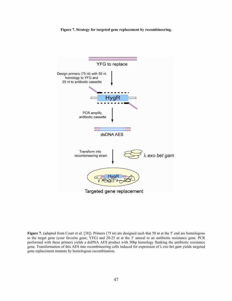
Figure 7. Strategy for targeted gene replacement by recombineering.
Figure 7. (adapted from Court et al. [38]). Primers (75 nt) are designed such that 50 nt at the 5 end are homologous to the target gene (your favorite gene; YFG) and 20-25 nt at the 3 anneal to an antibiotic resistance gene. PCR performed with these primers yields a dsDNA AES product with 50bp homology flanking the antibiotic resistance gene. Transformation of this AES into recombineering cells induced for expression of λ exo bet gam yields targeted gene replacement mutants by homologous recombination.
47
The dependence on host RecA, as well as on the individual Red proteins, was examined
by measuring targeted gene replacement frequencies in strains missing any one of these
[142,240]. A 10-fold drop was observed in recA strains, indicating only a modest role for
RecA in λ Red-mediated recombination. Deletion of any one of the Red- or Gam-encoding genes
results in zero transformants as compared to 4,000 in a strain with all three. The dependence on
Gam or the requirement for a recBCD strain was observed in other studies [135,142]. However,
a recent study showed that Gam is not required for recombineering of dsDNA substrates,
although it increases recombineering frequencies ~10-fold [43].
1.3.2.2 Recombineering with ssDNA substrates
Point mutagenesis by recombineering was an important development that requires the
simplest of manipulations. The ability to construct single nucleotide changes has numerous
applications, including the study of specific amino acid effects on protein function and structure.
This has been accomplished in E. coli on the chromosome, plasmids, and BACs using short
ssDNA substrates [52,219]. Since SSAPs – like λ Beta – can bind and recombine short segments
of ssDNA, point mutations can be made with synthetic ssDNA substrates. Oligonucleotides are
synthesized containing a point mutation and are transformed into electrocompetent
recombineering cells induced for λ Red/Gam expression. Point mutations by ssDNA
recombineering are incorporated at a sufficiently high frequency to eliminate the need for
selection (as high as 6% of the total survivors of electroporation in some experiments [52]).
However, this number seems to be achieved only rarely, and more typical frequencies are 0.1% -
0.5%. Alternative strategies for introduction of point mutations have been developed that include
a selection step [141]. The target gene is marked first with an antibiotic resistance gene and sacB
48

by targeted gene replacement. This allele is subsequently targeted by recombineering using a
ssDNA that deletes the selection markers and simultaneously incorporates a point mutation;
negative selection for cells that have lost sacB, which are able to grow on sucrose, identifies the
mutant recombinant. Other strategies for identifying point mutants exist, such as the use of
specialized PCR screens (mismatch amplification mutation assay; discussed in section 3.4.6) and
inactivation of mismatch repair.
Recombineering can also be used to make deletions and small insertions. Deletion of the
galK gene using a ssDNA substrate was shown to be as efficient as making a point mutation
[52]. These ssDNA substrates can also be used to delete larger regions; this is particularly useful
for the removal of antibiotic resistance and sacB genes in mutant strains [223]. Small insertions
can be made, although the frequency of recombination decreases as the length of the insertion
increases (tested up to 60 nt) [244]. The recombineering technology can likely be used for
numerous other applications, and more developments will probably arise in the future.
The frequency of incorporation of point mutations is highly correlated with the activity of
the E. coli methyl-directed mismatch repair (MMR) system [37]. Since the MMR proteins
function to correct errors during DNA replication, repair of the recombineered point mutation
back to wild type can occur at high frequencies by MMR. Elimination of MMR by mutation of
mutH, mutL, mutS, uvrD results in an increase in ssDNA recombineering (25- to 60-fold).
Recombineering frequencies with ssDNA were found to correlate with the pattern of MMR
activity, such that certain mismatches are more frequently corrected than others. Ultimately,
mutS strains are recommended for increasing ssDNA recombineering frequencies for point
mutagenesis in up to 25% of viable cells following transformation [37].
49
The dependence of ssDNA recombineering on the length of the ssDNA substrate has also
been tested. In one study, maximal numbers of point mutants were obtained in strains expressing
Beta with a 70 nt substrate; shortening these to 60, 50, or 40 nt resulted in a large drop in
recombination frequency (approximately four orders of magnitude), and lengths of 20 nt did not
recombine [52]. The 10-fold decrease in recombination observed with oligonucleotides
shortened from 40 nt to 30 nt [52] likely reflects the length requirement for Beta binding to
ssDNA (36 nt) [144]. RecT was also found to recombine longer ssDNAs substrates more
efficiently (>30 nt) [244]. In addition, homology was required on both sides of the point
mutation, and placement of the point mutation at either the 3 or 5 end of the ssDNA substrate
did not produce recombinants [244]. However, shifting the point mutation toward the 3 end such
that more homology was present on the 5 side was more successful than the opposite scenario.
These data suggest a requirement for binding of the SSAP on both sides of the substrate. It is
also noteworthy that annealing two complementary oligonucleotides did increase recombineering
frequencies slightly in some assays compared to using either oligonucleotide independently
[244].
A variety of mutant host strains were tested by Zhang et al. to determine the contribution
of host recombination proteins, and it was found that strains with sbcBC mutations were more
deficient for ssDNA recombineering than wild type [244]. In contrast to targeted gene
replacements with dsDNA substrates, recombineering of a ssDNA substrate was not at all
dependent on RecA [52,244].
It has been demonstrated that only λ Beta (or RecT) is necessary and sufficient for
ssDNA recombineering [52,241,244]. However, another study with RecET and λ Exo/Beta
showed a slight increase in frequency when the cognate exonuclease was included with the
50
SSAP [244]. This is further evidence of a specific protein-protein interaction between these pairs
of proteins [142]. Since SSAPs are found in a plethora of prokaryotes and eukaryotes, it was
postulated that this type of recombination could be extended into other systems, and indeed it
was shown that both λ Beta and RecT function in mouse ES cells to promote ssDNA
recombineering [244]. The P22 Erf protein was also shown to function in ssDNA
recombineering similar to λ Beta and RecT, although the P22 system was not found to support
dsDNA recombineering [244]. In one study, deletion of gam resulted in approximately a five-
fold decrease in ssDNA recombination frequency [52]. It is not clear why Gam is required for
maximal recombination since Beta binds and protects ssDNA from nuclease attack. Yu et al.
hypothesized that perhaps the ssDNA nuclease activity of RecBCD still has a slight negative
effect. This observation was contradicted by another study in which no difference in ssDNA
recombineering was found in the presence or absence of RecBC or λ Gam [244].
A strand bias was observed in correlation with ssDNA recombineering frequencies.
Using oligonucleotides that anneal to both strands of the chromosome at six different loci, it was
found that the oligonucleotide that annealed to the template for lagging strand (discontinuous)
DNA replication (referred to as the ‘lagging strand’) was most efficient [52]. The biases toward
ssDNAs targeting the lagging versus the leading strand ranged from 2- to 50-fold. This supports
the hypothesis that the direction of DNA replication at the target locus directly influences the
recombination frequency of ssDNAs, since the lagging strand likely has more single-stranded
regions exposed to which a ssDNA substrate (bound by Beta) could anneal and recombine [38].
However, other cellular processes such as transcription, MMR, or other DNA repair systems that
function with strand-specificity could conceivably generate exposed regions of ssDNA for
pairing predominantly one strand at a particular locus, resulting in a strand bias. Numerous
51

reports that examined recombination with ssDNA substrates in yeast and mammalian cells
present data that transcription plays a large role in the strand biases [83,117]. Therefore, Li and
colleagues examined the effects of these different factors on ssDNA recombination [113]. They
conclude that, in E. coli, MMR and DNA replication are the major contributors to the observed
strand biases, with little to no influence from other cellular processes such as transcription.
Therefore, the current model for ssDNA recombination in E. coli, the ‘annealing-
integration’ model, suggests that the ssDNA anneals to the lagging strand and DNA polymerase
and ligase complete the reaction to join this ssDNA to the template (Figure 8A). Further,
sequence-specific effects can be dominant to the role of DNA replication for mutations that are
corrected by the MMR system. This model could also be extended to examine recombination of
dsDNA substrates, in which the resected, SSAP-bound 3 ends could also anneal to the lagging
strand during DNA replication [38]. Previously, it was thought that dsDNA substrates were
recombined either by strand annealing or strand invasion [106], but these mechanisms imply an
indirect role for DNA replication to provide exposed ssDNA surfaces for recombination.
Alternatively, while recombineering of dsDNA substrates is likely different than that which
occurs with λ phage DNA recombination, a direct role of DNA replication would connect
observations made of the two processes.
One model that has more experimental support is called the ‘replisome invasion and
template switch’ mechanism (Figure 8B) [180]. This suggests that the SSAP-bound 3 ssDNA
end that is annealed to the lagging strand actually becomes a template for continuous (leading)
strand synthesis. Replication continues through this substrate, and the lagging strand portion of
the fork is released. However, this leaves several subsequent details unresolved, such as the fate
of the unreplicated lagging strand half of the fork. A more likely model suggests that replication
52

does not continue through the substrate, but terminates at the dsDNA junction, and is completed
by ligation (not shown). Following this, recombination of the second resected end results in
replacement of the wild type template with the dsDNA substrate (K. Murphy, personal
communication). These models are both currently being further tested.
53
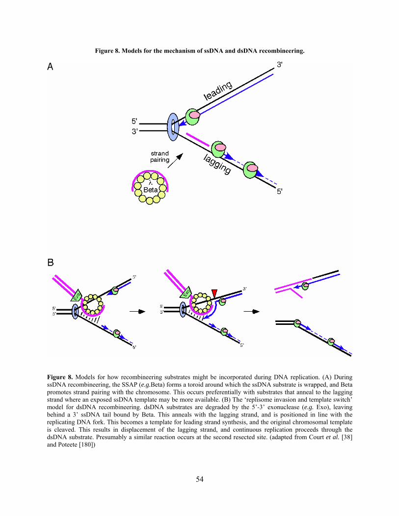
Figure 8. Models for the mechanism of ssDNA and dsDNA recombineering.
Figure 8. Models for how recombineering substrates might be incorporated during DNA replication. (A) During ssDNA recombineering, the SSAP (e.g.Beta) forms a toroid around which the ssDNA substrate is wrapped, and Beta promotes strand pairing with the chromosome. This occurs preferentially with substrates that anneal to the lagging strand where an exposed ssDNA template may be more available. (B) The ‘replisome invasion and template switch’ model for dsDNA recombineering. dsDNA substrates are degraded by the 5’-3’ exonuclease (e.g. Exo), leaving behind a 3’ ssDNA tail bound by Beta. This anneals with the lagging strand, and is positioned in line with the replicating DNA fork. This becomes a template for leading strand synthesis, and the original chromosomal template is cleaved. This results in displacement of the lagging strand, and continuous replication proceeds through the dsDNA substrate. Presumably a similar reaction occurs at the second resected site. (adapted from Court et al. [38] and Poteete [180])
54
1.4 SPECIFIC AIMS OF THIS STUDY
The development of a simple and efficient system for genetics would greatly benefit the
mycobacterial research community. Numerous factors inherent to mycobacterial cell growth and
cell wall structure prevent simple handling and manipulation of the mycobacteria. However, the
relatively high levels of illegitimate recombination compared to homologous recombination in
M. tuberculosis and other slow-growers is the primary limiting factor to the application of
conventional genetic techniques in these bacteria. The current methods for targeted gene
replacement are designed to circumvent illegitimate recombination by modifying the AES or its
delivery into the host cell. It is striking, however, that none of these have focused on increasing
the levels of homologous recombination within the bacterial cell. The historical success of
adapting mycobacteriophages and their proteins for manipulation of their mycobacterial hosts
has led to the hypothesis that mycobacteriophage-encoded recombination proteins could be
introduced into the mycobacterial cell to improve the efficiency of homologous recombination
and thereby promote allelic exchange for mutagenesis purposes. The success of the λ Red
recombination system for recombineering in E. coli further supported this notion and provided a
basis for initial experimental design. Therefore the focus of my thesis research has been to utilize
mycobacteriophage-encoded recombination proteins to develop a recombineering system for the
mycobacteria.
55
1.4.1 Specific Aim 1: Bioinformatic and biochemical analysis of mycobacteriophage
Che9c-encoded RecET homologues.
Mycobacteriophage-encoded homologues of the E. coli Rac prophage RecET proteins are rare in
mycobacteriophages; only Che9c was found to encode homologues of both. In vitro biochemical
analysis of Che9c gp60 and gp61 demonstrates that they possess exonuclease activity and DNA
binding activities, respectively, similar to RecET. These data are presented in Chapter 2, and
some of the experiments have been published [227].
1.4.2 Specific Aim 2: Development of a mycobacterial recombineering system using
mycobacteriophage Che9c-encoded recombination proteins.
Che9c gp60 and gp61 have biochemical properties reminiscent of a coordinated recombination
system that functions via the single strand annealing pathway. Expression of these proteins in
mycobacterial strains yields a substantial increase in homologous recombination. This has
provided an efficient genetic tool that has been successfully used to construct gene replacement
mutants and point mutants in the genomes of both M. smegmatis and M. tuberculosis, and likely
is applicable to other mycobacterial species. Chapter 3 describes the development of the
mycobacterial recombineering system and the various technical applications, the majority of
which have been published [227-229].
56
1.4.3 Specific Aim 3: Identification of additional mycobacteriophage-encoded
recombination systems.
Sequencing of more than 50 mycobacteriophage genomes has revealed several additional gene
candidates that may encode functional recombination proteins; these are present in the genomes
of phages Giles, Halo, Wildcat, and also prophages in the genome of M. avium and
Mycobacterium abscessus. In vivo analysis of several of the putative SSAPs, as well as λ Beta
and RecT, demonstrate that the Che9c gp61 functions most efficiently in mycobacteria.
Mycobacteriophage TM4 also appears to encode a recombination system, although the genes
responsible have not thus far been identified by bioinformatic analysis. Experimental analysis of
TM4 cosmid recombination sheds some light on the mechanism of TM4 recombination in vivo.
These experiments and the implications of the results are discussed in Chapter 4.
57
2.0 MYCOBACTERIOPHAGE CHE9C ENCODES RecE AND RecT HOMOLOGUES
2.1 INTRODUCTION
Bacteriophages are an extremely diverse group of organisms, and at an estimated 1031 total
phage particles, they are more abundant than any other life form in the biosphere [77]. Phages
can be found in a variety of environments along with their bacterial hosts, and interactions
between phages and bacterial populations foster copious amounts of genetic exchange. This
contributes to a large pool of shared genetic elements [76] and has a significant impact on the
evolution of bacteria, particularly on mechanisms of pathogenicity and acquisition of virulence
genes [233]. Phages are often grouped based on their morphology, host-range, and other types of
limited characteristics. However, it has become apparent that relationships among phages are
better represented and understood through examination of their gene similarity and organization
and by grouping them in ways that account for both their high level of diversity and the
independent origin of their genes [108].
Although the number of well-characterized phages is a miniscule fraction of the total
population, more than 500 phage genomes have been sequenced to date [73]. A significant
proportion of these include the group of phages that infect the mycobacteria: the
mycobacteriophages. More than 50 mycobacteriophage genomes have been sequenced
[73,126,165,170] (and G. Hatfull, unpublished data), revealing a mosaic architecture reminiscent
58

of that originally observed in the lamboid phages and in other phages [165]. In this way, when
comparing phages, similar genes are often staggered amongst genes have been acquired in a
different way, and are organized in a modular organization. The unique combination of genes
and gene clusters in this manner – with little to no sequence homology at the gene boundaries –
is evidence that illegitimate recombination plays an important role in genetic exchange of
functional genetic elements [126]. An alternative hypothesis has been suggested in a recent study
that homeologous recombination – recombination between sequences that are related but are
divergent – contributed to genetic mosaicism in phage λ [123]. Further, Martinsohn et al. suggest
that the λ Red recombination system contributes to this recombination substantially more than
the host rec proteins. However, the contribution of this particular type of recombination may not
be the common contributing factor in other phages, and the observations made in this article
could be limited to a small number of phages. Since the presence and/or activity of these types of
recombination systems has not been carefully examined in many phages, the effect and
prevalence of homeologous recomibination is unclear.
Bioinformatic analysis of mycobacteriophage genomes indicates that the genes encoding
phage structural and assembly proteins are typically organized in similarly ordered operons, and
therefore their function can often be inferred from previously characterized genes [165].
However, approximately half of the mycobacteriophage ORFs do not have detectable similarity
to known genes from either phages or other organisms, and their function is unknown [71]. Of
the mycobacteriophage genes that do have homologues, 90% of these are found in other
mycobacteriophages, indicating that these organisms exchange DNA more frequently amongst
themselves than with their bacterial hosts or other phages. A large proportion of the genes that
have detectable similarity to known genes are found in multiple mycobacteriophages, while a
59
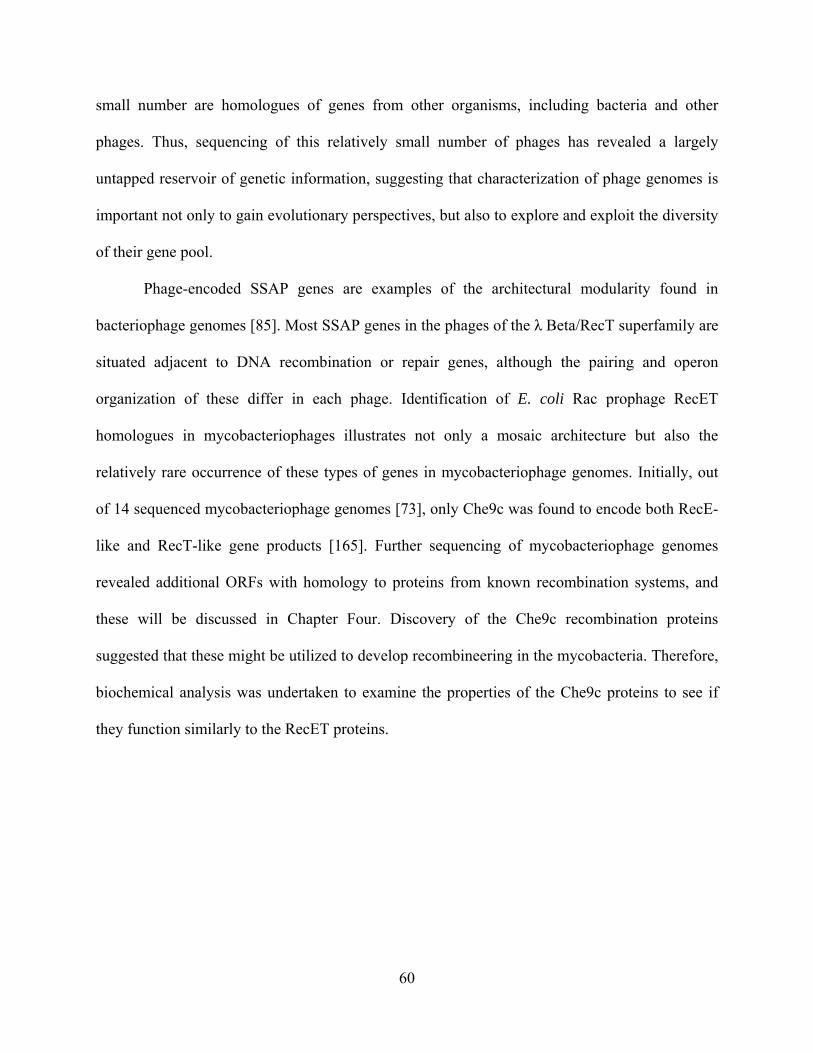
small number are homologues of genes from other organisms, including bacteria and other
phages. Thus, sequencing of this relatively small number of phages has revealed a largely
untapped reservoir of genetic information, suggesting that characterization of phage genomes is
important not only to gain evolutionary perspectives, but also to explore and exploit the diversity
of their gene pool.
Phage-encoded SSAP genes are examples of the architectural modularity found in
bacteriophage genomes [85]. Most SSAP genes in the phages of the λ Beta/RecT superfamily are
situated adjacent to DNA recombination or repair genes, although the pairing and operon
organization of these differ in each phage. Identification of E. coli Rac prophage RecET
homologues in mycobacteriophages illustrates not only a mosaic architecture but also the
relatively rare occurrence of these types of genes in mycobacteriophage genomes. Initially, out
of 14 sequenced mycobacteriophage genomes [73], only Che9c was found to encode both RecE-
like and RecT-like gene products [165]. Further sequencing of mycobacteriophage genomes
revealed additional ORFs with homology to proteins from known recombination systems, and
these will be discussed in Chapter Four. Discovery of the Che9c recombination proteins
suggested that these might be utilized to develop recombineering in the mycobacteria. Therefore,
biochemical analysis was undertaken to examine the properties of the Che9c proteins to see if
they function similarly to the RecET proteins.
60

2.2 BIOINFORMATIC ANALYSES OF MYCOBACTERIOPHAGES REVEALS A
PUTATIVE RECOMBINATION SYSTEM
Through BLAST analyses [4], it was observed that identifiable recombination systems are rare in
the mycobacteriophages, and only one phage encodes proteins that are distantly related to RecE
and RecT of the E. coli Rac prophage (Figure 9). Che9c gp60 shares 28% identity with the C-
terminal region of RecE. This encompasses a nuclease domain belonging to the RecB family,
while the N-terminus of RecE is not necessary for its exonuclease activity [31]. The N-terminal
two-thirds of gp61 (residues 28-237; Figure 9A) have 29% identity to RecT, whereas the C-
terminal third of gp61 (residues 238-353) only has detectable similarity to the corresponding
region of a predicted M. avium RecT protein (discussed in Chapter Four) and no other known
proteins. A multiple sequence alignment performed with Che9c gp61 and the proteins identified
by Iyer et al. as members of the λ Beta/RecT superfamily shows conservation of a core domain
(200 amino acids) and a similar predicted secondary structure (Figure 9B), indicating that gp61
is indeed a member of this superfamily of SSAPs [85]. Much like the Rac prophage RecET
system, no Gam homologues have thus far been identified in any of the sequenced
mycobacteriophages.
61
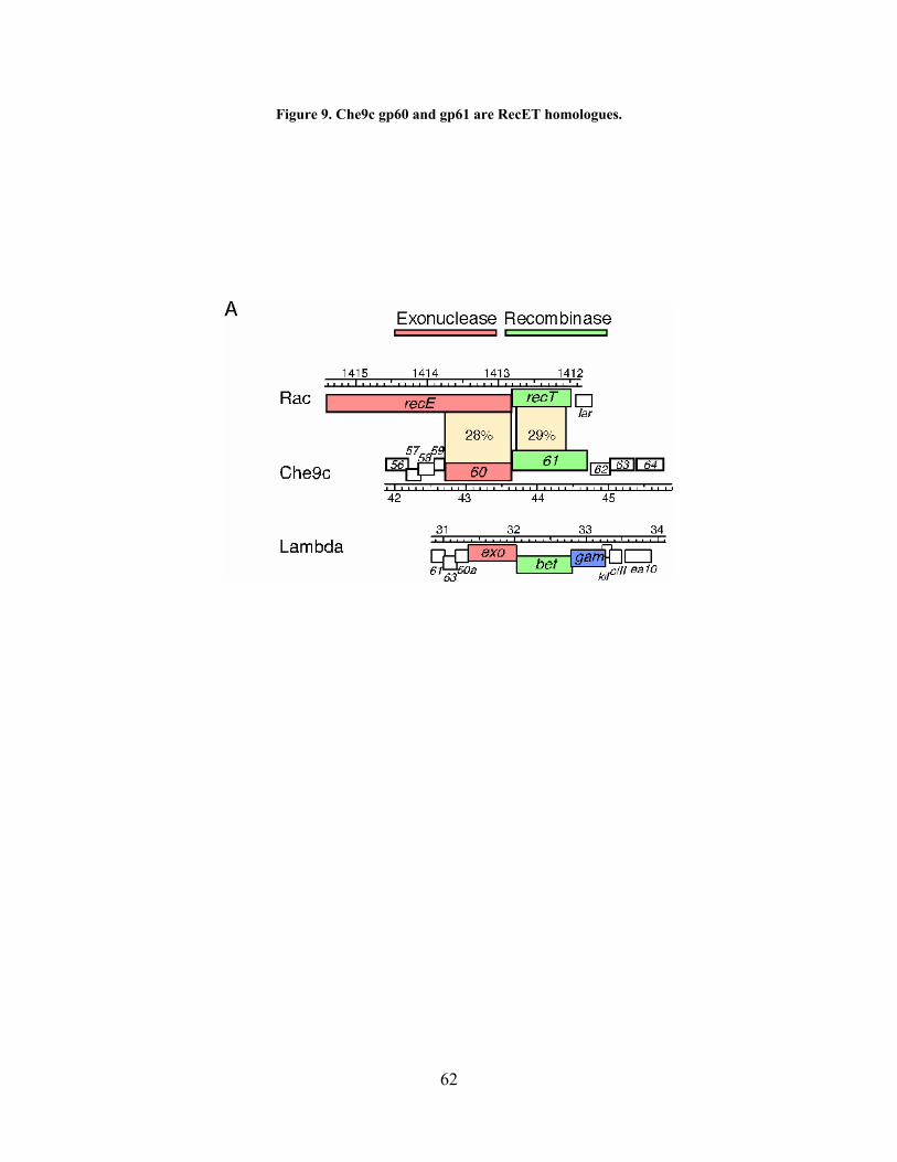
Figure 9. Che9c gp60 and gp61 are RecET homologues.
62

63

Figure 9. (A) Che9c gp60 is a RecE homologue, while Che9c gp61 is a RecT homologue. Exonucleases are indicated in red, and SSAPs (recombinases) are indicated in green. E. coli Rac prophage genes and Che9c genes are transcribed from left to right, while the λ genes are transcribed right to left. (B) Multiple sequence alignments were performed with all protein sequences used by Iyer et al. [85], and conserved regions are shown. The T-coffee program was used to align Che9c gp61 (outlined in blue) with the λ Beta/RecT protein family members [148], and this was manually incorporated into the alignment made by Iyer et al. Secondary structure predictions (using JPred) for gp61 were also conserved (shown in blue at the top) [40]. Similar residues are highlighted that were found by Iyer et al. to be conserved greater than 85%: h, hydrophobic; l, aliphatic; a, aromatic; o, alcohol; c, charged; +. basic; -, acidic; p, polar; b, big; s, small; u, tiny.
64

2.3 PURIFICATION OF CHE9C GP60 AND GP61 PROTEINS
To determine if the Che9c proteins function similarly to their RecET homologues, gp60 and
gp61 proteins containing C-terminal 6x-histadine tags were over-expressed and purified from E.
coli lysates by nickel-affinity chromatography. SDS-PAGE analysis indicated that purified
samples of recombinant gp61 were nearly homogeneous, while recombinant gp60 samples
retained small amounts of contaminating host proteins (Figure 10A). Therefore, a mock-
purification was performed with E. coli extracts from a strain containing an empty vector, and it
was observed that these samples contained similar host proteins to the gp60 preparation (Figure
10B). This mock-purified protein sample was used for biochemical assays alongside gp60 as a
negative control.
65
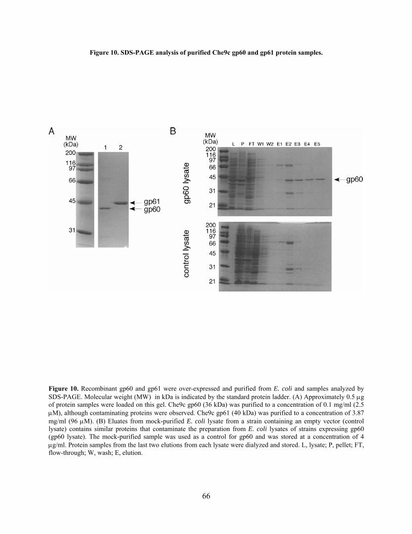
Figure 10. SDS-PAGE analysis of purified Che9c gp60 and gp61 protein samples.
Figure 10. Recombinant gp60 and gp61 were over-expressed and purified from E. coli and samples analyzed by SDS-PAGE. Molecular weight (MW) in kDa is indicated by the standard protein ladder. (A) Approximately 0.5 g of protein samples were loaded on this gel. Che9c gp60 (36 kDa) was purified to a concentration of 0.1 mg/ml (2.5 M), although contaminating proteins were observed. Che9c gp61 (40 kDa) was purified to a concentration of 3.87 mg/ml (96 M). (B) Eluates from mock-purified E. coli lysate from a strain containing an empty vector (control lysate) contains similar proteins that contaminate the preparation from E. coli lysates of strains expressing gp60 (gp60 lysate). The mock-purified sample was used as a control for gp60 and was stored at a concentration of 4 g/ml. Protein samples from the last two elutions from each lysate were dialyzed and stored. L, lysate; P, pellet; FT, flow-through; W, wash; E, elution.
66

2.4 CHE9C GP60 IS AN EXONUCLEASE
Phage λ Exo and E. coli RecE are highly processive enzymes that degrade linear dsDNA in a 5
to 3 direction [89,116]. To determine if gp60 has exonuclease activity, three in vitro assays were
developed [227]. First, gp60 was observed to degrade short radiolabeled dsDNA substrates (100
bp) similarly to λ Exo, while no degradation was seen in negative control reactions (Figure 11A).
Notably, it was observed that serial dilutions of gp60 did not yield the expected step-wise
decrease in activity, but rather even a 2-fold dilution resulted in very little degradation activity,
which may be due to protein inactivation in dilution buffers. Because the observed activity of
gp60 could conceivably also be attributed to a contaminating phosphatase that would remove the
radioactive phosphate, a similar assay with linearized plasmid dsDNA substrates was used and
visualized by agarose gel electrophoresis. Incubation of gp60 with this dsDNA substrate also
resulted in degradation, while negative control reactions with mock-purified protein did not
(Figure 11B). Finally, the observed exonuclease activity was shown to be limited to substrates
with dsDNA ends, since neither supercoiled or nicked open circle dsDNA substrates were
degraded by gp60 (Figure 11C). These data demonstrate that Che9c gp60 has exonuclease
activity similar to λ Exo and RecE.
67
Figure 11. In vitro assays demonstrate exonuclease activity of Che9c gp60.
Figure 11. (A) Exonuclease activity was assayed by incubating Che9c gp60, λ Exo, or control protein extract with 32P-labeled dsDNA (100 bp) for 5 minutes at room temperature, and the reactions analyzed by polyacrylamide gel electrophoresis. Reactions contained either no protein (–), or two-fold serial dilutions as indicated. Reactions with the highest protein concentrations contained Che9c gp60 at 0.2 μM or 5 U of λ Exo (NEB). The control protein extract was prepared from mock induced cells and the highest concentration corresponds to approximately 0.1 μg/ml. (B) Exonuclease activity of Che9c gp60 (0.2 M), λ Exo (1 U/10 l), or control protein was assayed in reactions with a 3 kbp linearized plasmid DNA substrate (0.8 nM), incubated for increasing amounts of time (t = 0, 2, 5, 7, or 10 minutes), and analyzed by agarose gel electrophoresis. The marker (M) indicates sizes in kbp. (C) Exonuclease activity of circular versus linear substrates was assayed. Che9c gp60 (final concentration 0.2 μM) or λ Exo (5 U) was incubated for increasing times (0, 5, and 10 min) similarly to (B) with a 3 kbp dsDNA substrate (2 nM) that was either supercoiled closed circular or linear (as indicated) and the products analyzed by agarose gel electrophoresis.
68
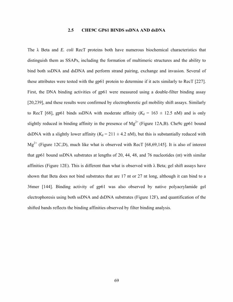
2.5 CHE9C GP61 BINDS ssDNA AND dsDNA
The λ Beta and E. coli RecT proteins both have numerous biochemical characteristics that
distinguish them as SSAPs, including the formation of multimeric structures and the ability to
bind both ssDNA and dsDNA and perform strand pairing, exchange and invasion. Several of
these attributes were tested with the gp61 protein to determine if it acts similarly to RecT [227].
First, the DNA binding activities of gp61 were measured using a double-filter binding assay
[20,239], and these results were confirmed by electrophoretic gel mobility shift assays. Similarly
to RecT [68], gp61 binds ssDNA with moderate affinity (Kd = 163 ± 12.5 nM) and is only
slightly reduced in binding affinity in the presence of Mg2+ (Figure 12A,B). Che9c gp61 bound
dsDNA with a slightly lower affinity (Kd = 211 ± 4.2 nM), but this is substantially reduced with
Mg2+ (Figure 12C,D), much like what is observed with RecT [68,69,145]. It is also of interest
that gp61 bound ssDNA substrates at lengths of 20, 44, 48, and 76 nucleotides (nt) with similar
affinities (Figure 12E). This is different than what is observed with λ Beta; gel shift assays have
shown that Beta does not bind substrates that are 17 nt or 27 nt long, although it can bind to a
36mer [144]. Binding activity of gp61 was also observed by native polyacrylamide gel
electrophoresis using both ssDNA and dsDNA substrates (Figure 12F), and quantification of the
shifted bands reflects the binding affinities observed by filter binding analysis.
69

Figure 12. Che9c gp61 binds ssDNA and dsDNA.
Figure 12. Purified gp61 protein at varying concentrations (0, 0.2, 0.3, 0.7, 1.3, 2.0, 2.7, 3.2 M) was incubated with 66.7 nM 32P-labeled ssDNA or dsDNA in binding assay buffer and analyzed either by double-filter binding assays (A-E) or native polyacrylamide gel electrophoresis (F). These experiments (without Mg2+) were repeated in triplicate for both ssDNA (A) and dsDNA (C) and the data analyzed on SigmaPlot to determine binding constants. Reactions were also assayed with ssDNA (B) or dsDNA (D) containing 0 mM MgCl2 (●), 5 mM MgCl2 (○), or 10 mM MgCl2 (▼). (E) ssDNA substrates of different lengths were tested (0 mM MgCl2) and are depicted in the legend. (F) For gel shift assays, the same reactions from using either ssDNA (A) or dsDNA (C) were run on a native 8% polyacrylamide gel and analyzed.
70
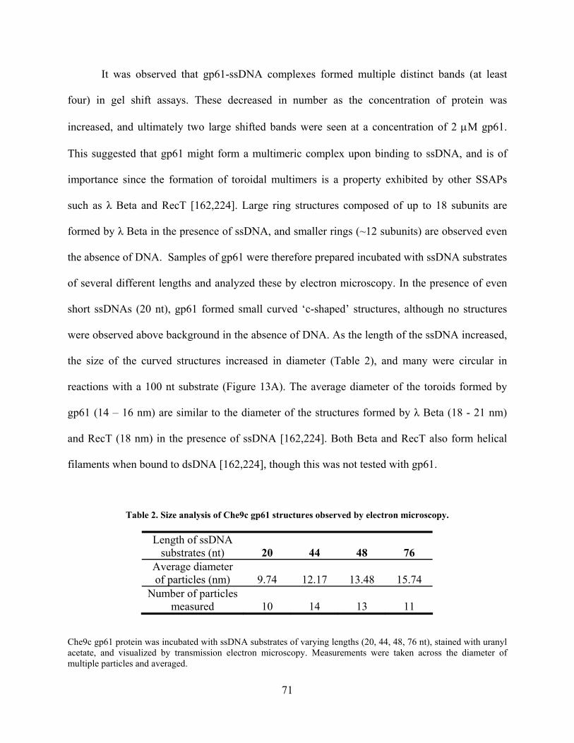
It was observed that gp61-ssDNA complexes formed multiple distinct bands (at least
four) in gel shift assays. These decreased in number as the concentration of protein was
increased, and ultimately two large shifted bands were seen at a concentration of 2 M gp61.
This suggested that gp61 might form a multimeric complex upon binding to ssDNA, and is of
importance since the formation of toroidal multimers is a property exhibited by other SSAPs
such as λ Beta and RecT [162,224]. Large ring structures composed of up to 18 subunits are
formed by λ Beta in the presence of ssDNA, and smaller rings (~12 subunits) are observed even
the absence of DNA. Samples of gp61 were therefore prepared incubated with ssDNA substrates
of several different lengths and analyzed these by electron microscopy. In the presence of even
short ssDNAs (20 nt), gp61 formed small curved ‘c-shaped’ structures, although no structures
were observed above background in the absence of DNA. As the length of the ssDNA increased,
the size of the curved structures increased in diameter (Table 2), and many were circular in
reactions with a 100 nt substrate (Figure 13A). The average diameter of the toroids formed by
gp61 (14 – 16 nm) are similar to the diameter of the structures formed by λ Beta (18 - 21 nm)
and RecT (18 nm) in the presence of ssDNA [162,224]. Both Beta and RecT also form helical
filaments when bound to dsDNA [162,224], though this was not tested with gp61.
Table 2. Size analysis of Che9c gp61 structures observed by electron microscopy.
Length of ssDNA substrates (nt) 20 44 48 76
Average diameter of particles (nm) 9.74 12.17 13.48 15.74
Number of particles measured 10 14 13 11
Che9c gp61 protein was incubated with ssDNA substrates of varying lengths (20, 44, 48, 76 nt), stained with uranyl acetate, and visualized by transmission electron microscopy. Measurements were taken across the diameter of multiple particles and averaged.
71
Like λ Beta, the E. coli RecT protein also forms multimers in the absence of DNA that
are visible by electron microscopy, although no structures could be seen with gp61 using simple
negative staining. Gel filtration analysis of RecT (originally called p33) indicates that it forms a
tetramer [68]. Therefore, analytical gel filtration was used to determine the state of gp61 in
solution. Three concentrations of recombinant gp61 protein were run on a Superdex gel filtration
column that had been standardized with both low and high molecular weight proteins. As the
concentration of gp61 was increased from 5 M to 25 M, the size of the complex increased. At
5 M, gp61 eluted at a time corresponding to approximately 70 kDa, which is roughly twice the
size of the predicted molecular weight of gp61 (40 kDa). At 10 M, it eluted at 102 kDa, and at
25 M it eluted at 143 kDa. Although these data do not fit exactly with the predicted size of
multimers of gp61 (e.g., 80 kDa, 120 kDa, or 160 kDa), the native molecular weight of the
standards varied slightly on this column (±14 kDa) (Figure 13B). Thus, it appears that at the
highest concentration tested, gp61 likely forms a tetramer, much like what has been observed for
RecT (concentration not given for RecT experiment; [68]). Additionally, increasing the salt
(NaCl) concentration in the buffer from 100 mM to 300 mM did not change the size of the gp61
complexes eluted by gel filtration (data not shown). This indicates that the multimerization of
gp61 in solution is not likely the result of non-specific protein-protein interactions; further
anlaysis would be required to completely rule out this possibility. Reactions containing gp61
incubated with ssDNA eluted in the void volume, indicating that they were larger than the pore
size of this column, which is consistent with gp61-ssDNA complex formation (data not shown).
Finally, while these data are not conclusive, they support the hypothesis that gp61 forms
multimers in the absence of DNA.
72
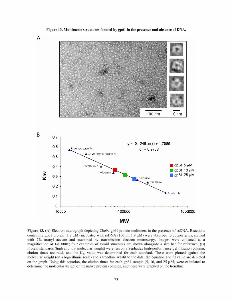
Figure 13. Multimeric structures formed by gp61 in the presence and absence of DNA.
Figure 13. (A) Electron micrograph depicting Che9c gp61 protein multimers in the presence of ssDNA. Reactions containing gp61 protein (1.2 M) incubated with ssDNA (100 nt; 1.9 M) were absorbed to copper grids, stained with 2% uranyl acetate and examined by transmission electron microscopy. Images were collected at a magnification of 140,000x; four examples of toroid structures are shown alongside a size bar for reference. (B) Protein standards (high and low molecular weight) were run on a Sephadex high-performance gel filtration column, elution times recorded, and the Kav value was determined for each standard. These were plotted against the molecular weight (on a logarithmic scale) and a trendline wasfit to the data; the equation and fit value are depicted on the graph. Using this equation, the elution times for each gp61 sample (5, 10, and 25 M) were calculated to determine the molecular weight of the native protein complex, and these were graphed on the trendline.
73
2.6 CONCLUSIONS
The mosaic architecture commonly observed in bacteriophages is exemplified by phage-encoded
SSAPs and their cognate exonucleases [85] in that there is no apparent consistency with the
specific pairing of these proteins. Further, SSAP-exonuclease recombination systems are rare in
sequenced mycobacteriophages. Bioinformatic analyses identified mycobacteriophage Che9c
gp60 and gp61 as homologues of the E. coli Rac prophage RecE and RecT proteins. Finding
these genes in mycobacteriophages was somewhat suprising; previous analyses found RecT-like
proteins predominantly in the low GC% Gram-positive bacteria [85], while the mycobacteria
have a high G+C% (~65-67%) [22]. However, the Che9c-encoded proteins were distantly related
to the E. coli RecET proteins with less than 30% amino acid identity conserved. Similarly to the
organization of these genes seen in other phages, the mycobacteriophage-encoded SSAP is
located next to a predicted exonuclease.
Desite the weak identity observed for these proteins, the biochemical properties of Che9c
gp60/gp61 support the finding that they are RecET homologues. Che9c gp60 has dsDNA
exonuclease activity that does not act on circular substrates, which has been observed for RecE.
Che9c gp61 binds ssDNA and dsDNA with affinities and Mg2+ dependencies similar to RecT,
and it forms toroids in the presence of ssDNA. Interestingly, gp61 binds to ssDNA substrates as
small as 20 nt with higher affinity than longer substrates. Although it is reported that Beta cannot
bind substrates shorter than 36 nucleotides [144], more recent experiments indicate that it does
indeed bind smaller lengths of ssDNA (D. Court, personal communication).
Numerous additional in vitro assays could be performed with each of these proteins to
further characterize their activities, such as strand pairing and strand exchange assays for gp61.
These assays were attempted but were not successful for control reactions. It would also be
74
helpful to analyze gp61 by native gradient gel electrohoresis to confirm the gel filtration data
which suggest it is a trimer or tetramer in its native state. Also, RecT DNA binding is affected by
salt (NaCl) concentrations greater than 50 M [145]. It was suggested that the varying salt and
Mg2+ requirements for ssDNA and dsDNA binding may reflect different types of binding [145],
which is supported by the observation that ssDNA- and dsDNA-gp61 complexes form toroids
and helical filaments, respectively [224]. Additional experiments could therefore be performed
with gp61 in buffers containing Mg2+ and/or NaCl to determine their effect on multimer
formation. MgCl2 is absolutely required for formation of λ Beta multimers in the absence of
DNA, and addition of Mg2+ to reactions with Beta and ssDNA appears to stabilize the formation
of large rings [162]. However, Beta also requires Mg2+ for ssDNA binding [144], whereas RecT
and gp61 do not [145]. In fact, RecT multimers are inhibited with high concentrations of MgCl2,
though a low concentration (0.3 mM) is required for the formation of small circles but not rod-
like structures [224]. Therefore varying Mg2+ concentration or testing other cations might
enhance gp61 multimerization, and this could be assayed in the future.
Collectively, the biochemical data clearly demonstrate that gp60 and gp61 function
equivalently to the E. coli λ Red and RecET systems in all assays tested. Thus, these proteins
have properties consistent with their utility as a means to develop a recombineering system in the
mycobacteria using mycobacteriophage-encoded proteins.
75

3.0 DEVELOPMENT OF THE MYCOBACTERIAL RECOMBINEERING SYSTEM
3.1 INTRODUCTION
Recombineering is a widely used system for mutagenesis in E. coli [38], which has recently been
extended to other Gram-negative bacteria, such as Salmonella and Shigella [42,185]. A variety of
techniques can be performed with ease including gene replacements, point mutations, deletions,
small insertions, in vivo cloning, and modifications of bacterial artificial chromosomes and
genomic libraries [36,38,199]. Recombineering exploits the potent recombination activities of
the λ Red proteins and is a simple means of increasing homologous recombination in the host
bacterium with minimal DNA manipulations or screening.
Recombineering is most commonly used for construction of allelic gene replacements
and point mutations on either the bacterial chromosome or on BACs. Substrates for gene
replacements contain an antiobiotic resistance gene of choice (and a sacB gene for counter-
selection if desired) with short lengths of homology to the gene target on either end (Figure 7)
[223]. These strains can be easily unmarked with ssDNA substrates that have homology flanking
the antibiotic and sacB cassettes with selection for sucrose resistance. Optimum numbers of
recombinants (up to 105 out of 108 viable cells) are obtained with 100 – 300 ng of the AES.
Recombination of the dsDNA AES substrates is dependent on Exo and Beta, and the addition of
Gam increases recombineering efficiencies significantly [135,240].
76

Mutagenesis with ssDNA substrates is a particularly useful strategy that permits
numerous types of mutations to be introduced into bacterial and phage genomes [52,151]. This
recombination only requires the Beta protein, although some experiments have shown that the
presence of Exo (even with ssDNA substrates) can slightly enhance recombination [244].
Maximal recombination frequencies are achieved with 70 nt ssDNAs [52], and these substrates
are designed with the mutation(s) centered such that they anneal to the lagging strand for DNA
replication, since these are more efficiently recombined [52].
The E. coli recombineering strains that are typically used express the λ Red/Gam proteins
under control of the temperature-sensitive repressor CI857 in a defective λ prophage. Following
mutagenesis, the prophage can be removed or the mutation can be moved to a clean background
by generalized transduction with phage P1 [223]. Alternatively, there are various versions of
recombineering plasmids with temperature-senstive replicons that can be transferred into
different strains and quickly removed [42]. These appear to work just as well as the prophage
version, and can be used in other Gram-negative bacteria. Recombineering strains are induced at
mid-logarithmic phase for 15 minutes and prepared for transformation; this expression time is
optimal for recombineering but is short enough that cell death does not occur from the toxic
effects of Gam [240].
Clearly, the strategic use of the λ Red proteins to develop the widely-applicable
recombineering technology in E. coli sets a precedent for improving genetic techniques in other
bacterial systems. Mycobacterial researchers in particular would benefit enormously from a
simple and efficient system for genetics – such as recombineering – that alleviates time-
consuming DNA manipulations and screening procedures. However, the λ Red proteins, while
useful for E. coli and related Gram-negative organisms, reportedly are not functional in the
77

mycobacteria, which is not surprising considering the divergence between these two groups of
bacteria. From the bioinformatic and biochemical data, the mycobacteriophage-encoded RecET
homologues appeared to be the most likely candidates for developing a recombineering system
for the mycobacteria. Accordingly, expression of the Che9c proteins gp60 and gp61 elevates
homologous recombination in both fast- and slow-growing mycobacteria. This facilitates the use
of recombineering-based strategies for mutagenesis of mycobacterial and mycobacteriophage
genomes. This chapter will discuss the development of the Che9c-based mycobacterial
recombineering system and its various applications.
3.2 EXPRESSION OF CHE9C RECOMBINATION GENES IN VIVO
In order to test the activity of Che9c gp60 and gp61 in vivo, various plasmids were constructed
that express these genes in the mycobacteria (see Table 15) [227]. Although genes 59 and 62
encode small proteins, it was possible that one of these might encode a Gam-like functional
protein that is not identifiable by bioinformatic analysis. Therefore, this region of Che9c was
cloned under the control of the M. smegmatis acetamidase promoter (Pacetamidase) [44,157] on an
extrachromosomally-replicating parent vector (pLAM12) to create pJV24 (Figure 14). The
acetamidase promoter is comprised of the upstream region of the M. smegmatis acetamidase
gene regulon and terminates at the start codon of the acetamidase gene (amiE) [157,190].
Therefore, placing the start codon of a gene at this locus results in translation from the ribosome
binding site (RBS) encoded by this cassette. Additional constructs were made in the parent
vector pLAM12 containing only genes 60 and 61; in this case, gene 60 was under translational
control from either its endogenous signals (pJV53; Figure 14) or those present in the acetamidase
78

promoter cassette (pJV63). Similar plasmids were constructed containing only gene 60 (pJV61
and pJV55) or gene 61 (pJV52 and pJV62), either under their own translation signals or those of
the acetamidase cassette, respectively. All of these plasmids were transformed into M. smegmatis
for further assays. Genes 59-62 were also cloned under control of the constitutive M. bovis BCG
hsp60 promoter (pJV23), but this plasmid did not produce transformants in M. smegmatis. It is
also noted that M. smegmatis:pJV63 grows slowly and does not grow on plates in the presence of
inducer (data not shown). This may be due to the somewhat toxic effects of gp60, since strains
expressing only this protein (even in the absence of induction) grow slowly compared to empty
vector control strains (data not shown).
79
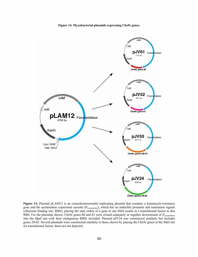
Figure 14. Mycobacterial plasmids expressing Che9c genes.
Figure 14. Plasmid pLAM12 is an extrachromosomally-replicating plasmid that contains a kanamycin-resistance gene and the acetamidase expression cassette (Pacetamidase), which has an inducible promoter and translation signals (ribosome binding site: RBS); placing the start codon of a gene at site NdeI results in a translational fusion to this RBS. For the plasmids shown, Che9c genes 60 and 61 were cloned separately or together downstream of Pacetamidase into the HpaI site with their endogenous RBSs included. Plasmid pJV24 was constructed similarly but includes genes 59-62. Several plasmids were constructed similarly to those shown by placing the Che9c genes at the NdeI site for translational fusion; these are not depicted.
80

Protein expression was monitored by western blot analysis for several of these strains
with polyclonal antibodies generated against purified gp61 protein (Figure 15) [227]. All strains
in which gene 61 was under control of Pacetamidase had detectable expression of gp61 after three hr
of induction with acetamide. It was seen that some strains expressed more gp61 than others,
although there was no correlation between expression levels and whether the endogenous
translation signals or signals from the acetamidase cassette were used (Figure 15 A,B; compare
strains with pJV53 to pJV63, and pJV52 to pJV62, respectively). Specifically, a strain with
pJV62 (Pacetamidase RBS) had three-fold less gp61 expression than pJV52 (endogenous RBS),
whereas the opposite was true for pJV53 (endogenous RBS) and pJV63 (Pacetamidase RBS). The
strain expressing gp59-gp62 (M. smegmatis:pJV24) consistently showed expression of gp61 in
the absence of acetamide (Figure 15A,C), and this may be due to leaky expression sometimes
observed with this promoter in succinate medium [157]. This level of protein expression was not
observed with any other M. smegmatis strain, and further there is no expression observed in
media containing ADC (see Materials and Methods) that is reported to be repressive of this
promoter (Figure 15D). Strains of M. tuberculosis containing the same plasmids were also tested
for protein expression, and it was observed that there was much more leaky expression of the
promoter in the absence of induction (Figure 15E).
81

Figure 15. Western blot analysis of mycobacterial strains expressing Che9c proteins.
Figure 15. Strains of either M. smegmatis (A-D) or M. tuberculosis (E) containing various plasmids were grown to mid-log phase and samples split; one culture was induced with 0.2% acetamide, and both were grown for 3 hours. Cell aliquots were normalized to OD600 and samples were run on SDS polyacylamide gels and analyzed by western blot with polyclonal anti-gp61 antibodies.
82
Cultures of M. smegmatis mc2155:pJV24 repeatedly showed a slight decrease in viability
(assayed by colony counts) after four hours of induction with acetamide that continued to decline
up to 24 hours, whereas mc2155:pJV53 did not (Figure 16A and data not shown). This may be
due to the increased levels of protein expression in mc2155:pJV24, as seen by western blot
(Figure 16B), or alternatively could result from the expression of Che9c gp59 and/or gp62. The
strain containing the empty control vector (mc2155:pLAM12) surprisingly grows more slowly
than strains expressing Che9c genes. Ultimately, three hours of induction appeared to give
adequate levels of protein expression without any potential toxic effects, and this also is
approximately the length of time required for M. smegmatis doubling in this media. Therefore,
these strains were tested for recombination activity in vivo using this induction procedure.
83
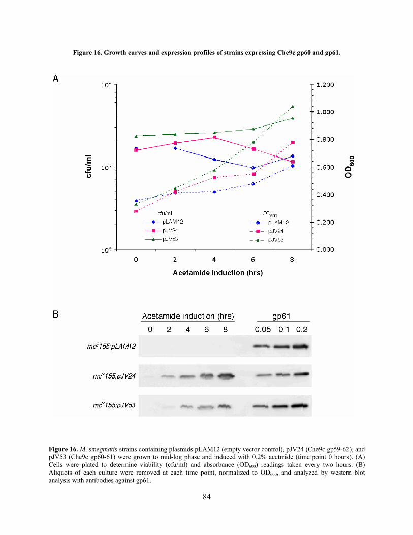
Figure 16. Growth curves and expression profiles of strains expressing Che9c gp60 and gp61.
Figure 16. M. smegmatis strains containing plasmids pLAM12 (empty vector control), pJV24 (Che9c gp59-62), and pJV53 (Che9c gp60-61) were grown to mid-log phase and induced with 0.2% acetmide (time point 0 hours). (A) Cells were plated to determine viability (cfu/ml) and absorbance (OD600) readings taken every two hours. (B) Aliquots of each culture were removed at each time point, normalized to OD600, and analyzed by western blot analysis with antibodies against gp61.
84
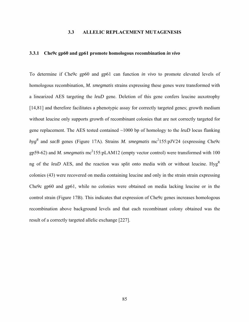
3.3 ALLELIC REPLACEMENT MUTAGENESIS
3.3.1 Che9c gp60 and gp61 promote homologous recombination in vivo
To determine if Che9c gp60 and gp61 can function in vivo to promote elevated levels of
homologous recombination, M. smegmatis strains expressing these genes were transformed with
a linearized AES targeting the leuD gene. Deletion of this gene confers leucine auxotrophy
[14,81] and therefore facilitates a phenotypic assay for correctly targeted genes; growth medium
without leucine only supports growth of recombinant colonies that are not correctly targeted for
gene replacement. The AES tested contained ~1000 bp of homology to the leuD locus flanking
hygR and sacB genes (Figure 17A). Strains M. smegmatis mc2155:pJV24 (expressing Che9c
gp59-62) and M. smegmatis mc2155:pLAM12 (empty vector control) were transformed with 100
ng of the leuD AES, and the reaction was split onto media with or without leucine. HygR
colonies (43) were recovered on media containing leucine and only in the strain strain expressing
Che9c gp60 and gp61, while no colonies were obtained on media lacking leucine or in the
control strain (Figure 17B). This indicates that expression of Che9c genes increases homologous
recombination above background levels and that each recombinant colony obtained was the
result of a correctly targeted allelic exchange [227].
85
Figure 17. Allelic gene replacement of the M. smegmatis leuD gene.
Figure 17. [227] (A) An AES targeting M. smegmatis leuD is depicted in this schematic; plasmid p0004S:leuD contains ~1000 bp of homology flanking a hygR and sacB gene, and this was linearized by restriction digest. (B) Strains M. smegmatis mc2155:pJV24 and M. smegmatis mc2155:pLAM12 were grown to mid-logarithmic phase, induced with acetamide for three hours, and electrocompetent cells prepared. 100 ng of the leuD AES were transformed into these strains, recovered for four hours, and the reaction split onto media in the presence or absence of leucine.
86

3.3.2 Recombineering requires both Che9c gp60 and gp61.
A similar assay targeting the M. smegmatis leuB gene was used to dissect the genetic
requirements for recombination, and this demonstrated that expression of both Che9c gp60 and
gp61 is necessary and sufficient for recombineering (Table 3) [227]. The presence of genes 59
and 62 in plasmid pJV24 repeatedly yielded similar recombineering frequencies to plasmid
pJV53, which expresses only genes 60 and 61 (Table 3 and Figure 19), and these two strains
gave the highest recombineering frequencies. Interestingly, although pJV63 produces higher
levels of protein expression than pJV53 (Figure 15A), this strain was reduced for recombination
activity. Based on these data, strain mc2155:pJV53 was used for most subsequent experiments
because it does not exhibit a viability defect phenotype.
Table 3. Recombineering requires both Che9c gp60 and gp61.
Strain (proteins encoded)a
Recovered colonies w/leucine b
Recovered colonies w/o leucinec
Cell competencyd (cfu/g DNA)
Recombineering frequencye (w/leucine)
mc2155:pLAM12 (control strain) 0 1 5.8 x 10⁵ 0
mc2155:pJV61 (gp60 only) 0 0 1.2 x 10⁶ 0
mc2155:pJV52 (gp61 only) 0 0 6.0 x 10⁵ 0
mc2155:pJV24 (gp59-62) 52 0 6.4 x 10⁵ 1.6 x 10⁻³
mc2155:pJV53 (gp60-61) 57 1 1.4 x 10⁶ 8.3 x 10⁻⁴
mc2155:pJV63 (gp60-61)f 7 0 4.8 x 10⁵ 2.9 x 10⁻⁴
a. Each strain contains an extrachromosomally-replicating plasmid expressing varying combinations of Che9c gp60 and gp61. b,c. Cells were transformed with 100 ng of an AES targeting leuB, and recovered cells were split on media with or without leucine supplement. d. Cell competency is determined as the cfu/g plasmid pPGA1, an integration-proficient vector providing hygromycin resistance, when 50 ng was transformed. e. Recombineering frequency (recombinant cfu/g DNA/cell competency) is shown for transformations with the leuB substrate (p0004S:leuB) and that are plated on leucine-supplemented media. f. mc2155:pJV63 expresses Che9c gp60/gp61 under control of the acetamidase promoter through a translational fusion to that cassette, in contrast to mc2155:pJV53 in which these genes are expressed from their endogenous signals.
87
3.3.3 Recombineering of the M. smegmatis groEL1 gene
Recombineering of other loci had similar results to those obtained at the leuD and leuB loci, and
genotypic analysis of the recombinants demonstrated that 90% or greater were correctly targeted
[227]. First, the groEL1 gene was targeted using an AES with ~500 bp of homology on each end
(Figure 18A), which was amplified by PCR using a circular AES as a template (see Figure 21A).
Colony PCR analysis shows that each of the ten colonies tested in this example were allelic gene
replacements of groEL1 (Figure 18B), and Southern blot analysis confirmed these results (Figure
18C). Additionally, several groEL1 mutant strains constructed by recombineering exhibited the
expected biofilm defects for this strain (data not shown) [149]. Shortening the homology lengths
of the groEL1 AES resulted in a decrease in recombination, such that less than ten colonies were
obtained with 50 bp homology regions (Figure 19), and only ~ 50% of these were correctly
targeted (data not shown). Not surprisingly, there is a low level of recombination activity in the
absence of induction due to leaky expression from the acetamidase promoter, which has been
observed in these experiments and others (data not shown, and K. Derbyshire, personal
communication). Extending the induction time from three hours up to ten hours only slightly
increased recombineering frequencies (less than two-fold, data not shown); therefore a three-
hour induction was used for all subsequent experiments. This recombination activity is
somewhat dependent on host RecA, since recombination frequencies were decreased five-fold in
an M. smegmatis recA strain (Table 4); the effect of M. smegmatis RecA dependence was small
compared to the 10-50 fold decrease observed in E. coli in recombineering assays [135,240].
88
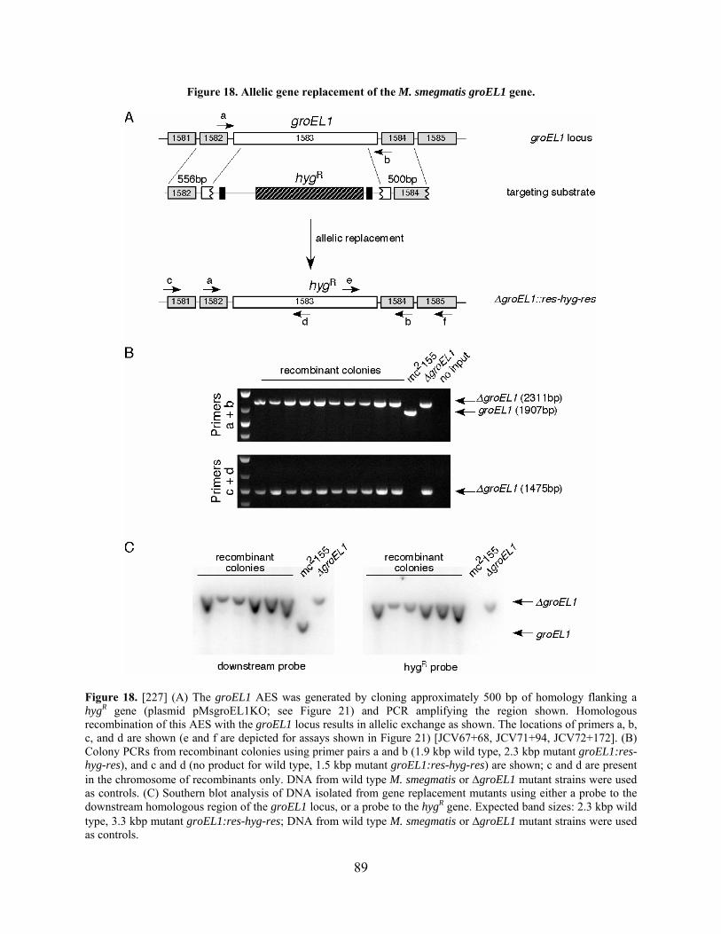
Figure 18. Allelic gene replacement of the M. smegmatis groEL1 gene.
Figure 18. [227] (A) The groEL1 AES was generated by cloning approximately 500 bp of homology flanking a hygR gene (plasmid pMsgroEL1KO; see Figure 21) and PCR amplifying the region shown. Homologous recombination of this AES with the groEL1 locus results in allelic exchange as shown. The locations of primers a, b, c, and d are shown (e and f are depicted for assays shown in Figure 21) [JCV67+68, JCV71+94, JCV72+172]. (B) Colony PCRs from recombinant colonies using primer pairs a and b (1.9 kbp wild type, 2.3 kbp mutant groEL1:res-hyg-res), and c and d (no product for wild type, 1.5 kbp mutant groEL1:res-hyg-res) are shown; c and d are present in the chromosome of recombinants only. DNA from wild type M. smegmatis or groEL1 mutant strains were used as controls. (C) Southern blot analysis of DNA isolated from gene replacement mutants using either a probe to the downstream homologous region of the groEL1 locus, or a probe to the hygR gene. Expected band sizes: 2.3 kbp wild type, 3.3 kbp mutant groEL1:res-hyg-res; DNA from wild type M. smegmatis or groEL1 mutant strains were used as controls.
89
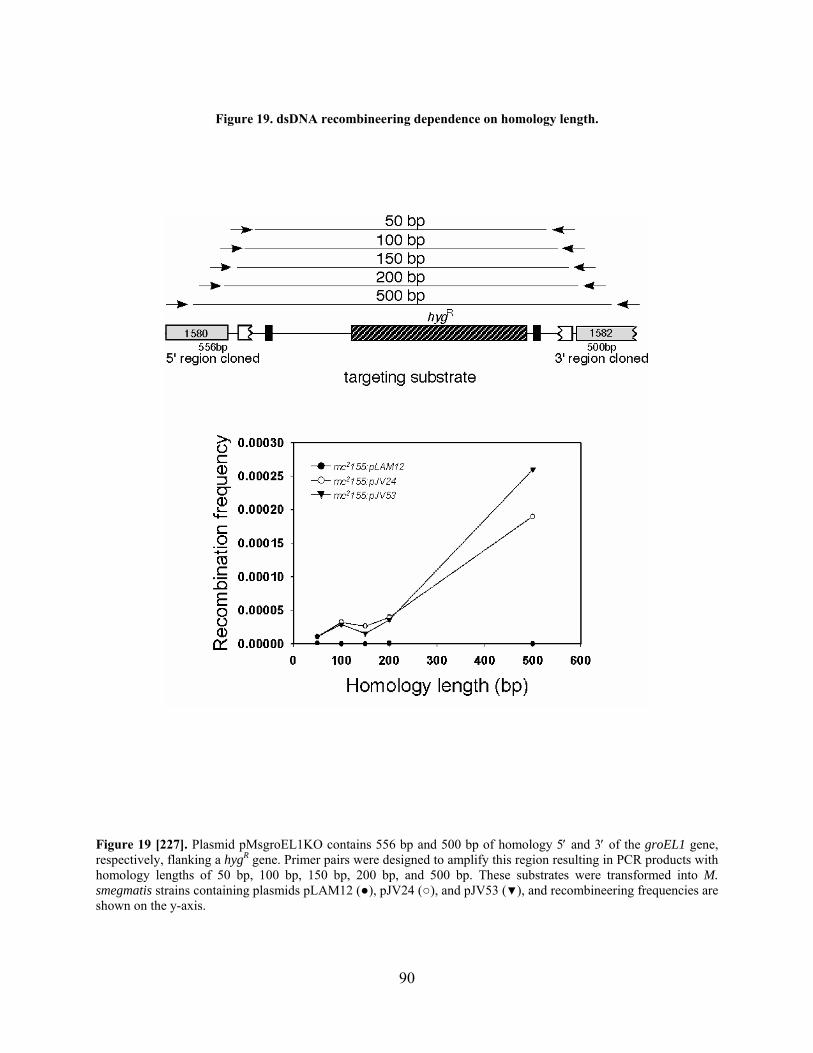
Figure 19. dsDNA recombineering dependence on homology length.
Figure 19 [227]. Plasmid pMsgroEL1KO contains 556 bp and 500 bp of homology 5 and 3 of the groEL1 gene, respectively, flanking a hygR gene. Primer pairs were designed to amplify this region resulting in PCR products with homology lengths of 50 bp, 100 bp, 150 bp, 200 bp, and 500 bp. These substrates were transformed into M. smegmatis strains containing plasmids pLAM12 (●), pJV24 (○), and pJV53 (▼), and recombineering frequencies are shown on the y-axis.
90

Table 4. dsDNA recombineering dependence on host RecA.
Strain Recovered colonies with groEL1 AESb
Cell competency (cfu/g)c
Recombineering frequencyd
mc2155:pJV53 226 6.0 x 10⁶ 3.8 x 10⁻⁴
mc2155:pJV53 recAa 99 1.3 x 10⁷ 7.6 x 10⁻⁵
a. The M. smegmatis recA strain was constructed by allelic gene replacement by recombineering and unmarked using resolvase, as described in the Materials and Methods. b. Electrocompetent cells of the two strains were transformed with 100 ng of the groEL1 AES (see Figure 18), and HygR colonies were recovered; the data represent the average of two experiments. c. Cell competency is determined as the cfu/g plasmid pJV39, an integration-proficient vector providing hygromycin resistance, when 50 ng was transformed. d. Recombineering frequency is calculated as the number of recombinant cfu per g DNA divided by the cell competency.
3.3.4 Recombineering frequencies are limited by DNA uptake efficiency.
Using 100 ng of the dsDNA substrates for allelic exchange typically produced between 50 and
200 recombinant colonies, and the number of colonies obtained by recombineering was directly
proportional to the ability of the electrocompetent cells to productively take up DNA (referred to
as ‘cell competency’). Control transformations with an integration-proficient plasmid were
performed with 50 ng to determine cell competency, and these values are reported as
transformants (colony forming units; cfu) per g DNA (Table 3, Table 4, and Table 5), which is
typically ~106 cfu/g. Therefore a ‘recombineering frequency’ is used to compare experiments;
this is calculated as the number of recombineering transformants per g DNA divided by the cell
competency. When using 100 ng of the AES, recombineering frequencies averaged 1-5 x 10⁻⁴
(Table 5). Increasing the amount of AES (up to one g) does not result in a higher
recombineering frequency (data not shown), but rather this can be accomplished by increasing
the competency of the cells by optimizing the protocol for electrocompetent cell preparation (see
Materials and Methods).
91
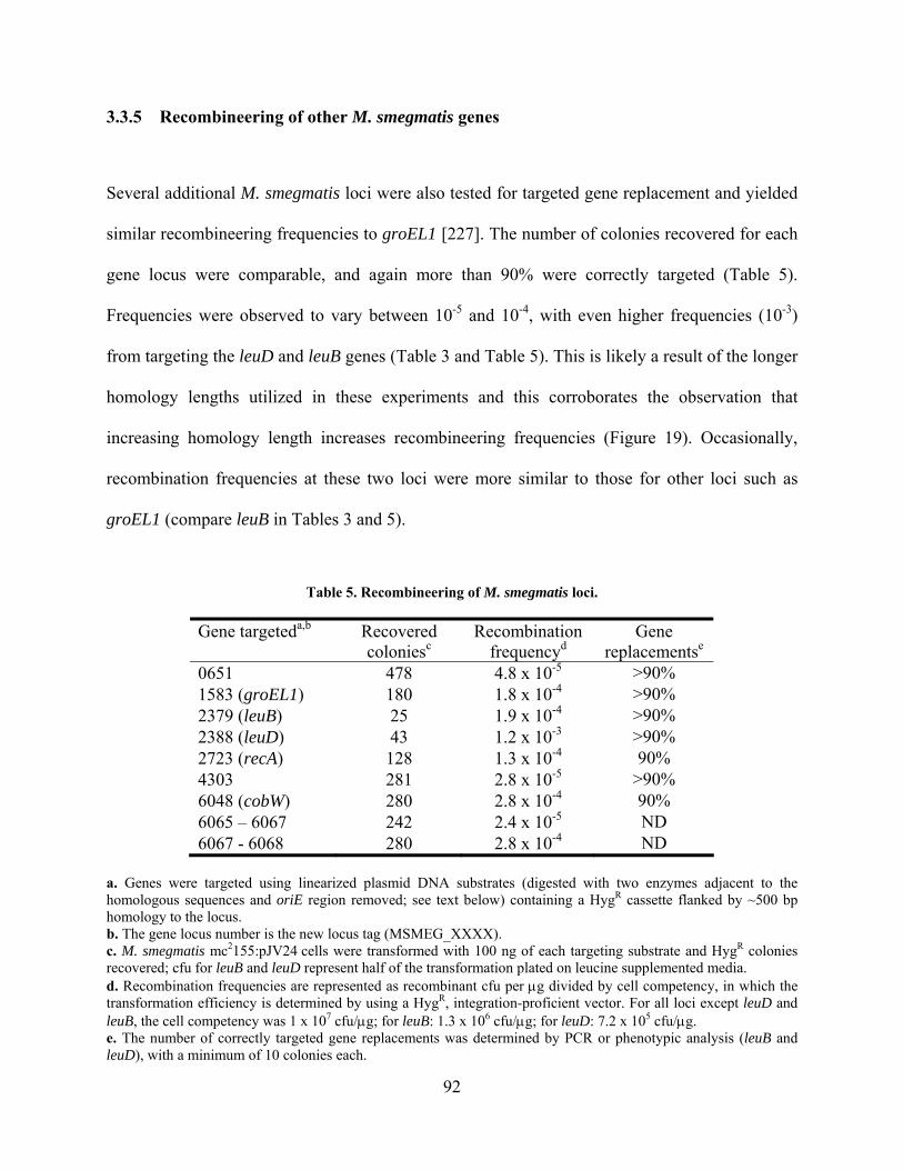
3.3.5 Recombineering of other M. smegmatis genes
Several additional M. smegmatis loci were also tested for targeted gene replacement and yielded
similar recombineering frequencies to groEL1 [227]. The number of colonies recovered for each
gene locus were comparable, and again more than 90% were correctly targeted (Table 5).
Frequencies were observed to vary between 10-5 and 10-4, with even higher frequencies (10-3)
from targeting the leuD and leuB genes (Table 3 and Table 5). This is likely a result of the longer
homology lengths utilized in these experiments and this corroborates the observation that
increasing homology length increases recombineering frequencies (Figure 19). Occasionally,
recombination frequencies at these two loci were more similar to those for other loci such as
groEL1 (compare leuB in Tables 3 and 5).
Table 5. Recombineering of M. smegmatis loci.
Gene targeteda,b Recovered coloniesc
Recombination frequencyd
Gene replacementse
0651 478 4.8 x 10⁻⁵ >90% 1583 (groEL1) 180 1.8 x 10⁻⁴ >90% 2379 (leuB) 25 1.9 x 10⁻⁴ >90% 2388 (leuD) 43 1.2 x 10⁻³ >90% 2723 (recA) 128 1.3 x 10⁻⁴ 90% 4303 281 2.8 x 10⁻⁵ >90% 6048 (cobW) 280 2.8 x 10⁻⁴ 90% 6065 – 6067 242 2.4 x 10⁻⁵ ND 6067 - 6068 280 2.8 x 10⁻⁴ ND
a. Genes were targeted using linearized plasmid DNA substrates (digested with two enzymes adjacent to the homologous sequences and oriE region removed; see text below) containing a HygR cassette flanked by ~500 bp homology to the locus. b. The gene locus number is the new locus tag (MSMEG_XXXX). c. M. smegmatis mc2155:pJV24 cells were transformed with 100 ng of each targeting substrate and HygR colonies recovered; cfu for leuB and leuD represent half of the transformation plated on leucine supplemented media. d. Recombination frequencies are represented as recombinant cfu per g divided by cell competency, in which the transformation efficiency is determined by using a HygR, integration-proficient vector. For all loci except leuD and leuB, the cell competency was 1 x 10⁷ cfu/g; for leuB: 1.3 x 10⁶ cfu/g; for leuD: 7.2 x 10⁵ cfu/g. e. The number of correctly targeted gene replacements was determined by PCR or phenotypic analysis (leuB and leuD), with a minimum of 10 colonies each.
92
3.3.6 Recombineering of the M. tuberculosis groEL1 gene
Since recombineering was successful in M. smegmatis, the effectiveness of this system was
tested in M. tuberculosis by targeting the groEL1 gene (Figure 20A) [227]. The results were
similar to those seen in M. smegmatis; ~150 recombinant colonies were obtained in an M.
tuberculosis H37Rv:pJV53 strain (Figure 20B and Table 6), yielding a recombineering
frequency of 1.7 x 10⁻⁴. Out of 16 colonies examined by Southern blot analysis, at least 14 were
correctly targeted to the groEL1 locus (Figure 20C). Although colonies were observed in a
control strain (H37Rv:pLAM12), these grew slowly (Figure 20B), arose at a much lower
frequency (8.1 x 10⁻⁶), and none were correctly targeted when examined by Southern blot
analysis (Figure 20D). However, the M. tuberculosis H37Rv:pJV53 strain showed protein
expression in the absence of acetamide induction (Figure 15E), which is not observed in M.
smegmatis (Figure 15A). This is not surprising, since previous studies have shown that M.
tuberculosis has a lower tolerance than M. smegmatis for plasmids containing the acetamidase
promoter cassette [25]. Therefore, as an additional experiment, cultures were grown in OADC
(see Materials and Methods), washed, and grown for 24 hours in media containing succinate and
acetamide prior to harvesting for electrocompetent cells. Recombinants were also correctly
targeted in these experiments (Figure 20C,D), and recombineering frequencies were similar;
however, cell competency was overall lower for these strains. Although protocols for preparing
electrocompetent cells of M. tuberculosis do not recommend storing cell aliquots at -80°C, this
did not have an effect on the overall recombineering frequency. However, freezing cells did
lower cell competency (Table 6), which has been observed previously [80]. Additionally, using a
93

PCR-generated groEL1 AES yielded approximately five-fold more recombinants than a
pMtbgroEL1 plasmid AES linearized by restriction digest (Table 6), and a higher proportion of
the PCR colonies were correctly targeted (Figure 20D). The incorrect targeting of the PacI-
digested groEL1 AES was likely due to targeting to the pJV53 plasmid via the homology at the
oriE region (see below). Using PCR-generated substrates also reduced the background in the
control strain (Table 6).
Table 6. Recombineering frequencies from targeted gene replacement of the M. tuberculosis groEL1.
Recombineering frequencies with each AESb
Strain and growth mediaa pYUB854
PacI-digest pMtbgroEL1KO
PacI-digest pMtbgroEL1KO
PCR H37Rv:pLAM12, OADC 1.7 x 10⁻⁵ 3.3 x 10⁻⁵ 5.6 x 10⁻⁶
H37Rv:pLAM12, succinate 1.3 x 10⁻⁵ 1.2 x 10⁻⁵ 8.1 x 10⁻⁶
H37Rv:pJV53, OADC 3.3 x 10⁻⁵ 3.3 x 10⁻⁵ 2.1 x 10⁻⁴
H37Rv:pJV53, succinate 2.7 x 10⁻⁵ 3.7 x 10⁻⁵ 1.7 x 10⁻⁴
H37Rv:pJV53, frozen aliquotsc ND ND 4.3 x 10⁻⁴
a. Strains were grown to mid-logarithmic phase in media and induced for 24 hours as follows: either grown initially to mid-logarithmic phase in media (1) containing OADC, washed, and induced in succinate and acetamide media, or (2) containing succinate, and subsequently acetamide added for induction. b. The AESs were prepared either by restriction digest with PacI of plasmids pYUB854 (no homology) or pMtbgroEL1KO (groEL1), or by PCR-amplification. c. Electrocompetent cell aliquots (cells grown in succinate media) were frozen at -80°C for two weeks, thawed, and transformed with the PCR-amplified groEL1 AES.
94

Figure 20. Allelic replacement of the M. tuberculosis groEL1 gene by recombineering.
Figure 20 [227]. (A) Schmetic of the M. tuberculosis groEL1 gene locus and the groEL1 AES. (B) Pictures of the recovered colonies in M. tuberculosis strains from transformations with 100 ng of groEL1 AES. (C) Southern blot analysis of the M. tuberculosis groEL1 gene locus of DNA isolated from 16 recombinant strains made by recombineering with a PCR-generated groEL1 AES in a pJV53 strain. The probe anneals to the downstream homology region of the groEL1 AES; expected band sizes: 7.5 kbp wild type; 10.2 kbp mutant. DNA from wild type and groEL1 M. tuberculosis strains (mutant constructed by specialized transduction [14]) were used as controls. (D) Southern blot of DNA isolated from recombinants: pJV53 or pLAM12 cultures grown either in succinate and induced with acetamide, or in OADC and washed into succ./acet. The AES was generated by PacI digest or PCR.
95

3.3.7 Recombineering efficiently targets replicating plasmids.
Throughout these experiments, the linearized AESs were generated by plasmid digest, with the
exception of the groEL1 AES, which was typically generated by PCR amplification. In one case,
the groEL1 AES plasmid was digested with an enzyme that cut near the E. coli origin of
replication (oriE), and a large increase in the number of recombinant colonies (104) was
observed (Figure 21A,B). This was not due to the presence of non-homologous regions at the
ends of the substrate (data not shown), nor was it dependent on the presence of homologous
sequences (pYUB854; Figure 21B). Recombinant colonies were analyzed by PCR with primers
that anneal in the AES homologies and thus produced two PCR products (wild type and mutant)
if the AES has been incorporated at a locus other than groEL1. These two PCR products were
seen at a low frequency with either AflII- or NcoI-digested AESs (Figure 21C). Conversely, all
colonies yielded two PCR products from transformations with PciI-digested plasmid AESs
targeting the groEL1 or MSMEG4308 genes (pMsgroEL1KO and pMs4308KO; Figure 21C,D).
These observations are likely connected to the presence of a region encompassing the
oriE that is homologous to the extrachromosomal pJV24 plasmid (Figure 21A). Digest in this
region (with PciI) results in a linear AES with significant lengths of homology to pJV24 on each
end. However, removal of the backbone of the plasmid produces colonies that are all correctly
targeted to the chromosomal groEL1 locus instead of the plasmid (Figure 21E). Analysis of
plasmids electroduced from PciI-generated colonies showed that 64% of the plasmids (originally
KanR only) were KanR/HygR (Figure 21F), and restriction digests revealed the presence of
additional DNA sequences in these plasmids (Figure 21G). These data clearly demonstrate that
extrachromosomal plasmids can be targeted by dsDNA recombineering, and that the plasmid
backbone of circular AES constructs must be removed if chromosomal targeting is desired [227].
96
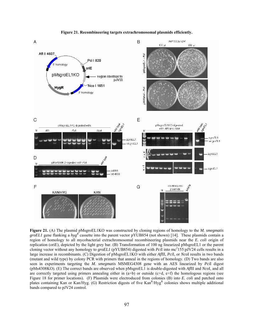
Figure 21. Recombineering targets extrachromosomal plasmids efficiently.
Figure 21. (A) The plasmid pMsgroEL1KO was constructed by cloning regions of homology to the M. smegmatis groEL1 gene flanking a hygR cassette into the parent vector pYUB854 (not shown) [14]. These plasmids contain a region of homology to all mycobacterial extrachromosomal recombineering plasmids near the E. coli origin of replication (oriE), depicted by the light grey bar. (B) Transformation of 100 ng linearized pMsgroEL1 or the parent cloning vector without any homology to groEL1 (pYUB854) digested with PciI into mc2155:pJV24 cells results in a large increase in recombinants. (C) Digestion of pMsgroEL1KO with either AflII, PciI, or NcoI results in two bands (mutant and wild type) by colony PCR with primers that anneal in the regions of homology. (D) Two bands are also seen in experiments targeting the M. smegmatis MSMEG4308 gene with an AES linearized by PciI digest (pMs4308KO). (E) The correct bands are observed when pMsgroEL1 is double-digested with AflII and NcoI, and all are correctly targeted using primers annealing either in (a+b) or outside (c+d, e+f) the homologous regions (see Figure 18 for primer locations). (F) Plasmids were electroduced from colonies (B) into E. coli and patched onto plates containing Kan or Kan/Hyg. (G) Restriction digests of five KanR/HygR colonies shows multiple additional bands compared to pJV24 control.
97

The data presented in this chapter demonstrate that recombineering with dsDNA
substrates is a simple and efficient method of constructing gene replacement mutants in both M.
smegmatis and M. tuberculosis [227]. The success of this technique further suggested that other
types of mutagenesis might be accomplished using recombineering, and these will be discussed
in the following section.
3.4 POINT MUTAGENESIS
3.4.1 ssDNA recombineering of replicating plasmids requires only Che9c gp61.
To determine if short ssDNA substrates (oligonucleotides) could be used to make point
mutations on mycobacterial genomes, a simple assay was developed using extrachromosomally-
replicating plasmids as targets for recombination [228]. The chosen target gene was a mutated
version of the hygR gene containing two consecutive amber mutations that inactivate its function
(hygS); the assay therefore tests if a HygR phenotype could be restored by ssDNA recombineering
at the mutated locus. The hygS gene was cloned into various plasmids expressing Che9c gp60,
gp61, or both (Figure 22A, Table 7), and electrocompetent cells were prepared of M. smegmatis
strains containing these plasmids. Complementary substrates were synthesized that were 100 nt
long (Table 16; JCV198, JCV199) and were homologous to the mutated region of hygS, with the
mutations that restore wild type sequence in the center. Transformation of M. smegmatis strains
expressing gp61 or both gp60/gp61 with either of these oligonucleotides resulted in more than
103 HygR colonies, whereas strains expressing only gp60 or containing an empty vector had only
background numbers of colonies (<25) (Figure 22B, Table 7). Transformations with
98
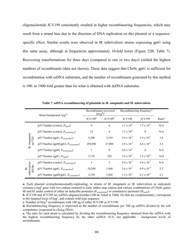
oligonucleotide JCV199 consistently resulted in higher recombineering frequencies, which may
result from a strand bias due to the direction of DNA replication on this plasmid or a sequence-
specific effect. Similar results were observed in M. tuberculosis strains expressing gp61 using
this same assay, although at frequencies approximately 10-fold lower (Figure 22B, Table 7).
Recovering transformations for three days (compared to one or two days) yielded the highest
numbers of recombinants (data not shown). These data suggest that Che9c gp61 is sufficient for
recombination with ssDNA substrates, and the number of recombinants generated by this method
is 100- to 1000-fold greater than for what is obtained with dsDNA substrates.
Table 7. ssDNA recombineering of plasmids in M. smegmatis and M. tuberculosis.
Recombinants recovered (HygR)c
Recombineering frequencyd
Strain background, hygS a
JCV198b JCV199b JCV198 JCV199 Ratioe
pJV73amber (control, Phsp60) 6 6 1.7 x 10⁻⁵ 1.7 x 10⁻⁵ N/A
pJV74amber (control, Pacetamidase) 23 0 7.1 x 10⁻⁶ 0 N/A
pJV75amber (gp61, Pacetamidase) 5,300 3,310 3.9 x 10⁻² 2.4 x 10⁻² 1.6
pJV76amber (gp60/gp61, Pacetamidase) 294,000 67,000 2.9 x 10⁻² 6.6 x 10⁻³ 4.3
pJV77amber (gp60, Pacetamidase) 1 0 8.0 x 10⁻⁶ 0 N/A
M. s
meg
mat
is
mc2 15
5
pJV78amber (gp61, Phsp60) 2,710 250 3.5 x 10⁻³ 3.2 x 10⁻⁴ 10.8
pJV74amber (control, Pacetamidase) 3 3 9.4 x 10⁻⁷ 9.4 x 10⁻⁷ N/A
pJV75amber (gp61, Pacetamidase) 10,200 1,960 3.6 x 10⁻³ 6.9 x 10⁻⁴ 5.2 M.
tube
rcul
osis
H
37R
v
pJV76amber (gp60/gp61, Pacetamidase) 2,130 1,020 1.3 x 10⁻³ 6.1 x 10⁻⁴ 2.1
a. Each plasmid (extrachromosomally-replicating; in strains of M. smegmatis or M. tuberculosis as indicated) contains a hygS gene with two codons mutated to early amber stop codons and various combinations of Che9c genes 60 and 61 under control of either an inducible promoter (Pacetamidase) or constitutive promoter (Phsp60). b. JCV198 and JCV199 are ssDNA oligonucleotides (100 nt; listed in Table 16) that are complementary, correspond to the mutated locus of hygS, and contain wild type sequence. c. Number of HygR recombinants with 100 ng of either JCV198 or JCV199. d. Recombineering frequency is expressed as the number of recombinants per 100 ng ssDNA divided by the cell competency (expressed in cfu/g DNA). e. The ratio for each strain is calculated by dividing the recombineering frequency obtained from the ssDNA with the highest recombineering frequency by the other ssDNA. N/A: not applicable – background levels of recombinants.
99
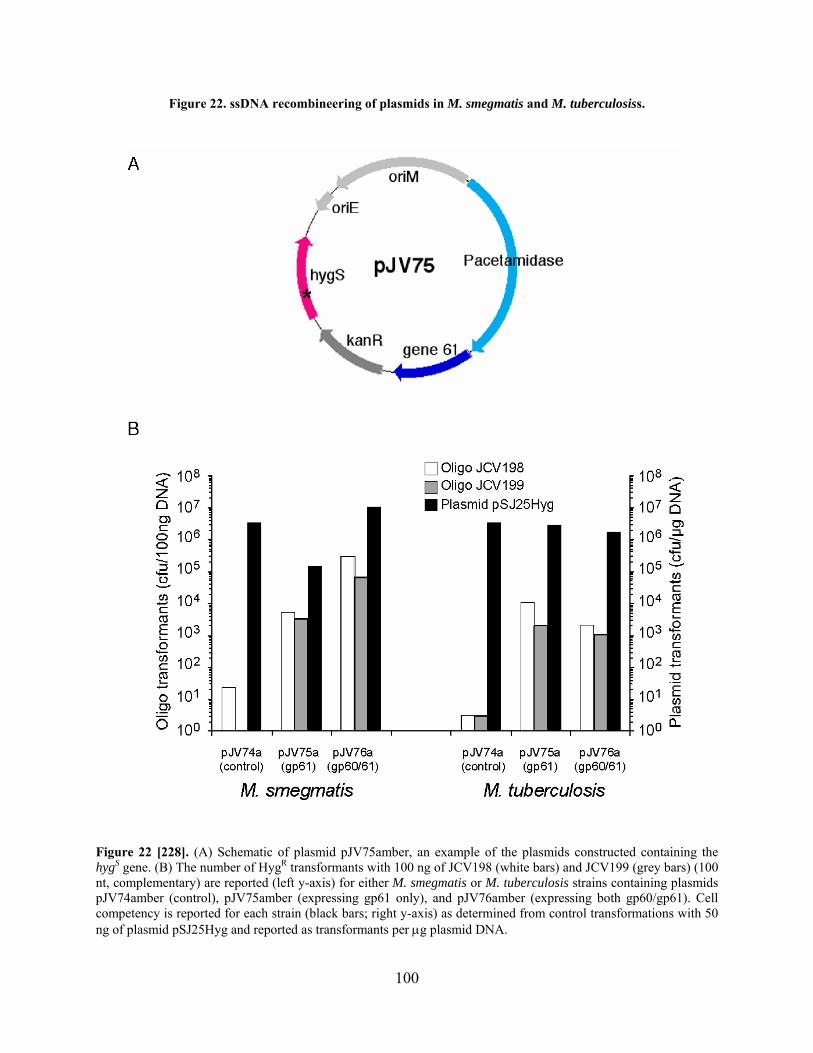
Figure 22. ssDNA recombineering of plasmids in M. smegmatis and M. tuberculosiss.
Figure 22 [228]. (A) Schematic of plasmid pJV75amber, an example of the plasmids constructed containing the hygS gene. (B) The number of HygR transformants with 100 ng of JCV198 (white bars) and JCV199 (grey bars) (100 nt, complementary) are reported (left y-axis) for either M. smegmatis or M. tuberculosis strains containing plasmids pJV74amber (control), pJV75amber (expressing gp61 only), and pJV76amber (expressing both gp60/gp61). Cell competency is reported for each strain (black bars; right y-axis) as determined from control transformations with 50 ng of plasmid pSJ25Hyg and reported as transformants per g plasmid DNA.
100
3.4.2 Introducing point mutations in the M. smegmatis chromosome by ssDNA
recombineering
To determine if ssDNA recombineering could be used to target the chromosome, the same hygS
gene was inserted into the chromosome of M. smegmatis at two loci using L5 and Bxb1
integration-proficient vectors [95,110]. The L5 and Bxb1 attB sites are located on different sides
of the M. smegmatis chromosome and are approximately the same distance from the origin of
replication (Figure 23A); Bxb1 is located 1.67 megabasepairs (mbp) 3 of the origin, and L5 is
2.22 mbp 5 of the origin (at 4.76 mbp). The hygS cassette was also inserted in both orientations
at each locus in order to examine the possible strand bias seen with plasmid targeting (Figure
23B). Using these four strains, the same oligonucleotides (JCV198 and JCV199) were tested for
ssDNA recombineering in an M. smegmatis mc2155:pJV62 background, which expresses Che9c
gp61 with transcription and translation signals from the acetamidase cassette. HygR colonies
were recovered using either oligonucleotide, although a strand bias was observed that, in some
cases, was more than 1000-fold (Figure 23C, Table 8). This strand bias correlates with the
direction of DNA replication at each locus, such that oligonucleotides that anneal to the lagging
strand consistently generated higher recombination frequencies than those annealing to the
leading strand. The dif site for replication termination is predicted to be at 3.41 mbp on the M.
smegmatis chromosome [74,75], and bi-directional replication from the origin to the terminus
correlates with the data from this assay (Figure 23). Interestingly, for each locus, one orientation
resulted in a much smaller strand bias (<3-fold; Table 8, pJV89amber and pJV94amber); lower
numbers of transformants were obtained with the oligonucleotide annealing to the lagging strand
(JCV198), and higher numbers of transformants were found with the leading strand
101

oligonucleotide (JCV199) as compared to the other orientation. This effect was consistent in all
strain backgrounds tested (Table 9) but was only seen with these particular integrated targets for
ssDNA recombineering. This may be due either to the presence of additional genetic elements on
the integrating plasmid that interfere with recombination or to a sequence-specific effect.
Table 8. ssDNA recombineering of a hygS gene in the M. smegmatis chromosome.
Recombinants recovered (HygR)
Recombineering frequency c Strain backgrounda
pJV62 (gp61), hygS JCV198 b JCV199 b JCV198 JCV199 Ratio d
pJV89amber 1,220 670 3.2 x 10⁻³ 1.7 x 10⁻³ 1.8
pJV91amber 5 20,400 8.5 x 10⁻⁶ 3.5 x 10⁻² 4,080
pJV92amber 15 27,600 8.4 x 10⁻⁵ 1.6 x 10⁻¹ 1,840
pJV94amber 1,760 700 4.7 x 10⁻³ 1.9 x 10⁻³ 2.5
a. Each strain contains a hygS gene integrated at either the Bxb1 attB locus (pJV89amber, pJV91amber) or the L5 attB locus (pJV92amber, pJV94amber). b. JCV198 and JCV199 are ssDNA oligonucleotides (100 nt; listed in Table 16) that are complementary, correspond to the mutated locus of hygS, and contain wild type sequence. c. Recombineering frequency is expressed as the number of recombinants per 100 ng ssDNA divided by the cell competency (expressed in cfu/g DNA). d. The ratio for each strain is calculated by dividing the recombineering frequency obtained from the ssDNA with the highest recombineering frequency by the other ssDNA.
Recombineering of the hygS gene at these two loci was compared in strain backgrounds
containing plasmids expressing either Che9c gp60/gp61 (pJV53) or only gp61, either from its
endogenous translation signals (pJV62) or those of the acetamidase cassette (pJV52).
Recombineering frequencies were approximately 10-fold higher in a pJV62 strain than in a
pJV52 strain (Table 9), and notably, pJV62 expresses slightly lower levels of gp61 compared to
pJV52 (Figure 15B). Therefore, strains expressing Che9c gp61 from plasmid pJV62 were used
in most ssDNA recombineering experiments.
102
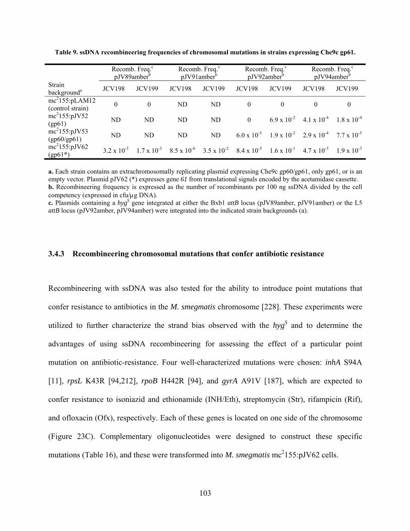
Table 9. ssDNA recombineering frequencies of chromosomal mutations in strains expressing Che9c gp61.
Recomb. Freq.c
pJV89amberb Recomb. Freq.c
pJV91amberb Recomb. Freq.c
pJV92amberb Recomb. Freq.c
pJV94amberb
Strain backgrounda JCV198 JCV199 JCV198 JCV199 JCV198 JCV199 JCV198 JCV199
mc2155:pLAM12 (control strain)
0 0 ND ND 0 0 0 0
mc2155:pJV52 (gp61)
ND ND ND ND 0 6.9 x 10⁻² 4.1 x 10⁻⁴ 1.8 x 10⁻⁴
mc2155:pJV53 (gp60/gp61)
ND ND ND ND 6.0 x 10⁻⁵ 1.9 x 10⁻² 2.9 x 10⁻⁴ 7.7 x 10⁻⁵
mc2155:pJV62 (gp61*)
3.2 x 10⁻³ 1.7 x 10⁻³ 8.5 x 10⁻⁶ 3.5 x 10⁻² 8.4 x 10⁻⁵ 1.6 x 10⁻¹ 4.7 x 10⁻³ 1.9 x 10⁻³
a. Each strain contains an extrachromosomally replicating plasmid expressing Che9c gp60/gp61, only gp61, or is an empty vector. Plasmid pJV62 (*) expresses gene 61 from translational signals encoded by the acetamidase cassette. b. Recombineering frequency is expressed as the number of recombinants per 100 ng ssDNA divided by the cell competency (expressed in cfu/g DNA). c. Plasmids containing a hygS gene integrated at either the Bxb1 attB locus (pJV89amber, pJV91amber) or the L5 attB locus (pJV92amber, pJV94amber) were integrated into the indicated strain backgrounds (a).
3.4.3 Recombineering chromosomal mutations that confer antibiotic resistance
Recombineering with ssDNA was also tested for the ability to introduce point mutations that
confer resistance to antibiotics in the M. smegmatis chromosome [228]. These experiments were
utilized to further characterize the strand bias observed with the hygS and to determine the
advantages of using ssDNA recombineering for assessing the effect of a particular point
mutation on antibiotic-resistance. Four well-characterized mutations were chosen: inhA S94A
[11], rpsL K43R [94,212], rpoB H442R [94], and gyrA A91V [187], which are expected to
confer resistance to isoniazid and ethionamide (INH/Eth), streptomycin (Str), rifampicin (Rif),
and ofloxacin (Ofx), respectively. Each of these genes is located on one side of the chromosome
(Figure 23C). Complementary oligonucleotides were designed to construct these specific
mutations (Table 16), and these were transformed into M. smegmatis mc2155:pJV62 cells.
103
Recombinant drug-resistant colonies were recovered at similar frequencies to those
observed with hygS experiments (Figure 23B, Table 10); background levels of drug-resistance
were similar to those reported in previous studies [11,94,187]. Targeting the lagging strand was
most efficient for each gene (~105 colonies), which is consistent with the data from hygS
targeting experiments and implicates a role for DNA replication in ssDNA recombination.
Interestingly, the strand biases varied in size; the gene most proximal to the origin of replication
(gyrA) had a strand bias of 36,000-fold, whereas the gene closest to dif (inhA) had a bias of 5-
fold. Overall numbers of recombinants from targeting the rpsL gene were 10-fold lower than for
other loci, although the strand bias (7,800-fold) was intermittent between gyrA and inhA. The
bias for rpoB could not be determined accurately because the background level of spontaneous
RIF-resistance masked the level of recombineering with the leading strand oligonucleotide
(Figure 23C, Table 10). In addition, not only was the strand bias small at inhA, but this did not
result from a decrease in colonies from the oligonucleotide targeting the lagging strand. Rather,
there was an increase in recombinants with a leading strand oligonucleotide at this locus as
compared to the others.
104
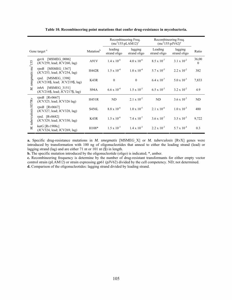
Table 10. Recombineering point mutations that confer drug-resistance in mycobacteria.
Recombineering Freq. (mc2155:pLAM12)c
Recombineering Freq. (mc2155:pJV62)c
Gene target a Mutationb leading
strand oligo lagging
strand oligo Leading
strand oligo lagging
strand oligo Ratio
gyrA [MSMEG_0006] (JCV259, lead; JCV260, lag)
A91V 1.4 x 10⁻⁶ 4.0 x 10⁻⁶ 8.5 x 10⁻⁷ 3.1 x 10⁻² 36,000
rpoB [MSMEG_1367] (JCV253, lead; JCV254, lag)
H442R 1.5 x 10⁻⁴ 1.0 x 10⁻⁴ 5.7 x 10⁻⁵ 2.2 x 10⁻² 382
rpsL [MSMEG_1398] (JCV218§, lead; JCV219§, lag)
K43R 0 0 6.4 x 10⁻⁷ 5.0 x 10⁻³ 7,833
M. s
meg
mat
is m
c2 155
inhA [MSMEG_3151] (JCV216§, lead; JCV217§, lag)
S94A 6.6 x 10⁻⁴ 1.5 x 10⁻³ 6.5 x 10⁻³ 3.2 x 10⁻² 4.9
rpoB [Rv0667] (JCV325, lead; JCV326 lag)
H451R ND 2.1 x 10⁻⁵ ND 3.6 x 10⁻³ ND
rpoB [Rv0667] (JCV327, lead; JCV328, lag)
S456L 8.8 x 10⁻⁶ 1.0 x 10⁻⁵ 2.1 x 10⁻⁶ 1.0 x 10⁻³ 480
rpsL [Rv0682] (JCV329, lead; JCV330, lag)
K43R 1.5 x 10⁻⁶ 7.4 x 10⁻⁷ 3.6 x 10⁻⁷ 3.5 x 10⁻³ 9,722
M. t
uber
culo
sis
H37
Rv
katG [Rv1908c] (JCV324, lead; JCV269, lag)
H108* 1.5 x 10-3 1.4 x 10⁻³ 2.2 x 10⁻³ 5.7 x 10⁻⁴ 0.3
a. Specific drug-resistance mutations in M. smegmatis [MSMEG_X] or M. tuberculosis [RvX] genes were introduced by transformation with 100 ng of oligonucleotides that anneal to either the leading strand (lead) or lagging strand (lag) and are either 71 nt or 101 nt (§) in length. b. The specific mutation introduced by the oligonucleotide (oligo) is indicated; *, amber. c. Recombineering frequency is determine by the number of drug-resistant transformants for either empty vector control strain (pLAM12) or strain expressing gp61 (pJV62) divided by the cell competency. ND; not determined. d. Comparison of the oligonucleotides: lagging strand divided by leading strand.
105
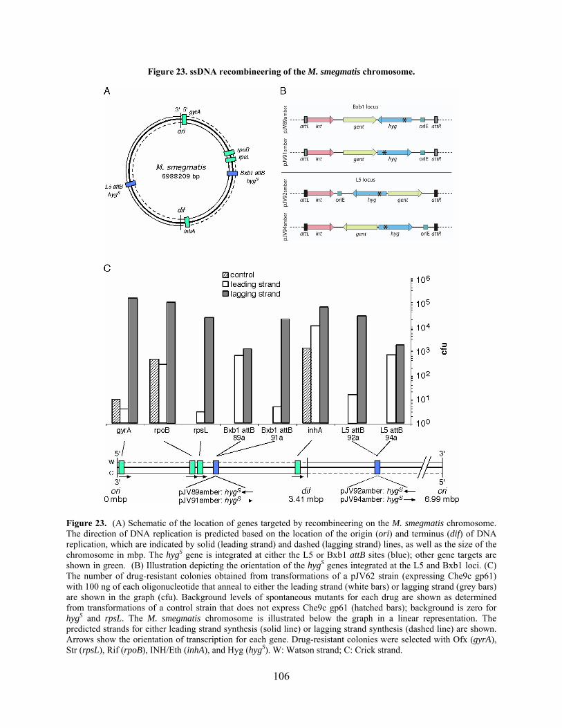
Figure 23. ssDNA recombineering of the M. smegmatis chromosome.
Figure 23. (A) Schematic of the location of genes targeted by recombineering on the M. smegmatis chromosome. The direction of DNA replication is predicted based on the location of the origin (ori) and terminus (dif) of DNA replication, which are indicated by solid (leading strand) and dashed (lagging strand) lines, as well as the size of the chromosome in mbp. The hygS gene is integrated at either the L5 or Bxb1 attB sites (blue); other gene targets are shown in green. (B) Illustration depicting the orientation of the hygS genes integrated at the L5 and Bxb1 loci. (C) The number of drug-resistant colonies obtained from transformations of a pJV62 strain (expressing Che9c gp61) with 100 ng of each oligonucleotide that anneal to either the leading strand (white bars) or lagging strand (grey bars) are shown in the graph (cfu). Background levels of spontaneous mutants for each drug are shown as determined from transformations of a control strain that does not express Che9c gp61 (hatched bars); background is zero for hygS and rpsL. The M. smegmatis chromosome is illustrated below the graph in a linear representation. The predicted strands for either leading strand synthesis (solid line) or lagging strand synthesis (dashed line) are shown. Arrows show the orientation of transcription for each gene. Drug-resistant colonies were selected with Ofx (gyrA), Str (rpsL), Rif (rpoB), INH/Eth (inhA), and Hyg (hygS). W: Watson strand; C: Crick strand.
106

Colonies from inhA targeting experiments were analyzed by PCR-amplification and
sequencing of the inhA gene; all contained the S94A mutation, whereas colonies from negative
control transformations did not (data not shown). An oligonucleotide that incorporates a
synonymous third-base change at the same locus (inhA S94 codon) did not yield INHR
transformants above background levels. Recombination with ssDNA appears to be independent
of host RecA (Table 11), unlike recombination with dsDNA substrates (Table 4). Collectively,
these data indicate that introduction of the inhA S94A and other mutations arose from
specifically-targeted recombination events that are dependent on Che9c gp61 and not general
mutagenesis.
Table 11. ssDNA recombineering dependence on host RecA.
Strain background Target: inhAb Target: rpsLb Recombineering frequency inhA
Recombineering frequency rpsL
mc2155:pLAM12 (control) 1,630 1 3.2 x 10⁻⁴ 2.0 x 10⁻⁷
mc2155:pJV62 (gp61) 115,000 6,600 1.4 x 10⁻¹ 8.1 x 10⁻³
mc2155:pJV62 recAa (gp61) 362,000 29,800 9.1 x 10⁻² 7.5 x 10⁻³
a. This M. smegmatis recA strain was constructed by K.G. Papavinasasundaram and colleagues [155] and is HygR . b. Oligonucleotides (100 ng) targeting the lagging strand containing either the inhA S94A (JCV217) or rpsL K43R (JCV219) point mutations were transformed into the M. smegmatis strains listed and either INHR or StrR transformants were selected, respectively. The number of transformants and recombineering frequencies for each target are shown.
Similar results were obtained in M. tuberculosis in which ssDNAs were designed to
introduce point mutations in rpoB (S456L and H451R), rpsL (K43R), and katG (H108amber;
INHR [191,209]) (Figure 24, Table 10). Drug-resistant colonies (up to 104) were obtained with
oligonucleotides that anneal to the lagging strand, with large strand biases up to ~9,700-fold; the
background of katG prevented an accurate comparison of leading and lagging strand efficiencies.
However, the recombineering frequencies were 5- to 30-fold lower as compared to those
observed in M. smegmatis (Table 10), consistent with the plasmid-targeting results.
107

Figure 24. ssDNA recombineering of the M. tuberculosis chromosome.
Figure 24. Similar to Figure 23 for the M. smegmatis chromosome, the locations of the M. tuberculosis genes targeted by ssDNA recombineering are depicted. (A) The location of the rpsL, rpoB, and katG genes on the M. tuberculosis chromosome, as well as ori and dif are shown on this schematic. Predicted leading (solid line) and lagging (dashed line) strands are indicated. (B) The number of drug-resistant colonies obtained from transformations of a pJV62 strain (expressing Che9c gp61) with 100 ng of each oligonucleotide that anneals to either the leading strand (white bars) or lagging strand (grey bars) are shown in the graph (cfu). Background levels of drug-resistant mutants are determined from transformations of a control strain that does not express Che9c gp61 (hatched bars). The rpoB mutation in this graph is S456L. Drug-resistant colonies were selected on Str (rpsL), RIF (rpoB), and INH (katG).
108
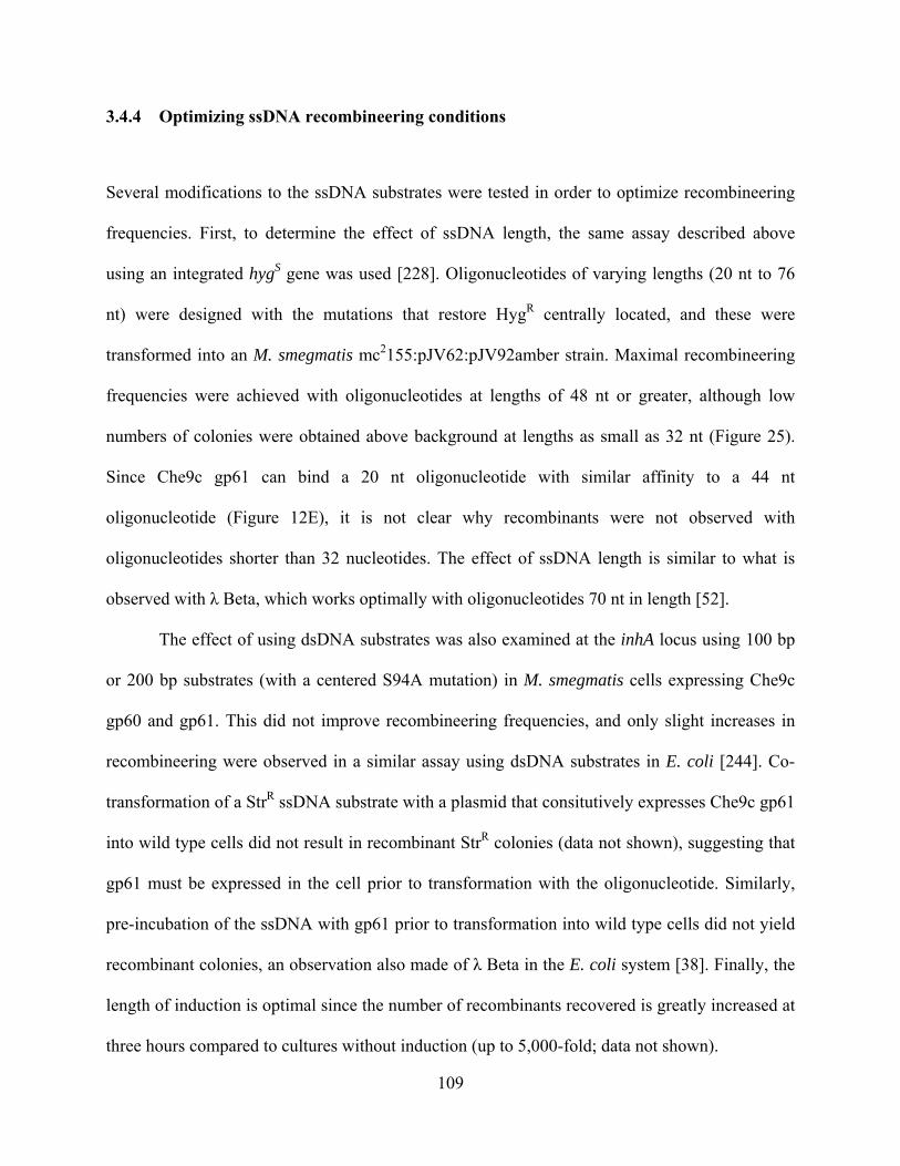
3.4.4 Optimizing ssDNA recombineering conditions
Several modifications to the ssDNA substrates were tested in order to optimize recombineering
frequencies. First, to determine the effect of ssDNA length, the same assay described above
using an integrated hygS gene was used [228]. Oligonucleotides of varying lengths (20 nt to 76
nt) were designed with the mutations that restore HygR centrally located, and these were
transformed into an M. smegmatis mc2155:pJV62:pJV92amber strain. Maximal recombineering
frequencies were achieved with oligonucleotides at lengths of 48 nt or greater, although low
numbers of colonies were obtained above background at lengths as small as 32 nt (Figure 25).
Since Che9c gp61 can bind a 20 nt oligonucleotide with similar affinity to a 44 nt
oligonucleotide (Figure 12E), it is not clear why recombinants were not observed with
oligonucleotides shorter than 32 nucleotides. The effect of ssDNA length is similar to what is
observed with λ Beta, which works optimally with oligonucleotides 70 nt in length [52].
The effect of using dsDNA substrates was also examined at the inhA locus using 100 bp
or 200 bp substrates (with a centered S94A mutation) in M. smegmatis cells expressing Che9c
gp60 and gp61. This did not improve recombineering frequencies, and only slight increases in
recombineering were observed in a similar assay using dsDNA substrates in E. coli [244]. Co-
transformation of a StrR ssDNA substrate with a plasmid that consitutively expresses Che9c gp61
into wild type cells did not result in recombinant StrR colonies (data not shown), suggesting that
gp61 must be expressed in the cell prior to transformation with the oligonucleotide. Similarly,
pre-incubation of the ssDNA with gp61 prior to transformation into wild type cells did not yield
recombinant colonies, an observation also made of λ Beta in the E. coli system [38]. Finally, the
length of induction is optimal since the number of recombinants recovered is greatly increased at
three hours compared to cultures without induction (up to 5,000-fold; data not shown).
109
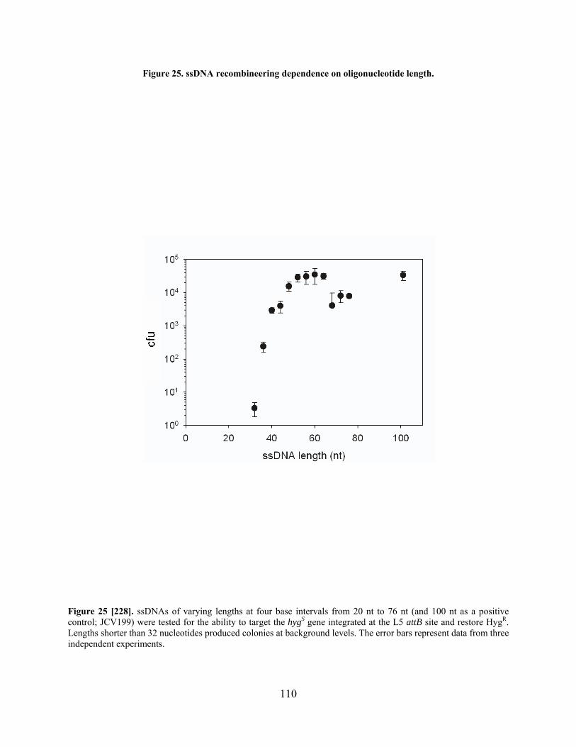
Figure 25. ssDNA recombineering dependence on oligonucleotide length.
Figure 25 [228]. ssDNAs of varying lengths at four base intervals from 20 nt to 76 nt (and 100 nt as a positive control; JCV199) were tested for the ability to target the hygS gene integrated at the L5 attB site and restore HygR. Lengths shorter than 32 nucleotides produced colonies at background levels. The error bars represent data from three independent experiments.
110

3.4.5 Development of a co-transformation strategy to select against non-transformable
cells
The recombineering experiments performed with ssDNA substrates demonstrated that selectable
point mutations could be made on either the chromosome or on extrachromosomal plasmid in M.
smegmatis and M. tuberculosis [228]. However, most point mutations are not selectable by drug-
resistance or other phenotypes, and instead require genotypic analysis to identify the mutant
allele. Although recombination of ssDNA substrates is very efficient, approximately only one
point mutant is recovered out of 1,000 viable cells. However, this frequency is similar to that
observed in a standard plasmid transformation. Since the limiting factor appeared to be the
competency of the cells, and not the frequency of recombination, it was reasoned that non-
selectable point mutants could be recovered if the non-transformed cells could be removed from
the population to be screened.
A co-transformation strategy was therefore tested in which a HygR plasmid and an INHR
(inhA S94A) oligonucleotide were electroporated into M. smegmatis cells expressing Che9c gp61
and selected on media containing Hyg, INH, or both. Notably, INHR/HygR mutants were
identified from colonies selected only on Hyg at a ~10% frequency (Figure 26A). This frequency
was obtained using saturating amounts (500 ng) of either the plasmid or oligonucleotide, and 100
ng of the other substrate (Figure 26B). Similar co-selection frequencies were also obtained
regardless of the type of HygR plasmid, either integrating (Bxb1, L5, Giles) or replicating. This
tactic was also successful using a double-oligonucleotide transformation: one oligonucleotide to
introduce the desired point mutation (e.g. inhA S94A; JCV217; INHR) and the other to repair a
hygS mutation (JCV198) present on the extrachromosomal plasmid (pJV75amber). This resulted
in a slightly lower co-selection frequency (~3-5%) but did not require the introduction of an
111

additional plasmid Optimal levels of co-selection were obtained with 200-500 ng of the INHR
oligonucleotide and 50-100 ng of the HygR oligonucleotide (Figure 26C); increasing the HygR
oligonucleotide to 500 ng dropped frequencies ~10-fold. Finally, four hours recovery yielded
optimal co-selection frequencies (7.2%), whereas shortening the time (1 hour, 1.2%) or
lengthening the time more than 8 hours (overnight, 2.8%) did not improve recovery of doubly-
resistant INH/Hyg colonies.
112
Figure 26. Optimizing recovery of point mutations by co-transformation of a HygR substrate.
Figure 26 [228]. (A) Colonies from transformations with a HygR plasmid (pSJ25Hyg) and an INHR oligonucleotide (JCV217) were selected only on Hyg and patched onto INH/Hyg media. (B) Varying amounts of the HygR plasmid and the INHR oligonucleotide (10, 25, 50, 100, 250, or 500 ng) were co-transformed and plated on Hyg, INH, or Hyg/INH. The key indicates the substrate held constant at 500 ng (while the other substrate quantity was varied) and the type of antibiotic selection for each reaction. (C) Co-transformations with varying amounts of the HygR oligonucleotide (JCV198) and INHR oligonucleotide (JCV217) were plated on Hyg, INH, or Hyg/INH. The key indicates the substrate and quantity held constant, and the antibiotic selection for each reaction.
113
3.4.6 Point mutagenesis in the absence of selection
The results of the co-selection experiments suggested that mutant alleles could be easily
identified at a high frequency when selection for plasmid transformants eliminates the non-
transformable majority of the cell population. To test this idea, the same experiment was
performed in which the INHR oligonucleotide and HygR plasmid were co-transformed, but the
inhA mutation was not selected. Rather, the cells were diluted multiple times in liquid media
containing Hyg in a culture block. Each culture well was plated to determine the number of
starting cells; in this experiment, each well contained ~70 HygR cells at the time of dilution.
Culture wells were then grown to saturation, and the inhA locus examined for each well by
mismatch amplification mutation assay PCR (MAMA-PCR) [30,219]. Mutant alleles (even a
single base change) are identified in a large wild type population by this technique (Figure 27A),
in which the primers and PCR conditions are optimized such that only mutant alleles are
amplified. Using this method, at least one mutant inhA allele was identified in each culture well
(Figure 27B). Homogenous mutant colonies were identified at a frequency of 3-4% from these
culture wells by plating for single colonies and selecting for INHR (Figure 27C). It is noted that
this is slightly decreased from experiments in which colonies are selected directly following
transformation on solid media (5-10%). However, this is likely due to variation between
experiments, since Hyg/INHR colonies were recovered at similar frequencies before and after
outgrowth in culture wells in a different experiment (5% and 4.7%, respectively).
This technique was also tested with an additional gene locus, the blaS gene in M.
smegmatis. The oligonucleotide, which was designed to introduce two consecutive amber
mutations in blaS, was co-transformed with a HygR plasmid, and the cells were diluted into
liquid Hyg media as described above. MAMA-PCR identified a mutant allele in each culture
114

well (out of ~50 HygR cells) (Figure 27D), and subsequent MAMA-PCR analysis identified pure
mutant strains that arose from the plating of a positive culture well (Figure 27E). This
experiment corroborated the previous experiments with inhA, in that mutant cells were present at
~3% out of HygR cells. Only two rounds of PCR were required to identify three blaS mutants (43
PCR reactions). Alternatively, the experiment could likely be altered such that the transformed
cells are diluted in Hyg media, grown to saturation, and plated for single colonies for MAMA-
PCR analysis. Interestingly, the early amber mutations did not confer the expected ampicillin-
sensitive phenotype for any of the three strains constructed; the blaS locus was analyzed by PCR
and sequencing and the correct mutations were present (data not shown).
In addition, an M. smegmatis pyrF point mutant that introduces an early stop codon
(Q61amber) was also constructed using co-transformation. Since inactivation of pyrF results in
5-FOA resistance and uracil auxotrophy, this mutant strain could also be used for co-selection in
which uracil prototrophy acts as a positive selection, confirmed by 5-FOA sensitivity. The strain
was constructed by double-oligonucleotide co-transformation, and three mutants were identified
as UraS and 5-FOAR out of 100 HygR colonies. Two of these were confirmed by PCR and
sequencing of the pyrF gene (data not shown).
Experiments targeting two additional non-selectable loci (M. smegmatis groEL1 and
leuD) were also attempted but were unsuccessful (data not shown). Several modifications were
tested, including using oligonucleotides targeting both DNA strands, dsDNA substrates (100 and
200 bp), increasing the recovery time of the transformation or the amount of substrate, but no
mutants were found by MAMA-PCR in any of the examined samples. A similar result was found
during attempts to make a selectable point mutation in M. smegmatis embB (I289M) that should
confer ethambutol resistance [112,214], although recombinant colonies were never obtained
115

above background levels. This may be due to sequence-specific effects, and experiments
introducing different mutations at the same or different nucleotides could be performed to
examine this possibility.
Finally, this strategy was tested on the M. tuberculosis leuD gene using co-transformation
with a HygR plasmid, and mutants were identified by MAMA-PCR (Figure 27F). The co-
selection frequency in M. tuberculosis, estimated at 0.5% - 1%, is lower than that observed for
most M. smegmatis loci. However, it should be noted that co-selection frequencies for the M.
smegmatis rpsL locus are ~0.3 – 0.5%, since overall numbers of recombinants are ~10-fold lower
at this target. Therefore, the low frequencies at the M. tuberculosis leuD target might be due to
generally lower co-transformation or recombination frequencies, or sequence-specific effects.
These data jointly demonstrate that both selectable and non-selectable point mutations can be
constructed in M. smegmatis and M. tuberculosis.
116
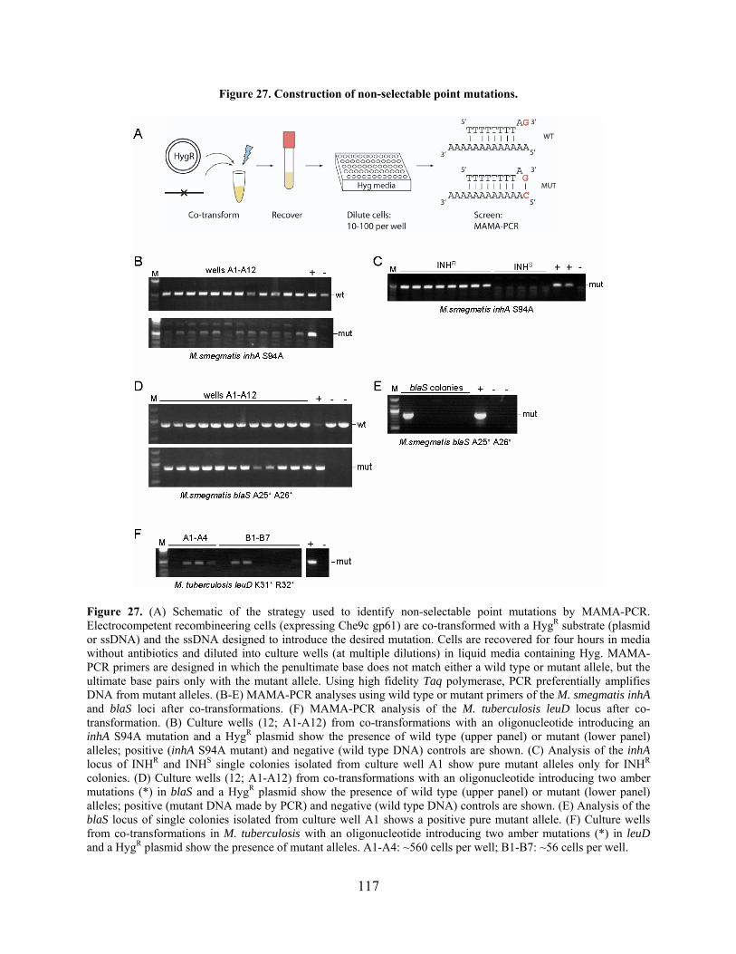
Figure 27. Construction of non-selectable point mutations.
Figure 27. (A) Schematic of the strategy used to identify non-selectable point mutations by MAMA-PCR. Electrocompetent recombineering cells (expressing Che9c gp61) are co-transformed with a HygR substrate (plasmid or ssDNA) and the ssDNA designed to introduce the desired mutation. Cells are recovered for four hours in media without antibiotics and diluted into culture wells (at multiple dilutions) in liquid media containing Hyg. MAMA-PCR primers are designed in which the penultimate base does not match either a wild type or mutant allele, but the ultimate base pairs only with the mutant allele. Using high fidelity Taq polymerase, PCR preferentially amplifies DNA from mutant alleles. (B-E) MAMA-PCR analyses using wild type or mutant primers of the M. smegmatis inhA and blaS loci after co-transformations. (F) MAMA-PCR analysis of the M. tuberculosis leuD locus after co-transformation. (B) Culture wells (12; A1-A12) from co-transformations with an oligonucleotide introducing an inhA S94A mutation and a HygR plasmid show the presence of wild type (upper panel) or mutant (lower panel) alleles; positive (inhA S94A mutant) and negative (wild type DNA) controls are shown. (C) Analysis of the inhA locus of INHR and INHS single colonies isolated from culture well A1 show pure mutant alleles only for INHR colonies. (D) Culture wells (12; A1-A12) from co-transformations with an oligonucleotide introducing two amber mutations (*) in blaS and a HygR plasmid show the presence of wild type (upper panel) or mutant (lower panel) alleles; positive (mutant DNA made by PCR) and negative (wild type DNA) controls are shown. (E) Analysis of the blaS locus of single colonies isolated from culture well A1 shows a positive pure mutant allele. (F) Culture wells from co-transformations in M. tuberculosis with an oligonucleotide introducing two amber mutations (*) in leuD and a HygR plasmid show the presence of mutant alleles. A1-A4: ~560 cells per well; B1-B7: ~56 cells per well.
117
3.5 OTHER APPLICATIONS OF RECOMBINEERING
An additional attractive use of recombineering is the construction of unmarked, in-frame deletion
mutants, and this can also be used for removing antibiotic resistance markers from gene
replacement mutants. To determine if mycobacterial recombineering could be used to make
deletions, the M. smegmatis leuD gene was targeted with substrates that delete the same region
as previous allelic replacement experiments at this locus (Figure 17A). However, in this case,
short ssDNA (100 nt) or dsDNA (100 bp or 200 bp) substrates were used that did not contain an
antibiotic marker for selection. In addition, dsDNA substrates were tested because previous
experiments targeting mycobacteriophage genomes indicated that these substrates were more
efficient for deletion construction than ssDNAs (L. Marinelli, manuscript in preparation). The
co-transformation strategy was used for this experiment since it had been successful for
constructing point mutations. Colonies were analyzed by two methods, either by plating directly
following transformation recovery, or by diluting cells in liquid media containing Hyg. In one
experiment, colonies plated directly following transformation (with a 100 bp leuD substrate and
HygR oligonucleotide) were replica plated onto media lacking leucine to identify leucine
auxotrophs, and a single mutant was identified at a frequency of ~0.5% (Figure 28B).
In other experiments, culture wells were screened for the presence of the leuD deletion
mutant. Mutants from experiments with an oligonucleotide that anneals to the lagging strand
could be identified by MAMA-PCR at a low frequency (~0.2%), while no mutants were
observed using a leading strand oligonucleotide (Figure 28C). Conversely, mutants were easily
identified from transformations with 100 bp and 200 bp substrates using MAMA-PCR analysis,
which indicated the presence of the mutant allele at a high frequency (Figure 28C). However,
mutants can still be identified using a less sensitive PCR technique (with primers that anneal
118

outside the deletion locus). For example, in one experiment in which a HygR plasmid was co-
transformed with a 200 bp susbtrate, at least one mutant was identified out of eight culture wells
(~10 cells per well; Figure 28D). Upon plating these colonies from the positive culture well, two
mutants were identified and confirmed out of ten tested (Figure 28E). It should be noted that the
frequencies observed by MAMA-PCR and standard PCR were inconsistent, which likely reflects
the detection of much smaller quantities of the mutant allele by MAMA-PCR. Futher, mutant
identification was simplified at this particular locus by screening for leucine auxotrophy.
However, mutants were readily identified by using co-transformation and PCR screening
techniques, and this technique is likely applicable to other genes. Additionally, it appears that
using 200 bp substrates gives the highest recombineering frequencies, much like what is
observed for mycobacteriophage recombineering (L. Marinelli, manuscript in preparation).
119
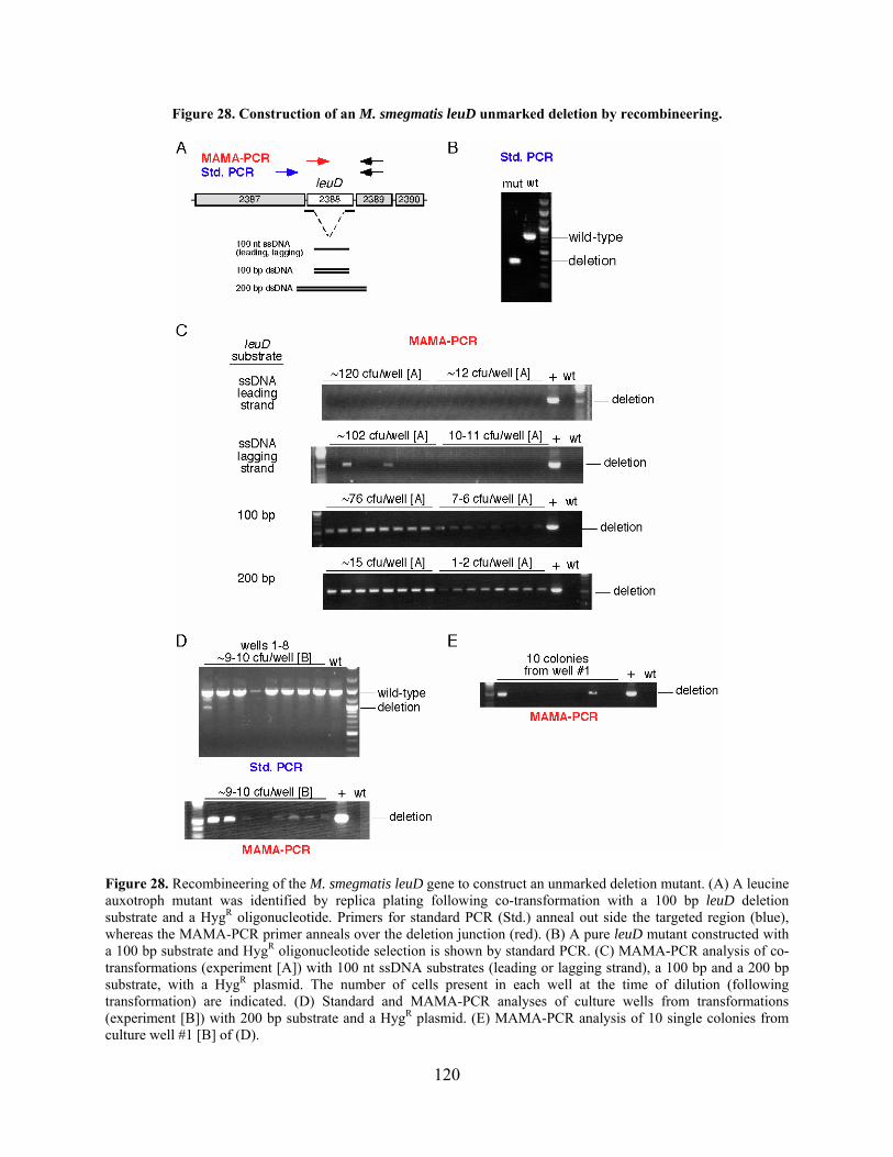
Figure 28. Construction of an M. smegmatis leuD unmarked deletion by recombineering.
Figure 28. Recombineering of the M. smegmatis leuD gene to construct an unmarked deletion mutant. (A) A leucine auxotroph mutant was identified by replica plating following co-transformation with a 100 bp leuD deletion substrate and a HygR oligonucleotide. Primers for standard PCR (Std.) anneal out side the targeted region (blue), whereas the MAMA-PCR primer anneals over the deletion junction (red). (B) A pure leuD mutant constructed with a 100 bp substrate and HygR oligonucleotide selection is shown by standard PCR. (C) MAMA-PCR analysis of co-transformations (experiment [A]) with 100 nt ssDNA substrates (leading or lagging strand), a 100 bp and a 200 bp substrate, with a HygR plasmid. The number of cells present in each well at the time of dilution (following transformation) are indicated. (D) Standard and MAMA-PCR analyses of culture wells from transformations (experiment [B]) with 200 bp substrate and a HygR plasmid. (E) MAMA-PCR analysis of 10 single colonies from culture well #1 [B] of (D).
120

3.6 CONCLUSIONS
The Che9c recombineering system has successfully been used to construct gene replacements,
point mutations, and gene deletions in the genomes of both M. smegmatis and M. tuberculosis. It
is an efficient method for mutagenesis that is generally applicable to chromosomal and plasmid
loci and is likely to be useful in other mycobacterial species. The Che9c proteins function
similarly to the λ Red and RecET proteins in E. coli both in vitro and for recombineering in vivo.
In fact, overall recombineering frequencies are similar between the two systems once differences
in DNA uptake efficiencies are taken into account [52,240].
3.6.1 Recombineering: a powerful technique for constructing gene replacement mutants
in the mycobacteria
Targeted gene knockouts can be made simply with linear AESs generated from circular plasmid
constructs containing ~500 bp or more of homology to the target gene flanking an antibiotic
resistance gene. These can be linearized either by PCR-amplification or double-restriction digest,
ensuring removal of the plasmid backbone (Figure 29). Using 100 ng of these substrates
generates a sufficient number of mutant colonies at every non-essential gene locus tested thus
far, with more than 90% the desired mutants. Although gene knockouts were obtained using 50
bp of homology, this is not a recommended strategy due to the large decrease in recombineering
frequency observed with these substrates. It is clear that the competency of the cells to take up
DNA is an important criterion, and cells must be prepared with care. However, sufficiently
competent cells were routinely made using a simple protocol without addition of glycine or other
suggested supplements. As expected, the recovery of the desired gene replacement mutants is
121
dependent on expression of Che9c gp60 and gp61, and this sufficiently increased homologous
recombination in M. tuberculosis, such that few recombinants arose from illegitimate
recombination in these experiments.
Targeted gene replacement mutagenesis has obvious benefits for the potential of making
large-scale ordered gene deletion mutants in the genomes of M. tuberculosis and M. smegmatis.
Not only would this provide mutant strains for various experimental purposes, but it would also
supplement the data regarding gene essentiality from previous genome-wide studies [200].
Additionally, nonsense mutations could be introduced into putative essential genes by ssDNA
recombineering to confirm essentiality and to avoid the polar effects of gene replacements or
transposon insertions.
122

Figure 29. Construction of a recombieering AES for allelic gene replacement mutagenesis.
Figure 29. Diagram of the recommended procedure for generating recombineering AESs. The regions at the 5 and 3 ends of the targeted gene locus are amplified by PCR, such that the final products contain unique restriction sites (A, B, C, and D) for directional cloning flanking an antibiotic resistance gene (e.g. HygR). The PCR products and the cloning vector are digested with all four enzymes and simultaneously ligated together. Selection of HygR E. coli transformants generally yields a sufficient number of clones (albeit less than a standard two-way ligation), from which DNA is prepared and analyzed. Correctly cloned plasmids are digested with the two enzymes whose unique sites are at the distal ends of the homologous regions (A and D). Reactions are cleaned-up (removal of the plasmid backbone is unnecessary) and transformed into mycobacterial cells.
123

3.6.2 Recombineering of selectable and non-selectable point mutations
The mycobacterial ssDNA recombineering technology enables the construction of isogenic
strains that differ by a single point mutation without direct selection, a technique that is
unparalleled by any other approach. Point mutations can be introduced in mycobacterial
chromosomes, replicating plasmids, and lytically-replicating mycobacteriophage genomes at
high frequencies. Since only Che9c gp61 is required for ssDNA recombination, point mutants
can be constructed without the potential toxic effects of expressing gp60. Because ssDNA
substrates can be synthesized commercially, these experiments merely require design and
purchase of ssDNAs of a minimum recommended length of 48 nucleotides, which eliminates the
requirement for plasmid construction or other complex DNA manipulations. In addition, the
double-oligonucleotide co-transformation strategy enables the construction of non-selectable
point mutations, such that strains are completely unmarked following removal of the
recombineering plasmid. Remarkably, the E. coli and mycobacterial recombineering systems
perform comparably under optimal conditions for each system – MMR-defective, Gam-
expressing strains and co-selective transformation, respectively – such that point mutants can be
identified in the absence of direct selection at a high frequency (10-25%, respectively)
[37,52,228].
The ssDNA recombineering technology could be specifically applied for determining the
role of mutations that confer drug-resistance, particularly in regard to clinical research on the
origins of XDR M. tuberculosis strains. Since most mutations are identified in combination with
other mutations in M. tuberculosis strains, it is necessary to re-introduce each single mutation
into a clean genetic background to determine its specific contribution to the strain’s drug-
susceptibility profile. However, this was previously not feasible due to the lack of generalized
124

transducing phages for M. tuberculosis. Only recently was the inhA S94A mutant strain
constructed alongside an isogenic wild type strain by specialized transduction [232]. Similarly,
the current study used ssDNA recombineering to test four characterized mutations for their
ability to confer antibiotic resistance as reported, especially in genes such as inhA that have high
levels of spontaneous mutagenesis and drug-resistance. For example, the gyrA A91V mutation
had previously been identified in vitro along with other mutations in the gyrA gene, and this was
not transduced into a clean genetic background [187]. The results of this study confirmed that
this mutation is sufficient for generating resistance to ofloxacin. It is anticipated that similar
experiments utilizing this technology will contribute to drug studies as a counterpart to detection
and characterization of mutations conferring drug-resistance in M. tuberculosis clinical isolates.
3.6.3 Unique attributes of the mycobacterial recombineering system
Che9c gp61 functions similarly to λ Beta for ssDNA recombineering such that only the SSAP is
required, recombination is independent of host RecA, and it is affected by the direction of DNA
replication at the targeted locus. In both systems, ssDNA substrates that target (anneal to) the
lagging strand DNA are more efficient than those that target the leading strand [52]. However,
the strand biases in mycobacterial ssDNA recombineering assays at certain chromosomal loci are
surprisingly quite sizeable (greater than 1000-fold in some cases), which is in stark contrast to
the 2- to 50-fold bias in E. coli with λ Red recombineering [52]. It is clear that ssDNAs can
recombine with the leading strand – since this was observed when targeting plasmids – but this is
not consistently observed above background mutational frequencies or the limitations of DNA
transformation. It is noted that experiments targeting plasmids in mycobacteria produce more
modest strand biases, and this may be related to the mechanism of replication on the plasmid-
125

encoded origin of replication. Furthermore, the size of the strand bias decreased from the origin
to the terminus, suggesting that regions near the replication terminus are replicated by forks
traveling in both directions. Overall, the dissimilarity in strand bias magnitude observed in
mycobacteria and E. coli may relect with fundamental differences in DNA replication, which is
not well-characterized in the mycobacteria. Specifically, this could be related to availability of
ssDNA regions near replication forks for ssDNA recombineering substrates, or alternatively, the
interaction of the SSAP-bound ssDNA with host proteins involved in DNA replication.
Mycobacteria do not encode homologues of the mutLS MMR, nor do they appear to have
a functional MMR system [213], which otherwise might influence the efficiency of generating
certain basepair mismatches [113]. Thus, DNA replication appears to be the major factor
contributing to the frequency of ssDNA recombineering in mycobacteria. However,
transformation efficiency has a limiting effect on mycobacterial recombineering, and it is not
clear if this masks any other contributing factors. For instance, there were several attempts to
make chromosomal point mutations that were unsuccessful, and this may be due to undetermined
sequence-specific effects at those loci. Also, it is not clear why the highest observed co-selection
frequency is 10%. Although the frequency was not expected to surpass 50% since only one
strand of the chromosome is targeted, it is surprising that it is as low as 5-10%, and the reason
for this is not known.
An interesting difference between the E. coli and mycobacterial recombineering systems
is the structure of the substrates required for making deletions. Experiments targeting both the
mycobacterial chromosome and mycobacteriophage genomes for deletions demonstrated that
dsDNA substrates yield better recombineering frequencies than ssDNA substrates, contrary to
what is recommended in E. coli protocols for this type of mutagenesis [223]. In E. coli, ssDNA
126
substrates are clearly sufficient for making deletions at a high frequency [52,223], although the
efficiency of dsDNA substrates for the same mutations has not been rigorously studied.
Ultimately, 200 bp dsDNA substrates are recommended for constructing deletion mutants since
these were more efficient than 100 bp substrates for both mycobacterial and mycobacteriophage
genomes (see below). This is less suprising, however, since it reflects the correlation between an
increase in homology length and an increase in recombineering frequency, which has been
observed previously with both mycobacterial and E. coli recombineering systems [142,229,240].
3.6.4 Other uses for mycobacterial recombineering
An ideal use of recombineering is to unmark allelic gene replacement mutants, which would also
be useful in the mycobacteria. This is accomplished in E. coli by using a ssDNA substrate with
homology to the regions to be deleted, typically flanking an antibiotic resistance gene (used to
select the gene knockout) and a SacB cassette for negative selection. Interestingly, as described
above, short dsDNA substrates were more efficient than either ssDNA for making an unmarked
deletion mutant in M. smegmatis. In-frame internal deletions and insertion of small tags for
protein purification would also be useful strategies for assessing protein function. Although the
ability to make these types of mutations in mycobacterial genomes has not been tested, it is
likely that this will be successful, given the techniques that have been performed with the
mycobacterial recombineering system on phages.
Experiments performed by Laura Marinelli have demonstrated that the mycobacterial
recombineering system could also be used to target lytically-replicating mycobacteriophage
genomes (manuscript in preparation). Phage mutagenesis is accomplished using a co-
transformation strategy (similar to experiments described above) in which the phage DNA is
127

transformed into recombineering-proficient cells along with the substrate to make the mutation.
Several types of mutations have been constructed in different phages, including point mutations,
unmarked deletions, and small insertions, and even a mutation that introduced a deletion and
point mutation simultaneously. The ability to make mutants in virtually any mycobacteriophage
will facilitate the study of uncharacterized phage genes and systematic analysis of genes essential
for phage propagation, among other uses.
3.6.5 Potential for optimizing the Che9c recombineering system
As with any newly developed system, there are a number of parameters that could be tested to
further optimize the conditions and increase the number of recombinants recovered. It is clear
that the level of protein expression plays a role in recombineering frequencies; however, higher
levels of gp61 expression did not necessarily correlate with an increase in recombineering. This
is observed in the E. coli system as well [135,137,244], and using a 5:1 ratio of Beta:Exo
produces the best results (K. Murphy, personal communication). An approximation of this with
the mycobacterial recombineering system was roughly attempted by placing Che9c 60 under
control of the Pacetamidase and 61 under the Phsp60. This setup did not increase dsDNA
recombineering frequencies, which is not surprising since constitutive expression of gp61 did not
increase ssDNA recombineering. It is likely that testing other promoter combinations would
produce better results. For example, using another inducible promoter such as the Tet
operator/UV15 promoter system [51,67] to control expression might work well. Alternatively,
the current induction conditions could be modified, such as by altering the concentration of
acetamide.
128
Another attractive approach for potential improvement to the mycobacterial system is to
determine if the λ Gam or another functional analogue (T4 gp2, P22 Abc1/2) might function in
mycobacteria to block host nuclease degradation. However, due to the specific protein-protein
interactions of Gam or Abc1/2 with RecBCD that are required for activity, these particular
proteins may not be the ideal solution. Instead, it might be more beneficial to co-opt a system in
which the proteins block degradation by a different mechanism, such as T4 gp2 or Mu Gam
which bind and protect the ends of dsDNAs [2,6]. Alternatively, recombineering could be tested
in recBCD mutant strains, although this limits the strain background. It is also not known if other
mycobacterial nucleases act on dsDNA substrates (such as AddA), and therefore, mutation of
host genes is not preferred. However, to ascertain the effect of nuclease mutations, as well as to
begin to characterize the role of RecBCD and AddA in mycobacteria, some of these experiments
have been performed and are described in the Appendix. Briefly, deletion of recB only modestly
improved recombination frequencies (3- to 5-fold), and one assay with λ Gam did not improve
recombineering frequencies. However, the recB strain holds potential for certain
recombineering methods in which the genetic background is of less importance, such as with
recombineering of phage genomes. This strain could therefore improve the frequency of mutant
allele recovery in these assays, making mutant isolation easier and more efficient.
Importantly, even though the Che9c recombineering system does not encode a Gam-like
protein, it has worked sufficiently well for making gene replacement mutants at every non-
essential locus thus far tested in the Hatfull lab (>20 genes). This is striking in that inhibition of
RecBCD (either with Gam or using a recBCD strain background) increases recombineering
with the λ Red system at least 10-fold [43], and in some studies was found to be required
[135,240,242]. This appears to be similar for RecET, such that Gam is not required but merely
129

enhances recombination efficiency [242]. It would therefore be interesting to examine the effect
of Gam (or an analogous protein) on Che9c-mediated recombineering.
One concern is the potential for genetic rearrangements due to leaky expression of the
Che9c genes in M. tuberculosis, when these cells are grown in succinate media without
acetamide. This expression pattern has been observed previously in mycobacterial strains
expressing proteins from this promoter cassette, but importantly, expression was repressed in
media containing ADC [157]. Although Che9c gp60/gp61 expression has not been monitored in
M. tuberculosis recombineering strains grown in ADC, it is likely that expression is decreased (if
not repressed completely) in ADC media as compared to succinate media. Recombinants were
obtained at similar frequencies when M. tuberculosis recombineering cells were grown in ADC,
washed, and incubated in succinate/acetamide media, albeit the cell competency dropped for
both strains tested. This therefore represents an alternative approach to possibly eliminate
expression of the Che9c proteins prior to the required 24 hour induction.
Finally, other mycobacteriophage-encoded recombination systems were identified from
sequencing and characterization. Although the Che9c proteins work sufficiently well for the
types of mutagenesis thus far tested, other phages might encode recombination proteins with
higher levels of activity. Therefore, the following chapter will examine the activity of phage-
encoded recombination proteins in the mycobacteria.
130
4.0 IDENTIFICATION AND CHARACTERIZATION OF OTHER
BACTERIOPHAGE RECOMBINASES
4.1 INTRODUCTION
Recombinases that function in the single strand annealing recombination pathway are found in
many bacteriophages, although only a few have been well-studied. SSAPs are typically identified
in operons adjacent to an exonuclease similarly to λ Red and E. coli RecET. These genes exhibit
the mosaic pattern broadly observed in phage genomes, such that different genes encoding
SSAPs and exonucleases are mixed [85]. For example, in phages SPP1 (of B. subtilis) and A118
(of Listeria monocytogenes), an Exo-like gene is found with a RecT-like gene. Several different
exonucleases have been found in these systems, including proteins of the type II restriction
enzyme fold (like λ Exo) and the type EndoVII fold [85]. Further, the gene order within the
operon is not consistent, such that either the SSAP or the exonuclease can be transcribed first
[43]. In some cases, like phage SPP1, other ORFs are predicted to lie between the genes
encoding the exonuclease and recombinase [85]. Therefore, it appears that the organization of
these recombination genes does not follow a particular pattern, but these can typically be
identified based on similarity to other phage-encoded systems.
The apparent species-specificity of these recombination proteins is of particular interest
with regard to the development and optimization of recombineering systems for bacteria other
131
than E. coli. A recent study by Datta and colleagues examined several putative SSAPs from
phages that infect various bacterial hosts for activity in E. coli [43]. Using ssDNA
recombineering as an assay, they observed that several SSAPs function with similar efficiency to
λ Beta (~107 colonies), and not surprisingly, these proteins are predominantly from phages of
Gram-negative bacteria. Several SSAPs from other phages that infect Gram-positive bacteria are
able to introduce point mutations with moderate success (105 to 106 colonies). Notably, B.
subtilis phage SPP1 gp35 and mycobacteriophage Che9c gp61 had the lowest recombination
efficiencies (103 and 104 colonies, respectively). Also, a direct comparison of λ Beta and E. coli
Rac prophage RecT showed that RecT functions ~30-fold worse in this assay. These data
collectively suggest that there is a correlation between protein activity and organism relatedness,
such that these phage-encoded proteins do not function as well in more distantly-related bacteria.
Although the basis of this is unclear, it is possibly due to the ability of these proteins to interact
specifically with host proteins during recombination, such as the components of the DNA
replication machinery.
In the same study, it was also observed that the B. subtilis phage SPP1 gp34.1 and gp35
proteins, which are λ Exo and RecT homologues, respectively [85], promote dsDNA
recombineering in E. coli [43]. Conversely, the L. monocytogenes phage A118 gp47 and gp48
proteins (also λ Exo and RecT homologues, respectively [85]) did not have dsDNA activity,
although the gp47 had ssDNA recombineering activity [43]. Interestingly, the genes encoding
SPP1 gp34.1 and gp35 are separated by three predicted ORFs, whereas A118 gp47 and gp48 are
adjacent. These data suggest that different pairs of recombination proteins can be identified in
phages, although they may not be located together and not all are necessarily active in this type
of assay.
132
The finding that SSAPs from the same bacterial hosts – specifically, λ Beta and E. coli
RecT – have different levels of activity has emphasized the need to examine other
mycobacteriophage candidates for recombination activity. Mycobacteriophages are extremely
diverse, and a large proportion of ORFs (~50%) do not have recognizable sequence similarity to
known genes and are therefore of unknown function [165]. Currently, more than 50
mycobacteriophages have been sequenced, contributing to a vast reservoir of genetic information
in which to search for SSAP-like genes. The Che9c-encoded recombinase and exonuclease were
identified out of the first 14 sequenced mycobacteriophages. However, analysis of the more
recently sequenced phages has revealed additional putative homologous recombination systems,
and these will be discussed and characterized in this chapter.
Another approach to identifying phage-encoded recombinases is to assay directly for
recombination activity, particularly with phages in which recombination proteins cannot be
readily identified bioinformatically. During the construction of the first shuttle phasmids, Jacobs
and colleagues observed that mycobacteriophage TM4 cosmid libraries recombined at a high
frequency in vivo [86]. These TM4 cosmids are chimeric constructs in which an E. coli plasmid
is randomly ligated to large fragments of the phage TM4 genome. These typically contain a
nearly complete phage genomic molecule (containing an E. coli plasmid) with a small deletion of
a portion of the phage DNA (see Figure 4). Most of these cosmids cannot be propagated
individually as phages and are non-infectious (as assayed by plaque formation); however, those
that can were further utilized as shuttle phasmids [13,14,86]. Strikingly, when plaques resulting
from transformation with a pool of cosmids were analyzed, it was found that only one plaque out
of 400 still contained the E. coli plasmid; the rest contained intact wild type TM4 genomic DNA.
This suggested that TM4 encodes a recombination system, although none of the ORFs have
133

similarity to known recombination proteins [165]. Therefore, the same assay utilizing TM4
cosmid libraries was used to characterize the putative recombination system of this phage.
4.2 BIOINFORMATIC ANALYSIS OF OTHER MYCOBACTERIOPHAGE
RECOMBINATION SYSTEMS
The results of BLAST analyses [4] using known recombination proteins as queries suggest that
mycobacteriophages Halo, Giles, and BPs encode putative recombination systems that include
recombinases in the λ Beta/RecT SSAP superfamily (Figure 30). Since Halo and BPs are 100%
identical in the region containing these genes (99% overall), this analysis focused on the Halo
proteins. Halo gp42 is 46% identical to Che9c gp60 and 30% identical to the C-terminus of
RecE. However, analyses with Halo gp43 indicate that it is much more distantly related to other
phage-encoded RecT proteins (~13% identity), and this was only identified after two rounds of
PSI-BLAST. Additionally, the Halo gp43 protein was purified in a similar manner to Che9c
gp61, and its DNA binding properties were analyzed by filter binding assays. Preliminary results
indicated that gp43 does bind ssDNA (data not shown), although these experiments need to be
repeated to determine the binding constant.
Unlike Halo, BPs, and Che9c, mycobacteriophage Giles does not have a RecE-like
homologue; instead, gp52 contains a domain from the YqaJ family of phage-encoded
exonucleases. The putative SSAP in Giles, gp53, is also not easily identifiable, but it has 30%
amino acid identity to Halo gp43. These two proteins do appear to be members of the λ
Beta/RecT SSAP family (Figure 30B), although more distantly related than Che9c gp61.
134
In the process of studying genes related to Che9c 60 and 61, it was observed that M.
avium contains a prophage that encodes similar proteins (Figure 30A). M. avium MAV_0829
shares 29% amino acid identity with Che9c gp61; it is annotated as a ‘RecT/YqaK’ protein and
has 40% identity to E. coli RecT. Not surprisingly, the gene adjacent to this (MAV_0830) is
predicted to encode an exonuclease; it is 41% identical to Che9c gp60 and 23% identical to
RecE. Further BLAST analysis also identified an Erf-like protein, gp64, encoded by
mycobacteriophages Wildcat and Cjw1; Wildcat gp64 is 21% identical to P22 Erf and Cjw1
gp70 is 15% identical to Erf (not shown). However, none of their adjacent genes has similarity to
known recombination proteins. A prophage in M. abscessus is also predicted to encode a protein
that is a distant relative of Erf (MAB_1744; 18% identity; not shown). Collectively, these data
provided additional candidates to test for recombination activity in mycobacteria.
135
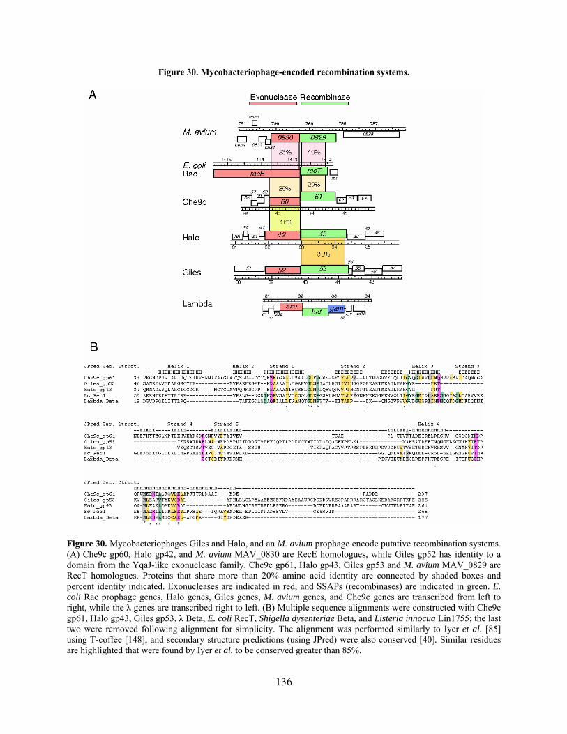
Figure 30. Mycobacteriophage-encoded recombination systems.
Figure 30. Mycobacteriophages Giles and Halo, and an M. avium prophage encode putative recombination systems. (A) Che9c gp60, Halo gp42, and M. avium MAV_0830 are RecE homologues, while Giles gp52 has identity to a domain from the YqaJ-like exonuclease family. Che9c gp61, Halo gp43, Giles gp53 and M. avium MAV_0829 are RecT homologues. Proteins that share more than 20% amino acid identity are connected by shaded boxes and percent identity indicated. Exonucleases are indicated in red, and SSAPs (recombinases) are indicated in green. E. coli Rac prophage genes, Halo genes, Giles genes, M. avium genes, and Che9c genes are transcribed from left to right, while the λ genes are transcribed right to left. (B) Multiple sequence alignments were constructed with Che9c gp61, Halo gp43, Giles gp53, λ Beta, E. coli RecT, Shigella dysenteriae Beta, and Listeria innocua Lin1755; the last two were removed following alignment for simplicity. The alignment was performed similarly to Iyer et al. [85] using T-coffee [148], and secondary structure predictions (using JPred) were also conserved [40]. Similar residues are highlighted that were found by Iyer et al. to be conserved greater than 85%.
136

4.3 COMPARISON OF SSAP ACTIVITY IN M. SMEGMATIS
Recombineering with ssDNA substrates provides a simple assay for recombination activity in
vivo. In particular, drug-resistance mutations that give low background can be introduced in the
M. smegmatis chromosome (discussed in section 3.4.3). Several SSAPs were tested for activity,
including Halo gp43 and Giles gp53. In addition, E. coli RecT and λ Beta were analyzed in order
to determine their activity in this distantly related bacterium, as well as to compare these results
to those from a similar study by Datta et al. performed in E. coli [43]. The SSAP genes – Che9c
61, Halo 43, Giles 53, E. coli recT, and λ bet – were under control of the Pacetamidase promoter
cassette (plasmid pLAM12) such that translation was derived from either their endogenous
signals (pJV52, pJV103, pJV145, pJV104, and pJV105) or from signals in the acetamidase
promoter cassette (NdeI site; see Figure 14) (pJV62, pJV106, pJV116, pJV107, and pJV108).
M. smegmatis strains containing each of these plasmids were induced for expression the
same way as all previous recombineering strains with the Che9c proteins. Reverse-transcription
PCR (RT-PCR) analysis was used to examine expression from Pacetamidase for many of the
constructs expressing genes from their endogenous translation signals (Figure 31A). Western
blot analysis was performed on strains expressing Halo gp43 (Figure 31B), but was not suitable
for some strains. Antibodies were not available for E. coli RecT, and the λ Beta antibodies (a gift
from D. Court) had high background signal in M. smegmatis cells that masked any potential
protein expression. Neither RT-PCR nor western blots were performed on Giles gp53 protein
expressing strains. Strains expressing λ bet and E. coli recT both had detectable levels of RNA
after induction with acetamide, indicating that at least transcription from this promoter is active
in these constructs. However, this does not rule out any potential translation or protein instability
137
problems that could occur in this assay. Similar to Che9c gp61 (Figure 15), protein expression
was also observed for the Halo gp43 (Figure 31B) by western blot.
Following these analyses, in order to test in vivo recombineering activity, each strain was
transformed with oligonucleotides that introduced point mutations in the inhA, rpsL, and gyrA
loci and recombinant colonies were selected. There were several surprising observations from
these assays (Figure 31C, Table 12). First, Halo gp43 had a significant level of ssDNA
recombineering activity, whereas Giles gp53 did not. This may be due to a lack of adequate
expression, which can be tested in the future by RT-PCR and/or western blot analysis. Second,
E. coli RecT had a high level of activity that was similar to Halo gp43, although not as high as
Che9c gp61. Strains expressing λ Beta did not produce recombinant colonies above background,
which is expected given anecdotal reports that the λ proteins are not active in mycobacteria. In
addition, the strain that expressed RecT from the translation signals of the acetamidase promoter
cassette had higher levels of activity (~10-fold) than the strain expressing RecT from its own
translation signals (Figure 31C); this was also observed for Che9c gp61 in this and in previous
assays (Table 9).
These data suggest that Che9c gp61 has the highest level of ssDNA recombineering
activity in mycobacterial cells. Further, although Halo gp43 and Giles gp53 are 30% identical,
strains expressing the Halo protein produced recombinants, whereas strains containing the Giles
constructs did not. Finally, there is a substantial difference between the activities of E. coli phage
SSAPs in the mycobacteria, such that λ Beta cannot recombine ssDNAs, while RecT is
moderately efficient.
138
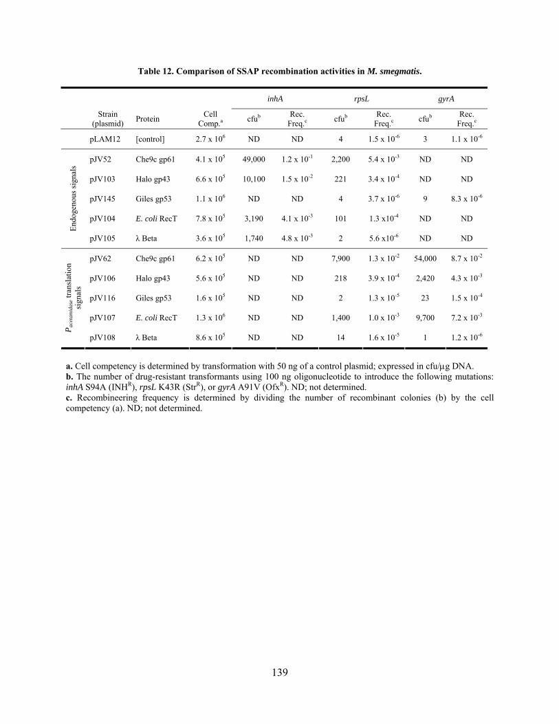
Table 12. Comparison of SSAP recombination activities in M. smegmatis.
inhA rpsL gyrA
Strain (plasmid) Protein
Cell Comp.a cfub
Rec. Freq.c cfub
Rec. Freq.c cfub
Rec. Freq.c
pLAM12 [control] 2.7 x 10⁶ ND ND 4 1.5 x 10⁻⁶ 3 1.1 x 10⁻⁶
pJV52 Che9c gp61 4.1 x 10⁵ 49,000 1.2 x 10⁻¹ 2,200 5.4 x 10⁻³ ND ND
pJV103 Halo gp43 6.6 x 10⁵ 10,100 1.5 x 10⁻² 221 3.4 x 10⁻⁴ ND ND
pJV145 Giles gp53 1.1 x 10⁶ ND ND 4 3.7 x 10⁻⁶ 9 8.3 x 10⁻⁶
pJV104 E. coli RecT 7.8 x 10⁵ 3,190 4.1 x 10⁻³ 101 1.3 x10⁻⁴ ND ND
End
ogen
ous
sign
als
pJV105 λ Beta 3.6 x 10⁵ 1,740 4.8 x 10⁻³ 2 5.6 x10⁻⁶ ND ND
pJV62 Che9c gp61 6.2 x 10⁵ ND ND 7,900 1.3 x 10⁻² 54,000 8.7 x 10⁻²
pJV106 Halo gp43 5.6 x 10⁵ ND ND 218 3.9 x 10⁻⁴ 2,420 4.3 x 10⁻³
pJV116 Giles gp53 1.6 x 10⁵ ND ND 2 1.3 x 10⁻⁵ 23 1.5 x 10⁻⁴
pJV107 E. coli RecT 1.3 x 10⁶ ND ND 1,400 1.0 x 10⁻³ 9,700 7.2 x 10⁻³
Pac
etam
idas
e tra
nsla
tion
si
gnal
s
pJV108 λ Beta 8.6 x 10⁵ ND ND 14 1.6 x 10⁻⁵ 1 1.2 x 10⁻⁶
a. Cell competency is determined by transformation with 50 ng of a control plasmid; expressed in cfu/g DNA. b. The number of drug-resistant transformants using 100 ng oligonucleotide to introduce the following mutations: inhA S94A (INHR), rpsL K43R (StrR), or gyrA A91V (OfxR). ND; not determined. c. Recombineering frequency is determined by dividing the number of recombinant colonies (b) by the cell competency (a). ND; not determined.
139
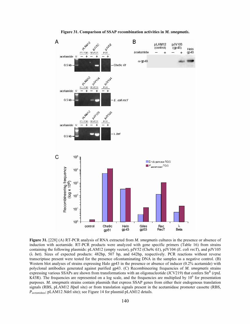
Figure 31. Comparison of SSAP recombination activities in M. smegmatis.
Figure 31. [228] (A) RT-PCR analysis of RNA extracted from M. smegmatis cultures in the presence or absence of induction with acetamide. RT-PCR products were analyzed with gene specific primers (Table 16) from strains containing the following plasmids: pLAM12 (empty vector), pJV52 (Che9c 61), pJV104 (E. coli recT), and pJV105 (λ bet). Sizes of expected products: 482bp, 507 bp, and 642bp, respectively. PCR reactions without reverse transcriptase present were tested for the presence ofcontaminating DNA in the samples as a negative control. (B) Western blot analyses of strains expressing Halo gp43 in the presence or absence of inducer (0.2% acetamide) with polyclonal antibodies generated against purified gp43. (C) Recombineering frequencies of M. smegmatis strains expressing various SSAPs are shown from transformations with an oligonucleotide (JCV219) that confers StrR (rpsL K43R). The frequencies are represented on a log scale, and the frequencies are multiplied by 106 for presentation purposes. M. smegmatis strains contain plasmids that express SSAP genes from either their endogenous translation signals (RBS, pLAM12 HpaI site) or from translation signals present in the acetamidase promoter cassette (RBS, Pacetamidase; pLAM12 NdeI site); see Figure 14 for plasmid pLAM12 details.
140
4.4 CHARACTERIZATION OF A PUTATIVE RECOMBINATION SYSTEM IN
MYCOBACTERIOPHAGE TM4
Although the number of sequenced genomes continues to increase, recent PSI-BLAST analysis
of the predicted gene products encoded by mycobacteriophage TM4 still did not reveal any clues
as to which genes might provide the recombination activity. While some gene products had short
regions of similarity to proteins with known recombination activity, they were not good
candidates or were not located next to genes that were likely to be part of a recombination
system. For example, gp54 (93 amino acids) has similarity to a region of the YqaJ-like
exonuclease protein of Bacillus cereus. However, gp53 has only low levels of sequence
similarity with hypothetical transpeptidase or dehydrogenase proteins, whereas the other adjacent
gene, gp56, is predicted to encode a protein only 29 amino acids in length with no sequence
similarity to known proteins. In another case, gp59 has only 17% sequence identity to a putative
RecB family exonuclease from a Thermus phage. Again, the adjacent genes are not likely
candidates; gp57 is predicted to encode a DinG helicase, gp58 has an esterase_lipase domain,
and gp60 is a small protein (57 amino acids) without similarity to any proteins in the database. It
is possible that there are different start sites for some of these genes that would alter the analysis.
However, based on the annotated genes, these do not appear to encode bona fide recombinase or
exonuclease homologues.
Therefore, to further examine the recombination phenotype observed for the TM4 cosmid
molecules, a new TM4 cosmid library was constructed (as described [86] and in Figure 4, except
a HygR plasmid was inserted instead of an AmpR plasmid). These molecules contained the E. coli
plasmid inserted in a region of the TM4 genome that is either essential (non-viable phage =
cosmid) or non-essential (true shuttle phasmid). TM4 cosmid DNA was isolated from individual
141

E. coli HygR colonies and examined by analytical restriction digest and sequencing to determine
the structure of the cosmid. A set of cosmids was obtained by three rounds of screening, and the
location of the E. coli plasmid and size of the TM4 deletion region were determined; these are
illustrated in Figure 32. As a reference, four shuttle phasmids that were isolated by Jacobs and
colleagues are also depicted in Figure 32 [13,14,86,87,210]. In addition, HygR colonies
(~27,000) were pooled, and DNA was prepared in order to repeat the experiments performed by
Jacobs et al. [86].
142

Figure 32. Diagram of the TM4 cosmid library.
Figure 32. Each solid line on the schematic represents a TM4 cosmid (TM4cosX) or shuttle phasmid (phX). Dotted lines at the ends indicate that the molecule is connected at the termini; otherwise the E. coli plasmid connects the circle. The ‘blank’ spaces indicate the region deleted in the cosmid, as well as the location of the E. coli plasmid. A linear representation of the TM4 genome is depicted below (in kbp). Shuttle phasmids were made by Jacobs and colleagues [13,14,86]. The purple box indicates the region deleted in both TM4cos7 and TM4cos20 that renders them incapable of recombination.
143

Recombination experiments were performed in which cosmid pairs were co-transformed
(500 g each) into wild type M. smegmatis, recovered for 30 minutes, and plated as top agar
lawns with additional M. smegmatis cells. Plaque numbers were recorded for each
transformation. It was observed that only cosmid pairs that represented the full genome between
them (i.e., non-overlapping deletions) could produce plaques (Table 13), supporting the
hypothesis that these molecules undergo recombination in vivo. Since most cosmids had large
deletions of their genome (~9 kbp), it is not surprising that the individual cosmids could not
propagate as phages. Pairs of cosmids with overlapping deletions did not produce plaques, likely
because the common deleted region was essential. As expected, plaques were also obtained from
transformations with DNA of the ‘pooled’ cosmid library. Notably, two cosmids, TM4cos7 and
TM4cos20, were not able to recombine with any other cosmids, even though the complete
genome was represented in all pairs tested (Table 13). However, it is interesting that small
numbers of plaques were obtained with these pairs, whereas zero plaques were consistently
obtained with pairs that were not expected to recombine. This deficiency in recombination is
likely due to the presence of a cis-acting element in this region that is required for recombination
and/or DNA replication. The region encodes only one small gene in entirety, 71, but does also
include the 3 half of gene 70, which is predicted to encode the DNA primase. Therefore, the cis-
acting element could be an origin of replication, which is often located in the region of the
genome that encodes DNA replication proteins in other phages and in bacteria [58,99].
144

Table 13. Recombination between TM4 cosmids as measured by plaque formation.
Individual Pairs w/
overlapping deletions
Pairs w/ non-overlapping deletions
TM4Cos7 and
TM4Cos20 pairs
TM4Cos#a Pfu per g DNAb TM4Cos#a Pfu per
g DNAb TM4Cos#a Pfu per g DNAb TM4Cos#a Pfu per g
DNAb
7 0 8 + 11 0 8 + 9 235 7 + 8 2
8 0 9 + 49 0 9 + 11 353 7 + 9 3
9 0 9 + 53 0 9 + 12 252 7 + 11 0
11 0 14 + 13 0 9 + 42 263 7 + 14 2
12 0 8 + 14 491 14 + 20 2
13 0 11 + 49 248
14 0
20 0
42 0
49 0
53 0 TM4 DNA
(pfu per g)c 1 x 10⁴
Pooled DNA (pfu per g)
320
a. Each cosmid was assigned a number during screening: TM4cosX. b. The number of plaques (pfu) per g total DNA is shown from transformations with DNA from either single cosmids, pairs of cosmids, or a pooled library. c. Wild type TM4 DNA (200 ng) was used as a positive control and is represented as pfu/g.
Analysis of plaques resulting from transformations with pooled cosmid DNA or pairs of
cosmids showed only the presence of wild type TM4 DNA (Figure 33A). These recombinant
plaques did not show the presence of the E. coli plasmid (assayed by PCR), and DNA prepared
from the plaques displayed a restriction pattern identical to wild type (Figure 33B). No true
shuttle phasmids were identified out of 14 plaques screened from the pooled library, which is
expected since these were recovered from the previous study at a very low frequency (~0.25%).
Further, the average size of the deletions in the cosmids was ~9 kbp, which is much larger than
the deletions found in shuttle phasmids, such as phAE87 (305 bp) and phAE159 (~5856 bp) [14],
and is therefore more likely to have removed essential genes.
145

Recombination between the TM4 cosmids could conceivably be derived from either host
or phage recombination protein activity. Therefore, similar assays were performed in recA and
recB M. smegmatis strains (gifts of K.G. Papavinasasundaram and K. Derbyshire, respectively)
to determine the role of host recombination. Recombinant wild type plaques were obtained in
both strains using pairs of cosmids to assess recombination (Figure 33C), but unpredictably,
recombination levels were consistently higher in the recA and recB strains (~2-fold)
compared to wild type. These data demonstrate that TM4 cosmid molecules recombine in vivo to
yield wild type TM4 DNA independently of host RecA and RecB, which suggests that TM4
encodes a recombination system.
146
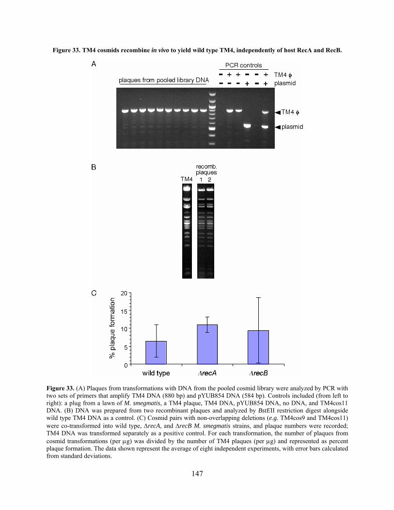
Figure 33. TM4 cosmids recombine in vivo to yield wild type TM4, independently of host RecA and RecB.
Figure 33. (A) Plaques from transformations with DNA from the pooled cosmid library were analyzed by PCR with two sets of primers that amplify TM4 DNA (880 bp) and pYUB854 DNA (584 bp). Controls included (from left to right): a plug from a lawn of M. smegmatis, a TM4 plaque, TM4 DNA, pYUB854 DNA, no DNA, and TM4cos11 DNA. (B) DNA was prepared from two recombinant plaques and analyzed by BstEII restriction digest alongside wild type TM4 DNA as a control. (C) Cosmid pairs with non-overlapping deletions (e.g. TM4cos9 and TM4cos11) were co-transformed into wild type, recA, and recB M. smegmatis strains, and plaque numbers were recorded; TM4 DNA was transformed separately as a positive control. For each transformation, the number of plaques from cosmid transformations (per g) was divided by the number of TM4 plaques (per g) and represented as percent plaque formation. The data shown represent the average of eight independent experiments, with error bars calculated from standard deviations.
147
A number of other experimental approaches for identifying the TM4 recombination
proteins were attempted without success. First, an M. smegmatis TM4 genomic library was
constructed in which TM4 fragments were cloned in the pLAM12 vector under control of
Pacetamidase, and a library of these were transformed into wild type M. smegmatis. These cells were
induced for expression and prepared similarly to Che9c gp60/gp61-expressing cells.
Recombination activity was assayed by ssDNA recombineering using oligonucleotides that
introduce point mutations that confer drug-resistance. However, transformation of the pooled
library cells did not produce recombinant colonies in duplicate experiments. Therefore, as a
second approach, individual segments of the TM4 genome (~3 kbp, excluding known structural
genes) were cloned in pLAM12. However, only two out of ten plasmids successfully
transformed M. smegmatis. This suggests that even the leaky expression of the acetamidase
promoter is sufficient to cause toxicity with some of these genes, and therefore a different
promoter or vector may be required. A similar result that may be a result of leaky expression was
observed in experiments with Halo genes 41-44 cloned under the acetamidase promoter. It was
observed that constructs that expressed Halo gp41-44 on a replicating vector (pLAM12 parent)
grew very slowly, while an integrated version was better tolerated (data not shown). These
experiments – both the M. smegmatis library and the individual TM4 clones – could be repeated
in a different vector background.
148
4.5 CONCLUSIONS
4.5.1 Mycobacteriophage-encoded recombination systems
Thus far, only seven known or putative recombination systems have been identified by
bioinformatic analyses in mycobacteriophages and prophages out of 51 sequenced phages and all
mycobacterial sequences in the NCBI database. However, more in-depth PSI-BLAST analysis
using putative recombination proteins from phages of related bacteria as queries may uncover
additional mycobacteriophage genes. The observation that TM4 likely encodes a recombination
system that is not recognizable by sequence similarity suggests that these proteins are probably
present in other phages but are, thus far, unidentified. The putative Giles recombination system
could have easily been overlooked without careful scrutiny if not for the similarity between Giles
gp53 and Halo gp43, and these genes were only recognized because the gene adjacent to Halo
gp43 is a recognizable RecE homologue. Approximately 50% of the mycobacteriophage ORFs
do not have sequence identity to proteins with known function from other organisms, but many
are similar to other mycobacteriophage-encoded genes. Therefore, identification of additional
phage-encoded recombination proteins – either by bioinformatic or experimental analyses – may
reveal the presence of these in more mycobacteriophages.
Among the mycobacteriophage-encoded SSAPs that were examined in vivo, Che9c gp61
demonstrated the highest level of recombineering activity, and Halo gp43 functioned less well
with an 8- to 30-fold reduction in activity. Surprisingly, however, Giles gp53 did not produce
recombinant colonies above background levels, even though it shares 30% amino acid identity
with Halo gp43. However, since protein expression was not confirmed, these results are not
conclusive. It would therefore be interesting to examine the other putative SSAP proteins for
149

activity in mycobacteria. These results are reminiscent of those from the study by Datta et al. in
which λ Beta and E. coli RecT demonstrated a stark difference in recombineering efficiency
[43], even though they both are encoded by E. coli phages. This further supports the notion that
development of genetic tools such as this may require characterization of multiple
bacteriophages to increase the available phage gene pool in which to search for recombination
proteins.
The role of these proteins in the mycobacteriophages is unknown, and the question of
whether these proteins are essential in phages Che9c, Halo, and Giles is currently being tested in
the Hatfull lab. Further, their role (if any) in phage propagation cannot necessarily be inferred
based on data from other phages. The activities of these proteins vary in other phages, although it
is common that recombination deficient phages are decreased in burst size [53,218]. For
example, one function of the λ Red system is likely to increase DNA synthesis by generating
additional circular genomes from linear concatemers [53,106], whereas the P22 system
circularizes the genome upon entry into the host cell [237,238]. Further investigation of the
prevalence of SSAP genes in mycobacteriophage genomes and their function in vivo will yield
better insights into their biological relevance and diversity.
4.5.2 SSAP species-specificity
From this study and that performed by Datta and colleagues [43], it is apparent that there is a
distinct difference in recombination activity when the same SSAPs are tested in M. smegmatis
and E. coli. Although this could be due to experimental variation, it is more likely a result of the
inherent species-specific nature of these proteins. Recombination proteins encoded by Gram-
negative bacteria tended to have the highest activities in E. coli, whereas proteins from phages
150
infecting more distantly related hosts displayed decreased activity. In M. smegmatis, expression
of Che9c gp61 facilitated the highest recombineering frequencies, with Halo gp43 and E. coli
RecT moderately lower in activity (30- and 10-fold, respectively). Therefore, the finding that
Che9c gp61 functions at a low level in E. coli is clearly not due to an inherently poor activity of
this protein, which was suggested by Datta et al. [43], but instead is probably due to expression
in a distantly-related organism. However, the data from the two studies correlated in the general
observation that the SSAPs – specifically λ Beta and Che9c gp61 – displayed the highest
activities above others tested in the native bacterial hosts of the phages from which they were
derived. It is interesting to note that E. coli RecT had a high level of activity in M. smegmatis,
whereas λ Beta was not active. This is in contrast to observations made in E. coli where λ Beta
functioned substantially better than RecT in E. coli.
Overall, it appears that SSAP proteins function optimally in bacteria that are closely
related to the hosts of their respective phages. This could be due to specific interactions with host
proteins that occur during recombination. One plausible hypothesis is that the SSAP – and
possibly the exonuclease – interacts with components of the DNA replication machinery, as
replication is a process that has a direct effect on λ Red-, RecET-, and Che9c gp60/gp61-
mediated recombination efficiency [52,113,228,244]. A potential candidate for this interacting
partner is the host SSB because it is associated with ssDNA during DNA synthesis, and this is
therefore the suggested target for SSAP-ssDNA complex recombination. Therefore, extension of
the recombineering technology to other organisms may require identification of a recombination
system encoded by a host-specific phage in order to produce functional protein interactions for
optimal activity.
151

The SSAPs examined in this study have yet to be tested in other mycobacterial species,
such as M. tuberculosis. Recombineering with ssDNA – though not with dsDNA – mediated by
Che9c gp61 in M. tuberculosis is decreased 5- to 30-fold as compared to M. smegmatis in the
same assays. This could also be due to slight differences between mycobacterial species in host
protein-SSAP interactions that result in a decreased recombination efficiency. It is therefore
possible that a different mycobacteriophage-encoded SSAP, such as Halo gp43, may improve
recombineering frequencies in M. tuberculosis.
4.5.3 The TM4 recombination system
The in vivo recombination assay with TM4 cosmids observed by Jacobs et al. was repeated in
this study, both with pools of the entire library of molecules and with pairs of cosmids [86]. No
true shuttle phasmids were identified, though this is not surprising due to the average size of the
deletions. Cosmid recombination was independent of the activities of the RecBCD complex, as
well as the major host recombination protein, RecA. Therefore it appears that the activity is
derived from phage-encoded proteins, although there may be other host proteins that are
required. It is striking that an analysis of the TM4 genome does not reveal any pairs of proteins
with sequence similarity to known recombination proteins. This suggests that the proteins
required for recombination of the TM4 cosmids may be a new family of recombinases and/or
exonucleases, and potentially these genes are located in separate regions of the genome.
Experiments designed to screen for the recombination proteins were not initially successful, but
the data provided a basis for altering the experimental setup. Further, a test screen should be
performed with a phage genome – such as Che9c – that is known to encode recombination
proteins. If the region of the Che9c genome that encodes gp61 can be identified in this type of
152

screen, this lends support to the utility of this experiment for other phages without recognizable
recombination homologues.
During the course of experiments with TM4 cosmids, a putative cis-acting element was
discovered in the region of the genome between 42,796 bp and 44,854 bp. This is most likely the
location of the origin of replication. Two pieces of evidence support this hypothesis; first, this
region includes the putative DNA primase gene (70), and in other phages – such as λ – the origin
of replication is present in the region encoding genes required for DNA replication [58]. Second,
DNA replication plays a critical role in recombination in the single strand annealing pathway –
such as with λ Red – by providing recombinogenic substrates [215,216,222]. Phage proteins
required for DNA replication should be provided in trans by the other cosmid, but the deletion of
the cosmid origin of replication cannot be compensated and may severely limit recombination. It
is also possible that recombination still occurs at low levels, as evidenced by the small numbers
of plaques observed in recombination assays with cosmids deleted for this region. Further
experimentation would be required to clearly identify the TM4 origin of replication and/or this
cis-acting element.
153

5.0 DISCUSSION
5.1 MYCOBACTERIAL RECOMBINEERING
Bacteriophages have long demonstrated their utility for advancing tools for genetics and
molecular biology in their bacterial hosts. Some of the more well-known examples of this are
DNA ligase, T4 polymerase, and various restriction enzymes. This is further exemplified by the
use of phage-encoded recombination proteins for the recombineering technology originally
developed in E. coli, and later extended for use in other Gram-negative bacteria. The
mycobacteriophages are no exception; the sequencing and characterization of these phages has
provided a vast reservoir of genes to study and exploit for materials such as integration-proficient
plasmids, selectable markers, and most recently, the mycobacterial recombineering system. The
development of this system will allow members of the mycobacterial research community to
perform genetic manipulations with an efficiency that is unparalleled by any other technique.
Gene replacement mutagenesis by recombineering requires the same amount of DNA cloning
and cell preparation as the minimum amount required for any other technique. Construction of
the AES merely requires the standard synthesis of a linear substrate with ~500 bp homology
flanking an antibiotic resistance cassette. No further manipulations of the AES are required, nor
are the rounds of screening needed for some methods, since 90% of the mutants generated by
recombineering are correctly targeted. Electrocompetent cell aliquots of the mycobacterial
154
recombineering strain (containing plasmid pJV53 or a similar construct) can be prepared in
advance and stored, which minimizes experimental preparation. Further, recombineering of point
mutations does not require any plasmid construction, since the short ssDNA substrates can be
synthesized commercially. Importantly, mutations that are not directly selectable can be made at
a relatively high frequency (3-5%) by using a co-transformation technique. Removal of the
recombineering plasmid can also be simplified by using a sacB gene for counter-selection.
Another potential use of recombineering is the deletion of sequences, such as entire genes,
internal domains, or the antibiotic resistance genes in marked gene replacement mutants. This
has been demonstrated by deleting most of the M. smegmatis leuD gene, and likely can be used
for other purposes.
5.1.1 Future applications of mycobacterial recombineering
The mycobacteriophage Che9c-encoded recombination system has provided a means for
improving genetic techniques in mycobacteria, and it is likely that further extension of this
technology will be made for other purposes. Targeted gene replacement mutagenesis has obvious
potential for making complete gene deletion sets for M. tuberculosis and M. smegmatis, a feat
that otherwise would be too time-consuming with available methods. Not only would this
provide mutant strains for various experimental purposes, but it would also supplement the data
pertaining to gene essentiality from previous genome-wide studies [200].
Additionally, nonsense mutations could be introduced into putative essential genes to
assay essentiality and gene function. The initial experimental design to test this approach
involved the use of a nonsense codon suppressor tRNA gene derived from mycobacteriophage
L5, which has been shown previously to suppress amber mutations in mycobacteria [60]. The
155
amber suppressor gene has been cloned such that its expression should be controlled by the Tet
inducible promoter [51], although suppression of amber mutations has thus far been
unsuccessful. However, alternative expression systems could be tested that would provide tightly
controlled induction or repression. Nonsense mutations could then be introduced into test genes
by ssDNA recombineering, and the viability of the mutant strain could be assessed in the
presence or absence of nonsense suppressor gene expression. Although this approach has yet to
be tested, it offers the potential for analysis of gene essentiality at an individual locus or genome-
wide level. Finally, mutagenesis by ssDNA recombineering allows point mutations to be inserted
in isogenic strains for direct and uncomplicated comparisons. This is more beneficial compared
to previous methodologies that typically required gene deletion followed by complementation,
and therefore analyses were not performed under endogenous conditions. This can be
specifically applied for determining the role of mutations that confer drug-resistance, which may
aid in research on the origins of XDR M. tuberculosis strains.
The extension of this technology for mutagenesis of mycobacteriophage genomes
recently has provided a simple method for future genomic and proteomic study of phages (L.
Marinelli, manuscript in preparation). For example, current experiments are testing if
recombineering can be used to insert His-tags onto phage genes to facilitate simple purification
of tagged proteins directly from infected cells. Several phages containing either point mutations
or deletions have been constructed and are also currently being studied. In addition, gene
essentiality can be tested, which has been demonstrated in a proof-of-principle experiment
involving the deletion of the lysA gene of mycobacteriophage Giles.
156

5.2 MYCOBACTERIOPHAGE-ENCODED RECOMBINATION PROTEINS: A
MODEL FOR DEVELOPMENT OF A RECOMBINEERING SYSTEM
The mycobacteriophages are a fascinating group of organisms that have greatly contributed to
our knowledge of evolution, morphologic and genetic diversity, biochemisty, and the biological
consequences of phage-host interactions. Phage genome sequencing contributes to the expanding
gene pool, a useful source for studies of gene function and the development of genetic tools. At
the beginning of this project, the only sequenced mycobacteriophages encoding putative
homologous recombination systems were Che9c and Halo, and therefore only these were
available for study. Subsequently, phages BPs and Giles were sequenced, and similar proteins
were identified, while careful PSI-BLAST analyses continue to reveal additional putative
recombinases. Also of interest are the prophages that appear to be present in the genomes of M.
avium and M. abscessus and encode SSAP recombinase homologues, as well as
mycobacteriophages Wildcat and Cjw1. Although none of these proteins were examined any
further in this study, they are also potential candidates for recombinase activity in vivo.
Interestingly, the mycobacteriophage-encoded recombinases that were tested had varying
levels of activity in the M. smegmatis ssDNA recombineering assay. Fortuitously, the first
recombinase used, Che9c gp61, exhibits the highest levels of recombination activity in vivo thus
far. Halo gp43 is slightly less efficient, and Giles gp53 did not show any activity in these assays,
although in this case expression was not confirmed. This first suggests that identification of only
one phage-encoded recombination protein may not be sufficient for development of a
recombineering system in other bacteria. Instead, these findings support the notion that
identification and analysis of multiple phage-encoded proteins is preferable in order to optimize
recombineering frequencies. In light of the species-specificity observed for both the E. coli and
157

mycobacterial phage-encoded recombination proteins, it is clear that optimal levels of
recombineering can best be achieved through isolation and sequencing of host-specific
bacteriophages. Therefore, it is likely that recombineering systems can be developed in virtually
any genetically tractable bacterium for which at least basic genetic tools – such as plasmids and
expression cassettes – have been described.
An important consideration is the question as to why the mycobacteriophage-encoded
recombinases display varying levels of activity in M. smegmatis, particularly since they all
appear to belong to the same SSAP superfamily. One attractive explanation is that they each
function optimally in the preferred host bacterium of their respective phages. The species-
specific nature of these proteins – observed broadly between phage-encoded proteins of
distantly-related host bacteria such as E. coli and M. smegmatis – likely affects activity even in
closely related bacteria of the same genus. Che9c gp61, for example, shows decreased
recombination efficiency in M. tuberculosis compared to M. smegmatis. The basis of the
differing activity levels may be attributed to specific recombinase-host protein interactions –
during processes such as DNA replication – that are required for optimal recombineering. The
role of replication is particularly interesting to consider with regard to the fast- and slow-growing
mycobacteria. Although this is not well-studied, the rate, processivity, and/or regulation of DNA
replication in M. tuberculosis is probably dissimilar to M. smegmatis, which may have a
profound effect on recombineering frequencies. Therefore, perhaps expression of another
recombinase will be more suitable for recombineering in M. tuberculosis or other mycobacteria.
Halo gp43 is a particularly interesting candidate because Halo can infect M. tuberculosis (T.
Sampson, personal communication). This logic can also be applied to other bacteria. While the λ
158

Red proteins may function sufficiently in some Gram-negative bacteria, developing
recombineering in others may require testing of additional host-specific phage-encoded systems.
It is likely that additional homologues of known recombination proteins will be found as
more phages and bacteria are sequenced. Possibly more interesting, however, are the phage
genes that are not detectably related to known recombinases, but still function similarly. The
genes that encode the recombination system of TM4 remain anonymous, and even a recent
analysis did not reveal any likely candidates. Clearly, a screen will be necessary to identify these
proteins. This tactic could then be used to develop recombineering in any bacterial system for
which phages have been isolated but recombination proteins are not recognizable (or if the phage
is not sequenced). The simplest approach appears to be the construction of a phage genomic
library in several different vector backbones (integrating or replicating), potentially with
different promoters in order to test varying expression levels. Subsequently, the library of
bacterial cells containing these plasmids would be screened for activity using ssDNA
recombineering of an allele conferring a drug-resistant phenotype. To test this, a screen should
be performed first using a phage genome that is known to encode recombination proteins, such
as Che9c or Halo. If this is successful, it would lend support to the use of this approach as a
broadly applicable method for identifying phage recombinases, potentially one that could be
used for phages of other bacteria.
159

6.0 MATERIALS AND METHODS
6.1 REAGENTS AND BUFFERS
6.1.1 Growth media
7H9 broth: 4.7 g Middlebrook 7H9 powder (Difco) was dissolved in 900 ml dH2O and 5 ml
40% glycerol. This was autoclaved, and 100 ml ADC (see below), 2.5 ml 20% Tween 80 (if
desired), and antibiotics were aseptically added as required. For growth of M. tuberculosis, 5 ml
oleic acid per liter was added.
7H9 induction medium: 4.7 g 7H9 powder (Difco) was dissolved in 900 ml dH2O and 5 ml
40% glycerol. This was autoclaved, and 100 ml dH2O, 10 ml 20% succinate, 2.5 ml 20% Tween,
and Kanamycin (see below) were aseptically added.
7H10 agar: 19 g Middlebrook 7H10 powder (Difco) was dissolved in 900 ml dH2O, and 12.5 ml
40% glycerol and 4 drops anti-bubble (Pourite) were added. This was autoclaved, and 100 ml
ADC and antibiotics as required were aseptically added. For growth of M. tuberculosis, 5 ml
oleic acid per liter was added.
7H11 agar: 21 g Middlebrook 7H11 powder (Difco) was dissolved in 900 ml dH2O, and 12.5 ml
40% glycerol and 4 drops anti-bubble (Pourite) were added. This was autoclaved, and 100 ml
160

ADC, plus 5 ml oleic acid (or 100 ml OADC, BDL), and antibiotics were aseptically added as
required.
Mycobacterial top agar (MBTA): 4.7 g Middlebrook 7H9 powder (Difco) and 7 g Bacto Agar
were dissolved in 900 ml dH2O and autoclaved.
ADC: 20 g dextrose and 8.5 g NaCl were dissolved in 950 ml dH2O. 50 g Albumin (Spectrum
Biochem) was added and stirred with no heat until dissolved. This was filter-sterilized through a
0.22-m-pore membrane and stored at 4C.
20% Tween 80: Tween 80 was dissolved at 20% (v/v) by heating to 56C, filtered through a
0.22-m-pore membrane, and stored at 4C. This was used at a final concentration of 0.05% in
liquid media.
20% acetamide: Acetamide (Sigma) was dissolved at 20% in dH2O, filtered through a 0.22-m-
pore membrane, and stored at 4C. This was used at a final concentration of 0.2% in media.
20% succinate: Sodium succinate dibasic hexahydrate (succinic acid, Sigma S9637) was
dissolved at 20% in dH2O, filtered through a 0.22-m-pore membrane, and stored at 4C. This
was used at a final concentration of 0.2% in media.
Oleic acid: Oleic acid (Sigma) was dissolved at 10 mg/ml in dH2O by heating the ampule in
37C water bath and stirring into dH2O with heat until completely dissolved. 1 g NaOH was
added and stirred until dissolved, and the solution was filtered through a 0.22-m-pore
membrane and stored in 10 ml aliquots at -20C. Used at a final concentration of 50 g/ml in M.
tuberculosis media.
Luria-Bertani broth (LB broth): 20 g LB broth (Difco) was dissolved in 1 L dH2O. This was
autoclaved and antibiotics were added aseptically when required.
161
Luria-Bertani agar (LB agar): 35 g LB agar (Difco) was dissolved in 1 L dH2O, and 4 drops
anti-bubble (Pourite) were added. This was autoclaved, and antibiotics were added aseptically
when required.
Tryptic Soy Broth (TSB): 30 g TSB (Difco) was dissolved in 1 L dH2O. This was autoclaved,
and antibiotics were added aseptically when required.
6.1.2 Antibiotics and Supplements
Carbenicillin: (Cb, Sigma) was dissolved at 50 mg/ml in dH2O, filtered through a 0.22-m-pore
membrane, and stored at 4C.
Chloramphenicol: (CM, Sigma) was dissolved at 100 mg/ml in 100% ethanol and stored at 4C.
Cycloheximide: (Chx, Sigma) was dissolved at 10 mg/ml in dH2O, filtered through a 0.22-m-
pore membrane, and stored at 4C.
Ethambutol: (EMB, Sigma) was dissolved at 50 mg/ml in dH2O, filtered through a 0.22-m-
pore membrane, and stored at 4C.
Ethionamide: (ETH, Sigma) was dissolved at 50 mg/ml in 100% DMSO and stored at 4C.
Gentamicin: (Gent, sulfate salt, Sigma) was dissolved 10 mg/ml in dH2O, filtered through a
0.22-m-pore membrane, and stored at -20C in 1 ml aliquots.
Hygromycin B: (Hyg, Sigma) was dissolved at 100 mg/ml in dH2O, filtered through a 0.22-m-
pore membrane, and stored at -20C in 1 ml aliquots.
Isoniazid: (INH, isonicotinic hydrazide, Sigma) was dissolved at 50 mg/ml in dH2O, filtered
through a 0.22-m-pore membrane, and stored at 4°C. Solutions were made fresh and used
within one week.
162

Isopropyl -D-1-thiogalactopyranoside: (IPTG) was resuspended to either 1 M or 0.1 M in
dH2O, filtered through a 0.22-m-pore membrane, and stored at 4°C.
Kanamycin: (Kan, Sigma) was dissolved at 50 mg/ml in dH2O, filtered through a 0.22-m-pore
membrane, and stored at 4C.
Leucine: (LEU, Sigma) was dissolved at 10 mg/ml in dH2O, filtered through a 0.22-m-pore
membrane, and stored at 4C.
Ofloxacin: (OFX, Sigma) was dissolved at 50 mg/ml in 1 N NaOH, filtered through a 0.22-m-
pore membrane, and stored at 4C. Solutions were made fresh and used within one week.
Pantothenate: DL-Pantothenic acid (PAN, Sigma) was dissolved at 100 mg/ml in dH2O, filtered
through a 0.22-m-pore membrane, and stored at -20C.
Rifampicin: (RIF, Sigma) was dissolved at 50 mg/ml in 100% DMSO and stored at 4C
wrapped in foil. Solutions were made fresh and used within one week.
Streptomycin: (Str, Sigma) was dissolved at 50 mg/ml in dH2O, filtered through a 0.22-m-pore
membrane, and stored at 4C. Solutions were made fresh and used within one week.
Tetracycline: (Tet, Sigma) was dissolved at 5 mg/ml in dH2O, filtered through a 0.22-m-pore
membrane, and stored at -20C. Tetracycline is light sensitive – media was prepared only at the
time needed.
Uracil: (URA, Sigma) was dissolved at 200 mM (22.41 mg/ml; 112.1 MW) in 1 N NaOH,
filtered through a 0.22-m-pore membrane, and stored at 4C.
X-gal: 100 mg (entire bottle from Invitrogen) was dissolved at 50 mg/ml in 100% DMF (2 ml),
wrapped in foil and stored at -20°C.
163
5-Fluoro-orotic acid: (5-FOA, US Biologicals) was added to agar media prior to autoclaving at
1 mg/ml. Powder goes into solution throughout autoclave cycle.
6.1.3 Laboratory reagents and stock solutions
0.1 M CaCl2: 11.1 g CaCl2 was dissolved in 1 L dH2O and autoclaved.
100X Denhardt’s solution: 2 g BSA, 2 g Ficoll, and 2 g Polyvinyl pyrrolidine were dissolved in
100 ml dH2O, filtered through a 0.22-m-pore membrane, and stored at -20°C.
0.1 M DTT: Dithiothreitol was dissolved in dH2O at 0.1 M (1.54 g per 10 ml; 154 MW), filtered
through a 0.22-m-pore membrane, and stored at -20°C in 1 ml aliquots.
0.5 M EDTA: 93.06 g disodium EDTA (Na2EDTA) was dissolved in 400 ml dH2O on low heat,
and pH was adjusted to 7.5 with NaOH (~50 ml 0.5M NaOH). Volume was brought to 500 ml
and autoclaved.
0.1 M Na2EDTA pH 8.8: 37.2 g of disodium EDTA (Fisher) was dissolved in 1 L dH2O, and pH
was adjusted to 8.8 with NaOH.
40% Glycerol: 400 ml Glycerol was mixed in 600 ml dH2O and autoclaved.
40% Glucose: 400 g dextrose was dissolved in 750 ml dH2O and autoclaved. Water was added
first and dextrose was slowly added to dissolve.
0.25 M HCl: Stock solution HCl (11 M) was diluted to 0.25 M by adding 22.7 stock HCl per one
L of dH2O.
0.5 N KOH: 28.1 g of KOH was dissolved in 1 L dH2O and stored at room temperature.
1 M MgCl2: 203 g MgCl2 hexahydrate (or 95.21 g anhydrous) was dissolved in 1 L dH2O and
autoclaved.
1 M MgSO4: 120.37 g MgSO4 was dissolved in 1 L dH2O and autoclaved.
164
5 M NaCl: 292 g NaCl was dissolved in 1 L dH2O, heated on low to dissolve, and autoclaved.
3 M NaOAc: 408.1 g NaOAc was dissolved in 400 ml dH2O on low heat and pH adjusted to 5.2
with glacial acetic acid (~200 ml). Volume was brought to 1 L and autoclaved.
5 N NaOH: 200 g of NaOH was dissolved in 1 L dH2O.
0.4 N NaOH: 80 ml of 5 N NaOH was brought to 1 L with dH2O.
Phage Buffer: 4 g NaCl was dissolved in 980 ml dH2O, and 10 ml of 1 M Tris pH 7.5 and 10
mL of 1 M MgSO4 were added and autoclaved.
Phenylmethanesulphonylfluoride: (PMSF) was dissolved in isopropanol to 100 mM. The tube
was wrapped in foil and stored at -20°C.
Proteinase K: proteinase K was dissolved at 10 mg/ml in dH2O, filtered through a 0.22-m-pore
membrane, and stored at -20C in 1 ml aliquots.
10% SDS: 100 g SDS was dissolved in 1 L dH2O, filtered through a 0.22-m-pore membrane,
and stored at room temperature.
20X SSC: 175.32 g NaCl and 88.23 g sodium citrate were dissolved in 1 L dH2O and pH was
adjusted to 7.0 with HCl.
1 M Tris pH 7.5: 121 g Trizma base was dissolved in 800 ml dH2O, and pH was adjusted to 7.5
with HCl. Volume was brought to 1 L and autoclaved.
1 M Tris pH 8.0: 121 g Trizma base was dissolved in 800 ml dH2O, and pH was adjusted to 8.0
with HCl. Volume was brought to 1 L and autoclaved.
Tris-buffered saline ± Tween: (TBS, TBS-T) 25 mM Tris pH 8.0, 125 mM NaCl, ± 0.1%
Tween.
Tris-EDTA (TE): 10 ml of 1 M Tris, pH 7.5 and 2.5 ml of 0.5 M EDTA were mixed in 987.5 ml
dH2O and autoclaved. Final solution: 10 mM Tris pH 7.5, 1.25 mM EDTA.
165

6.1.4 Gel electrophoresis
6.1.4.1 Agarose gel electrophoresis
20% Ficoll dye: 2 g Ficoll was heated on low heat to dissolve in 8 ml dH2O. 1 ml 1% (w/v)
bromophenol blue and 1 ml 1% (w/v) xylene cyanol were added.
Tris Borate EDTA (TBE): 121.1 g Trizma base, 51.25 g Boric Acid, and 3.72 g EDTA were
dissolved in ~890 ml to make a 10X solution. 1X = 100 mM Tris (121.1 MW), 83 mM Boric
Acid (61.83 MW), and 1 mM EDTA (372.24 MW).
6.1.4.2 Polyacrylamide gel electrophoresis
Coomassie Blue Stain: 2.5 g Coomassie Blue was dissolved in 450 ml methanol, and 90 ml
acetic acid was added and volume brought to 1 L dH2O. This was filtered and stored at room
temperature.
Glycerol loading dye: 0.25% (w/v) bromophenol blue, 0.25% (w/v) xylene cyanol, and 30%
glycerol
Protein gel running buffer (10X): 144 g glycine, 30 g Trizma base, and 10 g SDS were
dissolved in 1 L dH2O by mixing SDS first, and pH was adjusted to 8.3.
4X SDS-PAGE loading dye: 125 mM Tris pH 7.5, 20% glycerol, 2% SDS, 5% -
mercaptoethanol, 0.1% bromophenol blue dye. In 8 ml, add 2.6 ml dH2O, 1 ml 0.5 M Tris pH
7.5, 1.6 ml neat glycerol, 1.6 ml 10% SDS, 0.4 ml -mercaptoethanol, 0.8 ml 1% bromophenol
blue.
166

SDS-PAGE Laemmli gels: For 2 small 10% separating gels: 5 ml dH2O, 2.5 ml 4X separating
buffer, 2.5 ml 40% acrylamide:bisacrylamide (29:1), 10 l TEMED, 40 l 10% (w/v) APS. For 2
small stacking 4.5% stacking gels: 3 ml dH2O, 1.25 ml 4X stacking buffer, 0.5 ml 40%
acrylamide:bisacrylamide (29:1), 15 l TEMED, 25 l 10% APS.
4X SDS-PAGE separating buffer: 18.17 g Trizma base was dissolved in ~80 ml dH2O, and 4
ml 10% SDS was added. pH was adjusted to 8.8 with HCl (~2.5 ml), and volume was brought to
100 ml.
4X SDS-PAGE stacking buffer: 6.06 g Trizma base was dissolved in ~80 ml dH2O, and 4 ml
10% SDS was added. pH was adjusted to 6.8 with HCl, and volume was brought to 100 ml.
TBE polyacrylamide native gels: For 1 large 8% gel: 42 ml dH2O, 6 ml 10X TBE buffer, 12 ml
40% acrylamide:bisacrylamide (29:1), 21 l TEMED, 420 l 10% APS.
6.1.5 Assay buffers
Annealing buffer: 50 mM Tris pH 7.5, 100 mM NaCl
Binding assay reaction buffer: 33 mM Tris pH 7.5, 13 mM MgCl2, 1.8 mM DTT, 88 g/ml
BSA
Binding assay wash buffer: 33 mM Tris pH 7.5, 13 mM MgCl2, 1.8 mM DTT
Exonuclease assay buffer: 20 mM Tris, pH 8.0, 10 mM MgCl2, 10 mM -mercaptoethanol
Genomic DNA prep CTAB solution: 4.1 g NaCl was dissolved in 90 ml dH2O, and 10 g
Cetrimide was added, stirring. This was incubated at 65°C until in solution, and stored at room
temperature.
167

Gel filtration equilibration buffer: 33 mM Tris pH 7.5, 100 mM NaCl
Genomic DNA prep GTE solution: 25 mM Tris pH 8.0, 10 mM EDTA, 50 mM glucose
Protein dilution buffer: 10 mM Tris pH 7.5, 1 mM DTT, 1 mg/ml BSA
Protein purification lysis buffer: 50 mM Tris pH 8.0, 300 mM NaCl, 5% glycerol, 1 mM
PMSF
Protein purification wash buffer: 50 mM Tris pH 8.0, 300 mM NaCl, 5% glycerol, 0-20 mM
imidazole
Protein purification elution buffer: 50 mM Tris pH 8.0, 300 mM NaCl, 5% glycerol, 20-200
mM imidazole
Protein storage buffer: 20 mM Tris pH 8.0, 0.1 mM EDTA, 0.1 mM DTT, 50 mM NaCl, 50%
glycerol
Southern blot pre-hybridization buffer: 6X SSC, 2X Denhardt’s solution, 0.1% SDS
Southern blot hybridization buffer: 6X SSC, 20 mM NaPO4, pH 7.5, 5% PEG 8000 (Sigma)
Southern blot wash buffer 1: 2X SSC, 0.1% SDS
Southern blot wash buffer 2: 0.2X SSC, 0.1% SDS
Western blot transfer buffer: 48 mM Tris, 39 mM Glycine, 0.037% SDS, 20% methanol
6.2 PLASMID CLONING
6.2.1 Plasmid maintenance in E. coli strains
Plasmid constructs were maintained in either E. coli DH5 or GC5 (GeneChoice; similar
genotype to DH5) strains. When required, BL21(DE3) pLysS cells (Invitrogen) were used for
168
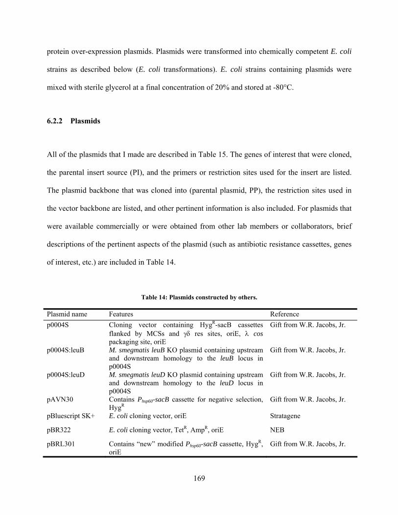
protein over-expression plasmids. Plasmids were transformed into chemically competent E. coli
strains as described below (E. coli transformations). E. coli strains containing plasmids were
mixed with sterile glycerol at a final concentration of 20% and stored at -80°C.
6.2.2 Plasmids
All of the plasmids that I made are described in Table 15. The genes of interest that were cloned,
the parental insert source (PI), and the primers or restriction sites used for the insert are listed.
The plasmid backbone that was cloned into (parental plasmid, PP), the restriction sites used in
the vector backbone are listed, and other pertinent information is also included. For plasmids that
were available commercially or were obtained from other lab members or collaborators, brief
descriptions of the pertinent aspects of the plasmid (such as antibiotic resistance cassettes, genes
of interest, etc.) are included in Table 14.
Table 14: Plasmids constructed by others.
Plasmid name Features Reference
p0004S Cloning vector containing HygR-sacB cassettes flanked by MCSs and res sites, oriE, cos packaging site, oriE
Gift from W.R. Jacobs, Jr.
p0004S:leuB M. smegmatis leuB KO plasmid containing upstream and downstream homology to the leuB locus in p0004S
Gift from W.R. Jacobs, Jr.
p0004S:leuD M. smegmatis leuD KO plasmid containing upstream and downstream homology to the leuD locus in p0004S
Gift from W.R. Jacobs, Jr.
pAVN30 Contains Phsp60-sacB cassette for negative selection, HygR
Gift from W.R. Jacobs, Jr.
pBluescript SK+ E. coli cloning vector, oriE Stratagene
pBR322 E. coli cloning vector, TetR, AmpR, oriE NEB
pBRL301 Contains “new” modified Phsp60-sacB cassette, HygR, oriE
Gift from W.R. Jacobs, Jr.
169
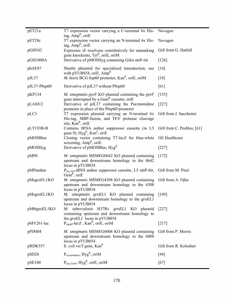
pET21a T7 expression vector carrying a C-terminal 6x His-tag, AmpR, oriE
Novagen
pET28c T7 expression vector carrying an N-terminal 6x His-tag, AmpR, oriE
Novagen
pGH542 Expresses resolvase constitutively for unmarking gene knockouts, TetR, oriE, oriM
Gift from G. Hatfull
pGH1000A Derivative of pMOSHyg containing Giles attP-int [126]
phAE87 Shuttle phasmid for specialized transduction; use with pYUB854, oriE, AmpR
[14]
pJL37 M. bovis BCG hsp60 promoter, KanR, oriE, oriM [18]
pJL37-Phsp60 Derivative of pJL37 without Phsp60 [61]
pKP134 M. smegmatis pyrF KO plasmid containing the pyrF gene interrupted by a GentR cassette, oriE
[155]
pLAM12 Derivative of pJL37 containing the Pacetamidase promoter in place of the Phsp60 promoter
[227]
pLC3 T7 expression plasmid carrying an N-terminal 6x His-tag, MBP-fusion, and TEV protease cleavage site, KanR, oriE
Gift from J. Sacchetini
pLT193B-B Contains tRNA amber suppressor cassette (in L5 gene 9), HygR, KanS, oriE
Gift from C. Peebles; [61]
pMOSBlue Cloning vector containing T7-lacZ for blue-white screening, AmpR, oriE
GE Healthcare
pMOSHyg Derivative of pMOSBlue, HygR [227]
pMP6 M. smegmatis MSMEG0642 KO plasmid containing upstream and downstream homology to the 0642 locus in pYUB854
[172]
pMPambar Pmyc1tet-tRNA amber suppressor cassette, L5 attP-Int, GentR, oriE
Gift from M. Piuri
pMsgroEL1KO M. smegmatis MSMEG4308 KO plasmid containing upstream and downstream homology to the 4308 locus in pYUB854
Gift from A. Ojha
pMsgroEL1KO M. smegmatis groEL1 KO plasmid containing upstream and downstream homology to the groEL1 locus in pYUB854
[149]
pMtbgroEL1KO M. tuberculosis H37Rv groEL1 KO plasmid containing upstream and downstream homology to the groEL1 locus in pYUB854
[227]
pMV261-lac Phsp60-lacZ , KanR, oriE, oriM [217]
pPJM04 M. smegmatis MSMEG6008 KO plasmid containing upstream and downstream homology to the 6008 locus in pYUB854
Gift from P. Morris
pRDK557 E. coli recT gene, KanR Gift from R. Kolodner
pSD26 Pacetamidase, HygR, oriM [44]
pSE100 Pmyc1tetO, HygR, oriE, oriM [67]
170
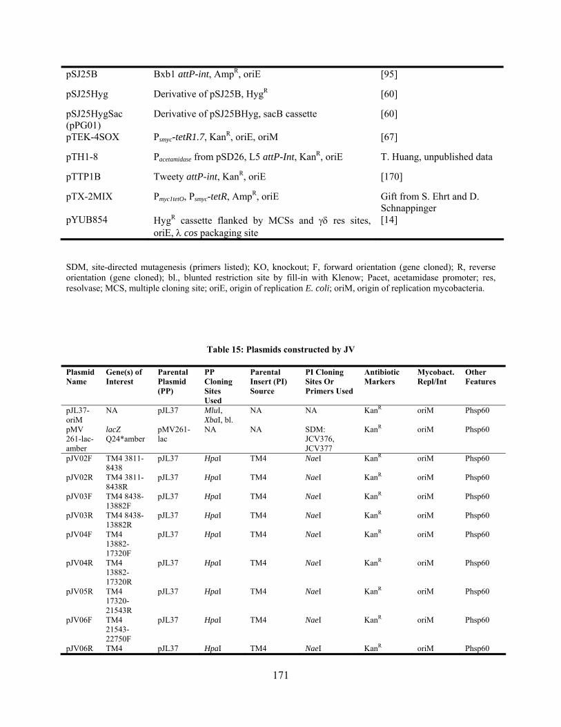
pSJ25B Bxb1 attP-int, AmpR, oriE [95]
pSJ25Hyg Derivative of pSJ25B, HygR [60]
pSJ25HygSac (pPG01)
Derivative of pSJ25BHyg, sacB cassette [60]
pTEK-4SOX Psmyc-tetR1.7, KanR, oriE, oriM [67]
pTH1-8 Pacetamidase from pSD26, L5 attP-Int, KanR, oriE T. Huang, unpublished data
pTTP1B Tweety attP-int, KanR, oriE [170]
pTX-2MIX Pmyc1tetO, Psmyc-tetR, AmpR, oriE Gift from S. Ehrt and D. Schnappinger
pYUB854 HygR cassette flanked by MCSs and res sites, oriE, cos packaging site
[14]
SDM, site-directed mutagenesis (primers listed); KO, knockout; F, forward orientation (gene cloned); R, reverse orientation (gene cloned); bl., blunted restriction site by fill-in with Klenow; Pacet, acetamidase promoter; res, resolvase; MCS, multiple cloning site; oriE, origin of replication E. coli; oriM, origin of replication mycobacteria.
Table 15: Plasmids constructed by JV
Plasmid Name
Gene(s) of Interest
Parental Plasmid (PP)
PP Cloning Sites Used
Parental Insert (PI) Source
PI Cloning Sites Or Primers Used
Antibiotic Markers
Mycobact. Repl/Int
Other Features
pJL37-oriM
NA pJL37 MluI, XbaI, bl.
NA NA KanR oriM Phsp60
pMV 261-lac-amber
lacZ Q24*amber
pMV261-lac
NA NA SDM: JCV376, JCV377
KanR oriM Phsp60
pJV02F TM4 3811-8438
pJL37 HpaI TM4 NaeI KanR oriM Phsp60
pJV02R TM4 3811-8438R
pJL37 HpaI TM4 NaeI KanR oriM Phsp60
pJV03F TM4 8438-13882F
pJL37 HpaI TM4 NaeI KanR oriM Phsp60
pJV03R TM4 8438-13882R
pJL37 HpaI TM4 NaeI KanR oriM Phsp60
pJV04F TM4 13882-17320F
pJL37 HpaI TM4 NaeI KanR oriM Phsp60
pJV04R TM4 13882-17320R
pJL37 HpaI TM4 NaeI KanR oriM Phsp60
pJV05R TM4 17320-21543R
pJL37 HpaI TM4 NaeI KanR oriM Phsp60
pJV06F TM4 21543-22750F
pJL37 HpaI TM4 NaeI KanR oriM Phsp60
pJV06R TM4 pJL37 HpaI TM4 NaeI KanR oriM Phsp60
171

21543-22750R
pJV07F TM4 22750-29001F
pJL37 HpaI TM4 NaeI KanR oriM Phsp60
pJV07R TM4 22750-29001R
pJL37 HpaI TM4 NaeI KanR oriM Phsp60
pJV09F TM4 34461-39007F
pJL37 HpaI TM4 NaeI KanR oriM Phsp60
pJV09R TM4 34461-39007R
pJL37 HpaI TM4 NaeI KanR oriM Phsp60
pJV11F TM4 42691-45427F
pJL37 HpaI TM4 NaeI KanR oriM Phsp60
pJV11R TM4 42691-45427R
pJL37 HpaI TM4 NaeI KanR oriM Phsp60
pJV15 TetR cassette
pSJ25B DraI, HindIII, bl.
pBR322 AvaI, HindIII, bl.
TetR Bxb1 attP/Int
pJV16 lacZ 5' piece pJV15 BsaAI pMV261-lac PCR: JCV07, JCV08
TetR Bxb1 attP/Int
pJV17 sacB cassette
pJV16 BsaAI pAVN30 PCR: sacBF/R TetR Bxb1 attP/Int
sacB
pJV18 PmlI site pJV17 NA NA SDM: JCV23, JCV24
TetR Bxb1 attP/Int
pJV19 EcoRV site pTH1-8 NA NA SDM: JCV21, JCV22
CbR, KanR L5 attP/Int Pacet
pJV20 lacZ 3' piece pJV18 PmlI pMV261-lac PCR: JCV09, JCV10
TetR Bxb1 attP/Int
sacB
pJV21 TM4 "7-20 region"
pBR322 BsaAI TM4 PCR: JCV01, JCV03
TetR
pJV23 Che9c genes 59-62
pJL37 HpaI Che9c PCR: JCV19, JCV20
KanR oriM Phsp60
pJV24 Che9c genes 59-62
pLAM12 HpaI Che9c PCR: JCV19, JCV20
KanR oriM Pacet
pJV25 Che9c genes 59-62
pJV19 EcoRV Che9c PCR: JCV19, JCV20
KanR L5 attP/Int Pacet
pJV26 Che9c genes 59-62
pJL37-Phsp60
HpaI Che9c PCR: JCV19, JCV20
KanR oriM
pJV27 Msmeg recA upstream homol.
pYUB854 XbaI, StuI M. smegmatis
PCR: JCV15, JCV16
HygR res sites
pJV28 Msmeg recA downstream homol.; final recA KO plasmid
pJV27 HindIII, bl.
M. smegmatis
PCR: JCV17, JCV18
HygR res sites
pJV29 pJV28+ shuttle phasmid phAE87
phAE87 PacI pJV28 PacI HygR shuttle phasmid
pJV30F TM4 "5-16kb region"
pJL37 HpaI TM4 KpnI, bl. KanR oriM Phsp60
pJV30R TM4 "5-16kb region"
pJL37 HpaI TM4 KpnI, bl. KanR oriM Phsp60
pJV30F- NA pJV30F HindIII, NA NA KanR oriM
172
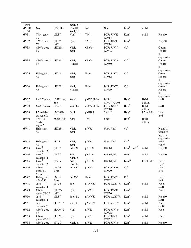
Hsp60 XbaI, bl. pJV30R-Hsp60
NA pJV30R HindIII, XbaI, bl.
NA NA KanR oriM
pJV31 TM4 gene 70
pJL37 HpaI TM4 PCR: JCV13, JCV14
KanR oriM Phsp60
pJV32 TM4 gene 70
pJL37-oriM
HpaI TM4 PCR: JCV13, JCV14
KanR Phsp60
pJV33 Che9c gene 60
pET21a NdeI, XhoI
Che9c PCR: JCV47, JCV48
CbR C-term His tag; T7 expression
pJV34 Che9c gene 61
pET21a NdeI, HindIII
Che9c PCR: JCV49, JCV50
CbR C-term His tag; T7 expression
pJV35 Halo gene 42
pET21a NdeI, XhoI
Halo PCR: JCV51, JCV52
CbR C-term His tag; T7 expression
pJV36 Halo gene 43
pET21a NdeI, HindIII
Halo PCR: JCV53, JCV59
CbR C-term His tag; T7 expression
HygR lacZ 5' piece pSJ25HygSac
XmnI pJV37 pMV261-lac PCR: JCV07,JCV08
Bxb1 attP/Int
sacB
pJV38 lacZ 3' piece HygR Bxb1 attP/Int
SapI, bl. pMV261-lac pJV37 PCR: JCV09, JCV10
sacB
pJV39 L5 attP/Int cassette, R
pMOSHyg DraI pMH94 SalI, bl. HygR L5 attP/Int Interr. lacZ
pJV40 TM4 "5-16kb region"
pSJ25Hyg KpnI TM4 KpnI HygR Bxb1 attP/Int
pJV41 Halo gene 42
pET28c NdeI, XhoI
pJV35 NdeI, XhoI CbR N and C-term His tag; T7 expression
NdeI, XhoI CmR pJV42 Halo gene 42
pLC3 NdeI, XhoI
pJV35 MBP-fusion
pJV43 GentR cassette, R
pJL37 BamHI pKP134 BamHI KanR, GentR oriM Phsp60
pJV44 GentR cassette, R
SpeI, NheI, bl.
pKP134 BamHI, bl. pJL37 GentR oriM Phsp60
pJV45 GentR cassette, R
pJV39 AatII, ClaI, bl.
pKP134 BamHI, bl. GentR L5 attP/Int Interr. HygR
pJV46 Che9c genes 59-62, R
pMOS Blue
EcoRV pJV23 PCR: JCV19, JCV20
CbR Interr. lacZ
pJV47 Halo genes 41-44, R
pMOS Blue
EcoRV Halo PCR: JCV61, JCV62
CbR Interr. lacZ
pJV48 sacB cassette, R
pJV24 SpeI pAVN30 PCR: sacBF/R KanR oriM Pacet, sacB
pJV49 Che9c genes 59-62
pJL37-oriM
HpaI pJV23 PCR: JCV19, JCV20
KanR Pacet
pJV50 sacB cassette, R
pJL37 SpeI, bl. pAVN30 PCR: sacBF/R KanR oriM Phsp60, sacB
pJV51 sacB cassette, R
pLAM12 SpeI, bl. pAVN30 PCR: sacBF/R KanR oriM Pacet, sacB
pJV52 Che9c gene 61
pLAM12 HpaI pJV23 PCR: JCV49, JCV78
KanR oriM Pacet
pJV53 Che9c genes 60-61
pLAM12 HpaI pJV23 PCR: JCV47, JCV78
KanR oriM Pacet
pJV54 Che9c gene pJV50 NheI, bl. pJV23 PCR: JCV49, KanR oriM Phsp60,
173
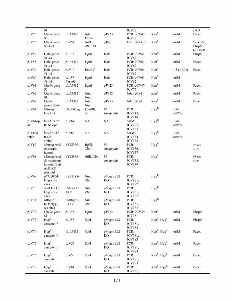
61 JCV78 sacB pJV55 Che9c gene
60 pLAM12 NdeI,
EcoRI pJV23 PCR: JCV47,
JCV77 KanR oriM Pacet
pJV56 Che9c gene 60/acet
pJV54 NotI, XbaI, bl.
pJV61 DraI, NheI, bl. KanR oriM Pacet-60; Phsp60-61; sacB
pJV57 Halo genes 41-44
pJL37 HpaI Halo PCR: JCV61, JCV62
KanR oriM Phsp60
pJV58 Halo genes 41-44
pLAM12 HpaI Halo PCR: JCV61, JCV62
KanR oriM Pacet
pJV59 Halo genes 41-44
pJV19 EcoRV Halo PCR: JCV61, JCV62
KanR L5 attP/Int Pacet
pJV60 Halo genes 41-44
pJL37-Phsp60
HpaI Halo PCR: JCV61, JCV62
KanR oriM
pJV61 Che9c gene 60
pLAM12 HpaI pJV23 PCR: JCV47, JCV77
KanR oriM Pacet
pJV62 Che9c gene 61
pLAM12 NdeI, NheI
pJV52 NdeI, NheI KanR oriM Pacet
pJV63 Che9c genes 60-61
pLAM12 NdeI, NheI
pJV53 NdeI, NheI KanR oriM Pacet
pJV64 Msmeg leuD , R
pSJ25Hyg HindIII, bl.
M. smegmatis
PCR: JCV113, JCV118
HygR Bxb1 attP/Int
pJV64opal
leuD R15* R16* opal
pJV64 NA NA SDM: JCV132, JCV133
HygR Bxb1 attP/Int
pJV64amber
leuD K31* R32* amber
pJV64 NA NA SDM: JCV134, JCV135
HygR Bxb1 attP/Int
pJV67 Msmeg recB upstream homol.
pYUB854 BglII, XhoI
M. smegmatis
PCR: JCV136, JCV137
HygR res sites
pJV68 Msmeg recB downstream homol; final recB KO plasmid
pYUB854 AflII, XbaI M. smegmatis
PCR: JCV138, JCV139
HygR res sites
pJV69 pYUB854: Hyg - res sites
pYUB854 NheI, XbaI
pMsgroEL1KO
PCR: JCV181, JCV182
HygR
pJV70 groEL KO: Hyg - res sites
pMsgroEL1KO
NheI, XbaI
pMsgroEL1KO
PCR: JCV181, JCV182
HygR
pJV71 MtbgroEL KO: Hyg - res sites
pMtbgroEL1KO
NheI, XbaI
pMsgroEL1KO
PCR: JCV181, JCV182
HygR
pJV72 Che9c gene 61
pJL37 HpaI pJV23 PCR: JCV49, JCV78
KanR oriM Phsp60
pJV73 HygR cassette, F
pJL37 SpeI pMsgroEL1KO
PCR: JCV181, JCV183
KanR, HygR oriM Phsp60
pJV74 HygR cassette, F
pLAM12 SpeI pMsgroEL1KO
PCR: JCV181, JCV183
KanR, HygR oriM Pacet
pJV75 HygR cassette, F
pJV52 SpeI pMsgroEL1KO
PCR: JCV181, JCV183
KanR, HygR oriM Pacet
pJV76 HygR cassette, F
pJV53 SpeI pMsgroEL1KO
PCR: JCV181, JCV183
KanR, HygR oriM Pacet
pJV77 HygR cassette, F
pJV61 SpeI pMsgroEL1KO
PCR: JCV181,
KanR, HygR oriM Pacet
174

JCV183 pJV78 HygR
cassette, F pJV72 SpeI pMsgroEL1
KO PCR: JCV181, JCV183
KanR, HygR oriM Phsp60
pJV73 amber
Hyg D15* D16* amber
pJV73 NA NA SDM: JCV184, JCV185
KanR oriM Phsp60; HygS
pJV74 amber
Hyg D15* D16* amber
pJV74 NA NA SDM: JCV184, JCV185
KanR oriM Pacet; HygS
pJV75 amber
Hyg D15* D16* amber
pJV75 NA NA SDM: JCV184, JCV185
KanR oriM Pacet; HygS
pJV76 amber
Hyg D15* D16* amber
pJV76 NA NA SDM: JCV184, JCV185
KanR oriM Pacet; HygS
pJV77 amber
Hyg D15* D16* amber
pJV77 NA NA SDM: JCV184, JCV185
KanR oriM Pacet; HygS
pJV78 amber
Hyg D15* D16* amber
pJV78 NA NA SDM: JCV184, JCV185
KanR oriM Phsp60; HygS
pJV73 opal
Hyg D15* D16* opal
pJV73 NA NA SDM: JCV186, JCV187
KanR oriM Phsp60; HygS
pJV74 opal
Hyg D15* D16* opal
pJV74 NA NA SDM: JCV186, JCV187
KanR oriM Pacet; HygS
pJV75 opal
Hyg D15* D16* opal
pJV75 NA NA SDM: JCV186, JCV187
KanR oriM Pacet; HygS
pJV76 opal
Hyg D15* D16* opal
pJV76 NA NA SDM: JCV186, JCV187
KanR oriM Pacet; HygS
pJV77 opal
Hyg D15* D16* opal
pJV77 NA NA SDM: JCV186, JCV187
KanR oriM Pacet; HygS
pJV78 opal
Hyg D15* D16* opal
pJV78 NA NA SDM: JCV186, JCV187
KanR oriM Phsp60; HygS
pJV79 Bxb1 attL(CT)-50, F
pMOSBlueHyg
NdeI, bl. attL(CT)-50mer, attL(CT)-50mer AP
NA HygR, CbR
pJV80 Bxb1 attL(CT)-50, R
pJV69 XbaI, bl. attL(CT)-50mer, attL(CT)-50mer AP
NA HygR
pJV81 Bxb1 attR(CT)-50, F
pJV79 SmaI attR(CT)-50mer, attR(CT)-50mer AP
NA HygR, CbR Bxb1 attL/attR(CT) flanking HygR
pJV82 Bxb1 attR(CT)-50, R
pJV80 NheI, bl. attR(CT)-50mer, attR(CT)-50mer AP
NA HygR
pJV83 marinum ATCC927 recA, F
pMOS Blue
EcoRV M. marinum ATCC
PCR: JCV194, JCV197
CbR
pJV84 marinum ATCC927
pYUB854 XbaI, bl. M. marinum ATCC
PCR: JCV194,
HygR res sites
175
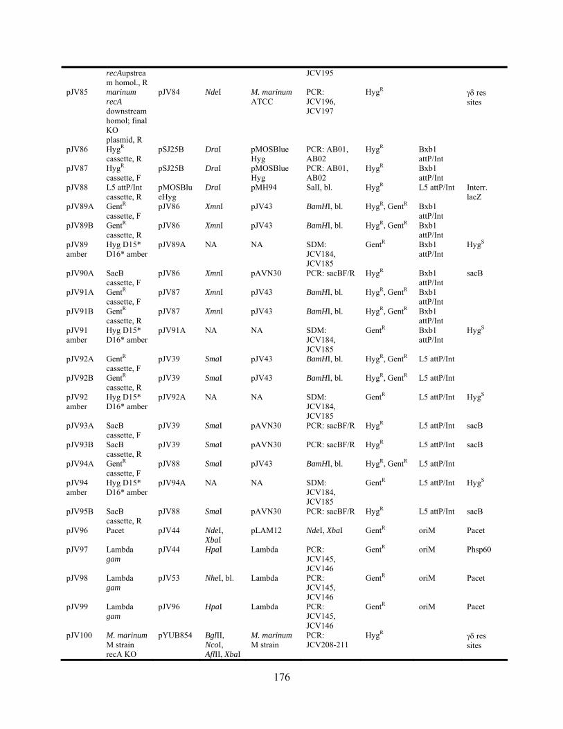
recAupstream homol., R
JCV195
pJV85 marinum recA downstream homol; final KO plasmid, R
pJV84 NdeI M. marinum ATCC
PCR: JCV196, JCV197
HygR res sites
pJV86 HygR cassette, R
pSJ25B DraI pMOSBlue Hyg
PCR: AB01, AB02
HygR Bxb1 attP/Int
pJV87 HygR cassette, F
pSJ25B DraI pMOSBlue Hyg
PCR: AB01, AB02
HygR Bxb1 attP/Int
pJV88 L5 attP/Int cassette, R
pMOSBlueHyg
DraI pMH94 SalI, bl. HygR L5 attP/Int Interr. lacZ
pJV89A GentR cassette, F
pJV86 XmnI pJV43 BamHI, bl. HygR, GentR Bxb1 attP/Int
pJV89B GentR cassette, R
pJV86 XmnI pJV43 BamHI, bl. HygR, GentR Bxb1 attP/Int
pJV89 amber
Hyg D15* D16* amber
pJV89A NA NA SDM: JCV184, JCV185
GentR Bxb1 attP/Int
HygS
pJV90A SacB cassette, F
pJV86 XmnI pAVN30 PCR: sacBF/R HygR Bxb1 attP/Int
sacB
pJV91A GentR cassette, F
pJV87 XmnI pJV43 BamHI, bl. HygR, GentR Bxb1 attP/Int
pJV91B GentR cassette, R
pJV87 XmnI pJV43 BamHI, bl. HygR, GentR Bxb1 attP/Int
pJV91 amber
Hyg D15* D16* amber
pJV91A NA NA SDM: JCV184, JCV185
GentR Bxb1 attP/Int
HygS
pJV92A GentR cassette, F
pJV39 SmaI pJV43 BamHI, bl. HygR, GentR L5 attP/Int
pJV92B GentR cassette, R
pJV39 SmaI pJV43 BamHI, bl. HygR, GentR L5 attP/Int
pJV92 amber
Hyg D15* D16* amber
pJV92A NA NA SDM: JCV184, JCV185
GentR L5 attP/Int HygS
pJV93A SacB cassette, F
pJV39 SmaI pAVN30 PCR: sacBF/R HygR L5 attP/Int sacB
pJV93B SacB cassette, R
pJV39 SmaI pAVN30 PCR: sacBF/R HygR L5 attP/Int sacB
pJV94A GentR cassette, F
pJV88 SmaI pJV43 BamHI, bl. HygR, GentR L5 attP/Int
pJV94 amber
Hyg D15* D16* amber
pJV94A NA NA SDM: JCV184, JCV185
GentR L5 attP/Int HygS
pJV95B SacB cassette, R
pJV88 SmaI pAVN30 PCR: sacBF/R HygR L5 attP/Int sacB
pJV96 Pacet pJV44 NdeI, XbaI
pLAM12 NdeI, XbaI GentR oriM Pacet
pJV97 Lambda gam
pJV44 HpaI Lambda PCR: JCV145, JCV146
GentR oriM Phsp60
pJV98 Lambda gam
pJV53 NheI, bl. Lambda PCR: JCV145, JCV146
GentR oriM Pacet
pJV99 Lambda gam
pJV96 HpaI Lambda PCR: JCV145, JCV146
GentR oriM Pacet
pJV100 M. marinum M strain recA KO
pYUB854 BglII, NcoI, AflII, XbaI
M. marinum M strain
PCR: JCV208-211
HygR res sites
176

pJV101 M. smegmatis recD KO
pYUB854 BlgII, HindIII, AflII, XbaI
M. smegmatis
PCR: JCV212-215
HygR res sites
pJV102 attL-hygR-attR
pJV39 XbaI, KpnI
pJV81 XbaI, KpnI HygR L5 attP/Int Bxb1 attL-INT/ attR(CT) flanking HygR
pJV103 Halo gene 43
pLAM12 HpaI pJV57 PCR: JCV53, JCV226
KanR oriM Pacet
pJV104 E. coli recT pLAM12 HpaI pRDK557 PCR: JCV230, JCV237
KanR oriM Pacet
pJV105 Lambda bet pLAM12 HpaI Lambda DNA
PCR: JCV227, JCV228
KanR oriM Pacet
pJV106 Halo gene 43
pLAM12 NdeI, EcoRI
pJV57 PCR: JCV53, JCV226
KanR oriM Pacet
pJV107 E. coli recT pLAM12 NdeI, EcoRI, bl.
pRDK557 PCR: JCV229, JCV237
KanR oriM Pacet
pJV108 Lambda bet pLAM12 NdeI, EcoRI
Lambda DNA
PCR: JCV227, JCV228
KanR oriM Pacet
pJV109 Bxb1 attL (CT)-INT
pJV81 SpeI, HpaI, bl.
Bxb1 lysogen
PCR: attL(CT)-50, JCV243
HygR, CbR Bxb1 attL-INT/ attR(CT) flanking HygR
pJV110 Bxb1 attL (CT)-INT
pJV102 SpeI, HpaI, bl.
Bxb1 lysogen
PCR: attL(CT)-50, JCV243
HygR L5 attP/Int Bxb1 attL-INT/ attR(CT) flanking HygR
pJV111 Msmeg katG (6384) KO
pYUB854 AflII, XbaI, BglI, HindIII
M. smegmatis
PCR: JCV249-252
HygR res sites
pJV112 Phsp60-lacZ pJV45F HindIII, XbaI
pMV261-lac HindIII, XbaI GentR L5 attP/Int lacZ, Interr. HygR
pJV113R
sacB-new (no Phsp60), R
pJV53 NheI pBRL301 PCR: JCV288, JCV289
KanR oriM Pacet, Che9c 60-61
pJV114F
sacB-new (no Phsp60), F
pJV62 NheI pBRL301 PCR: JCV288, JCV289
KanR oriM Pacet, Che9c 61
pJV114R
sacB-new (no Phsp60), R
pJV62 NheI pBRL301 PCR: JCV288, JCV289
KanR oriM Pacet, Che9c 61
pJV115F
sacB-new (no Phsp60), F
pJV75 amber
NheI pBRL301 PCR: JCV288, JCV289
KanR oriM Pacet, Che9c 61, HygS
pJV115R
sacB-new (no Phsp60), R
pJV75 amber
NheI pBRL301 PCR: JCV288, JCV289
KanR oriM Pacet, Che9c 61, HygS
pJV116 Giles gene 53
pLAM12 NdeI, EcoRI
Giles PCR: JCV338, JCV339
KanR oriM Pacet
pJV117 Halo gene 43, alternative
pLAM12 NdeI, EcoRI
Halo PCR: JCV379, JCV226
KanR oriM Pacet
177
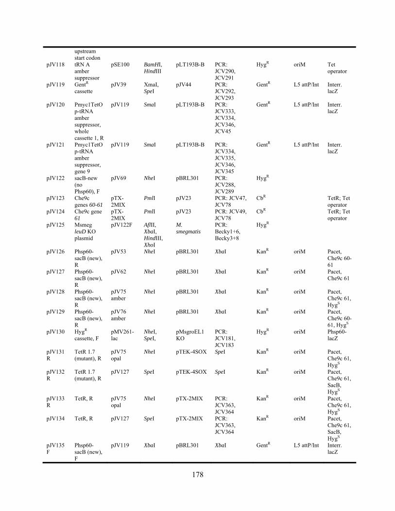
upstream start codon
pJV118 tRN A amber suppressor
pSE100 BamHI, HindIII
pLT193B-B PCR: JCV290, JCV291
HygR oriM Tet operator
pJV119 GentR cassette
pJV39 XmaI, SpeI
pJV44 PCR: JCV292, JCV293
GentR L5 attP/Int Interr. lacZ
pJV120 Pmyc1TetOp-tRNA amber suppressor, whole cassette 1, R
pJV119 SmaI pLT193B-B PCR: JCV333, JCV334, JCV346, JCV45
GentR L5 attP/Int Interr. lacZ
pJV121 Pmyc1TetOp-tRNA amber suppressor, gene 9
pJV119 SmaI pLT193B-B PCR: JCV334, JCV335, JCV346, JCV345
GentR L5 attP/Int Interr. lacZ
pJV122 sacB-new (no Phsp60), F
pJV69 NheI pBRL301 PCR: JCV288, JCV289
HygR
pJV123 Che9c genes 60-61
pTX-2MIX
PmlI pJV23 PCR: JCV47, JCV78
CbR TetR; Tet operator
pJV124 Che9c gene 61
pTX-2MIX
PmlI pJV23 PCR: JCV49, JCV78
CbR TetR; Tet operator
pJV125 Msmeg leuD KO plasmid
pJV122F AflII, XbaI, HindIII, XhoI
M. smegmatis
PCR: Becky1+6, Becky3+8
HygR
pJV126 Phsp60-sacB (new), R
pJV53 NheI pBRL301 XbaI KanR oriM Pacet, Che9c 60-61
pJV127 Phsp60-sacB (new), R
pJV62 NheI pBRL301 XbaI KanR oriM Pacet, Che9c 61
pJV128 Phsp60-sacB (new), R
pJV75 amber
NheI pBRL301 XbaI KanR oriM Pacet, Che9c 61, HygS
pJV129 Phsp60-sacB (new), R
pJV76 amber
NheI pBRL301 XbaI KanR oriM Pacet, Che9c 60-61, HygS
pJV130 HygR cassette, F
pMV261-lac
NheI, SpeI,
pMsgroEL1KO
PCR: JCV181, JCV183
HygR oriM Phsp60-lacZ
pJV131R
TetR 1.7 (mutant), R
pJV75 opal
NheI pTEK-4SOX SpeI KanR oriM Pacet, Che9c 61, HygS
pJV132R
TetR 1.7 (mutant), R
pJV127 SpeI pTEK-4SOX SpeI KanR oriM Pacet, Che9c 61, SacB, HygS
pJV133R
TetR, R pJV75 opal
NheI pTX-2MIX PCR: JCV363, JCV364
KanR oriM Pacet, Che9c 61, HygS
pJV134 TetR, R pJV127 SpeI pTX-2MIX PCR: JCV363, JCV364
KanR oriM Pacet, Che9c 61, SacB, HygS
pJV135F
Phsp60-sacB (new), F
pJV119 XbaI pBRL301 XbaI GentR L5 attP/Int Interr. lacZ
178
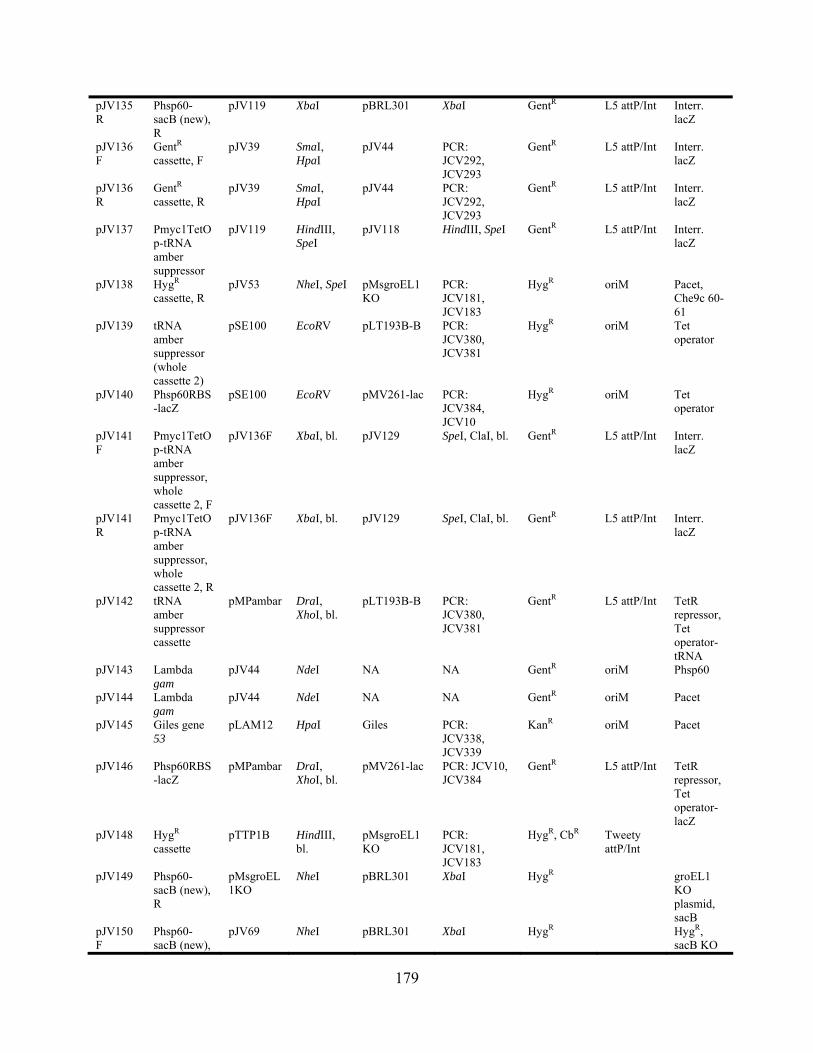
pJV135R
Phsp60-sacB (new), R
pJV119 XbaI pBRL301 XbaI GentR L5 attP/Int Interr. lacZ
pJV136F
GentR cassette, F
pJV39 SmaI, HpaI
pJV44 PCR: JCV292, JCV293
GentR L5 attP/Int Interr. lacZ
pJV136R
GentR cassette, R
pJV39 SmaI, HpaI
pJV44 PCR: JCV292, JCV293
GentR L5 attP/Int Interr. lacZ
pJV137 Pmyc1TetOp-tRNA amber suppressor
pJV119 HindIII, SpeI
pJV118 HindIII, SpeI GentR L5 attP/Int Interr. lacZ
pJV138 HygR cassette, R
pJV53 NheI, SpeI pMsgroEL1KO
PCR: JCV181, JCV183
HygR oriM Pacet, Che9c 60-61
pJV139 tRNA amber suppressor (whole cassette 2)
pSE100 EcoRV pLT193B-B PCR: JCV380, JCV381
HygR oriM Tet operator
pJV140 Phsp60RBS-lacZ
pSE100 EcoRV pMV261-lac PCR: JCV384, JCV10
HygR oriM Tet operator
pJV141F
Pmyc1TetOp-tRNA amber suppressor, whole cassette 2, F
pJV136F XbaI, bl. pJV129 SpeI, ClaI, bl. GentR L5 attP/Int Interr. lacZ
pJV141R
Pmyc1TetOp-tRNA amber suppressor, whole cassette 2, R
pJV136F XbaI, bl. pJV129 SpeI, ClaI, bl. GentR L5 attP/Int Interr. lacZ
pJV142 tRNA amber suppressor cassette
pMPambar DraI, XhoI, bl.
pLT193B-B PCR: JCV380, JCV381
GentR L5 attP/Int TetR repressor, Tet operator-tRNA
pJV143 Lambda gam
pJV44 NdeI NA NA GentR oriM Phsp60
pJV144 Lambda gam
pJV44 NdeI NA NA GentR oriM Pacet
pJV145 Giles gene 53
pLAM12 HpaI Giles PCR: JCV338, JCV339
KanR oriM Pacet
pJV146 Phsp60RBS-lacZ
pMPambar DraI, XhoI, bl.
pMV261-lac PCR: JCV10, JCV384
GentR L5 attP/Int TetR repressor, Tet operator-lacZ
pJV148 HygR cassette
pTTP1B HindIII, bl.
pMsgroEL1KO
PCR: JCV181, JCV183
HygR, CbR Tweety attP/Int
pJV149 Phsp60-sacB (new), R
pMsgroEL1KO
NheI pBRL301 XbaI HygR groEL1 KO plasmid, sacB
pJV150F
Phsp60-sacB (new),
pJV69 NheI pBRL301 XbaI HygR HygR, sacB KO
179
F plasmid pJV150R
Phsp60-sacB (new), R
pJV69 NheI pBRL301 XbaI HygR HygR, sacB KO plasmid
6.2.3 Cloning procedures
6.2.3.1 Preparation of the insert and vector for plasmid constructions
Plasmid cloning was performed using DNA inserts generated either by PCR-
amplification or restriction digest of the parental insert source as described in Table 15. PCR
reactions were set up as described below (PCR), and the products were cleaned up using the
QIAquick PCR cleanup protocol (QIAGEN), eluting in 50 l EB buffer. Restriction digests of
either plasmid DNA or PCR DNA were used to obtain the vector backbone or desired insert.
Restriction enzymes were from NEB exclusively, and digests were performed according to
manufacturer’s instructions for preferred buffers and reaction conditions, typically in 50 l
reaction volumes. Restriction digests were incubated at the required temperature for 2 hr for
plasmid DNAs and 4 hr for PCR products. Following digestion, the vector backbone digest was
heat-killed (if possible) and treated with 1 l calf intestinal phosphatase (CIP; NEB) to remove
the 5 phosphate and prevent ligation of the vector to itself. The digest reactions were then run
on 0.8% agarose gels, the bands containing the desired DNA fragments were extracted, and these
were cleaned up using the QIAquick gel extraction protocol (QIAGEN). The DNA was
quantified by analyzing 2 l on an agarose gel and comparing to the GC #1 quantitative DNA
ladder (GeneChoice) using Quantity One 4.6 software.
In cases in which a PCR product was cloned into a blunt site in the parental plasmid, the
PCR product was treated with T4 polynucleotide kinase (Roche) and the accompanying buffer
180
directly in the PCR reaction (without cleanup) for 1 hr at 37C. Plasmids and inserts that
required blunt ends were treated with the Klenow enzyme (NEB). The DNA digest was first
heat-killed (if possible), and subsequently 0.5 M dNTPs, 1X buffer were added, and 0.5 l of
Klenow in a total reaction volume of 60 l. The reactions were incubated at room temperature
for 15 mins, and these were immediately cleaned up using the QIAquick gel extraction protocol
for enzymatic cleanup (QIAGEN) or run on an agarose gel to be gel extracted.
6.2.3.2 Ligations and transformations
Ligations were performed with the Fast-link DNA ligase enzyme (Epicentre) in 15 l
volumes using 1.5 l ATP for sticky-end ligations and 0.75 l ATP for blunt-end ligations.
These were incubated at room temperature for 2 hr or longer and heat-killed at 75°C for 15 mins.
The heat-killed ligations were transformed into E. coli GC5 cells as described below (E. coli
transformations). All plasmids that were constructed were checked by restriction digest with two
different enzymes, and the insert was sequenced.
6.3 PCR
Primers were designed typically with melting temperatures at or above 60°C (if possible) to
simplify amplification from the high-G+C% mycobacterial or mycobacteriophage templates. If
restriction sites were used, these were engineered in the center of the oligonucleotide by
changing nucleotides when required, with perfect homology to the template flanking the site.
Oligonucleotides were synthesized as described above (DNA substrates) and resuspended in TE
181
buffer to 100 M stock solutions upon receipt of the lyophilized pellet of DNA. Working
solutions were 10 M, and all stock solutions were stored at -20°C.
PCR reactions were performed as described [7], typically using Pfu polymerase
(Stratagene) and when necessary, Pfu Turbo (for extended length targets longer than 4 kbp).
Reactions (50 l) contained template DNA (5-10 ng), 0.5 M primers, 0.2 mM dNTPs, 1X
buffer, 0-5% DMSO (typically 5%), and 1-5 U polymerase. Cycling conditions were set with an
initial 95°C denaturation for 5 min, and 25 cycles of denaturation (95°C for 30 sec), annealing
(varying temperature, 30 sec), and extension (72°C, varying length). Annealing temperatures
used were 2°C lower than the lowest primer melting temperature, and the extension was equal to
approximately 1 min per 1 kbp of desired product. A final extension at 72°C for 7 mins was
used, followed by cooling to 4°C. The 72°C extension temperature was used only for Pfu
polymerase; it was adjusted to 68°C for all Taq polymerases.
6.3.1 Colony PCR
PCRs used as a screening method (see section 7.10.4) typically were performed with Taq DNA
polymerase (NEB). Colonies were resuspended using sterile toothpicks in 200 l dH2O, vortexed
vigorously, boiled for 5 min, and vortexed again, and a volume equal to 1/10 the total PCR
reaction volume was used. Reaction volumes often were decreased to 25 l for screening PCRs.
6.3.2 MAMA-PCR
Mismatch amplification mutation assay PCR (MAMA-PCR) [30,219] was used to identify
mutant alleles – either point mutations or deletions – in a population consisting primarily of wild
182

type alleles. Primers (~20 nt) were designed in which the 3 ultimate base of the primer matched
the mutant sequence. For point mutations, this was the most 3 of the mutated bases; for
deletions, this was at the junction of the new deletion allele locus. Typically, the penultimate
base was also changed such that neither base would anneal at a wild type locus, and only the
ultimate base would anneal at a mutant locus (Figure X). Reactions were performed using
Platinum Taq High Fidelity DNA Polymerase (Invitrogen), in which 100 l of culture or
resuspended colonies were used for colony PCR as described above. Reaction conditions were as
described above for basic PCR with 2 mM MgSO4 added to increase the fidelity of the
polymerase.
6.3.3 Reverse transcription-PCR
Reverse transcription-PCR (RT-PCR) was performed essentially as described [228]. RNA was
extracted and purified from M. smegmatis cultures using the RiboPure-Bacteria kit (Ambion).
Each sample was disrupted with the mini bead-beater (30 sec) following an 8 min vortexing on
the vortex adapter. RNA samples were treated with DNaseI, as well as RNaseOUT to kill any
contaminating RNase enzymes; DNase Inactivation reagent was used following DNase
treatment. RNA aliquots were analyzed on agarose gels, quantified and stored at -80°C. RT-PCR
reactions were performed with the Qiagen OneStep RT-PCR kit (Qiagen) with equal amounts of
RNA and analyzed on agarose gels. Control PCR reactions using Pfu polymerase (Stratagene)
were used as controls to assess the presence of contaminating DNA in the RNA samples.
183

6.3.4 Sequencing
Sequencing of plasmids or PCRs was done either through the DNA synthesis facility of the
University of Pittsburgh or GeneWiz company. Typically 5-8 l of plasmid miniprep DNA was
used (~800 ng), and either 3.2 pmols (DNA synthesis facility) or 8 pmols (Genewiz) of primer
were used. Sequencing primers were designed 100 bp upstream of the region to be sequenced.
6.3.5 Site-directed mutagenesis (SDM)
The QuikChange site-directed mutagenesis (and sometimes the XL kit; Stratagene) was used for
all experiments. Primers were typically PAGE-purified, and PCR conditions were used as
recommended by the manufacturer.
6.4 DNA SUBSTRATES
All oligonucleotides (Table 16) were purchased from Integrated DNA Technologies, and when
required (lengths exceeding 30 nt), the oligonucleotides were PAGE-purified. When necessary,
oligonucleotides were radiolabeled with [-32P]-ATP (Perkin-Elmer) and T4 polynucleotide
kinase (Roche) for 30 min at 37°C and purified on ProbeQuant G-50 Micro Columns
(Amersham) to remove unincorporated label. When short (50 – 100 bp) dsDNA substrates were
required, complementary oligonucleotides were annealed at 0.4 M in annealing buffer (50 mM
Tris, pH 7.5 and 100 mM NaCl) by incubating for 5 min at 95C in a water bath and slow
cooling overnight to room temperature.
184
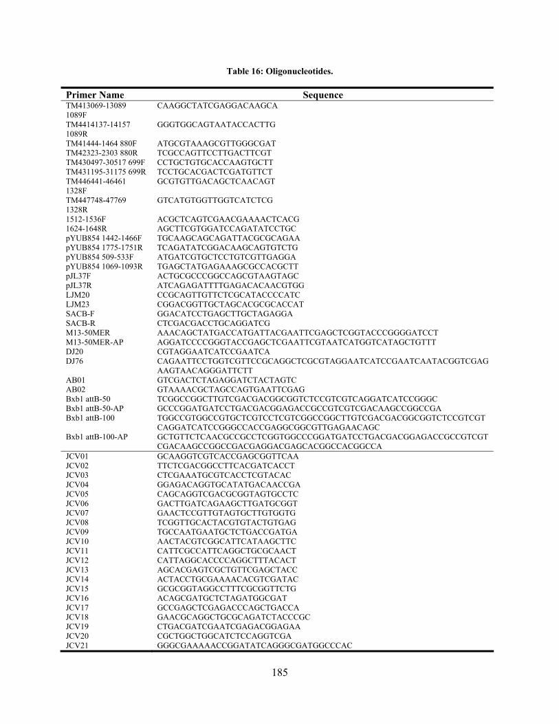
Table 16: Oligonucleotides.
Primer Name Sequence TM413069-13089 1089F
CAAGGCTATCGAGGACAAGCA
TM4414137-14157 1089R
GGGTGGCAGTAATACCACTTG
TM41444-1464 880F ATGCGTAAAGCGTTGGGCGAT TM42323-2303 880R TCGCCAGTTCCTTGACTTCGT TM430497-30517 699F CCTGCTGTGCACCAAGTGCTT TM431195-31175 699R TCCTGCACGACTCGATGTTCT TM446441-46461 1328F
GCGTGTTGACAGCTCAACAGT
TM447748-47769 1328R
GTCATGTGGTTGGTCATCTCG
1512-1536F ACGCTCAGTCGAACGAAAACTCACG 1624-1648R AGCTTCGTGGATCCAGATATCCTGC pYUB854 1442-1466F TGCAAGCAGCAGATTACGCGCAGAA pYUB854 1775-1751R TCAGATATCGGACAAGCAGTGTCTG pYUB854 509-533F ATGATCGTGCTCCTGTCGTTGAGGA pYUB854 1069-1093R TGAGCTATGAGAAAGCGCCACGCTT pJL37F ACTGCGCCCGGCCAGCGTAAGTAGC pJL37R ATCAGAGATTTTGAGACACAACGTGG LJM20 CCGCAGTTGTTCTCGCATACCCCATC LJM23 CGGACGGTTGCTAGCACGCGCACCAT SACB-F GGACATCCTGAGCTTGCTAGAGGA SACB-R CTCGACGACCTGCAGGATCG M13-50MER AAACAGCTATGACCATGATTACGAATTCGAGCTCGGTACCCGGGGATCCT M13-50MER-AP AGGATCCCCGGGTACCGAGCTCGAATTCGTAATCATGGTCATAGCTGTTT DJ20 CGTAGGAATCATCCGAATCA DJ76 CAGAATTCCTGGTCGTTCCGCAGGCTCGCGTAGGAATCATCCGAATCAATACGGTCGAG
AAGTAACAGGGATTCTT AB01 GTCGACTCTAGAGGATCTACTAGTC AB02 GTAAAACGCTAGCCAGTGAATTCGAG Bxb1 attB-50 TCGGCCGGCTTGTCGACGACGGCGGTCTCCGTCGTCAGGATCATCCGGGC Bxb1 attB-50-AP GCCCGGATGATCCTGACGACGGAGACCGCCGTCGTCGACAAGCCGGCCGA Bxb1 attB-100 TGGCCGTGGCCGTGCTCGTCCTCGTCGGCCGGCTTGTCGACGACGGCGGTCTCCGTCGT
CAGGATCATCCGGGCCACCGAGGCGGCGTTGAGAACAGC Bxb1 attB-100-AP GCTGTTCTCAACGCCGCCTCGGTGGCCCGGATGATCCTGACGACGGAGACCGCCGTCGT
CGACAAGCCGGCCGACGAGGACGAGCACGGCCACGGCCA JCV01 GCAAGGTCGTCACCGAGCGGTTCAA JCV02 TTCTCGACGGCCTTCACGATCACCT JCV03 CTCGAAATGCGTCACCTCGTACAC JCV04 GGAGACAGGTGCATATGACAACCGA JCV05 CAGCAGGTCGACGCGGTAGTGCCTC JCV06 GACTTGATCAGAAGCTTGATGCGGT JCV07 GAACTCCGTTGTAGTGCTTGTGGTG JCV08 TCGGTTGCACTACGTGTACTGTGAG JCV09 TGCCAATGAATGCTCTGACCGATGA JCV10 AACTACGTCGGCATTCATAAGCTTC JCV11 CATTCGCCATTCAGGCTGCGCAACT JCV12 CATTAGGCACCCCAGGCTTTACACT JCV13 AGCACGAGTCGCTGTTCGAGCTACC JCV14 ACTACCTGCGAAAACACGTCGATAC JCV15 GCGCGGTAGGCCTTTCGCGGTTCTG JCV16 ACAGCGATGCTCTAGATGGCGAT JCV17 GCCGAGCTCGAGACCCAGCTGACCA JCV18 GAACGCAGGCTGCGCAGATCTACCCGC JCV19 CTGACGATCGAATCGAGACGGAGAA JCV20 CGCTGGCTGGCATCTCCAGGTCGA JCV21 GGGCGAAAAACCGGATATCAGGGCGATGGCCCAC
185
JCV22 GTGGGCCATCGCCCTGATATCCGGTTTTTCGCCC JCV23 GACCAAAATCCCTTCACGTGAGTTTTCGTTCCAC JCV24 GTGGAACGAAAACTCACGTGAAGGGATTTTGGTC JCV25 TACGCGCCGTGTAAGGGCACGCAGA JCV26 GGCAGCGAGGACAACTTGAGCCGTC JCV27 GGTTACGATGCGCCCATCTACACCA JCV28 CACTCGCTTTAATGATGATTTCAGC JCV29 CTGCATGGTCAGGTCATGGATGAGC JCV30 TGCGCCAAGCTTCCTGCTGAACATC JCV31 CATACACGGTGCCTGACTGCGTTAG JCV32 GTCAGATGCGGGATGGCGTGGGACG JCV33 CAAAACAGGCGGCAGTAAGGCGGTCG JCV34 GAGTTGGTAGCTCTTGATCCGGCAAAC JCV35 GACCCCGAGGTGCACGGGCTGAAGGC JCV36 CGGCTCTACGCCGACTGCATGGAATC JCV37 CTACCAGGGCATCGTCAAACTGTTC JCV38 CAACGACGCCGATTGGTTCAAGTTCC JCV39 CTTCAGCAGAGCGCAGATATCAAATACTGTCCTTC JCV40 GAAGGACAGTATTTGATATCTGCGCTCTGCTGAAG JCV41 CGTAGTTAGGCCACCACGTGAAGAACTCTGTAGCACC JCV42 GGTGCTACAGAGTTCTTCACGTGGTGGCCTAACTACG JCV43 GTGGTTTGTTTGCCGGATCACGTGCTACCAACTCTTTTTCC JCV44 GGAAAAAGAGTTGGTAGCACGTGATCCGGCAAACAAACCAC JCV45 CTGGCTTCAGCAGAGCGCATATGCCAAATACTGTCCTTC JCV46 GAAGGACAGTATTTGGCATATGCGCTCTGCTGAAGCCAG JCV47 ACGAGATCGGCGGCCGCATATGAGT JCV48 TGCTTGGTGACAGCACTCGAGGCCATA JCV49 AAGGGGATTACACATATGGCTGAAA JCV50 TGGTTCGCCGGGGAAGCTTTGCGTT JCV51 TGATGAACGGGCCCCGCATATGAC JCV52 TGTTTGGTGCCTCTCGAGTGCGGT JCV53 TGAGGAAGGCACCAAACCATATGACC JCV54 TGAATCAGTCCCAAAGCTTCTCGT JCV55 GACGGCTCAAGTTGTCCTCGCTGCC JCV56 TTTCGCTAAATACTGGCAGGCGTTT JCV57 CACATGTTCTTTCCTGCGTTATCCC JCV58 GCTAACTCACATTAATTGCGTTGCG JCV59 GAATCAGTCCCAAAGCTTCTCGTTCCCTTCAC JCV60 GAATCAGTCCCAGAATTCCTCGTTCCCTTCAC JCV61 GTCGACGAACTGCTCGAACGACTCAT JCV62 GTGAGTCGCGGAACATCGCGAAGCAC JCV63 CTGCGCCGTTACCACCGCTGCGTTC JCV64 GATATCGACCCAAGTACCGCCACCT JCV65 CTCTCATGTATAGAGTGCTAGGTGGC JCV66 GGTCGTGGTTTCGCCGCCAGGAGCGGA JCV67 GTGACACCGTCATCTACAGCAAGTACGGC JCV68 CGGTCTCGCAGGCCTCGAAAAACCGCTCAG JCV69 GTCCGGGGCGGTACGCGTAGGCGTCTGAAAG JCV70 CTCCGTCGTCAGGATCATCCGGGCCAC JCV71 AGCGGCTCCCAGAATTCCTGGTCGTTC JCV72 GCGAACTGCTCGCCTTCACCTTCCTGCAC JCV73 GTCCGGGGCGGTACGCGTAGGCGTCTGAAAGAGACTTATGAGCAATCTAGGGGGATCC
GATAAATCAATCTAAAG JCV74 CTCCGTCGTCAGGATCATCCGGGCCACCGAGGCGGCGTTGAGAACAGGCAGGTCGACT
CTAGAGGATCTACT JCV75 TCGCGCCCACCACGACACCGAACGG JCV76 GACGTCACCGTCGCGCAGTCGATCGT JCV77 GTGCCTTGGGCGAATTCTGCTTGGT JCV78 GCAGATCAACCACCGCGTCGGAATTCGC JCV79 GAAATTCCGCTGGGAGATGGACAACA JCV80 CGACCAGTAGATCGATGTACCAGG JCV81 GTGTCAACGGCGAGTGCTACCTC
186
JCV82 CGAGCGCCTGCGACTGTGACAGG JCV83 GTCCCGAAGAACGATGGCATTTACG JCV84 CACCAGCAGCAGGCGTTCTACGAAG JCV85 GATTCCGCGGGAAGCCGGAAGAGGCT JCV86 GTGAAGAAGTCGGTCGTCGGAAACA JCV87 GAAAACCCGGATGCGGCAAGCACTC JCV88 CGAACGCGCCGAAATCTATCTGTCC JCV89 CACGGCCTCGACGGGTCCGTCGACCA JCV90 GCGTCCACCTGCTGTTTTGCCTTCA JCV91 GTGCGGCGCAGCACCATGACGTTGT JCV92 CTCGTGCTCGGCCGGGCACGCAGGCG JCV93 GTTCCCACTCGGAGGCAGGCAGA JCV94 CGGCGACGTATCGGCGCACCTCAT JCV95 GGGGATCCGATAAATCAATCTAAAG JCV96 GCAGGTCGACTCTAGAGGATCTACT JCV97 CTTATGAGCAATCTAGGGGGATCCGATAAATCAATCTAAAG JCV98 CGGCGTTGAGAACAGGCAGGTCGACTCTAGAGGATCTACT JCV99 GGATCGATGTCGACTGCCAGGCATCA JCV100 CCAGTGAATTCGAGCTCGGTACCC JCV101 CGCCAAGCTCTAATACGACTCACTA JCV102 CGACGTTGTAAAACGACGGCCAGTGA JCV103 GTCCGGGGCGGTACGCGTAGGCGTCTGAAAGAGACTTATGAGCAATCTAGGGATCGAT
GTCGACTGCCAGGCATCA JCV104 CTCCGTCGTCAGGATCATCCGGGCCACCGAGGCGGCGTTGAGAACAGGCCCAGTGAAT
TCGAGCTCGGTACCC JCV105 GGTCGCGACGAATCGAAAACGGTGCGA JCV106 CAGCGTCGCCGCCGCGATGTTCTAT JCV107 GCTTCCACCGGTTGCCTGCGCGTTCT JCV108 GACGCGTTCGCCACACACCAGTGCGT JCV109 GATCACCTCGTCGTCCACGCCCTT JCV110 CACGGGCACAGCAGGATGAGGTC JCV111 CAAGCGCGACGGAGACGGCCAGGGTA JCV112 CACCTGGTGGATATCGAGTCCGGT JCV113 CTGCCGCCCGGCGTGAGCGCCAAGGA JCV114 GGACTGGTGGGCGCCGACTATCTGA JCV115 GTGCACCAACGGCCGGATCGAGGAT JCV116 GAGGTCGGCTTGACCTTCACGGTCT JCV117 GAAAGCTCGCGTCCCCTGCAGATCTA JCV118 CGCAGTTTCAGGCGAAGCTAGCGAA JCV119 CATCGGTGTCCCGCTGCGGCGGTCCAA JCV120 CGAGCCGCCTCGTAGGCCTCGATCTGA JCV121 GTGTCCCGCTGCGGCGGTCCAACGTCGACACCGACCAGATCATCCCGGCGGGGCCTTA
CGCTGCGGAAACTCGATCAGATCGAGGCCTACGAGGCGGCTC JCV122 GAGCCGCCTCGTAGGCCTCGATCTGATCGAGTTTCCGCAGCGTAAGGCCCCGCCGGGAT
GATCTGGTCGGTGTCGACGTTGGACCGCCGCAGCGGGACAC JCV123 GACTCTAGAGGATCTACTAGTCATA JCV124 GAATTCGAGCTCGGTACCCGGGGAT JCV125 TACGACTCACTATAGGGAAAGCTTG JCV126 CTGGATCCACGAAGCTTCCCATGGT JCV127 AGAGCTCACCTAGGTATCTAGAAC JCV128 GTGTCACCGGTGTTCGGTGACCGACT JCV129 ACCATGGGAAGCTTCGTGGATCCAGCTCCAGCACCGTGGTGGTGTTCGGT JCV130 GTTCTAGATACCTAGGTGAGCTCTGTATCTGTTCGTACGTGGCATGTGC JCV131 CGTCAGCCCTTGCGAGAGCGCAA JCV132 CATCCCGGCGGTCTACCTGTAGTAGGTGACCCGAACGGGTTTCG JCV133 CGAAACCCGTTCGGGTCACCTACTACAGGTAGACCGCCGGGATG JCV134 CCGGCATCGGTGTCCCGCTGTGATGATCCAACGTCGACACCGACC JCV135 GGTCGGTGTCGACGTTGGATCATCACAGCGGGACACCGATGCCGG JCV136 GTGTCACAGATCTTCGGTGACCGACT JCV137 CGCTGGCCTCGAGCACCGTGGTGGTGTT JCV138 GCGACGCATCTAGATGGTGTGCTGTAT JCV139 CGTCAGCCCTTAAGAGAGCGCAA
187

JCV140 ACCATGGGAAGCTTCGTGGATCCAGCGCTGGCCTCCAGCACCGTGGTGGTGTT JCV141 CTTCAGCAGATCCACCGCCTCGGT JCV142 TGCCGAAATCCGGTCCGGCGACAA JCV143 GATGCGCGACATCGCCGTGGACA JCV144 ACGCCAGCTGGCGAAAGGGGGATGT JCV145 CGTCGACGCTTATAAACATATGGATATT JCV146 CGACACGTTCAGCAAGCTTCCCAG JCV147 CACCAGTGAAGGGAACATATGACCA JCV148 GAACATCGCGAAGCTTGCCGCGCAC JCV149 GTCCGGGGCGGTACGCGTAGGCGTCTGAA JCV150 GGTCTCCGTCGTCAGGATCATCCGGGC JCV151 GTAATCACTCGTGTTCACCGCCCC JCV152 TGGTGGCCGTGGCCGTGCTCGTCCT JCV153 GAGGAGTACCTGATCCTGTCGGCCC JCV154 CAAGCGGACCGGGGGTGTCGCGTGA JCV155 ACACCGTCATCTACAGCAAGTACG JCV156 TCGAAAAACCGCTCAGCGGCGCCCGCA JCV157 CCCACGAAGGAGAAGCGTGATGGAGGCTTTCACCACTCACACCGGCATCGGTGTCCCG
CTGCGGCGGTCCAA JCV158 CGCCGAAGGCCCGGATCAGGCCGGGAGAGTGCGCGGTTTCCAGGCCGGACGAGCCGCC
TCGTAGGCCTCGATCTG JCV159 CGAACTGCTCGCCTTCACCTTCCT JCV160 CTGTCCGCACCGCGGTCAGGCGTTG JCV161 CAGCAACGCCAACAGCCGTGCCACGGT JCV162 CCGTACGTCTCGAGGAATTCCTGCAGGATATCTGGATCCACGAAGCTTCCGGTCTCCGT
CGTCAGGATCATCCGGGC JCV163 TCGTTCATCCATAGTTGCCTGACTCCCAGTCCGTAATACGACTCACTTAAGTCCGGGGC
GGTACGCGTAGGCGTCTGAA JCV164 GGTCTCCGTCGTCAGGATCATCCGGGCCACCGAGGCGGCGTTGAGAACAGGACACGAC
TTATCGCCACTGGCAGC JCV165 GTCCGGGGCGGTACGCGTAGGCGTCTGAAAGAGACTTATGAGCAATCTAGGGAAACGA
CAGGTGCTGAAAGCGAGCT JCV166 CATGCGTTAGAGGTCGGTGAGCCCT JCV167 CACCGTCATCTACAGCAAGTACG JCV168 CTCATCTATCCGCCCGGGATAGCAT JCV169 AAGTGGTAATTCGGACGGTTCCG JCV170 GTGAGGAGTTTCATTCACCGTGATA JCV171 GCTGGACGTTGCGGAGGGTGACA JCV172 CTCACCGGTGAACGGGTGTTCGAT JCV173 GTGCGGCGTGATCGCCGTCGGTAGC JCV174 GATGCCAACGGGCCCGCCGGCACCA JCV175 GCTGACCGGAGTTCAGTGCGCGTG JCV176 CTGCGTAGAGGAGCCTGATGAGCAA JCV177 CTTGCTGATGCCCGAACCCAGCGCGAT JCV178 GAGACCGACCTGAAGAAGCGCAAGGA JCV179 GAAAGGCCGGTGCGGTGAAGGTTTT JCV180 CATGCACATCGGCGGGTTCGAGGATCT JCV181 GATTTAGGATACATGCTAGCCACCT JCV182 ACCGCAGCACCATCTAGAACGTCCC JCV183 ACCGCAGCGCTAGCGAGAACGTCCC JCV184 CGACCGTATTGATTCGTAGTAGTCCTACGCGAGCCTG JCV185 CAGGCTCGCGTAGGACTACTACGAATCAATACGGTCG JCV186 CGACCGTATTGATTCGTGATGATCCTACGCGAGCCTGC JCV187 GCAGGCTCGCGTAGGATCATCACGAATCAATACGGTCG JCV188 CTTCTCGACCGTATTGATTCGTAGTAGTCCTACGCGAGCCTGCGG JCV189 CCGCAGGCTCGCGTAGGACTACTACGAATCAATACGGTCGAGAAG JCV190 CCGTATTGATTCGGATGATTGATGAGCGAGCCTGCGGAACGACC JCV191 GGTCGTTCCGCAGGCTCGCTCATCAATCATCCGAATCAATACGG JCV192 GAGTTGGTAGCTCTTGATCCGGCA JCV193 CTCCACGAGCTGCCCGTGGAGGACT JCV194 GAACTACGCCCTGGCTCAAGACGCA JCV195 CGGGATGATCGAAATCGGTTGCCGT
188

JCV196 GGCTTCATCCGGAAATCCGGTGCGT JCV197 GCTTTTCGCGCCTGAGCTTGGTCCT JCV198 CCGCTGTGACACAAGAATCCCTGTTACTTCTCGACCGTATTGATTCGGATGATTCCTAC
GCGAGCCTGCGGAACGACCAGGAATTCTGGGAGCCGCTGGC JCV199 GCCAGCGGCTCCCAGAATTCCTGGTCGTTCCGCAGGCTCGCGTAGGAATCATCCGAATC
AATACGGTCGAGAAGTAACAGGGATTCTTGTGTCACAGCGG JCV200 GACGAAAGGGCCTCGTGATACGCCTA JCV201 CGAGCTCGGTACCCGGACATCCTGA JCV202 CGATGTCGACTGCCAGGCATCAAAT JCV203 GTTGGTGGGTGGCCGTGCATGTGAT JCV204 TCAGGATGTCCGGGTACCGAGCTCGGGAAGCGGAAGTGCGCGCTGCGATA JCV205 ATTTGATGCCTGGCAGTCGACATCGGTCAGCCGTTGCGCCACATGCACAT JCV206 CTGCGTGGTGGACGGTTCGCACGGTTGT JCV207 CATTCGCCATTCAGGCTGCGCAACT JCV208 GAACTAAGATCTGGCTCAAGACGCA JCV209 CGGGATGATCGACCATGGTTGCCGT JCV210 GGCTTCATCTAGAAATCCGGTGCGT JCV211 GCTTTTCGCGCTTAAGCTTGGTCCT JCV212 GTTGGTGGGAGATCTTGCATGTGAT JCV213 GGAAGCGGAAGCTTGCGCTGCGATA JCV214 GTCAGCCGTCTAGACACATGCACAT JCV215 CTGCGTGGTGGACGCTTAAGACGGTTGT JCV216 GGATCACCGCCGAGATCGGTGAGGGCAACAAGATCGACGGTGTGGTGCACGCGATCGG
GTTCATGCCGCAGAGCGGTATGGGCATCAACCCGTTCTTCGAC JCV217 GTCGAAGAACGGGTTGATGCCCATACCGCTCTGCGGCATGAACCCGATCGCGTGCACC
ACACCGTCGATCTTGTTGCCCTCACCGATCTCGGCGGTGATCC JCV218 CAGCCCGCAGCGTCGCGGCGTGTGCACGCGCGTTTACACCACCACTCCGAGGAAGCCG
AACTCGGCGCTCCGGAAGGTCGCGCGCGTGAAGCTGACCAGCC JCV219 GGCTGGTCAGCTTCACGCGCGCGACCTTCCGGAGCGCCGAGTTCGGCTTCCTCGGAGTG
GTGGTGTAAACGCGCGTGCACACGCCGCGACGCTGCGGGCTG JCV220 TGTGGTCACCGACCAGATCGACTACCTGACCGCCGACGAGGAGGACCGCCATGTCGTG
GCGCAGGCCAACTCGCCGACCGACGAGAACGGCCGCTTCACCG JCV221 CGGTGAAGCGGCCGTTCTCGTCGGTCGGCGAGTTGGCCTGCGCCACGACATGGCGGTC
CTCCTCGTCGGCGGTCAGGTAGTCGATCTGGTCGGTGACCACA JCV222 TGTGGTCACCGACCAGATCGACTACCTGACCGCCGACGAGGAGGACCGCCGCGTCGTG
GCGCAGGCCAACTCGCCGACCGACGAGAACGGCCGCTTCACCG JCV223 CGGTGAAGCGGCCGTTCTCGTCGGTCGGCGAGTTGGCCTGCGCCACGACGCGGCGGTC
CTCCTCGTCGGCGGTCAGGTAGTCGATCTGGTCGGTGACCACA JCV224 GCTGGTGTAGTCGTGGCCGTTTCGAT JCV225 CATTGTCGAAGCTGTTGGATGCGGA JCV226 GCTGGTCCTGAATTCAGTCCCATGGT JCV227 CAGAGGTATAAAACATATGAGTACTGCACT JCV228 GCAGGAGAATTCCCGGTGTCATGCT JCV229 TGACTAAGCAACCACCAATCGCAA JCV230 GAATATGCAAATGACTAAGCAACCACC JCV231 GACAGGCCTACTCGAAGGCAAGCGCA JCV232 GCGCTCTTGGCGACGGTCATCCAGT JCV233 GCCAACCATTCAGCAGCTGGTCCGCA JCV234 GCTCTTCTCCTTCTTCGCGCCATAGC JCV235 CCGGCCTCGAGGTCCGCGACGTGCA JCV236 CACCAGCGGTGCCTCGCTGCGCACCA JCV237 ACACCGCCAGGCTGAATTATTCCTCTG JCV238 GATCACACCCGTGATCACAGCCCAATTCACCACTCCCGAAAGGAAATGCACACACAAC
CACCTGGACGCCCAGGCCGGCCTCACAGCCGGCCTGGGCGTT JCV239 AACGCCCAGGCCGGCTGTGAGGCCGGCCTGGGCGTCCAGGTGGTTGTGTGTGCATTTCC
TTTCGGGAGTGGTGAATTGGGCTGTGATCACGGGTGTGATC JCV240 CACAGCCCAATTCACCACTCCCGAAAGGAAATGCACACACAACCACCTGGACGCCCAG
GCCGGCCTCACA JCV241 GATGCCCATACCGCTCTGCGGCATGAACCCGATCGCGTGCACCACACCGTCGATCTTGT
TGCCCTCACCGA JCV242 GATGCCCATACCGCTCTGCGGCATGAACCCGATGGAGTGCACCACACCGTCGATCTTGT
TGCCCTCACCGA
189
JCV243 GCTTTTCTGCGTTCTCGGGTAGCCGCT JCV244 CAACAGCGCTAGCATCCTTGAGAGTT JCV245 CAGGTTCGGTGGCGCGCTACGAATCT JCV246 CGGCCAGCACGATGCGCGCGGATGCGT JCV247 CCGCTCTGCGGCATGAACCCGATCGCGTGCACCACACCGTCGATCTTGTTG JCV248 GCATGAACCCGATCGCGTGCACCACACCGTC JCV249 CGCACGCTTAAGCCGACACGGTCAT JCV250 CACGGTTTCTAGAACCGGCCACCGGA JCV251 CGCGTCGAAGCTTCAGCTGCGGGCCCT JCV252 GTGCGCGCAAGATCTCGCCCAGCAGCA JCV253 CATGGACCAGAACAACCCGCTGTCGGGTCTGACCCGCAAGCGTCGTCTTTCGGCGCTGG
GCCCCGGCGGTC JCV254 GACCGCCGGGGCCCAGCGCCGAAAGACGACGCTTGCGGGTCAGACCCGACAGCGGGTT
GTTCTGGTCCATG JCV255 CACTACGGACCGCTGTTCATCCGCATGGCCTGGCAGGCCGCGGGCACCTACCGCGTCA
GTGACGGCCGCGG JCV256 CCGCGGCCGTCACTGACGCGGTAGGTGCCCGCGGCCTGCCAGGCCATGCGGATGAACA
GCGGTCCGTAGTG JCV257 CACTACGGACCGCTGTTCATCCGCATGGCCTGGTAGGCCGCGGGCACCTACCGCGTCAG
TGACGGCCGCGG JCV258 CCGCGGCCGTCACTGACGCGGTAGGTGCCCGCGGCCTACCAGGCCATGCGGATGAACA
GCGGTCCGTAGTG JCV259 CGAGACGATGGGTAACTACCATCCGCACGGCGACGTCTCGATCTACGACACCCTGGTC
CGCATGGCCCAGC JCV260 GCTGGGCCATGCGGACCAGGGTGTCGTAGATCGAGACGTCGCCGTGCGGATGGTAGTT
ACCCATCGTCTCG JCV261 GTGATCGGCGCCAACTCGTCCGACGACGGCTACATGCTGCAGATGGCGCGCACGGCCG
AGCACGCGGGCTA JCV262 TAGCCCGCGTGCTCGGCCGTGCGCGCCATCTGCAGCATGTAGCCGTCGTCGGACGAGTT
GGCGCCGATCAC JCV241 GATGCCCATACCGCTCTGCGGCATGAACCCGATCGCGTGCACCACACCGTCGATCTTGT
TGCCCTCACCGA JCV263 CGAACAAACCGTCCTCGAAACCCGTTCGGGTCACCTACTACAGGTAGACCGCCGGGAT
GATCTGGTCGGTGTCGA JCV264 TCGGGGCGGGCAACAAGCTCGACGGGGTGGTGCATGCGATTGGGTTCATGCCGCAGAC
CGGGATGGGCATC JCV265 GATGCCCATCCCGGTCTGCGGCATGAACCCAATCGCATGCACCACCCCGTCGAGCTTGT
TGCCCGCCCCGA JCV266 GTCGAAGAACGGGTTGATGCCCATCCCGGTCTGCGGCATGAACCCAATCGCATGCACC
ACCCCGTCGAGCTTGTTGCCCGCCCCGATCGCCTCGGTCACCC JCV267 CACTACGGGCCGCTGTTTATCCGGATGGCGTGGCAGGCTGCCGGCACCTACCGCATCCA
CGACGGCCGCGG JCV268 GTATGGCACCGGAACCGGTAAGGACGCGATCACCACCGGCATCGAGGTCGTATGGACG
AACACCCCGACGA JCV269 GCCACTACGGGCCGCTGTTTATCCGGATGGCGTGGTAGTAGGCCGGCACCTACCGCATC
CACGACGGCCGCGGCGG JCV270 GTGCCCGAGCAACACCCACCCATTACAGAAACCACCACCGGAGCCGCTAGCAACGGCT
GTCCCGTCGTGGGAAGTTCGTGCAGGACTTCGTCGCTGCCTGGGACAAGGTGATGAACCTCGACAGGTTCGACGTGCGCTGA
JCV271 GACCACCTCGCAGCCGTGGTGGCCCGCCGACTACGGCCACTACGGGCCGCTGTTTATCCGGATGGCGTGGGCACCTACCGCATCCACGACGGCCGCGGCGGCGCCGGGGGCGGCATGCAGCGGTTCGCGCCGCTTAACAG
JCV272 CGAACAAGCCGTCCTCGAAACCGGTTCGGGTGACCTACTACAGAAAGACCGCGGGAATGATCTGATCGGTGTCGA
JCV273 ATCACGTCGAGCGGCGGCGGCGCCGCAGCGGCGGCCTACTAGATCTCCGACAGGCTCACCGAGGCTTCACGCGCGG
JCV274 TTATTTTTGACACCAGACCAACTGGTAATGGTAGCGACCGGCGCTCAGCTGGAATTCCGCCGATACTGACGTTTGAGGGGACGACGACAGTATCGGCCTCAGGAAGATCGCACTCCAGCCAGCTTTCCGGCACCGCTTCT
JCV275 GCATCTGCCAGTTTGAGGGGACGACGACAGTATCGGATCGCACTCCAGCCAGCTTTCCGGCACCGCTTCT
JCV276 GGATAGGTCACGTTGGTGTAGATGGGCGCATCGTAGATCGCACTCCAGCCAGCTTTCCG
190
GCACCGCTTCT JCV277 TGCATCTGCCAGTTTGAGGGGACGACGACAGTATCCTACTAAGGAAGATCGCACTCCA
GCCAGCTTTCCGGCACCG JCV278 GATCCGCACCGTCGAGCAGTCCGACA JCV279 GGCTCGACTACCGTTTCGGATTGCT JCV280 GCCATCAATGAAAGAGCAACTGGCA JCV281 CCAGCTTTACTCAGGCTGCGCACCA JCV282 GGTGATGCCAGCGATGCGCAGTTCA JCV283 CAGGAATCCAAGAGCTTTTACTGCTT JCV284 GAAAGCTTCGAATTCTGCAGCTGGATC JCV285 CCAAGACAATTGCGGATCCCGTCGT JCV286 CGGCGATCCGGTCGTCGACGGGAGCGGCGGAAGCCTACTACATACGCACACCGGCGGC
CGCCATCACTGCCAGGG JCV287 TGTCGCGGTGCAGCAGCACCAGCGCGTCTTCGAGATCTGCTTCCTGCGAGTCGAAGTCG
GTGACGAAGTAG JCV288 GACAATTGCGGAGCTAGCCATGGACAT JCV289 GTCATACGCGGCTAGCGGATCCCGTTA JCV290 GCCTTCGGATCCTCCCCTGACGTGTA JCV291 CGTTGTAAGCTTCGGGTGGATGTCA JCV292 GCCATCATGGCCGCGGTGATCAGCTA JCV293 GGTACCGAGCACTAGTTGACATAAGC JCV294 TGTGCGTATGCCGAC JCV295 TGTGCGTATGTAGTA JCV296 CAGTGCACGCCGAGTTCGGGCAGCA JCV297 GGTCTACCTGAAGCG JCV298 GGTCTACCTGTAGTA JCV299 GTGCGCCAGAGATAACCGCCTTGAACT JCV300 CGACGGTGTGGTGCACT JCV301 CGACGGTGTGGTGCAGG JCV302 CGTAGATCACGGTGCCGGTGGT JCV303 GATGCCCATACCGCTCTGCGGCATGAACCCGATCGCATGGACCACACCGTCGATCTTGT
TGCCCTCACCGA JCV304 CGACGGTGTGGTCCATG JCV305 GACTCGCAGGAAGCAGT JCV306 GACTCGCAGGAAGCACA JCV307 CACGATGCCCTCCTCGACCGCT JCV308 GACTCGCAGGAAGCAGATCTCGAAGAC JCV309 GCCGGTGTGCGTATGTAGTAGGCTTCCGCCG JCV310 CCGGCGGTCTACCTGTAGTAGGTGACCCGAA JCV311 CCGGCGGTCTACCTGAAGCG JCV312 CCGGCGGTCTACCTGTAGTA JCV313 GCCGGTGTGCGTATGCCGAC JCV314 GCCGGTGTGCGTATGTAGTA JCV315 GTCCTCCCTATCAGTGATAGATA JCV316 CTACTTCGTCACCGACTTCGACTCGCAGGAAGCAGATCTCGAAGACGCGCTGGTGCTGC
TGCACCGCGACA JCV317 TACCTCGAGGTCACCGAGGGCGTCGGGTTCGACAAGGGCTTCCTGTCGGCCTACTTCGT
CACCGACTTCGACTCG JCV318 CTCGGCGACCTTCTCCAGCAGCGGCAGCAGATCCGGCAGCGAGCTGATCTTGTCGCGGT
GCAGCAGCACCAGCGC JCV319 GCTGGAACTCGACGTGCAGAACGAGGAGCACCTGTCGACTCTGGCCGACCGGATCACC
GCCGAGATCGGTGAGGG JCV320 TACGAGTACGCCGAGATGTGGATGCCCTTGGACACATCCTCGTACGGCGCGTCGAAGA
ACGGGTTGATGCCCATA JCV321 GACGAATCTCTCACGACGCAGTGT JCV322 TCAGCGCACGTCGAACCTGTCGAGGTTCATCACCTTGTCCCAGGCAGCGACGAAGTCCT
GCACGAACTTCCCACGACGGGACAGCCGTTGCTAGCGGCTCCGGTGGTGGTTTCTGTAATGGGTGGGTGTTGCTCGGGCAC
JCV323 CTGTTAAGCGGCGCGAACCGCTGCATGCCGCCCCCGGCGCCGCCGCGGCCGTCGTGGATGCGGTAGGTGCCCACGCCATCCGGATAAACAGCGGCCCGTAGTGGCCGTAGTCGGCGGGCCACCACGGCTGCGAGGTGGTC
JCV324 CCGCCGCGGCCGTCGTGGATGCGGTAGGTGCCGGCCTACTACCACGCCATCCGGATAA
191

ACAGCGGCCCGTAGTGGC JCV325 CATGGACCAGAACAACCCGCTGTCGGGGTTGACCCGCAAGCGCCGACTGTCGGCGCTG
GGGCCCGGCGGTC JCV326 GACCGCCGGGCCCCAGCGCCGACAGTCGGCGCTTGCGGGTCAACCCCGACAGCGGGTT
GTTCTGGTCCATG JCV327 CCCGCTGTCGGGGTTGACCCACAAGCGCCGACTGTTGGCGCTGGGGCCCGGCGGTCTGT
CACGTGAGCGTG JCV328 CACGCTCACGTGACAGACCGCCGGGCCCCAGCGCCAACAGTCGGCGCTTGTGGGTCAA
CCCCGACAGCGGG JCV329 TGGTGTATGCACCCGCGTGTACACCACCACTCCGAGGAAGCCGAACTCGGCGCTTCGG
AAGGTTGCCCGCG JCV330 CGCGGGCAACCTTCCGAAGCGCCGAGTTCGGCTTCCTCGGAGTGGTGGTGTACACGCG
GGTGCATACACCA JCV331 GCCATCCTGACGGATGGCCT JCV332 GCATGCGGATCGTGCTCATT JCV333 CGCCGCCCGAAATGAGCACGATCCGCATGCCACCGCACCCATCAGAGATGGT JCV334 TAAAAAAGGGGACCTCTAGGGTCCCCAATTAATTAGTTGTTCCTTTCGGGTGGATGTCA JCV335 CGCCGCCCGAAATGAGCACGATCCGCATGCGCACACACCCCTGACTCCTGCTA JCV336 CGAGCCTGCGGAACGACTAGGAATTCTGGGAGCCG JCV337 CGGCTCCCAGAATTCCTAGTCGTTCCGCAGGCTCG JCV338 GAAGGTGACCAAACCATATGACCACCA JCV339 CATGCTGGAATTCGGGGCGATCATT JCV340 TGCTGACATGCGGGCGTAGCTCAATGGTAGAGCCCTAGTCTTCCAAACTAGCTACGCGG
GTTCGATTCCCGTCGCCCGCTCGGTAGGGACCGCCACGTGCGATTTAGGATACATGCTAGCCACCT
JCV341 GTGCGCCGATTTCTGCACCACGGTCGTGATCTGCGACGAACCACGACCTTGGTGCAGAAATCGCGGGGGCAGTTGAGCACTCGGCAACGAAAAAGGGACCACCGCAGCGCTAGCGAGAACGTCCC
JCV342 ACCAGATCATCCCGGCGGTC JCV343 ACCAGATCATCCCGGCGGGG JCV344 CAGAAGGCCATCCTGACGGATGGCCT JCV345 GCATGCGGATCGTGCTCATTTCGG JCV346 GATTAGCTAAGCAGAAGGCCATCCT JCV347 CTACGCGAGCCTGCGGAACGACTAGGAATTCTGGGAGCCGCTGGC JCV348 GCCAGCGGCTCCCAGAATTCCTAGTCGTTCCGCAGGCTCGCGTAG JCV349 GCTCGACGGGGTGGTGCAAT JCV350 GCTCGACGGGGTGGTGCAAG JCV351 CAGACAGCAGCGCGCACACCGTCTT JCV352 GCTCGACGGGGTGGTGCATGCGATTGGGT JCV353 CCCGCGGTCTTTCTGAAGCG JCV354 CCCGCGGTCTTTCTGTAGTA JCV355 GAGTTTCCGCAGCGTAAGGGCTAT JCV356 CCCGCGGTCTTTCTGTAGTAGGTCAC JCV357 GATCCAATATTACTAGTAGATCTCGT JCV358 GACGTCTTAATTAATATGCATCAAT JCV359 GACGTCTTAATTAATATGCATCAATTGATTTA JCV360 GATCCAATATTACTAGTAGATCTCGTAATATTG JCV361 GAATAGAGGTCCGCTGTGACATAGGAATCCCTGTTACTTCTC JCV362 GAGAAGTAACAGGGATTCCTATGTCACAGCGGACCTCTATTC JCV363 GACCTCTAGGGTCCCCAGCTGGCTAG JCV364 CACGGCCGTGACGCTAGCGACGATCCA JCV365 AAAACGATTGTCATTATCGTACGACGGTACCGCACGACGAAGGAGAGTCAATGGCTCG
CAACGAGATCCGGCCCATCGTGAAGCTGCGGTCCACTGCGGG JCV366 CCCGCAGTGGACCGCAGCTTCACGATGGGCCGGATCTCGTTGCGAGCCATTGACTCTCC
TTCGTCGTGCGGTACCGTCGTACGATAATGACAATCGTTTT JCV367 AGAAGGTCTGATGGCTCGCAACGAGATCCGGCCCATCGTGAAGCTGCGGTATGGCGAA
GAAGTCGAAGATTGTCAAGAACGAGCAGCGGCGAGAACTGGT JCV368 ACCAGTTCTCGCCGCTGCTCGTTCTTGACAATCTTCGACTTCTTCGCCATACCGCAGCTT
CACGATGGGCCGGATCTCGTTGCGAGCCATCAGACCTTCT JCV369 AAATACGATCCGGTCCTGCGCCGCCACGTCGAGTTCCGCGAGGAACGCTGATGGCAGT
CAAGAAGTCCAGAAAGCGCACGGCCGCAACTGAACTCAAGAA JCV370 TTCTTGAGTTCAGTTGCGGCCGTGCGCTTTCTGGACTTCTTGACTGCCATCAGCGTTCCT
192

CGCGGAACTCGACGTGGCGGCGCAGGACCGGATCGTATTT JCV371 GAGATGGCGCATCGCGGCGAGTTGCCCGGTGTGCGGAAGGCGAGTTGGTGGGCGTGCG
GTATGACACCATCGGTGCCGAAGGCGACTGCGGATCGAGGAA JCV372 TTCCTCGATCCGCAGTCGCCTTCGGCACCGATGGTGTCATACCGCACGCCCACCAACTC
GCCTTCCGCACACCGGGCAACTCGCCGCGATGCGCCATCTC JCV373 GTGGCTGACGAGCAGGGTGCCAGGT JCV374 CACCGATGGTGTCATACCGCACGCCT JCV375 ATCATCCCGGCGGGGCCTTACGCT JCV376 GGAAAACCCTGGCGTTACCTAGCTTAATCGCCTTGCAG JCV377 CTGCAAGGCGATTAAGCTAGGTAACGCCAGGGTTTTCC JCV378 GTCTCTGACGAGCGGGAGAACCCA JCV379 GAGCTTCAACCCACCATATGAGGAAGGCA JCV380 GTCACGACCGGTTGTGTGAGCCAGA JCV381 CTGTCCTCGTTGGGTACCGAGCTCGA JCV382 CGACGAAGGAGAGTCAATCT JCV383 CGACGAAGGAGAGTCAATCG JCV384 GATCCGGAGGAATCACTTCGCAATG JCV385 TGATGTCGCTGCAGGAACTGCACAGCGAACTGGGGTCGCGCCGGTCATGACGGGCCCA
CCACGCGACAGCGCCATTGCGCCGACAATCGTCGGTCCAACG JCV386 CGTTGGACCGACGATTGTCGGCGCAATGGCGCTGTCGCGTGGTGGGCCCGTCATGACC
GGCGCGACCCCAGTTCGCTGTGCAGTTCCTGCAGCGACATCA JCV387 CTCGGCCGCGGTGCAGGGCATCGAGGCCGGCATCCGCGGCGACATCGGCGTGATGTCG
CTGCAGGAACTGCACAG JCV388 GCACAGGGCTGCCGGCTCGGACACATCTGAAATCCGGCTCCTACCTGAGCCGTTGGAC
CGACGATTGTCGGCGCA JCV389 CATCCCGTGCGTCACCACGGTGCA JCV390 GTGATGGACTGCGCCGCAGGCGAACT JCV391 GGCGACCAGCACTACCGGCGTACGCATGGGACCTCCCGGTTTGCTTATTGAAAACGATT
GTCATTATCGTACGAC JCV392 GGTCCGGGTCGTTGCGGCGATTCTTGCGGGTGACGTACGTGTATCCGGTTCCCGCAGTG
GACCGCAGCTTCACGA JCV393 CCGTAGGCCTCGAAGAACGCGACCTACGGTTTGACGATGGCGAAGCCCGCG JCV394 CCGTATGACTCAAAAAACGCCACCTACGGTTTGACCACCGCGAAATCAGCG JCV395 CCCCGCCGAACCGTAGGCCTCGAAGAACGCGACCTACGGTTTGACGATGGCGAAGCCC
GCGAACGCCGCGA JCV396 GGCGTCGACCGTCAGGCCCCAGGCGTTCAAGAGCTACTAATGCGGGTCGATGCCGGGG
CACAGCGGCCCGCGCG JCV397 TCCTCCCACTCCCTCGGTCTCACCT JCV398 TCGGCCGCGGTGCAGGGCATCGAGG JCV399 CGAAGAGGCCGACCCGATTGAAGGGGATTACATCTATGGCTGAAAATGCTGGGCCCAA
CGCATGAGCGCCCCGGCGAACCACGACGCGGTGGTTGATCTG JCV400 ACCGTCGGCGACGTCGTCACCGACAGCTATATCTACGACACCGACCCGCTCGAAGAGG
CCGACCCGATTGAAGGG JCV401 GGCCTCGGCGTAGTGGTACGTCGGCGGGCGCGTCATGACTGCACCACCTGCAGATCAA
CCACCGCGTCGTGGTTC JCV402 GACGTGGTCACGATCAGCCTGCCCT JCV403 CAACTTGAACGCGATCGCGGGCACG JCV404 GCTCCGGGCTCGCAGCAGCGGGCTT JCV405 CAACGCAATTAATGTGAGTTAGCTCA JCV406 GGGTGATGTCCGGGGCGGTACGCGTAGGCGTCTGAAAGAGACTTATGAGCGTTCTCAA
CGCCGCCTCGGTGGCCCGGATGATCCTGACGACGGAGACCGC JCV407 GCGGTCTCCGTCGTCAGGATCATCCGGGCCACCGAGGCGGCGTTGAGAACGCTCATAA
GTCTCTTTCAGACGCCTACGCGTACCGCCCCGGACATCACCC JCV408 GCGCAGTCGATCGTCGACGCGGTCGCGCAGGCGAACCGGGAGGCGGATCCGGCGGCGC
GCGACGGCGATCCGGTGGGCCCGTTCGGTGTCGTGGTGGGCG JCV409 CGCCCACCACGACACCGAACGGGCCCACCGGATCGCCGTCGCGCGCCGCCGGATCCGC
CTCCCGGTTCGCCTGCGCGACCGCGTCGACGATCGACTGCGC JCV410 GAGGAGTCAACCCCATATGATGTCGAT JCV411 GGTCGGCGAATTCCGTTGTCATGTC JCV412 AGCTGCACATATGCGCTGCGCCCGT JCV413 GGATAGGTCAGGAATTCGCCGGTCA JCV414 GTCGCGCCATATGGTGCAGATTCT
193

JCV415 GGTGGCGCTGAATTCGTCGTACGTCA JCV416 GAGGTGGCGTCCATATGCACATGCA JCV417 CGTCTGCTGCTTGAATTCGGTCACAG JCV418 CGGATGCTCACGGATCCGATCTGCCA JCV419 GATCTTGGCGATGAATTCGCTGCGCT JCV420 GAGGGCGGCATATGAGCGCCGA JCV421 CTGTCCCTATCGAATTCGGCCTGGT JCV422 GGCGCATATGAGCCGCGAGCTGCT JCV423 CTTCGCCTCGTCGAATTCCGGGTCGT JCV424 GTGCAATGACATATGAGTGACGGAC JCV425 CATCGTTTGTGAATTCTCGCTTGAA JCV426 GGATCGACATATGAAGCGCACCAGGA JCV427 CTCGCCGGAATTCTGCTACGGGTCGA JCV428 GCGGTCGACATATGCTGAGCGTGCAGCC JCV429 CTGCATAACCGGAATTCAGGGCGTGT
6.5 PROTEIN PURIFICATION
C-terminally 6x-his-tagged Che9c gp60, Che9c gp61, and Halo gp43 proteins were purified by
over-expression from T7-inducible vectors in E. coli BL21(DE3) pLysS (Invitrogen)
transformed with pJV33 and pJV34, respectively, using nickel-affinity chromatography. In
addition, empty vector pET21a was also transformed into BL21(DE3) pLysS cells and prepared
in parallel as a negative control.
Che9c gp60 was purified as follows: cells containing pJV33 were grown at 37C to an
OD600 of 0.4. Che9c gp60 expression was induced by addition of 1 mM IPTG, and cells were
harvested by centrifugation after 4 hr growth at 30C. Cell pellets were resuspended in lysis
buffer at 5 ml per gram wet weight and sonicated 6-8x at bursts of 10 s with 1 min cooling in
between bursts. Cell lysate was centrifuged at 13,000 rpm for 30 min at 4C, and the supernatant
was incubated with 750 l Ni-NTA agarose resin (Qiagen; washed 2x with dH2O) rocking at 4C
for 1.5 h. This was applied to a column and washed 2x with 20x column volumes of wash buffer
containing 10 mM imidazole (first wash) or 20 mM imidazole (second wash). Protein was eluted
194
4x with 1 column volume elution buffer. Eluted fractions were analyzed by SDS-PAGE, and the
fractions containing the least contaminating proteins were dialyzed against storage buffer.
Che9c gp61 purification was performed similarly except 2.5 ml Ni-NTA agarose resin
was used, washes did not contain imidazole, and elutions were performed with a gradient of
imidazole concentrations (20, 40, 60, 80, 100, 150, 200, and 250 mM imidazole). Elution
fractions with 150 and 200 mM imidazole were dialyzed against storage buffer. Proteins were
analyzed by SDS-PAGE and quantified by Bradford protein assay (Bio-rad).
Halo gp43 protein purification was performed similarly to that for gp61 except 5 ml of
resin was used. Two combinations of elution fractions (80 and 100; 150 and 200 mM imidazole)
were dialyzed against storage buffer and analyzed and quantified as above.
6.5.1.1 Antibody synthesis
Anti-gp61 and anti-gp43 antibodies were synthesized by Pocono Rabbit Farms.
Screening bleeds were initially tested from rabbits and found to have very high background on
wild type M. smegmatis cell extracts. Therefore, multiple mouse screening bleeds were tested,
and three mice were chosen for each protein (Che9c gp61 and Halo gp43). Approximately 1 –
1.5 mg of nickel-affinity purified protein was sent to Pocono Rabbit Farms, and mouse bleeds
were obtained. Mice S12 and S14 were inoculated with Che9c gp61 protein (mouse S13 died),
and mice S19, S20, and S21 were inoculated with Halo gp43 protein. Titermax adjuvant was
used in place of Freund’s, which is not suitable for mycobacteria. Approximately four bleeds
were collected for each mouse, and subsequently the final bleeds were extracted. All antibodies
were stored in 5 l aliquots at -80°C.
195
6.6 IN VITRO ASSAYS
6.6.1 Exonuclease assays
Exonuclease activity of Che9c gp60 was determined as described [227] by three assays - A, B,
and C - in which Exo (5 U/l; NEB) was used as a positive control for DNA degradation,
while mock purified pET21a vector control protein preparations were used as a negative control.
Assays A and B were used to determine the exonuclease activity of Che9c gp60 on linear
dsDNA, while assay C was used to determine its activity on supercoiled or nicked open-circle
dsDNA.
In assay A, reaction mixtures (50 l) contained 8 nM 32P-labeled 100 bp Bxb1 attB, 40
nM unlabeled 100 bp Bxb1 attB (see Table 16), and gp60 or control proteins in exonuclease
assay buffer. Che9c gp60 (10.6 pmol/l), Exo, and mock purified control protein were added at
1 l per reaction of serially diluted concentrations (neat, 2x, 4x, 8x, 16x, and 32x in protein
dilution buffer) with respect to the stock concentration of each protein. Reactions were
incubated for 5 min at room temperature and stopped with final concentrations of 10 mM EDTA
and 0.5% SDS and addition of glycerol loading dye (0.25% bromophenol blue, 0.25% xylene
cyanol, and 30% glycerol). Samples were analyzed by gel electrophoresis on 8% native TBE
polyacrylamide gels, dried on Whatman paper for 1.5 hr at 85C, and exposed to BioMax
Maximum Resolution film (Kodak) overnight.
In assay B, reaction mixtures (50 l) contained 0.8 nM pBluescript SK+ (Stratagene)
linear dsDNA (linearized by digesting with EcoRV) and protein in exonuclease assay buffer.
Che9c gp60 and mock-purified protein were added at equal volumes (1 l) from parallel
196
purifications, with gp60 at a final concentration of 212 nM; Exo was added at 5 U per reaction.
Negative control reactions were routinely performed using 1 l storage buffer instead of enzyme.
Reactions were incubated for 0 – 10 min at room temperature, stopped with EDTA and SDS as
above, and loading dye was added. 25 l of each reaction was analyzed by gel electrophoresis in
0.8% agarose gels, visualized with ethidium bromide (0.4 g/ml) and photographed using a Bio-
Rad Gel Doc 2000 and Quantity One 4.6 software.
In assay C, reaction mixtures (20 l) contained either 2 nM linearized or 2 nM
circularized pBluescript SK+ and protein in exonuclease assay buffer. Che9c gp60 was added to
a final concentration of 530 nM by addition of 1 l protein and 1 l mock-purified control
protein was used; Exo was added at 5 U per reaction. Reactions were incubated for 0 – 10 min
at room temperature, stopped by addition of EDTA and SDS as above, and loading dye added.
Reactions were analyzed on agarose gels as with assay B.
6.6.2 DNA binding assays
6.6.2.1 Double-filter binding assay
Filter-binding assays were used to determine the DNA binding activity of Che9c gp61
and Halo gp43 [227]. A double-filter binding assay was utilized [20,239] in which nitrocellulose
filter membranes were stacked on top of DEAE-cellulose filters and filtered by means of a FH
225 V filter manifold system (GE Healthcare). Protein and protein/DNA complexes are bound by
the nitrocellulose filter on the top, and the unbound DNA is captured by the DEAE filter below,
so that the total DNA counts per sample are accounted for. DEAE-cellulose filters, grade DE81
(Whatman), were prepared as described [20] in which the membranes were treated with 0.1 M
197
Na2EDTA pH 8.8 for 10 min rocking at room temperature, followed by 3-10 min washes in 500
mM NaCl. The filters are then briefly treated with 500 mM NaOH (less than 30 sec), washed 15
times with dH2O, washed with TBS buffer, and stored at 4°C in that same buffer. Protran BA85
nitrocellulose filters (Whatman/Schleicher & Schuell) were prepared by removing the blue
packaging papers, and incubating the filters in 500 mM KOH on a rocking platform for 25 min
(time critical) at room temperature. The filters were washed 15 times with dH2O, washed twice
with TBS buffer, and stored at 4C in TBS buffer.
DNA binding reactions (30 l) contained 32P-labeled Bxb1 ss-attB (50 nt or 100 nt; Table
16), enzyme, and binding assay buffer similar to the conditions described for assaying DNA
binding of RecT [68]. Reactions containing dsDNA (~66.7 nM) used an annealed 32P-labeled 50
bp attB substrate (Table 16) which was gel-extracted from a native 8% TBE polyacrylamide gel
(exposed to film for 30 sec to locate probe), electroeluted in dialysis membrane (SpectraPor,
MWCO 1000) in TBE at 100 v for 45 min, phenol:chloroform extracted and ethanol precipitated.
In most reactions, 90% of the DNA added was cold, and 10% was used ‘hot’ to reduce the
amount of radioactivity use. The concentrations of gp61 used were in the range of 0.2 – 3.7 M
(diluted in protein dilution buffer), and storage buffer was used as a ‘no protein’ control.
Reactions were incubated at 37C for 20 mins and filtered. Each reaction (25 l) was filtered and
washed once with wash buffer. Filters were individually placed in 5 ml ScintSafe 30%
scintillation fluid (Fisher) and the radioactive counts per minute (CPM) were determined by
counting in a scintillation counter, with 1 min counting per sample. Percent DNA bound was
determined by the following equation:
%DNAbound CPMNC
CPMNC CPMDEAE
198
CNC is the CPM retained on the nitrocellulose filters (protein and protein/DNA complexes), and
CPMDEAE is the CPM on DEAE filters (unbound DNA), and this is further normalized to
negative controls in which no protein was added. Binding constants were determined by non-
linear regression using SigmaPlot 8.0. Error bars represent the standard deviation from three
independent experiments.
6.6.2.2 Gel shift assay
Gel shift assays were performed with the DNA binding reaction conditions similar to
those described for the filter-binding assays using either ss-attB (50 nt) or ds-attB substrates
(Table 16). DNA binding reactions were loaded on native 8% TBE polyacrylamide gels with
glycerol loading dye, dried on Whatman paper for 1.5 hr at 85C, and exposed to BioMax
Maximum Resolution film (Kodak) overnight or on a Fujifilm phosphoimager plate.
Phosphoimager plates were scanned with a Fujifilm FLA-5100, and quantitation of shifted bands
was performed using MultiGauge software.
6.6.3 Electron Microscopy
Samples were prepared and analyzed by transmission electron microscopy as described [20] with
reactions (50 l) containing purified Che9c gp61 [227] at a concentration of 50 g/ml (1.2 M)
and 1.95 M ss-attB DNA (100 nt; Table 16) or oligonucleotides of various lengths. Briefly, the
reactions were absorbed to glow-discharged, formvar carbon-coated 400 mesh copper grids, and
these were negatively stained with 2% uranyl acetate. Images were collected at a magnification
of 140,000x.
199

6.6.4 Gel filtration
Analytical gel filtration of purified Che9c gp61 protein was performed using a Superdex 200
10/300 GL high performance gel filtration column (Tricorn, Amersham) controlled by the
System Gold high-pressure liquid chromatography system (Beckman Coulter, Inc.) as previously
described [101]. The column was standardized using a gel filtration calibration kit (Amersham)
in which both low molecular and high molecular weight protein standards were run in duplicate
to determine their respective elution volumes. Standards included: ribonuclease A (13.7 kDa),
chymotrypsinogen (25 kDa), ovalbumin (43 kDa), albumin (67 kDa), aldolase (158 kDa),
catalase (232 kDa), and apoferritin (443 kDa). Tryptophan and Barnyard Phage particles were
used to determine the bed and void volumes, respectively. Approximately 330 g of each
standard was used for calibration at 100 l volumes. The column was first equilibrated in one
column volume (approximately 25 ml) of equilibration buffer (33 mM Tris, pH 7.5, 100 mM
NaCl) at a flow-rate of 0.2 ml/min. Protein standards or gp61 protein were loaded through a 100
l loop and eluted at a flow-rate of 0.5 ml/min. Elution volumes of proteins (recorded as the time
of peak absorbance) were monitored at a wavelength of 280 nm using protein fluorescence and,
as an additional reading, on a Jasco fluorescence detector. The Kav values (fractional retentions
of the samples [68]) for each protein standard were calculated based on the following equation:
Kav = Ve - Vo
Vt - Vo
Ve is the elution volume of the protein, Vo is the column void volume, and Vt is the total bed
volume of the column. The Kav value for each protein standard was plotted against the molecular
200

weight (Daltons) on a logarithmic scale, and a trendline was determined based on the standards.
To assay the size of gp61 protein complexes, gp61 protein was tested at concentrations of 5 M,
10 M, and 25 M. The elution volumes were collected for each, and the Kav values were
determined. Using the equation from the slope of the line, and solving for y, the molecular
weight for each gp61 reaction was determined. The column error was determined for the
molecular weight of the protein standards compared to the value obtained using the slope
equation in order to determine the range of accuracy for the gp61 reactions.
6.7 WESTERN BLOT ANALYSIS
Che9c gp61 and Halo gp43 protein expression were monitored using western blot analysis [227].
M. smegmatis cells were collected, resuspended in SDS-PAGE buffer and normalized to OD600.
To do this, the OD600 was measured (OD per ml), the cells pelleted, and resuspended to a
concentration of 0.05 ODs per l. Strain samples (0.5 ODs of cells) and purified protein (0.5 g)
as positive controls were loaded on 10% SDS-polyacrylamide gels following boiling for 3 – 5
min at 95°C in 4x SDS-PAGE loading dye diluted down to 1-2x. Proteins were transferred to
Sequi-blot PVDF membranes (Bio-rad) using a Bio-rad semi-dry transfer cell in transfer buffer
(48 mM Tris, 39 mM glycine, 0.037% SDS, 20% methanol) at 0.8 mAmps per cm2 of gel. The
blots were allowed to dry, rewetted with methanol, and blocked in 5% milk in TBS-T for 1 hr
rocking at room temperature. These were probed with either mouse anti-gp61 polyclonal
antibodies or mouse anti-gp43 polyclonal antibodies in 1% milk in TBS-T (1:5,000) and rocked
for 1 hr at room temperature. The blots were then washed 3 times for 5 min in TBS-T, and
201

subsequently probed with sheep anti-mouse horseradish peroxidase-linked secondary antibody in
0.5% milk in TBS-T (1:15,000) for 45 min rocking at room temperature. The blots were washed
again and submerged in 14 ml Western Lightening Reagent (Perkin-Elmer) for 2 mins to detect
the secondary antibody. Blots were visualized using a Fujifilm LAS-3000.
6.8 SOUTHERN BLOT ANALYSIS
6.8.1 Genomic DNA preparation from Mycobacterial cultures
DNA was isolated from saturated M. smegmatis and M. tuberculosis cultures as described [229].
Briefly, 10 ml of culture was centrifuged at 2,000 x g for 20 min, and the cell pellet was
resuspended in 1 ml GTE Solution (25 mM Tris-HCl pH 8.0, 10 mM EDTA, 50 mM glucose).
This was transferred to a microcentrifuge tube and pelleted for 10 min, and resuspended in 450
µl GTE Solution. To this, 50 l of lysozyme solution (10 mg/ml in Tris pH 8.5) was added,
gently mixed, and incubated at 37°C overnight. 100 µl 10% SDS was then added and gently
mixed, and 50 µl 10 mg/ml Proteinase K (Sigma) was added and mixed gently; this was
incubated at 55°C for 20 to 40 min. 200 µl 5M NaCl was then added and gently mixed, and 160
µl of CTAB (preheated at 65C) was added and mixed gently. This was incubated at 65°C for 10
min. Finally, an equal volume (~1 ml) chloroform:isoamyl alcohol (24:1) was added to the tube,
the aqueous phase containing the DNA was extracted and precipitated similar to the procedure
for preparing mycobacteriophage genomic DNA (see below), and this was stored at -20C. When
applicable, the BSL3 M. tuberculosis strains were only removed from the BSL3 lab following
extraction with chloroform:isoamyl alcohol.
202
6.8.2 Southern blotting procedures
Southern blots were performed as described [7] to determine the genotype of a putative mutant
strain, such as an allelic replacement mutant. The predicted sequence of the mutant strain was
used to choose restriction sites that would result in different sized fragments (1 – 10 kbp) when
the DNA was digested, making it possible to distinguish between wild type and mutant strains.
Chromosomal DNA was isolated as described above and DNA digests (20 l) were incubated 8
hr – O/N and run on an agarose gel to separate fragments.
The DNA was transferred to a nylon membrane using alkaline transfer conditions
followed by downward capillary transfer. To prepare the gel, it was rinsed in dH2O and placed in
0.25 M HCl on a rocking platform at room temperature for 30 min. The gel was rinsed with
dH2O and placed in 0.4 M NaOH on a rocking platform at room temperature for 20 min to
denature the DNA. To setup for the downward capillary transfer, a stack of paper towels (~2
inches) topped with four pieces of Whatman paper (all cut to the size of the gel) were stacked
next to a dish containing transfer buffer (0.4 M NaOH). The nylon membrane was placed on top
of the Whatman paper (one piece wetted with transfer buffer), and the gel was gently placed on
top of the nylon membrane, removing bubbles. Three wet pieces of Whatman were placed on top
of the gel, and a long piece of Whatman was placed on top of this that extended down into the
dish of transfer buffer. Two glass gel plates were placed on top of the stack to prevent
evaporation and to provide some weight, and this was left to transfer for 2 hr or longer. After
transfer, the wells were marked with a pencil, and the membrane air-dried. DNA was cross-
linked to the nylon by using a Stratalinker UV Crosslinker (Stratagene) on auto-crosslink mode.
203

Membranes were treated with 20 ml of a pre-hybridization solution in a roller bottle at
50°C for 2 hr, and at this time the radioactive probe was prepared. A dsDNA substrate (typically
500 bp of PCR product) was used for hybridization to detect genotypic differences between
strains tested by Southern blot. The DNA was denatured by boiling for 5 min at 95°C and cooled
on ice, and a random primers labeling reaction (50 l) was used to radiolabel the substrate. The
reaction (ssDNA substrate, random primers mix, dTTP, dCTP, dGTP, [-32P]dATP, and Klenow
enzyme) was incubated for 1 hr at 30°C, denatured at 95°C, and cooled on ice. Denatured probe
and 1 mg denatured sheared salmon sperm DNA were added to 10 ml hybridization solution. The
membrane was transferred to this solution and incubated in a roller bottle overnight at 65°C. The
membrane was washed one time in wash solution 1 for 20 min at room temperature rolling, and
two times in wash solution 2 for 20 min., first at room temperature and then at 65°C rolling. The
membrane was exposed to film overnight and developed.
6.9 BACTERIAL STRAINS, GROWTH CONDITIONS, AND MANIPULATIONS
6.9.1 Escherichia coli
6.9.1.1 Strains/Media
E. coli DH5, E. coli GC5, and E. coli 5 strains were grown in LB broth or on LB agar
plates supplemented with kanamycin (Kan, 20 g/ml), hygromycin (Hyg, 150 g/ml),
carbenicillin (Cb, 50 g/ml), tetracycline (Tet, 6.5 g/ml), chloramphenicol (15 g/ml),
204
gentamicin (Gent, 15 g/ml), and/or x-gal (40 g/ml) as required. Strains were stored in 20%
glycerol at -80°C.
6.9.1.2 Transformations
Transformations of all chemically competent E. coli cells were performed according to
manufacturers’ instructions for GC5 or DH5 cells. Briefly, DNA was added to 50 l cell
aliquots on ice for 30 min, heat-shocked at 42C for 45 sec, recovered in 450 l TSB for 1 hr,
and plated on selective media. Electrocompetent cells (typically XL1-Blue) were transformed by
thawing cell aliquots on ice, adding DNA to cells, and incubating on ice for 10 min. Cells were
transferred to chilled 0.2 M cuvettes (Bio-rad) and transformed with a Bio-Rad Gene Pulser II
set at 2.5 kV, 200 , and 25 F. Cells were recovered in 1 ml TSB broth for 1 hr at 37°C, and
plated on selective media.
6.9.2 Mycobacterium smegmatis mc2155
6.9.2.1 Strains/Media
The high-efficiency transformation strain M. smegmatis mc2155 was used for all
manipulations [211]. M. smegmatis was grown in 7H9 broth (Difco) supplemented with 10%
ADC, 0.2% glycerol, and 0.05% tween, and on 7H10 agar (Difco) plates supplemented with 10%
ADC and 0.5% glycerol as described [18] unless otherwise mentioned. When required, media
was supplemented with the following: Kan (20 g/ml), Hyg (150 g/ml), Cb (50 g/ml), Chx
(10 g/ml), Tet (2.5 g/ml), Gent (10 g/ml plates; 2.5 g/ml liquid), isoniazid (INH; 25 g/ml),
ethionamide (ETH; 25 g/ml), rifampicin (Rif; 200 g/ml), streptomycin (Str; 20 g/ml),
205

ofloxacin (Ofx; 0.5 g/ml), ethambutol (10 g/ml), x-gal (40 g/ml), 5-fluoroorotic acid (5-
FOA; 1 mg/ml), uracil (0.2 mM), and/or leucine (100 g/ml). Single colonies were picked and
routinely inoculated from streak plates into 3 ml 7H9 broth with ADC, tween, and the
appropriate antibiotic, and these were incubated with shaking at 37°C until saturated. Strains
were stored at -80°C in 20% glycerol, and were streaked on 7H10 plates directly from these
frozen glycerol stocks when required. M. smegmatis strains constructed are listed in Table 17.
Table 17. M. smegmatis strains.
Strain background
Relevant mutation(s)
Replicating plasmid
Antibiotic resistance
Recombineering substrate used to construct strain
M. smegmatis mc2155
0642:res-hygR-res pJV24 KanR, HygR AES; pMP6
M. smegmatis mc2155
4308:res-hygR-res pJV24 KanR, HygR AES; pMs4308
M. smegmatis mc2155
6008:res-hygR-res pJV24 KanR, HygR AES; pPJM04
M. smegmatis mc2155
groEL1:res-hygR-res
pJV24 KanR, HygR AES; pMsgroEL1
M. smegmatis mc2155
groEL1:res-sacB-hygR-res
pJV53 KanR, HygR AES; pJV149
M. smegmatis mc2155
leuB:res-sacB-hygR-res
pJV53
KanR, HygR AES; p0004SleuB
M. smegmatis mc2155
leuD pJV76amber KanR, HygR 100bp dsDNA
M. smegmatis mc2155
leuD:res-sacB-hygR-res
pJV24 KanR, HygR AES; p0004SleuD
M. smegmatis mc2155
pyrF:gentR pJV98 KanR, GentR AES; pKP134
M. smegmatis mc2155
recA pJV53 KanR Unmarked with res; removed pGH542
M. smegmatis mc2155
recA:res-hygR-res pJV53 KanR, HygR AES; pJV28
M. smegmatis mc2155
recB pJV53 KanR Unmarked with res; removed pGH542
M. smegmatis mc2155
recB:res-hygR-res pJV53 KanR, HygR AES; pJV68
M. smegmatis mc2155
recD pJV53 KanR Unmarked with res; removed pGH542
M. smegmatis mc2155
recD:res-hygR-res pJV53 KanR, HygR AES; pJV101
M. smegmatis blaS 25* 26* pJV62 KanR ssDNA (JCV286)
206

mc2155 M. smegmatis mc2155
gyrA A91V pJV62 KanR, OFXR ssDNA (JCV260)
M. smegmatis mc2155
inhA S94A pJV62 KanR, INHR ssDNA (JCV217)
M. smegmatis mc2155
rpoB H442R pJV62 KanR, RIFR ssDNA (JCV254)
M. smegmatis mc2155
rpsL K43R pJV62 KanR, StrR ssDNA (JCV219)
These are strains with specifically engineered mutations that were constructed by recombineering; the type of recombineering substrate used for each mutation is described. This list does not include strains constructed merely by introducing a replicating or integrating plasmid. Abbreviations: AES, allelic exchange substrate; res, resolvase site.
6.9.2.2 Competent cell preparations
Electrocompetent cells of M. smegmatis were made as described [18,227]. Briefly,
cultures were grown to an OD600 = 0.8 – 1.0 and placed on ice for 30 min to 2 hr. These were
centrifuged at 5,000 rpm for 10 min at 4°C, the supernatant was discarded, and the pellets were
washed with ½ the original volume of ice-cold 10% glycerol. Centrifugation and washing of the
cell pellets was repeated 2-3 times using 1/4, 1/8, and 1/10 volumes for washes. The final cell
suspension was in 10% glycerol at approximately 1/15 – 1/25 the original volume, or between an
OD600 = 5.5 – 7.0. After variation of experimental conditions, it seems that there is a window of
cell concentration in which the highest level of competency can be achieved. Additionally, using
larger wash volumes (i.e., ½, ½, and ¼ in succession) and larger culture volumes (>50 ml) results
in better cell competency. Cell aliquots were placed on dry ice and frozen at -80°C until use.
6.9.2.3 Transformations
Transformations of electrocompetent cells were performed by thawing competent cell
aliquots on ice, using approximately 100 l per transformation. DNA was added to cells, mixed
207
gently, allowed to incubate on ice for 10 min, and the cell mixture was transferred to chilled 0.2
M cuvettes (Bio-rad). Cells were transformed with a Bio-Rad Gene Pulser II set at 2.5 kV, 1000
, and 25 F, typically with time constants above 20. Transformed cells were recovered for 2 hr
or longer in 7H9 broth with ADC and tween shaking at 37°C. These were plated on 7H10
selective media, and incubated at 37°C for 3 – 5 days.
6.9.2.4 Assay for UV sensitivity
M. smegmatis strains to be tested for their phenotype following UV exposure were grown
in the desired medium to an OD600 = 0.8. The assay was performed in two ways. In one
approach, 1 ml of the culture was placed in a sterile Petri dish and exposed to UV at levels
between 50 – 300 J/m2 using the Stratalinker UV Crosslinker. The cells were subsequently
serially diluted and plated on solid media. Alternatively, serial dilutions of the cultures were
plated first and then subjected to UV treatment. Following either experiment, the plates were
incubated at 37°C and colony numbers recorded wild type and recA strains were always used as
positive and negative controls, respectively.
6.9.3 Mycobacterium tuberculosis
6.9.3.1 Strains/Media
M. tuberculosis H37Rv and M. tuberculosis mc27000 were used for all manipulations.
M. tuberculosis mc27000 is a derivative of H37Rv in which the RD1 region and panCD were
both deleted, resulting in a pan- phenotype [150]. M. tuberculosis was grown in 7H9 broth
(Difco) supplemented with 10% OADC (ADC plus oleic acid, BDL), 0.5% glycerol, and 0.05%
208
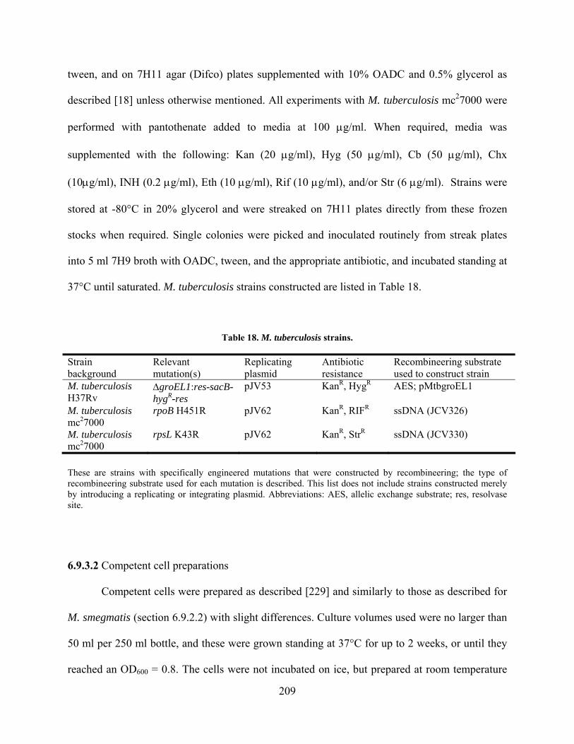
tween, and on 7H11 agar (Difco) plates supplemented with 10% OADC and 0.5% glycerol as
described [18] unless otherwise mentioned. All experiments with M. tuberculosis mc27000 were
performed with pantothenate added to media at 100 g/ml. When required, media was
supplemented with the following: Kan (20 g/ml), Hyg (50 g/ml), Cb (50 g/ml), Chx
(10g/ml), INH (0.2 g/ml), Eth (10 g/ml), Rif (10 g/ml), and/or Str (6 g/ml). Strains were
stored at -80°C in 20% glycerol and were streaked on 7H11 plates directly from these frozen
stocks when required. Single colonies were picked and inoculated routinely from streak plates
into 5 ml 7H9 broth with OADC, tween, and the appropriate antibiotic, and incubated standing at
37°C until saturated. M. tuberculosis strains constructed are listed in Table 18.
Table 18. M. tuberculosis strains.
Strain background
Relevant mutation(s)
Replicating plasmid
Antibiotic resistance
Recombineering substrate used to construct strain
M. tuberculosis H37Rv
groEL1:res-sacB-hygR-res
pJV53 KanR, HygR AES; pMtbgroEL1
M. tuberculosis mc27000
rpoB H451R pJV62 KanR, RIFR ssDNA (JCV326)
M. tuberculosis mc27000
rpsL K43R pJV62 KanR, StrR ssDNA (JCV330)
These are strains with specifically engineered mutations that were constructed by recombineering; the type of recombineering substrate used for each mutation is described. This list does not include strains constructed merely by introducing a replicating or integrating plasmid. Abbreviations: AES, allelic exchange substrate; res, resolvase site.
6.9.3.2 Competent cell preparations
Competent cells were prepared as described [229] and similarly to those as described for
M. smegmatis (section 6.9.2.2) with slight differences. Culture volumes used were no larger than
50 ml per 250 ml bottle, and these were grown standing at 37°C for up to 2 weeks, or until they
reached an OD600 = 0.8. The cells were not incubated on ice, but prepared at room temperature
209

by pelleting and washing with 10% glycerol and finally resuspending in 10% glycerol in the
same manner as for M. smegmatis. The cells were typically not frozen but used immediately for
transformation. Extra cell aliquots were frozen and stored at -80°C until use.
6.9.3.3 Transformations
M. tuberculosis cells were transformed using the conditions described for M. smegmatis
above (section 6.9.2.3 and [229]), except for the following: the cells were never incubated on ice,
and the cells were recovered – following transformation – in 7H9 broth supplemented with
OADC and tween for 1 – 3 days standing at 37°C. Transformations were plated on selective
media and incubated at 37°C for 20 – 30 days.
6.10 RECOMBINEERING PROTOCOLS
6.10.1 Strain growth and media
6.10.1.1 M. smegmatis
M. smegmatis mc2155 recombineering strains were made as described [227-229] by
transforming the pJV plasmids into wild type electrocompetent cells and plating on 7H10/Kan
media. The transformants were streaked for single isolates, inoculated into 3 ml cultures of
7H9/ADC/tween/Kan, grown shaking at 37°C until saturated, and frozen at -80°C. These were
then sub-cultured for growing competent cell batches.
To grow competent cells, recombineering strains were inoculated in 7H9 induction
medium (7H9, 0.2% succinate, Kan, and tween) to an OD600 = 0.010 – 0.025 approximately 15
210

hr prior to the desired preparation time and incubated shaking at 37°C. The media was prepared
by bringing the 90 ml of 7H9 up to 100 ml with dH2O, and adding 1 ml of 20% succinate
(succinic acid dibasic sodium salt) to a final concentration of 0.2%, and these were grown to an
OD600 = 0.4 – 0.5, acetamide added to a final concentration of 0.2% (to induce gene expression),
and grown for 3 hr shaking at 37°C. The competent cells were then prepared as described above
(section 6.9.2.2).
Electrocompetent cells of recombineering strains were transformed with the
recombineering substrate as described above (section 6.9.2.3). Cells were recovered in 7H9 with
ADC and tween for 4 hr (unless otherwise described) and plated on selective media, always with
Kan present in addition to the specific antibiotic required for each recombineering protocol.
6.10.1.2 M. tuberculosis
M. tuberculosis H37Rv and M. tuberculosis mc27000 recombineering strains were made
as described [227-229]. Plasmids for recombineering were transformed into wild type
electrocompetent cells prepared as described above (sections 6.9.2.2 and 6.9.2.3) and plated on
7H11/Kan media (plus pantothenate for mc27000). The colonies were inoculated into 5 ml
cultures of 7H9/ADC/tween/Kan (plus pantothenate for mc27000), grown standing at 37°C until
saturated, and frozen at -80°C. These cultures were then sub-cultured for growing competent cell
batches.
Recombineering strains were subcultured into 50 ml of 7H9 induction medium (7H9,
0.2% succinate, Kan, tween, and pantothenate for mc27000) to an OD600 = 0.01 – 0.025 and
incubated standing at 37°C for approximately 10 days. Once the cultures reached OD600 = 0.45 –
0.50, acetamide was added to a final concentration of 0.2%, and the cells were grown at 37C
overnight (>16 hrs). Electrocompetent cells were prepared as described above (section 6.9.3.2).
211
The cells were transformed as described above (section 6.9.3.3) with the recombineering
substrate. Transformed cells were recovered in 7H9 plus OADC and tween (plus pantothenate
for mc27000) for >16 hr (unless otherwise described) and plated on 7H11/Kan selective media
containing antibiotics specific to each protocol below.
6.10.2 Recombineering substrates: synthesis and preparation
6.10.2.1 Gene replacements
To construct substrates for making allelic replacement mutants or “gene knockouts
(KOs),” allelic exchange substrates (AESs) or “KO substrates” were constructed for each target
gene (see Figure 29). These contain homologous sequences upstream and downstream of the
gene and were cloned flanking an antibiotic resistance cassette. Primers were designed to
amplify ~500 bp regions of homology at the 5 and 3 ends of the gene, typically designed such
that ~100 bp at each end of the target gene is intact following gene replacement. Primers also
were engineered to contain specific restriction enzyme sites to facilitate directional cloning. The
PCR products were cloned into a vector flanking a hyg-resistance cassette, typically either
pYUB854 (containing res sites for unmarking), pJV69 (pYUB854 without res sites), or
pJV150 (pJV69 plus a sacB cassette). The cloning was often performed as a 4-way ligation in
which the cloning vector was digested with all four restriction enzymes, corresponding to the
sites in the PCR primers, and these two pieces were ligated simultaneously to the two digested
PCR products to yield one final plasmid (confirmed by analytical restriction digest).
The vector (containing the homologous sequences) was linearized by restriction digest
with two enzymes, preferably the two enzymes used to clone at the most distal regions of the
212
targeting substrate. This yielded two fragments; one containing the hygR cassette flanked by the
two homologous regions, and the other fragment containing the oriE backbone. Alternatively,
the section with the homologous regions was amplified by PCR (such as for the pMsgroEL1KO
substrate). The digest reaction or PCR reaction was cleaned up to remove enzyme using the
QIAquick gel extraction protocol for enzymatic cleanup (QIAGEN), and DNA was eluted in
dH2O (in order to minimize salt for transformations). The linear DNA containing the homology
was quantified by agarose gel electrophoresis or UV spectrometry.
6.10.2.2 Point mutations
Substrates for making point mutations are ssDNA oligonucleotides. The shortest
recommended length is 48 nucleotides (Figure 25; [228]), although longer substrates (70 nt – 100
nt) were used for some experiments. The mutation(s) to be introduced was centered in the
oligonucleotide. From experimental evidence it was determined that oligonucleotides that are
complementary to the lagging strand for DNA synthesis work better than those that anneal to the
leading strand. The oligonucleotides were synthesized as described above (DNA substrates) and
resuspended in TE buffer upon receipt of the lyophilized DNA pellet, typically to 1 M. The
sequences of oligonucleotides used in this study are summarized in Table 16.
6.10.2.3 Unmarked deletions
Recommended substrates for deletions are 200 bp, with 100 bp of homology on each side
of the deletion locus based on a method previously described [172]. First, a 100 nt
oligonucleotide was designed with 50 nt of homology on each end. Primers, called “extenders,”
were designed that contained 50 nucleotides at the 5 end that had homology to the target gene
213
followed by 25 nt that annealed to the template (100 nt oligonucleotide). The final PCR product
was 200 bp and contained 100 bp of homology upstream and downstream of the target
gene/region (Figure 28).
6.10.3 Construction of mutants
6.10.3.1 Gene replacements
Electrocompetent cells of strains containing plasmid pJV53 (or a similar plasmid
containing Che9c 60-61) were transformed with 100 ng targeting substrate DNA as described
above (6.9.2.3 and 6.9.3.3). The transformations were recovered by incubating at 37C in 7H9
broth containing ADC and tween, and OADC if for M. tuberculosis. For M. smegmatis, the cells
were recovered for 4 hrs, and for M. tuberculosis, the cells were recovered 1-3 days.
Following recovery, the entire reaction (~1 ml) was plated on 7H10 or 7H11 agar plates
(containing Kan and Hyg, and oleic acid for M. tuberculosis), and incubated at 37C until
colonies were of sufficient size for sub-culturing (~5 days for M. smegmatis, 3-4 weeks for M.
tuberculosis). Typically, between 50-200 recombinant colonies were recovered. All batches of
competent cells were tested for cell competency by transforming (in a separate aliquot of cells)
50 ng of a HygR integration-proficient vector (either pJV39 or pSJ25Hyg), plating on 7H10/Hyg
plates, and determining the number of cfu per g of plasmid DNA. The number of viable cells in
each transformation reaction was determined by plating serial dilutions of the cell competency
control reaction on 7H10/Kan media.
214

6.10.3.2 Point mutations
Selectable point mutations in either the mycobacterial chromosome or on
extrachromosomal plasmids were generated by transforming the ssDNA substrate (containing the
point mutation) into electrocompetent recombineering cells. The strain background was typically
pJV62 or a derivative, since only the recombinase (gp61) is required for recombination with
ssDNA. 100 ng of ssDNA substrate was transformed as described above (6.9.2.3 and 6.9.3.3),
and the cells were recovered in 7H9 broth with ADC, tween, and OADC for M. tuberculosis, at
37°C. For M. smegmatis, cultures were recovered shaking for 4 hrs, and for M. tuberculosis they
were recovered for 3 days standing. The recovered cells were diluted, plated on selective media,
and incubated for 4-5 days (M. smegmatis) or 3 weeks (M. tuberculosis) at 37°C. Cell
competency and viability counts were determined as described above for gene replacements.
For non-selectable mutations, transformations with ssDNAs were performed as above
using excess ssDNA compared with a HygR, integration-proficient plasmid (pJV39 or
pSJ25Hyg) or a ssDNA that could also be recombined to confer HygR (JCV198 ssDNA in
backgrounds with hygS). Optimal results were obtained with 500 ng of the mutating ssDNA and
100 ng of the HygR selectable element, respectively. Following recovery (4 hr for M. smegmatis,
3 days for M. tuberculosis), the cells were diluted in 7H9 broth (plus ADC/OADC and tween)
containing Hyg and Kan to approximately 10–100 HygR cells per well (1 ml media) in a sterile
96-well culture block. These dilutions were simultaneously plated on 7H10/Hyg/Kan agar plates
to determine HygR cell counts. The cultures were incubated (shaking at 250 rpm for M.
smegmatis; standing for M. tuberculosis) at 37°C to an OD600 = 1.0 and screened by colony PCR
(MAMA-PCR). Each culture well containing a mutant allele was plated for single colonies and
re-screened by colony PCR to identify the isolated mutant.
215
6.10.3.3 Unmarked deletions
To make unmarked deletions, 100 – 200 ng of the dsDNA substrate was co-transformed
with 50 – 100 ng of a HygR-selectable substrate, either a HygR plasmid or another ssDNA that
could confer HygR (JCV198 in a hygS background strain). The cells were recovered as described
for non-selectable point mutations; the transformation was either plated on Hyg media or were
serially diluted in 7H9 broth with Hyg. Transformants or liquid culture dilutions were screened
by PCR or identifying phenotype (e.g. leucine auxotrophy for leuD mutants) for the presence of
the desired mutation. Each culture containing the mutation was plated for single colonies and re-
screened by PCR to identify the mutant strain.
6.10.4 Analysis of recombinant colonies
6.10.4.1 Gene replacements
Colonies recovered from transformations with the targeting substrate were analyzed by
either colony PCR or Southern blot to confirm the genotype of the strain. For colony PCR, the
colonies were either allowed to grow to a large size or were patched onto a fresh plate to get
enough cells for the PCR. Primers were designed within the homologous regions of the targeting
substrate to determine if the gene locus contained the HygR resistance cassette or if it was wild
type, which would result in differently sized PCR products. Colony PCR was performed as
described above (6.3.1).
For Southern blot analysis, colonies were inoculated into ~10 ml 7H9 broth containing
ADC, tween, Kan, and Hyg (and OADC for M. tuberculosis). The cultures were incubated at
37C for 3 days (M. smegmatis) or 10 days (M. tuberculosis) until the culture had a substantial
amount of visible growth. The cells were collected and genomic DNA was prepared as described
216
above (6.8.1). Southern blot analysis was performed as described above (6.8) on each
recombinant strain to be tested, using the pJV53 strain as a control. Probes were synthesized
using primers either to the upstream or downstream homologous region in order to determine if
the gene locus contained the HygR resistance cassette or if it was wild type. Alternatively, a
probe to the HygR cassette was also used.
6.10.4.2 Point mutations
Point mutations that were selectable (such as the rpsL K43R mutation) were confirmed
by sequencing. PCR was performed with primers that amplified the gene locus, and this DNA
was cleaned up using QIAquick PCR purification (QIAGEN) and sequenced as described above
using the same primers.
For large-scale identification of non-selectable point mutations, culture wells or single
colonies were screened by MAMA-PCR with primers designed to distinguish between wild type
and mutant alleles as described above (6.3.2) [30,219]. Primers were synthesized for both the
mutant allele and the wild type allele as a control. Positive control templates for mutant alleles
were synthesized by PCR-amplification by one of four methods: 1) using the recombineering
substrate that was used to make the point mutation as a forward primer, 2) synthesizing a new
primer that contained the mutation as a forward primer, 3) using the mutant MAMA-PCR
screening primer to amplify from a wild type template but with the Pfu polymerase that could
read through the non-matching 3 end of the wild type template, or 4) PCR amplifying a known
mutant with sequencing primers.
217

6.10.4.3 Unmarked deletions
Deletion mutants were identified by PCR from a colony or culture using primers that
flanked the deleted region, which were designed to amplify either wild type or mutant loci. The
deletion mutants would therefore yield a smaller-sized PCR product. Colony PCR was
performed as described above (6.3.1), and cultures that contained mutant alleles were plated for
single colonies and re-screened to identify the isolated mutant strain.
6.10.5 Strain unmarking
Gene replacement strains that were constructed by recombineering often contained -res sites
for removing the interrupting HygR cassette (if the targeting substrate was constructed in a
pYUB854 vector backbone). Alternatively, dsDNA recombineering was used to remove the
HygR cassette, but only in conjunction with a sacB cassette for negative selection (if the targeting
substrate was constructed in a pJV150 vector backbone). Ultimately, the pJV53 plasmid (or
similar recombineering plasmid) was occasionally removed from the strain by serial dilution, or
by using sacB as a negative selection in strains that contain a pJV recombineering plasmid with a
sacB cassette (e.g. pJV48, pJV126).
6.10.5.1 Removing HygR by -resolvase
Unmarking of M. smegmatis recombinant replacement strains using resolvase was
accomplished by transforming the strain with pGH542, a TetR plasmid that constitutively
expresses the resolvase. Electrocompetent cells of the recombinant strain were prepared,
transformed with 50 ng of plasmid pGH542, and plated on 7H10 agar plates containing Tet. The
plates are incubated at 37C for 3-4 days until colonies are of sufficient size for sub-culturing.
218
Julie
Typewritten Text
Julie
Typewritten Text
Julie
Typewritten Text

The recovered colonies were patched onto multiple selective 7H10 agar plates containing
antibiotics in the order as follows: 1) Hyg, 2) Tet, 3) Cb/Chx, and were incubated at 37C for 3-4
days. Colonies that are HygS and TetR are therefore “unmarked” and the removal of the HygR
cassette can be verified by colony PCR at that locus.
The pGH542 plasmid was removed from this strain at the same time as the
recombineering plasmid following the protocol described below (6.10.5.2). However,
occasionally it was desired to retain the recombineering plasmid but remove the pGH542
plasmid, and in these cases, the recombineering plasmid was selected during the serial dilutions,
whereas the pGH542 was not (media containing Kan and not Tet). Ultimately, strains that were
determined (by patching on multiple selective plates) to be TetS, KanS, and HygS were retained
and stored.
6.10.5.2 Removing the recombineering plasmid
In cases in which it was desired to remove the recombineering plasmid from a
recombinant M. smegmatis strain, this was subcultured into a 10 ml culture of 7H9 media
(containing only ADC, tween, and Cb and Chx) and incubated with shaking at 37C until it
reached saturation (~2 days). This culture was subcultured into another 10 ml culture exactly as
above, using 1 l of the culture (1:10,000) and incubated with shaking at 37C until saturation
(~2 days). Dilutions (10-4 – 10-7) of this culture were plated on 7H10 agar plates (containing Cb
and Chx only). The recovered colonies were patched onto multiple selective 7H10 agar plates
containing antibiotics in the order as follows: 1) Hyg, 2) Tet, 3) Kan, 4) Cb/Chx only. The plates
were incubated at 37C for 3-4 days. KanS colonies were saved and stored.
219
6.11 MYCOBACTERIOPHAGE MANIPULATIONS
6.11.1 Mycobacteriophage lysate preparation
Mycobacteriophages TM4 and Che9c were propagated on M. smegmatis mc2155 as described
[198]. M. smegmatis cultures were grown in ADC (no tween) until saturated in baffled flasks.
For typical small-plate lawns, 1.5 ml MBTA was mixed with 1.5 ml 7H9/ADC/CaCl2 and 300 l
M. smegmatis cultures, and this was solidified and used for spot-tests with serially-diluted phage.
For plate infections, serially-diluted phage were added to the 300 l of cells, incubated standing
at 37°C for 20 mins and subsequently plated with MBTA and 7H9 as above. Lysates were
prepared by flooding plates (5-8 ml) with phage buffer plus CaCl2 at room temperature for 2 hrs,
or overnight at 4°C. These were collected, the debris removed by centrifugation at 3500 x g, and
the supernatant was filtered (0.22 M filters) and stored at 4°C. For large-plates, 5 ml MBTA, 5
ml 7H9/ADC/CaCl2 and 1 ml M. smegmatis cells were used.
6.11.1.1 Large-scale preparation of mycobacteriophage CsCl stock
TM4 was prepared in which 30 large plates were made of TM4 infections that yielded
“webbed” lawns (approximately 6000 pfu total per large plate). Lysates were prepared, and these
were treated either by the conventional PEG precipitation protocol and continuous CsCl gradient
purification [198], or using the following protocol.
220
For the modified TM4 “large prep protocol,” the lysates collected from 30 large plates
were centrifuged in four Ti75 rotor tubes (~60 ml per tube) at 20,000 x g in the ultracentrifuge
for 1.5 hr (phage buffer was used to balance tubes). The supernatant was removed, and the phage
pellet was resuspended in 2 ml phage buffer (plus 1 mM CaCl2) overnight standing at 4°C. The
phages were collected by gently swirling the pellet, and this was titered. At this stage, either a
continuous CsCl or step CsCl gradient was used to purify the phage from any cell debris. The
step gradient yielded better results, and this was performed by layering the following solutions in
this order: 1) 8 ml phage into one tube, 8 ml phage buffer in another tube (as a balance), 2)
Pasteur pipeting 1 ml 10% glycerol under phage/phage buffer, 3) 1.5 ml of 1.4 mg/ml CsCl
under glycerol, and 4) 1.5 ml of 1.6 mg/ml CsCl under that. These tubes were centrifuged at
30,000 x g in a swinging bucket SW41 rotor for 1.5 hrs. The phage band was extracted with a
syringe, and the phage were dialyzed twice against 500 ml phage buffer with 1 mM CaCl2 at 4°C
for 4 hr each time.
6.11.1.2 Genomic DNA isolation from mycobacteriophage stock
Genomic DNA was isolated as described [198]. Briefly, ~500 l of dialyzed phage were
extracted with buffer-equilibrated phenol repeatedly (with TE back-extraction) until protein was
removed. This was followed by an extraction with phenol:chloroform:isoamyl alcohol (25:24:1)
and another extraction with chloroform. The DNA was ethanol precipitated, washed, and
resuspended in TE.
6.11.1.3 Small-scale genomic DNA isolation from lysates
To make small amounts of phage DNA, 2-3 small plate lysates were prepared, and the
phage was pelleted by incubating on ice with equal volumes of saturated ammonium sulfate for
221
1-3 hr [198]. The phage were pelleted at 3500 x g and resuspended in phage lysis buffer. This
was treated with proteinase K at 10 g/ml for 2 hr at 37°C and subsequently phenol extracted as
described above. It is important to note that the aqueous layer in the first extraction is underneath
the organic layer due to the presence of the lysis buffer. To fix this, an equal volume of TE was
added to the phage lysis buffer following treatment with proteinase K; this resulted in a shift of
the aqueous layer to the top for all extractions.
6.11.2 TM4 Cosmid library construction
Approximately 4 g TM4 genomic DNA was ligated to itself to form concatemers using T4
DNA ligase overnight at room temperature; this facilitated connection of the otherwise linear
ends. This was partially digested with a frequently-cutting enzyme to yield approximately 40-45
kbp fragments; this was accomplished best with a 15 min digest with Sau3AI at 37°C. This was
immediately placed on ice, phenol extracted, ethanol precipitated, and resuspended in 10 l TE.
The DNA was then ligated to digested pYUB854 DNA cut with Bgl II (ends compatible with
Sau3AI) using the FastLink ligase (Epicentre) for 15 mins at room temperature, and packaged
into heads using Gigapack III Gold Packaging Extract (Stratagene) according to
manufacturer’s instructions. HB101 cells were grown and infected with the -packaged
molecules, and colonies were selected on Hyg (resistance conferred by pYUB854). Colonies
were miniprepped, and these were analyzed by restriction digest and sequencing to identify the
segment of TM4 that was cloned into the cosmid. Alternatively, pools of E. coli HB101 colonies
were prepared together by scraping colonies into 25 ml LB broth plus Hyg, growing to
saturation, and midi-prepping the DNA (QIAGEN).
222

6.11.3 TM4 cosmid recombination assays
TM4 cosmids that were defined by analytical restriction digest and sequencing were used to
assay for recombination in vivo in M. smegmatis. Individual cosmids or pooled cosmid preps
were transformed into electrocompetent M. smegmatis as described above (6.9.2.2 and 6.9.2.3),
except tween was not used in the cultures. Concentrated stocks of cosmid DNAs were critical for
good transformation frequencies; 1 g total DNA was transformed (500 ng each cosmid if in
pairs), and 50-200 ng TM4 DNA was used as a control. Transformations were recovered in 1 ml
LB broth for 30 min and plated in 300 l M. smegmatis cells with 0.5 ml 7H9 and 1.5 ml MBTA
on 7H10 agar plates. These were incubated at 37°C overnight, and plaque numbers recorded for
single cosmid transformations versus pair-wise combinations. PCR assays to detect TM4 phage
DNA (band = 880 bp) and pYUB854 DNA (band = 584 bp) simultaneously contained primers to
both (TM41444-1464 880F, TM42323-2303 880R; pYUB854 509-533F, pYUB854 1069-
1093R) and were conducted as described above (6.3).
223
APPENDIX A
THE ROLE OF HOST NUCLEASES IN MYCOBACTERIAL RECOMBINEERING
A.1 INTRODUCTION
A.1.1 The E. coli RecBCD complex and λ Gam
The RecBCD complex in E. coli functions for recombinational repair of double-strand DNA
breaks and broken replication forks (reviewed by A. Kuzminov [106] and by Amundsen and
Smith [5]). It is a highly processive multienzyme complex that possesses strong helicase activity
and ATP-dependent dsDNA and ssDNA exonuclease activities. During repair, the enzyme
degrades dsDNA in a 5 to 3 direction, and following recognition of a Chi site (by RecD),
RecBCD stimulates RecA polymerization on the 3 ssDNA tail (along with SSB), which
promotes strand invasion at homologous targets for recombination. However, the RecBCD
complex also degrades foreign dsDNA molecules – such as the ends of the λ genome – after
digestion by restriction enzymes upon entry into the cell. More than 40 years of research have
been dedicated to examining RecBCD, providing a detailed understanding of the genetic and
224

biochemical properties of this very complex enzyme [106]. However, this appendix will focus on
a small portion of this work and discuss only the most pertinent aspects of its biology.
Mutant E. coli strains of recB, recC, and recD display different phenotypes in response to
DNA damage induced by UV, as well as in regard to their recombination activity [106]. Null
recB or recC mutations render cells sensitive to UV and have decreased overall viability (~30%),
whereas recD mutants survive like wild type after UV treatment [133]. The UV phenotype of
recBCD mutant strains can be suppressed by expression of λ Exo/Beta, and the viability of this
strain is even increased ~10-fold compared to wild type [135]. Further, the nuclease activities of
RecBCD are eliminated in a either a recB or recD mutant strain [5]. However, in a recD mutant
strain, some of the helicase activity is retained, although it is decreased because RecD provides
the faster helicase subunit [5]. These recD strains are described as ‘hyper-recombination’
mutants [106] because they constitutively load RecA for homologous recombination. This
phenotype essentially mimics the activity of RecBCD following recognition of Chi, which is
attributed to the removal of the RecD subunit after Chi; the presence of RecD can actually inhibit
RecA polymerization [5]. Interestingly, there is a specific recB mutation (recB1080) that
abolishes nuclease activity but can still unwind DNA; however, this is incapable of
recombination due to the presence of RecD [5].
The λ Gam protein inhibits all known activities of RecBCD by binding to the RecB
subunit and preventing the complex from binding to dsDNA ends [39,93,122,133,138,176].
However, Gam expression does not cause all the examined phenotypes of a recBCD strain.
This is due to the presence of a portion of RecBCD that is not bound by Gam and is therefore
able to degrade dsDNA [138]. There is also some evidence that Gam interacts with SbcC, though
this is not well studied [102]. Because of these activities, dsDNA is protected from nuclease
225
attack in the presence of Gam, which makes λ Red recombination more efficient
[38,142,240,241]. Therefore, although Gam is not essential for phage λ propagation [53] or
recombineering [43,241], expression of Gam in recombineering assays with dsDNA increases
frequencies ~10-fold [43]. Gam expression in wild type cells yields a UV-sensitive phenotype
that mimics a recBC strain (~10-fold decrease in viability compared to wild type) [135].
However, Gam does not increase the sensitivity of either recBC or recD strains, which suggests
that the UV phenotype is a result of Gam interaction only with RecBCD and not any other
complex [135].
Other phages encode proteins that function analogously to λ Gam, such that they protect
linear dsDNA ends from degradation, although the means by which they accomplish this differs.
For example, both phage Mu (Gam) [2] and phage T4 (gp2) [6,115,208] have proteins that bind
dsDNA ends, whereas λ Gam specifically inactivates RecBCD. Conversely, the Abc proteins of
P22 actually modify the activity of RecBCD in order to exploit its 5-3 ssDNA exonuclease
activity for P22 Erf-mediated recombination [136,176]. A decrease in host nuclease activity has
been observed following infection with several additional dsDNA phages [195], which is most
likely being facilitated by similar types of proteins.
A.1.2 Mycobacteria encode both RecBCD and AddA homologues.
Recent bioinformatic analysis of several mycobacterial genomes has revealed a number of genes
that encode homologues of the B. subtilis AddA protein (Figure 34, D. Ennis and G. Cromie,
personal communication). B. subtilis AddA is a part of the AddAB enzyme complex. These
proteins are functionally analogous to E. coli RecBCD in vivo [98], although the complexes have
slightly different biochemical properties. These genes were originally believed to be restricted to
226
Gram-positive bacteria [32], but recently they have also been identified in the Gram-negative
bacteria, Rhizobium etli [245] and Coxiella burnetii (D. Ennis, personal communication). The
AddA subunit has homology to RecB and contains a similar nuclease domain, as well as helicase
and ATPase domains. The AddB subunit does not have homology to RecC or RecD, but it does
have ATPase and nuclease domains that are slightly divergent in sequence compared to AddA
[32].
It is apparent that the list of genomes that encode these proteins is not likely complete,
and also that some bacteria may encode both types of enzymes. For example, M. tuberculosis
was listed in the review by Chedin et al. as having only a ‘RecBCD-type enzyme’ [32], but
bioinformatic data suggests that AddA homologues are also present. Further, there is an E. coli
RecD homologue present in both B. subtilis and Lactococcus lactis, as well as in other bacteria
that contain neither RecBC nor AddAB. Collectively, these findings suggest that there is still
much to learn about the roles of these proteins in bacteria that encode homologues of both types
of enzymes.
The putative mycobacterial AddA proteins are highly conserved between species (>60%
amino acid identity) and are similar to the B. subtilis AddA protein at specific regions, including
the ATPase domains (Walker A and B motifs) and RecB nuclease domains (Figure 34). For
example, M. smegmatis MSMEG_1943 has 24% and 27% identity to B. subtilis AddA at its N-
and C-terminus, respectively. However, BLAST analysis with B. subtilis AddB does not identify
any obvious homologues in the mycobacteria. Instead, these analyses identify multiple genes in
each mycobacterial genome with similarity to AddA, and some of these are found in pairs (e.g.
MSMEG_1943 and MSMEG_1941). Typically, the gene with the highest degree of identity was
aligned in Figure 34A, and the adjacent gene was aligned in Figure 34B.
227
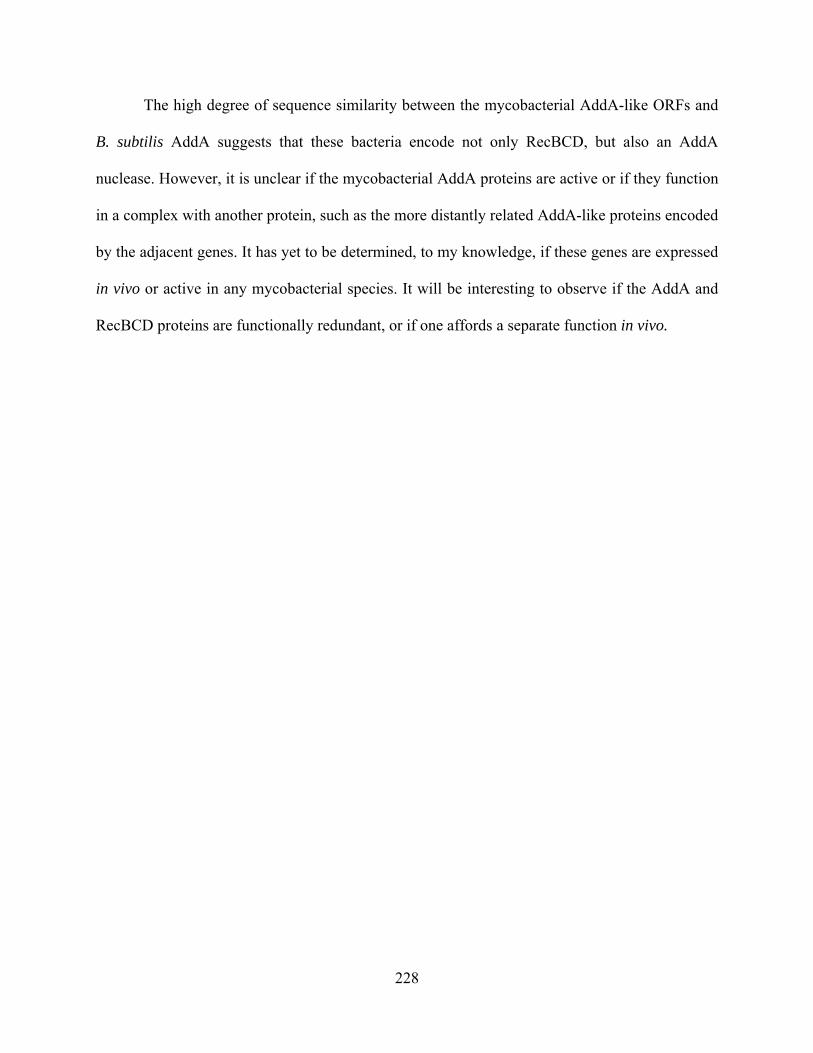
The high degree of sequence similarity between the mycobacterial AddA-like ORFs and
B. subtilis AddA suggests that these bacteria encode not only RecBCD, but also an AddA
nuclease. However, it is unclear if the mycobacterial AddA proteins are active or if they function
in a complex with another protein, such as the more distantly related AddA-like proteins encoded
by the adjacent genes. It has yet to be determined, to my knowledge, if these genes are expressed
in vivo or active in any mycobacterial species. It will be interesting to observe if the AddA and
RecBCD proteins are functionally redundant, or if one affords a separate function in vivo.
228

229
Figure 34. Multiple sequence alignments of putative mycobacterial and B. subtilis AddA proteins.
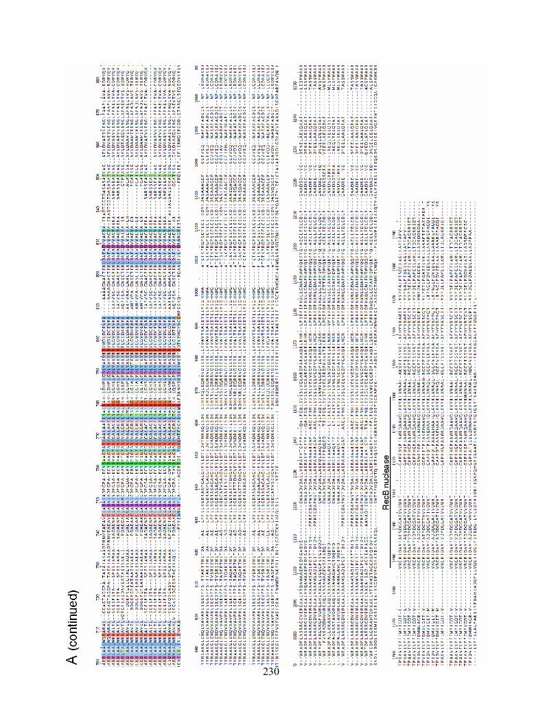
230
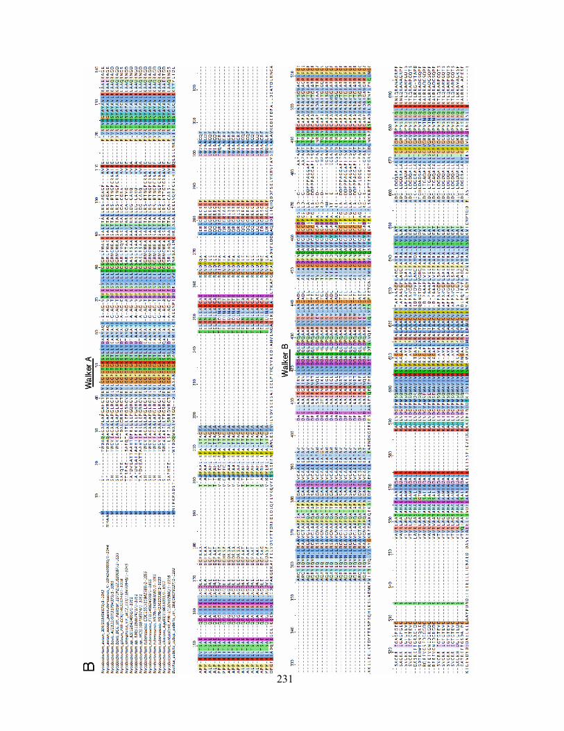
231
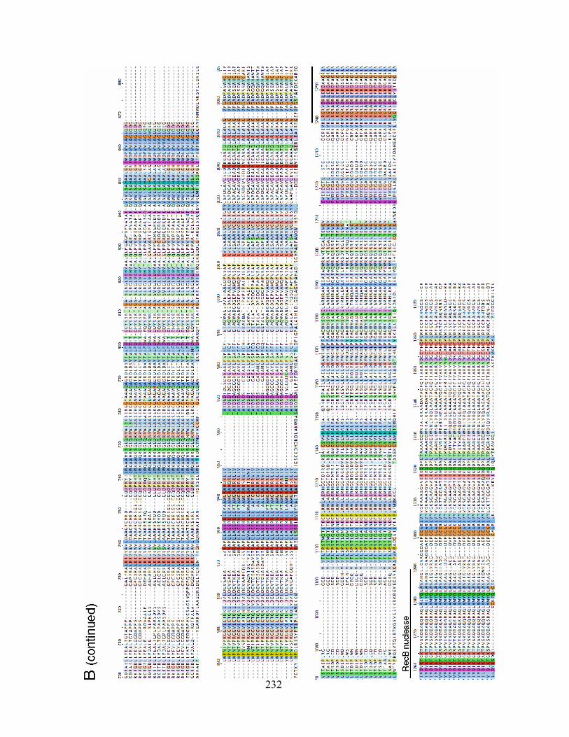
232
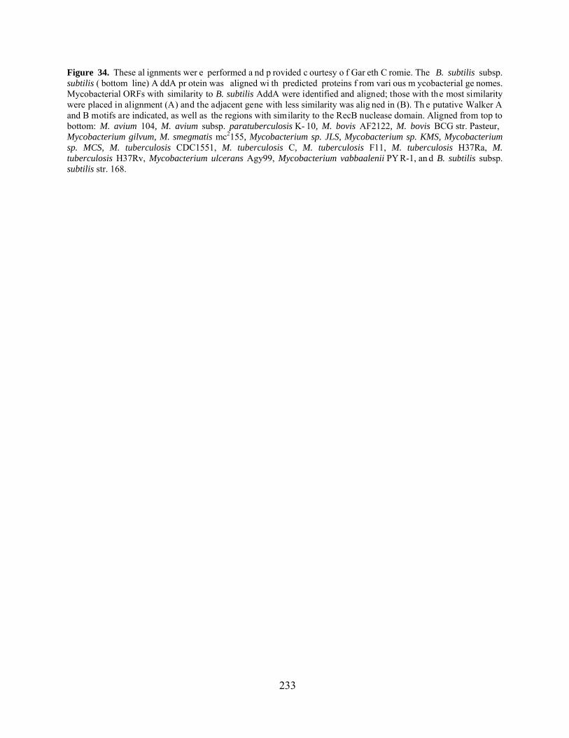
233
Figure 34. These al ignments wer e performed a nd p rovided c ourtesy o f Gar eth C romie. The B. subtilis subsp. subtilis ( bottom line) A ddA pr otein was aligned wi th predicted proteins f rom vari ous m ycobacterial ge nomes. Mycobacterial ORFs with similarity to B. subtilis AddA were identified and aligned; those with the most similarity were placed in alignment (A) and the adjacent gene with less similarity was alig ned in (B). Th e putative Walker A and B motifs are indicated, as well as the regions with similarity to the RecB nuclease domain. Aligned from top to bottom: M. avium 104, M. avium subsp. paratuberculosis K- 10, M. bovis AF2122, M. bovis BCG str. Pasteur, Mycobacterium gilvum, M. smegmatis mc2155, Mycobacterium sp. JLS, Mycobacterium sp. KMS, Mycobacterium sp. MCS, M. tuberculosis CDC1551, M. tuberculosis C, M. tuberculosis F11, M. tuberculosis H37Ra, M. tuberculosis H37Rv, Mycobacterium ulcerans Agy99, Mycobacterium vabbaalenii PY R-1, an d B. subtilis subsp. subtilis str. 168.

A.1.3 Recombineering in E. coli: the effect of host RecBCD
The degree to which λ Gam is required to inactivate host nuclease activity for λ Red
recombineering with dsDNA substrates differs in varying reports. In studies by K. Murphy and
Yu et al., the data indicated that Gam expression (or inactivation of recBCD by mutation) was
absolutely required for recovery of gene replacement mutants [135,240]. This was observed
using long (~1 kbp) dsDNA substrates, with either 50 bp or 1 kbp homology lengths. However,
later studies using similar length or shorter substrates showed only an ~10-fold decrease in
efficiency in the absence of Gam [43,241]. There does not appear to be a difference in
recombineering frequencies between a recBCD strain or Gam-expressing strain [135], but there
are clearly advantages to using Gam to facilitate recombineering in any background. Finally,
there is only a modest decrease (~5-fold) in ssDNA recombineering frequencies in the absence of
Gam [52].
This appendix will discuss the results of the preliminary experiments that have been
performed in M. smegmatis recB and recD strains, as well as assays in which the expression
of the λ gam gene is controlled by the acetamidase promoter in M. smegmatis. These strains were
assayed for both UV sensitivity and dsDNA recombineering activity. While these experiments
have provided some data regarding the activity of M. smegmatis RecBCD, additional
experiments will be required to fully understand the role of the mycobacterial nucleases.
234
A.2 RECOMBINEERING ACTIVITY IN REC- M. SMEGMATIS STRAINS
A.2.1 Recombineering in recB and recD strains
The mycobacteria encode both RecBCD and AddA proteins, as discussed above. The RecBCD
genes are grouped together, likely in one operon, in the chromosome of M. smegmatis (Figure
35A), the organism in which all these experiments were performed. Conceivably, one approach
to increase recombineering frequencies is to inhibit the potentially negative effects of host
nucleases. With regard to RecBCD, a recD mutant would be ideal because, at least in E. coli, it
has wild type viability, does not lose viability after treatment with DNA damaging agents, and
retains the helicase and recombination-stimulating activities of the complex [5,106]. In order to
study the role of RecBCD more thoroughly, M. smegmatis mutant strains were constructed that
contained gene replacements of either the recB and recD genes, and were subsequently
unmarked by resolvase (Figure 35B,C). These strains were then tested for dsDNA
recombineering by targeting the M. smegmatis groEL1 gene (section 3.3.3), and the data were
compared to recombineering frequencies obtained in a wild type genetic background.
The recB strain demonstrated a slight increase in recombineering activity, typically
between 3- and 5-fold compared to wild type (Figure 35D, Table 19). Since reports in E. coli
vary regarding the effect of RecBCD, it is difficult to directly compare these data. However, it
was surprising that the increase was only 5-fold, particularly because the smallest effect observed
in E. coli is a 10-fold increase in efficiency [43]. The recD strain was also slightly increased for
recombineering activity, although to a lesser extent than recB (~2-fold increase). These results
235

indicate that while recB and recD may have a small effect on recombineering efficiency, their
activities do not drastically inhibit recombination with dsDNA substrates.
236

Figure 35. Recombineering frequencies in recB and recD M. smegmatis strains.
Figure 35. (A) Schematic of the M. smegmatis chromosome at the region encoding the recBCD genes. (B) Southern blot analysis of DNA prepared from four individual recB gene replacement mutants made by recombineering; wild type DNA is used as a control (4,077 bp and 5,175 bp, respectively). Also shown, colony PCR analysis of 10 recB mutants that were unmarked with resolvase; the hygR marked mutant strain is used as a control (1,396 bp and 3,130 bp, respectively). (C) Colony PCR analysis of both unmarked and marked recD gene replacement mutants made by recombineering, using wild type DNA as a control (401 bp, 2,138 bp, and 1,787 bp, respectively). (D) Recombineering frequencies from experiments targeting the M. smegmatis groEL1 gene in wild type, recB, and recD M. smegmatis mc2155 strains containing plasmid pJV53. Frequencies are represented on a log scale and multiplied by 105 for presentation purposes. The data shown are the averages of three experiments for wild type and recB, and two experiments for recD; error bars represent standard deviation.
237

A.2.2 Expression of λ gam in M. smegmatis
In order to determine the activity of λ Gam in mycobacteria, this gene was cloned either singly
under Pacetamidase (pJV99) or Phsp60 (pJV97) or together with the Che9c genes 60 and 61 under
Pacetamidase (pJV98). In the latter case, the gam gene plus 19 bp of sequence upstream of the start
codon were cloned downstream (158 bp) of Che9c 61 in plasmid pJV53. Competent cells of an
M. smegmatis strain containing the pJV98 plasmid (Che9c 60 and 61, λ gam) were prepared
similarly to Che9c recombineering strains and tested for dsDNA recombineering activity using
groEL1 as a target gene (sections 6.10.1.1 and 3.3.3). The results did not show a difference in
recombineering frequency as compared to the typical pJV53 strain that expresses only Che9c
genes (Table 19). This may be due to expression problems, protein instability, or potentially the
Gam protein is inactive in M. smegmatis.
Table 19. Recombineering frequencies in recB, recD, and Gam-expressing M. smegmatis strains
Strain (proteins encoded)a Recombinant coloniesb
Cell competencyc
Recombineering frequencyc Ratioe
mc2155:pJV53 (Che9c gp60/61) 226 6.0 x 10⁶ 3.8 x 10⁻⁴ N/A
mc2155:pJV53 recB (Che9c gp60/61) 254 1.7 x 10⁶ 1.5 x 10⁻³ 3.9
mc2155: pJV53 recD (Che9c gp60/61) 30 5.3 x 10⁵ 5.6 x 10⁻⁴ 1.5
mc2155:pJV98 (Che9c gp60/61, λ Gam)
123 3.7 x 10⁶ 3.3 x 10⁻⁴ 0.9
a. The M. smegmatis recA strain was constructed by allelic gene replacement with recombineering and unmarked using resolvase, as described in the Materials and Methods. b. Electrocompetent cells of the strains were transformed with 100 ng of the groEL1 AES (see Figure 18), and HygR colonies were recovered; the data represent the average of two experiments. c. Cell competency is determined as the cfu/g plasmid pJV39, an integration-proficient vector providing hygromycin resistance, when 50 ng was transformed. d. Recombineering frequency is calculated as the number of recombinant cfu per g DNA divided by the cell competency. e. The ratio is calculated by dividing the recombineering frequency of the test strain (recB, recD, or pJV98) by the recombineering frequency of the wild type mc2155:pJV53 strain.
238
A.3 EXAMINATION OF THE UV PHENOTYPE OF M. SMEGMATIS STRAINS
The experiments performed with the various M. smegmatis strains (recB, recD, or Gam-
expressing) suggested, somewhat inconclusively, that RecBCD has a minor effect (if at all) on
recombineering frequencies. To further assess the activity of RecBCD, and specifically to
determine the effect of expressing Gam, these strains were subjected to UV treatment to assay
for sensitivity to DNA damage. As expected, the recA strain consistently showed a decrease in
viability that ranged from 100- to 1,000-fold (depending on the assay; Figure 36 and data not
shown). However, the recA percent survival is higher (100-fold) in this experiment and others
compared to previous studies with this strain (~0.1% after treatment with 39 J/m2; [155]).
Surprisingly, even with a high level of UV treatment (300 J/m2), the recB strain showed
viability similar to wild type (Figure 36). Two different recB strains have been constructed
(mc2155:pJV53 recB, Figure 36, and mc2155 recB, a gift from K. Derbyshire), and both
demonstrate wild type viability following UV treatment (data not shown). Further, the recD
strain also showed wild type viability, but this is similar to what is observed in E. coli for a recD
null mutant [133].
With regard to λ Gam, the mc2155:pJV99 strain (Pacetamidase:gam) appeared to have a
slight viability decrease (3.5-fold) compared to the control (mc2155:pJV96) in this particular
experiment. However, repetition of this assay did not show the same defect. Further, a similar
strain in which gam is expressed from the constitutive hsp60 promoter did not show a decrease in
viability. These assays suggest that although recB strains are increased in recombination, they
are not UV-sensitive. In addition, the strains expressing λ Gam do not appear to have either a UV
or recombination phenotype.
239
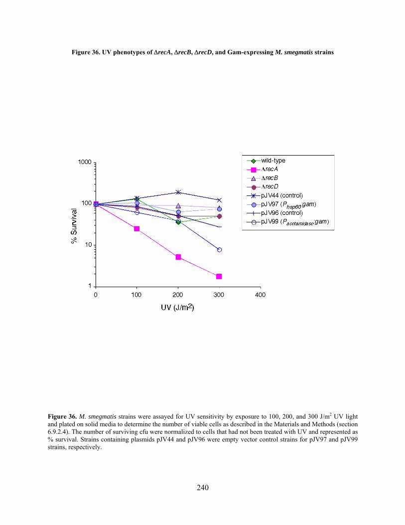
Figure 36. UV phenotypes of recA, recB, recD, and Gam-expressing M. smegmatis strains
Figure 36. M. smegmatis strains were assayed for UV sensitivity by exposure to 100, 200, and 300 J/m2 UV light and plated on solid media to determine the number of viable cells as described in the Materials and Methods (section 6.9.2.4). The number of surviving cfu were normalized to cells that had not been treated with UV and represented as % survival. Strains containing plasmids pJV44 and pJV96 were empty vector control strains for pJV97 and pJV99 strains, respectively.
240
A.4 PRELIMINARY CONCLUSIONS
These experiments sought to assay for activity of the M. smegmatis RecBCD enzyme by two
methods: (1) observing the UV phenotype of recB, recD, and Gam-expressing strains, and (2)
by determining the recombineering frequencies in these strains. The UV sensitivity assay
described above has been used previously in M. smegmatis with wild type and recA strains,
although the recA strain survived ~100-fold better in this experiments than reported previously
[155]. It is likely that this is due to variations in experimental procedures, since all repetitions of
this assay consistently showed the same level of killing for both wild type and recA strains.
Additional assays could be tested, such as mitomycin C sensitivity, which may have more
reproducible results. Further, a standard gene replacement of M. smegmatis groEL1 was tested in
each strain to determine the frequency of the recombination event compared to wild type.
The assays performed with λ Gam did not indicate that it had any effect on
recombineering frequency or viability following UV treatment. It was not determined if the
protein was expressed properly following induction, and it is also possible that the protein
product was unstable or could not interact with the RecBCD complex. If protein expression
and/or activity is an issue, alternative constructs could be tested. Another, possibly better,
approach is to test additional proteins that inhibit nuclease activity that do not require protein-
protein interactions, such as T4 gp2 or Mu Gam.
In E. coli, a recB strain is decreased 30% in viability, ~100-fold in recombination
activity, and 10-fold in survival in assays following treatment with DNA damaging agents
[106,133]. These characteristics were not observed in the two M. smegmatis recB strains; both
behaved similarly to wild type with regard to overall viability and survival following UV
241

treatment. There are two likely, possibly independent, explanations for this. First, the two recB
strains – which were made separately – could have acquired suppressor mutations that restored
UV-resistance, but retained the positive effect on recombineering frequency conferred by recB.
One category of suppressor mutations of recBC in E. coli occurs in the sbcB and sbcC genes.
These mutations restore wild type levels of recombination, DNA repair, and viability by
inactivating the SbcCD and ExoI exonucleases. [104,118,221]. Suppressors of recBC are also
located in the sbcA gene only in certain strains of E. coli, which activate expression of recET
(discussed in section 1.2.3). Therefore it is possible that the two M. smegmatis recB strains
used in these studies contained suppressor mutations that restored UV-resistance but were still
deficient in dsDNA exonuclease activity such that recombineering frequencies were increased. It
is not clear if mycobacteria, specifically M. smegmatis, encode sbcBC-like genes in which
suppressor mutations could arise. It appears that M. marium encodes a protein with similarity to
the SbcC of several Bacillus species, although it appears to be interrupted by a large (~475
amino acids) internal domain of unknown function. Further, the M. marinum SbcC does not have
similarity to other mycobacterial proteins, and no SbcB or SbcC homologues have been
identified thus far in other mycobacterial species.
An alternative possibility is that the AddA homologues – which are two different proteins
for each mycobacterial genome – identified by bioinformatics form an active enzymatic complex
that compensates for RecBCD. Specifically, the UV-resistant phenotype of the M. smegmatis
recB strain could be a result of AddA-like activity. In addition, there may be activities of the
RecBCD complex that are not completely complemented by AddA, such as dsDNA exonuclease
activity. This could explain why only a moderate increase in recombineering frequencies was
observed in the recB strain compared with the minimum of 10-fold increase observed in E. coli.
242
Additional experiments will be required to determine the role of the putative AddA proteins in
the recombination and UV-sensitive phenotypes of M. smegmatis recB.
Ultimately, the recB strain showed a 3.7-fold increase in recombineering frequency,
while the recD strain improved frequencies to a lesser extent. However, the recB strain could
be useful for some recombineering approaches, particularly those in which phage genomes are
the targets and the cell’s genetic background is not of critical importance. In these assays, a
relatively small number of phage DNA molecules are productively taken up by cells to produce
plaques in a typical phage recombineering experiment (~100-200), and mutants are isolated at a
frequency of 10-40%, or 1 out of 12-18 plaques. Therefore, increasing the frequency of
recombination in the cells by using a recB strain would enable easier identification of mutants
for this particular application of recombineering, and potentially others.
243
BIBLIOGRAPHY
1. World Health Organization. (2008) Global tuberculosis control report - surveillance, planning, financing 2008. http://www.who.int/tb/publications/global_report/2008/en/index.html
2. Abraham, ZH and N Symonds (1990) Purification of overexpressed gam gene protein
from bacteriophage Mu by denaturation-renaturation techniques and a study of its DNA-binding properties. Biochem J 269(3): 679-84.
3. Aldovini, A, RN Husson and RA Young (1993) The uraA locus and homologous
recombination in Mycobacterium bovis BCG. J Bacteriol 175(22): 7282-9. 4. Altschul, SF, TL Madden, AA Schaffer, J Zhang, Z Zhang, W Miller and DJ Lipman
(1997) Gapped BLAST and PSI-BLAST: a new generation of protein database search programs. Nucleic Acids Res 25(17): 3389-402.
5. Amundsen, SK and GR Smith (2003) Interchangeable parts of the Escherichia coli
recombination machinery. Cell 112(6): 741-4. 6. Appasani, K, DS Thaler and EB Goldberg (1999) Bacteriophage T4 gp2 interferes with
cell viability and with bacteriophage lambda Red recombination. J Bacteriol 181(4): 1352-5.
7. Ausubel, FM, R Brent, RE Kingston, DD Moore, JG Siedman, JA Smith and K Struhl
(2005) Current protocols in molecular biology. John Wiley & Sons, Inc. 8. Azad, AK, TD Sirakova, LM Rogers and PE Kolattukudy (1996) Targeted replacement
of the mycocerosic acid synthase gene in Mycobacterium bovis BCG produces a mutant that lacks mycosides. Proc Natl Acad Sci U S A 93(10): 4787-92.
244

9. Balasubramanian, V, MS Pavelka, Jr., SS Bardarov, J Martin, TR Weisbrod, RA
McAdam, BR Bloom and WR Jacobs, Jr. (1996) Allelic exchange in Mycobacterium tuberculosis with long linear recombination substrates. J Bacteriol 178(1): 273-9.
10. Banaiee, N, M Bobadilla-del-Valle, PF Riska, S Bardarov, Jr., PM Small, A Ponce-de-
Leon, WR Jacobs, Jr., GF Hatfull and J Sifuentes-Osornio (2003) Rapid identification and susceptibility testing of Mycobacterium tuberculosis from MGIT cultures with luciferase reporter mycobacteriophages. J Med Microbiol 52(Pt 7): 557-61.
11. Banerjee, A, E Dubnau, A Quemard, V Balasubramanian, KS Um, T Wilson, D Collins,
G de Lisle and WR Jacobs, Jr. (1994) inhA, a gene encoding a target for isoniazid and ethionamide in Mycobacterium tuberculosis. Science 263(5144): 227-30.
12. Barbour, SD, H Nagaishi, A Templin and AJ Clark (1970) Biochemical and genetic
studies of recombination proficiency in Escherichia coli. II. Rec+ revertants caused by indirect suppression of rec- mutations. Proc Natl Acad Sci U S A 67(1): 128-35.
13. Bardarov, S, J Kriakov, C Carriere, S Yu, C Vaamonde, RA McAdam, BR Bloom, GF
Hatfull and WR Jacobs, Jr. (1997) Conditionally replicating mycobacteriophages: a system for transposon delivery to Mycobacterium tuberculosis. Proc Natl Acad Sci U S A 94(20): 10961-6.
14. Bardarov, S, S Bardarov Jr, Jr., MS Pavelka Jr, Jr., V Sambandamurthy, M Larsen, J
Tufariello, J Chan, G Hatfull and WR Jacobs Jr, Jr. (2002) Specialized transduction: an efficient method for generating marked and unmarked targeted gene disruptions in Mycobacterium tuberculosis, M. bovis BCG and M. smegmatis. Microbiology 148(Pt 10): 3007-17.
15. Baulard, A, L Kremer and C Locht (1996) Efficient homologous recombination in fast-
growing and slow-growing mycobacteria. J Bacteriol 178(11): 3091-8. 16. Baulard, A, L Kremer, P Supply, D Vidaud, JM Bidart, D Bellet and C Locht (1996) A
new series of mycobacterial expression vectors for the development of live recombinant vaccines. Gene 176(1-2): 149-54.
17. Bi, B, N Rybalchenko, EI Golub and CM Radding (2004) Human and yeast Rad52
proteins promote DNA strand exchange. Proc Natl Acad Sci U S A 101(26): 9568-72. 18. Bibb, LA and GF Hatfull (2002) Integration and excision of the Mycobacterium
tuberculosis prophage-like element, phiRv1. Mol Microbiol 45(6): 1515-26. 19. Bloom, BR (1994) Tuberculosis. Washington, D.C.: ASM Press.
245

20. Bochman, ML and A Schwacha (2007) Differences in the single-stranded DNA binding activities of MCM2-7 and MCM467: MCM2 and MCM5 define a slow ATP-dependent step. J Biol Chem 282(46): 33795-804.
21. Borsuk, S, TA Mendum, MQ Fagundes, M Michelon, CW Cunha, J McFadden and OA
Dellagostin (2007) Auxotrophic complementation as a selectable marker for stable expression of foreign antigens in Mycobacterium bovis BCG. Tuberculosis (Edinb) 87(6): 474-80.
22. Brodin, P, C Demangel and ST Cole (2005) Introduction to functional genomics of the
Mycobacterium tuberculosis complex. In Tuberculosis and the tubercle bacillus. Vol. Eisenach, K, ST Cole, WR Jacobs, Jr. and D McMurray. Washington, D.C.: ASM Press, pp. 143-153.
23. Brooks, K and AJ Clark (1967) Behavior of lambda bacteriophage in a recombination
deficienct strain of Escherichia coli. J Virol 1(2): 283-93. 24. Brosch, R and MA Behr (2005) Comparative genomics and evolution of Mycobacterium
bovis BCG. In Tuberculosis and the tubercle bacillus. Vol. Eisenach, K, ST Cole, WR Jacobs, Jr. and D McMurray. Washington, D.C.: ASM Press, pp. 155-164.
25. Brown, AC and T Parish (2006) Instability of the acetamide-inducible expression vector
pJAM2 in Mycobacterium tuberculosis. Plasmid 55(1): 81-6. 26. Calmette, A and C Guerin (1920) Nouvelles recherches experimentales sur la vaccination
des bovides contre la tuberculose. Ann Inst Pasteur 34: 553-560. 27. Carriere, C, PF Riska, O Zimhony, J Kriakov, S Bardarov, J Burns, J Chan and WR
Jacobs, Jr. (1997) Conditionally replicating luciferase reporter phages: improved sensitivity for rapid detection and assessment of drug susceptibility of Mycobacterium tuberculosis. J Clin Microbiol 35(12): 3232-9.
28. Cassuto, E, T Lash, KS Sriprakash and CM Radding (1971) Role of exonuclease and
protein of phage lambda in genetic recombination. V. Recombination of lambda DNA in vitro. Proc Natl Acad Sci U S A 68(7): 1639-43.
29. Cassuto, E and CM Radding (1971) Mechanism for the action of lambda exonuclease in
genetic recombination. Nat New Biol 229(1): 13-6. 30. Cha, RS, H Zarbl, P Keohavong and WG Thilly (1992) Mismatch amplification mutation
assay (MAMA): application to the c-H-ras gene. PCR Methods Appl 2(1): 14-20. 31. Chang, HW and DA Julin (2001) Structure and function of the Escherichia coli RecE
protein, a member of the RecB nuclease domain family. J Biol Chem 276(49): 46004-10.
246
32. Chedin, F and SC Kowalczykowski (2002) A novel family of regulated helicases/nucleases from Gram-positive bacteria: insights into the initiation of DNA recombination. Mol Microbiol 43(4): 823-34.
33. Chu, CC, A Templin and AJ Clark (1989) Suppression of a frameshift mutation in the
recE gene of Escherichia coli K-12 occurs by gene fusion. J Bacteriol 171(4): 2101-9. 34. Clark, AJ (1974) Progress toward a metabolic interpretation of genetic recombination of
Escherichia coli and bacteriophage lambda. Genetics 78(1): 259-71. 35. Cole, ST, R Brosch, J Parkhill, T Garnier, C Churcher, D Harris, SV Gordon, K
Eiglmeier, S Gas, CE Barry, 3rd, F Tekaia, K Badcock, D Basham, D Brown, T Chillingworth, R Connor, R Davies, K Devlin, T Feltwell, S Gentles, N Hamlin, S Holroyd, T Hornsby, K Jagels, A Krogh, J McLean, S Moule, L Murphy, K Oliver, J Osborne, MA Quail, MA Rajandream, J Rogers, S Rutter, K Seeger, J Skelton, R Squares, S Squares, JE Sulston, K Taylor, S Whitehead and BG Barrell (1998) Deciphering the biology of Mycobacterium tuberculosis from the complete genome sequence. Nature 393(6685): 537-44.
36. Copeland, NG, NA Jenkins and DL Court (2001) Recombineering: a powerful new tool
for mouse functional genomics. Nat Rev Genet 2(10): 769-79. 37. Costantino, N and DL Court (2003) Enhanced levels of lambda Red-mediated
recombinants in mismatch repair mutants. Proc Natl Acad Sci U S A 100(26): 15748-53. 38. Court, DL, JA Sawitzke and LC Thomason (2002) Genetic engineering using
homologous recombination. Annu Rev Genet 36: 361-88. 39. Court, R, N Cook, K Saikrishnan and D Wigley (2007) The crystal structure of lambda-
Gam protein suggests a model for RecBCD inhibition. J Mol Biol 371(1): 25-33. 40. Cuff, JA, ME Clamp, AS Siddiqui, M Finlay and GJ Barton (1998) JPred: a consensus
secondary structure prediction server. Bioinformatics 14(10): 892-3. 41. Dahl, JL (2005) Scanning electron microscopy analysis of aged Mycobacterium
tuberculosis cells. Can J Microbiol 51(3): 277-81. 42. Datta, S, N Costantino and DL Court (2006) A set of recombineering plasmids for gram-
negative bacteria. Gene 379: 109-15. 43. Datta, S, N Costantino, X Zhou and DL Court (2008) Identification and analysis of
recombineering functions from Gram-negative and Gram-positive bacteria and their phages. Proc Natl Acad Sci U S A 105(5): 1626-31.
247

44. Daugelat, S, J Kowall, J Mattow, D Bumann, R Winter, R Hurwitz and SH Kaufmann (2003) The RD1 proteins of Mycobacterium tuberculosis: expression in Mycobacterium smegmatis and biochemical characterization. Microbes Infect 5(12): 1082-95.
45. Davis, EO, SG Sedgwick and MJ Colston (1991) Novel structure of the recA locus of
Mycobacterium tuberculosis implies processing of the gene product. J Bacteriol 173(18): 5653-62.
46. Davis, EO, PJ Jenner, PC Brooks, MJ Colston and SG Sedgwick (1992) Protein splicing
in the maturation of M. tuberculosis recA protein: a mechanism for tolerating a novel class of intervening sequence. Cell 71(2): 201-10.
47. Davis, EO, HS Thangaraj, PC Brooks and MJ Colston (1994) Evidence of selection for
protein introns in the recAs of pathogenic mycobacteria. Embo J 13(3): 699-703. 48. Davis, EO, B Springer, KK Gopaul, KG Papavinasasundaram, P Sander and EC Bottger
(2002) DNA damage induction of recA in Mycobacterium tuberculosis independently of RecA and LexA. Mol Microbiol 46(3): 791-800.
49. Donnelly-Wu, MK, WR Jacobs, Jr. and GF Hatfull (1993) Superinfection immunity of
mycobacteriophage L5: applications for genetic transformation of mycobacteria. Mol Microbiol 7(3): 407-17.
50. Ehrlich, SD, H Bierne, E d'Alencon, D Vilette, M Petranovic, P Noirot and B Michel
(1993) Mechanisms of illegitimate recombination. Gene 135(1-2): 161-6. 51. Ehrt, S, XV Guo, CM Hickey, M Ryou, M Monteleone, LW Riley and D Schnappinger
(2005) Controlling gene expression in mycobacteria with anhydrotetracycline and Tet repressor. Nucleic Acids Res 33(2): e21.
52. Ellis, HM, D Yu, T DiTizio and DL Court (2001) High efficiency mutagenesis, repair,
and engineering of chromosomal DNA using single-stranded oligonucleotides. Proc Natl Acad Sci U S A 98(12): 6742-6.
53. Enquist, LW and A Skalka (1973) Replication of bacteriophage lambda DNA dependent
on the function of host and viral genes. I. Interaction of red, gam and rec. J Mol Biol 75(2): 185-212.
54. Fenton, AC and AR Poteete (1984) Genetic analysis of the erf region of the
bacteriophage P22 chromosome. Virology 134(1): 148-60. 55. Fishel, RA, AA James and R Kolodner (1981) recA-independent general genetic
recombination of plasmids. Nature 294(5837): 184-6. 56. Frischkorn, K, P Sander, M Scholz, K Teschner, T Prammananan and EC Bottger (1998)
Investigation of mycobacterial recA function: protein introns in the RecA of pathogenic
248
mycobacteria do not affect competency for homologous recombination. Mol Microbiol 29(5): 1203-14.
57. Frischkorn, K, B Springer, EC Bottger, EO Davis, MJ Colston and P Sander (2000) In
vivo splicing and functional characterization of Mycobacterium leprae RecA. J Bacteriol 182(12): 3590-2.
58. Furth, ME and SH Wickner (1983) Lambda DNA replication. In Lambda II. Vol.
Hendrix, RW, JW Roberts, FW Stahl and RA Wiesberg. Cold Spring Harbor, NY: Cold Spring Harbor Laboratory, pp. 145-174.
59. Garbe, TR, J Barathi, S Barnini, Y Zhang, C Abou-Zeid, D Tang, R Mukherjee and DB
Young (1994) Transformation of mycobacterial species using hygromycin resistance as selectable marker. Microbiology 140 (Pt 1): 133-8.
60. Ghosh, P, LR Wasil and GF Hatfull (2006) Control of Phage Bxb1 Excision by a Novel
Recombination Directionality Factor. PLoS Biol 4(6): e186. 61. Ghosh, P, LA Bibb and GF Hatfull (2008) Two-step site selection for serine-integrase-
mediated excision: DNA-directed integrase conformation and central dinucleotide proofreading. Proc Natl Acad Sci U S A 105(9): 3238-43.
62. Gicquel-Sanzey, B, J Moniz-Pereira, M Gheorghiu and J Rauzier (1989) Structure of
pAL5000, a plasmid from M. fortuitum and its utilization in transformation of mycobacteria. Acta Leprol 7 Suppl 1: 208-11.
63. Gillen, JR, AE Karu, H Nagaishi and AJ Clark (1977) Characterization of the
deoxyribonuclease determined by lambda reverse as exonuclease VIII of Escherichia coli. J Mol Biol 113(1): 27-41.
64. Gillen, JR, DK Willis and AJ Clark (1981) Genetic analysis of the RecE pathway of
genetic recombination in Escherichia coli K-12. J Bacteriol 145(1): 521-32. 65. Gopaul, KK, PC Brooks, JF Prost and EO Davis (2003) Characterization of the two
Mycobacterium tuberculosis recA promoters. J Bacteriol 185(20): 6005-15. 66. Gottesman, MM, ME Gottesman, S Gottesman and M Gellert (1974) Characterization of
bacteriophage lambda reverse as an Escherichia coli phage carrying a unique set of host-derived recombination functions. J Mol Biol 88(2): 471-87.
67. Guo, XV, M Monteleone, M Klotzsche, A Kamionka, W Hillen, M Braunstein, S Ehrt
and D Schnappinger (2007) Silencing Mycobacterium smegmatis by using tetracycline repressors. J Bacteriol 189(13): 4614-23.
249

68. Hall, SD, MF Kane and RD Kolodner (1993) Identification and characterization of the Escherichia coli RecT protein, a protein encoded by the recE region that promotes renaturation of homologous single-stranded DNA. J Bacteriol 175(1): 277-87.
69. Hall, SD and RD Kolodner (1994) Homologous pairing and strand exchange promoted
by the Escherichia coli RecT protein. Proc Natl Acad Sci U S A 91(8): 3205-9. 70. Hatfull, GF, L Barsom, L Chang, M Donnelly-Wu, MH Lee, M Levin, C Nesbit and GJ
Sarkis (1994) Bacteriophages as tools for vaccine development. Dev Biol Stand 82: 43-7. 71. Hatfull, GF (2004) Mycobacteriophages and tuberculosis. In Tuberculosis. Vol. Eisenach,
K, ST Cole, WR Jacobs, Jr. and D McMurray. Washington, D.C.: ASM Press, pp. 203-218.
72. Hatfull, GF (2005) Mycobacteriophages: pathogenesis and applications. In Phages: their
role in bacterial pathogenesis and biotechnology. Vol. Waldor, MK, DI Friedman and SL Adhya. Washington, D.C.: ASM Press, pp. 238-255.
73. Hatfull, GF, ML Pedulla, D Jacobs-Sera, PM Cichon, A Foley, ME Ford, RM Gonda, JM
Houtz, AJ Hryckowian, VA Kelchner, S Namburi, KV Pajcini, MG Popovich, DT Schleicher, BZ Simanek, AL Smith, GM Zdanowicz, V Kumar, CL Peebles, WR Jacobs, Jr., JG Lawrence and RW Hendrix (2006) Exploring the mycobacteriophage metaproteome: phage genomics as an educational platform. PLoS Genet 2(6): e92.
74. Hendrickson, H and JG Lawrence (2006) Selection for chromosome architecture in
bacteria. J Mol Evol 62(5): 615-29. 75. Hendrickson, H and JG Lawrence (2007) Mutational bias suggests that replication
termination occurs near the dif site, not at Ter sites. Mol Microbiol 64(1): 42-56. 76. Hendrix, RW, MC Smith, RN Burns, ME Ford and GF Hatfull (1999) Evolutionary
relationships among diverse bacteriophages and prophages: all the world's a phage. Proc Natl Acad Sci U S A 96(5): 2192-7.
77. Hendrix, RW (2002) Bacteriophages: evolution of the majority. Theor Popul Biol 61(4):
471-80. 78. Hermans, J, C Martin, GN Huijberts, T Goosen and JA de Bont (1991) Transformation of
Mycobacterium aurum and Mycobacterium smegmatis with the broad host-range gram-negative cosmid vector pJRD215. Mol Microbiol 5(6): 1561-6.
79. Hill, SA, MM Stahl and FW Stahl (1997) Single-strand DNA intermediates in phage
lambda's Red recombination pathway. Proc Natl Acad Sci U S A 94(7): 2951-6.
250

80. Hinds, J, E Mahenthiralingam, KE Kempsell, K Duncan, RW Stokes, T Parish and NG Stoker (1999) Enhanced gene replacement in mycobacteria. Microbiology 145 (Pt 3): 519-27.
81. Hondalus, MK, S Bardarov, R Russell, J Chan, WR Jacobs, Jr. and BR Bloom (2000)
Attenuation of and protection induced by a leucine auxotroph of Mycobacterium tuberculosis. Infect Immun 68(5): 2888-98.
82. Husson, RN, BE James and RA Young (1990) Gene replacement and expression of
foreign DNA in mycobacteria. J Bacteriol 172(2): 519-24. 83. Igoucheva, O, V Alexeev and K Yoon (2001) Targeted gene correction by small single-
stranded oligonucleotides in mammalian cells. Gene Ther 8(5): 391-9. 84. Ikeda, H, K Shiraishi and Y Ogata (2004) Illegitimate recombination mediated by
double-strand break and end-joining in Escherichia coli. Adv Biophys 38: 3-20. 85. Iyer, LM, EV Koonin and L Aravind (2002) Classification and evolutionary history of
the single-strand annealing proteins, RecT, Redbeta, ERF and RAD52. BMC Genomics 3(1): 8.
86. Jacobs, WR, Jr., M Tuckman and BR Bloom (1987) Introduction of foreign DNA into
mycobacteria using a shuttle phasmid. Nature 327(6122): 532-5. 87. Jacobs, WR, Jr., SB Snapper, L Lugosi, A Jekkel, RE Melton, T Kieser and BR Bloom
(1989) Development of genetic systems for the mycobacteria. Acta Leprol 7 Suppl 1: 203-7.
88. Jacobs, WR, Jr., RG Barletta, R Udani, J Chan, G Kalkut, G Sosne, T Kieser, GJ Sarkis,
GF Hatfull and BR Bloom (1993) Rapid assessment of drug susceptibilities of Mycobacterium tuberculosis by means of luciferase reporter phages. Science 260(5109): 819-22.
89. Joseph, JW and R Kolodner (1983) Exonuclease VIII of Escherichia coli. II. Mechanism
of action. J Biol Chem 258(17): 10418-24. 90. Joseph, JW and R Kolodner (1983) Exonuclease VIII of Escherichia coli. I. Purification
and physical properties. J Biol Chem 258(17): 10411-7. 91. Kalpana, GV, BR Bloom and WR Jacobs, Jr. (1991) Insertional mutagenesis and
illegitimate recombination in mycobacteria. Proc Natl Acad Sci U S A 88(12): 5433-7. 92. Karakousis, G, N Ye, Z Li, SK Chiu, G Reddy and CM Radding (1998) The beta protein
of phage lambda binds preferentially to an intermediate in DNA renaturation. J Mol Biol 276(4): 721-31.
251
93. Karu, AE, Y Sakaki, H Echols and S Linn (1975) The gamma protein specified by bacteriophage gamma. Structure and inhibitory activity for the recBC enzyme of Escherichia coli. J Biol Chem 250(18): 7377-87.
94. Karunakaran, P and J Davies (2000) Genetic antagonism and hypermutability in
Mycobacterium smegmatis. J Bacteriol 182(12): 3331-5. 95. Kim, AI, P Ghosh, MA Aaron, LA Bibb, S Jain and GF Hatfull (2003)
Mycobacteriophage Bxb1 integrates into the Mycobacterium smegmatis groEL1 gene. Mol Microbiol 50(2): 463-73.
96. Kmiec, E and WK Holloman (1981) Beta protein of bacteriophage lambda promotes
renaturation of DNA. J Biol Chem 256(24): 12636-9. 97. Knipfer, N, A Seth and TE Shrader (1997) Unmarked gene integration into the
chromosome of Mycobacterium smegmatis via precise replacement of the pyrF gene. Plasmid 37(2): 129-40.
98. Kooistra, J, BJ Haijema and G Venema (1993) The Bacillus subtilis addAB genes are
fully functional in Escherichia coli. Mol Microbiol 7(6): 915-23. 99. Kornberg, A and TA Baker (1992) DNA Replication. New York: W. H. Freeman and
Company. 100. Kovall, R and BW Matthews (1997) Toroidal structure of lambda-exonuclease. Science
277(5333): 1824-7. 101. Krzysiak, TC, T Wendt, LR Sproul, P Tittmann, H Gross, SP Gilbert and A Hoenger
(2006) A structural model for monastrol inhibition of dimeric kinesin Eg5. Embo J 25(10): 2263-73.
102. Kulkarni, SK and FW Stahl (1989) Interaction between the sbcC gene of Escherichia coli
and the gam gene of phage lambda. Genetics 123(2): 249-53. 103. Kumar, RA, MB Vaze, NR Chandra, M Vijayan and K Muniyappa (1996) Functional
characterization of the precursor and spliced forms of RecA protein of Mycobacterium tuberculosis. Biochemistry 35(6): 1793-802.
104. Kushner, SR, H Nagaishi, A Templin and AJ Clark (1971) Genetic recombination in
Escherichia coli: the role of exonuclease I. Proc Natl Acad Sci U S A 68(4): 824-7. 105. Kushner, SR, H Nagaishi and AJ Clark (1974) Isolation of exonuclease VIII: the enzyme
associated with sbcA indirect suppressor. Proc Natl Acad Sci U S A 71(9): 3593-7. 106. Kuzminov, A (1999) Recombinational repair of DNA damage in Escherichia coli and
bacteriophage lambda. Microbiol Mol Biol Rev 63(4): 751-813, table of contents.
252

107. Labidi, A, HL David and D Roulland-Dussoix (1985) Restriction endonuclease mapping
and cloning of Mycobacterium fortuitum var. fortuitum plasmid pAL5000. Ann Inst Pasteur Microbiol 136B(2): 209-15.
108. Lawrence, JG, GF Hatfull and RW Hendrix (2002) Imbroglios of viral taxonomy: genetic
exchange and failings of phenetic approaches. J Bacteriol 184(17): 4891-905. 109. Lee, EC, D Yu, J Martinez de Velasco, L Tessarollo, DA Swing, DL Court, NA Jenkins
and NG Copeland (2001) A highly efficient Escherichia coli-based chromosome engineering system adapted for recombinogenic targeting and subcloning of BAC DNA. Genomics 73(1): 56-65.
110. Lee, MH, L Pascopella, WR Jacobs, Jr. and GF Hatfull (1991) Site-specific integration of
mycobacteriophage L5: integration-proficient vectors for Mycobacterium smegmatis, Mycobacterium tuberculosis, and bacille Calmette-Guerin. Proc Natl Acad Sci U S A 88(8): 3111-5.
111. Lee, S, J Kriakov, C Vilcheze, Z Dai, GF Hatfull and WR Jacobs, Jr. (2004) Bxz1, a new
generalized transducing phage for mycobacteria. FEMS Microbiol Lett 241(2): 271-6. 112. Lety, MA, S Nair, P Berche and V Escuyer (1997) A single point mutation in the embB
gene is responsible for resistance to ethambutol in Mycobacterium smegmatis. Antimicrob Agents Chemother 41(12): 2629-33.
113. Li, XT, N Costantino, LY Lu, DP Liu, RM Watt, KS Cheah, DL Court and JD Huang
(2003) Identification of factors influencing strand bias in oligonucleotide-mediated recombination in Escherichia coli. Nucleic Acids Res 31(22): 6674-87.
114. Li, Z, G Karakousis, SK Chiu, G Reddy and CM Radding (1998) The beta protein of
phage lambda promotes strand exchange. J Mol Biol 276(4): 733-44. 115. Lipinska, B, AS Rao, BM Bolten, R Balakrishnan and EB Goldberg (1989) Cloning and
identification of bacteriophage T4 gene 2 product gp2 and action of gp2 on infecting DNA in vivo. J Bacteriol 171(1): 488-97.
116. Little, JW (1967) An exonuclease induced by bacteriophage lambda. II. Nature of the
enzymatic reaction. J Biol Chem 242(4): 679-86. 117. Liu, L, MC Rice, M Drury, S Cheng, H Gamper and EB Kmiec (2002) Strand bias in
targeted gene repair is influenced by transcriptional activity. Mol Cell Biol 22(11): 3852-63.
118. Lloyd, RG and C Buckman (1985) Identification and genetic analysis of sbcC mutations
in commonly used recBC sbcB strains of Escherichia coli K-12. J Bacteriol 164(2): 836-44.
253
119. Luisi-DeLuca, C, AJ Clark and RD Kolodner (1988) Analysis of the recE locus of
Escherichia coli K-12 by use of polyclonal antibodies to exonuclease VIII. J Bacteriol 170(12): 5797-805.
120. Malyarchuk, S, D Wright, R Castore, E Klepper, B Weiss, AJ Doherty and L Harrison
(2007) Expression of Mycobacterium tuberculosis Ku and Ligase D in Escherichia coli results in RecA and RecB-independent DNA end-joining at regions of microhomology. DNA Repair (Amst) 6(10): 1413-24.
121. Marklund, BI, DP Speert and RW Stokes (1995) Gene replacement through homologous
recombination in Mycobacterium intracellulare. J Bacteriol 177(21): 6100-5. 122. Marsic, N, S Roje, I Stojiljkovic, E Salaj-Smic and Z Trgovcevic (1993) In vivo studies
on the interaction of RecBCD enzyme and lambda Gam protein. J Bacteriol 175(15): 4738-43.
123. Martinsohn, JT, M Radman and MA Petit (2008) The lambda red proteins promote
efficient recombination between diverged sequences: implications for bacteriophage genome mosaicism. PLoS Genet 4(5): e1000065.
124. Matsuura, S, J Komatsu, K Hirano, H Yasuda, K Takashima, S Katsura and A Mizuno
(2001) Real-time observation of a single DNA digestion by lambda exonuclease under a fluorescence microscope field. Nucleic Acids Res 29(16): E79.
125. McFadden, J (1996) Recombination in mycobacteria. Mol Microbiol 21(2): 205-11. 126. Morris, P, LJ Marinelli, D Jacobs-Sera, RW Hendrix and GF Hatfull (2008) Genomic
characterization of mycobacteriophage giles: evidence for phage acquisition of host DNA by illegitimate recombination. J Bacteriol 190(6): 2172-82.
127. Movahedzadeh, F, MJ Colston and EO Davis (1997) Determination of DNA sequences
required for regulated Mycobacterium tuberculosis RecA expression in response to DNA-damaging agents suggests that two modes of regulation exist. J Bacteriol 179(11): 3509-18.
128. Movahedzadeh, F, MJ Colston and EO Davis (1997) Characterization of Mycobacterium
tuberculosis LexA: recognition of a Cheo (Bacillus-type SOS) box. Microbiology 143 (Pt 3): 929-36.
129. Muniyappa, K and CM Radding (1986) The homologous recombination system of phage
lambda. Pairing activities of beta protein. J Biol Chem 261(16): 7472-8. 130. Muniyappa, K, MB Vaze, N Ganesh, M Sreedhar Reddy, N Guhan and R Venkatesh
(2000) Comparative genomics of Mycobacterium tuberculosis and Escherichia coli for recombination (rec) genes. Microbiology 146 (Pt 9): 2093-5.
254

131. Murphy, KC, L Casey, N Yannoutsos, AR Poteete and RW Hendrix (1987) Localization
of a DNA-binding determinant in the bacteriophage P22 Erf protein. J Mol Biol 194(1): 105-17.
132. Murphy, KC, AC Fenton and AR Poteete (1987) Sequence of the bacteriophage P22 anti-
recBCD (abc) genes and properties of P22 abc region deletion mutants. Virology 160(2): 456-64.
133. Murphy, KC (1991) Lambda Gam protein inhibits the helicase and chi-stimulated
recombination activities of Escherichia coli RecBCD enzyme. J Bacteriol 173(18): 5808-21.
134. Murphy, KC (1994) Biochemical characterization of P22 phage-modified Escherichia
coli RecBCD enzyme. J Biol Chem 269(36): 22507-16. 135. Murphy, KC (1998) Use of bacteriophage lambda recombination functions to promote
gene replacement in Escherichia coli. J Bacteriol 180(8): 2063-71. 136. Murphy, KC (2000) Bacteriophage P22 Abc2 protein binds to RecC increases the 5'
strand nicking activity of RecBCD and together with lambda bet, promotes Chi-independent recombination. J Mol Biol 296(2): 385-401.
137. Murphy, KC, KG Campellone and AR Poteete (2000) PCR-mediated gene replacement
in Escherichia coli. Gene 246(1-2): 321-30. 138. Murphy, KC (2007) The lambda Gam protein inhibits RecBCD binding to dsDNA ends.
J Mol Biol 371(1): 19-24. 139. Muttucumaru, DG and T Parish (2004) The molecular biology of recombination in
Mycobacteria: what do we know and how can we use it? Curr Issues Mol Biol 6(2): 145-57.
140. Muyrers, JP, Y Zhang, G Testa and AF Stewart (1999) Rapid modification of bacterial
artificial chromosomes by ET-recombination. Nucleic Acids Res 27(6): 1555-7. 141. Muyrers, JP, Y Zhang, V Benes, G Testa, W Ansorge and AF Stewart (2000) Point
mutation of bacterial artificial chromosomes by ET recombination. EMBO Rep 1(3): 239-43.
142. Muyrers, JP, Y Zhang, F Buchholz and AF Stewart (2000) RecE/RecT and
Redalpha/Redbeta initiate double-stranded break repair by specifically interacting with their respective partners. Genes Dev 14(15): 1971-82.
143. Muyrers, JP, Y Zhang and AF Stewart (2000) ET-cloning: think recombination first.
Genet Eng (N Y) 22: 77-98.
255
144. Mythili, E, KA Kumar and K Muniyappa (1996) Characterization of the DNA-binding
domain of beta protein, a component of phage lambda red-pathway, by UV catalyzed cross-linking. Gene 182(1-2): 81-7.
145. Noirot, P and RD Kolodner (1998) DNA strand invasion promoted by Escherichia coli
RecT protein. J Biol Chem 273(20): 12274-80. 146. Noirot, P, RC Gupta, CM Radding and RD Kolodner (2003) Hallmarks of homology
recognition by RecA-like recombinases are exhibited by the unrelated Escherichia coli RecT protein. Embo J 22(2): 324-34.
147. Norman, E, OA Dellagostin, J McFadden and JW Dale (1995) Gene replacement by
homologous recombination in Mycobacterium bovis BCG. Mol Microbiol 16(4): 755-60. 148. Notredame, C, DG Higgins and J Heringa (2000) T-Coffee: A novel method for fast and
accurate multiple sequence alignment. J Mol Biol 302(1): 205-17. 149. Ojha, A, M Anand, A Bhatt, L Kremer, WR Jacobs, Jr. and GF Hatfull (2005) GroEL1:
A Dedicated Chaperone Involved in Mycolic Acid Biosynthesis during Biofilm Formation in Mycobacteria. Cell 123(5): 861-73.
150. Ojha, AK, AD Baughn, D Sambandan, T Hsu, X Trivelli, Y Guerardel, A Alahari, L
Kremer, WR Jacobs, Jr. and GF Hatfull (2008) Growth of Mycobacterium tuberculosis biofilms containing free mycolic acids and harbouring drug-tolerant bacteria. Mol Microbiol.
151. Oppenheim, AB, AJ Rattray, M Bubunenko, LC Thomason and DL Court (2004) In vivo
recombineering of bacteriophage lambda by PCR fragments and single-strand oligonucleotides. Virology 319(2): 185-9.
152. Orr-Weaver, TL, JW Szostak and RJ Rothstein (1981) Yeast transformation: a model
system for the study of recombination. Proc Natl Acad Sci U S A 78(10): 6354-8. 153. Paget, E and J Davies (1996) Apramycin resistance as a selective marker for gene
transfer in mycobacteria. J Bacteriol 178(21): 6357-60. 154. Papavinasasundaram, KG, F Movahedzadeh, JT Keer, NG Stoker, MJ Colston and EO
Davis (1997) Mycobacterial recA is cotranscribed with a potential regulatory gene called recX. Mol Microbiol 24(1): 141-53.
155. Papavinasasundaram, KG, MJ Colston and EO Davis (1998) Construction and
complementation of a recA deletion mutant of Mycobacterium smegmatis reveals that the intein in Mycobacterium tuberculosis recA does not affect RecA function. Mol Microbiol 30(3): 525-34.
256
156. Papavinasasundaram, KG, C Anderson, PC Brooks, NA Thomas, F Movahedzadeh, PJ Jenner, MJ Colston and EO Davis (2001) Slow induction of RecA by DNA damage in Mycobacterium tuberculosis. Microbiology 147(Pt 12): 3271-9.
157. Parish, T, E Mahenthiralingam, P Draper, EO Davis and MJ Colston (1997) Regulation
of the inducible acetamidase gene of Mycobacterium smegmatis. Microbiology 143 (Pt 7): 2267-76.
158. Parish, T, BG Gordhan, RA McAdam, K Duncan, V Mizrahi and NG Stoker (1999)
Production of mutants in amino acid biosynthesis genes of Mycobacterium tuberculosis by homologous recombination. Microbiology 145 (Pt 12): 3497-503.
159. Parish, T and NG Stoker (2000) Use of a flexible cassette method to generate a double
unmarked Mycobacterium tuberculosis tlyA plcABC mutant by gene replacement. Microbiology 146 (Pt 8): 1969-75.
160. Pashley, C and NG Stoker (2000) Plasmids in mycobacteria. In Molecular genetics of
mycobacteria. Vol. Hatfull, GF and WR Jacobs, Jr. Washington, D.C.: ASM Press, pp. 55-68.
161. Pashley, CA, T Parish, RA McAdam, K Duncan and NG Stoker (2003) Gene
replacement in mycobacteria by using incompatible plasmids. Appl Environ Microbiol 69(1): 517-23.
162. Passy, SI, X Yu, Z Li, CM Radding and EH Egelman (1999) Rings and filaments of beta
protein from bacteriophage lambda suggest a superfamily of recombination proteins. Proc Natl Acad Sci U S A 96(8): 4279-84.
163. Pavelka, MS, Jr. and WR Jacobs, Jr. (1999) Comparison of the construction of unmarked
deletion mutations in Mycobacterium smegmatis, Mycobacterium bovis bacillus Calmette-Guerin, and Mycobacterium tuberculosis H37Rv by allelic exchange. J Bacteriol 181(16): 4780-9.
164. Pearson, RE, S Jurgensen, GJ Sarkis, GF Hatfull and WR Jacobs, Jr. (1996) Construction
of D29 shuttle phasmids and luciferase reporter phages for detection of mycobacteria. Gene 183(1-2): 129-36.
165. Pedulla, ML, ME Ford, JM Houtz, T Karthikeyan, C Wadsworth, JA Lewis, D Jacobs-
Sera, J Falbo, J Gross, NR Pannunzio, W Brucker, V Kumar, J Kandasamy, L Keenan, S Bardarov, J Kriakov, JG Lawrence, WR Jacobs, Jr., RW Hendrix and GF Hatfull (2003) Origins of highly mosaic mycobacteriophage genomes. Cell 113(2): 171-82.
166. Pelicic, V, JM Reyrat and B Gicquel (1996) Expression of the Bacillus subtilis sacB gene
confers sucrose sensitivity on mycobacteria. J Bacteriol 178(4): 1197-9.
257
167. Pelicic, V, JM Reyrat and B Gicquel (1996) Positive selection of allelic exchange mutants in Mycobacterium bovis BCG. FEMS Microbiol Lett 144(2-3): 161-6.
168. Pelicic, V, JM Reyrat and B Gicquel (1996) Generation of unmarked directed mutations
in mycobacteria, using sucrose counter-selectable suicide vectors. Mol Microbiol 20(5): 919-25.
169. Pelicic, V, M Jackson, JM Reyrat, WR Jacobs, Jr., B Gicquel and C Guilhot (1997)
Efficient allelic exchange and transposon mutagenesis in Mycobacterium tuberculosis. Proc Natl Acad Sci U S A 94(20): 10955-60.
170. Pham, TT, D Jacobs-Sera, ML Pedulla, RW Hendrix and GF Hatfull (2007) Comparative
genomic analysis of mycobacteriophage Tweety: evolutionary insights and construction of compatible site-specific integration vectors for mycobacteria. Microbiology 153(Pt 8): 2711-23.
171. Pierce, EA, Q Liu, O Igoucheva, R Omarrudin, H Ma, SL Diamond and K Yoon (2003)
Oligonucleotide-directed single-base DNA alterations in mouse embryonic stem cells. Gene Ther 10(1): 24-33.
172. Piuri, M and GF Hatfull (2006) A peptidoglycan hydrolase motif within the
mycobacteriophage TM4 tape measure protein promotes efficient infection of stationary phase cells. Mol Microbiol 62(6): 1569-85.
173. Poteete, AR and AC Fenton (1983) DNA-binding properties of the Erf protein of
bacteriophage P22. J Mol Biol 163(2): 257-75. 174. Poteete, AR, RT Sauer and RW Hendrix (1983) Domain structure and quaternary
organization of the bacteriophage P22 Erf protein. J Mol Biol 171(4): 401-18. 175. Poteete, AR and AC Fenton (1984) Lambda red-dependent growth and recombination of
phage P22. Virology 134(1): 161-7. 176. Poteete, AR, AC Fenton and KC Murphy (1988) Modulation of Escherichia coli RecBCD
activity by the bacteriophage lambda Gam and P22 Abc functions. J Bacteriol 170(5): 2012-21.
177. Poteete, AR, AC Fenton and AV Semerjian (1991) Bacteriophage P22 accessory
recombination function. Virology 182(1): 316-23. 178. Poteete, AR and AC Fenton (1993) Efficient double-strand break-stimulated
recombination promoted by the general recombination systems of phages lambda and P22. Genetics 134(4): 1013-21.
258

179. Poteete, AR (2001) What makes the bacteriophage lambda Red system useful for genetic engineering: molecular mechanism and biological function. FEMS Microbiol Lett 201(1): 9-14.
180. Poteete, AR (2008) Involvement of DNA replication in phage lambda Red-mediated
homologous recombination. Mol Microbiol 68(1): 66-74. 181. Radding, CM (1970) The role of exonuclease and beta protein of bacteriophage lambda
in genetic recombination. I. Effects of red mutants on protein structure. J Mol Biol 52(3): 491-9.
182. Radding, CM and DM Carter (1971) The role of exonuclease and beta protein of phage
lambda in genetic recombination. 3. Binding to deoxyribonucleic acid. J Biol Chem 246(8): 2513-8.
183. Raj, CV and T Ramakrishnan (1970) Transduction in Mycobacterium smegmatis. Nature
228(5268): 280-1. 184. Ramakrishnan, L, HT Tran, NA Federspiel and S Falkow (1997) A crtB homolog
essential for photochromogenicity in Mycobacterium marinum: isolation, characterization, and gene disruption via homologous recombination. J Bacteriol 179(18): 5862-8.
185. Ranallo, RT, S Barnoy, S Thakkar, T Urick and MM Venkatesan (2006) Developing live
Shigella vaccines using lambda Red recombineering. FEMS Immunol Med Microbiol 47(3): 462-9.
186. Rand, L, J Hinds, B Springer, P Sander, RS Buxton and EO Davis (2003) The majority of
inducible DNA repair genes in Mycobacterium tuberculosis are induced independently of RecA. Mol Microbiol 50(3): 1031-42.
187. Revel, V, E Cambau, V Jarlier and W Sougakoff (1994) Characterization of mutations in
Mycobacterium smegmatis involved in resistance to fluoroquinolones. Antimicrob Agents Chemother 38(9): 1991-6.
188. Reyrat, JM, FX Berthet and B Gicquel (1995) The urease locus of Mycobacterium
tuberculosis and its utilization for the demonstration of allelic exchange in Mycobacterium bovis bacillus Calmette-Guerin. Proc Natl Acad Sci U S A 92(19): 8768-72.
189. Riska, PF, Y Su, S Bardarov, L Freundlich, G Sarkis, G Hatfull, C Carriere, V Kumar, J
Chan and WR Jacobs, Jr. (1999) Rapid film-based determination of antibiotic susceptibilities of Mycobacterium tuberculosis strains by using a luciferase reporter phage and the Bronx Box. J Clin Microbiol 37(4): 1144-9.
259
190. Roberts, G, DG Muttucumaru and T Parish (2003) Control of the acetamidase gene of Mycobacterium smegmatis by multiple regulators. FEMS Microbiol Lett 221(1): 131-6.
191. Rouse, DA, JA DeVito, Z Li, H Byer and SL Morris (1996) Site-directed mutagenesis of
the katG gene of Mycobacterium tuberculosis: effects on catalase-peroxidase activities and isoniazid resistance. Mol Microbiol 22(3): 583-92.
192. Russell, CB, DS Thaler and FW Dahlquist (1989) Chromosomal transformation of
Escherichia coli recD strains with linearized plasmids. J Bacteriol 171(5): 2609-13. 193. Rybalchenko, N, EI Golub, B Bi and CM Radding (2004) Strand invasion promoted by
recombination protein beta of coliphage lambda. Proc Natl Acad Sci U S A 101(49): 17056-60.
194. Sacchettini, JC, EJ Rubin and JS Freundlich (2008) Drugs versus bugs: in pursuit of the
persistent predator Mycobacterium tuberculosis. Nat Rev Microbiol 6(1): 41-52. 195. Sakaki, Y (1974) Inactivation of the ATP-dependent DNase of Escherichia coli after
infection with double-stranded DNA phages. J Virol 14(6): 1611-2. 196. Sander, P, A Meier and EC Bottger (1995) rpsL+: a dominant selectable marker for gene
replacement in mycobacteria. Mol Microbiol 16(5): 991-1000. 197. Sarkis, GJ, WR Jacobs, Jr. and GF Hatfull (1995) L5 luciferase reporter
mycobacteriophages: a sensitive tool for the detection and assay of live mycobacteria. Mol Microbiol 15(6): 1055-67.
198. Sarkis, GJ and GF Hatfull (1998) Mycobacteriophages. Methods Mol Biol 101: 145-73. 199. Sarov, M, S Schneider, A Pozniakovski, A Roguev, S Ernst, Y Zhang, AA Hyman and
AF Stewart (2006) A recombineering pipeline for functional genomics applied to Caenorhabditis elegans. Nat Methods 3(10): 839-44.
200. Sassetti, CM, DH Boyd and EJ Rubin (2001) Comprehensive identification of
conditionally essential genes in mycobacteria. Proc Natl Acad Sci U S A 98(22): 12712-7. 201. Sawitzke, JA, LC Thomason, N Costantino, M Bubunenko, S Datta and DL Court (2007)
Recombineering: in vivo genetic engineering in E. coli, S. enterica, and beyond. Methods Enzymol 421: 171-99.
202. Scollard, DM, LB Adams, TP Gillis, JL Krahenbuhl, RW Truman and DL Williams
(2006) The continuing challenges of leprosy. Clin Microbiol Rev 19(2): 338-81. 203. Semerjian, AV, DC Malloy and AR Poteete (1989) Genetic structure of the bacteriophage
P22 PL operon. J Mol Biol 207(1): 1-13.
260

204. Shinohara, A, M Shinohara, T Ohta, S Matsuda and T Ogawa (1998) Rad52 forms ring structures and co-operates with RPA in single-strand DNA annealing. Genes Cells 3(3): 145-56.
205. Shiraishi, K, K Hanada, Y Iwakura and H Ikeda (2002) Roles of RecJ, RecO, and RecR
in RecET-mediated illegitimate recombination in Escherichia coli. J Bacteriol 184(17): 4715-21.
206. Shulman, MJ, LM Hallick, H Echols and ER Signer (1970) Properties of recombination-
deficient mutants of bacteriophage lambda. J Mol Biol 52(3): 501-20. 207. Signer, ER and J Weil (1968) Recombination in bacteriophage lambda. I. Mutants
deficient in general recombination. J Mol Biol 34(2): 261-71. 208. Silverstein, JL and EB Goldberg (1976) T4 DNA injection. II. Protection of entering
DNA from host exonuclease V. Virology 72(1): 212-23. 209. Slayden, RA and CE Barry, 3rd (2000) The genetics and biochemistry of isoniazid
resistance in mycobacterium tuberculosis. Microbes Infect 2(6): 659-69. 210. Snapper, SB, L Lugosi, A Jekkel, RE Melton, T Kieser, BR Bloom and WR Jacobs, Jr.
(1988) Lysogeny and transformation in mycobacteria: stable expression of foreign genes. Proc Natl Acad Sci U S A 85(18): 6987-91.
211. Snapper, SB, RE Melton, S Mustafa, T Kieser and WR Jacobs, Jr. (1990) Isolation and
characterization of efficient plasmid transformation mutants of Mycobacterium smegmatis. Mol Microbiol 4(11): 1911-9.
212. Springer, B, YG Kidan, T Prammananan, K Ellrott, EC Bottger and P Sander (2001)
Mechanisms of streptomycin resistance: selection of mutations in the 16S rRNA gene conferring resistance. Antimicrob Agents Chemother 45(10): 2877-84.
213. Springer, B, P Sander, L Sedlacek, WD Hardt, V Mizrahi, P Schar and EC Bottger (2004)
Lack of mismatch correction facilitates genome evolution in mycobacteria. Mol Microbiol 53(6): 1601-9.
214. Sreevatsan, S, KE Stockbauer, X Pan, BN Kreiswirth, SL Moghazeh, WR Jacobs, Jr., A
Telenti and JM Musser (1997) Ethambutol resistance in Mycobacterium tuberculosis: critical role of embB mutations. Antimicrob Agents Chemother 41(8): 1677-81.
215. Stahl, FW, MM Stahl and RE Malone (1978) Red-mediated recombination of phage in
a recA- recB- host. Mol Gen Genet 159: 207-211. 216. Stahl, MM, L Thomason, AR Poteete, T Tarkowski, A Kuzminov and FW Stahl (1997)
Annealing vs. invasion in phage lambda recombination. Genetics 147(3): 961-77.
261

217. Stover, CK, VF de la Cruz, TR Fuerst, JE Burlein, LA Benson, LT Bennett, GP Bansal, JF Young, MH Lee, GF Hatfull and et al. (1991) New use of BCG for recombinant vaccines. Nature 351(6326): 456-60.
218. Susskind, MM and D Botstein (1978) Molecular genetics of bacteriophage P22.
Microbiol Rev 42(2): 385-413. 219. Swaminathan, S, HM Ellis, LS Waters, D Yu, EC Lee, DL Court and SK Sharan (2001)
Rapid engineering of bacterial artificial chromosomes using oligonucleotides. Genesis 29(1): 14-21.
220. Takahashi, N and I Kobayashi (1990) Evidence for the double-strand break repair model
of bacteriophage lambda recombination. Proc Natl Acad Sci U S A 87(7): 2790-4. 221. Templin, A, SR Kushner and AJ Clark (1972) Genetic analysis of mutations indirectly
suppressing recB and recC mutations. Genetics 72(2): 105-15. 222. Thaler, DS, MM Stahl and FW Stahl (1987) Evidence that the normal route of
replication-allowed Red-mediated recombination involves double-chain ends. Embo J 6(10): 3171-6.
223. Thomason, L, DL Court, M Bubunenko, N Costantino, H Wilson, S Datta and A
Oppenheim (2007) Recombineering: genetic engineering in bacteria using homologous recombination. Curr Protoc Mol Biol Chapter 1: Unit 1 16.
224. Thresher, RJ, AM Makhov, SD Hall, R Kolodner and JD Griffith (1995) Electron
microscopic visualization of RecT protein and its complexes with DNA. J Mol Biol 254(3): 364-71.
225. Tolun, G (2007) More than the sum of its parts: physical and mechanistic coupling in the
phage lambda red recombinase. University of Miami. Ph.D. dissertation. 226. Turenne, CY, R Wallace, Jr. and MA Behr (2007) Mycobacterium avium in the
postgenomic era. Clin Microbiol Rev 20(2): 205-29. 227. van Kessel, JC and GF Hatfull (2007) Recombineering in Mycobacterium tuberculosis.
Nat Methods 4(2): 147-52. 228. van Kessel, JC and GF Hatfull (2008) Efficient point mutagenesis in mycobacteria using
single-stranded DNA recombineering: characterization of antimycobacterial drug targets. Mol Microbiol 67(5): 1094-107.
229. van Kessel, JC and GF Hatfull (2008) Mycobacterial recombineering. Methods Mol Biol
435: 203-15.
262

230. Venkatesh, R, N Ganesh, N Guhan, MS Reddy, T Chandrasekhar and K Muniyappa (2002) RecX protein abrogates ATP hydrolysis and strand exchange promoted by RecA: insights into negative regulation of homologous recombination. Proc Natl Acad Sci U S A 99(19): 12091-6.
231. Venkatesh, TV and CM Radding (1993) Ribosomal protein S1 and NusA protein
complexed to recombination protein beta of phage lambda. J Bacteriol 175(6): 1844-6. 232. Vilcheze, C, F Wang, M Arai, MH Hazbon, R Colangeli, L Kremer, TR Weisbrod, D
Alland, JC Sacchettini and WR Jacobs, Jr. (2006) Transfer of a point mutation in Mycobacterium tuberculosis inhA resolves the target of isoniazid. Nat Med 12(9): 1027-9.
233. Wagner, PL and MK Waldor (2002) Bacteriophage control of bacterial virulence. Infect
Immun 70(8): 3985-93. 234. Wang, J and KM Derbyshire (2004) Plasmid DNA transfer in Mycobacterium smegmatis
involves novel DNA rearrangements in the recipient, which can be exploited for molecular genetic studies. Mol Microbiol 53(4): 1233-41.
235. Wards, BJ and DM Collins (1996) Electroporation at elevated temperatures substantially
improves transformation efficiency of slow-growing mycobacteria. FEMS Microbiol Lett 145(1): 101-5.
236. Warming, S, N Costantino, DL Court, NA Jenkins and NG Copeland (2005) Simple and
highly efficient BAC recombineering using galK selection. Nucleic Acids Res 33(4): e36. 237. Weaver, S and M Levine (1977) Recombinational circularization of Salmonella phage
P22 DNA. Virology 76(1): 29-38. 238. Weaver, S and M Levine (1977) The timing of erf-mediated recombination in replication,
lysogenization, and the formation of recombinant progeny by Salmonella phage P22. Virology 76(1): 19-28.
239. Wong, I and TM Lohman (1993) A double-filter method for nitrocellulose-filter binding:
application to protein-nucleic acid interactions. Proc Natl Acad Sci U S A 90(12): 5428-32.
240. Yu, D, HM Ellis, EC Lee, NA Jenkins, NG Copeland and DL Court (2000) An efficient
recombination system for chromosome engineering in Escherichia coli. Proc Natl Acad Sci U S A 97(11): 5978-83.
241. Yu, D, JA Sawitzke, H Ellis and DL Court (2003) Recombineering with overlapping
single-stranded DNA oligonucleotides: testing a recombination intermediate. Proc Natl Acad Sci U S A 100(12): 7207-12.
263
264
242. Zhang, Y, F Buchholz, JP Muyrers and AF Stewart (1998) A new logic for DNA engineering using recombination in Escherichia coli. Nat Genet 20(2): 123-8.
243. Zhang, Y, JP Muyrers, G Testa and AF Stewart (2000) DNA cloning by homologous
recombination in Escherichia coli. Nat Biotechnol 18(12): 1314-7. 244. Zhang, Y, JP Muyrers, J Rientjes and AF Stewart (2003) Phage annealing proteins
promote oligonucleotide-directed mutagenesis in Escherichia coli and mouse ES cells. BMC Mol Biol 4(1): 1.
245. Zuniga-Castillo, J, D Romero and JM Martinez-Salazar (2004) The recombination genes
addAB are not restricted to gram-positive bacteria: genetic analysis of the recombination initiation enzymes RecF and AddAB in Rhizobium etli. J Bacteriol 186(23): 7905-13.