
Recombinant Flagellin and Incomplete Freund’s Adjuvant ...
10
ISSN: 2357-0547 (Print) Research Article / JAPR ISSN: 2357-0539 (Online) Elhosary et al., 2020, 4 (3), 101-110 http://aprh.journals.ekb.eg/ 101 Recombinant Flagellin and Incomplete Freund’s Adjuvant Potentiate the Vaccine Efficacy of the Iron Acquisition Protein (HitA) of Pseudomonas aeruginosa Mona A. Elhosary 1 , Nadia El Guink 1 , Amany AbdelBary 2 , Hamida M. Aboushleib 1 , Mohammed Bahey-El-Din 1 * 1 Department of Microbiology and Immunology, Faculty of Pharmacy, Alexandria University, Alexandria, Egypt. 2 Department of Pathology, Faculty of Medicine, Alexandria University, Alexandria, Egypt. *Corresponding author: Mohammed Bahey-El-Din, Department of Microbiology and Immunology, Faculty of Pharmacy, Alexandria University, Alexandria, Egypt.. Tel. +2034868482 Email address: [email protected] Submitted on: 18-05-2020; Revised on: 06-06-2020; Accepted on: 15-06-2020 ABSTRACT Objectives: Pseudomonas aeruginosa is a notorious bacterial pathogen that can cause a variety of infections with high morbidity and mortality. The increasing multi-drug resistance of this pathogen makes it urgent to develop an effective vaccine. This study aimed at evaluation of the immunoadjuvant effect of flagellin (FliC) of Salmonella enterica and incomplete Freund’s adjuvant (IFA) on the protective vaccine efficacy of P. aeruginosa recombinant iron acquisition protein (HitA). Methods: In this work, recombinant HitA, FliC and fused FliC-F-HitA proteins were expressed in Escherichia coli after cloning their respective genes into pQE31 plasmid vector. The proteins were purified using metal affinity chromatography. The immunoadjuvant effect of FliC was examined upon mixing as well as fusion with HitA antigen of P. aeruginosa in the presence of incomplete Freund’s adjuvant (IFA). This was tested by active immunization followed by challenge using P. aeruginosa murine infection model. Results: Two weeks after the last immunization dose, serum samples were tested for antibody response which showed significant HitA-specific IgG antibody response in all immunized groups compared to control groups. A significant reduction in bacterial burden of lungs from mice immunized with HitA/FliC/IFA mixture was observed after challenge. Opsonophagocytic assay and liver histopathological examination confirmed the previous results. Conclusion: Overall, HitA recombinant protein is considered a promising vaccine candidate against P. aeruginosa upon mixing with S. enterica flagellin protein FliC and IFA. Keywords: Pseudomonas aeruginosa; Vaccine; HitA; Flagellin; FliC; Iron acquisition protein INTRODUCTION Pseudomonas aeruginosa is an opportunistic, ubiquitous, Gram negative rod-shaped bacterium. It can cause serious infections especially in immunocompromised patients such as cancer, burn, diabetic and cystic fibrosis patients 1 . It can cause respiratory tract infection, urinary tract infections (UTIs), bacteremia, and soft tissue infections 1 . Treatment of P. aeruginosa infected patients constitutes a difficult challenge due to high intrinsic resistance to antibiotics because of numerous multiple drug efflux To cite this article: Elhosary, M. A.; El Guink, N.; AbdelBary, A.; Aboushleib, H. M.; Bahey-El-Din, M. Recombinant Flagellin and Incomplete Freund’s Adjuvant Potentiate the Vaccine Efficacy of the Iron Acquisition Protein (HitA) of Pseudomonas aeruginosa. J. Adv. Pharm. Res. 2020, 4 (3), 101-110. DOI: 10.21608/aprh.2020.30543.1108
Transcript of Recombinant Flagellin and Incomplete Freund’s Adjuvant ...

ISSN: 2357-0547 (Print) Research Article / JAPR
ISSN: 2357-0539 (Online) Elhosary et al., 2020, 4 (3), 101-110
http://aprh.journals.ekb.eg/
101
Recombinant Flagellin and Incomplete Freund’s Adjuvant Potentiate
the Vaccine Efficacy of the Iron Acquisition Protein (HitA)
of Pseudomonas aeruginosa
Mona A. Elhosary1, Nadia El Guink1, Amany AbdelBary2, Hamida M. Aboushleib1, Mohammed Bahey-El-Din1*
1Department of Microbiology and Immunology, Faculty of Pharmacy, Alexandria University, Alexandria, Egypt.
2Department of Pathology, Faculty of Medicine, Alexandria University, Alexandria, Egypt.
*Corresponding author: Mohammed Bahey-El-Din, Department of Microbiology and Immunology, Faculty of
Pharmacy, Alexandria University, Alexandria, Egypt.. Tel. +2034868482
Email address: [email protected]
Submitted on: 18-05-2020; Revised on: 06-06-2020; Accepted on: 15-06-2020
ABSTRACT
Objectives: Pseudomonas aeruginosa is a notorious bacterial pathogen that can cause a variety of infections with high
morbidity and mortality. The increasing multi-drug resistance of this pathogen makes it urgent to develop an effective
vaccine. This study aimed at evaluation of the immunoadjuvant effect of flagellin (FliC) of Salmonella enterica and
incomplete Freund’s adjuvant (IFA) on the protective vaccine efficacy of P. aeruginosa recombinant iron acquisition
protein (HitA). Methods: In this work, recombinant HitA, FliC and fused FliC-F-HitA proteins were expressed in
Escherichia coli after cloning their respective genes into pQE31 plasmid vector. The proteins were purified using metal
affinity chromatography. The immunoadjuvant effect of FliC was examined upon mixing as well as fusion with HitA
antigen of P. aeruginosa in the presence of incomplete Freund’s adjuvant (IFA). This was tested by active immunization
followed by challenge using P. aeruginosa murine infection model. Results: Two weeks after the last
immunization dose, serum samples were tested for antibody response which showed significant HitA-specific IgG
antibody response in all immunized groups compared to control groups. A significant reduction in bacterial burden of
lungs from mice immunized with HitA/FliC/IFA mixture was observed after challenge. Opsonophagocytic
assay and liver histopathological examination confirmed the previous results. Conclusion: Overall, HitA recombinant
protein is considered a promising vaccine candidate against P. aeruginosa upon mixing with S. enterica flagellin protein
FliC and IFA.
Keywords: Pseudomonas aeruginosa; Vaccine; HitA; Flagellin; FliC; Iron acquisition protein
INTRODUCTION
Pseudomonas aeruginosa is an opportunistic,
ubiquitous, Gram negative rod-shaped bacterium. It can
cause serious infections especially in
immunocompromised patients such as cancer, burn,
diabetic and cystic fibrosis patients1. It can cause
respiratory tract infection, urinary tract infections
(UTIs), bacteremia, and soft tissue infections1.
Treatment of P. aeruginosa infected patients constitutes
a difficult challenge due to high intrinsic resistance
to antibiotics because of numerous multiple drug efflux
To cite this article: Elhosary, M. A.; El Guink, N.; AbdelBary, A.; Aboushleib, H. M.; Bahey-El-Din, M.
Recombinant Flagellin and Incomplete Freund’s Adjuvant Potentiate the Vaccine Efficacy of the Iron Acquisition
Protein (HitA) of Pseudomonas aeruginosa. J. Adv. Pharm. Res. 2020, 4 (3), 101-110. DOI:
10.21608/aprh.2020.30543.1108
ISSN: 2357-0547 (Print) Research Article / JAPR
ISSN: 2357-0539 (Online) Elhosary et al., 2020, 4 (3), 101-110
http://aprh.journals.ekb.eg/
102
pumps and low permeability of the outer membrane of
the pathogen2. It also has the capability of forming
biofilms on surfaces of medical devices such as
catheters, ventilators, in addition to mucosal
membranes3. Up till now, no licensed vaccine is
available against P. aeruginosa infection despite the
tremendous efforts made during the last four decades4.
Different types of vaccines were developed using
different cell associated and secreted P. aeruginosa
antigens such as the mucoid substance, high molecular
mass alginate components, surface exposed antigens
(O and H antigens), polysaccharides, polysaccharide-
protein conjugates, outer membrane proteins
F and I, the type III secretion system component PcrV,
pili and live attenuated vaccines5–7. The challenge of
developing a protective vaccine against this
pathogen is due to many factors such as high serotype
variability of immunogenic epitopes and low
immunogenicity of conserved epitopes6, in addition to
the high variability of host immune response due to
different health conditions of people at risk to P.
aeruginosa infection7. Using immunoadjuvants with
vaccines, especially subunit ones, is very important to
elicit strong immune response following
immunization8. Flagellin of Salmonella enterica
serovar Typhimurium is a potent Toll-like receptor-5
(TLR-5) agonist, and its immunostimulatory activity is
well-reported9. Incomplete Freund’s adjuvant
(IFA) is another important immunoadjuvant that is
composed of mineral oil and a surfactant called
mannide monooleate10. IFA is mixed with aqueous
solution of antigen to form a water-in-oil emulsion. It is
commonly used as immunoadjuvant in animal
experiments to elicit strong antibody response against
co-administered vaccine antigens10.
Many virulence factors contribute to
P. aeruginosa pathogenicity, but the iron acquisition
systems plays an important role in the virulence of this
pathogen especially in chronic infections11–13.
This creates an interesting field of research for the
potential use of iron acquisition proteins as vaccine
candidates against P. aeruginosa infection.
The iron acquisition periplasmic protein HitA
plays an important role in iron acquisition by P.
aeruginosa and other Gram-negative bacteria14,15. We
recently reported on the potential of HitA as a P.
aeruginosa vaccine candidate in combination with
Bacillus Calmette-Guérin (BCG) as an
immunoadjuvant16. We herein elaborate on that work by
testing further immunoadjuvants, namely recombinant
flagellin and incomplete Freund’s adjuvant (IFA).
MATERIAL AND METHODS
Microorganisms
P. aeruginosa standard strain (ATCC 9027)
and Salmonella enterica subsp. enterica serovar
Typhimurium (ATCC 14028) were used for PCR
amplification of hitA gene and fliC gene, respectively.
E. coli DH5α and E. coli M15 (pREP4) were used as
intermediate cloning and expression hosts, respectively.
Culture media, antibiotics and incubation conditions
Luria Bertani (LB) broth (10 g/l Tryptone
(Oxoid, USA), 5 g/l Yeast extract (Oxoid, USA), and
10 g/l Sodium chloride (MP Biomedicals, France)) was
used for inoculation of all bacteria used in this study.
LB agar was prepared by adding agar agar (2% w/v) to
prepared LB broth before autoclaving. Ampicillin
(Epico, Egypt) and kanamycin sulfate (Sigma Aldrich,
USA) were added to the culture media at final
concentration of 100μg/mL and 25μg/mL whenever
required. All liquid cultures used in this study were
incubated for 18h at 37°C and 200 rpm except for
cultures of induction for protein expression which were
incubated for 3h at 37°C and 200rpm.
In silico analysis of HitA immunogenicity
The immunogenicity of HitA antigen (protein
accession NP_253376.1) was analyzed using two online
antigen prediction tools, namely, VaxiJen
(http://www.ddg-
pharmfac.net/vaxijen/VaxiJen/VaxiJen.html) and
AntigenPro program from SCRATCH tools
(http://scratch.proteomics.ics.uci.edu/ ).
PCR amplification and splicing by overlap extension
of hitA and fliC genes:
The DNA sequence of hitA gene and fliC gene
(Accession no. NC_002516.2 and KF589316.1) were
checked for the presence of signal peptide sequence
using SignalP 4.1 Server (http://www.cbs.dtu.dk/
services/SignalP-4.1/). Oligonucleotide primers were
designed to exclude the coding sequence of HitA signal
peptide during polymerase chain reaction (PCR)
amplification. Table 1 summarizes the oligonucleotide
primers used in this study. P. aeruginosa and S.
enterica colonies, isolated on LB agar plates, were used
to amplify hitA (927 bp) and fliC (1488bp) genes by
colony PCR, respectively. For hitA amplification,
primers 1 and 2 were used while primers 3 and 4 were
used for fliC amplification. Fusion of fliC and hitA
genes was carried out using the splicing by overlap
extension (SOE) technique17. Briefly, fliC was
amplified with primers 3 and 5 while hitA was
amplified with primers 6 and 2. The two amplicons
were gel extracted after agarose gel electrophoresis then
mixed at equal molar ratio to act as a template for a
third PCR reaction using primers 3 and 2 to result in the
fusion amplicon fliC-F-hitA. Figure 1 shows a
diagrammatic illustration of the PCR amplification
procedures of target amplicons. MyTaqTM hot start
ISSN: 2357-0547 (Print) Research Article / JAPR
ISSN: 2357-0539 (Online) Elhosary et al., 2020, 4 (3), 101-110
http://aprh.journals.ekb.eg/
103
Table 1. Oligonucleotide primers used in the current study
Primer code Primer name Primer sequence (5’ to 3’) a
1 hitA forward primer ATTCGGATCCGGATCCCGTCACCCTTACCCT (BamHI)
2 hitA reverse primer ATTCAAGCTTTCAATTCAGGCCAACGTCGC (HindIII)
3 fliC forward primer
ATCCGGATCCGATGGCACAAGTCATTAATAC (BamHI)
4 fliC reverse primer
ATCGAAGCTTTTAACGCAGTAAAGAGAGGA (HindIII)
5 fliC reverse primer with hitA overhang
GGGTGACGGGATCACGCAGTAAAGAGAGGACGT
6 hitA forward primer with fliC overhang TCTCTTTACTGCGTGATCCCGTCACCCTTACCCT
a When applicable, recognition sites of restriction enzymes are underlined and enzyme name is mentioned between parentheses. For primers 5 and
6, overhangs are underlined.
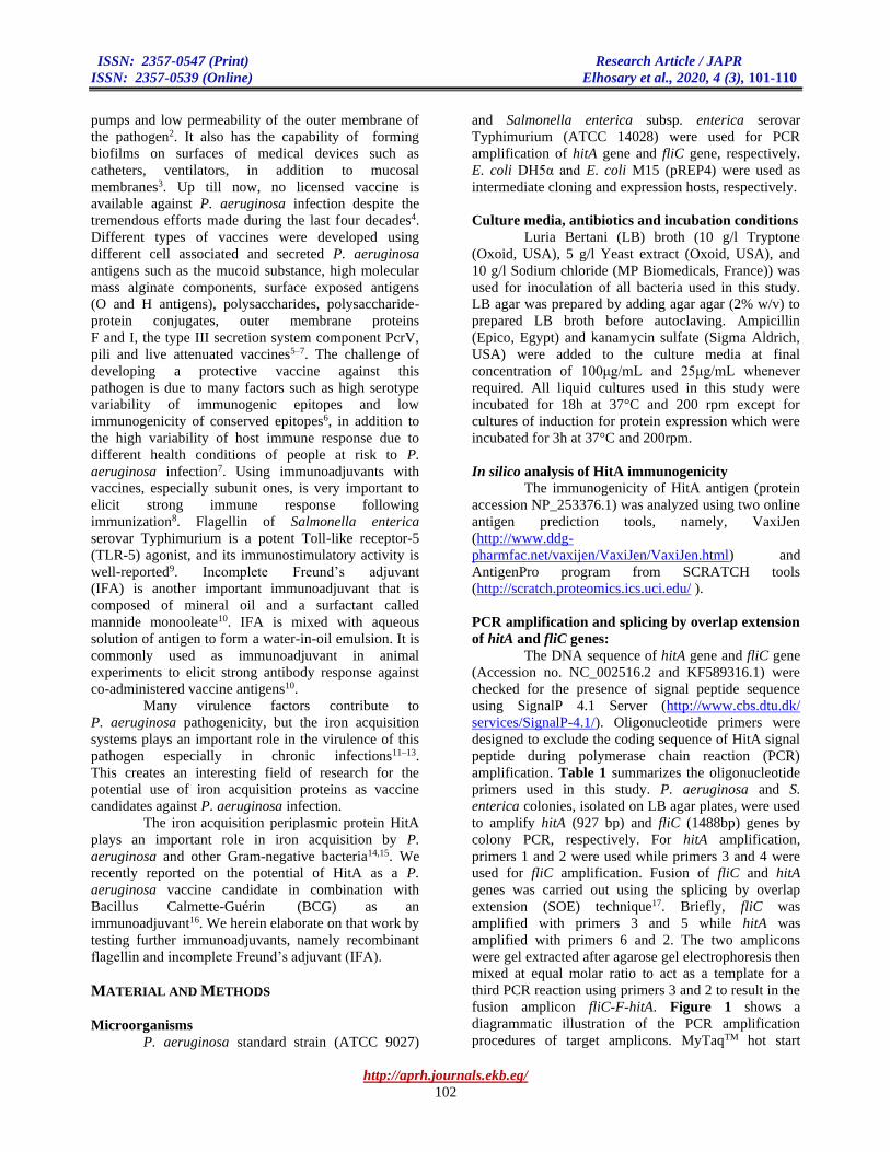
Figure 1. Simplified illustration of PCR amplification of target amplicons. (a) and (b) represent amplification of fliC and
hitA, respectively, using corresponding gene-specific primers. (c) shows the steps of splicing by overlap extension (SOE)
technique to construct the fusion gene fliC-F-hitA. Primer numbers (according to Table 1) are mentioned adjacent to the
arrows representing each pertinent used primer. The three amplicons resulting from (a), (b) and (c) were digested with
BamHI and HindIII and ligated to a similarly digested pQE31 plasmid for cloning and expression of the corresponding
protein products.
Master Mix (BIOLINE, UK) was used in all PCR
reactions following manufacturer’s instructions.
Plasmid pQE31 as well as all amplicons were digested
with BamHI and HindIII. Ligation of each amplicon to
pQE31 was fulfilled individually using T4 DNA ligase
(NEB, USA). Chemically competent E. coli DH5α
cells were transformed with the ligation reactions and
positive clones were selected by inoculation on
ampicillin plates. One transformed colony with each
constructed plasmid was selected and grown for
plasmid extraction. Chemically competent E. coli M15
(pREP4) was transformed with the created plasmids and
positive clones were selected on ampicillin/kanamycin
LB agar plates. The constructed plasmids, pQE31-hitA,
pQE31-fliC and pQE31-fliC-F-hitA, were extracted and
sent for insert sequencing by GATC Biotech
sequencing services (Germany).
Protein Expression of recombinant HitA, FliC and
fusion FliC-F-HitA
Induction of protein expression was carried out
as outlined in the QiaExpressionist™ manual of Qiagen
(Germany). A fresh overnight culture of E. coli
(pREP4) harboring one of the constructed plasmids was
hitA
fliC
(a)
(b)
(c)
hitAfliC
hitAfliC
fliC-F-hitA fusion gene
fliC and hitA amplified separately resulting in amplicons with complementary overhangs
3
4
1
2
3
3
2
2
fliC and hitA amplicons mixed at equimolar ratio and PCR repeated with primers 3 and 2 only
5
6
ISSN: 2357-0547 (Print) Research Article / JAPR
ISSN: 2357-0539 (Online) Elhosary et al., 2020, 4 (3), 101-110
http://aprh.journals.ekb.eg/
104
used to inoculate 200mL LB broth supplemented with
ampicillin and kanamycin. The culture was incubated at
37°C with vigorous shaking till reaching optical density
at 600nm (OD600) of 0.4-0.6. This was followed by
induction with isopropyl β-D-1-thiogalactopyranoside
(Melford, UK) at a final concentration of 1mM. After
induction of recombinant protein expression, cells were
harvested by centrifugation, washed with sterile 0.9%
saline and the bacterial pellet was kept at -20°C until
protein purification was carried out.
For protein purification, the frozen bacterial
pellet was thawed on ice for 15min and re-suspended in
10 mL of denaturing lysis buffer (100mM NaH2PO4,
10mM Tris.Cl and 8M Urea in distilled water; pH 8)
and left for lysis at 25°C for 1h at 80rpm horizontal
shaking. After bacterial lysis, cell lysate was collected
after centrifugation at 4°C for 30min at 12000 xg and
recombinant His-tagged antigens were purified using
Nickel-NitriloTriacetic Acid (Ni-NTA) agarose
(Qiagen, Germany) under denaturing conditions (The
QiaExpressionist™, Qiagen, Germany). Purified
recombinant antigens were analyzed by sodium
dodecylsulfate polyacrylamide gel electrophoresis
(SDS-PAGE) using Acrylamide/Bis-acrylamide 40%
(37.5:1) (Noragen Biotek corp., Canada) in the
preparation of separating gel (12%) and stacking gel
(4%). SDS-PAGE gel was stained using Coomassie
brilliant blue R-250 dye (MP Biomedicals, USA) and
destained using a solution of 200mL glacial acetic acid,
100mL methanol and 700mL distilled water. To
confirm that purified antigens are His-tagged proteins,
western blot assay was performed as previously
described18 using mouse anti-His tag antibody
(Biolegend, USA) as the primary antibody. The
secondary antibodies used were alkaline phosphatase-
labeled antibody to mouse IgG (KPL, USA) in case of
HitA antigen and peroxidase-labeled antibody to mouse
IgG in case of FliC and FliC-F-HitA. Detection was
performed using BCIP/NBT phosphatase substrate (1-
component) (KPL, USA) to detect HitA and 3, 3'-
diaminobenzidine tetrahydrochloride (DAB) (Bio Basic
Inc., Canada) to detect FliC and FliC-F-HitA. In
addition, western blot against HitA protein was also
carried out using mouse anti-Pseudomonas aeruginosa
serum (obtained from mice immunized with sublethal
P. aeruginosa infection) as the primary antibody in
order to check HitA immunogenicity. Recombinant
protein eluates were refolded using PD-10 desalting
columns (GE Healthcare, USA) containing Sephadex™
G-25 medium following manufacturer instructions.
Mice immunization with recombinant antigens
Eight-week-old female Swiss albino mice (20-
25g) were purchased from Theodor Bilharz Research
Institute, Giza, Egypt. The mice were divided into
groups (8 mice/group). All animal procedures were
approved by the Animal Care and Use Committee
(ACUC) of the Faculty of Pharmacy, Alexandria
University. All animal experiments were performed
following institutional and international ethical
standards. In this study, two adjuvants were used,
recombinant FliC antigen and incomplete Freund’s
adjuvant (IFA). Five groups of mice were used to assess
the protective effect of HitA recombinant protein
against P. aeruginosa infection using different
immunization regimens. Group 1 mice were immunized
with a mixture of HitA (10μg), FliC (6μg) and IFA,
group 2 mice were immunized with the fusion protein
FliC-F-HitA (10μg) and IFA, group 3 mice were
immunized with HitA (10μg) and IFA, group 4 mice
were injected with IFA in saline and group 5 were
immunized with HitA (10μg) only. Each group was
immunized by subcutaneous injection with 200μL of
prepared regimen followed by two booster doses at two-
week intervals. Two weeks after the last booster dose,
blood samples were collected from mice by the
submandibular bleeding method19, and sera were kept
at -20°C for subsequent analysis.
Indirect enzyme linked immunosorbent assay
(ELISA)
Refolded HitA recombinant antigen was
diluted to a final concentration of 10μg/mL with coating
buffer (2.65g sodium carbonate and 2.1g sodium
bicarbonate per 500mL sterile distilled water, pH 9.6).
Diluted antigen was used for coating 96-well high
binding ELISA plate (Greiner Bio One, Germany)
(100μl/well). Coated plate was incubated statically
overnight at 4°C. After coating, the plate was washed 3
times with phosphate-buffered saline (PBS) followed by
blocking with 5% skimmed milk 3h. After blocking, the
plate was washed 3 times with PBS and 100μl of
diluted mouse serum (1:300 dilution in blocking buffer)
were added and left for 1h. After thorough washing,
peroxidase-labeled anti-mouse IgG antibody (KPL,
USA) was added for 1h. After washing the plate, 100μL
of 3,3',5,5'-Tetramethylbenzidine (TMB) Microwell
Peroxidase Substrate System (KPL, USA) were added
to each well and left for color development in darkness
for 20min at room temperature. This was followed by
adding 50μL of 1M sulfuric acid (stopping solution)
and measuring the absorbance at 450nm using
microplate reader (BIOTEK, USA).
Opsonophagocytic assay
Serum samples were collected 13 days after
the last vaccine booster before the bacterial challenge.
The opsonophagocytosis stimulating activity of serum
samples isolated from immunized mice were examined
as previously described16,20. Briefly, P. aeruginosa was
inoculated into RPMI 1640 medium containing 10% v/v
fetal bovine serum (FBS) and incubated at 37°C for

ISSN: 2357-0547 (Print) Research Article / JAPR
ISSN: 2357-0539 (Online) Elhosary et al., 2020, 4 (3), 101-110
http://aprh.journals.ekb.eg/
105
18h. Grown bacterial cells were harvested by
centrifugation at 4°C followed by washing with sterile
saline and final resuspension in 2mL sterile saline.
Human peripheral polymorphonuclear leukocytes
(PMN) were freshly isolated from blood of a healthy
human volunteer using Histopaque®-1077 (Sigma
Aldrich, USA) according to manufacturer’s instructions
and following informed consent by the blood donor.
The isolated PMNs were resuspended into 500μL of
RPMI 1640 medium, and cell viability was determined
by trypan blue exclusion method. PMN cells were
diluted to a final count of 5X10⁵ cells/ml using the same
medium. Reaction mixture was prepared by mixing 5μL
of diluted bacterial suspension (1500 colony forming
units (CFU)), 20μL pooled mouse serum (heat
inactivated at 56°C for 10min), 15μL PMNs (7500
cells) and 20μL active complement (fresh human
serum). Samples were taken at zero time and 40
minutes later following shaking incubation at 37°C.
Bacterial count was determined by plating tenfold serial
dilutions onto LB agar plates where the grown colonies
after incubation at 37°C for 18h were counted and
original count was calculated. Result was calculated as
percentage killing using the formula: [(count of control
– count of immunized)/count of control] X100.
Mice challenge with P. aeruginosa
Mice in this study were challenged two weeks
after the last immunization dose by intraperitoneal
injection of 200μL bacterial suspension equivalent to
108 CFU. Inoculum of P. aeruginosa was prepared from
overnight culture in RPMI 1640 medium containing
10% FBS. Mice were sacrificed 48h after the challenge
for organ examination. Lungs were isolated for bacterial
bioburden determination. Isolated lungs were
homogenized in 3mL sterile saline, tenfold serially
diluted and then plated on LB agar plates. Livers were
also isolated and fixed in 10% formalin and examined
microscopically after hematoxylin and eosin (H&E)
staining for histopathological changes due to P.
aeruginosa infection. Parameters examined were focal
lytic necrosis, bile ductular proliferation, congestion,
Kupffer cells hyperplasia, steatosis and ballooning
degeneration of hepatocytes and periportal
inflammation21,22. Scoring systems were used for
quantitative comparison between immunized and non-
immunized mice23,24.
Statistical analysis
ELISA results of IgG antibody response in
different mice groups were analyzed using one-way
analysis of variance (ANOVA) followed by post-test
(Tukey-Kramer test). Lung bioburden and liver
histopathological scores were analyzed using Kruskal
Wallis non-parametric test and Dunn’s post-hoc test.
Statistical analysis tests were performed using
GraphPad InStat 3. P value was considered significant
when less than 0.05.
RESULTS
Immunogenicity prediction, PCR amplification and
splicing by overlap extension
Bioinformatic analysis of HitA protein by
VaxiJen and AntigenPro antigen prediction tools
revealed antigenic score of 0.48 (above the threshold of
0.4) and antigenic probability of 0.788, respectively.
Amplification of hitA and fliC with gene-specific
primers was carried out by colony PCR on P.
aeruginosa and S. enterica isolated colonies,
respectively. SOE technique was successfully employed
to create the fusion gene fliC-F-hitA. Agarose gel
electrophoresis revealed the amplicons at their expected
sizes: hitA gene (927bp), fliC gene (1488bp) and the
fusion gene fliC-F-hitA (2415bp) (Figure 2). E. coli
(pREP4) clones harboring the different constructed
plasmids were confirmed by gene-specific PCR
reactions and DNA sequencing.
Figure 2. Agarose gel electrophoresis of gene amplicons
(a) PCR amplified hitA (927bp) (Lane 1) using gene-
specific primers and P. aeruginosa colony PCR. (b) PCR
amplified fliC (1488bp) (Lane 1) using gene-specific
primers and an isolated S. enterica colony PCR. (c) PCR
amplified fusion gene fliC-F-hitA using primers 3 and 2
showing a band at the expected size of 2415bp (Lane 1).
Lane 2 represents DNA ladder.
Successful antigen expression in E. coli (pREP4)
hosts
Recombinant expressed antigens were purified
using Ni-NTA agarose to capture His-tagged antigens
followed by washing and elution of pure antigens.
Purified antigens were checked by SDS-PAGE as
shown in Figure 3. All the three recombinant antigens;
(a) (b) (c)

ISSN: 2357-0547 (Print) Research Article / JAPR
ISSN: 2357-0539 (Online) Elhosary et al., 2020, 4 (3), 101-110
http://aprh.journals.ekb.eg/
106
HitA, FliC and FliC-F-HitA, appeared at their expected
sizes: 34, 53 and 88 kDa, respectively. Recombinant
antigens identity, as being His-tagged, was confirmed
by western blot analysis using mouse anti-His tag
antibodies (Figure 4). Furthermore, mouse anti-
Pseudomonas aeruginosa serum antibodies reacted
positively with HitA in western blot confirming
immunogenicity of HitA upon P. aeruginosa infection
(data not shown).
Figure 3. SDS-PAGE of the recombinant expressed
antigens. (a) SDS-PAGE of purified HitA antigen (34
kDa). (b) SDS-PAGE of purified FliC (53 kDa) (lane 1)
and the fusion FliC-F-HitA protein (about 88 kDa)
(lane 2).
Figure 4. Western blot to detect His-tagged antigens. (a)
Western blot of HitA using mouse anti-His tag antibody as
primary antibody and detection by BCIP/NBT phosphatase
chromogenic substrate. (b) Western blot of FliC (lane 1)
and the fusion FliC-F-HitA protein (lane 2) using mouse
anti-His tag antibody as primary antibody and detection by
3, 3'-diaminobenzidine tetrahydrochloride (DAB)
peroxidase chromogenic substrate.
Active immunization elicits HitA-specific IgG
antibodies which enhance P. aeruginosa
opsonophagocytosis by PMNs
Indirect ELISA results showed a significant
HitA-specific IgG antibody response in immunized
groups 1, 2 and 3. On the other hand, negligible IgG
response was observed in the IFA negative control
group 4, and in group 5 immunized with HitA alone
without adjuvants (Figure 5). Opsonophagocytic assay
demonstrated that serum antibodies from immunized
groups 1, 2 and 3 enhanced the phagocytic activity of
PMNs. No P. aeruginosa count was recovered from
opsonophagocytic reactions containing serum from
immunized mice reflecting 100% percentage killing
compared to the negative control group and the group
given HitA alone.
Figure 5. Indirect ELISA results showing HitA-specific
immune response. Murine HitA-specific IgG antibody
was measured two-weeks after the last vaccine booster.
The illustrated results are at serum dilution of 1/300.
Error bars represent mean reading+/- SEM (standard
error of the mean).
Immunized mice are protected against P. aeruginosa
challenge
Animal groups were challenged with P.
aeruginosa intraperitoneally two weeks after the last
vaccine booster. After 48h, mice were sacrificed to
determine bacterial count in lungs. There was a
significant reduction in bacterial burden of lungs in
group 1 compared to group 4 and 5 (P < 0.01) as shown
in Figure 6. There was no statistically significant lung
protection in other immunized groups.
Upon examining the liver sections of the
immunized groups, they showed preserved lobular
architecture with no cirrhotic changes or fibrosis,
no/minimal focal lytic necrosis, low portal
inflammation, no bile ductular proliferative
inflammation, minimal central or portal veins
congestion and no Kupffer cells hyperplasia (Table 2
and Figure 7). There was a significant reduction in
20
30
40 50 60
7090
kDa kDa
30
40 50 60
(a) (b)
1 2
20
30
40 50 60 70
kDa
90
20
30
40 50 60 70
kDa
90
(a) (b)
1 2
0
0.5
1
1.5
2
2.5
3
3.5
4
HitA/FliC/IFA Fusion/IFA HitA/IFA IFA HitA
Ab
sorb
an
ce a
t 4
50
nm
*********
ISSN: 2357-0547 (Print) Research Article / JAPR
ISSN: 2357-0539 (Online) Elhosary et al., 2020, 4 (3), 101-110
http://aprh.journals.ekb.eg/
107
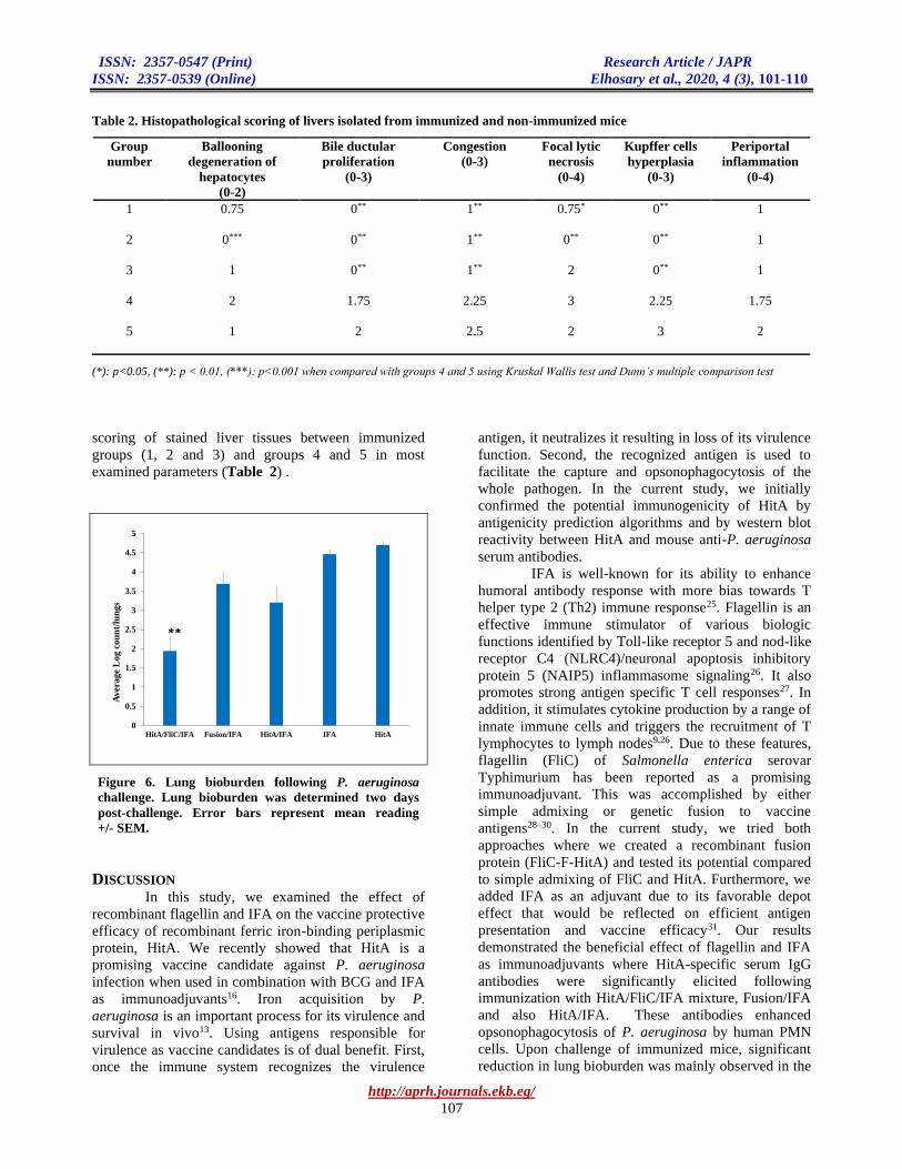
Table 2. Histopathological scoring of livers isolated from immunized and non-immunized mice
Group
number
Ballooning
degeneration of
hepatocytes
(0-2)
Bile ductular
proliferation
(0-3)
Congestion
(0-3)
Focal lytic
necrosis
(0-4)
Kupffer cells
hyperplasia
(0-3)
Periportal
inflammation
(0-4)
1
0.75 0** 1** 0.75* 0** 1
2
0*** 0** 1** 0** 0** 1
3
1 0** 1** 2 0** 1
4
2 1.75 2.25 3 2.25 1.75
5 1 2 2.5 2 3 2
(*): p<0.05, (**): p < 0.01, (***): p<0.001 when compared with groups 4 and 5 using Kruskal Wallis test and Dunn’s multiple comparison test
scoring of stained liver tissues between immunized
groups (1, 2 and 3) and groups 4 and 5 in most
examined parameters (Table 2) .
Figure 6. Lung bioburden following P. aeruginosa
challenge. Lung bioburden was determined two days
post-challenge. Error bars represent mean reading
+/- SEM.
DISCUSSION In this study, we examined the effect of
recombinant flagellin and IFA on the vaccine protective
efficacy of recombinant ferric iron-binding periplasmic
protein, HitA. We recently showed that HitA is a
promising vaccine candidate against P. aeruginosa
infection when used in combination with BCG and IFA
as immunoadjuvants16. Iron acquisition by P.
aeruginosa is an important process for its virulence and
survival in vivo13. Using antigens responsible for
virulence as vaccine candidates is of dual benefit. First,
once the immune system recognizes the virulence
antigen, it neutralizes it resulting in loss of its virulence
function. Second, the recognized antigen is used to
facilitate the capture and opsonophagocytosis of the
whole pathogen. In the current study, we initially
confirmed the potential immunogenicity of HitA by
antigenicity prediction algorithms and by western blot
reactivity between HitA and mouse anti-P. aeruginosa
serum antibodies.
IFA is well-known for its ability to enhance
humoral antibody response with more bias towards T
helper type 2 (Th2) immune response25. Flagellin is an
effective immune stimulator of various biologic
functions identified by Toll-like receptor 5 and nod-like
receptor C4 (NLRC4)/neuronal apoptosis inhibitory
protein 5 (NAIP5) inflammasome signaling26. It also
promotes strong antigen specific T cell responses27. In
addition, it stimulates cytokine production by a range of
innate immune cells and triggers the recruitment of T
lymphocytes to lymph nodes9,26. Due to these features,
flagellin (FliC) of Salmonella enterica serovar
Typhimurium has been reported as a promising
immunoadjuvant. This was accomplished by either
simple admixing or genetic fusion to vaccine
antigens28–30. In the current study, we tried both
approaches where we created a recombinant fusion
protein (FliC-F-HitA) and tested its potential compared
to simple admixing of FliC and HitA. Furthermore, we
added IFA as an adjuvant due to its favorable depot
effect that would be reflected on efficient antigen
presentation and vaccine efficacy31. Our results
demonstrated the beneficial effect of flagellin and IFA
as immunoadjuvants where HitA-specific serum IgG
antibodies were significantly elicited following
immunization with HitA/FliC/IFA mixture, Fusion/IFA
and also HitA/IFA. These antibodies enhanced
opsonophagocytosis of P. aeruginosa by human PMN
cells. Upon challenge of immunized mice, significant
reduction in lung bioburden was mainly observed in the
0
0.5
1
1.5
2
2.5
3
3.5
4
4.5
5
HitA/FliC/IFA Fusion/IFA HitA/IFA IFA HitA
Av
era
ge
Lo
g c
ou
nt/
lun
gs
**

ISSN: 2357-0547 (Print) Research Article / JAPR
ISSN: 2357-0539 (Online) Elhosary et al., 2020, 4 (3), 101-110
http://aprh.journals.ekb.eg/
108
Figure 7. Histopathological examination of livers from different animal groups. (a) liver section in an immunized case
(group 1) showing normal liver architectures, central veins with mild peri-portal inflammation (arrows) (x100). (b) a higher
power view of the previous case showing mild focal peri-portal neutrophilic infiltration and mild diffuse micro-vesicular
steatosis (x400). Liver sections in non-immunized case (group 4) showing (c): marked peri-portal inflammation, arrows
(x100), (d): marked congestion, arrows (x100), (e): numerous foci of focal lytic necrosis; (f): higher power view of (e) showing
the focal lytic necrosis where the necrotic hepatocytes are replaced by aggregates of neutrophils, arrows (x400).
f e
d c
b a
ISSN: 2357-0547 (Print) Research Article / JAPR
ISSN: 2357-0539 (Online) Elhosary et al., 2020, 4 (3), 101-110
http://aprh.journals.ekb.eg/
109
group immunized with HitA/FliC/IFA mixture. The
bioburden reduction was not statistically significant in
lungs of other immunized groups (i.e. Fusion/IFA and
HitA/IFA). A possible explanation of the failure of the
Fusion protein (i.e. FliC-F-HitA) to reduce the lung
bioburden might be the low immunizing dose given to
mice. Fusion protein (FliC-F-HitA) was given at a dose
of 10µg. Based on the molecular weights of FliC and
HitA, the contribution of HitA in this fusion dose
represented only 4 µg out of the 10µg injected. On the
other hand, the other groups, HitA/FliC/IFA and
HitA/IFA, were immunized with a higher dose of 10µg
HitA. Using a higher dose of the fusion protein might
stimulate a more protective efficacy. Furthermore, it
was previously reported that the position of the antigen
fused to flagellin played a critical role in
immunogenicity outcome9. The antigen can be fused to
the N-terminal or the C-terminal of flagellin or can be
inserted into the hypervariable region of flagellin9. In
our work, we fused HitA to the C-terminal end of
flagellin. Trying the other two fusion options is worthy
of further investigation. The failure of HitA/IFA,
compared to HitA/FliC/IFA, to protect the lungs
highlights the critical role of flagellin in augmenting the
protective immune response. Investigation of detailed
cytokine profile in the different animal groups should
clarify the exact role/mechanism of flagellin
immunostimulation. In our study, flagellin (FliC),
whether fused or unfused, was administered at low
doses (below 10 μg/mouse) as recommended by
previous studies9,28,29. Low flagellin doses (1-10μg)
were shown to be effective as vaccine adjuvant,
reflecting its potent immunostimulatory effect9,28,29.
Histopathological examination of livers of immunized
mice revealed significant improvement when compared
with livers of control groups. This was observed in
most of the tested histopathological parameters (Table
2). It is noteworthy that group 5 immunized with HitA
alone without adjuvants were not protected against
challenge and no significant HitA-specific antibody
response was observed. This was quite expected since it
is well-reported that recombinant subunit antigens are
weakly immunogenic and need immunoadjuvants to be
effective as vaccines8.
CONCLUSION
Overall, HitA ferric iron binding periplasmic
protein is considered a promising vaccine candidate
against P. aeruginosa. Flagellin, in combination with
IFA, played an important role as immunoadjuvant to
augment the immune response to HitA. Future studies
combining HitA with other P. aeruginosa antigens
could improve the vaccine protective outcome.
Acknowledgements
The authors declare no external funding
was included. The authors would like to thank Prof. Dr.
Amal Khalil for help and support.
Conflict of interest
The authors declare that there is no conflict of
interest regarding the publication of this paper.
REFERENCES
1. Bassetti, M.; Vena, A.; Croxatto, A.; Righi, E.;
Guery, B. How to manage Pseudomonas
aeruginosa infections. Drugs Context. 2018, 7.
2. Azam, M.W.; Khan, A.U. Updates on the
pathogenicity status of Pseudomonas aeruginosa.
Drug Discov. Today. 2019, 24 (1), 350–359.
3. Lee, K.; Yoon, S.S. Pseudomonas aeruginosa
Biofilm, a Programmed Bacterial Life for Fitness.
J. Microbiol. Biotechnol. 2017, 27 (6),1053–64.
4. Merakou, C.; Schaefers, M.M.; Priebe G.P.
Progress toward the elusive Pseudomonas
aeruginosa vaccine. Surg Infect (Larchmt). 2018,
19 (8), 757–768.
5. Stanislavsky, E.S.; Lam, J. S. Pseudomonas
aeruginosa antigens as potential vaccines. FEMS
Microbiol. Rev. 1997, 21 (3), 243–77.
6. Priebe, G.P.; Goldberg, J.B. Vaccines for
Pseudomonas aeruginosa: A long and winding
road. Expert Rev. Vaccines. 2014, 13 (4), 507–519.
7. Grimwood, K.; Kyd, J.M.; Owen, S.J.; Massa,
H.M.; Cripps, A.W. Vaccination against respiratory
Pseudomonas aeruginosa infection. Hum. Vaccines
Immunother. 2015, 11 (1), 14–20.
8. Kurella, S.; Manocha, M.; Sabhnani, L.; Thomas,
B.; Rao, D.N. New age adjuvants and delivery
systems for subunit vaccines. Indian J. Clin.
Biochem. 2000, 15, 83–100.
9. Mizel, S.B.; Bates, J.T. Flagellin as an Adjuvant:
Cellular Mechanisms and Potential. J. Immunol.
2010, 185, 5677–82.
10. Jennings, V.M. Review of Selected Adjuvants
Used in Antibody Production. ILAR J. 1995, 37 (3),
119–125.
11. Vasil, M.L.; Ochsner, U.A. The response of
Pseudomonas aeruginosa to iron: Genetics,
biochemistry and virulence. Mol. Microbiol.1999,
34 (3), 399–413.
12. Visca, P.; Leoni, L.; Wilson, M.J.; Lamont, I.L.
Iron transport and regulation, cell signalling and
genomics: Lessons from Escherichia coli and
Pseudomonas. Mol. Microbiol. 2002, 45 (5),
1177–1190.
13. Ben Haj Khalifa, A.;Moissenet, D.; Vu Thien, H.;
Khedher, M. Virulence factors in Pseudomonas

ISSN: 2357-0547 (Print) Research Article / JAPR
ISSN: 2357-0539 (Online) Elhosary et al., 2020, 4 (3), 101-110
http://aprh.journals.ekb.eg/
110
aeruginosa: mechanisms and modes of regulation.
Ann. Biol. Clin. (Paris). 2011, 69 (4), 393–403.
14. Adhikari, P.; Kirby, S.D.; Nowalk, A.J.; Veraldi,
K.L.; Schryvers, A.B.; Mietzner, T.A. Biochemical
characterization of a Haemophilus influenzae
periplasmic iron transport operon. J. Biol. Chem.
1995, 270, 25142–9.
15. Nde, C.W.; Jang, H.J.; Toghrol, F.; Bentley, W.E.
Toxicogenomic response of Pseudomonas
aeruginosa to ortho-phenylphenol. BMC
Genomics. 2008, 9, 473.
16. Elhosary, M.A.; Bahey-El-Din, M.; AbdelBary, A.;
El Guink, N.; Aboushleib, H.M. Immunization with
the ferric iron-binding periplasmic protein HitA
provides protection against Pseudomonas
aeruginosa in the murine infection model. Microb.
Pathog. 2019, 131, 181–5.
17. Horton, R.M.; Hunt, H.D.; Ho, S.N.; Pullen, J.K.;
Pease, L.R. Engineering hybrid genes without the
use of restriction enzymes: gene splicing by
overlap extension. Gene. 1989, 77, 61–8.
18. Mossallam, S.F.; Amer, E.I.; Ewaisha, R.E.; Khalil,
A.M.; Aboushleib, H.M.; Bahey-El-Din, M. Fusion
protein comprised of the two schistosomal
antigens, Sm14 and Sm29, provides significant
protection against Schistosoma mansoni in murine
infection model. BMC Infect. Dis. 2015, 15.
19. Golde, W.T.; Gollobin, P.; Rodriguez, L.L. A
rapid, simple, and humane method for
submandibular bleeding of mice using a lancet.
Lab. Anim. (NY). 2005, 34, 39–43.
20. Kurupati, P.; Ramachandran, N.P.; Poh, C.L.
Protective efficacy of DNA vaccines encoding
outer membrane protein A and OmpK36 of
Klebsiella pneumoniae in mice. Clin. Vaccine
Immunol. 2011, 18 (1), 82–8.
21. Schümann, J.; Angermüller, S.; Bang, R.; Lohoff,
M.; Tiegs, G. Acute hepatotoxicity of
Pseudomonas aeruginosa exotoxin A in mice
depends on T cells and TNF. J. Immunol. 1998,
155, 4829–37.
22. Zuheir Majeed, S.; Ghadban Auda, I.; Muhammed
Ali Salman, I.; Auda, G. Histopathological
Changes of Some Internal Organs of Mice Injected
with Exotoxin a at Low Concentrations. J. Biol.
Agric. Healthc. 2014, 4 (9), 28–33.
23. Brunt, E.M.; Tiniakos, D.G. Histopathology of
nonalcoholic fatty liver disease. World J.
Gastroenterol. 2010, 16, 5286–96.
24. Ishak, K.; Baptista, A.; Bianchi, L.; Callea, F.; De
Groote, J.; Gudat, F.; Denk, H.; Desmet, V.; Korb,
G.; MacSween, R.N.; Phillips, MJ. Histological
grading and staging of chronic hepatitis. J.
Hepatol. 1995, 22 (6), 696–9.
25. Chang, J.C.C.; Diveley, J.P.; Savary, J.R.; Jensen,
F.C. Adjuvant activity of incomplete Freund’s
adjuvant. Adv. Drug Deliv. Rev.1998, 32, 173–86.
26. Lin, K.H.; Chang, L.S.; Tian, C.Y.; Yeh, Y.C.;
Chen, Y.J.; Chuang, T.H.; Liu.,S.J.; Leng, C.H.
Carboxyl-terminal fusion of E7 into Flagellin shifts
TLR5 activation to NLRC4/NAIP5 activation and
induces TLR5-independent anti-tumor immunity.
Sci. Rep. 2016, 6 (1), 1–10.
27. Bates, J.T.; Graff, A.H.; Phipps, J.P.; Grayson,
J.M.; Mizel, S.B. Enhanced antigen processing of
flagellin fusion proteins promotes the antigen-
specific CD8 + T cell response independently of
TLR5 and MyD88 . J. Immunol. 2011, 186 (11),
6255–62.
28. Bargieri, D.Y.; Rosa, D.S.; Braga, C.J.M.;
Carvalho, B.O.; Costa, F.T.M., Espíndola, N.M.,
Vaz, A.J.; Soares, I.S.; Ferreira, L.C.S.; Rodrigues,
M.M. New malaria vaccine candidates based on the
Plasmodium vivax Merozoite Surface Protein-1 and
the TLR-5 agonist Salmonella Typhimurium FliC
flagellin. Vaccine. 2008, 26, 6132–42.
29. Honko, A.N.; Sriranganathan, N.; Lees, C.J.;
Mizel, S.B. Flagellin is an effective adjuvant for
immunization against lethal respiratory challenge
with Yersinia pestis. Infect. Immun. 2006, 74 (2),
1113–1120.
30. Xiao, X.; Zhang, Y.; Liu, J.; Wei, Q.; Yin, X.
Immunoenhancement with flagellin as an adjuvant
to whole-killed rabies vaccine in mice. Arch. Virol.
2016, 161 (3), 685–91.
31. Koh, Y.T.; Higgins, S.A.; Weber, J.S.; Kast, W.M.
Immunological consequences of using three
different clinical/laboratory techniques of
emulsifying peptide-based vaccines in incomplete
Freund’s adjuvant. J. Transl. Med. 2006, 4, 42.


