Chapter 9 Major Histocompatibility Complex (MHC) Chapter 9 Major Histocompatibility Complex (MHC)
Upload
tim-elliottCategory
view
212download
0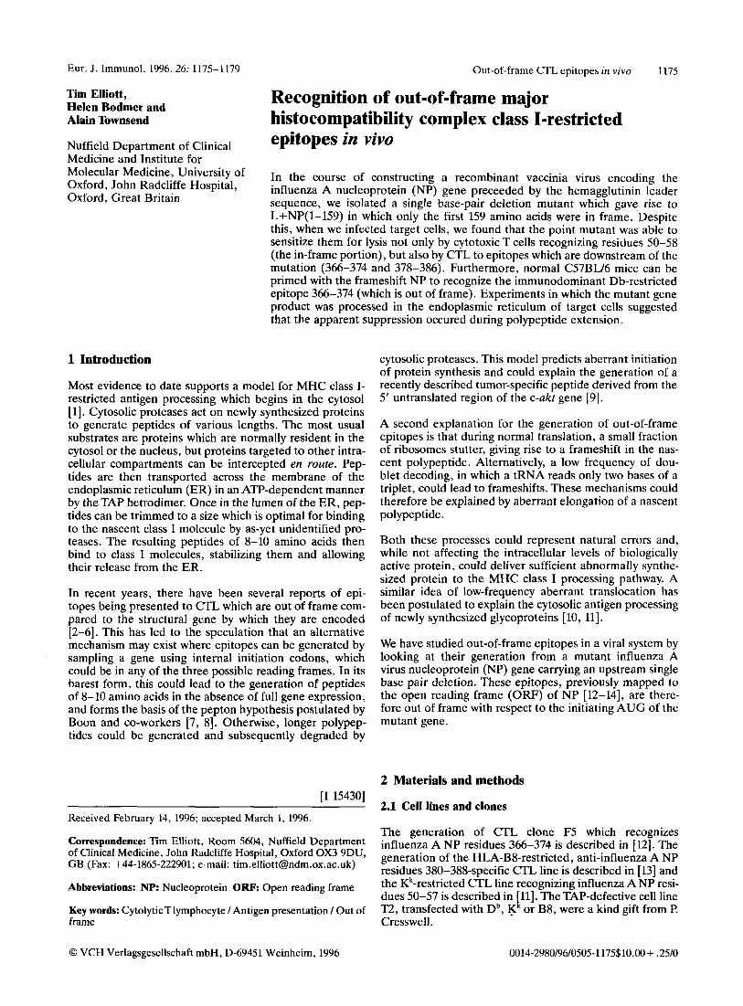
Eur. J. Immunol. 1996.26: 1175-1179 Out-of-frame CTL epitopes in vivo 1175
Tim Elliott, Helen Bodmer and Alain Townsend
Nuffield Department of Clinical Medicine and Institute for Molecular Medicine, University of Oxford, John Radcliffe Hospital, Oxford, Great Britain
Recognition of out-of-frame major histocompatibility complex class I-restricted epitopes in vivo
In the course of constructing a recombinant vaccinia virus encoding the influenza A nucleoprotein (NP) gene preceeded by the hemagglutinin leader sequence, we isolated a single base-pair deletion mutant which gave rise to L+NP(l-159) in which only the first 159 amino acids were in frame. Despite this, when we infected target cells, we found that the point mutant was able to sensitize them for lysis not only by cytotoxic T cells recognizing residues 50-58 (the in-frame portion), but also by CTL to epitopes which are downstream of the mutation (366-374 and 378-386). Furthermore, normal C57BU6 mice can be primed with the frameshift NP to recognize the immunodominant Db-restricted epitope 366-374 (which is out of frame). Experiments in which the mutant gene product was processed in the endoplasmic reticulum of target cells suggested that the apparent suppression occured during polypeptide extension.
1 Introduction
Most evidence to date supports a model for MHC class I- restricted antigen processing which begins in the cytosol [l]. Cytosolic proteases act on newly synthesized proteins to generate peptides of various lengths. The most usual substrates are proteins which are normally resident in the cytosol or the nucleus, but proteins targeted to other intra- cellular compartments can be intercepted en route. Pep- tides are then transported across the membrane of the endoplasmic reticulum (ER) in an ATP-dependent manner by the TAP hetrodimer. Once in the lumen of the ER, pep- tides can be trimmed to a size which is optimal for binding to the nascent class I molecule by as-yet unidentified pro- teases. The resulting peptides of 8-10 amino acids then bind to class I molecules, stabilizing them and allowing their release from the ER.
In recent years, there have been several reports of epi- topes being presented to CTL which are out of frame com- pared to the structural gene by which they are encoded [2-61. This has led to the speculation that an alternative mechanism may exist where epitopes can be generated by sampling a gene using internal initiation codons, which could be in any of the three possible reading frames. In its barest form, this could lead to the generation of peptides of 8-10 amino acids in the absence of full gene expression, and forms the basis of the pepton hypothesis postulated by Boon and co-workers [7, 81. Otherwise, longer polypep- tides could be generated and subsequently degraded by
cytosolic proteases. This model predicts aberrant initiation of protein synthesis and could explain the generation of a recently described tumor-specific peptide derived from the 5' untranslated region of the c-akt gene [9].
A second explanation for the generation of out-of-frame epitopes is that during normal translation, a small fraction of ribosomes stutter, giving rise to a frameshift in the nas- cent polypeptide. Alternatively, a low frequency of dou- blet decoding, in which a tRNA reads only two bases of a triplet, could lead to frameshifts. These mechanisms could therefore be explained by aberrant elongation of a nascent polypeptide.
Both these processes could represent natural errors and, while not affecting the intracellular levels of biologically active protein, could deliver sufficient abnormally synthe- sized protein to the MHC class I processing pathway. A similar idea of low-frequency aberrant translocation has been postulated to explain the cytosolic antigen processing of newly synthesized glycoproteins [lo, 111.
We have studied out-of-frame epitopes in a viral system by looking at their generation from a mutant influenza A virus nucleoprotein (NP) gene carrying an upstream single base pair deletion. These epitopes, previously mapped to the open reading frame (OW) of NP [12-141, are there- fore out of frame with respect to the initiating AUG of the mutant gene.
2 Materials and methods [I 154301
Received February 14, 1996; accepted March 1, 1996.
Correspondence: Tim Elliott, Room 5604, Nuffield Department of Clinical Medicine, John Radcliffe Hospital, Oxford OX3 9DU, GB (Fax: +44-186222901; e-mail: [email protected])
Abbreviations: NP: Nucleoprotein O R F Open reading frame
Key words: CytolyticTlyrnphocyte I Antigen presentation I Out of frame
2.1 Cell lines and clones
The generation of CTL clone F5 which recognizes influenza A NP residues 366-374 is described in [12]. The generation of the HLA-B&restricted, anti-influenza A NP residues 380-388-specific CTL line is described in [ 131 and the Kk-restricted CTL line recognizing influenza A NP resi- dues 50-57 is described in 11 . The TAP-defective cell line T2, transfected with Db, K or B8, were a kind gift from P. Cresswell.
1 . 1
0 VCH Verlagsgesellschaft mbH, D-69451 Weinheim, 1996 0014-2980/96/0505-1175$10.00 + .25/0

1176 T. Elliott et al. Eur. J. Immunol. 1996.26: 1175-1179
2.2 Construction of recombinant vaccinia viruses A C T A G C T A G B a b c d
Recombinant vaccinia viruses were constructed exactly as described in [ll]. Genes were introduced into the vaccinia genome by homologous recombination with the thymidine kinase gene using the shuttle vector psc11.30R.2. Recom- binant viruses were plaque-purified four times with the aid of the p-galactosidase reporter gene. The last two rounds of purification indicated that 100% of tha viral plaques contained the P-galactosidase reporter gene.
2.3 Immunoprecipitation
Immunoprecipitations were carried out exactly as described [ll] with 5 x 10' labeled T2 cells infected for 1 h with recombinant vaccinia at a multiplicity of infection (m.0.i.) of 3 during incubation in methionine-free medium. Metabolic labeling with [35S] methionine was then carried out for 1 h prior to lysing the cells and pre- clearing overnight with fixed Staphylococcus aureus. Influenza nucleoporotein was precipitated with mAb 4-7- 18 [ E l , and HLA was precipitated with mAb W6/32 (a gift from A. Michael).
2.4 CTL assay
CTL activity was tested by the standard "Cr-release assay using 1 x lo4 labeled target cells. Targets were infected for 1 h at a m.0.i. of 5 , 3 h prior to the start of the assay, and *'Cr labeling was for 1 h. Where sensitization by synthetic peptides was measured, uninfected 51Cr-labeled target cells were pulsed for 1 h at 37C with the appropriate peptide then washed before being used in the CTL assay.
2.5 Priming mice with recombinant vaccinia
For each experiment, two 9-week-old female C57BLI6 mice were injected intraperitoneally with 1 x lo7 PFU of recombinant or wild-type WR vaccinia. After 9 days, the dispersed pooled splenocytes were cultured at a density of 3 x lo6 ml-' with 4 X lo7 irradiated autologous spleno- cytes pulsed with 10 pM NP 366-374 in a total volume of 3 ml Iscove's MEM supplemented with 10 % FCS and 2 mM L-glutamine. Restimulation with peptide in this way was as effective as restimulation with influenza A infected auto- logous feeders.
3 Results and discussion
3.1 Isolation of a frameshifted nucleoprotein gene
While creating a construct encoding the NP gene preceded by the hemagglutinin leader sequence (L+NP), we iso- lated a subclone which had a single base pair deletion at coding base A478 (Fig. 1A). The resides in a Bam HI site and its deletion results in the corresponding loss of this site (Fig. 1B). The mutant gene L+NP(l-159) has an ORF encoding the first 159 amino acids of NP plus 6 residues in the new reading frame before a stop codon is reached (Fig. 1C). We constructed recombinant vaccinia viruses containing either the wild-type L+NP or the frameshifted
c
C 478 465 t CGC ACC GGC ATCLGUCCC AGG ATG TGC TCT L+NP
I56 I19 R T G M D P R M C S
L+NP(1-159) CGC ACC GGC ATG GTC CCA GGA TGT GCT CTC R T G M V P G C A L
Figure 1. (A) Plasmid psc11.302.R containing L+NP(l-159) (right sequence) and the authentic NP sequence (left-sequence) were sequenced across the site of the deletion by the dideoxy method. The arrow indicates the position of A478 which is deleted in L+NP(l-159). This deletion results in the loss of a Bam HI site (underlined in C), shown in (B) where PCR products including the leader sequence to coding base 1097 of NP, from either psc11.302.R L+NP (lanes a and c) or psc11.302.R L+NP(l-159) (lanes b and d) were digested for 2 h with Eco 01091 (lanes a and b) or Bam HI (lanes c and d) and run on an ethidium bromide- stained 2 % agarose gel. Digestion with Eco 01091 results in two fragments of 333 and 818 bp reflecting cleavage at base 279 of NP. Digestion of L+NP with Bam HI results in three fragments of 216, 314 and 621 bp, reflecting cleavage at bases 260 and 476 of NP. In L+NP (1-159), the latter site has clearly been lost, resulting in only two fragments of 314 and 837 bp. (C) Nucleotide and amino acid sequence around the deletion seen in L+NP(l-159), showing the novel ORF of six amino acids. Note that the numbering sys- tem starts at Met 1 of NP and not Met 1 of the leader sequence.
a b c d
NP + 4 HC
4 *
4 p2-m
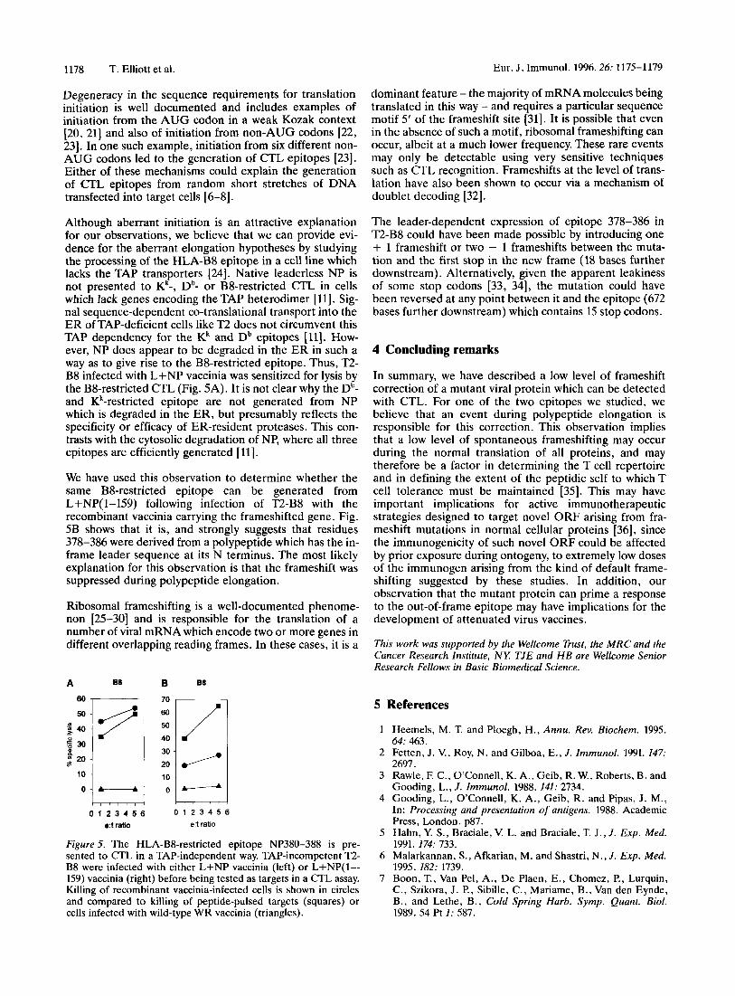
Figure 2. Metabolically labeled T2 cells infected with either L+NP vaccinia (lanes a and b) or L+NP(l-159) vaccinia (lanes c and d) were lysed and immunoprecipitated with either the anti- NP mAb 4-7-18 ([ll] lanes a and c) or the anti-HLA mAb W6/32 (lanes b and d). The position of L+NP, MHC class I heavy chain (HC) and P2-microglobulin are marked. Human invariant chain, which consistently co-precipitates with HLA-BS1 in T2 [37] is marked with an asterisk. The autoradiogram shown in this figure was greatly overexposed to aid in the detection of very low levels of NP expression.
L+NP(l-159) and used these to infect metabolically- labeled T2 cells. Infection of 1 X lo7 cells with the wild- type L+NP recombinant vaccinia gave rise to an immu- noprecipitable metabolically labeled product of 38-kDa using an anti-NP mAb (Fig. 2). This has been shown to be an ER-resident glycosylated form of NP ([l l] and TJE, unpublished observations). No protein product could be identified by immunoprecipitation from a lysate of cells

Eur. J. lmmunol. 1996.26: 1175-1179
infected with L+NP( 1-159) recombinant vaccinia (Fig. 2). Immunohistochemical staining of cells infected with L+NP(l-159) vaccinia with a polyclonal sheep anti-NP antiserum, however, revealed a reticular staining pattern (data not shown). Taken together, these results suggest that the truncated protein was cotranslationally translo- cated into the ER where it either adopted a conformation that was undetectable with the mAb or was too rapidly degraded to be detectable.
3.2 Recognition of the frameshifted gene-encoded nucleoprotein by CTL
Despite the apparent lack of expression of full-length NP from the frameshifted gene, we found that when target cells were infected with L+NP( 1-159) vaccinia, they were sensitized for lysis not only by H-2Kk-restricted CTL, which recognize residues 50-58 encoded within the truncated protein, but also by H2-Db-restricted CTL recognizing residues 366-374 and HLA-B8-restricted CTL specific for residues 378-386 as shown in Fig. 3. The Db- and B8- restricted epitopes reside 621 and 657 bases downstream of the frameshift and are out of frame with respect to the initiating AUG. The possibility that sensitization was due to the presence of a rare revertant clone is unlikely, since the recombinant vaccinia was selected after four rounds of plaque purification. With respect to CTL recognition, therefore, the frameshift appeared to have been sup- pressed. Despite our inability to detect any full-length L+NP synthesis from the L+NP(l-159) gene, these results suggest is that the mutation in L+NP(l-159) is ignored or corrected sufficiently frequently to produce sensitizing amounts of NP 366-374 and NP 380-388.
3.3 Priming mice against the out-of-frame epitope
It is now known that a very small number of MHC class I/ peptide complexes are required at the target cell surface for recognition by secondary Cm [16, 171. To determine whether sufficient NP 366-374 could be made in vivo to
70
60
._ u) 50 2 .40
5 30
s
u)
H 20
10
0
1 2 3 4 5 e:t ratio
0 1 2 3 4 5 6 e:t ratio
88 80
10
0 1 2 3 4 5 e:t ratio
Figure 3. Sensitization of target cells with L+NP(l-159) vac- cinia. L-Db cells, which express both Kk and Db, and an EBV- transformed B cell line expressing HLA-B8, were tested for their ability to present peptide at 10 nM (squares), or vaccinia-derived antigen following infection with L+NP(l-159) vaccinia (circles). Control lysis following infection with wild-type WR vaccinia is also shown (triangles). The peptides used were the K'-restricted epitope SDYEGRLI (left), the Db-restricted epitope ASNENM- DAM (center) and the BS-restricted epitope ELSRYWAI (right).
Out-of-frame CTL epitopes in vivo 1177
prime a CTL response, we primed C57BL16 mice with L+NP vaccinia or L+NP(l-159) vaccinia and 7 days later, restimulated splenocytes with either influenza virus or peptide-pulsed autologous splenocytes. After 5 days, the cultures were tested for their ability to lyse Db-bearing tar- get cells pulsed with the epitope 366-374. Fig. 4 shows that an appreciable response to this epitope was mounted in animals immunized with the frameshifted NP-vaccinia which was about half as vigorous as the response animals immunized with the same number of L+NP vaccinia. This response was equivalent to that mounted by animals immunized with 1 x lo4 - 1 X lo5 PFU of L+NP vaccinia (data not shown). Suppression of the frameshift in vivo therefore occurs to a degree which is sufficient to prime a CTL response, but the efficiency in generating the in- frame epitope appears to be between 100 and 10oO times lower from the out-of-frame gene compared to the in- frame gene.
This observation could be relevant to the design of atten- uated virus vaccines. For example, Desrosiers and co- workers [18] have recently described an attenuated SIV in which a deletion in the nef gene introduces a frameshift beyond nucleotide 345 (out of 571) and renders the virus nonpathogenic. Vaccination of animals with the attenuated virus apparently protects them from infection with SIV. Interestingly, three immunodominant epitopes [ 191 reside downstream of the deletion in nef. Although it is not known whether the vaccinated animals respond to these epitopes, our results with L+NP(l-159) vaccinia suggest that it is possible.
3.4 Leadepdependent processing in T2 suggests a mechanism of aberrant elongation
The mechanism(s) by which out-of-frame epitopes could be generated fall into two broad categories as outlined above: the aberrant initiation and the aberrant elongation hypotheses. Transcriptional processing events are also a possible mechanism.
L+NPvacc
35 1
rn $ 5
0 -5 I
0 10 20 e:t ratio
L+NP(l-lSS)vacc WRVacc
10 5 '.: -5 0 0 10 20
e:t ratio
15 10
0 5w -5 L
0 10 20 e:t ratio
Figure 4. Priming mice with L+NP(l-159) to elicit Db-restricted, NP366-374 specific CTL. Splenocytes from C57BU6 mice primed with either L+NP vaccinia (left), L+NP( 1-159) vaccinia (center) or wild-type WR-vaccinia (right) were tested for their ability to recognize target cells pulsed with either NP366-374 (circles) or NP50-57 as a control (squares) after secondary stimulation in vitro with NP366-374. The responses shown in this figure were seen in nine of ten mice immunized with L+NP(l-159) vaccinia.
1178 T. Elliott et al. Eur. J . Immunol. 1996.26: 1175-1179
Degeneracy in the sequence requirements for translation initiation is well documented and includes examples of initiation from the AUG codon in a weak Kozak context [20, 211 and also of initiation from non-AUG codons [22, 231. In one such example, initiation from six different non- AUG codons led to the generation of CTL epitopes [23]. Either of these mechanisms could explain the generation of CTL epitopes from random short stretches of DNA transfected into target cells [6-81.
Although aberrant initiation is an attractive explanation for our observations, we believe that we can provide evi- dence for the aberrant elongation hypotheses by studying the processing of the HLA-B8 epitope in a cell line which lacks the TAP transporters [24]. Native leaderless NP is not presented to Kk-, Db- or B8-restricted CTL in cells which lack genes encoding the TAP heterodimer [ 111. Sig- nal sequence-dependent co-translational transport into the ER of TAP-deficient cells like T2 does not circumvent this TAP dependency for the Kk and Db epitopes [U]. How- ever, NP does appear to be degraded in the ER in such a way as to give rise to the B8-restricted epitope. Thus, T2- B8 infected with L f N P vaccinia was sensitized for lysis by the B8-restricted CTL (Fig. 5A). It is not clear why the Db- and Kk-restricted epitope are not generated from NP which is degraded in the ER, but presumably reflects the specificity or efficacy of ER-resident proteases. This con- trasts with the cytosolic degradation of NP, where all three epitopes are efficiently generated [l l] .
We have used this observation to determine whether the same B8-restricted epitope can be generated from L+NP(l-159) following infection of n - B 8 with the recombinant vaccinia carrying the frameshifted gene. Fig. 5B shows that it is, and strongly suggests that residues 378-386 were derived from a polypeptide which has the in- frame leader sequence at its N terminus. The most likely explanation for this observation is that the frameshift was suppressed during polypeptide elongation.
Ribosomal frameshifting is a well-documented phenome- non [25-301 and is responsible for the translation of a number of viral mRNA which encode two or more genes in different overlapping reading frames. In these cases, it is a
A BB B 88
60 I , 70 I
0 1 2 3 4 5 6 0 1 2 3 4 5 6 e:t ratio e:t ratio
Figure 5. The HLA-B%restricted epitope NP380-388 is pre- sented to CTL in a TAP-independent way. TAP-incompetent T2- B8 were infected with either L+NP vaccinia (left) or L+NP(l-- 159) vaccinia (right) before being tested as targets in a CTL assay. Killing of recombinant vaccinia-infected cells is shown in circles and compared to killing of peptide-pulsed targets (squares) or cells infected with wild-type WR vaccinia (triangles).
dominant feature - the majority of mRNA molecules being translated in this way - and requires a particular sequence motif 5' of the frameshift site [31]. It is possible that even in the absence of such a motif, ribosomal frameshifting can occur, albeit at a much lower frequency. These rare events may only be detectable using very sensitive techniques such as CTL recognition. Frameshifts at the level of trans- lation have also been shown to occur via a mechanism of doublet decoding [32].
The leader-dependent expression of epitope 378-386 in n - B 8 could have been made possible by introducing one + 1 frameshift or two - 1 frameshifts between the muta- tion and the first stop in the new frame (18 bases further downstream). Alternatively, given the apparent leakiness of some stop codons [33, 341, the mutation could have been reversed at any point between it and the epitope (672 bases further downstream) which contains 15 stop codons.
4 Concluding remarks
In summary, we have described a low level of frameshift correction of a mutant viral protein which can be detected with CTL. For one of the two epitopes we studied, we believe that an event during polypeptide elongation is responsible for this correction. This observation implies that a low level of spontaneous frameshifting may occur during the normal translation of all proteins, and may therefore be a factor in determining the T cell repertoire and in defining the extent of the peptidic self to which T cell tolerance must be maintained [35]. This may have important implications for active immunotherapeutic strategies designed to target novel ORF arising from fra- meshift mutations in normal cellular proteins [36], since the immunogenicity of such novel ORF could be affected by prior exposure during ontogeny, to extremely low doses of the immunogen arising from the kind of default frame- shifting suggested by these studies. In addition, our observation that the mutant protein can prime a response to the out-of-frame epitope may have implications for the development of attenuated virus vaccines.
This work was supported by the Wellcome Trust, the MRC and the Cancer Research Institute, NY: TJE and H B are Wellcome Senior Research Fellows in Basic Biomedical Science.
5 References
1 Heemels, M. T. and Ploegh, H., Annu. Rev. Biochem. 1995. 64: 463.
2 Fetten, J. V., Roy, N. and Gilboa, E., J. Immunol. 1991. 147: 2697.
3 Rawle, F. C., O'Connell, K. A., Geib, R. W., Roberts, B. and Gooding, L., J. Immunol. 1988. 141: 2734.
4 Gooding, L., O'Connell, K. A., Geib, R. and Pipas, J. M., In: Processing and presentation of antigens. 1988. Academic Press, London. p87.
5 Hahn, Y. S., Braciale, V. L. and Braciale, T. J . , J. Exp. Med. 1991. 174: 733.
6 Malarkannan, S., Afkarian, M. and Shastri, N., J. Exp. Med. 1995. 182: 1739.
7 Boon, T., Van Pel, A., De Plaen, E., Chomez, P., Lurquin, C., Szikora, J. P., Sibille, C., Mariame, B., Van den Eynde, B., and Lethe, B., Cold Spring Harb. Symp. Quant. Biol. 1989. 54 Pt I: 587.

Eur. J. Immunol. 1996.26: 1175-1179 Out-of-frame CTL epitopes in vivo 1179
8 Boon, T. and Van Pel, A., Irnrnunogenetics 1989. 29: 75. 9 Uenka, A., Ono, T., Akisawa, T., Wada, H. , Yasuda, T. and
Nakayama, E . , J. Exp. Med. 1994.180: 1599. 10 Townsend, A. R., Bastin, J., Could, K. and Brownlee, G. G.,
Nature 1986. 324: 575. 11 Elliott, T., Willis, A., Cerundolo, V. and Townsend, A., J.
Exp. Med. 1995. 181: 1481. 12 Townsend, A. R., Rothbard, J., Gotch, E M., Bahadur, G.,
Wraith, D. and McMichael, A. J., Cell 1986. 44: 959. 13 Tussey, L. G., Rowland-Jones, S., Zheng, T. S., Androlewicz,
M. J., Cresswell, l?, Frelinger, J . and McMichael, A. J., Immunity 1995. 3: 65.
14 Bastin, J., Rothbard, J., Davey, J., Jones, I. andTownsend, A. , J. Exp. Med. 1987. 165: 1508.
15 Townsend, A. R., Gotch, E M. and Davey, J., Cell 1985. 42: 457.
16 Christinck, E. R., Luscher, M. A., Barber, B. H. and Wil- liams, D. B., Nature 1991. 352: 67.
17 Kageyama, S., Tsomides, T. J., Sykulev, Y. and Eisen, H. N., J. Imrnunol. 1995. 154: 567.
18 Daniel, M. D., Kirchhoff, E, Czajak, S. C., Sehgal, P. K. and Desrosiers, R. C., Science 1995. 258: 1938.
19 Bourgault, I., Chirat, F., Tartar, A., Levy, J. l?, Guillet, J. G. and Venet, A. , J . Immunol. 1994. 152: 2530.
20 Kozak, M., J. Cell Biol. 1995. 108: 229. 21 Chenik, M., Chebli, K. and Blondel, D., J. virol. 1995. 69:
707. 22 Becerra, S. P., Rose, J. A., Hardy, M., Baroudy, B. M. and
Anderson, C. W., Proc. Natl. Acad. Sci. USA 1985.82: 7919. 23 Shastri, N., Nguyen, V. and Gonzalez, F., J. Biol. Chem.
1995. 270: 1088.
24 Alexander, J., Payne, J . A. , Murray, R., Frelinger, J. A. and Cresswell, P., Irnrnunogenetics 1989. 29: 380.
25 Hatfield, D. and Oroszlan S., Trends Biochern. Sci. 1990. 15: 186.
26 Jacks, T., Power, M. D. , Masiarz, F. R., Luciw, P. A., Barr, P. J. and Varmus, H. E., Nature 1988. 331: 280.
27 Jacks, T., Townsley, K., Varmus, H . E. and Majors, J., Proc. Natl. Acad. Sci. USA 1987. 84: 4298.
28 Jacks, T. and Varmus, H. E., Science 1985.230: 1237. 29 Moore, R., Dixon, M., Smith, R., Peters, G. and Dickson,
C., J. virol. 1987. 61: 480. 30 Inoue, J., Watanabe, T., Sato, M., Oda, A., Toyoshima, K.,
Yoshida, M. and Seiki, M., virology 1986. 150: 187. 31 Jacks, T., Madhani, H. D., Masiarz, F. R. and Varmus, H. E.,
Cell 1988. 55: 447. 32 Hizi, A., Henderson, L. E. , Copeland, T. D., Sowder, R. C.,
Hixson, C. V. and Oroszlan, S. , Proc. Natl. Acad. Sci. USA 1987. 84: 7041.
33 Yoshinaka, Y., Katoh, I., Copeland, T. D. and Oroszlan, S., J. virol. 1985. 55: 870.
34 Yoshinaka, Y., Katoh, I., Copeland, T. D. and Oroszlan, S. , Proc. Natl. Acad. Sci. USA 1985. 82: 1618.
35 Bevan, M. J., Hogquist, K. A. and Jameson, S. C., Science 1994. 264: 796.
36 Townsend, A., Ohlen, C., Rogers, M., Edwards, J., Mukher- jee, S. and Bastin, J., Nature 1994. 371: 662.
37 Cerundolo, V., Elliott, T., Elvin, J., Bastin, J. and Townsend, A.. Eur. J. Immunol. 1992.22: 2243.