Recitation and Lab # 05 15 - Rutgers Universityadvis/300_phys_PDFs/300_15_recitation5.pptx.pdf ·...
12
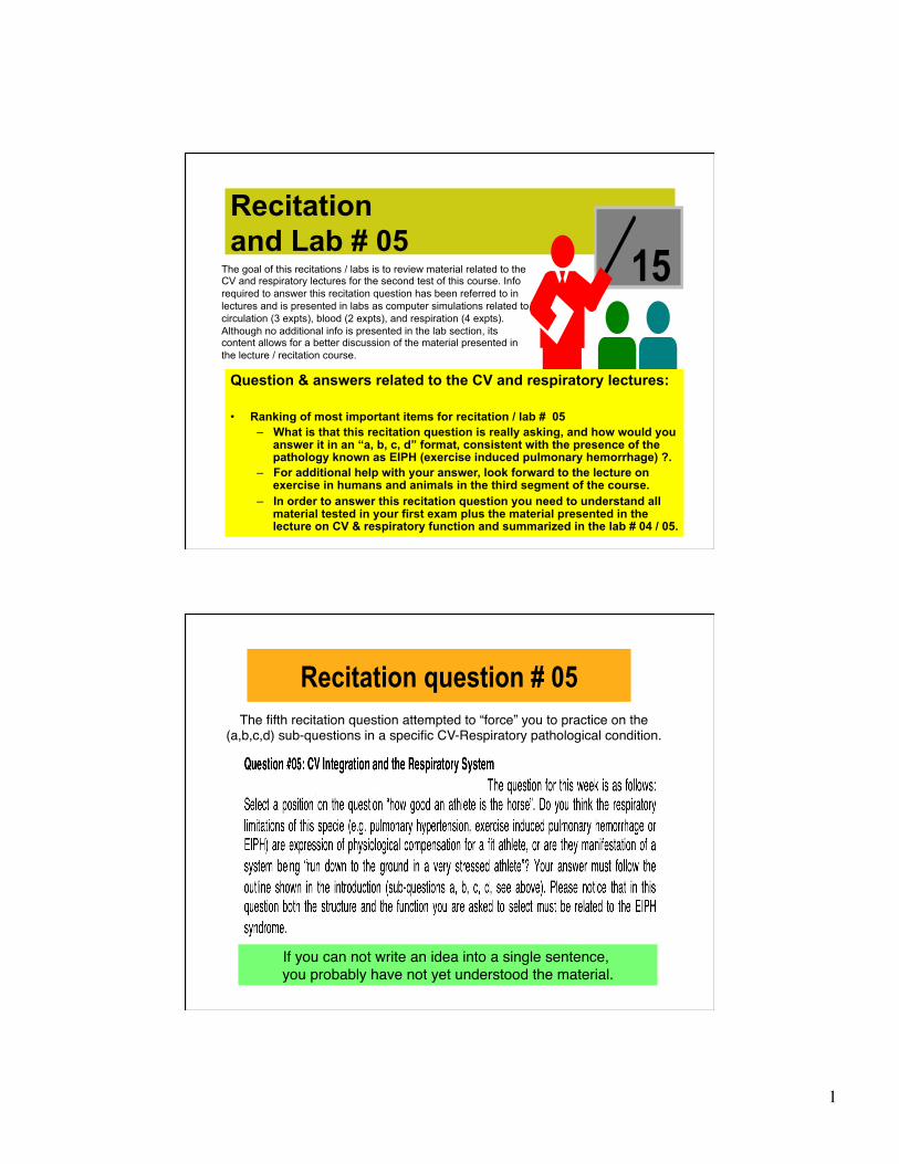
1 Recitation and Lab # 05 15 The goal of this recitations / labs is to review material related to the CV and respiratory lectures for the second test of this course. Info required to answer this recitation question has been referred to in lectures and is presented in labs as computer simulations related to circulation (3 expts), blood (2 expts), and respiration (4 expts). Although no additional info is presented in the lab section, its content allows for a better discussion of the material presented in the lecture / recitation course. Question & answers related to the CV and respiratory lectures: • Ranking of most important items for recitation / lab # 05 – What is that this recitation question is really asking, and how would you answer it in an “a, b, c, d” format, consistent with the presence of the pathology known as EIPH (exercise induced pulmonary hemorrhage) ?. – For additional help with your answer, look forward to the lecture on exercise in humans and animals in the third segment of the course. – In order to answer this recitation question you need to understand all material tested in your first exam plus the material presented in the lecture on CV & respiratory function and summarized in the lab # 04 / 05. Recitation question # 05 The fifth recitation question attempted to “force” you to practice on the (a,b,c,d) sub-questions in a specific CV-Respiratory pathological condition. If you can not write an idea into a single sentence, you probably have not yet understood the material.
Transcript of Recitation and Lab # 05 15 - Rutgers Universityadvis/300_phys_PDFs/300_15_recitation5.pptx.pdf ·...
1
Recitation and Lab # 05 15 The goal of this recitations / labs is to review material related to the
CV and respiratory lectures for the second test of this course. Info required to answer this recitation question has been referred to in lectures and is presented in labs as computer simulations related to circulation (3 expts), blood (2 expts), and respiration (4 expts). Although no additional info is presented in the lab section, its content allows for a better discussion of the material presented in the lecture / recitation course.
Question & answers related to the CV and respiratory lectures: • Ranking of most important items for recitation / lab # 05
– What is that this recitation question is really asking, and how would you answer it in an “a, b, c, d” format, consistent with the presence of the pathology known as EIPH (exercise induced pulmonary hemorrhage) ?.
– For additional help with your answer, look forward to the lecture on exercise in humans and animals in the third segment of the course.
– In order to answer this recitation question you need to understand all material tested in your first exam plus the material presented in the lecture on CV & respiratory function and summarized in the lab # 04 / 05.
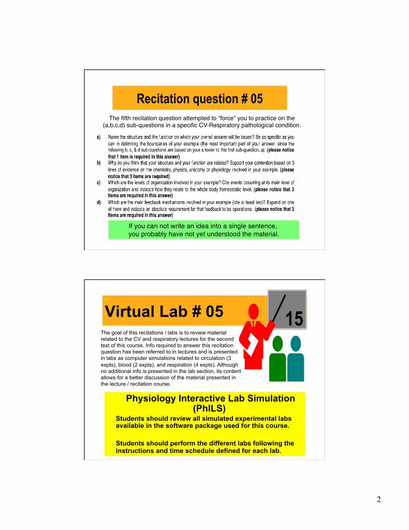
Recitation question # 05 The fifth recitation question attempted to “force” you to practice on the
(a,b,c,d) sub-questions in a specific CV-Respiratory pathological condition. "
If you can not write an idea into a single sentence, "you probably have not yet understood the material.
2
Recitation question # 05 The fifth recitation question attempted to “force” you to practice on the
(a,b,c,d) sub-questions in a specific CV-Respiratory pathological condition. "
If you can not write an idea into a single sentence, "you probably have not yet understood the material.
Virtual Lab # 05
Physiology Interactive Lab Simulation (PhILS)
Students should review all simulated experimental labs available in the software package used for this course.
Students should perform the different labs following the instructions and time schedule defined for each lab.
15 The goal of this recitations / labs is to review material related to the CV and respiratory lectures for the second test of this course. Info required to answer this recitation question has been referred to in lectures and is presented in labs as computer simulations related to circulation (3 expts), blood (2 expts), and respiration (4 expts). Although no additional info is presented in the lab section, its content allows for a better discussion of the material presented in the lecture / recitation course.
3
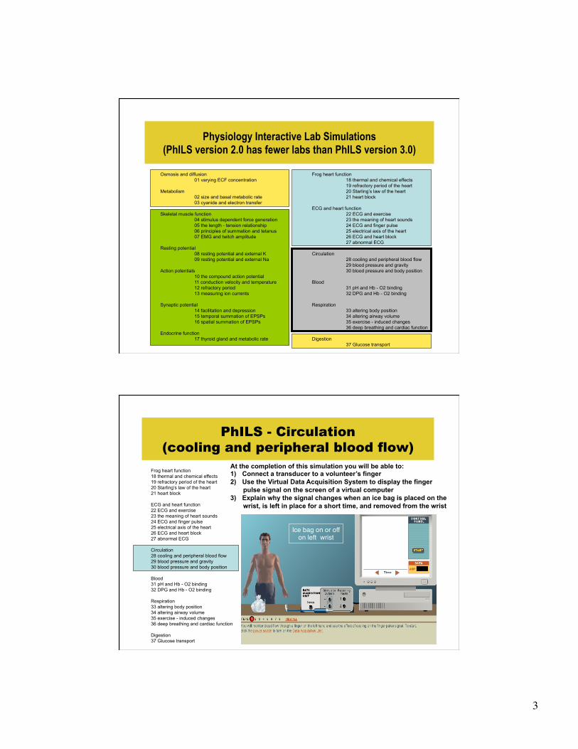
Physiology Interactive Lab Simulations (PhILS version 2.0 has fewer labs than PhILS version 3.0)
Osmosis and diffusion 01 varying ECF concentration
Metabolism
02 size and basal metabolic rate 03 cyanide and electron transfer
Skeletal muscle function
04 stimulus dependent force generation 05 the length - tension relationship 06 principles of summation and tetanus 07 EMG and twitch amplitude
Resting potential
08 resting potential and external K 09 resting potential and external Na
Action potentials
10 the compound action potential 11 conduction velocity and temperature 12 refractory period 13 measuring ion currents
Synaptic potential
14 facilitation and depression 15 temporal summation of EPSPs 16 spatial summation of EPSPs
Endocrine function
17 thyroid gland and metabolic rate
Frog heart function 18 thermal and chemical effects 19 refractory period of the heart 20 Starling’s law of the heart 21 heart block
ECG and heart function
22 ECG and exercise 23 the meaning of heart sounds 24 ECG and finger pulse 25 electrical axis of the heart 26 ECG and heart block 27 abnormal ECG
Circulation
28 cooling and peripheral blood flow 29 blood pressure and gravity 30 blood pressure and body position
Blood
31 pH and Hb - O2 binding 32 DPG and Hb - O2 binding
Respiration
33 altering body position 34 altering airway volume 35 exercise - induced changes 36 deep breathing and cardiac function
Digestion
37 Glucose transport
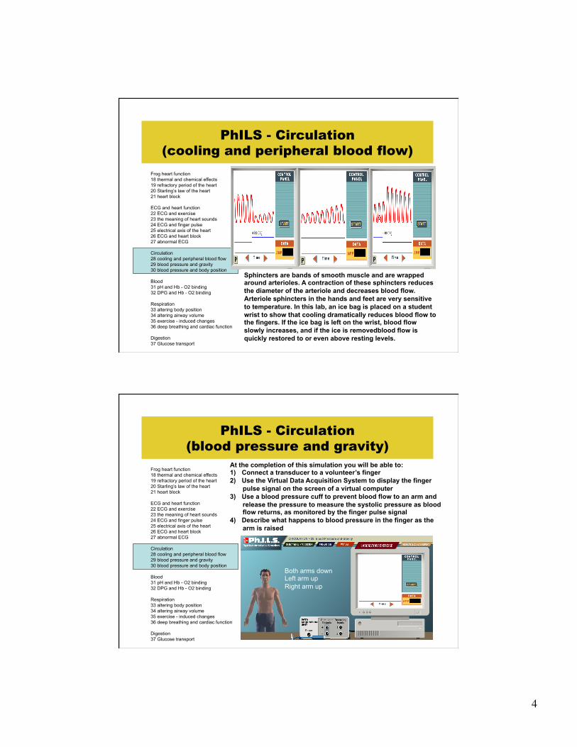
PhILS - Circulation (cooling and peripheral blood flow)
Frog heart function 18 thermal and chemical effects 19 refractory period of the heart 20 Starling’s law of the heart 21 heart block ECG and heart function 22 ECG and exercise 23 the meaning of heart sounds 24 ECG and finger pulse 25 electrical axis of the heart 26 ECG and heart block 27 abnormal ECG Circulation 28 cooling and peripheral blood flow 29 blood pressure and gravity 30 blood pressure and body position Blood 31 pH and Hb - O2 binding 32 DPG and Hb - O2 binding Respiration 33 altering body position 34 altering airway volume 35 exercise - induced changes 36 deep breathing and cardiac function Digestion 37 Glucose transport
At the completion of this simulation you will be able to: 1) Connect a transducer to a volunteer’s finger 2) Use the Virtual Data Acquisition System to display the finger
pulse signal on the screen of a virtual computer 3) Explain why the signal changes when an ice bag is placed on the
wrist, is left in place for a short time, and removed from the wrist
Ice bag on or off on left wrist"
4
PhILS - Circulation (cooling and peripheral blood flow)
Frog heart function 18 thermal and chemical effects 19 refractory period of the heart 20 Starling’s law of the heart 21 heart block ECG and heart function 22 ECG and exercise 23 the meaning of heart sounds 24 ECG and finger pulse 25 electrical axis of the heart 26 ECG and heart block 27 abnormal ECG Circulation 28 cooling and peripheral blood flow 29 blood pressure and gravity 30 blood pressure and body position Blood 31 pH and Hb - O2 binding 32 DPG and Hb - O2 binding Respiration 33 altering body position 34 altering airway volume 35 exercise - induced changes 36 deep breathing and cardiac function Digestion 37 Glucose transport
Sphincters are bands of smooth muscle and are wrapped around arterioles. A contraction of these sphincters reduces the diameter of the arteriole and decreases blood flow. Arteriole sphincters in the hands and feet are very sensitive to temperature. In this lab, an ice bag is placed on a student wrist to show that cooling dramatically reduces blood flow to the fingers. If the ice bag is left on the wrist, blood flow slowly increases, and if the ice is removedblood flow is quickly restored to or even above resting levels.
PhILS - Circulation (blood pressure and gravity)
Frog heart function 18 thermal and chemical effects 19 refractory period of the heart 20 Starling’s law of the heart 21 heart block ECG and heart function 22 ECG and exercise 23 the meaning of heart sounds 24 ECG and finger pulse 25 electrical axis of the heart 26 ECG and heart block 27 abnormal ECG Circulation 28 cooling and peripheral blood flow 29 blood pressure and gravity 30 blood pressure and body position Blood 31 pH and Hb - O2 binding 32 DPG and Hb - O2 binding Respiration 33 altering body position 34 altering airway volume 35 exercise - induced changes 36 deep breathing and cardiac function Digestion 37 Glucose transport
At the completion of this simulation you will be able to: 1) Connect a transducer to a volunteer’s finger 2) Use the Virtual Data Acquisition System to display the finger
pulse signal on the screen of a virtual computer 3) Use a blood pressure cuff to prevent blood flow to an arm and
release the pressure to measure the systolic pressure as blood flow returns, as monitored by the finger pulse signal
4) Describe what happens to blood pressure in the finger as the arm is raised
Both arms down Left arm up Right arm up

5
PhILS - Circulation (blood pressure and gravity)
Frog heart function 18 thermal and chemical effects 19 refractory period of the heart 20 Starling’s law of the heart 21 heart block ECG and heart function 22 ECG and exercise 23 the meaning of heart sounds 24 ECG and finger pulse 25 electrical axis of the heart 26 ECG and heart block 27 abnormal ECG Circulation 28 cooling and peripheral blood flow 29 blood pressure and gravity 30 blood pressure and body position Blood 31 pH and Hb - O2 binding 32 DPG and Hb - O2 binding Respiration 33 altering body position 34 altering airway volume 35 exercise - induced changes 36 deep breathing and cardiac function Digestion 37 Glucose transport
Both arms down Left arm up Right arm up
cuff"
How would this pattern (both arms down) change (sistolic pressure and pulse amplitude), if the right or left arm is raised?"
A force must be applied to move a fluid through a tube. The heart is a pump that provides pressure to push blood through the blood vessels of the circulatory system. The pressure of the fluid will decline if it flows against the force of gravity. This lab demonstrate that elevating a hand above the head decreases the blood pressure and blood flow.
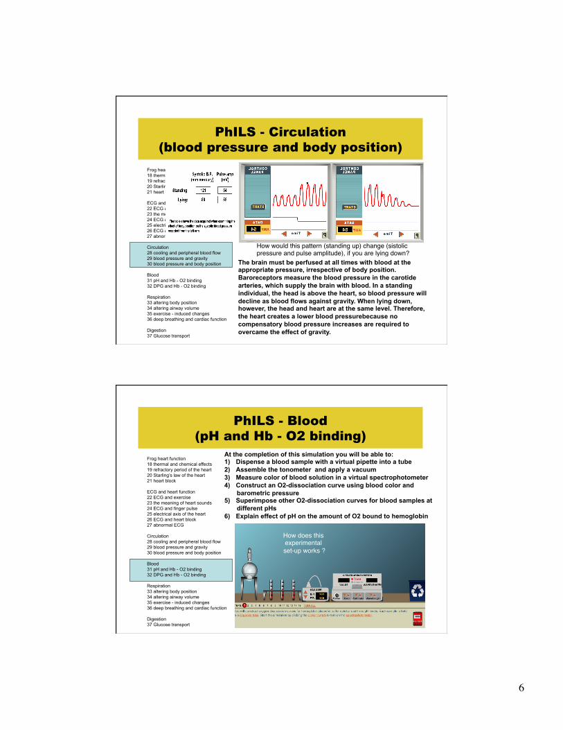
PhILS - Circulation (blood pressure and body position)
Frog heart function 18 thermal and chemical effects 19 refractory period of the heart 20 Starling’s law of the heart 21 heart block ECG and heart function 22 ECG and exercise 23 the meaning of heart sounds 24 ECG and finger pulse 25 electrical axis of the heart 26 ECG and heart block 27 abnormal ECG Circulation 28 cooling and peripheral blood flow 29 blood pressure and gravity 30 blood pressure and body position Blood 31 pH and Hb - O2 binding 32 DPG and Hb - O2 binding Respiration 33 altering body position 34 altering airway volume 35 exercise - induced changes 36 deep breathing and cardiac function Digestion 37 Glucose transport
At the completion of this simulation you will be able to: 1) Connect a transducer to a volunteer’s finger 2) Use the Virtual Data Acquisition System to display the finger
pulse signal on the screen of a virtual computer 3) Use a blood pressure cuff to prevent blood flow to an arm and
release the pressure to measure the systolic pressure as blood flow returns, as monitored by the finger pulse signal
4) Describe what happens to blood pressure when the volunteer lies down
6
PhILS - Circulation (blood pressure and body position)
Frog heart function 18 thermal and chemical effects 19 refractory period of the heart 20 Starling’s law of the heart 21 heart block ECG and heart function 22 ECG and exercise 23 the meaning of heart sounds 24 ECG and finger pulse 25 electrical axis of the heart 26 ECG and heart block 27 abnormal ECG Circulation 28 cooling and peripheral blood flow 29 blood pressure and gravity 30 blood pressure and body position Blood 31 pH and Hb - O2 binding 32 DPG and Hb - O2 binding Respiration 33 altering body position 34 altering airway volume 35 exercise - induced changes 36 deep breathing and cardiac function Digestion 37 Glucose transport
How would this pattern (standing up) change (sistolic pressure and pulse amplitude), if you are lying down?"
The brain must be perfused at all times with blood at the appropriate pressure, irrespective of body position. Baroreceptors measure the blood pressure in the carotide arteries, which supply the brain with blood. In a standing individual, the head is above the heart, so blood pressure will decline as blood flows against gravity. When lying down, however, the head and heart are at the same level. Therefore, the heart creates a lower blood pressurebecause no compensatory blood pressure increases are required to overcame the effect of gravity.
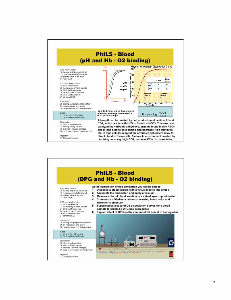
PhILS - Blood (pH and Hb - O2 binding)
Frog heart function 18 thermal and chemical effects 19 refractory period of the heart 20 Starling’s law of the heart 21 heart block ECG and heart function 22 ECG and exercise 23 the meaning of heart sounds 24 ECG and finger pulse 25 electrical axis of the heart 26 ECG and heart block 27 abnormal ECG Circulation 28 cooling and peripheral blood flow 29 blood pressure and gravity 30 blood pressure and body position Blood 31 pH and Hb - O2 binding 32 DPG and Hb - O2 binding Respiration 33 altering body position 34 altering airway volume 35 exercise - induced changes 36 deep breathing and cardiac function Digestion 37 Glucose transport
At the completion of this simulation you will be able to: 1) Dispense a blood sample with a virtual pipette into a tube 2) Assemble the tonometer and apply a vacuum 3) Measure color of blood solution in a virtual spectrophotometer 4) Construct an O2-dissociation curve using blood color and
barometric pressure 5) Superimpose other O2-dissociation curves for blood samples at
different pHs 6) Explain effect of pH on the amount of O2 bound to hemoglobin
How does this experimental set-up works ?"
7
Frog heart function 18 thermal and chemical effects 19 refractory period of the heart 20 Starling’s law of the heart 21 heart block ECG and heart function 22 ECG and exercise 23 the meaning of heart sounds 24 ECG and finger pulse 25 electrical axis of the heart 26 ECG and heart block 27 abnormal ECG Circulation 28 cooling and peripheral blood flow 29 blood pressure and gravity 30 blood pressure and body position Blood 31 pH and Hb - O2 binding 32 DPG and Hb - O2 binding Respiration 33 altering body position 34 altering airway volume 35 exercise - induced changes 36 deep breathing and cardiac function Digestion 37 Glucose transport
PhILS - Blood (pH and Hb - O2 binding)
pH = pK + log HCO3 PCO2
A low pH can be created by cell production of lactic acid and CO2, which reacts with H2O to form H + HCO3. This reaction catalyzed by carbonic anhydrase, enzyme found inside RBCs. The H ions bind to beta chains and decrease Hb’s affinity to O2. In high cellular respiration, arterioles sphincters relax to direct blood to these cells. Factors in environment created by respiring cells, e.g. high CO2, increase O2 – Hb dissociation.
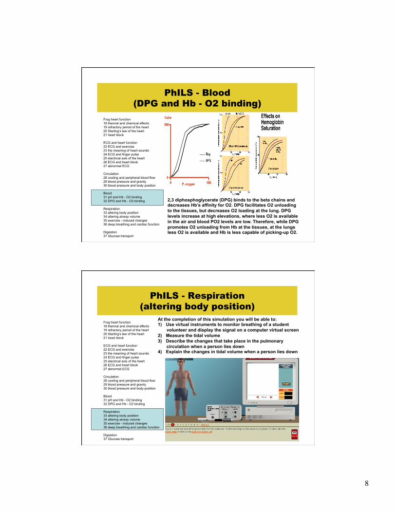
PhILS - Blood (DPG and Hb - O2 binding)
Frog heart function 18 thermal and chemical effects 19 refractory period of the heart 20 Starling’s law of the heart 21 heart block ECG and heart function 22 ECG and exercise 23 the meaning of heart sounds 24 ECG and finger pulse 25 electrical axis of the heart 26 ECG and heart block 27 abnormal ECG Circulation 28 cooling and peripheral blood flow 29 blood pressure and gravity 30 blood pressure and body position Blood 31 pH and Hb - O2 binding 32 DPG and Hb - O2 binding Respiration 33 altering body position 34 altering airway volume 35 exercise - induced changes 36 deep breathing and cardiac function Digestion 37 Glucose transport
At the completion of this simulation you will be able to: 1) Dispense a blood sample with a virtual pipette into a tube 2) Assemble the tonometer and apply a vacuum 3) Measure color of blood solution in a virtual spectrophotometer 4) Construct an O2-dissociation curve using blood color and
barometric pressure 5) Superimpose a second O2-dissociation curves for a blood
sample to which 2,3 DPG has been added 6) Explain effect of DPG on the amount of O2 bound to hemoglobin
8
PhILS - Blood (DPG and Hb - O2 binding)
Frog heart function 18 thermal and chemical effects 19 refractory period of the heart 20 Starling’s law of the heart 21 heart block ECG and heart function 22 ECG and exercise 23 the meaning of heart sounds 24 ECG and finger pulse 25 electrical axis of the heart 26 ECG and heart block 27 abnormal ECG Circulation 28 cooling and peripheral blood flow 29 blood pressure and gravity 30 blood pressure and body position Blood 31 pH and Hb - O2 binding 32 DPG and Hb - O2 binding Respiration 33 altering body position 34 altering airway volume 35 exercise - induced changes 36 deep breathing and cardiac function Digestion 37 Glucose transport
2,3 diphosphoglycerate (DPG) binds to the beta chains and decreases Hb’s affinity for O2. DPG facilitates O2 unloading to the tissues, but decreases O2 loading at the lung. DPG levels increase at high elevations, where less O2 is available in the air and blood PO2 levels are low. Therefore, while DPG promotes O2 unloading from Hb at the tissues, at the lungs less O2 is available and Hb is less capable of picking-up O2.
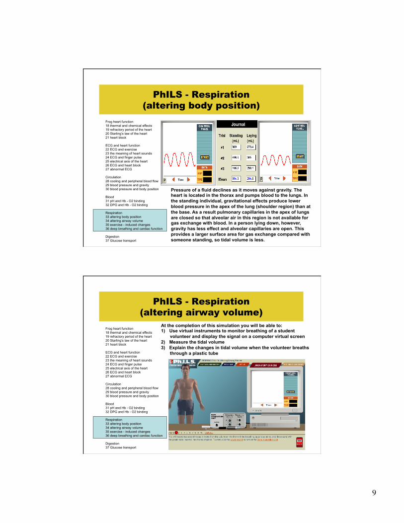
PhILS - Respiration (altering body position)
Frog heart function 18 thermal and chemical effects 19 refractory period of the heart 20 Starling’s law of the heart 21 heart block ECG and heart function 22 ECG and exercise 23 the meaning of heart sounds 24 ECG and finger pulse 25 electrical axis of the heart 26 ECG and heart block 27 abnormal ECG Circulation 28 cooling and peripheral blood flow 29 blood pressure and gravity 30 blood pressure and body position Blood 31 pH and Hb - O2 binding 32 DPG and Hb - O2 binding Respiration 33 altering body position 34 altering airway volume 35 exercise - induced changes 36 deep breathing and cardiac function Digestion 37 Glucose transport
At the completion of this simulation you will be able to: 1) Use virtual instruments to monitor breathing of a student
volunteer and display the signal on a computer virtual screen 2) Measure the tidal volume 3) Describe the changes that take place in the pulmonary
circulation when a person lies down 4) Explain the changes in tidal volume when a person lies down
9
PhILS - Respiration (altering body position)
Frog heart function 18 thermal and chemical effects 19 refractory period of the heart 20 Starling’s law of the heart 21 heart block ECG and heart function 22 ECG and exercise 23 the meaning of heart sounds 24 ECG and finger pulse 25 electrical axis of the heart 26 ECG and heart block 27 abnormal ECG Circulation 28 cooling and peripheral blood flow 29 blood pressure and gravity 30 blood pressure and body position Blood 31 pH and Hb - O2 binding 32 DPG and Hb - O2 binding Respiration 33 altering body position 34 altering airway volume 35 exercise - induced changes 36 deep breathing and cardiac function Digestion 37 Glucose transport
Pressure of a fluid declines as it moves against gravity. The heart is located in the thorax and pumps blood to the lungs. In the standing individual, gravitational effects produce lower blood pressure in the apex of the lung (shoulder region) than at the base. As a result pulmonary capillaries in the apex of lungs are closed so that alveolar air in this region is not available for gas exchange with blood. In a person lying down, however, gravity has less effect and alveolar capillaries are open. This provides a larger surface area for gas exchange compared with someone standing, so tidal volume is less.
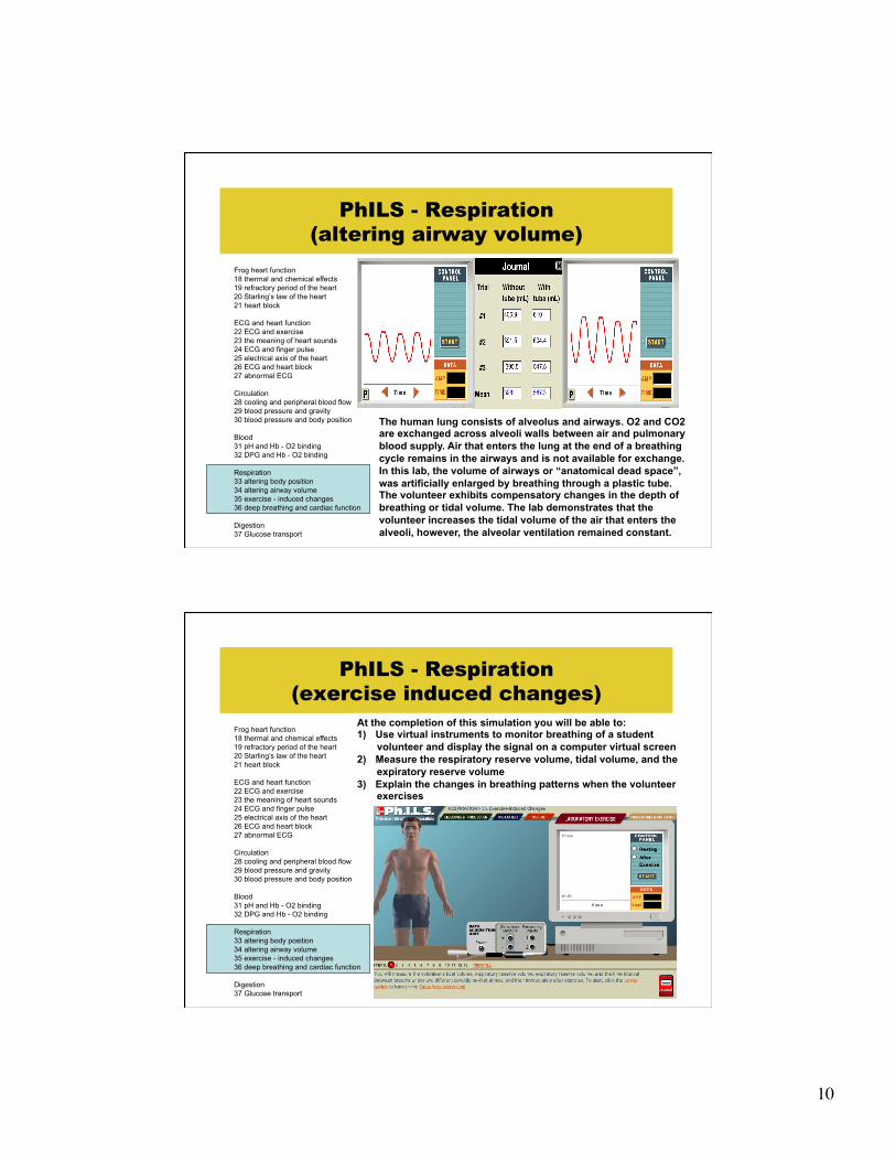
PhILS - Respiration (altering airway volume)
Frog heart function 18 thermal and chemical effects 19 refractory period of the heart 20 Starling’s law of the heart 21 heart block ECG and heart function 22 ECG and exercise 23 the meaning of heart sounds 24 ECG and finger pulse 25 electrical axis of the heart 26 ECG and heart block 27 abnormal ECG Circulation 28 cooling and peripheral blood flow 29 blood pressure and gravity 30 blood pressure and body position Blood 31 pH and Hb - O2 binding 32 DPG and Hb - O2 binding Respiration 33 altering body position 34 altering airway volume 35 exercise - induced changes 36 deep breathing and cardiac function Digestion 37 Glucose transport
At the completion of this simulation you will be able to: 1) Use virtual instruments to monitor breathing of a student
volunteer and display the signal on a computer virtual screen 2) Measure the tidal volume 3) Explain the changes in tidal volume when the volunteer breaths
through a plastic tube
10
PhILS - Respiration (altering airway volume)
Frog heart function 18 thermal and chemical effects 19 refractory period of the heart 20 Starling’s law of the heart 21 heart block ECG and heart function 22 ECG and exercise 23 the meaning of heart sounds 24 ECG and finger pulse 25 electrical axis of the heart 26 ECG and heart block 27 abnormal ECG Circulation 28 cooling and peripheral blood flow 29 blood pressure and gravity 30 blood pressure and body position Blood 31 pH and Hb - O2 binding 32 DPG and Hb - O2 binding Respiration 33 altering body position 34 altering airway volume 35 exercise - induced changes 36 deep breathing and cardiac function Digestion 37 Glucose transport
The human lung consists of alveolus and airways. O2 and CO2 are exchanged across alveoli walls between air and pulmonary blood supply. Air that enters the lung at the end of a breathing cycle remains in the airways and is not available for exchange. In this lab, the volume of airways or “anatomical dead space”, was artificially enlarged by breathing through a plastic tube. The volunteer exhibits compensatory changes in the depth of breathing or tidal volume. The lab demonstrates that the volunteer increases the tidal volume of the air that enters the alveoli, however, the alveolar ventilation remained constant.
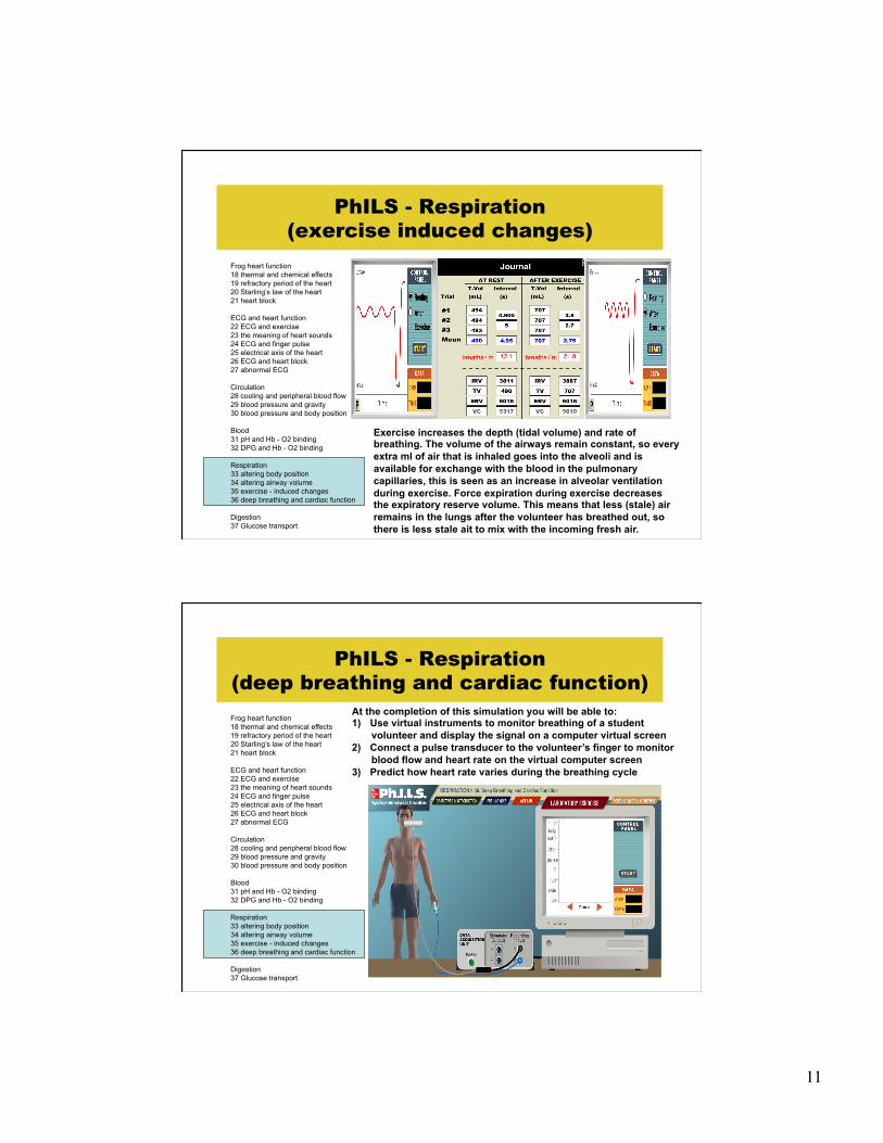
PhILS - Respiration (exercise induced changes)
Frog heart function 18 thermal and chemical effects 19 refractory period of the heart 20 Starling’s law of the heart 21 heart block ECG and heart function 22 ECG and exercise 23 the meaning of heart sounds 24 ECG and finger pulse 25 electrical axis of the heart 26 ECG and heart block 27 abnormal ECG Circulation 28 cooling and peripheral blood flow 29 blood pressure and gravity 30 blood pressure and body position Blood 31 pH and Hb - O2 binding 32 DPG and Hb - O2 binding Respiration 33 altering body position 34 altering airway volume 35 exercise - induced changes 36 deep breathing and cardiac function Digestion 37 Glucose transport
At the completion of this simulation you will be able to: 1) Use virtual instruments to monitor breathing of a student
volunteer and display the signal on a computer virtual screen 2) Measure the respiratory reserve volume, tidal volume, and the
expiratory reserve volume 3) Explain the changes in breathing patterns when the volunteer
exercises
11
PhILS - Respiration (exercise induced changes)
Frog heart function 18 thermal and chemical effects 19 refractory period of the heart 20 Starling’s law of the heart 21 heart block ECG and heart function 22 ECG and exercise 23 the meaning of heart sounds 24 ECG and finger pulse 25 electrical axis of the heart 26 ECG and heart block 27 abnormal ECG Circulation 28 cooling and peripheral blood flow 29 blood pressure and gravity 30 blood pressure and body position Blood 31 pH and Hb - O2 binding 32 DPG and Hb - O2 binding Respiration 33 altering body position 34 altering airway volume 35 exercise - induced changes 36 deep breathing and cardiac function Digestion 37 Glucose transport
Exercise increases the depth (tidal volume) and rate of breathing. The volume of the airways remain constant, so every extra ml of air that is inhaled goes into the alveoli and is available for exchange with the blood in the pulmonary capillaries, this is seen as an increase in alveolar ventilation during exercise. Force expiration during exercise decreases the expiratory reserve volume. This means that less (stale) air remains in the lungs after the volunteer has breathed out, so there is less stale ait to mix with the incoming fresh air.
PhILS - Respiration (deep breathing and cardiac function) Frog heart function 18 thermal and chemical effects 19 refractory period of the heart 20 Starling’s law of the heart 21 heart block ECG and heart function 22 ECG and exercise 23 the meaning of heart sounds 24 ECG and finger pulse 25 electrical axis of the heart 26 ECG and heart block 27 abnormal ECG Circulation 28 cooling and peripheral blood flow 29 blood pressure and gravity 30 blood pressure and body position Blood 31 pH and Hb - O2 binding 32 DPG and Hb - O2 binding Respiration 33 altering body position 34 altering airway volume 35 exercise - induced changes 36 deep breathing and cardiac function Digestion 37 Glucose transport
At the completion of this simulation you will be able to: 1) Use virtual instruments to monitor breathing of a student
volunteer and display the signal on a computer virtual screen 2) Connect a pulse transducer to the volunteer’s finger to monitor
blood flow and heart rate on the virtual computer screen 3) Predict how heart rate varies during the breathing cycle

12
PhILS - Respiration (deep breathing and cardiac function) Frog heart function 18 thermal and chemical effects 19 refractory period of the heart 20 Starling’s law of the heart 21 heart block ECG and heart function 22 ECG and exercise 23 the meaning of heart sounds 24 ECG and finger pulse 25 electrical axis of the heart 26 ECG and heart block 27 abnormal ECG Circulation 28 cooling and peripheral blood flow 29 blood pressure and gravity 30 blood pressure and body position Blood 31 pH and Hb - O2 binding 32 DPG and Hb - O2 binding Respiration 33 altering body position 34 altering airway volume 35 exercise - induced changes 36 deep breathing and cardiac function Digestion 37 Glucose transport
Compare inhalation vs "exhalation for:" "Intrathoraxic pressure (P)""Veins negative P in heart""Heart rate""Stroke volume (estimated)""Venous return to the heart"
The breathing cycle consists of a period of inhalation and exhalation. During inhalation, the volume of the thorax increases and the pressure inside the thorax decreases. This draw air into the lung and also pull blood towards the heart which is located inside the thorax. One theory maintains that the increase in venous return produces a faster stretching of the wall of the atria, and this increases heart rate. During exhalation, the volume of the thorax decreases and pressure of the thorax increases. This pushes air out of the lungs and increases venous return. The result is a decrease in heart rate.