Recent Advances in Drosophila Models of Charcot-Marie ...
19
International Journal of Molecular Sciences Review Recent Advances in Drosophila Models of Charcot-Marie-Tooth Disease Fukiko Kitani-Morii 1,2, * and Yu-ichi Noto 2 1 Department of Molecular Pathobiology of Brain Disease, Kyoto Prefectural University of Medicine, Kyoto 6028566, Japan 2 Department of Neurology, Kyoto Prefectural University of Medicine, Kyoto 6028566, Japan; [email protected] * Correspondence: [email protected]; Tel.: +81-75-251-5793 Received: 31 August 2020; Accepted: 6 October 2020; Published: 8 October 2020 Abstract: Charcot-Marie-Tooth disease (CMT) is one of the most common inherited peripheral neuropathies. CMT patients typically show slowly progressive muscle weakness and sensory loss in a distal dominant pattern in childhood. The diagnosis of CMT is based on clinical symptoms, electrophysiological examinations, and genetic testing. Advances in genetic testing technology have revealed the genetic heterogeneity of CMT; more than 100 genes containing the disease causative mutations have been identified. Because a single genetic alteration in CMT leads to progressive neurodegeneration, studies of CMT patients and their respective models revealed the genotype-phenotype relationships of targeted genes. Conventionally, rodents and cell lines have often been used to study the pathogenesis of CMT. Recently, Drosophila has also attracted attention as a CMT model. In this review, we outline the clinical characteristics of CMT, describe the advantages and disadvantages of using Drosophila in CMT studies, and introduce recent advances in CMT research that successfully applied the use of Drosophila, in areas such as molecules associated with mitochondria, endosomes/lysosomes, transfer RNA, axonal transport, and glucose metabolism. Keywords: Charcot-Marie-Tooth disease (CMT); Drosophila melanogaster; human disease model; neurodegeneration; peripheral neuropathy 1. Introduction 1.1. Clinical Features of CMT Charcot-Marie-Tooth disease (CMT) is the most common inherited peripheral neuropathy. The average prevalence of CMT is reported to be about 1 in 2,500 people [1]; however, the CMT prevalence rate varies markedly among epidemiological studies due to a large variety of CMT symptoms [2]. At present, the reported prevalence of CMT among Europeans is about 10–30 per 100,000 people, while that in the East Asia is 5.3–10.8 per 100,000 people [3–6]. The diagnosis of CMT is based on clinical symptoms, electrophysiological studies, genetic testing, and nerve biopsy [7]. CMT is usually juvenile-onset, and typical symptoms are slow, progressive muscle weakness and sensory disturbance in a distal dominant pattern. Patients show clumsiness, foot deformity (such as pes cavus), and gait disturbance. Some patients show additional symptoms such as hearing loss and scoliosis [8]. Interestingly, there are accumulating case reports of cerebral white matter abnormalities mainly in X-linked CMT type 1, and recently, abnormal diffusion-tensor imaging on brain MRI was shown to correlate with clinical disability in various CMT subgroups, suggesting subclinical central nervous system involvement in addition to clinical peripheral neuropathy [9–11]. Most patients develop symptoms in childhood, while there are marked individual differences in the severity and Int. J. Mol. Sci. 2020, 21, 7419; doi:10.3390/ijms21197419 www.mdpi.com/journal/ijms
Transcript of Recent Advances in Drosophila Models of Charcot-Marie ...
International Journal of
Molecular Sciences
Review
Recent Advances in Drosophila Models ofCharcot-Marie-Tooth Disease
Fukiko Kitani-Morii 1,2,* and Yu-ichi Noto 2
1 Department of Molecular Pathobiology of Brain Disease, Kyoto Prefectural University of Medicine,Kyoto 6028566, Japan
2 Department of Neurology, Kyoto Prefectural University of Medicine, Kyoto 6028566, Japan;[email protected]
* Correspondence: [email protected]; Tel.: +81-75-251-5793
Received: 31 August 2020; Accepted: 6 October 2020; Published: 8 October 2020�����������������
Abstract: Charcot-Marie-Tooth disease (CMT) is one of the most common inherited peripheralneuropathies. CMT patients typically show slowly progressive muscle weakness and sensory lossin a distal dominant pattern in childhood. The diagnosis of CMT is based on clinical symptoms,electrophysiological examinations, and genetic testing. Advances in genetic testing technologyhave revealed the genetic heterogeneity of CMT; more than 100 genes containing the diseasecausative mutations have been identified. Because a single genetic alteration in CMT leads toprogressive neurodegeneration, studies of CMT patients and their respective models revealed thegenotype-phenotype relationships of targeted genes. Conventionally, rodents and cell lines haveoften been used to study the pathogenesis of CMT. Recently, Drosophila has also attracted attention asa CMT model. In this review, we outline the clinical characteristics of CMT, describe the advantagesand disadvantages of using Drosophila in CMT studies, and introduce recent advances in CMTresearch that successfully applied the use of Drosophila, in areas such as molecules associated withmitochondria, endosomes/lysosomes, transfer RNA, axonal transport, and glucose metabolism.
Keywords: Charcot-Marie-Tooth disease (CMT); Drosophila melanogaster; human disease model;neurodegeneration; peripheral neuropathy
1. Introduction
1.1. Clinical Features of CMT
Charcot-Marie-Tooth disease (CMT) is the most common inherited peripheral neuropathy.The average prevalence of CMT is reported to be about 1 in 2,500 people [1]; however, the CMTprevalence rate varies markedly among epidemiological studies due to a large variety of CMTsymptoms [2]. At present, the reported prevalence of CMT among Europeans is about 10–30 per100,000 people, while that in the East Asia is 5.3–10.8 per 100,000 people [3–6]. The diagnosis of CMTis based on clinical symptoms, electrophysiological studies, genetic testing, and nerve biopsy [7].CMT is usually juvenile-onset, and typical symptoms are slow, progressive muscle weakness andsensory disturbance in a distal dominant pattern. Patients show clumsiness, foot deformity (such aspes cavus), and gait disturbance. Some patients show additional symptoms such as hearing loss andscoliosis [8]. Interestingly, there are accumulating case reports of cerebral white matter abnormalitiesmainly in X-linked CMT type 1, and recently, abnormal diffusion-tensor imaging on brain MRI wasshown to correlate with clinical disability in various CMT subgroups, suggesting subclinical centralnervous system involvement in addition to clinical peripheral neuropathy [9–11]. Most patientsdevelop symptoms in childhood, while there are marked individual differences in the severity and
Int. J. Mol. Sci. 2020, 21, 7419; doi:10.3390/ijms21197419 www.mdpi.com/journal/ijms
Int. J. Mol. Sci. 2020, 21, 7419 2 of 19
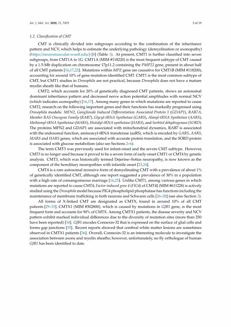
progression rate even in those with the same genetic alteration [12]. A nerve conduction study isperformed for patients to estimate the background pathology of CMT. A decreased nerve conductionvelocity (NCV) (< 38 m/s) indicates CMT1. The main pathological feature of CMT1 is a destructionof the myelin sheath, which is produced by Schwann cells. On the other hand, the presence ofdecreased compound muscle and sensory action potentials with normal NCV (>38 m/s) indicatesCMT2, whose pathological feature is primary axonal damage. The intermediate NCV (30-45 m/s) isassociated with the mixed pathology of damaged myelin (demyelination) and axon (axonopathy) [13].Genetic testing has made marked progress in recent years, and more than 100 genes containingcausative mutations have been identified with the widespread use of next-generation sequencingtechnologies, revealing significant genetic heterogeneity of CMT [2,14,15] (Figure 1). Previous studiesshowed that about 60% of CMT patients received a definitive diagnosis by genetic testing, andover 90% of genetically diagnosed patients show an alteration in one of the following four genes:Peripheral Myelin Protein 22-kDa (PMP22), Gap Junction Beta 1 (GJB1), Myelin Protein Zero (MPZ), andMitofusin 2 (MFN2) [2,16,17]. However, in the Mediterranean area (e.g., southern Italy and easternSpain), mutations in Ganglioside-induced differentiation-associated protein 1 (GDAP1) are the third mostcommon genetic diagnosis of CMT after PMP22 duplication and mutations in GJB1 [18,19]. No GDAP1mutation was identified in a German cohort, suggesting that the geographical area affects the geneticdistribution [20]. Nerve biopsy, which was previously the key diagnostic step, is being replacedby genetic testing, but it is still important in atypical cases [7]. Typical findings of nerve biopsy indemyelinating-type CMT are onion bulb formation and a thinned myelin sheath, which are the resultof repeated de- and remyelination, and these findings are uniformly observed throughout the nerve.In contrast, axonopathy leads to a decreased number of axons and the disappearance of Schwann cellsin the absence of demyelination [21].
Int. J. Mol. Sci. 2020, 21, x FOR PEER REVIEW 2 of 17
performed for patients to estimate the background pathology of CMT. A decreased nerve conduction velocity (NCV) (< 38 m/s) indicates CMT1. The main pathological feature of CMT1 is a destruction of the myelin sheath, which is produced by Schwann cells. On the other hand, the presence of decreased compound muscle and sensory action potentials with normal NCV (> 38 m/s) indicates CMT2, whose pathological feature is primary axonal damage. The intermediate NCV (30-45 m/s) is associated with the mixed pathology of damaged myelin (demyelination) and axon (axonopathy) [13]. Genetic testing has made marked progress in recent years, and more than 100 genes containing causative mutations have been identified with the widespread use of next-generation sequencing technologies, revealing significant genetic heterogeneity of CMT [2,14,15] (Figure 1). Previous studies showed that about 60% of CMT patients received a definitive diagnosis by genetic testing, and over 90% of genetically diagnosed patients show an alteration in one of the following four genes: Peripheral Myelin Protein 22-kDa (PMP22), Gap Junction Beta 1 (GJB1), Myelin Protein Zero (MPZ), and Mitofusin 2 (MFN2) [2,16,17]. However, in the Mediterranean area (e.g., southern Italy and eastern Spain), mutations in Ganglioside-induced differentiation-associated protein 1 (GDAP1) are the third most common genetic diagnosis of CMT after PMP22 duplication and mutations in GJB1 [18,19]. No GDAP1 mutation was identified in a German cohort, suggesting that the geographical area affects the genetic distribution [20]. Nerve biopsy, which was previously the key diagnostic step, is being replaced by genetic testing, but it is still important in atypical cases [7]. Typical findings of nerve biopsy in demyelinating-type CMT are onion bulb formation and a thinned myelin sheath, which are the result of repeated de- and remyelination, and these findings are uniformly observed throughout the nerve. In contrast, axonopathy leads to a decreased number of axons and the disappearance of Schwann cells in the absence of demyelination [21].
Figure 1. Schematic summary showing various Charcot-Marie-Tooth disease (CMT)-related genes and pathways in the peripheral nerve. If each gene has multiple functions, the most representative one is described. The enlarged box shows a cross-sectional view of the peripheral nerve.
1.2. Classification of CMT
CMT is clinically divided into subgroups according to the combination of the inheritance pattern and NCV, which helps to estimate the underlying pathology (demyelination or axonopathy)
Figure 1. Schematic summary showing various Charcot-Marie-Tooth disease (CMT)-related genes andpathways in the peripheral nerve. If each gene has multiple functions, the most representative one isdescribed. The enlarged box shows a cross-sectional view of the peripheral nerve.
Int. J. Mol. Sci. 2020, 21, 7419 3 of 19
1.2. Classification of CMT
CMT is clinically divided into subgroups according to the combination of the inheritancepattern and NCV, which helps to estimate the underlying pathology (demyelination or axonopathy)(https://neuromuscular.wustl.edu/) [8] (Table 1). At present, CMT1 is further classified into sevensubgroups, from CMT1A to 1G. CMT1A (MIM #118220) is the most frequent subtype of CMT causedby a 1.5-Mb duplication on chromosome 17p11.2 containing the PMP22 gene, present in about halfof all CMT patients [16,17,22]. Mutations within MPZ gene are causative for CMT1B (MIM #118200),accounting for around 10% of gene-mutation-identified CMT. CMT1 is the most common subtype ofCMT, but CMT1 studies in Drosophila are not practical, because Drosophila does not have a maturemyelin sheath like that of humans.
CMT2, which accounts for 20% of genetically diagnosed CMT patients, shows an autosomaldominant inheritance pattern and decreased nerve action potential amplitudes with normal NCV(which indicates axonopathy) [16,17]. Among many genes in which mutations are reported to causeCMT2, research on the following important genes and their functions has markedly progressed usingDrosophila models: MFN2, Ganglioside Induced Differentiation Associated Protein 1 (GDAP1), RAB7A,Member RAS Oncogene Family (RAB7), Glycyl-tRNA Synthetase (GARS), Alanyl-tRNA Synthetase (AARS),Methionyl-tRNA Synthetase (MARS), Histidyl-tRNA synthetase (HARS), and Sorbitol dehydrogenase (SORD).The proteins MFN2 and GDAP1 are associated with mitochondrial dynamics, RAB7 is associatedwith the endosomal function, aminoacyl-tRNA transferase (aaRS), which is encoded by GARS, AARS,MARS and HARS genes, which are associated with accurate protein translation, and the SORD proteinis associated with glucose metabolism (also see Sections 2–6).
The term CMT3 was previously used for infant-onset and the severe CMT subtype. However,CMT3 is no longer used because it proved to be a severe form of early-onset CMT1 or CMT4 by geneticanalysis. CMT3, which was historically termed Dejerine–Sottas neuropathy, is now known as thecomponent of the hereditary neuropathies with infantile onset [23,24].
CMT4 is a rare autosomal recessive form of demyelinating CMT with a prevalence of about 1%of genetically identified CMT, although one report suggested a prevalence of 30% in a populationwith a high rate of consanguineous marriage [16,25]. Unlike CMT1, among various genes in whichmutations are reported to cause CMT4, Factor-induced gene 4 (FIG4) of CMT4J (MIM #611228) is activelystudied using the Drosophila model because FIG4 phospholipid phosphatase has functions including themaintenance of membrane trafficking in both neurons and Schwann cells [26–28] (see also Section 3).
All forms of X-linked CMT are designated as CMTX, found in around 10% of all CMTpatients [29–33]. CMTX1 (MIM #302800), which is caused by mutations in GJB1 gene, is the mostfrequent form and accounts for 90% of CMTX. Among CMTX1 patients, the disease severity and NCVpattern exhibit marked individual differences due to the diversity of mutation sites (more than 250have been reported) [34]. GJB1 encodes Connexin-32 that is expressed on the surface of glial cells andforms gap junctions [35]. Recent reports showed that cerebral white matter lesions are sometimesobserved in CMTX1 patients [36]. Overall, Connexin-32 is an interesting molecule to investigate theassociation between axons and myelin sheaths; however, unfortunately, no fly orthologue of humanGJB1 has been identified to date.
Int. J. Mol. Sci. 2020, 21, 7419 4 of 19
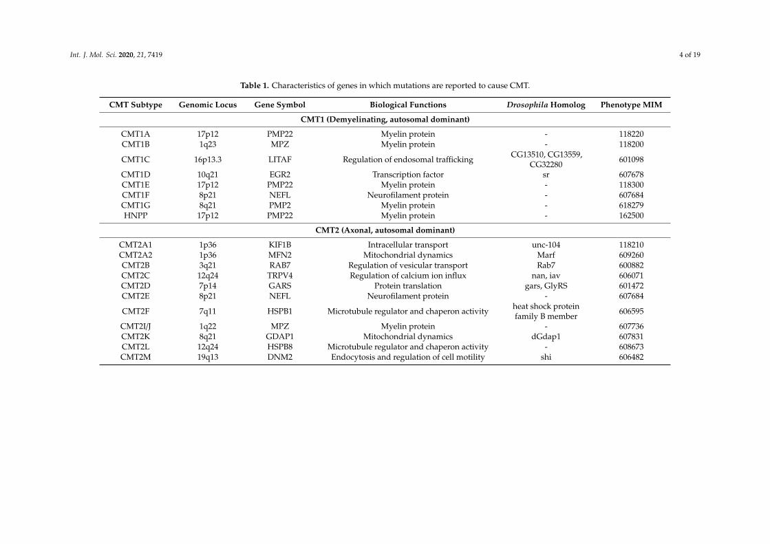
Table 1. Characteristics of genes in which mutations are reported to cause CMT.
CMT Subtype Genomic Locus Gene Symbol Biological Functions Drosophila Homolog Phenotype MIM
CMT1 (Demyelinating, autosomal dominant)
CMT1A 17p12 PMP22 Myelin protein - 118220CMT1B 1q23 MPZ Myelin protein - 118200
CMT1C 16p13.3 LITAF Regulation of endosomal trafficking CG13510, CG13559,CG32280 601098
CMT1D 10q21 EGR2 Transcription factor sr 607678CMT1E 17p12 PMP22 Myelin protein - 118300CMT1F 8p21 NEFL Neurofilament protein - 607684CMT1G 8q21 PMP2 Myelin protein - 618279HNPP 17p12 PMP22 Myelin protein - 162500
CMT2 (Axonal, autosomal dominant)
CMT2A1 1p36 KIF1B Intracellular transport unc-104 118210CMT2A2 1p36 MFN2 Mitochondrial dynamics Marf 609260CMT2B 3q21 RAB7 Regulation of vesicular transport Rab7 600882CMT2C 12q24 TRPV4 Regulation of calcium ion influx nan, iav 606071CMT2D 7p14 GARS Protein translation gars, GlyRS 601472CMT2E 8p21 NEFL Neurofilament protein - 607684
CMT2F 7q11 HSPB1 Microtubule regulator and chaperon activity heat shock proteinfamily B member 606595
CMT2I/J 1q22 MPZ Myelin protein - 607736CMT2K 8q21 GDAP1 Mitochondrial dynamics dGdap1 607831CMT2L 12q24 HSPB8 Microtubule regulator and chaperon activity - 608673CMT2M 19q13 DNM2 Endocytosis and regulation of cell motility shi 606482
Int. J. Mol. Sci. 2020, 21, 7419 5 of 19
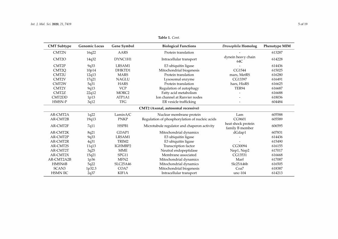
Table 1. Cont.
CMT Subtype Genomic Locus Gene Symbol Biological Functions Drosophila Homolog Phenotype MIM
CMT2N 16q22 AARS Protein translation - 613287
CMT2O 14q32 DYNC1H1 Intracellular transport dynein heavy chain64C 614228
CMT2P 9q33 LRSAM1 E3 ubiquitin ligase - 614436CMT2Q 10p14 DHKTD1 Mitochondrial biogenesis CG1544 615025CMT2U 12q13 MARS Protein translation mars, MetRS 616280CMT2V 17q21 NAGLU Lysosomal enzyme CG13397 616491CMT2W 5q31 HARS Protein translation hars, HisRS 616625CMT2Y 9q13 VCP Regulation of autophagy TER94 616687CMT2Z 22q12 MORC2 Fatty acid metabolism - 616688
CMT2DD 1p13 ATP1A1 Ion channel at Ranvier nodes - 618036HMSN-P 3q12 TFG ER vesicle trafficking - 604484
CMT2 (Axonal, autosomal recessive)
AR-CMT2A 1q22 LaminA/C Nuclear membrane protein Lam 605588AR-CMT2B 19q13 PNKP Regulation of phosphorylation of nucleic acids CG9601 605589
AR-CMT2F 7q11 HSPB1 Microtubule regulator and chaperon activity heat shock proteinfamily B member 606595
AR-CMT2K 8q21 GDAP1 Mitochondrial dynamics dGdap1 607831AR-CMT2P 9q33 LRSAM1 E3 ubiquitin ligase - 614436AR-CMT2R 4q31 TRIM2 E3 ubiquitin ligase - 615490AR-CMT2S 11q13 IGHMBP2 Transcription factor CG30094 616155AR-CMT2T 3q25 MME Neutral endopeptidase Nep1, Nep2 617017AR-CMT2X 15q21 SPG11 Membrane associated CG13531 616668
AR-CMT2A2B 1p36 MFN2 Mitochondrial dynamics Marf 617087HMSN6B 5q22 SLC25A46 Mitochondrial dynamics Slc25A46b 616505SCAN3 1p32.3 COA7 Mitochondrial biogenesis Coa7 618387
HSMN IIC 2q37 KIF1A Intracellular transport unc-104 614213
Int. J. Mol. Sci. 2020, 21, 7419 6 of 19
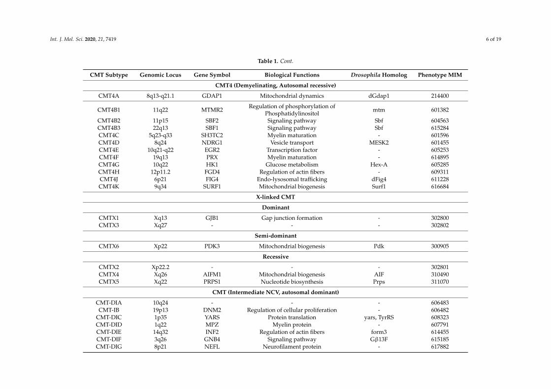
Table 1. Cont.
CMT Subtype Genomic Locus Gene Symbol Biological Functions Drosophila Homolog Phenotype MIM
CMT4 (Demyelinating, Autosomal recessive)
CMT4A 8q13-q21.1 GDAP1 Mitochondrial dynamics dGdap1 214400
CMT4B1 11q22 MTMR2 Regulation of phosphorylation ofPhosphatidylinositol mtm 601382
CMT4B2 11p15 SBF2 Signaling pathway Sbf 604563CMT4B3 22q13 SBF1 Signaling pathway Sbf 615284CMT4C 5q23-q33 SH3TC2 Myelin maturation - 601596CMT4D 8q24 NDRG1 Vesicle transport MESK2 601455CMT4E 10q21-q22 EGR2 Transcription factor - 605253CMT4F 19q13 PRX Myelin maturation - 614895CMT4G 10q22 HK1 Glucose metabolism Hex-A 605285CMT4H 12p11.2 FGD4 Regulation of actin fibers - 609311CMT4J 6p21 FIG4 Endo-lysosomal trafficking dFig4 611228CMT4K 9q34 SURF1 Mitochondrial biogenesis Surf1 616684
X-linked CMT
Dominant
CMTX1 Xq13 GJB1 Gap junction formation - 302800CMTX3 Xq27 - - - 302802
Semi-dominant
CMTX6 Xp22 PDK3 Mitochondrial biogenesis Pdk 300905
Recessive
CMTX2 Xp22.2 - - - 302801CMTX4 Xq26 AIFM1 Mitochondrial biogenesis AIF 310490CMTX5 Xq22 PRPS1 Nucleotide biosynthesis Prps 311070
CMT (Intermediate NCV, autosomal dominant)
CMT-DIA 10q24 - - - 606483CMT-IB 19p13 DNM2 Regulation of cellular proliferation - 606482
CMT-DIC 1p35 YARS Protein translation yars, TyrRS 608323CMT-DID 1q22 MPZ Myelin protein - 607791CMT-DIE 14q32 INF2 Regulation of actin fibers form3 614455CMT-DIF 3q26 GNB4 Signaling pathway Gβ13F 615185CMT-DIG 8p21 NEFL Neurofilament protein - 617882
Int. J. Mol. Sci. 2020, 21, 7419 7 of 19
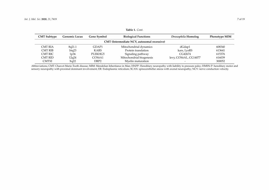
Table 1. Cont.
CMT Subtype Genomic Locus Gene Symbol Biological Functions Drosophila Homolog Phenotype MIM
CMT (Intermediate NCV, autosomal recessive)
CMT RIA 8q21.1 GDAP1 Mitochondrial dynamics dGdap1 608340CMT RIB 16q23 KARS Protein translation kars, LysRS 613641CMT RIC 1p36 PLEKHG5 Signaling pathway CG42674 615376CMT RID 12q24 COX6A1 Mitochondrial biogenesis levy, COX6AL, CG14077 616039
CMTXI Xq22 DRP2 Myelin maturation - 300052
Abbreviations; CMT: Charcot-Marie-Tooth disease; MIM: Mendelian Inheritance in Man; HNPP: Hereditary neuropathy with liability to pressure palsy; HMSN-P: hereditary motor andsensory neuropathy with proximal dominant involvement; ER: Endoplasmic reticulum; SCAN: spinocerebellar ataxia with axonal neuropathy; NCV: nerve conduction velocity.
Int. J. Mol. Sci. 2020, 21, 7419 8 of 19
1.3. Various CMT Models
Until now, various models such as rodents, the zebrafish, fruit fly, yeast, cell lines and inducedpluripotent stem cells (iPSCs) have been developed for CMT modeling [37–57]. Rodent CMT modelshave advanced understanding of neuropathy; however, regarding MFN2 of CMT2A, which is the mostfrequent subtype of axonal CMT, it used to be difficult to reproduce the phenotype in rodent due toembryonic lethality [58]. In addition, such models are expensive to maintain and are subject to ethicalrestrictions. Except for the Cartagena Protocol on Biosafety, there are currently no ethical or socialrestrictions on experiments with Drosophila (https://bch.cbd.int/protocol). Regarding the iPSC models,it is also an important tool for analyzing tissues not suitable for biopsy, such as nerves, but thereis a limitation whereby the results of in vitro experiments are markedly influenced by the cultureconditions. Compared with rodents and iPSCs, the strengths of Drosophila as a laboratory animal are asfollows: fewer ethical restrictions, a short lifecycle, a large number of genetically homogenous offspring,low maintenance cost and established genetic engineering techniques [59,60]. In the Drosophila CMTmodel, motor deficit in CMT patients is reproduced by a decline in climbing ability during the adultstage and in crawling ability during the larval stage [61]. The locomotive ability of adult flies is assessedby a negative geotaxis assay, first described as climbing activity [62]. Gently tapping a vial containingflies causes them to drop to the bottom, and flies are recorded as they climb up the wall of the vial.The percentage of flies that reach a certain height within a specified time is measured and statisticallyanalyzed. On the other hand, the crawling ability of larvae is used as a method to measure larvallocomotive ability [63]. In the larval crawling assay, larvae in the third instar stage are placed on an agarplate and recorded, and the speed of larval migration and distance are statistically analyzed. AlthoughDrosophila does not have a mature myelin sheath, the basic structure and physiology of the axons arehighly conserved between humans and Drosophila, and progress has been made mainly in axonal CMTresearch. Here, we introduce Drosophila models that aided in elucidating the pathophysiology of CMT.
2. Drosophila CMT Models for Investigating Aberrant Mitochondrial Dynamics
Mitochondria are highly dynamic organelles that are responsible for cell viability. Mitochondriahave various functions, such as ATP production, apoptosis, regulation of calcium signaling andreactive oxygen species (ROS) production. Mitochondria continuously repeat fusion and fission tomaintain their homeostasis and functions. Mitochondrial fusion promotes diffusion of the matrixcontent and dilution of oxidized metabolism and damaged mitochondrial DNA, whereas fission isan important process in mitophagy [64]. Mitochondrial fusion and fission are complex processes,and how mitochondrial abnormalities cause disease is not fully understood. However, mutants offollowing mitochondrial molecules are known to associated with CMT and have been actively studiedusing Drosophila: MFN2, ganglioside-induced differentiation associated protein 1 (GDAP1), and solutecarrier family 25 member 46 (SLC25A46).
2.1. MFN2
MFN2 is a GTPase localized to the outer mitochondrial membrane and forms a dimer duringthe process of mitochondrial fusion [65]. Mutations of the MFN2 gene are causative for CMT2A2(MIM #609260), which is the most common genotype of inherited axonal-type neuropathy. A previousstudy showed that mammalian MFN2 mutations impaired axonal transport of mitochondria [66].Failure to meet the demand for ATP at the distal axon is one possible cause of neurodegeneration inaxonal CMT. Knockdown of Mitochondrial assembly regulatory factor (Marf ), the Drosophila homolog ofhuman MFN2, also led to phenotypes as follows: (1) motor dysfunction (reduced climbing ability)that rescued with knock-in of the human wild-type MFN2 but not with MFN2 with R94Q mutation(one of the most common mutations associated with CMT2A), (2) impaired transport of mitochondriato the distal part of the axon and (3) fragmented and clustered mitochondria [67]. These phenotypescould reproduce key symptoms of patients. The Marf knockdown strain also showed fragmented
Int. J. Mol. Sci. 2020, 21, 7419 9 of 19
endoplasmic reticulum (ER) cisternae and increased levels of ER stress markers such as X-box bindingprotein 1 (Xbp1) and binding immunoglobulin protein (BiP), suggesting that ER stress is also involvedin the pathogenicity of Marf knockdown [67].
Regarding the relationship between mitochondrial morphology and function, Trevisan et al.reported that the neural function was dependent on the capacity of mitochondrial energy production,but was independent of their morphology and distribution [68]. They reported that the singleknockdown of Marf or Optic atrophy gene 1 (Opa1), each encoding a key molecule for mitochondrialfusion, caused mitochondrial fragmentation, impaired transport of mitochondria, impaired ATPproduction, and increased lethality in adult flies. Interestingly, double knockdown of genes Marf andDynamin related protein 1 (Drp1), which encoded another GTPase necessary for mitochondrial fission,improved the capacity for ATP production and survival rate but did not rescue aberrant mitochondrialmorphology and distribution. On the other hand, double knockdown of Opa1 and Drp1 rescued aberrantmitochondrial morphology and distribution, but it did not improve the capacity for ATP productionor viability. These results indicate that neuronal cell viability depends on mitochondrial functionsrather than the mitochondrial distribution or morphology. In terms of the relationship betweenthe mitochondrial function and morphology, El Fissi et al. reported the diversity of the impairedmitochondrial function and morphology depending on the mutation site in Marf. Interestingly, all Marftransgenic flies showed reduced climbing ability; flies carrying mutations within the GTPase domain(corresponds to R94Q and T105M in MFN2) of Marf showed unfused and aggregated mitochondria.On the other hand, flies carrying mutations within the helix bundle 1 domain (corresponds to R364Wand L76P in MFN2) of Marf showed enhanced mitochondrial fusion and giant mitochondria, rescued byoverexpression of the fission factor encoding the gene Drp1 [69]. From these results, not only impairedmitochondrial fusion but also excessive fusion of mitochondria may underlie CMT caused by mutantMFN2, and the diversity of the mutation site in the MFN2 gene and various functional alterations ofMFN2 protein caused by each MFN2 mutation may lead to the marked individual differences noted inCMT2A patients.
With respect to the development of treatment, Garrido-Maraver et al. showed that folatemetabolism-related gene expression was upregulated in Marf knockdown Drosophila. Although oralfolate supplementation did not have a therapeutic effect on this model, overexpression of one geneinvolved in folate metabolism reduced mortality and ameliorated locomotor deficits in Marf knockdownDrosophila [70]. These results may help to develop new therapeutic targets. Consequently, Marf/MFN2is one of the most important molecules in elucidating the effects of mitochondrial abnormality onhuman disease, and it is expected to continue to be actively studied in Drosophila.
2.2. GDAP1
Mutations within the GDAP1 gene are causative for CMT4A (MIM #214400, classified intodemyelinating CMT) or CMT2K (MIM #607831, classified into axonal CMT) [71,72]. GDAP1 is atransmembrane protein present in the outer mitochondrial membrane of both neurons and Schwanncells, similar in structure to glutathione S-transferase (GST) but without GST activity [73,74]. GDAP1 isinvolved in regulation of the mitochondrial morphology and function, but much remains unknown.GDAP1 gene mutations in patients with an autosomal recessive inheritance pattern were reported toimpair mitochondrial fission; however, impaired fusion was also observed in the presence of GDAP1mutations in those with an autosomal dominant inheritance pattern [73,74]. López Del Amo et al.showed that knockdown of dGdap1, which is a homolog of human GDAP1, caused degeneration ofthe fly’s retina and muscle, and these phenotypes were rescued by human GDAP1 expression [75].Both knockdown and overexpression of dGdap1 resulted in reduced climbing ability. Knockdownof dGdap1 also resulted in mitochondrial aggregation and large, elongated mitochondria in muscle.On the other hand, overexpression of dGdap1 resulted in a decrease in the size of mitochondria andcluster formation of mitochondria in the retina. Additionally, with both the overexpression andknockdown of dGdap1, early inactivation of the insulin pathway followed by reduced carbohydrate
Int. J. Mol. Sci. 2020, 21, 7419 10 of 19
degradation and increased β-oxidation of lipids were observed [75,76]. These dGdap1-based resultssuggest that impaired energy metabolism due to mitochondrial dysfunction may be closely associatedwith neurodegeneration.
2.3. SLC25A46
SLC25A46 is a transmembrane protein that exists in the outer mitochondrial membrane and isconsidered to be involved in mitochondrial fission. Mutations within the SLC25A46 gene are causativefor hereditary motor and sensory neuropathy (HMSN) type 6B (MIM #616505), and patients exhibit theautosomal recessive inherited form of axonal neuropathy and optic nerve atrophy [50]. The SLC25A46knockdown mouse model presented with optic nerve atrophy, axonal degeneration in the peripheralnervous system, giant mitochondria and severe ataxia due to shedding of Purkinje cells in thecerebellum [77]. There are two SLC25A46 paralogues in Drosophila, SLC25A46a [53] and SLC25A46b [54].Previous reports showed that knockdown of each of SLC25A46a and SLC25A46b in Drosophila similarlyled to motor deficit such as reduced crawling and climbing abilities, a shortened synaptic length in theneuromuscular junction (NMJ), decreased ATP production and ROS accumulation [78,79].
It was also reported that human SLC25A46b and Histone deacetylase 1 (HDAC1) interacted witheach other based on bioinfomatics analysis, and that Drosophila Histone deacetylase RPD3 (Rpd3), ahomolog of human HDAC1, encoded the protein which regulated the acetylation of histone H4K8 in theDrosophila SLC25A46b genomic region. The down-regulation of Rpd3 expression reduced motor deficitsand synaptic morphological abnormalities caused by dSCL25A46b knockdown [80]. These findingssuggest a novel perspective whereby not only genetic factors, but also epigenetic regulators, may beinvolved in neurodegeneration due to mitochondrial dysfunction.
3. Drosophila CMT Models for Investigating Membrane Trafficking Defects
3.1. FIG4
Mutations within the FIG4 gene are causative for CMT4J (MIM #611228) and amyotrophic lateralsclerosis (ALS) type 11 (MIM #612577) [81,82]. ALS is a progressive neurodegenerative diseasethat selectively damages motor neurons, resulting in motor deficits and fatal respiratory muscleparalysis. FIG4 encodes a phosphatase present on the surface of late endosomal membranes andit forms a protein complex with Fab1, which is a lipid kinase, and Vac14. This complex regulatesthe conversion between phosphatidylinositol 3,5-bisphosphate (PI [3,5] P2) and phosphatidylinositol3-phosphate (PI3P), with both being lipids making up membranes that are also responsible for signaltransduction [83]. However, recent studies of Drosophila revealed a different function of FIG4 otherthan phosphatase. In Drosophila, dFIG4 is a single homolog for human FIG4. Neuron-specific dFIG4knockdown Drosophila showed ALS-like symptoms such as reduced motor performance, an abnormalmorphology of NMJ in motor neurons, and a shortened lifespan. In addition, interestingly, these fliesalso showed enlarged lysosomes that were partially rescued by expression of the catalytically inactivedFIG4 mutant protein [84,85]. This suggests that dFIG4 functions independently of enzymatic activityto maintain lysosomal membrane homeostasis.
Furthermore, Muraoka et al. and Shimada et al., using genetic screening, showed that Drosophilalong non-coding RNA (lncRNA) interacted genetically with dFIG4 and that knockdown of this lncRNAimproved phenotypes including motor deficits such as reduced climbing ability and enlarged lysosomescaused by dFIG4 knockdown [86,87]. Up to now, exome analyses are mainly carried out for the geneticdiagnosis of CMT. However, these reports implicate the importance of information on non-proteincoding regions in the human genome to elucidate the pathogenesis of CMT. Although the role of theselncRNAs is not yet well-understood, lysosomal membrane metabolism by the FIG4 complex mayinvolve more molecules than previously considered.
Int. J. Mol. Sci. 2020, 21, 7419 11 of 19
3.2. RAB7
Mutations within RAB7 gene are causative for CMT2B (MIM #606071). Rab7 is a small GTPaseinvolved in late endosomal maturation [88,89]. Janssens et al. reported that CMT2B model Drosophila,which was expressing a CMT2B-causing Rab7 mutation in both alleles in all sensory neurons, showed areduced response to temperature and pain stimuli as well as motor deficit, being similar to thesymptoms of CMT2B patients. In the thermotaxis assay, the third instar larvae were placed in thecenter of a Petri dish with one side warmed to 30°C and the other maintained at 22°C. Then, less than10% of control larvae were on 30°C side, while 17.4% of CMT2B model larvae were on the 30°C side,suggesting impaired ability of larvae to perceive non-optimal temperatures in this model. In thenociception assay, the authors subjected larvae to touch with a soldering iron maintained at 43°Cas a pain stimulus, which caused a rolling motion in larvae. It took significantly longer to cause arolling motion in the CMT2B model larvae than in the control, and the proportion of larvae without arolling motion was also higher in the transgenic CMT2B model larvae. The authors then examinedthe impact of the CMT2B-causing Rab7 mutation on vesicle trafficking. Analysis of axonal transportin this transgenic CMT2B model larvae showed a reduced pausing time of RAB7-positive vesiclesin the axon [90]. This “reduced pausing time of RAB7-positive vesicles” was confirmed and furtherinvestigated in a study using the CMT2B model of Xenopus [91]. In this study, a reduced frequencyof pausing and an increased velocity of RAB7-positive axonal endosomes were observed in retinalcell axons of Xenopus carrying the CMT2B mutation. They also showed that these axonal endosomestransported the ribonucleoprotein particles, and that the CMT2B mutation of RAB7 reduced thesynthesis of axonal mitochondrial outer membrane proteins and altered both the axonal mitochondrialmorphology and mitochondrial membrane potential. These results indicate that the RAB7-positiveendosome may be the site of local translation in the axon and that such local protein synthesis may berequired to maintain the mitochondrial function [91]. These in vivo studies of CMT2B-causing RAB7mutations will continue to reveal new roles of endosomes and mitochondria in axonal integrity.
4. Drosophila CMT Models for Investigating Mutant Aminoacyl-tRNA Synthetases
Aminoacyl-tRNA synthetases (aaRS) are enzymes that bind specific amino acids to tRNAs in anATP-dependent manner. To date, mutants of six types of aaRS have been reported to underlie autosomaldominant axonal or intermediate CMT: Glycyl-tRNA synthetase (GARS) mutants for CMT2D (MIM#601472) and distal hereditary motor neuronopathy 5A (MIM #600287) [92]; Tyrosyl-tRNA synthetase(YARS) mutants for Dominant Intermediate-CMT, Type C (MIM #608323) [93]; Alanyl-tRNA synthetase(AARS) mutants for CMT2N (MIM #613287) [94]; Histidyl-tRNA synthetase (HARS) mutants forCMT2W (MIM #616625) [95]; Lysil-tRNA synthetase (KARS) mutants for CMT recessive intermediateB (CMTRIB, MIM #613641) [96]; Methionyl-tRNA synthetase (MARS) mutants for CMT2U (MIM#616280) [97].
Fly models of CMT caused by mutations in aaRS encoding genes reproduced the clinical phenotypesof CMT patients, such as an impaired motor function, axonal degeneration, muscle denervation andsynaptic dysfunction [51,98–101]; however, notably, the development of motor symptoms did notdepend on the enzymatic activity of aaRS in these models. Niehues et al. reported that Gars-mutatedDrosophila with impaired locomotion showed reduced global protein synthesis in peripheral neurons,and this reduced protein synthesis could not be rescued by overexpression of wild-type DrosophilaGars. It was suggested that reduced translation may be responsible for the CMT-related phenotypes inDrosophila, and that CMT-causing GARS mutations might have a toxic gain-of-function effect on proteintranslation [100]. Furthermore, Bervoets et al. showed that Drosophila models with CMT-causing Yarsmutations exhibited conformational changes in the nuclear YARS protein and over-activation of thetranscriptional regulator E2F1. It was suggested that aaRS may have a role as a transcriptional regulator.Regarding the therapeutic approach, both pharmacological prevention of the transfer of mutant YARSprotein from the cytoplasm to nucleus and genetic removal of mutant Yars from the nucleus improved
Int. J. Mol. Sci. 2020, 21, 7419 12 of 19
motor deficits and neural morphological abnormalities in Drosophila [102]. Other aaRSs are alsoexpected to have some signal-modulating effect, which may offer new therapeutic targets of CMT.
5. Drosophila CMT Models for Investigating Impaired Axonal Transport
Axonal transport is essential to maintain axonal homeostasis. Motor proteins transport variouscargos such as proteins and mRNA along cytoskeletal filaments. Three large superfamilies of motorprotein have been identified: kinesins, dyneins and myosins [103]. Among them, some mutantmembers of the kinesin and dynein families can cause hereditary neuropathy: Kinesin family member(KIF) 1Bβ for CMT2A1 (MIM #118210) [104], KIF1A for Hereditary Sensory and autonomic neuropathyIIC (MIM #614213) [105], and Dynein, cytoplasmic 1, heavy chain 1 (DYNC1H1) for CMT2O (MIM#614228) [106].
Regarding KIF1A, Kern et al. reported that Drosophila with the unc-104 (a synonym of KIF1A)hypomorphic mutant showed aberrant apposition of the active zone (AZ) and postsynaptic densities(PSDs) without synaptic retraction, indicating that the protein encoded by unc-104/KIF1A might regulatesynapse formation and maturation. The authors reported that unc-104/KIF1A knockdown resultedin about a 10-fold increase in PSDs unopposed by AZ compared with the wild-type [107]. Further,using the same mutant Drosophila, Zhang et al. revealed a decrease in component proteins of synapticvesicles (such as vesicular glutamate transporter (VGLUT) and cysteine string protein (CSP)) at NMJand their accumulation in neuronal cell bodies in the presence of unc-104/KIF1A knockdown. They alsoshowed that the level of Rab3 protein, which is KIF1A cargo and regulates the exocytosis of synapticvesicles, was markedly decreased in unc-104/KIF1A mutant NMJ. Interestingly, the overexpressionof RAB3 partially rescued aberrant apposition between pre- and postsynaptic structures without theimprovement of axonal transport itself, suggesting that the overproduction of Rab3 might enhancefeedback from postsynaptic structures, which was markedly impaired in unc-104/KIF1A mutantNMJ [108].
Recently, it was shown using an in vitro model that the KIF3 complex bound and transportedmRNAs and that the specific sequences of mRNAs increased the selectivity and efficiency oftransport [109]. Given the clinical characteristics of CMT such as distal dominancy, impaired synapsematuration caused by impaired intracellular transport may be a significant pathogenesis in CMT.
6. Drosophila CMT Model for Investigating Mutant Sorbitol Dehydrogenase
Sorbitol dehydrogenase (SORD) is one of the enzymes involved in the polyol pathway and isubiquitously expressed in mammalian tissues [110]. In the polyol pathway, glucose is convertedto sorbitol by aldose reductase and sorbitol is oxidized to fructose by SORD. Under hyperglycemicconditions, accumulations of sorbitol and fructose due to increased polyol pathway flux may contributeto tissue damage [111,112]. In 2020, Cortese et al. reported that biallelic mutations in SORD causedinherited neuropathies including CMT and distal hereditary motor neuropathy (MIM #618912) [113].Notably, in that paper, the pathogenic variant of SORD, c.757delG (p.Ala253GlnfsTer27), was shownto be the most frequent cause of the recessive form of inherited neuropathy (frequency of carrierswas ~3 per 1,000 individuals). Of 45 patients carrying biallelic mutations in SORD, 98% showeddistal dominant lower limb weakness and 43% showed sensory deficits (note: 65% of patientsshowed a reduced sensory action potential, suggesting asymptomatic sensory nerve impairment),and these patients had serum sorbitol levels more than 100 times higher than those of controls.Patient-derived fibroblasts also showed increased intracellular sorbitol. Furthermore, the pathogenicityof biallelic SORD mutations was validated by SORD deficiency models of Drosophila. Drosophila hastwo functional SORD proteins that are 90% identical to each other, named Sodh1 and Sodh2 [114].The authors reported that both Drosophila with neuron-specific knockdown of Sodh1 and Sodh2 andthat with homozygous Sodh2 loss-of-function mutation (Sodh2MB01265/MB01265) showed motor deficitand progressive neurodegeneration. In detail, these two Drosophila models showed reduced climbingability at a late stage (40 days after eclosion (DAE)) and the loss of photoreceptor terminals in the
Int. J. Mol. Sci. 2020, 21, 7419 13 of 19
lamina layer of compound eyes, and the extent of this structural abnormality ameliorated at 10 DAEcompared with 2 DAE. In addition, Sodh2MB01265/MB01265 mutant flies exhibited a four-fold increasein sorbitol concentrations in brain homogenates compared with controls. The authors concludedthat these results recapitulated the characteristics of patients, such as progressive motor deficit,neurodegeneration, and an elevated sorbitol level. Interestingly, the oral administration of inhibitorsof aldose reductase, which converts glucose to sorbitol, decreased sorbitol levels in brain homogenatesand improved the climbing ability and abnormal structure of photoreceptor terminals at a late stagein Sodh2MB01265/MB01265 mutant flies. Analysis of the Drosophila model, performed concurrently withidentification of the novel gene, helped to confirm that SORD mutations were pathogenic rather thansimply incidental findings.
7. Conclusions
The highly conserved neural structure between humans and Drosophila facilitates the study ofCMT in the Drosophila model. Especially, the motor symptoms of CMT patients are well-reproducedby the climbing and crawling assays of Drosophila. Studies of CMT using Drosophila models onlycommenced in 2009, and there are still many unstudied genes containing CMT-causing mutations inDrosophila. The study of genetic interactions, in which Drosophila excels, is an area of research that isdifficult using rodent models, and unique developments in this area will promote advancements inCMT research. It is our hope that the use of Drosophila will help facilitate such advancements.
Author Contributions: Conceptualization, F.K.-M. and Y.-i.N.; writing—original draft preparation, F.K.-M.;writing—review and editing, Y.-i.N. All authors have read and agreed to the published version of the manuscript.
Funding: This work was supported in part by a Grant-in-Aid (19K16924 to F.K-M.) from the Ministry of Education,Culture, Sports, Science and Technology (MEXT) of Japan and by the Research Committee of Charcot-Marie-ToothDisease (Grant Number: 17929553) of the Japan Agency for Medical Research and Development (AMED).
Conflicts of Interest: The authors declare no conflict of interest.
Abbreviations
CMT Charcot-Marie-Tooth diseaseHMSN Hereditary motor and sensory neuropathyALS Amyotrophic lateral sclerosisNMJAZPSDs
Neuromuscular junctionActive zonePostsynaptic densities
References
1. Skre, H. Genetic and clinical aspects of Charcot-Marie-Tooth’s disease. Clin. Genet. 1974, 6, 98–118. [CrossRef][PubMed]
2. Pareyson, D.; Saveri, P.; Pisciotta, C. New developments in Charcot-Marie-Tooth neuropathy and relateddiseases. Curr. Opin. Neurol. 2017, 30, 471–480. [CrossRef] [PubMed]
3. Barreto, L.C.; Oliveira, F.S.; Nunes, P.S.; de Franca Costa, I.M.; Garcez, C.A.; Goes, G.M.; Neves, E.L.;de Souza Siqueira Quintans, J.; de Souza Araujo, A.A. Epidemiologic Study of Charcot-Marie-Tooth Disease:A Systematic Review. Neuroepidemiology 2016, 46, 157–165. [CrossRef] [PubMed]
4. Lefter, S.; Hardiman, O.; Ryan, A.M. A population-based epidemiologic study of adult neuromusculardisease in the Republic of Ireland. Neurology 2017, 88, 304–313. [CrossRef]
5. Lousa, M.; Vazquez-Huarte-Mendicoa, C.; Gutierrez, A.J.; Saavedra, P.; Navarro, B.; Tugores, A.Genetic epidemiology, demographic, and clinical characteristics of Charcot-Marie-tooth disease in theisland of Gran Canaria (Spain). J. Peripher. Nerv. Syst. 2019, 24, 131–138. [CrossRef]
6. Park, H.J.; Choi, Y.C.; Oh, J.W.; Yi, S.W. Prevalence, Mortality, and Cause of Death in Charcot-Marie-ToothDisease in Korea: A Nationwide, Population-Based Study. Neuroepidemiology 2020, 54, 313–319. [CrossRef][PubMed]
Int. J. Mol. Sci. 2020, 21, 7419 14 of 19
7. Duchesne, M.; Mathis, S.; Richard, L.; Magdelaine, C.; Corcia, P.; Nouioua, S.; Tazir, M.; Magy, L.; Vallat, J.M.Nerve Biopsy Is Still Useful in Some Inherited Neuropathies. J. Neuropathol. Exp. Neurol. 2018, 77, 88–99.[CrossRef]
8. Pareyson, D.; Marchesi, C. Diagnosis, natural history, and management of Charcot-Marie-Tooth disease.Lancet Neurol. 2009, 8, 654–667. [CrossRef]
9. Lee, M.; Park, C.H.; Chung, H.K.; Kim, H.J.; Choi, Y.; Yoo, J.H.; Yoon, Y.C.; Hong, Y.B.; Chung, K.W.; Choi, B.O.;et al. Cerebral white matter abnormalities in patients with charcot-marie-tooth disease. Ann. Neurol. 2017,81, 147–151. [CrossRef]
10. Lu, Y.Y.; Lyu, H.; Jin, S.Q.; Zuo, Y.H.; Liu, J.; Wang, Z.X.; Zhang, W.; Yuan, Y. Clinical and Genetic Features ofChinese X-linked Charcot-Marie-Tooth Type 1 Disease. Chin. Med. J. (Engl) 2017, 130, 1049–1054. [CrossRef]
11. Hu, G.; Zhang, L.; Zhang, M.; Yang, C.; Nie, X.; Xiang, F.; Chen, L.; Dong, Z.; Yu, S. Novel gap junctionprotein beta-1 gene mutation associated with a stroke-like syndrome and central nervous system involvementin patients with X-linked Charcot-Marie-Tooth Type 1: A case report and literature review. Clin. Neurol.Neurosurg. 2019, 180, 68–73. [CrossRef] [PubMed]
12. Saifi, G.M.; Szigeti, K.; Snipes, G.J.; Garcia, C.A.; Lupski, J.R. Molecular mechanisms, diagnosis, and rationalapproaches to management of and therapy for Charcot-Marie-Tooth disease and related peripheralneuropathies. J. Investig. Med. 2003, 51, 261–283. [CrossRef] [PubMed]
13. Hattori, N.; Yamamoto, M.; Yoshihara, T.; Koike, H.; Nakagawa, M.; Yoshikawa, H.; Ohnishi, A.; Hayasaka, K.;Onodera, O.; Baba, M.; et al. Demyelinating and axonal features of Charcot-Marie-Tooth disease withmutations of myelin-related proteins (PMP22, MPZ and Cx32): A clinicopathological study of 205 Japanesepatients. Brain 2003, 126 Pt 1, 134–151. [CrossRef]
14. Pipis, M.; Rossor, A.M.; Laura, M.; Reilly, M.M. Next-generation sequencing in Charcot-Marie-Tooth disease:Opportunities and challenges. Nat. Rev. Neurol 2019, 15, 644–656. [CrossRef] [PubMed]
15. Rossor, A.M.; Carr, A.S.; Devine, H.; Chandrashekar, H.; Pelayo-Negro, A.L.; Pareyson, D.; Shy, M.E.;Scherer, S.S.; Reilly, M.M. Peripheral neuropathy in complex inherited diseases: An approach to diagnosis.J. Neurol. Neurosurg. Psychiatry 2017, 88, 846–863. [CrossRef]
16. Murphy, S.M.; Laura, M.; Fawcett, K.; Pandraud, A.; Liu, Y.T.; Davidson, G.L.; Rossor, A.M.; Polke, J.M.;Castleman, V.; Manji, H.; et al. Charcot-Marie-Tooth disease: Frequency of genetic subtypes and guidelinesfor genetic testing. J. Neurol. Neurosurg. Psychiatry 2012, 83, 706–710. [CrossRef]
17. Saporta, A.S.; Sottile, S.L.; Miller, L.J.; Feely, S.M.; Siskind, C.E.; Shy, M.E. Charcot-Marie-Tooth diseasesubtypes and genetic testing strategies. Ann. Neurol. 2011, 69, 22–33. [CrossRef]
18. Sivera, R.; Sevilla, T.; Vilchez, J.J.; Martinez-Rubio, D.; Chumillas, M.J.; Vazquez, J.F.; Muelas, N.; Bataller, L.;Millan, J.M.; Palau, F.; et al. Charcot-Marie-Tooth disease: Genetic and clinical spectrum in a Spanish clinicalseries. Neurology 2013, 81, 1617–1625. [CrossRef]
19. Manganelli, F.; Tozza, S.; Pisciotta, C.; Bellone, E.; Iodice, R.; Nolano, M.; Geroldi, A.; Capponi, S.; Mandich, P.;Santoro, L. Charcot-Marie-Tooth disease: Frequency of genetic subtypes in a Southern Italy population.J. Peripher. Nerv. Syst. 2014, 19, 292–298. [CrossRef]
20. Gess, B.; Schirmacher, A.; Boentert, M.; Young, P. Charcot-Marie-Tooth disease: Frequency of genetic subtypesin a German neuromuscular center population. Neuromuscul. Disord. 2013, 23, 647–651. [CrossRef]
21. Schroder, J.M. Neuropathology of Charcot-Marie-Tooth and related disorders. Neuromol. Med. 2006, 8, 23–42.[CrossRef]
22. Patel, P.I.; Roa, B.B.; Welcher, A.A.; Schoener-Scott, R.; Trask, B.J.; Pentao, L.; Snipes, G.J.; Garcia, C.A.;Francke, U.; Shooter, E.M.; et al. The gene for the peripheral myelin protein PMP-22 is a candidate forCharcot-Marie-Tooth disease type 1A. Nat. Genet. 1992, 1, 159–165. [CrossRef] [PubMed]
23. Baets, J.; Deconinck, T.; De Vriendt, E.; Zimon, M.; Yperzeele, L.; Van Hoorenbeeck, K.; Peeters, K.; Spiegel, R.;Parman, Y.; Ceulemans, B.; et al. Genetic spectrum of hereditary neuropathies with onset in the first year oflife. Brain 2011, 134 Pt 9, 2664–2676. [CrossRef]
24. Plante-Bordeneuve, V.; Said, G. Dejerine-Sottas disease and hereditary demyelinating polyneuropathy ofinfancy. Muscle Nerve 2002, 26, 608–621. [CrossRef]
25. Tazir, M.; Bellatache, M.; Nouioua, S.; Vallat, J.M. Autosomal recessive Charcot-Marie-Tooth disease:From genes to phenotypes. J. Peripher. Nerv. Syst. 2013, 18, 113–129. [CrossRef]
26. Nicot, A.S.; Laporte, J. Endosomal phosphoinositides and human diseases. Traffic 2008, 9, 1240–1249.[CrossRef] [PubMed]

Int. J. Mol. Sci. 2020, 21, 7419 15 of 19
27. Vaccari, I.; Carbone, A.; Previtali, S.C.; Mironova, Y.A.; Alberizzi, V.; Noseda, R.; Rivellini, C.; Bianchi, F.;Del Carro, U.; D’Antonio, M.; et al. Loss of Fig4 in both Schwann cells and motor neurons contributes toCMT4J neuropathy. Hum. Mol. Genet. 2015, 24, 383–396. [CrossRef] [PubMed]
28. Hu, B.; McCollum, M.; Ravi, V.; Arpag, S.; Moiseev, D.; Castoro, R.; Mobley, B.; Burnette, B.; Siskind, C.; Day, J.;et al. Myelin abnormality in Charcot-Marie-Tooth type 4J recapitulates features of acquired demyelination.Ann. Neurol. 2018, 83, 756–770. [CrossRef]
29. Shy, M.E.; Siskind, C.; Swan, E.R.; Krajewski, K.M.; Doherty, T.; Fuerst, D.R.; Ainsworth, P.J.; Lewis, R.A.;Scherer, S.S.; Hahn, A.F. CMT1X phenotypes represent loss of GJB1 gene function. Neurology 2007, 68,849–855. [CrossRef]
30. Ionasescu, V.V.; Trofatter, J.; Haines, J.L.; Summers, A.M.; Ionasescu, R.; Searby, C. X-linked recessiveCharcot-Marie-Tooth neuropathy: Clinical and genetic study. Muscle Nerve 1992, 15, 368–373. [CrossRef]
31. Kanhangad, M.; Cornett, K.; Brewer, M.H.; Nicholson, G.A.; Ryan, M.M.; Smith, R.L.; Subramanian, G.M.;Young, H.K.; Zuchner, S.; Kennerson, M.L.; et al. Unique clinical and neurophysiologic profile of a cohort ofchildren with CMTX3. Neurology 2018, 90, e1706–e1710. [CrossRef] [PubMed]
32. Kim, H.J.; Hong, S.H.; Ki, C.S.; Kim, B.J.; Shim, J.S.; Cho, S.H.; Park, J.H.; Kim, J.W. A novel locus for X-linkedrecessive CMT with deafness and optic neuropathy maps to Xq21.32-q24. Neurology 2005, 64, 1964–1967.[CrossRef] [PubMed]
33. Kennerson, M.L.; Yiu, E.M.; Chuang, D.T.; Kidambi, A.; Tso, S.C.; Ly, C.; Chaudhry, R.; Drew, A.P.; Rance, G.;Delatycki, M.B.; et al. A new locus for X-linked dominant Charcot-Marie-Tooth disease (CMTX6) is causedby mutations in the pyruvate dehydrogenase kinase isoenzyme 3 (PDK3) gene. Hum. Mol. Genet. 2013, 22,1404–1416. [CrossRef] [PubMed]
34. Vondracek, P.; Seeman, P.; Hermanova, M.; Fajkusova, L. X-linked Charcot-Marie-Tooth disease: Phenotypicexpression of a novel mutation Ile127Ser in the GJB1 (connexin 32) gene. Muscle Nerve 2005, 31, 252–255.[CrossRef]
35. Bergoffen, J.; Scherer, S.S.; Wang, S.; Scott, M.O.; Bone, L.J.; Paul, D.L.; Chen, K.; Lensch, M.W.; Chance, P.F.;Fischbeck, K.H. Connexin mutations in X-linked Charcot-Marie-Tooth disease. Science 1993, 262, 2039–2042.[CrossRef]
36. Paulson, H.L.; Garbern, J.Y.; Hoban, T.F.; Krajewski, K.M.; Lewis, R.A.; Fischbeck, K.H.; Grossman, R.I.;Lenkinski, R.; Kamholz, J.A.; Shy, M.E. Transient central nervous system white matter abnormality inX-linked Charcot-Marie-Tooth disease. Ann. Neurol. 2002, 52, 429–434. [CrossRef]
37. Hayasaka, K.; Himoro, M.; Sato, W.; Takada, G.; Uyemura, K.; Shimizu, N.; Bird, T.D.; Conneally, P.M.;Chance, P.F. Charcot-Marie-Tooth neuropathy type 1B is associated with mutations of the myelin P0 gene.Nat. Genet. 1993, 5, 31–34. [CrossRef]
38. Martini, R.; Zielasek, J.; Toyka, K.V.; Giese, K.P.; Schachner, M. Protein zero (P0)-deficient mice show myelindegeneration in peripheral nerves characteristic of inherited human neuropathies. Nat. Genet. 1995, 11,281–286. [CrossRef]
39. Huxley, C.; Passage, E.; Manson, A.; Putzu, G.; Figarella-Branger, D.; Pellissier, J.F.; Fontes, M. Constructionof a mouse model of Charcot-Marie-Tooth disease type 1A by pronuclear injection of human YAC DNA.Hum. Mol. Genet. 1996, 5, 563–569. [CrossRef]
40. Sereda, M.; Griffiths, I.; Puhlhofer, A.; Stewart, H.; Rossner, M.J.; Zimmerman, F.; Magyar, J.P.; Schneider, A.;Hund, E.; Meinck, H.M.; et al. A transgenic rat model of Charcot-Marie-Tooth disease. Neuron 1996, 16,1049–1060. [CrossRef]
41. Magyar, J.P.; Martini, R.; Ruelicke, T.; Aguzzi, A.; Adlkofer, K.; Dembic, Z.; Zielasek, J.; Toyka, K.V.; Suter, U.Impaired differentiation of Schwann cells in transgenic mice with increased PMP22 gene dosage. J. Neurosci.1996, 16, 5351–5360. [CrossRef] [PubMed]
42. Anzini, P.; Neuberg, D.H.; Schachner, M.; Nelles, E.; Willecke, K.; Zielasek, J.; Toyka, K.V.; Suter, U.; Martini, R.Structural abnormalities and deficient maintenance of peripheral nerve myelin in mice lacking the gapjunction protein connexin 32. J. Neurosci. 1997, 17, 4545–4551. [CrossRef] [PubMed]
43. Gillespie, C.S.; Sherman, D.L.; Fleetwood-Walker, S.M.; Cottrell, D.F.; Tait, S.; Garry, E.M.; Wallace, V.C.; Ure, J.;Griffiths, I.R.; Smith, A.; et al. Peripheral demyelination and neuropathic pain behavior in periaxin-deficientmice. Neuron 2000, 26, 523–531. [CrossRef]
Int. J. Mol. Sci. 2020, 21, 7419 16 of 19
44. Robertson, A.M.; Perea, J.; McGuigan, A.; King, R.H.; Muddle, J.R.; Gabreels-Festen, A.A.; Thomas, P.K.;Huxley, C. Comparison of a new pmp22 transgenic mouse line with other mouse models and human patientswith CMT1A. J. Anat. 2002, 200, 377–390. [CrossRef]
45. Grandis, M.; Leandri, M.; Vigo, T.; Cilli, M.; Sereda, M.W.; Gherardi, G.; Benedetti, L.; Mancardi, G.;Abbruzzese, M.; Nave, K.A.; et al. Early abnormalities in sciatic nerve function and structure in a rat modelof Charcot-Marie-Tooth type 1A disease. Exp. Neurol. 2004, 190, 213–223. [CrossRef]
46. Dequen, F.; Filali, M.; Lariviere, R.C.; Perrot, R.; Hisanaga, S.; Julien, J.P. Reversal of neuropathy phenotypesin conditional mouse model of Charcot-Marie-Tooth disease type 2E. Hum. Mol. Genet. 2010, 19, 2616–2629.[CrossRef]
47. Fledrich, R.; Schlotter-Weigel, B.; Schnizer, T.J.; Wichert, S.P.; Stassart, R.M.; Meyer zu Horste, G.; Klink, A.;Weiss, B.G.; Haag, U.; Walter, M.C.; et al. A rat model of Charcot-Marie-Tooth disease 1A recapitulatesdisease variability and supplies biomarkers of axonal loss in patients. Brain 2012, 135 Pt 1, 72–87. [CrossRef]
48. d’Ydewalle, C.; Krishnan, J.; Chiheb, D.M.; Van Damme, P.; Irobi, J.; Kozikowski, A.P.; Vanden Berghe, P.;Timmerman, V.; Robberecht, W.; Van Den Bosch, L. HDAC6 inhibitors reverse axonal loss in a mouse modelof mutant HSPB1-induced Charcot-Marie-Tooth disease. Nat. Med. 2011, 17, 968–974. [CrossRef]
49. Chapman, A.L.; Bennett, E.J.; Ramesh, T.M.; De Vos, K.J.; Grierson, A.J. Axonal Transport Defects in aMitofusin 2 Loss of Function Model of Charcot-Marie-Tooth Disease in Zebrafish. PLoS ONE 2013, 8, e67276.[CrossRef]
50. Abrams, A.J.; Hufnagel, R.B.; Rebelo, A.; Zanna, C.; Patel, N.; Gonzalez, M.A.; Campeanu, I.J.; Griffin, L.B.;Groenewald, S.; Strickland, A.V.; et al. Mutations in SLC25A46, encoding a UGO1-like protein, cause anoptic atrophy spectrum disorder. Nat. Genet. 2015, 47, 926–932. [CrossRef]
51. Storkebaum, E.; Leitao-Goncalves, R.; Godenschwege, T.; Nangle, L.; Mejia, M.; Bosmans, I.; Ooms, T.;Jacobs, A.; Van Dijck, P.; Yang, X.L.; et al. Dominant mutations in the tyrosyl-tRNA synthetase generecapitulate in Drosophila features of human Charcot-Marie-Tooth neuropathy. Proc. Nat. Acad. Sci. USA2009, 106, 11782–11787. [CrossRef] [PubMed]
52. Eschenbacher, W.H.; Song, M.; Chen, Y.; Bhandari, P.; Zhao, P.; Jowdy, C.C.; Engelhard, J.T.; Dorn, G.W., 2nd.Two rare human mitofusin 2 mutations alter mitochondrial dynamics and induce retinal and cardiacpathology in Drosophila. PLoS ONE 2012, 7, e44296. [CrossRef] [PubMed]
53. Saporta, M.A.; Dang, V.; Volfson, D.; Zou, B.; Xie, X.S.; Adebola, A.; Liem, R.K.; Shy, M.; Dimos, J.T. AxonalCharcot-Marie-Tooth disease patient-derived motor neurons demonstrate disease-specific phenotypesincluding abnormal electrophysiological properties. Exp. Neurol. 2015, 263, 190–199. [CrossRef] [PubMed]
54. Ohara, R.; Imamura, K.; Morii, F.; Egawa, N.; Tsukita, K.; Enami, T.; Shibukawa, R.; Mizuno, T.; Nakagawa, M.;Inoue, H. Modeling Drug-Induced Neuropathy Using Human iPSCs for Predictive Toxicology. Clin. Pharmacol.Ther. 2017, 101, 754–762. [CrossRef]
55. Kitani-Morii, F.; Imamura, K.; Kondo, T.; Ohara, R.; Enami, T.; Shibukawa, R.; Yamamoto, T.; Sekiguchi, K.;Toguchida, J.; Mizuno, T.; et al. Analysis of neural crest cells from Charcot-Marie-Tooth disease patientsdemonstrates disease-relevant molecular signature. Neuroreport 2017, 28, 814–821. [CrossRef]
56. Juneja, M.; Burns, J.; Saporta, M.A.; Timmerman, V. Challenges in modelling the Charcot-Marie-Toothneuropathies for therapy development. J. Neurol. Neurosurg. Psychiatry 2019, 90, 58–67. [CrossRef]
57. Rzepnikowska, W.; Kaminska, J.; Kabzinska, D.; Binieda, K.; Kochanski, A. A Yeast-Based Model forHereditary Motor and Sensory Neuropathies: A Simple System for Complex, Heterogeneous Diseases. Int. J.Mol. Sci. 2020, 21, 4277. [CrossRef]
58. Chen, H.; Detmer, S.A.; Ewald, A.J.; Griffin, E.E.; Fraser, S.E.; Chan, D.C. Mitofusins Mfn1 and Mfn2coordinately regulate mitochondrial fusion and are essential for embryonic development. J. Cell Biol. 2003,160, 189–200. [CrossRef]
59. Grunwald, D.J.; Eisen, J.S. Headwaters of the zebrafish—Emergence of a new model vertebrate. Nat. Rev.Genet. 2002, 3, 717–724. [CrossRef]
60. Venken, K.J.; Bellen, H.J. Emerging technologies for gene manipulation in Drosophila melanogaster. Nat. Rev.Genet. 2005, 6, 167–178. [CrossRef]
61. Nichols, C.D.; Becnel, J.; Pandey, U.B. Methods to assay Drosophila behavior. J. Vis. Exp 2012. [CrossRef][PubMed]
62. Le Bourg, E.; Lints, F.A. Hypergravity and aging in Drosophila melanogaster. 4. Climbing activity. Gerontology1992, 38, 59–64. [CrossRef] [PubMed]
Int. J. Mol. Sci. 2020, 21, 7419 17 of 19
63. Min, V.A.; Condron, B.G. An assay of behavioral plasticity in Drosophila larvae. J. Neurosci. Methods 2005,145, 63–72. [CrossRef] [PubMed]
64. Archer, S.L. Mitochondrial dynamics–mitochondrial fission and fusion in human diseases. N. Engl. J. Med.2013, 369, 2236–2251. [CrossRef]
65. Franco, A.; Kitsis, R.N.; Fleischer, J.A.; Gavathiotis, E.; Kornfeld, O.S.; Gong, G.; Biris, N.; Benz, A.; Qvit, N.;Donnelly, S.K.; et al. Correcting mitochondrial fusion by manipulating mitofusin conformations. Nature2016, 540, 74–79. [CrossRef]
66. Misko, A.L.; Sasaki, Y.; Tuck, E.; Milbrandt, J.; Baloh, R.H. Mitofusin2 mutations disrupt axonal mitochondrialpositioning and promote axon degeneration. J. Neurosci. 2012, 32, 4145–4155. [CrossRef]
67. Debattisti, V.; Pendin, D.; Ziviani, E.; Daga, A.; Scorrano, L. Reduction of endoplasmic reticulum stressattenuates the defects caused by Drosophila mitofusin depletion. J. Cell Biol. 2014, 204, 303–312. [CrossRef]
68. Trevisan, T.; Pendin, D.; Montagna, A.; Bova, S.; Ghelli, A.M.; Daga, A. Manipulation of MitochondriaDynamics Reveals Separate Roles for Form and Function in Mitochondria Distribution. Cell Rep. 2018, 23,1742–1753. [CrossRef]
69. El Fissi, N.; Rojo, M.; Aouane, A.; Karatas, E.; Poliacikova, G.; David, C.; Royet, J.; Rival, T. Mitofusingain and loss of function drive pathogenesis in Drosophila models of CMT2A neuropathy. EMBO Rep.2018, 19, e45241. [CrossRef]
70. Garrido-Maraver, J.; Celardo, I.; Costa, A.C.; Lehmann, S.; Loh, S.H.Y.; Martins, L.M. Enhancing folic acidmetabolism suppresses defects associated with loss of Drosophila mitofusin. Cell Death Dis. 2019, 10, 288.[CrossRef]
71. Baxter, R.V.; Ben Othmane, K.; Rochelle, J.M.; Stajich, J.E.; Hulette, C.; Dew-Knight, S.; Hentati, F.;Ben Hamida, M.; Bel, S.; Stenger, J.E.; et al. Ganglioside-induced differentiation-associated protein-1is mutant in Charcot-Marie-Tooth disease type 4A/8q21. Nat. Genet. 2002, 30, 21–22. [CrossRef] [PubMed]
72. Bouhouche, A.; Birouk, N.; Azzedine, H.; Benomar, A.; Durosier, G.; Ente, D.; Muriel, M.P.; Ruberg, M.;Slassi, I.; Yahyaoui, M.; et al. Autosomal recessive axonal Charcot-Marie-Tooth disease (ARCMT2):Phenotype-genotype correlations in 13 Moroccan families. Brain 2007, 130 Pt 4, 1062–1075. [CrossRef]
73. Niemann, A.; Ruegg, M.; La Padula, V.; Schenone, A.; Suter, U. Ganglioside-induced differentiation associatedprotein 1 is a regulator of the mitochondrial network: New implications for Charcot-Marie-Tooth disease.J. Cell Biol. 2005, 170, 1067–1078. [CrossRef] [PubMed]
74. Zimon, M.; Baets, J.; Fabrizi, G.M.; Jaakkola, E.; Kabzinska, D.; Pilch, J.; Schindler, A.B.; Cornblath, D.R.;Fischbeck, K.H.; Auer-Grumbach, M.; et al. Dominant GDAP1 mutations cause predominantly mild CMTphenotypes. Neurology 2011, 77, 540–548. [CrossRef] [PubMed]
75. Lopez Del Amo, V.; Seco-Cervera, M.; Garcia-Gimenez, J.L.; Whitworth, A.J.; Pallardo, F.V.; Galindo, M.I.Mitochondrial defects and neuromuscular degeneration caused by altered expression of Drosophila Gdap1:Implications for the Charcot-Marie-Tooth neuropathy. Hum. Mol. Genet. 2015, 24, 21–36. [CrossRef][PubMed]
76. Lopez Del Amo, V.; Palomino-Schatzlein, M.; Seco-Cervera, M.; Garcia-Gimenez, J.L.; Pallardo, F.V.;Pineda-Lucena, A.; Galindo, M.I. A Drosophila model of GDAP1 function reveals the involvement of insulinsignalling in the mitochondria-dependent neuromuscular degeneration. Biochim. Biophys. Acta Mol. BasisDis. 2017, 1863, 801–809. [CrossRef]
77. Li, Z.; Peng, Y.; Hufnagel, R.B.; Hu, Y.C.; Zhao, C.; Queme, L.F.; Khuchua, Z.; Driver, A.M.; Dong, F.;Lu, Q.R.; et al. Loss of SLC25A46 causes neurodegeneration by affecting mitochondrial dynamics and energyproduction in mice. Hum. Mol. Genet. 2017, 26, 3776–3791. [CrossRef]
78. Suda, K.; Ueoka, I.; Azuma, Y.; Muraoka, Y.; Yoshida, H.; Yamaguchi, M. Novel Drosophila model formitochondrial diseases by targeting of a solute carrier protein SLC25A46. Brain Res. 2018, 1689, 30–44.[CrossRef]
79. Ali, M.S.; Suda, K.; Kowada, R.; Ueoka, I.; Yoshida, H.; Yamaguchi, M. Neuron-specific knockdown ofsolute carrier protein SLC25A46a induces locomotive defects, an abnormal neuron terminal morphology,learning disability, and shortened lifespan. IBRO Rep. 2020, 8, 65–75. [CrossRef]
80. Suda, K.; Muraoka, Y.; Ortega-Yanez, A.; Yoshida, H.; Kizu, F.; Hochin, T.; Kimura, H.; Yamaguchi, M.Reduction of Rpd3 suppresses defects in locomotive ability and neuronal morphology induced by theknockdown of Drosophila SLC25A46 via an epigenetic pathway. Exp. Cell Res. 2019, 385, 111673. [CrossRef]
Int. J. Mol. Sci. 2020, 21, 7419 18 of 19
81. Chow, C.Y.; Zhang, Y.; Dowling, J.J.; Jin, N.; Adamska, M.; Shiga, K.; Szigeti, K.; Shy, M.E.; Li, J.; Zhang, X.;et al. Mutation of FIG4 causes neurodegeneration in the pale tremor mouse and patients with CMT4J. Nature2007, 448, 68–72. [CrossRef] [PubMed]
82. Chow, C.Y.; Landers, J.E.; Bergren, S.K.; Sapp, P.C.; Grant, A.E.; Jones, J.M.; Everett, L.; Lenk, G.M.;McKenna-Yasek, D.M.; Weisman, L.S.; et al. Deleterious variants of FIG4, a phosphoinositide phosphatase,in patients with ALS. Am. J. Hum. Genet. 2009, 84, 85–88. [CrossRef] [PubMed]
83. McCartney, A.J.; Zhang, Y.; Weisman, L.S. Phosphatidylinositol 3,5-bisphosphate: Low abundance,high significance. Bioessays 2014, 36, 52–64. [CrossRef] [PubMed]
84. Kyotani, A.; Azuma, Y.; Yamamoto, I.; Yoshida, H.; Mizuta, I.; Mizuno, T.; Nakagawa, M.; Tokuda, T.;Yamaguchi, M. Knockdown of the Drosophila FIG4 induces deficient locomotive behavior, shortening ofmotor neuron, axonal targeting aberration, reduction of life span and defects in eye development. Exp. Neurol.2016, 277, 86–95. [CrossRef] [PubMed]
85. Bharadwaj, R.; Cunningham, K.M.; Zhang, K.; Lloyd, T.E. FIG4 regulates lysosome membrane homeostasisindependent of phosphatase function. Hum. Mol. Genet. 2016, 25, 681–692. [CrossRef]
86. Muraoka, Y.; Nakamura, A.; Tanaka, R.; Suda, K.; Azuma, Y.; Kushimura, Y.; Lo Piccolo, L.; Yoshida, H.;Mizuta, I.; Tokuda, T.; et al. Genetic screening of the genes interacting with Drosophila FIG4 identified anovel link between CMT-causing gene and long noncoding RNAs. Exp. Neurol. 2018, 310, 1–13. [CrossRef]
87. Shimada, S.; Muraoka, Y.; Ibaraki, K.; Takano-Shimizu-Kouno, T.; Yoshida, H.; Yamaguchi, M. Identificationof CR43467 encoding a long non-coding RNA as a novel genetic interactant with dFIG4, a CMT-causinggene. Exp. Cell Res. 2020, 386, 111711. [CrossRef]
88. Auer-Grumbach, M.; De Jonghe, P.; Wagner, K.; Verhoeven, K.; Hartung, H.P.; Timmerman, V.Phenotype-genotype correlations in a CMT2B family with refined 3q13-q22 locus. Neurology 2000, 55,1552–1557. [CrossRef]
89. Langemeyer, L.; Frohlich, F.; Ungermann, C. Rab GTPase Function in Endosome and Lysosome Biogenesis.Trends Cell Biol. 2018, 28, 957–970. [CrossRef]
90. Janssens, K.; Goethals, S.; Atkinson, D.; Ermanoska, B.; Fransen, E.; Jordanova, A.; Auer-Grumbach, M.;Asselbergh, B.; Timmerman, V. Human Rab7 mutation mimics features of Charcot-Marie-Tooth neuropathytype 2B in Drosophila. Neurobiol. Dis. 2014, 65, 211–219. [CrossRef]
91. Cioni, J.M.; Lin, J.Q.; Holtermann, A.V.; Koppers, M.; Jakobs, M.A.H.; Azizi, A.; Turner-Bridger, B.;Shigeoka, T.; Franze, K.; Harris, W.A.; et al. Late Endosomes Act as mRNA Translation Platforms and SustainMitochondria in Axons. Cell 2019, 176, 56–72e15. [CrossRef] [PubMed]
92. Antonellis, A.; Ellsworth, R.E.; Sambuughin, N.; Puls, I.; Abel, A.; Lee-Lin, S.Q.; Jordanova, A.; Kremensky, I.;Christodoulou, K.; Middleton, L.T.; et al. Glycyl tRNA synthetase mutations in Charcot-Marie-Tooth diseasetype 2D and distal spinal muscular atrophy type V. Am. J. Hum. Genet. 2003, 72, 1293–1299. [CrossRef][PubMed]
93. Jordanova, A.; Irobi, J.; Thomas, F.P.; Van Dijck, P.; Meerschaert, K.; Dewil, M.; Dierick, I.; Jacobs, A.; DeVriendt, E.; Guergueltcheva, V.; et al. Disrupted function and axonal distribution of mutant tyrosyl-tRNAsynthetase in dominant intermediate Charcot-Marie-Tooth neuropathy. Nat. Genet. 2006, 38, 197–202.[CrossRef] [PubMed]
94. Latour, P.; Thauvin-Robinet, C.; Baudelet-Mery, C.; Soichot, P.; Cusin, V.; Faivre, L.; Locatelli, M.C.;Mayencon, M.; Sarcey, A.; Broussolle, E.; et al. A major determinant for binding and aminoacylation oftRNA(Ala) in cytoplasmic Alanyl-tRNA synthetase is mutated in dominant axonal Charcot-Marie-Toothdisease. Am. J. Hum. Genet. 2010, 86, 77–82. [CrossRef]
95. Vester, A.; Velez-Ruiz, G.; McLaughlin, H.M.; Program, N.C.S.; Lupski, J.R.; Talbot, K.; Vance, J.M.; Zuchner, S.;Roda, R.H.; Fischbeck, K.H.; et al. A loss-of-function variant in the human histidyl-tRNA synthetase (HARS)gene is neurotoxic in vivo. Hum. Mutat. 2013, 34, 191–199. [CrossRef]
96. McLaughlin, H.M.; Sakaguchi, R.; Liu, C.; Igarashi, T.; Pehlivan, D.; Chu, K.; Iyer, R.; Cruz, P.; Cherukuri, P.F.;Hansen, N.F.; et al. Compound heterozygosity for loss-of-function lysyl-tRNA synthetase mutations in apatient with peripheral neuropathy. Am. J. Hum. Genet. 2010, 87, 560–566. [CrossRef]
97. Gonzalez, M.; McLaughlin, H.; Houlden, H.; Guo, M.; Yo-Tsen, L.; Hadjivassilious, M.; Speziani, F.; Yang, X.L.;Antonellis, A.; Reilly, M.M.; et al. Exome sequencing identifies a significant variant in methionyl-tRNAsynthetase (MARS) in a family with late-onset CMT2. J. Neurol. Neurosurg. Psychiatry 2013, 84, 1247–1249.[CrossRef]
Int. J. Mol. Sci. 2020, 21, 7419 19 of 19
98. Ermanoska, B.; Motley, W.W.; Leitao-Goncalves, R.; Asselbergh, B.; Lee, L.H.; De Rijk, P.; Sleegers, K.;Ooms, T.; Godenschwege, T.A.; Timmerman, V.; et al. CMT-associated mutations in glycyl- andtyrosyl-tRNA synthetases exhibit similar pattern of toxicity and share common genetic modifiers inDrosophila. Neurobiol. Dis. 2014, 68, 180–189. [CrossRef]
99. Grice, S.J.; Liu, J.L.; Webber, C. Synergistic interactions between Drosophila orthologues of genes spanned byde novo human CNVs support multiple-hit models of autism. PLoS Genet. 2015, 11, e1004998. [CrossRef]
100. Niehues, S.; Bussmann, J.; Steffes, G.; Erdmann, I.; Kohrer, C.; Sun, L.; Wagner, M.; Schafer, K.; Wang, G.;Koerdt, S.N.; et al. Impaired protein translation in Drosophila models for Charcot-Marie-Tooth neuropathycaused by mutant tRNA synthetases. Nat. Commun. 2015, 6, 7520. [CrossRef]
101. Bussmann, J.; Storkebaum, E. Molecular pathogenesis of peripheral neuropathies: Insights from Drosophilamodels. Curr. Opin. Genet. Dev. 2017, 44, 61–73. [CrossRef] [PubMed]
102. Bervoets, S.; Wei, N.; Erfurth, M.L.; Yusein-Myashkova, S.; Ermanoska, B.; Mateiu, L.; Asselbergh, B.; Blocquel, D.;Kakad, P.; Penserga, T.; et al. Transcriptional dysregulation by a nucleus-localized aminoacyl-tRNA synthetaseassociated with Charcot-Marie-Tooth neuropathy. Nat. Commun. 2019, 10, 5045. [CrossRef] [PubMed]
103. Hirokawa, N.; Tanaka, Y. Kinesin superfamily proteins (KIFs): Various functions and their relevance forimportant phenomena in life and diseases. Exp. Cell Res. 2015, 334, 16–25. [CrossRef] [PubMed]
104. Zhao, C.; Takita, J.; Tanaka, Y.; Setou, M.; Nakagawa, T.; Takeda, S.; Yang, H.W.; Terada, S.; Nakata, T.;Takei, Y.; et al. Charcot-Marie-Tooth disease type 2A caused by mutation in a microtubule motor KIF1Bbeta.Cell 2001, 105, 587–597. [CrossRef]
105. Riviere, J.B.; Ramalingam, S.; Lavastre, V.; Shekarabi, M.; Holbert, S.; Lafontaine, J.; Srour, M.; Merner, N.;Rochefort, D.; Hince, P.; et al. KIF1A, an axonal transporter of synaptic vesicles, is mutated in hereditarysensory and autonomic neuropathy type 2. Am. J. Hum. Genet. 2011, 89, 219–230. [CrossRef]
106. Weedon, M.N.; Hastings, R.; Caswell, R.; Xie, W.; Paszkiewicz, K.; Antoniadi, T.; Williams, M.; King, C.;Greenhalgh, L.; Newbury-Ecob, R.; et al. Exome sequencing identifies a DYNC1H1 mutation in a largepedigree with dominant axonal Charcot-Marie-Tooth disease. Am. J. Hum. Genet. 2011, 89, 308–312.[CrossRef]
107. Kern, J.V.; Zhang, Y.V.; Kramer, S.; Brenman, J.E.; Rasse, T.M. The kinesin-3, unc-104 regulates dendritemorphogenesis and synaptic development in Drosophila. Genetics 2013, 195, 59–72. [CrossRef] [PubMed]
108. Zhang, Y.V.; Hannan, S.B.; Stapper, Z.A.; Kern, J.V.; Jahn, T.R.; Rasse, T.M. The Drosophila KIF1A Homologunc-104 Is Important for Site-Specific Synapse Maturation. Front. Cell Neurosci. 2016, 10, 207. [CrossRef]
109. Baumann, S.; Komissarov, A.; Gili, M.; Ruprecht, V.; Wieser, S.; Maurer, S.P. A reconstituted mammalianAPC-kinesin complex selectively transports defined packages of axonal mRNAs. Sci. Adv. 2020, 6, eaaz1588.[CrossRef]
110. El-Kabbani, O.; Darmanin, C.; Chung, R.P. Sorbitol dehydrogenase: Structure, function and ligand design.Curr. Med. Chem. 2004, 11, 465–476. [CrossRef]
111. Brownlee, M. Biochemistry and molecular cell biology of diabetic complications. Nature 2001, 414, 813–820.[CrossRef] [PubMed]
112. Garcia-Serrano, A.M.; Duarte, J.M.N. Brain Metabolism Alterations in Type 2 Diabetes: What Did We LearnFrom Diet-Induced Diabetes Models? Front. Neurosci. 2020, 14, 229. [CrossRef] [PubMed]
113. Cortese, A.; Zhu, Y.; Rebelo, A.P.; Negri, S.; Courel, S.; Abreu, L.; Bacon, C.J.; Bai, Y.; Bis-Brewer, D.M.;Bugiardini, E.; et al. Biallelic mutations in SORD cause a common and potentially treatable hereditaryneuropathy with implications for diabetes. Nat. Genet. 2020, 52, 473–481. [CrossRef] [PubMed]
114. Luque, T.; Hjelmqvist, L.; Marfany, G.; Danielsson, O.; El-Ahmad, M.; Persson, B.; Jornvall, H.;Gonzalez-Duarte, R. Sorbitol dehydrogenase of Drosophila. Gene, protein, and expression data showa two-gene system. J. Biol. Chem. 1998, 273, 34293–34301. [CrossRef]
© 2020 by the authors. Licensee MDPI, Basel, Switzerland. This article is an open accessarticle distributed under the terms and conditions of the Creative Commons Attribution(CC BY) license (http://creativecommons.org/licenses/by/4.0/).