Recent Advances in CT Image Reconstructionbouman/publications/orig-pdf/CRR... · Recent Advances in...
13
ADVANCES IN CT IMAGING (NJ PELC, SECTION EDITOR) Recent Advances in CT Image Reconstruction Jiang Hsieh • Brian Nett • Zhou Yu • Ken Sauer • Jean-Baptiste Thibault • Charles A. Bouman Received: 12 December 2012 / Accepted: 12 December 2012 Ó Springer Science+Business Media New York 2013 Abstract Over the past two decades, rapid system and hardware development of x-ray computed tomography (CT) technologies has been accompanied by equally exciting advances in image reconstruction algorithms. The algorithmic development can generally be classified into three major areas: analytical reconstruction, model-based iterative reconstruction, and application-specific recon- struction. Given the limited scope of this chapter, it is nearly impossible to cover every important development in this field; it is equally difficult to provide sufficient breadth and depth on each selected topic. As a compromise, we have decided, for a selected few topics, to provide suffi- cient high-level technical descriptions and to discuss their advantages and applications. Keywords Analytical reconstruction Á Filtered backprojection Á Cone-beam reconstruction Á Helical CT Á Iterative reconstruction Á Model-based iterative reconstruction Introduction Evolution and innovation in CT image reconstruction are often driven by advances in CT system designs, which in turn are driven by clinical demands. From a historical perspective, for example, the fan-beam axial filtered back- projection (FBP) algorithm was the dominant mode of image reconstruction for CT for decades until the intro- duction of helical or spiral CT, which forced the devel- opment of new reconstruction algorithms to compensate for helically-induced motion artifacts. The development of the helical scanner itself, on the other hand, was driven by the clinical desire to cover an entire human organ in a single breath-hold. The fan-beam helical algorithm was later replaced by the cone-beam FBP, necessitated by the introduction of the multi-slice scanner; while the multi- slice scanner itself was born to meet the clinical need for routinely achieving isotropic spatial resolution. With the huge success of CT as a diagnostic imaging modality, increased public awareness of CT-induced radiation, and the need to reduce the associated risks, inspired the development of low-dose clinical protocols and techniques which, in turn, accelerated the recent development of sta- tistical iterative reconstruction. Besides application-specific reconstruction techniques, image reconstruction for x-ray CT can be generally clas- sified into two major categories: analytical reconstruction and iterative reconstruction. The analytical reconstruction approaches, in general, try to formulate the solution in a J. Hsieh (&) Á B. Nett Á Z. Yu Á J.-B. Thibault General Electric Healthcare-Company, 3000 N. Grandview Blvd, W-1180, Waukesha, WI 53188, USA e-mail: [email protected] B. Nett e-mail: [email protected] Z. Yu e-mail: [email protected] J.-B. Thibault e-mail: [email protected] K. Sauer Department of Electrical Engineering, 275 Fitzpatrick Hall, University of Notre Dame, Notre Dame, IN 46556, USA e-mail: [email protected] C. A. Bouman Department of Electrical and Computer Engineering and Department of Biomedical Engineering, Purdue University, West Lafayette, IN 47907, USA e-mail: [email protected] 123 Curr Radiol Rep DOI 10.1007/s40134-012-0003-7
Transcript of Recent Advances in CT Image Reconstructionbouman/publications/orig-pdf/CRR... · Recent Advances in...
ADVANCES IN CT IMAGING (NJ PELC, SECTION EDITOR)
Recent Advances in CT Image Reconstruction
Jiang Hsieh • Brian Nett • Zhou Yu •
Ken Sauer • Jean-Baptiste Thibault •
Charles A. Bouman
Received: 12 December 2012 / Accepted: 12 December 2012
� Springer Science+Business Media New York 2013
Abstract Over the past two decades, rapid system and
hardware development of x-ray computed tomography
(CT) technologies has been accompanied by equally
exciting advances in image reconstruction algorithms. The
algorithmic development can generally be classified into
three major areas: analytical reconstruction, model-based
iterative reconstruction, and application-specific recon-
struction. Given the limited scope of this chapter, it is
nearly impossible to cover every important development in
this field; it is equally difficult to provide sufficient breadth
and depth on each selected topic. As a compromise, we
have decided, for a selected few topics, to provide suffi-
cient high-level technical descriptions and to discuss their
advantages and applications.
Keywords Analytical reconstruction �Filtered backprojection � Cone-beam reconstruction �Helical CT � Iterative reconstruction � Model-based
iterative reconstruction
Introduction
Evolution and innovation in CT image reconstruction are
often driven by advances in CT system designs, which in
turn are driven by clinical demands. From a historical
perspective, for example, the fan-beam axial filtered back-
projection (FBP) algorithm was the dominant mode of
image reconstruction for CT for decades until the intro-
duction of helical or spiral CT, which forced the devel-
opment of new reconstruction algorithms to compensate
for helically-induced motion artifacts. The development of
the helical scanner itself, on the other hand, was driven by
the clinical desire to cover an entire human organ in a
single breath-hold. The fan-beam helical algorithm was
later replaced by the cone-beam FBP, necessitated by the
introduction of the multi-slice scanner; while the multi-
slice scanner itself was born to meet the clinical need for
routinely achieving isotropic spatial resolution. With the
huge success of CT as a diagnostic imaging modality,
increased public awareness of CT-induced radiation, and
the need to reduce the associated risks, inspired the
development of low-dose clinical protocols and techniques
which, in turn, accelerated the recent development of sta-
tistical iterative reconstruction.
Besides application-specific reconstruction techniques,
image reconstruction for x-ray CT can be generally clas-
sified into two major categories: analytical reconstruction
and iterative reconstruction. The analytical reconstruction
approaches, in general, try to formulate the solution in a
J. Hsieh (&) � B. Nett � Z. Yu � J.-B. Thibault
General Electric Healthcare-Company, 3000 N. Grandview
Blvd, W-1180, Waukesha, WI 53188, USA
e-mail: [email protected]
B. Nett
e-mail: [email protected]
Z. Yu
e-mail: [email protected]
J.-B. Thibault
e-mail: [email protected]
K. Sauer
Department of Electrical Engineering, 275 Fitzpatrick Hall,
University of Notre Dame, Notre Dame, IN 46556, USA
e-mail: [email protected]
C. A. Bouman
Department of Electrical and Computer Engineering and
Department of Biomedical Engineering, Purdue University,
West Lafayette, IN 47907, USA
e-mail: [email protected]
123
Curr Radiol Rep
DOI 10.1007/s40134-012-0003-7

closed-form equation. Iterative reconstruction tries to for-
mulate the final result as the solution either to a set of
equations or the solution of an optimization problem,
which is solved in an iterative fashion (the focus of this
paper is more on the iterative solution of an optimization
problem). Naturally, there are pros and cons with each
approach. As a general rule of thumb, analytical recon-
struction is considered computationally more efficient
while iterative reconstruction can improve image quality. It
should be pointed out that many reconstruction algorithms
do not fall into these two categories in the strict sense.
These algorithms can be generally classified as hybrid
algorithms that leverage advanced signal-processing,
image-processing, and analytical reconstruction approa-
ches. Given the limited scope of this chapter, discussions
on these algorithms are omitted.
Analytical Reconstruction
Cone-Beam Step-and-Shoot Reconstruction
The concept of ‘‘cone-beam CT’’ is not as clearly defined as
one may think. In the early days of multi-slice CT develop-
ment, there was little doubt that a 4-slice scanner should be
called a multi-slice CT or multi-row CT, and not a cone-
beam CT. As the number of slices and the z-coverage
increase, the angle, h, formed by two planes defined by the
first and the last detector rows (Fig. 1) increases, and it
becomes increasingly difficult to decide where to draw the
line between a multi-slice scanner and a cone-beam scanner.
The step-and-shoot cone-beam (SSCB) acquisition refers
to the data collection process in which the source trajectory
forms a single circle. During the data acquisition, a patient
remains stationary while the x-ray source and detector rotate
about the patient. The advantage of SSCB is its simplicity
and robustness, especially when data collection needs to be
synchronized with a physiological signal, such as an EKG. If
the anticipated data acquisition is not aligned with the
desired physiological state, as in the case of arrhythmia in
coronary artery imaging, the CT system can simply wait for
the next physiological cycle to collect the desired data. This
acquisition results in a significant reduction in radiation dose
compared with low pitch helical acquisitions [1••, 2]. When
the z-coverage is sufficiently large, the entire organ, such as a
heart or a brain, can be covered during a single gantry rota-
tion to further minimize the probability of motion [3].
The inherent drawback of the SSCB is its incomplete
sampling since the data acquisition does not satisfy the
necessary condition for exact reconstruction [4]. This data
incompleteness can be viewed in terms of Radon space [5]
or in terms of the local Fourier transform of a given image
voxel. In the case of the SSCB acquisition, image artifacts
arise from three major causes: missing frequencies, fre-
quencies mishandled during the reconstruction, and axial
truncation. Missing frequencies (Fig. 2a, low in-plane and
high in z spatial frequencies) refers to the fact that there is a
small void region in the three-dimensional Fourier space of
the collected data for all image locations off the central
slice, and increasingly for more distant slices. Mishandled
frequencies (Fig. 2a, shaded region) refer to the issue of
improper handling of the redundant information in the
Fourier space. Axial truncation (Fig. 2b) is caused by the
geometry of cone beam sampling, in which the object is not
completely contained in the cone beam sampling area over
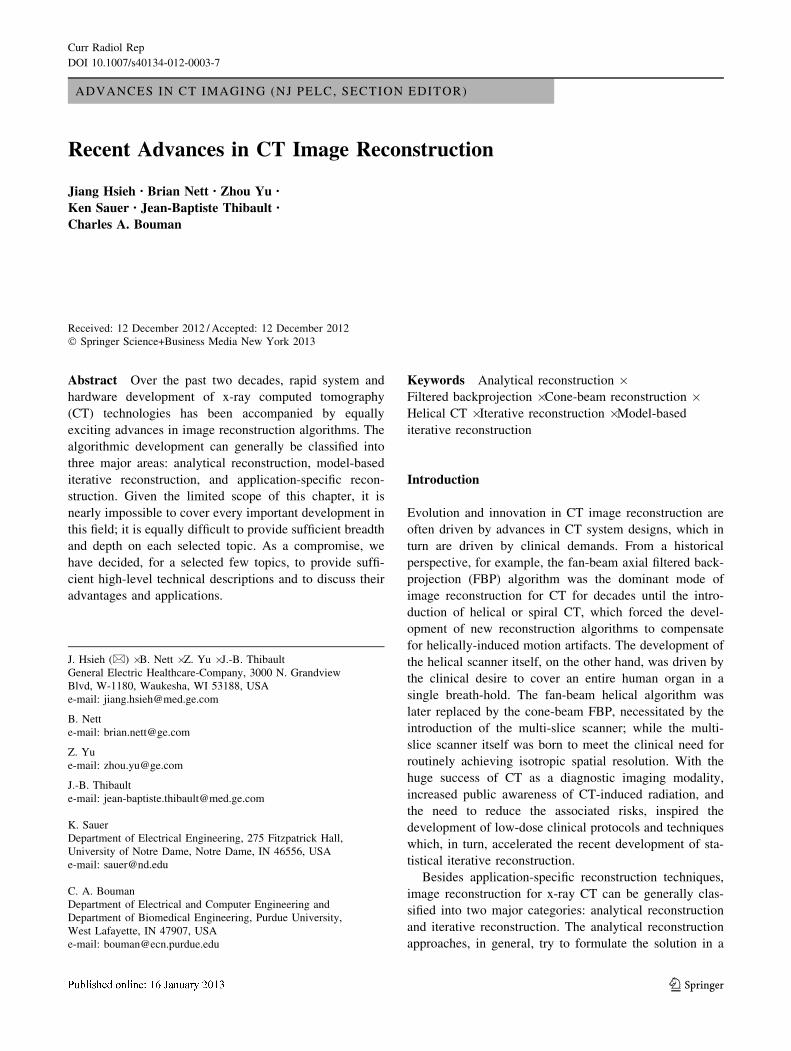
all views. Figure 3b, c depict coronal images of a CD-
phantom (similar to a Defrise phantom) scanned in heli-
cal and SSCB modes, respectively. Since parallel CDs
are placed horizontally with air gaps in between, the
Fig. 1 Illustration of cone-beam geometry
Fig. 2 Sampling challenges for step-and-shoot cone beam geometry
illustration of missing frequencies and mishandled data in Fourier
space (a) and axial truncation (b)
Curr Radiol Rep
123
corresponding coronal image should exhibit a pattern
similar to Venetian blinds. The image formed with the
SSCB mode clearly shows deficiencies for areas away from
the detector center-plane.
When the cone angle is moderate, various approximate
reconstruction algorithms have been shown to effectively
produce clinically acceptable images. One of the most
commonly used reconstruction algorithm is the Feldkamp–
Davis–Kress (FDK) reconstruction. This algorithm differs
from the conventional fan-beam reconstruction in replacing
the two-dimensional back-projection with a three-dimen-
sional back-projection to mimic the x-ray path as it tra-
verses the object, and in including a cosine weighting in the
cone direction to account for path length differences of
oblique rays [6]. Extensions from the FDK algorithm have
been introduced to solve one of the three major issues with
reconstruction from an arc trajectory.
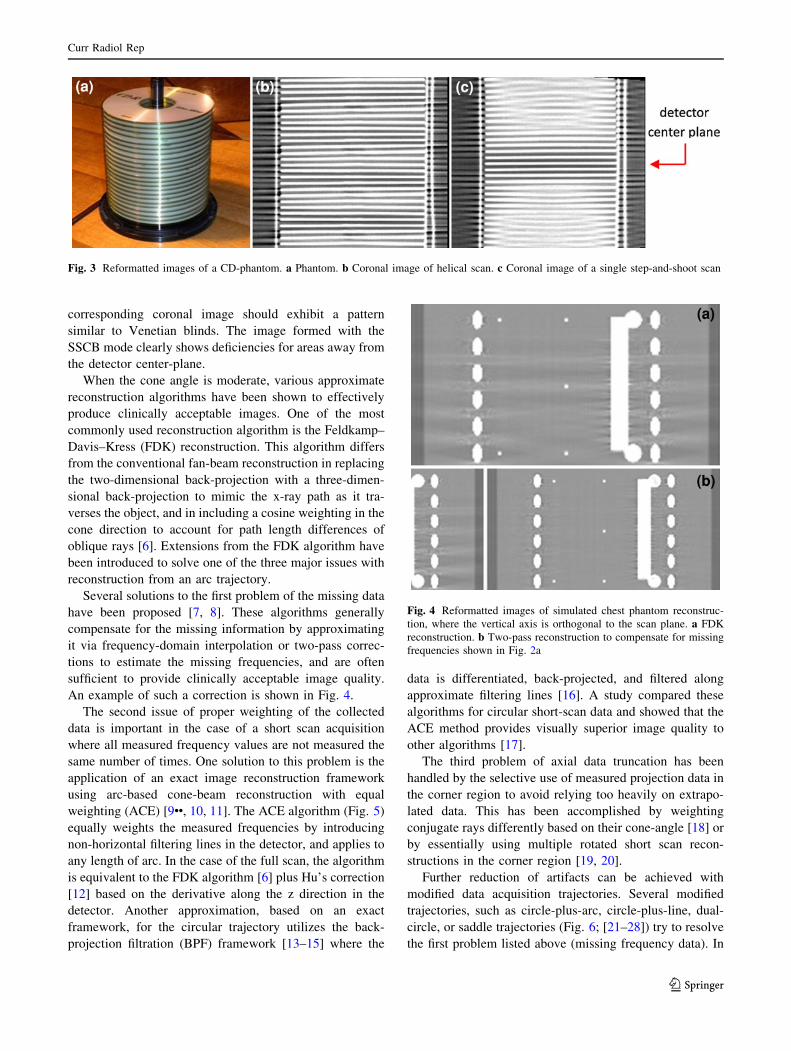
Several solutions to the first problem of the missing data
have been proposed [7, 8]. These algorithms generally
compensate for the missing information by approximating
it via frequency-domain interpolation or two-pass correc-
tions to estimate the missing frequencies, and are often
sufficient to provide clinically acceptable image quality.
An example of such a correction is shown in Fig. 4.
The second issue of proper weighting of the collected
data is important in the case of a short scan acquisition
where all measured frequency values are not measured the
same number of times. One solution to this problem is the
application of an exact image reconstruction framework
using arc-based cone-beam reconstruction with equal
weighting (ACE) [9••, 10, 11]. The ACE algorithm (Fig. 5)
equally weights the measured frequencies by introducing
non-horizontal filtering lines in the detector, and applies to
any length of arc. In the case of the full scan, the algorithm
is equivalent to the FDK algorithm [6] plus Hu’s correction
[12] based on the derivative along the z direction in the
detector. Another approximation, based on an exact
framework, for the circular trajectory utilizes the back-
projection filtration (BPF) framework [13–15] where the
data is differentiated, back-projected, and filtered along
approximate filtering lines [16]. A study compared these
algorithms for circular short-scan data and showed that the
ACE method provides visually superior image quality to
other algorithms [17].
The third problem of axial data truncation has been
handled by the selective use of measured projection data in
the corner region to avoid relying too heavily on extrapo-
lated data. This has been accomplished by weighting
conjugate rays differently based on their cone-angle [18] or
by essentially using multiple rotated short scan recon-
structions in the corner region [19, 20].
Further reduction of artifacts can be achieved with
modified data acquisition trajectories. Several modified
trajectories, such as circle-plus-arc, circle-plus-line, dual-
circle, or saddle trajectories (Fig. 6; [21–28]) try to resolve
the first problem listed above (missing frequency data). In
Fig. 3 Reformatted images of a CD-phantom. a Phantom. b Coronal image of helical scan. c Coronal image of a single step-and-shoot scan
Fig. 4 Reformatted images of simulated chest phantom reconstruc-
tion, where the vertical axis is orthogonal to the scan plane. a FDK
reconstruction. b Two-pass reconstruction to compensate for missing
frequencies shown in Fig. 2a
Curr Radiol Rep
123

Fig. 7, a phantom known to demonstrate such a problem is
shown for just a single-circle trajectory on the left and a
dual-circle trajectory on the right. Although these ‘circle
plus’ trajectories meet the conditions for mathematically
exact reconstruction, utilization of these trajectories in a
real clinical environment still faces significant challenges
because of many practical considerations: large mass of a
CT gantry, high gantry rotation speed, and desire to min-
imize overall data acquisition time.
Another relatively recent discovery is the fact that, in
order to reconstruct a given region of interest (ROI), one
does not need 180� plus the fan angle of the system [10,
29]. The 2D sufficiency condition is similar to Tuy’s
condition [4] for 3D exact reconstruction, in that any line
which can be drawn through the ROI must intersect the
source trajectory at least once. This condition can be
exploited to reduce the temporal window of acquisition for
off-center ROI imaging [30, 31].
Multi-Slice Helical Reconstruction
Since the introduction of helical scanning over two decades
ago, significant research efforts have been devoted to the
subject of cone-beam helical reconstruction, and the
majority of the clinical protocols have switched from step-
and-shoot to helical mode. In the helical mode of data
acquisition, the table is indexed along the z-axis while the
gantry rotates about the patient to collect projection
measurements.
Approaches to analytic helical cone beam reconstruction
can be generally divided into two major categories: exact
reconstruction methods and approximate reconstruction
methods. The exact reconstruction methods, as the name
implies, try to derive analytical solutions that match the
scanned object in mathematical precision if the input pro-
jections are the true line integrals [32]. One of the most
exciting developments in the past few years is the devel-
opment of exact FBP algorithms as first developed by
Katsevich [9••, 33••, 34]. This algorithm falls into the
Fig. 5 Slices through the Shepp Logan phantom with window
parameters of [0.98 1.05]. Note the improvement in the soft tissue
away from the central slice as well as the reduction in streaky cone-
beam artifacts (arrows) which are due to improper weighting. The
vertical direction in these images is the z direction, which is
orthogonal to the plane of the source trajectory
Fig. 6 Alternative source
trajectories to enable complete
sampling: circle-plus-line (a),
circle-plus-arc (b), dual-circle
(c), and saddle (d)
Fig. 7 An example of exact reconstruction from the source trajectory
of two concentric circles. On the left is the contribution to the final
image from the horizontal circle only. In the center, is contribution
from the vertical circle only and the sum is on the right. Reprinted
from [24], with permission
Curr Radiol Rep
123

category of FBP with a shift-invariant one-dimensional
filter, and the flow of this reconstruction algorithm is
similar to that of the fan-beam FBP. Since its invention,
many new algorithms have been proposed and developed
to allow more general trajectories, more efficient use of the
projection samples, and region-of-interest reconstruction
[14, 35–39]. The advantage of these algorithms is of course
their mathematical exactness. However, to date, these
algorithms have yet to be implemented widely in com-
mercial products, mainly due to their noise properties,
limited robustness to patient motion, and lack of flexibility.
For example, in a cardiac acquisition, a half-scan is typi-
cally used to improve the temporal resolution. In clinical
practice, a sub-volume of the heart (in z) needs to be
reconstructed for each half-scan acquisition to keep the
overall acquisition time to a minimum. This is difficult to
achieve with the exact reconstruction algorithms.
The second algorithm category is the approximate
reconstruction approaches derived from the original FDK
algorithm [40–48]. Each projection is weighted, filtered on
a row-by-row basis, and back-projected three-dimension-
ally into the object space. Although these algorithms are
approximate in nature, they do offer distinct advantages,
such as volume reconstruction in a single half-scan
acquisition. Because of their flexibility, their ability to
intrinsically handle data from long objects and their com-
putational efficiency, the approximate reconstruction
algorithms are still the dominant force behind most com-
mercial CT reconstruction engines.
It should be pointed out that the non-exact algorithms
often borrow techniques developed in the exact reconstruc-
tion algorithms. For example, the Katsevich algorithm per-
forms the projection filtration along a set of j-curves (instead
of row-by-row) as shown in Fig. 8, and each filtered ‘‘virtual
row’’ falls along a tangent to the helix. The same approach
can be incorporated into the approximate reconstruction to
significantly improve image quality. Figure 9 shows a
computer simulated phantom with (1) an approximate
algorithm with tangential filtering [47] and (2) the Katsevich
algorithm: comparable image quality between the exact and
approximate reconstruction algorithms can be observed.
Iterative Reconstruction
Over the years, many algorithmic approaches have been
proposed to reduce image artifacts and noise during the
image generation process [49–51]. Noise and artifact
reductions are necessary steps toward dose reduction in CT.
The recent burst of research activities in iterative recon-
struction can also be credited to the increased awareness of
the radiation dose generated by a CT scan [52–59], and
advancements in reconstruction hardware capability.
The use of an iterative reconstruction to solve the inverse
problem of x-ray computed tomography has a long history.
As a matter of fact, reconstruction of the very first clinical
image utilized an iterative technique called algebraic
reconstruction technique (ART) to invert a large matrixFig. 8 An example of the j-curves used in the filtering operation of
the Katsevich algorithm (helical pitch 87/64)
Fig. 9 Simulated body phantom with 64 9 0.625 mm detector
configuration and 63/64 helical pitch. a Modified FDK algorithm
with tangential filtering and 3D weighting. b Katsevich reconstruction
algorithm
Curr Radiol Rep
123

[60, 61]. Although the objective of ART at the time was to
find a solution to a complicated inverse problem and did not
involve complex modeling of the CT system, it nonetheless
demonstrated the efficacy of IR techniques for x-ray CT.
If we look beyond the CT reconstruction, statistical IR
has been used extensively in single photon emission com-
puted tomography (SPECT) and positron emission tomog-
raphy (PET) to combat photon starvation issues and to show
great benefits in noise reduction and improved accuracy in
the reconstructed images. Although the success of IR in
SPECT and PET seemingly implies a straightforward
transfer of such technology to CT, reality has shown that
such a transition has been met with great challenges due to
the significant differences between the modalities. First, in
x-ray CT, pre-processing and calibration steps are numer-
ous and critical to the production of CT images, and these
correction steps significantly change the statistical proper-
ties of the measured projection samples. If proper modeling
is not performed, the desired noise or dose reduction is
difficult to achieve. Second, spatial resolution is much
higher in x-ray CT than either SPECT or PET. Achieving
high spatial resolution in IR requires carefully modeling the
optics of the system as well as using an ‘‘edge-preserving’’
regularization design to control image properties and avoid
needless sacrifice of spatial resolution for noise reduction.
Third, due to the large amount of data and complexity of the
inverse CT problem and associated models, IR calls for the
design of robust and stable converged results by carefully
crafting an optimization cost function, the solution of which
is performed iteratively and typically demands long
reconstruction time. This has prevented wide application of
IR in clinical practice until recent advancements in com-
puter hardware and fast algorithms.
Unlike the analytical reconstruction algorithms where
each projection sample is weighted, filtered, and back-
projected to formulate an image, iterative reconstruction
arrives at the final solution in an iterative manner: the
initial reconstructed images are refined and modified iter-
atively until certain criteria are met. The criteria are written
in the form of a cost function to be minimized, which
measures the fit of the image to the data according to a
model of the imaging system. However, the enormous size
of the optimization problem as well as the complexity of
the model makes it difficult to solve, and the cost function
needs to be minimized iteratively. Each iteration typically
involves the forward- and back-projection of an interme-
diate image volume. The forward-projection step simulates
the x-ray interactions with the ‘‘object’’ (the intermediate
images) and produces a set of synthesized projections. The
synthesized projections are compared against the real
projection measurements collected by the CT scanner
according to a statistical metric, and the differences
between the two are attributed to the ‘‘error’’ or ‘‘bias’’ in
the intermediately estimated image volume. The error is
then used to update the intermediate image volume to
reduce the discrepancy between the image and the acquired
data. The new intermediate image is then forward-pro-
jected again in the next iteration, and, provided the choice
of an adequate cost function and a globally convergent
optimization algorithm, the image volume will converge to
an estimate near the minimizer of the cost function after
several iterations.
In the context of this chapter, due to limited space, we
consider only statistical IR as defined in an optimization
sense, whereby the reconstructed result is achieved by
minimizing a cost function that determines the desired
characteristics of the final image estimate as it relates to the
measurement data, and the iterative approach mostly deter-
mines the speed of the convergence to that solution. Other IR
formulations exist, which do not always provide the same
properties of predictable convergence and stable results.
Advantages and Requirements
To understand the advantages of a model-based iterative
reconstruction (MBIR) over other approaches, let us
examine a typical CT system and the assumptions being
made to derive the FBP algorithm, or analytical recon-
struction algorithms in general. Figure 10 depicts a sche-
matic diagram of a CT scanner with intentionally enlarged
x-ray source, detector, and image voxel for easier illus-
tration. In the derivation of an FBP algorithm, a series of
assumptions are made regarding the physical dimension of
the x-ray focal spot, the detector cell, and the image voxels
in order to make the mathematics manageable. Specifically,
Fig. 10 Schematic diagram of CT system
Curr Radiol Rep
123

the size of the x-ray focal spot is assumed to be infinitely
small and therefore can be approximated as a point source.
Although the detector-cell spacing is correctly considered
in the derivation of the reconstruction filters, the shape and
dimension of each detector cell are ignored, and all x-ray
photon interactions are assumed to take place at a point
located at the geometric center of the detector cell. Simi-
larly, the image voxel’s shape and size are ignored and
only the geometric center of the voxel is considered. These
assumptions lead to the formation of a single pencil beam
representing the line integral of the attenuation coefficients
along the path which connects the x-ray source and a
detector cell. In addition, each of the projection measure-
ments is assumed to be accurate and is not influenced by
the fluctuation induced by photon statistics or electronics
noise.
The CT system model traditionally used by analytical
algorithms clearly does not represent physical reality. For
most clinical CT scanners, typical focal spot size is about
1 mm 9 1 mm and detector cell spacing is slightly larger
than 1 mm with an active area of 80–90 % of the spacing.
The image voxel is normally modeled as rectangular in
shape with its dimensions determined by the reconstruction
field-of-view (FOV) and slice thickness (Due to limited
scope, other image voxel shapes, such as a blob, will not be
discussed). For example, for a 512 9 512 matrix size image
representing a 50 cm FOV and 5 mm slice thickness, the
reconstructed pixel is 0.98 mm 9 0.98 mm 9 5 mm in x,
y, and z. Because each projection sample is acquired over a
finite sampling duration (less than a millisecond on modern
scanners) with limited x-ray flux, each measurement at a
detector cell has finite photon statistics. The statistical
fluctuation is further complicated by the fact that the elec-
tronics used in the data acquisition system has an inherent
noise floor which contributes to the overall noise in the
measurement. There is little doubt that the simplifications
made in the derivation of an FBP should have an impact on
reconstructed image quality.
MBIR Algorithms
One way of incorporating an accurate system model while
overcoming the mathematical intractability of an analytical
reconstruction is to use a technique called model-based
iterative reconstruction, MBIR. In 1982, by incorporating
only the noise properties of emission tomography, an
expectation–maximization algorithm (EM) was proposed
for SPECT and was published in a classic paper [62••]. The
development of IR techniques for x-ray computed tomog-
raphy, however, started a few years later due to its tech-
nical complexities and the perception that less benefit was
possible than in emission CT [63•, 64••, 65, 66••, 67••,
68–70, 71•, 72–75].
The accuracy of modeling is critical to the quality of the
MBIR reconstruction. There are three main key models in
the IR algorithm: the forward model, the noise model, and
the image model. The forward model accounts for the
system optics and all geometry-related effects. An accurate
system model is necessary to ensure that modeling errors
do not grow during the iterative convergence process to
form artifacts in the reconstructed images One example of
an accurate forward model is casting of multiple pencil-
rays through the x-ray focal spot, the image voxel, and the
detector cell. These pencil-beams mimic different x-ray
photon paths going through the object, and the complicated
CT optics is approximated by a weighted summation of
many simplistic line integral models, as depicted in Fig. 11
[76]. One strategy to ensure an unbiased coverage of dif-
ferent points on the focal spot, different locations on a
detector cell, and different sub-regions inside an image
pixel is to sub-divide the focal spot, detector cell, and pixel
into equal-sized small elements. Needless to say, this
process is time consuming.
2An alternative approach is to model the ‘‘shadow’’ cast
by each image voxel onto the detector and rely on point-
response functions to perform the optics modeling, as
shown in Fig. 12 [71•, 77•]. In this approach, only one ray is
cast between the center of the focal spot and the center of
the image voxel to locate the point of intersection on the
detector, and a response function is used to distribute the
contribution of the image voxel to different detector cells
during the forward-projection process. The response func-
tion is non-stationary and changes with the voxel location to
account for different magnifications and orientations.
Compared to the ray casting approach, this method is
computationally more efficient, and can introduce advanced
Fig. 11 Illustration of modeling the system optics with ray casting
Curr Radiol Rep
123

system modeling such as detector effective area, cross-talk,
or focal spot energy distribution.
Note that accurate system modeling implies accounting
for spatially-varying behavior where the projection of an
image voxel onto the detector depends on its shape and
position relative to the x-ray source, contrary to simplistic
models used in FBP-type reconstruction. Using this accu-
rate system model, it is also important that the forward- and
back-projection operators remain adjoint to each other in
order to prevent the propagation of errors in the
reconstruction.
The modeling of noise distributions in the projections is
not as straightforward as one may think, complicated by
the fact that the CT projection measurement undergoes
complicated pre-processing and calibration processes prior
to its use in the reconstruction. Although the original x-ray
photon statistics follow approximately the Poisson distri-
bution (approximation due to the poly-energetic x-ray
source used in CT), the projection data after the calibration
process no longer exhibits this behavior. The same com-
plication applies to the electronic noise after the pre-pro-
cessing steps. If a simplistic model is used to approximate
the statistical properties, suboptimal results are likely to
result. It should be pointed out that the level of bias or error
introduced by the simplified model increases with the
reduction of the x-ray flux received at the detector, and the
image quality degradation increases as a result. This is
undesirable when considering the fact that one of the major
driving-forces behind MBIR is dose reduction, which leads
naturally to a reduced x-ray flux on the detector.
Once the projection noise is properly modeled, the
philosophy behind MBIR is to treat each projection sample
differentially based on its estimated noise variance. In
general, a higher confidence is placed on samples with high
signal-to-noise ratio (SNR) and lower weights are placed
on samples with low SNR. This is in direct contrast to the
noise treatment in analytical reconstruction where all
samples are treated equally from a statistical point of view.
The image model is somewhat more subjective. The
general approach is to model the image volumes as Markov
random fields, which results in a cost function term that
penalizes noise-induced intensity fluctuations in the
neighborhood of a voxel. By carefully examining the
human anatomy, one naturally concludes that the attenua-
tion characteristics of a voxel-size element inside the
human body are not completely isolated from or indepen-
dent of the surrounding elements. Therefore, if a voxel CT
number is far from its neighbors, it should be penalized and
modified. The term used to enforce such conditions in the
cost function is often called the regularization term in IR.
Although the concept looks simple, several challenges
need to be addressed in the design of the regularization
term. The differentiation of the noise from the real struc-
tures in the image can be quite difficult, since real object
structures are often buried in the noise and there is little
prior information available about the object itself. The
general approach taken by researchers is to construct an
‘‘edge-preserving prior’’ that preserves structural edges
[71•, 78, 79]. Another challenge is to maintain the uniform
image quality across the volume. In CT reconstruction, the
noise distribution in the image varies as a function of local
attenuation; highly attenuated regions tend to have higher
noise. Therefore, it requires a spatially varying design of the
regularization term to achieve uniform image resolution and
noise behavior [80]. Another difficulty is to properly bal-
ance the regularization and data likelihood terms in the cost
function to achieve the desired IQ: designing for robust IQ
behavior across a range of clinical scenarios without end-
user interaction can pose a significant challenge.
The MBIR cost function typically incorporates all the
models discussed above. High spatial resolution is usually
a direct result of an accurate forward model combined with
appropriate regularization. For demonstration, Fig. 13a, b
depict a clinical example of standard FBP and MBIR
reconstructions of the same projection data. The MBIR
image exhibits not only significantly reduced noise but also
much improved spatial resolution as demonstrated by the
visibility of fine anatomical structures.
It is clear from the discussion that fully modeled IR is
more computationally intensive than analytical recon-
struction methods. Although it provides superb image
quality in terms of spatial resolution, noise reduction, and
artifact correction, the time delay in the image generation
process is not negligible. In today’s clinical environment,
‘‘real time’’ image reconstruction is often demanded and
Fig. 12 Illustration of modeling the system optics with a response
function
Curr Radiol Rep
123

the CT operator expects all images will become available
shortly after the completion of the data acquisition.
Extensive research has been conducted over the years to
speed-up IR algorithms. Fast hardware implementation,
distributed parallel processing, and efficient algorithm
convergence properties are equally important to achieve
this goal. To speed up convergence, one approach is to
improve the update strategy at each iteration. There are two
types of update strategies: global updates, or local updates.
Algorithms using the global update strategy update the
entire image volume at once. These algorithms are rela-
tively easy to parallelize. However, since the inverse
problem is typically ill-conditioned based on the large
number of unknowns relative to the measurements and the
range of data statistics, simple global update algorithms
such as gradient descent and conjugate gradient tend to
require a large number of iterations to converge. Therefore,
various acceleration techniques such as preconditioner-
based methods [81] and ordered-subset (OS) methods [75]
have been proposed to accelerate the convergence of global
update algorithms. The OS algorithms perform their
updates based on a subset of the projection data: at each
iteration, image updates start with one subset of the data,
and rotate around all consecutive subsets until all projec-
tion data are used. Today, this acceleration technique is
used extensively in commercial PET and SPECT scanners.
Contrary to global updates, local updates focus on
updating a single voxel or a subset of voxels at a time. These
voxels are selected according to a pattern, possibly random,
and their modifications are estimated based on all projection
data. After the first set of voxels is updated, another set is
selected and updated in sequential fashion. An example of
such algorithm is the iterative-coordinate-descent (ICD)
algorithm [63•, 64••, 71•, 72]. If we define the single update
of all voxels in the image as one full-iteration, the local
update strategy generally takes only a few iterations to
reach acceptable convergence. The convergence speed can
be further improved by exploring the flexibility to update
certain pixels more frequently than others in the so-called
non-homogeneous update strategy [72, 82].
From an implementation point of view, however, it can
be difficult to parallelize a local update algorithm to run on
a large number of threads to take full advantage of massive
parallel computer architectures. The global update strategy
seems to be just the opposite: it is easier to parallelize, but
typically needs a larger number of iterations to reach
convergence. Historically, the computational complexity of
MBIR was one of the most important factors that prevented
its application to CT. Although the same issue is still
present today, the magnitude of the obstacle has been
significantly reduced. With renewed research interests, this
issue is likely to be resolved in the near future with the
combination of advanced computer architectures and
improved algorithm efficiency. It needs to be stated that the
investigation of MBIR for CT is still in its infancy and new
clinical applications of MBIR are likely to be discovered.
Discussion and Conclusion
Given the limited scope of this chapter, it has been nec-
essary to focus only on a few major areas of tomographic
reconstruction. Unfortunately, many exciting developments
to address special clinical needs could not be adequately
covered. For example, despite the novel scanner hardware
development, cardiac coronary artery imaging still poses
Fig. 13 Example of coronal
images reconstructed with
a FBP and b MBIR
Curr Radiol Rep
123

Fig. 14 Advanced motion correction examples (heart rate: 84 bpm). a Conventional FBP algorithm. b With advanced motion correction
Curr Radiol Rep
123
significant challenges to CT due to the presence of irregular
and rapid vessel motion. Motion-induced artifacts even
appear in patients with low heart-rates, resulting from the
relatively high velocities of some of the coronary arteries
[83]. Recent development of an advanced algorithm seems
to offer good solutions to address this issue [84••]. The
motion-correction algorithm characterizes the vessel
motion using images reconstructed from slightly different
cardiac phases and utilizes these motion vectors to correct
for the imaging artifacts. Figure 14 depicts an example of a
patient’s cardiac images without (a) and with (b) the
motion compensation. The improvement in image quality
is obvious.
New developments in other areas, such as the generation
of material-density images and monochromatic images in
dual-energy CT, variable-pitch helical acquisition and
reconstruction, metal artifact reduction algorithms, and
region-of-interest scanning and reconstruction, are equally
exciting. As technology evolves, it is expected that tomo-
graphic reconstruction algorithms and other advanced
algorithmic approaches will play an even greater role in CT.
Disclosure J. Hsieh, B. Nett, Z. Yu, and J.B. Thibault are
employees of General Electric Healthcare-Company; K. Sauer: none;
C.A. Bouman: none.
References
Papers of particular interest, published recently, have been
highlighted as:• Of importance•• Of major importance
1. •• Earls JP, Berman EL, Urban BA, et al. Prospectively gated
transverse coronary CT angiography versus retrospectively gated
helical technique: improved image quality and reduced radiation
dose. Radiology. 2008;246:742–53. One of the early approachesthat significantly reduce radiation dose for cardiac imaging
2. Hsieh J, Londt, Vass JM, et al. Step-and-shoot data acquisition
and reconstruction for cardiac x-ray computed tomography. Med
Phys. 2006;33(11):4236–48.
3. Mori S, Endo M, Obata T, et al. Clinical potentials of the pro-
totype 256-detector row CT-scanner. Acad Radiol. 2005;12(2):
148–54.
4. Tuy HK. An inversion formula for cone-beam reconstruction.
SIAM J Appl Math. 1983;43(3):546–52.
5. Grangeat P. Mathematical framework of cone-beam 3D recon-
struction via the first derivative of the Radon transform. In:
Herman GT. Louis AK, and Natterer F, Editors. Mathematical
Methods in Tomography. Berlin: Springer; 1497, Lecture Notes
in Mathematics, 1991;66–97.
6. Feldkamp LA, Davis LC, Kress JW. Practical cone-beam algo-
rithm. J Opt Soc Am. 1984;1(6):612–9.
7. Hsieh J. Computed tomography: principles, design artifacts, and
recent advances. Bellingham: SPIE; 2003.
8. Zeng K, Chen Z, Zhang L, Wang G. An error-reduction-based
algorithm for cone-beam computed tomography. Med Phys.
2004;31(12):3206–12.
9. •• Katsevich A. A general schedule for constructing inversion
algorithm for cone beam CT. Int J Math Math Sci.
2003;21:1305–21. An exact framework which preserves the fil-tered backprojection structure and which requires only 1D fil-tering operations in the detector.
10. Chen GH. A new framework of image reconstruction from fan
beam projections. Med Phys. 2003;30(6):1151–61.
11. Nett BE, Zhuang TL, Leng S, Chen GH. Arc-based cone-beam
reconstruction algorithm using an equal weighting scheme. J X-
ray Sci Tech. 2007;15(1):19–48.
12. Hu H. An improved cone-beam reconstruction algorithm for the
circular orbit. Scanning. 1996;18:572–81.
13. Zou Y, Pan X. Exact image reconstruction on PI lines from
minimum data in helical cone-beam CT. Phys Med Biol.
2004;49:941–59.
14. Zhuang T, Leng S, Nett BE, Chen GH. Fan-beam and cone-beam
image reconstruction via filtering the backprojection image of
differentiated projection data. Phys Med Biol. 2004;49:
5489–503.
15. Pack JD, Noo F, Clackdoyle R. Cone-beam reconstruction using
the backprojection of locally filtered projections. IEEE Trans
Med Imag. 2005;24(1):70–85.
16. Yu L, Zou Y, Sidky EY, et al. Region of interest reconstruction
from truncated data in circular an cone-beam CT. IEEE Trans
Med Imag. 2006;25(7):869–81.
17. Dennerlein F, Noo F, Hoppe S, et al. Evaluation of three ana-
lytical methods for reconstruction from cone-beam data on a
short circular scan. IEEE NSS. 2007;5:3933–8.
18. Tang X, Hsieh J, Hagiwara A, et al. A three-dimensional
weighted cone beam filtered backprojection (CB-FBP) algorithm
for image reconstruction in volumetric CT under a circular source
trajectory. Phys Med Biol. 2005;50(16):3889–905.
19. Komatsu S, et al. A combination-weighted Feldkamp—based
reconstruction algorithm for cone-beam CT. Phys Med Biol.
2006;51(16):3953–65.
20. Grimmer R, Oelhafen M, Elstrom U, Kachelriess M. Cone-beam
CT image reconstruction with extended z range. Med Phys.
2009;36(7):3363–70.
21. Zeng GL, Gullberg GT. A cone-beam tomography algorithm for
orthogonal circle-and-line orbit. Phys Med Biol. 1992;37:563–77.
22. Yang H, Li M, Koizumi KHK. View-independent reconstruction
algorithms for cone beam CT with general saddle trajectory. Phys
Med Biol. 2006;51:3865–84.
23. Katsevich A. Image reconstruction for a general circle-plus tra-
jectory. Inverse Prob. 2007;23(5):2223–30.
24. Zhuang T, Nett BE, Leng S, Chen G. A shift-invariant filtered
backprojection (FBP) cone-beam reconstruction algorithm for the
source trajectory of two concentric circles using an equal
weighting scheme. Phys Med Biol. 2006;51:3189–210.
25. Yan X, Leahy RM. Cone-beam tomography with circular, ellip-
tical, and spiral orbits. Phys Med Biol. 1992;37:493–506.
26. Defrise M, Clack R. A cone-beam reconstruction algorithm using
shift-variant filtering and cone-beam backprojection. IEEE Trans
Med Imag. 1994;13:186–95.
27. Kudo H, Saito T. Derivation and implementation of a cone-beam
reconstruction algorithm for nonplanar orbits. IEEE Trans Med
Imag. 1994;13(196):211.
28. Noo F, Clark R, White TA, Roney TJ. The dual-ellipse cross
vertex path for exact reconstruction of long objects in cone-beam
tomography. Phys Med Biol. 1998;43:797–810.
29. Noo F, Defrise M, Clackdoyle R, Kudo H. Image reconstruction
from fan-beam projections on less than half scan. Phys Med Biol.
2002;47:2525–46.
30. King M, Pan X, Yu L, Giger M. ROI reconstruction of motion-
contaminated data using a weighted backprojection-filtration
algorithm. Med Phys. 2006;33:1222–38.
Curr Radiol Rep
123

31. Nett BE, Leng S, Zambelli JN, Reeder SB, Speidel MA, Chen
GH. Temporally targeted imaging method applied to ECG-gated
computed tomography: preliminary phantom and in vivo expe-
rience. Acad Radiol. 2008;15(1):93–106.
32. Clark R, Defrise M. Overview of reconstruction algorithm for
exact cone-beam tomography. Proc SPIE. 1994;2299:230–41.
33. •• Katsevich A. Theoretically exact filtered backprojection type
inversion algorithm for spiral CT. SIAM J Appl Math.
2002;62:2012–26. An exact framework which preserves the fil-tered backprojection structure and which requires only 1D fil-tering operations in the detector.
34. Katsevich A. Improved exact FBP algorithm for spiral CT. Adv
Appl Math. 2004;32:607–81.
35. Chen G. An alternative derivation of Katsevich’s cone-beam
reconstruction formula. Med Phys. 2003;30:3217–26.
36. Pan X, Xia D, Zou Y, Yu L. A unified analysis of FBP-based
algorithms in helical cone-beam and circular cone- and fan-beam
scans. Phys Med Biol. 2004;49:4349–69.
37. Zou Y, Pan X. An extended data function and its generalized
backprojection for image reconstruction in helical cone-beam CT.
Phys Med Biol. 2004;49:N383–7.
38. Ye Y, Zhao S, Yu H, Wang G. A general exact reconstruction for
cone-beam CT via backprojection-filtration. IEEE Trans Med
Imag. 2005;24(9):1190–8.
39. Zhuang T, Chen G. New Families of exact fan-beam and cone-
beam image reconstruction formulae via filtering the backpro-
jection image of differentiated projection data along singly
measured lines. Inverse Prob. 2006;22:991–1006.
40. Wang G, Lin TH, Cheng P, Shinozaki DM. A general cone-beam
reconstruction algorithm. IEEE Trans Med Imag. 1993;12:
486–96.
41. Wang G, Liu Y, Lin TH, Cheng P. Half-scan cone-beam x-ray
microtomography formula. Scanning. 1994;16:216–20.
42. Hsieh J. A practical cone beam artifact correction algorithm. Proc
IEEE Nucl Sci Symp Med Imag Conf. 2000;15:71–4.
43. Silver M. High-helical-pitch cone-beam computed tomography.
Phys Med Biol. 1998;43:847–55.
44. Ning R, Chen B, Yu R, et al. Flat panel detector-based cone-beam
volume CT angiography imaging: system evaluation. IEEE Trans
Med Imag. 2000;19:949–63.
45. Bruder H, Kachelrieb M, Schaller S, Mertelmeier T. Performance
of approximate cone-beam reconstruction in multi-slice com-
puted tomography. SPIE Proc. 2000;3979:541.
46. Hsieh J, Tang X, Thibault JB, et al. Conjugate cone beam
reconstruction algorithm. Opt Eng. 2007;46(6):67001.
47. Tang X, Hsieh J, Nilsen RA, et al. A three-dimensional weighted
cone beam filtered backprojection (CB-FBP) algorithm for image
reconstruction in volumetric CT-helical scanning. Med Phys Biol.
2006;51(4):855–74.
48. Tang X, Hsieh J. Handling data redundancy in helical cone beam
reconstruction with a cone-angle-based window function and its
asymptotic approximation. Med Phys. 2007;34(6):1989–98.
49. Hsieh J. Adaptive streak artifact reduction in computed tomog-
raphy resulting from Excessive X-ray photon noise. Med Phys.
1998;25(11):2134–47.
50. La Riviere PJ. Penalized-likelihood sonogram smoothing for low-
dose CT. Med Phys. 2005;32:1676–83.
51. Kachelrieb M, Kalender WA. Empirical cupping correction: a
first-order raw data precorrection for cone-beam computed
tomography. Med Phys. 2006;33:1269–74.
52. Singh S, Kalra MK, Hsieh J, et al. Abdominal CT: comparison of
adaptive statistical iterative and filtered back projection recon-
struction techniques. Radiology. 2010;257:373–83.
53. Singh S, Kalra MK, Do S, et al. Comparison of hybrid and pure
iterative reconstruction techniques with conventional filtered
back projection: dose reduction potential in the abdomen.
J Comput Assist Tomogr. 2012;36(3):347–53.
54. Prakash P, Kalra MK, Ackman JB, et al. Diffuse lung disease: CT
of the chest with adaptive statistical iterative reconstruction
technique. Radiology. 2010;256(1):261–9.
55. Nelson RC, Feuerlein S, Boll DT. New iterative reconstruction
techniques for cardiovascular computed tomography: how do
they work, and what are the advantages and disadvantages?
J Cardiovasc Comput Tomogr. 2011;5:286–92.
56. Katsura M, Matsuda I, Akahane M, Sato J, Akai H, Yasaka K,
Kunimatsu A, Ohtomo K. Model-based iterative reconstruction
technique for radiation dose reduction in chest CT: comparison
with the adaptive statistical iterative reconstruction technique.
Eur Radiol. 2012;22(8):1613–23.
57. Husarik DB, Marin D, Samei E, et al. Radiation dose reduction in
abdominal computed tomography during the late hepatic arterial
phase using a model-based iterative reconstruction algorithm:
how low can we go? Invest Radiol. 2012;47(8):468–74.
58. Yamada Y, Jinzaki M, Tanami Y, et al. Model-based iterative
reconstruction technique for ultralow-dose computed tomography
of the lung: a pilot study. Invest Radiol. 2012;47(8):482–9.
59. Suzuki S, Machida H, Tanaka I, Ueno E. Measurement of vas-
cular wall attenuation: comparison of CT angiography using
model-based iterative reconstruction with standard filtered back-
projection algorithm CT in vitro. Euro J Radiol. 2012;81(11):
3348–53.
60. Hounsfield GN. A method of and apparatus for examination of a
body by radiation such as x or gamma radiation. The Patent
Office, London, Patent specification 1283915, 1972.
61. Herman GT, Lent A, Rowland S. ART: mathematics and appli-
cations, a report on the mathematical functions and on the
applicability to real data of algebraic reconstruction technique.
J Theor Biol. 1973;42(1):1–32.
62. •• Shepp LA, Vardi Y. Maximum likelihood reconstruction for
emission tomography. IEEE Trans Med Imag. 1982;MI-
1(2):113–22. One of the early papers introducing statisticalmodels to iterative reconstruction.
63. • Sauer K, Bouman CA. A local update strategy for iterative
reconstruction from projections. IEEE Trans Sign Proc.
1993;41:534–48. One of the earliest papers proposing a costfunction for iterative reconstruction using Bayesian framework.
64. •• Bouman CA, Sauer K. A unified approach to statistical
tomography using coordinate descent optimization. IEEE Trans
Imag Proc. 1996;5:480–92. A ICD-Newton Rapson method tosolve the optimization problem in iterative reconstruction.
65. Fessler JA. Statistical image reconstruction methods for trans-
mission tomography. In: Sonka M, Fitzpatrick JM, editors.
Handbook of medical imaging: medical image processing and
analysis. Bellingham: SPIE; 2000. p. 1–70.
66. •• Erdogan H, Fessler JA. Ordered subsets algorithms for trans-
mission tomography. Phys Med Biol. 1999;44:2835–51. Asimultaneous update algorithm known as separable paraboloidalsurrogates (SPS) to leverage OSEM widely used in emissiontomography to accelerate the convergence speed.
67. •• Browne JA, Holmes TJ. Developments with maximum likeli-
hood x-ray computed tomography. IEEE Trans Med Imag.
1992;11(1):40–52. One of the early papers to apply iterativereconstruction on real scan data (from industrial CT scanner)and access the image quality.
68. Wang G, Vannier MW, Cheng PC. Iterative x-ray cone beam
tomography for metal artifact reduction and local region recon-
struction. Microsc Microanal. 1999;5:58–65.
69. Elbakri IA, Fessler JA. Statistical image reconstruction for
polyenergetic x-ray computed tomography. IEEE Trans Med
Imag. 2002;21:89–99.
Curr Radiol Rep
123

70. Manglos SH, Gange GM, Krol A, et al. Transmission maximum-
likelihood reconstruction with ordered subsets for cone beam CT.
Phys Med Biol. 1995;40:1225–41.
71. • Thibault JB, Sauer KD, Bouman CA, Hsieh J. A three-dimen-
sional statistical approach to improved image quality for multi-
slice helical CT. Med Phys. 2007;34(11):4526–44. One of theearly papers to apply model-based statistical iterative recon-struction on multislice helical CT reconstruction.
72. Yu Z, Thibault JB, Bouman CA, et al. Fast model-based X-ray
CT reconstruction using spatially non-homogeneous ICD opti-
mization. IEEE Trans on Img Proc. 2011;20(1):161–75.
73. DeMan B, Nuyts DJP, Marchal G, Suetens P. An iterative max-
imum-likelihood polychromatic algorithm for CT. IEEE Trans
Med Imag. 2001;20:999–1008.
74. Nuyts J, DeMan B, Dupont P, et al. Iterative reconstruction for
helical CT: a simulation study. Phys Med Biol. 1998;43:729–37.
75. Kamphius C, Beekman FJ. Accelerated iterative transmission CT
reconstruction using an ordered subset convex algorithm. IEEE
Trans Med Imag. 1988;17(6):1101–5.
76. Thibault JB, Sauer K, Bouman C, Hsieh J. High quality iterative
image reconstruction for multi-slice helical CT. Proc Intl Conf
Fully 3D Recon Radiol Nuc Med 2003.
77. • DeMan B, Basu S. Distance-driven projection and backpro-
jection in three dimensions. Phys Med Biol. 2004;49(11):
2463–75. A paper proposed a novel system model known asdistance-driven model.
78. Geman S, Geman D. Stochastic relaxation, Gibbs distributions
and the Bayesian restoration of images. IEEE Trans Patt Anal
Mach Intell. 1984;PAMI-6:721–41.
79. Green P. Bayesian reconstruction from emission tomography data
using a modified EM algorithm. IEEE Trans Med Imag. 1990;
9(1):84–93.
80. Stayman JW, Fessler JA. Compensation for nonuniform resolu-
tion using penalized-likelihood reconstruction in space-variant
imaging systems. IEEE Trans Med Imag. 2004;23(3):269–84.
81. Chinn G, Huang SC. A general class of preconditioners for sta-
tistical iterative reconstruction of emission computed tomogra-
phy. IEEE Trans Med Imag. 1997;16(1):1–10.
82. Yu Z, Thibault JB, Bouman CA, et al. Edge Localized Iterative
Reconstruction for Computed Tomography. Proc 10th Intern
Meet Fully 3D Img Recon Radiol Nucl Med 2009.
83. Rohkohl C, Bruder H, Stierstorfer K, Flohr T. Improving best-
phase image quality in cardiac CT by motion correction with
MAM optimization. CT Meeting 2012, Salt Lake City, 2012.
84. •• Leipsic J, Labountry T, Hague CJ, et al. Effect of a novel
vendor-specific motion-correction algorithm on image quality
and diagnostic accuracy in persons undergoing coronary CT
angiography without rate-control medications. JCCT. 2012;6(3):
164–71. A clinical assessment of the first commercial offering ofa motion estimation and motion compensation algorithm for CT.
Curr Radiol Rep
123