Reagents and custom solutions for immunodiagnostic assay ......depth of technologies for...
84
Reagents and custom solutions for immunodiagnostic assay development
Transcript of Reagents and custom solutions for immunodiagnostic assay ......depth of technologies for...

Reagents and custom solutions for immunodiagnostic assay development

Naming convention on links need to be addressed before releasing to client. Double check color standards and fonts in graphs should all be Helvetica Neue not Universe.
Contents
Introduction 3
Capture surfaces 5Magnetic beads 5Latex beads 10Coated multiwell plates 13Custom plate coating service 17Biotin-binding proteins 18
Antibodies and detection probes 20Primary antibodies 21
ABfinity recombinant antibodies 23Secondary antibodies 25
Fluorescent and 25 enzyme-conjugated secondary antibodies Superclonal 26 secondary antibodies
CaptureSelect affinity ligands 27Custom antibody development and 29 production services ELISA products 32
ELISA kits 32Antibody pair kits 33
Biotin-binding protein conjugates 34
Linking mechanisms 40Crosslinkers 40PEGylation reagents 47Biotinylation reagents 49Biotin quantitation kits 53Pierce premium grade reagents 54 Fluorescent antibody and protein 56 labeling kits Fluorescent dyes 58Enzyme labeling kits 63
Nonspecific binding 65Blocking buffers 65Wash buffers 68Detergents 70Protein stabilizers 71
Detection substrates 72Chemiluminescent substrates 73Colorimetric substrates for 78 AP and HRPFluorescent substrates 80
Manufacturing capabilities 81

Introduction
Who understands your challenges? We do.Immunodiagnostic assay development and commercialization are challenging and time-consuming. Selecting the best combination of tools and technologies to create a high-performing, competitively positioned assay is demanding. We understand this challenge from our work with leading immunodiagnostics companies who face many of the same difficulties you do. Working with a broad array of companies gives us a unique perspective on this immunodiagnostics market, allowing us to see what differentiates the merely good companies from the industry leaders.
We see companies juggling costs, assay development time, and assay quality, which forces them to focus on certain aspects while sacrificing others. This balancing act can result in failures to fully maximize proprietary technology or delays to market, but assay development doesn’t have to be this way.
Then there are the other obstacles that you might face. What if you hit a reliability issue with the raw materials used in an assay already commercially available? That could result in the loss of market share and millions of dollars in revenue while the assay is redesigned. Or what happens if someone mistakenly performs an assay based on a raw material, for example, an antibody, that can’t be used for diagnostics? You have to start over, and unfortunately, we see this all the time.
And all you want is to create the best assays quickly and cost effectively. Does it have to be this hard?
The best solution—partnershipOne of the best ways to meet your goals is to partner with an experienced supplier. Think about it. You leverage your proprietary technology and in-house knowledge with the experience of a supplier who has worked with many of the leading immunodiagnostics companies and understands raw materials and new technologies. Using our experience for your benefit can be very powerful. It is the difference we see that separates the most successful companies because it enables them to break free of the cost, quality, and time paradigm. These are the companies who are developing high-quality assays, quickly and cost effectively.
Why choose Thermo Fisher Scientific?By choosing us, you gain an ally with a dedicated diagnostics partnering business that is always available for your projects. We have one of the largest breadth and depth of technologies for immunodiagnostic development, with integrated solutions that you can leverage. Our customized solutions enable you to get the best possible assay, and our experienced teams can help troubleshoot when you hit snags in development or post-launch. By partnering early in the development process, we can help you save time and produce the most optimal assay.
3

How to partner with usOur Licensing & Commercial Supply (LCS) team supports your assay development needs, providing access to our experts and providing the rights to commercially use our comprehensive tool kit that includes many different integrated product solutions. These tools will not only help improve the performance of your immunoassay or lateral flow assay, but can minimize development costs and lot-to-lot assay variation. We offer leading reagent products and technologies with superior levels of performance, together with high quality and lot-to-lot consistency expected by our customers.
We will provide samples from our broad product portfolio that best meet your requirements. Our immunodiagnostic development portfolio delivers a unique, integrated solution with all of the products necessary for developing an immunoassay, including:
Capture surfaces—magnetic beads, latex particles, and coated microplates that are the base support for the immunoassay.
Antibodies and detection probes—monoclonal, polyclonal, primary, secondary, and recombinant antibodies that bind the analyte of interest, link the analyte to the capture surface for detection fluorescently labeled. We also offer a comprehensive portfolio of over 1,000 ELISA products.
Linking mechanisms—crosslinkers, PEGylation, biotinylation, and labeling reagents that connect capture surfaces to antibodies, and antibodies to enzymes and dyes used with detection technologies.
Blocking buffers and detergents—a wide selection to minimize nonspecific binding in the immunoassay, improving assay sensitivity and dynamic range, while minimizing false positives.
Detection substrates—enzyme-triggered colorimetric, fluorescent, and chemiluminescent substrates.
If our off-the-shelf products do not meet your desired specifications, they can be further customized to suit your exact requirements. Our business development manager will determine if a license is required, and can offer special pricing for large-volume orders.
Selectproductsfrom our portfolio
Customizeproduct design as needed
Licensingand supply agreements
Manufactureproducts
Evaluateworkflow needs
Experienced representatives with technology and market experience
Extensive collection of technologies and tools for use in immunodiagnostic development
• Product knowledge• Process control
• cGMP/QSR • ISO 9001/13485• Private labeling• Custom packaging• Global distribution
≥5,000 patents and licenses
We are the single supplier to help you accelerate assay development and kit manufacturing.
4

Capture surfaces
For requests or inquiries on our products for immunodiagnostic assay development, please email [email protected]
Capture surfaces are the solid support for an immunoassay. We offer a range of capture surfaces including magnetic beads, latex beads, and surface-coated multiwell plates. We can also work with you to provide custom supports for your application.
Magnetic beadsDynabeads magnetic beads
Invitrogen™ Dynabeads™ products offer unique benefits for immunodiagnostic assay development. A comprehensive range of products is available, offering a choice of particle size and surface chemistry. Dynabeads magnetic beads offer superior uniformity of bead size, shape, and surface area to enable consistent performance and rapid liquid-phase reaction kinetics. The gentle, tube-based method does not require columns or centrifugation, and fits well with our detection chemistries (Figure 1).
Highlights:• Rapid kinetics
• Reproducibility
• Consistently high signal-to-noise ratios
• Chemical and physical durability
• Low coefficients of variation (CVs)
• Flexibility in assay format
• Ideal for automation
• Easy scale-up of solid-phase preparation
Capture surfaces
5

Flash protocol
Glow protocol
Read
Incubate
Read
Incubate
Ligand-coupledDynabeads magnetic beads
Dynabeads magnetic beadswith bound target
Surface-activatedDynabeads magnetic beads
Add your ligandAdd sample
containing targetMagneticseparation
Isolated, bead-bound targets areeasily washed and concentratedin a final volume of your choice
Add DynaLightSubstrate
Add DynaLightSubstrate
Add DynaLightTrigger Solution
Figure 1. Combine Invitrogen™ Dynabeads™ Epoxy beads and DynaLight™ Substrate for superior immunoassay flexibility. Surface-activated Dynabeads Epoxy beads use magnetic separation technology in sample processing. All steps take place in a single tube with few handling steps. Magnetic separation allows easy washing and concentration of your target material before using DynaLight Substrate in the detection step. DynaLight Substrate can be used in either glow or flash mode. The flash signal is generated with the addition of Invitrogen™ DynaLight™ Trigger Solution. For more details on DynaLight Substrate, see page 75.
Figure 2. Direct and indirect approach for magnetic separation. In direct capture, the target-specific ligand is bound to the Dynabeads magnetic beads and then added to the sample. For some applications, this enables reuse of the beads, thereby reducing costs. In indirect capture, the ligand is first allowed to bind to the target, prior to addition of the beads. This can be beneficial when the concentration of the target is low, the specific affinity is weak, or the binding kinetics are slow.
Direct capture Indirect capture
Biotinylated probe/ligand is first bound to the Dynabeads Streptavidin beads
Biotinylated probe/ligand is first added to the mixed starting sample
Add Dynabeads Streptavidin beads
Add sample
Magnetic separation
6

Capture surfaces
For requests or inquiries on our products for immunodiagnostic assay development, please email [email protected]
Table 1. Dynabeads products platform overview.
Magnetic beads platform
Characteristics Surface chemistry Precoupled Main application
Dynabeads™ M-450 (hydrophobic*)
4.5 µm
High magnetic content
Ideal for viscous solutions
Epoxy (neural) Anti-CD45: white blood cells
Anti-CD14: monocytes
Anti-Mouse IgG
Cell capture
Cell diagnostics development
Dynabeads™ M-280 (hydrophobic*)
2.8 µm
High surface-to-volume ratio
High loading of antibody
Wide application range
Tosylactivated (neutral)
Streptavidin (from tosylactivated)
Anti-Mouse IgG
Anti-Rabbit IgG
Immunodiagnostics development
Dynabeads™ M-270 (hydrophilic)
2.8 µm
High surface-to-volume ratio
Fast coupling, no need for blocking
Low background
Carboxylic acid (negatively charged)
Epoxy (neutral)
Streptavidin (from carboxylic acid)
Protein A
Protein G
Oligo(dT)25
Immunodiagnostics development
Immunoprecipitation
Molecular diagnostics development
Dynabeads™ MyOne™ (hydrophilic or hydrophobic)
1.1 µm
Highest surface-to-volume ratio
Fastest kinetics
Slow sedimentation
Tosylactivated carboxylic acid
Epoxy
Silane
Streptavidin (from tosylactivated and carboxylic acid)
Immunodiagnostics development
Molecular diagnostics development
Cell diagnostics development
* T he hydrophobicity is determined by the nature of the polymer that is used for coating the magnetized beads.
7

Dynabeads products for affinity binding
Dynabeads magnetic beads conjugated to streptavidin or secondary antibodies are available for affinity binding of capture antibody for easy preparation of nonovalent, antibody-conjugated beads. Invitrogen™ Dynabeads™ streptavidin is widely used for capturing, isolating, and handling biotinylated molecules.
• Invitrogen™ Dynabeads™ M-280 Streptavidin and Dynabeads™ MyOne™ Streptavidin T1 Beads
– Hydrophobic bead base blocked with BSA after binding of streptavidin
– Produces high signal in typical sandwich immunoassays
• Invitrogen™ Dynabeads™ M-270 Streptavidin and Dynabeads™ MyOne™ Streptavidin C1 Beads
– Hydrophilic carboxylated beads, no BSA used for blocking
– Produces lower nonspecific binding of small, hydrophobic molecules (dyes) and nucleic acids to the negatively charged beads
• Dynabeads magnetic beads with secondary antibodies (anti-mouse) are also available
Dynabeads products for covalent binding
The choice of surface chemistry will depend on the assay type, buffer chemistry, and requirements for the production process of antibody-conjugated beads.
• Tosylactivated
– High binding capacity for proteins
– Good results in most assay formats
– Overnight coupling and overnight blocking is required
• Carboxylic acid
– Fast coupling protocol
– Low tendency to form aggregates
– No blocking with protein needed
• Epoxy
– Optimized for antibody and antigen binding
– Medium coupling time, overnight wash recommended
– Neutral surface
Dynabeads M-270 Epoxy and MyOne Epoxy beads
Invitrogen™ Dynabeads™ M-270 Epoxy (2.8 µm) beads and Dynabeads™ MyOne™ Epoxy (1.1 µm) beads, have been designed to give low interference with the sample matrix and the bound protein. Low interference helps ensure high specific activity of the bound antibody and is advantageous when developing immunoassays where (peptide) antigens are bound to the surface. High specific activity of the bead-bound antibodies gives high capture efficiency, but is also cost effective, as less antibody may be required for the assay. Dynabeads Epoxy beads enable easy assay development with high signal-to-noise ratios and wide dynamic ranges, as well as great stability of bound antibodies. The beads are supplied as freeze-dried material, which is easily dispersed in a wide range of buffers due to the hydrophilic bead surfaces. As with all Dynabeads products, you can expect high quality and excellent batch-to-batch reproducibility.
Table 2. Available Dynabeads magnetic beads for affinity and covalent binding.
Dynabeads products for affinity binding
Product name Concentration
Dynabeads MyOne Streptavidin T1 10 mg/mL
Dynabeads MyOne Streptavidin C1 10 mg/mL
Dynabeads M-280 Streptavidin 10 mg/mL
Dynabeads M-270 Streptavidin 50 mg/mL
Dynabeads products for covalent binding
Product name Concentration
Dynabeads MyOne Epoxy Freeze dried
Dynabeads M-270 Epoxy Freeze dried
Dynabeads MyOne Tosylactivated 100 mg/mL
Dynabeads M-280 Tosylactivated 100 mg/mL
Dynabeads MyOne Carboxylic Acid 10 mg/mL
Dynabeads M-270 Carboxylic Acid 100 mg/mL
8

Capture surfaces
Panel A
Panel B
Figure 4. Performance of Dynabeads magnetic beads in human serum. (A) Scheme of the experimental setup. (B) Precision profile shows the coefficients of variation (%) as a function of I-T-C concentration. Continuous lines, M18-HRP conjugates; dashed lines, M18-AP conjugates. All Dynabeads magnetic beads show satisfactory background, signal, and low-end precision with HRP. Data were generated using a manual 96-well plate assay.
Figure 3. HRP-related products across the workflow.
Seamless incorporation into immunoassay workflows Dynabeads magnetic beads can be easily incorporated into immunoassay workflows (Figure 3). In combination with horseradish peroxidase (HRP) detection methods, Dynabeads magnetic beads offer superior performance (Figure 4).
9

For more information on Dynabeads magnetic beads, go to thermofisher.com/dynabeads
Table 3. Performance of HRP-related products in an immunoassay workflow.
Platform Products Performance* Comments
HRP conjugation methods
Thermo Scientific™ EZ-Link™ Maleimide Activated HRP ++ Similar performance for both kits, but EZ-Link Plus Activated Peroxidase is a faster and easier methodThermo Scientific™ EZ-Link™ Plus Activated Peroxidase +++
Substrates Thermo Scientific™ SuperSignal™ Femto Maximum Sensitivity Substrate
+++ SuperSignal Femto Maximum Sensitivity Substrate performs best
Thermo Scientific™ SuperSignal™ Pico Substrate ++
Thermo Scientific™ 1-Step Ultra TMB Substrate +
Blockers TBST, BSA ++ Better performance of StartingBlock T20 buffer in human serum
Thermo Scientific™ StartingBlock™ T20, normal mouse serum
+++
Dynabeads magnetic beads
Dynabeads M-280 Tosylactivated +++ High performance for all tested Dynabeads products in the HRP-based assayDynabeads M-270 Epoxy +++
Dynabeads M-270 Carboxylic Acid +++
Dynabeads M-280 Streptavidin +++
* Scale: more pluses equal better performance.
Latex beadsIDC™ surfactant-free microspheres We offer a wide selection of Invitrogen™ UltraClean™ surfactant-free microspheres (latex beads) for research and commercial applications. Latex beads are colloidal particles typically made of polystyrene (Figure 5). They are available as standard products in many different sizes and surface functionalities or can be tailored to your specifications. Latex beads can be easily modified with proteins such as antibodies or streptavidin via passive adsorption or covalent linkage.
Figure 5. 250 nm polystyrene latex particles.
Table 4. Latex beads applications.
Immunoassays • Agglutination tests (lateral flow) (Figure 6)
• Sandwich assays (ELISA)
• Particle capture
• Contrast agents
Flow cytometry • Instrument calibration
• Assay performance
• Particle capture assays (multiplexing)
Microscopy • Instrument calibration
• Assay performance
Fluid flow • Blood flow determination
• Microfluids
• Water flow
• Air flow (flow of airborne particles)
Cell biology • Tracing
• Cell differentiation
• Cell migration
HTS and HCS • Instrument calibration (excitation, emission, focus, etc.)
10

Capture surfaces
For requests or inquiries on our products for immunodiagnostic assay development, please email [email protected]
UltraClean high-activity latex beads
Invitrogen™ UltraClean™ high-activity latex beads are hydrophobic and surfactant-free. They are stabilized against aggregation by covalently linked charge groups (sulfate, carboxyl, amidine) and have ~95% of their surface available for passive adsorption of proteins.
AmidineAmidinated positively charged hydrophobic latex is particularly suitable for the preparation of latex intermediates. Amidine latex beads should be used in low to neutral pH environments and are available in a range of sizes and surface charge densities (70–1,000 Å2 per charged group).
Carboxyl Carboxyl charge-stabilized hydrophobic latex beads are available in a range of sizes and surface charge densities (70 Å2 per charged group down to 3,000 Å2 per charged group). These beads may be used either for physical adsorption of antigens or antibodies, or for covalent coupling of components to the particle surface.
Carboxyl/sulfate These hydrophobic polystyrene latex beads possess carboxyl and sulfate groups in comparable numbers. As a consequence, the total effective charge is pH-dependent. These beads have been designed for applications in which the reactivity of the carboxyl group combined with the charge stabilizing characteristics of the sulfate groups is beneficial. They are available in a range of proportions of surface charge groups to one another and particle size.
Sulfate These latex beads are stabilized by sulfate charges. Depending upon manufacturing conditions and particle size, the surface charge density of sulfate groups ranges from about one charged group for every 200 Å2 of particle surface down to one group for every 2,000 Å2 of surface.
The pKa of the sulfate group is <2; consequently, these particles are stable in acidic media and may be used in media of physiological ionic strength. Sulfate latex beads are suitable for calibration of particle size analysis equipment and appropriate for immunoassays that rely upon physical adsorption of antigens or antibodies.
Figure 6. Agglutination assay. Antibody-coated particles assemble via antigens. Detection is accomplished by visualization on a plate (size 0.7–1 µm), lateral flow assay using colored particles captured in a membrane (0.1–0.3 µm), or by optical density (turbidometric) measurement (<0.15 µm).
+ antigen
Clumping/aggregating of beads
Table 5. Latex beads are available in a wide range of surface functional types.
Physical Hydrophobic and hydrophilic
Chemical Functional group–SO4
–COOH–(NH2)2–NH2
–CHO–CH2Cl
TypeStrong acidWeak acidStrong baseWeak baseAldehydeChloromethyl
Sizes ~20 nm to 15 µm size range, surface dependent
11

UltraClean superactive latex beads
Invitrogen™ UltraClean™ superactive latex beads are hydrophilic and contain a very high density of functional groups for covalent coupling of proteins. The superactive layer is a three-dimensional layer, which increases the colloid stability of the particles and provides a ‘soft landing’ for proteins during interactions with the beads. There is less distortion of the protein structure than if it were physically adsorbed to a rigid surface.
Carboxylate-modified latex (CML) Carboxylate-modified latex beads are produced by copolymerizing carboxylic acid containing polymers. The result is a hydrophilic and somewhat ‘fluffy’ surface layer. The charge density of the CML particles ranges from about 10 Å2 to 100 Å2 per charged group.
Chloromethyl The chloromethyl latex bead has a high density of chloromethyl groups attached to the styrene monomeric unit. These surface functional groups react directly with amino groups in antibodies, antigens, or other ligands under mild aqueous conditions to yield a stable covalent product by a one-step process. The hydrophobic particles are stabilized by negatively-charged sulfate groups. This type of particle can be used at both high and low pH conditions.
Aldehyde/sulfate These hydrophilic super-active latex beads contain an abundance of aldehyde groups grafted to the surface of the polymer particle. Typical aldehyde density is ~50 Å2/group. The high density of aldehyde groups enables facile coupling of proteins and other materials to the latex particles in a one-step process. These particles are ideal candidates for a variety of applications in diagnostic assay development.
Aldehyde/amidine These particles are similar to aldehyde/sulfate latex beads, but contain a positively charged amidine functional group to provide colloidal stability.
Aliphatic amine
The aliphatic amine latex bead contains a high density of amine groups and can be used to covalently couple proteins. Location of the amine at the end of the spacer arm minimizes steric hindrance, thereby improving the kinetics of latex agglutination reactions. The particles are stabilized by the positively charged amine groups under low- to neutral-pH conditions. Care should be taken not to use them under high pH.
Specialty latexes A variety of specialty latexes and crosslinked particles, including NIST-traceable microspheres, are also available.
For more information on our latex beads, go to thermofisher.com/latexbeads
12

Capture surfaces
For requests or inquiries on our products for immunodiagnostic assay development, please email [email protected]
Coated multiwell platesWe offer a wide selection of high-performance, surface-coated plates (precoated and preblocked polystyrene 96-well and 384-well microplates) in clear, white, and black for use with standard or fluorescence plate readers. The choice of plate color depends on the detection chemistry. Clear polystyrene flat bottom plates are used for colorimetric assays, while black or white opaque plates are used for fluorescence and chemiluminescence applications. Each lot is functionally tested to help ensure minimal variance between wells and between plates. Custom-coated, multiwell plates are also available.
Table 6. Thermo Scientific™ Pierce™ coated polystyrene microplates.
Microplate coating Application
Protein A, G, or A/G For binding antibodies via their Fc regions
Protein L For binding Fab antibody fragments and single-chain variable fragments (scFvs) through the kappa light chain
Secondary antibodies For binding antibodies, as an alternative to Protein A, G, or L
NeutrAvidin protein or streptavidin
For binding biotinylated proteins, peptides, or nucleic acids; also available in black or white opaque microplates
Biotin For binding avidin, streptavidin, or Thermo Scientific™ Pierce™ NeutrAvidin™ biotin-binding protein
Ni2+ or glutathione For binding recombinantly expressed proteins containing polyhistidine or glutathione S-transferase (GST)
Maleic anhydride For binding large or small amine-containing molecules
Maleimide activated For binding sulfhydryl-containing molecules
Anti-GST For capturing proteins expressing glutathione S-transferase
13

NeutrAvidin Protein and Streptavidin coated Plates
Thermo Scientific™ Pierce™ Streptavidin and NeutrAvidin™ Protein Coated Plates are preblocked, ready-to-use coated plates for binding biotinylated antibodies or nucleic acid probes. The plates are available in standard binding capacity, high binding capacity, and high sensitivity formats.
Table 7. Comparison of Pierce NeutrAvidin Protein and Streptavidin Coated Plates. Detection ranges were determined using black plates of each product type and Thermo Scientific™ QuantaBlu™ Fluorogenic Peroxidase Substrate Kit.
High sensitivity (HS) High binding capacity (HBC)Standard binding capacity (SBC)
Application Detect low concentrations of biotinylated molecules
Detect high concentrations of biotinylated molecules
General ELISA screening applications
Biotinylated protein minimum size
>26 kDa >8 kDa >8 kDa
Detection range,NeutrAvidin plates
5–125 ng/mL 15–2,500 ng/mL 15–300 ng/mL
Detection range,streptavidin plates
5–300 ng/mL 62–10,000 ng/mL 31–1,250 ng/mL
14

Capture surfaces
For requests or inquiries on our products for immunodiagnostic assay development, please email [email protected]
Pierce Biotin Coated Plates Thermo Scientific™ Pierce™ Biotin Coated Plates can be used in immunoassays with NeutrAvidin, streptavidin, avidin, or other biotin-binding proteins. The plates are preblocked to help reduce nonspecific binding.
HBC
SBC
CHC
Biotinylated phosphopeptide (pM/well)
S/N
rat
io
10
8
6
4
2
00 5 10 15
Phosphopeptide Detection Assay Comparisonof Biotin-Binding Protein Coated Plates
Figure 7. Comparison of NeutrAvidin High Binding Capacity (HBC) Coated Plate, NeutrAvidin Standard Binding Capacity (SBC) Coated Plates, and another supplier’s streptavidin coated high binding capacity plates (CHC). Plates were incubated with various dilutions of biotinylated, phosphorylated peptide. After washing, the plates were incubated with mouse anti-phosphotyrosine antibody (1:1,000) and then detected using an anti-mouse-FITC conjugate (1:667). S/N = signal-to-noise ratio.
Pierce Protein A, G, A/G, and L Coated Plates
Thermo Scientific™ Pierce™ Protein A, G, A/G, and L Coated Plates provide alternatives to direct, passive adsorption methods for immobilizing antibodies for ELISA and other plate-based assay techniques. These plates are uniformly and stably coated with one of four popular immunoglobulin-binding proteins (Protein A, protein G, protein A/G, or protein L). They bind to the Fc region of antibodies, promoting optimal orientation for maximum antigen capture (Figure 8). Their consistent coating helps ensure minimal variation.
Passively bound antibody
Protein GA
bso
rban
ce a
t 45
0 n
m
Antibody added per well (ng)
0 0.16 0.312 0.625 1.25 2.5 5 10
0.8
0.7
0.6
0.5
0.4
0.3
0.2
0.1
0.0
Figure 8. Properly oriented antibodies retain higher activity.
Pierce Anti-GST Coated Plates
Thermo Scientific™ Pierce™ Anti-GST Coated Plates are polystyrene microplates coated with mouse monoclonal anti-GST antibody and preblocked for immediate use. Unlike glutathione-coated plates, these anti-GST plates effectively bind both native and denatured forms of GST. In most cases, prepurification of cell lysates is not necessary before screening and analysis of recombinant GST-tagged protein expression by ELISA using the plates.
15

Pierce Glutathione, Nickel, and Copper Coated Plates
Thermo Scientific™ Pierce™ Glutathione, Nickel, and Copper Coated Plates are used to capture and detect fusion proteins; glutathione-coated plates capture and detect glutathione S-transferase (GST) fusion proteins, whereas nickel and copper bind His-tagged fusion proteins (Figures 9 and 10).
Bo
un
d R
FU
PHT fusion protein applied (pmol)
8.9 pmol
36.5 pmol
Pierce Nickel Coated plates
Pierce HBC Copper Coated plates
0 0.1 0.2 0.3 0.4 0.5 0.6 0.7 0.8 0.9 1
60,000
50,000
40,000
30,000
20,000
10,000
0
Figure 9. Binding comparison of a histidine-tagged fluorescent fusion protein to standard Pierce Nickel Coated and Copper Coated High Binding Capacity (HBC) Plates. Pierce Copper Coated HBC Plates exhibit a four-fold greater capacity for binding purified polyhistidine-tagged protein when assayed using a 100 μL volume. Incubation time was two hours for binding.
Ab
sorb
ance
at
450
nm
ATF (ng)
Supplier's coated plate
Pierce Nickel Coated plates
0 1.15 3.125 6.25 12.5 25 50 100
1.0
0.8
0.6
0.4
0.2
0
Figure 10. Binding comparison of histidine-tagged ATF fusion protein to Pierce Nickel Coated Plates versus another supplier’s plate.
Pierce Amine and Sulfhydryl-Binding Plates
Thermo Scientific™ Pierce™ Maleic Anhydride Activated Plates allow covalent attachment of proteins and other primary amine–containing compounds to microplate wells. These plates are useful for immobilizing compounds that do not readily bind to plain polystyrene plates.
Thermo Scientific™ Pierce™ Maleimide Activated Plates are ideal for binding sulfhydryl-containing molecules that are difficult to coat onto polystyrene plates, such as peptides that contain a terminal cysteine. Our coated plates are an especially useful tool for assessing specific anti-hapten antibody titers during antibody production.
Pierce Antibody Coated Plates Thermo Scientific™ Pierce™ Antibody Coated Plates are useful for binding assays when available antibodies are in low quantities, denatured, or become inactive upon direct adsorption to polystyrene plates. Because these plates are IgG-specific, purified antibodies are not required. These plates may be used for direct, indirect, competitive, or sandwich assays.
• Anti-mouse plates: binding capacity: ~7 pmol IgG/well
• Anti-rabbit plates: binding capacity: ~12 pmol IgG/well
For more information on our coated plates, go to thermofisher.com/coatedplates
16

Capture surfaces
For requests or inquiries on our products for immunodiagnostic assay development, please email [email protected]
Custom plate coating serviceWe have developed novel chemistries and coatings for a number of applications, and have the capacity to process up to 5,000 microplates per day. Put our experience and expertise to work for you to develop the right combination of ligand, plate, and blocker for your application. We offer customized quality assurance tests to help ensure you receive reliable products that consistently meet your needs. Additionally, we can provide five test plates before we ship any lot of Thermo Scientific™ Pierce™ custom coated plates to you.
Plate coating options Choose any plate, ligand, and buffer combination.
Plate type (96- or 384-well)
Coating ligand Blocking buffer
Clear plates Antibodies Thermo Scientific™ SuperBlock™ Blocker
White plates Peptides StartingBlock Blocker
Black plates Fusion proteins Protein-free blocker
Clear bottom, black plates
Metal chelates Purified casein
Clear bottom, white plates
Biological polymers BSA
Filter plates Your own ligand Serum
Your own plate Your own blocker
Custom microplate packaging options
We can provide the appropriate packaging for your custom plates based on your usage. For example, your plates can be packaged for large screening applications (e.g., 25 plates/pack ready for stacking) or for inclusion in a kit for resale (e.g., single-pouch packages).
Partner with us to get the coated plate of your choiceDo you need• Coated 96- or 384-well plates, slides, or other coated
surfaces?
• A coated plate using a certain type of plate or a specific supplier’s plate?
• A specific surface chemistry that is not shown here?
We can helpGo to thermofisher.com to learn about our plate coating service. Outside the US, contact your local distributor or branch office.
17

Biotin-binding proteinsOur family of biotin-binding proteins includes streptavidin, avidin, and NeutrAvidin protein. Each protein binds four biotins per molecule with high affinity and selectivity. These proteins are ideal for solid phase modification for immunoassays.
• Streptavidin is most commonly used—it is non-glycosylated and exhibits low levels of nonspecific binding
• Avidin is a highly cationic glycoprotein with an isoelectric point of about 10—it can cause nonspecific background in some applications due to its positively charged residues and oligosaccharide components
• NeutrAvidin protein has been processed to remove the carbohydrate and lower its isoelectric point, resulting in reduced nonspecific background
Pierce streptavidin protein
Streptavidin is a biotin-binding protein that was originally isolated from Streptomyces avidinii. In contrast to avidin, streptavidin has no carbohydrate and has a mildly acidic pI of 5. Pierce streptavidin products use a recombinant form of streptavidin having a mass of 53,000 daltons and a near-neutral pI. Streptavidin is a tetrameric protein, with each subunit binding one molecule of biotin with affinity similar to that of avidin. Guanidinium chloride will dissociate avidin and streptavidin into subunits, but streptavidin is more resistant to dissociation.
NeutrAvidin biotin-binding proteinThermo Scientific™ NeutrAvidin™ biotin-binding protein is a form of avidin that has been processed to remove carbohydrate and to lower its isoelectric point, which can substantially decrease background due to nonspecific binding. The method used to deglycosylate the avidin retains its specific binding.
Table 8. Comparison of biotin-binding proteins.
Avidin StreptavidinNeutrAvidin protein
Molecular weight 67 kDa 53 kDa 60 kDa
Biotin-binding sites 4 4 4
Isoelectric point (pl) 10 6.8–7.5 6.3
Specificity Low High Highest
Affinity for biotin (Kd)
10-15 M 10-15 M 10-15 M
Nonspecific binding High Low Lowest
18

Capture surfaces
For requests or inquiries on our products for immunodiagnostic assay development, please email [email protected]
Pierce Avidin protein
Thermo Scientific™ Pierce™ Avidin is a purified avidin protein from hen egg whites that effectively binds biotin, a small molecule that is frequently used to tag antibodies and other probes for immunodetection methods.
Highlights:• Native avidin—glycoprotein from chicken egg whites,
67 kDa, pI = 10
• Soluble—glycosylation and high isoelectric point renders avidin more soluble than streptavidin
• Affordable—significantly less expensive than recombinant streptavidin
• ABC staining—avidin is usually preferred over streptavidin for avidin-biotin complex (ABC) staining methods in immunohistochemistry (IHC)
Avidin is a glycoprotein found in the egg white and tissues of birds, reptiles, and amphibia. This protein contains four identical subunits having a combined molecular mass of 67,000 to 68,000 daltons. Each subunit binds one molecule of biotin, and studies have shown that tryptophan and lysine are involved in the binding site for biotin. The sequencing of the subunit indicates it consists of 128 amino acids. Avidin has an isoelectric point of 10–10.5 and is very soluble in water and salt solutions. Avidin is stable over a wide range of pH values and temperatures. Extensive chemical modification has little effect on the activity of avidin, making it useful for detection and protein purification.
19

Our extensive line of antibodies includes specific polyclonal and monoclonal antibodies and Invitrogen™ ABfinity™ recombinant rabbit antibodies to probe, stain, locate, purify, and detect virtually any protein and posttranslational modification. Our offering of antibodies also includes secondary antibodies conjugated to the wide array of fluorescent and enzymatic labels, including Invitrogen™ Alexa Fluor™ and Thermo Scientific™ DyLight™ fluorescent dyes for high-sensitivity detection. We offer a comprehensive portfolio of over 1,000 ELISA products, ranging from antibody pairs to sensitive and accurate ELISA kits for over 800 targets. We also provide enzyme- and fluorescent dye–labeled streptavidin and avidin conjugates.
Over 50,000 products:• Monoclonal antibodies—mouse, rat, rabbit
• Polyclonal antibodies—goat, rabbit
• Conjugates for flow cytometry and cellular imaging
• Cleavage site–specific antibodies
• Antibodies to posttranslational modifications, including phosphorylation, acetylation, methylation, and more
Customization options:• Alexa Fluor fluorescent dyes
• DyLight fluorescent dyes
• Invitrogen™ fluorescent dyes
– Pacific Blue™, Pacific Green™, and Pacific Orange™ dyes
– pHrodo™ Red and pHrodo™ Green dyes
• Common enzymes such as horseradish peroxidase (HRP) and alkaline phosphatase (AP)
• Biotin
• Invitrogen™ Qdot™ nanocrystals
• Traditional fluorophores and labels
Antibodies and detection probes
20

Antibodies and detection probes
For requests or inquiries on our products for immunodiagnostic assay development, please email [email protected]
Primary antibodies We have a diverse collection of thousands of highly specific RUO, ASR, and IVD primary antibodies to targets for use in a variety of assays. These antibodies are validated using a combination of tests, including western blotting, immunocytochemistry, immunohistochemistry, ELISAs, turbidimetric, lateral flow, and flow cytometry assays.
Our portfolio includes antibodies directed against cluster of differentiation (CD) markers, cancer markers, immunology targets, stem cell markers, cell signaling targets, cytokines and chemokines, cell organelles and cell structure targets, cell junctions, and adhesion molecules, as well as other key targets involved in a variety of cellular processes.
Highlights:• Validated—extensive validation allows for highly specific
and consistent antibodies
• Robust—perform in many applications, including immunofluorescence, immunohistochemistry, and flow cytometry
• Selection—large menu of unique specificities
• Quality—most antibodies are produced under ISO 9001, with GMP available for most targets
• Convenient antibody search tool—use our antibody search tool at thermofisher.com/antibodies to find the primary antibody that suits your application
Applications:• ELISA
• Immunocytochemistry
• Immunofluorescence (Figures 1 and 2)
• Immunohistochemistry (Figure 3)
• Immunoprecipitation
• Agglutination
• Immunodiffusion
• Competition assay
• Flow cytometry
• ChIP assay
• Cytotoxicity assay
• Electron microscopy
• FACS
• FRET
• Fluorescent quenching
• Functional assay
• Infection
• Gel shift assay
• Hemagglutination assay
• Immunoradiometric assay
• Inhibition assay
• Neutralization
• Radioimmunoassay
• Western blot
21

Figure 1. Immunofluorescence analysis of Ku (p70/p80) (green) showing staining in the nucleus of HeLa cells. Formalin-fixed cells were permeabilized with 0.1% Triton™ X-100 reagent in TBS for 5–10 minutes and blocked with 3% BSA-PBS for 30 minutes at room temperature. Cells were probed with a Ku (p70/p80) monoclonal antibody in 3% BSA-PBS at a dilution of 1:200 and incubated overnight at 4°C in a humidified chamber. Cells were washed with PBST and incubated with a flourescent dye–conjugated secondary antibody in PBS at room temperature in the dark. F-actin (red) was stained with a red-flourescent phalloidin and nuclei (blue) were stained with DAPI. Images were taken at a magnification of 60x.
Figure 2. Immunofluorescence with Invitrogen™ epidermal growth factor receptor (EGFR) antibody (H11). Immunofluorescence analysis showing membrane-stained HeLa cells (green, right) with EGFR monoclonal antibody compared to a negative control without primary antibody (left). Cells were incubated with a fluorescent dye–conjugated secondary antibody. F-actin (red) was stained with a red-fluorescent phalloidin and nuclei (blue) were stained with Hoechst or DAPI dye.
Figure 3. Immunohistochemistry analysis of cytokeratin pan showing positive staining in the cytoplasm of paraffin-treated human colon carcinoma cells. To expose target proteins, an antigen retrieval method was performed using 10 mM sodium citrate (pH 6.0), then microwaved for 8–15 min. Following antigen retrieval, tissues were blocked in 3% hydrogen peroxide-methanol for 15 min at room temperature, washed with ddH2O and PBS, and probed with a Cytokeratin Pan monoclonal antibody in 3% BSA-PBS at a dilution of 1:100 overnight, and incubated at 4°C in a humidified chamber. Tissues were washed extensively with PBST and detection was performed using an HRP-conjugated secondary antibody followed by colorimetric detection using a DAB kit. Tissues were counterstained with hematoxylin and dehydrated with ethanol and xylene to prep for mounting.
For more information on our primary antibodies, go to thermofisher.com/antibodies
22

Antibodies and detection probes
For requests or inquiries on our products for immunodiagnostic assay development, please email [email protected]
ABfinity recombinant antibodies
Invitrogen™ ABfinity™ recombinant rabbit monoclonal antibodies are developed by immunizing animals, screening for functionality, and then cloning the immunogen-specific antibody genes into high-level mammalian expression vectors. The ABfinity monoclonal antibodies resemble rabbit monoclonals isolated from serum or produced by hybridomas, but they demonstrate greater specificity and sensitivity (Figure 4). Because ABfinity recombinant antibodies are derived from cloned DNA sequences of the heavy and light antibody chains, they are not susceptible to cell-line drift or lot-to-lot variation, thus allowing for peak specificity and performance in an animal-free system.
Highlights:• ABfinity monoclonal antibodies—specificity of mouse
monoclonals but with better performance across multiple applications
• Invitrogen™ ABfinity™ oligoclonal antibodies—a pool of monoclonal antibodies with the adaptability (or robustness) and sensitivity of a polyclonal combined with the specificity of a monoclonal
ABfinity monoclonals: highly consistent performance Monoclonal antibodies offer peak specificity for antibody tools; however, due to the nature of the production process required, lot-to-lot variation and cell-line drift are both potential issues. As a consequence, the performance of a traditional monoclonal antibody can change from lot to lot, requiring that you revalidate each lot before committing to valuable samples and time. Because ABfinity recombinant monoclonal antibodies (and ABfinity oligoclonal antibodies) are derived from cloned DNA sequences of the heavy and light antibody chains, they are not susceptible to cell-line drift or lot-to-lot variation, thus allowing for peak specificity and performance.
23

Figure 4. Comparison of the sensitivity and specificity of classic monoclonal and polyclonal antibodies to ABfinity recombinant monoclonal and oligoclonal antibodies.
ABfinity Antibodies are for Research Use Only.
For requests or inquiries on our products for immunodiagnostic assay development, please email [email protected]
ABfinity oligoclonals: the best of both worlds Polyclonal antibodies often show higher sensitivity than monoclonal antibodies because they recognize multiple antigenic sites on the target. However, lot-to-lot consistency is often a problem with standard polyclonal antibodies generated by immunizing an animal. Each immunization is likely to generate a different antibody profile, and therefore variation between lots can be very high. ABfinity recombinant oligoclonal antibodies comprise a variety of recombinant monoclonal antibodies, providing the best of both worlds—the sensitivity of a polyclonal antibody with the specificity of a monoclonal, all delivered with the consistency only found in a recombinant antibody.
The ABfinity oligoclonal antibody is functionally the same as a polyclonal antibody, recognizing multiple epitope sites on the target and therefore producing higher detection sensitivity for low-abundance targets when compared with monoclonal antibodies. The biggest advantage of the oligoclonal antibody, however, is that the identity of the light and heavy chains in the mixture is known, and this exact population can be produced in every lot, circumventing the biological variability typically associated with polyclonal antibody production.
Sp
ecifi
city
Sensitivity
High specificity
Low sensitivity
High specificity
High sensitivity
Low specificity
High sensitivity
ABfinityoligoclonalantibody
Rabbitpolyclonalantibody
ABfinitymonoclonalantibody
Mousemonoclonalantibody
Trade-off
24

Antibodies and detection probes
Secondary antibodies Secondary antibodies are used for the indirect detection of target antigens. Secondary antibodies can offer significant advantages such as increased sensitivity through the signal amplification and greater flexibility in labeling and detection options.
We have developed an extensive selection of high-quality conjugated and unconjugated secondary antibodies that can be used for fluorescent, colorimetric, and chemiluminescent detection of primary antibodies in a wide range of applications, such as cell imaging and flow cytometry. Our Alexa Fluor dye– and DyLight dye–conjugated secondary antibodies provide exceptionally bright and photostable conjugates. All of our secondary antibodies are designed to perform against intended species, across all applications.
A wide selection of highly cited research antibodies is available, many conjugated to a broad range of dyes and enzymes (Table 1):
• Alexa Fluor and DyLight fluorescent dyes
• Classic fluorescent dyes, such as FITC, R-phycoerythrin (RPE), and allophycocyanin (APC)
• Biotin
• Enzyme conjugates, such as HRP and AP
Fluorescent and enzyme-conjugated secondary antibodies
Fluorescent dye-conjugated secondary antibodies provide a much needed tool for identifying proteins in many applications including fluorescence cell imaging, western blotting, and immunohistochemistry. The advantages of using a fluorescently labeled secondary antibody include brighter signal, multiplexing capabilities, and ease of use. We offer a wide selection of fluorescent dye–conjugated secondary antibodies for your research, with over 60 different fluorescent dyes and dye combinations, and nanocrystals in a wide range of specific target and host species. In addition, we offer HRP- and AP-labeled antibodies.
Highlights: • Available with Alexa Fluor dyes, DyLight dyes, Qdot
nanocrystals, biotin, HRP, AP, and more
• Affinity-purified. Some antibodies are available in cross-adsorbed formats to minimize cross-reactivity
Table 1. Overview of available secondary antibodies and conjugate options.
Reactivity Alexa Fluor dyes DyLight dyesOther fluorescent conjugates
Enzyme & biotin-labeled conjugates Target Ig class
Anti-chicken Alexa Fluor 350 DyLight 350 FITC Enzyme labeled with HRP IgG
Anti-goat Alexa Fluor 405 DyLight 405 TRITC Enzyme labeled with AP IgM
Anti-mouse Alexa Fluor 488 DyLight 488 Rhodamine Biotin IgA
Anti-human Alexa Fluor 532 DyLight 550 Texas Red™ dye …and more IgG2a
Anti-rabbit Alexa Fluor 546 DyLight 594 Texas Red-X™ dye IgG1
Anti-rat Alexa Fluor 568 DyLight 633 RPE …and more
…and more Alexa Fluor 680 DyLight 650 APC
Alexa Fluor 647 DyLight 680 Qdot™ probes
Alexa Fluor 750 DyLight 755 Pacific dyes
DyLight 800 …and more
DyLight 800 4X PEG
For more information on our wide selection of secondary antibodies and conjugates, go to thermofisher.com/secondary-antibody-selection 25

Superclonal secondary antibodies
Invitrogen™ Superclonal™ secondary antibodies represent a breakthrough in recombinant antibody technology designed to provide precise and accurate detection of mouse, rabbit, and goat primary antibodies in a variety of applications. The proprietary screening and production process yields specific mixtures of recombinant goat or rabbit secondary antibodies that bind with the epitope-precision of monoclonal antibodies, while also achieving the multi-epitope coverage (e.g., H+L) and sensitivity of polyclonal antibodies. Each of our superclonal secondary antibodies is formulated and optimized to help achieve excellent results in ELISA, western blotting, and cell imaging.
Highlights:• Designed to eliminate cross-reactivity in detection of
primary antibodies (Figure 5)
• Developed as recombinant monoclonal antibodies to enable precise and accurate detection
• Formulated to recognize both heavy- and light-chain epitopes (H+L) of target IgG molecules
• Selected and optimized for use with cell imaging, ELISA, and western blotting applications
• Offered in four types: goat anti-mouse (GAM), goat anti-rabbit (GAR), rabbit anti-mouse (RAM), rabbit anti-goat (RAG) antibodies
• Available unconjugated and with biotin, HRP, or selected Alexa Fluor dye conjugates
Figure 5. Data demonstrating better performance of unconjugated Goat anti-Rabbit IgG (H+L) Superclonal secondary antibody and goat anti-rabbit IgG (H+L) polyclonal secondary antibody in an ELISA assay. (A) Indirect ELISA to detect recombinant antigens human IL-12ß, (B) S-RAGE, and (C) MIP-1ß using the respective primary antibody and serial dilutions (3-fold starting at 1 μg/mL; graphed in log scale) of Superclonal or polyclonal secondary antibody to test performance. HRP–conjugated rabbit anti-goat (H+L) Superclonal secondary antibody was used as a detector (1:2,500). A nonlinear regression analysis was performed. EC50 of the Superclonal antibody was 0.007 (IL-12ß), 0.003 (S-RAGE), and 0.005 (MIP-1ß) µg/mL, respectively, while the polyclonal antibody was 0.01 (IL-12ß), 0.01 (S-RAGE), and 0.02 (MIP-1ß) µg/mL, respectively.
For more information, go to thermofisher.com/superclonal
Conc. (μg/mL); log scale
OD
at
450
nm
-5 -4 -3 -2 -1 00
1
2
3
4Antigen: Rabbit anti S-RAGE Ab
Conc. (μg/mL); log scale
OD
at
450
nm
-5 -4 -3 -2 -1 00
1
2
3
4 Antigen: Rabbit anti-MIP-1 Ab
Conc. (μg/mL); log scale
OD
at
450
nm
-5 -4 -3 -2 -1 00
1
2
3
4 Antigen: Rabbit anti−IL-12β Ab
Superclonal antibody
Polyclonal antibody
160%
100%
50%
100%
Superclonal Alexa Fluor 488 dye
Polyclonal Alexa Fluor 488 dye
119%
100%
36%
100%
ABfinity α-Nucleosteminantibody
No primary control
Superclonal
Invitrogen™
Polyclonal
Polyclonalantibody
Superclonalantibody
Polyclonalantibody
Superclonalantibody
Polyclonalantibody
Superclonalantibody
Polyclonal antibody
Superclonalantibody
Rabbit 1º Ab GAR 2º Ab
RAG-HRP
substrate
Antigen
Superclonalantibody
Polyclonal antibody
High background
High background
ICC Flow
26

Antibodies and detection probes
For requests or inquiries on our products for immunodiagnostic assay development, please email [email protected]
CaptureSelect affinity ligands
Thermo Scientific™ CaptureSelect™ affinity ligands are camelid-derived single domain [VHH] antibody fragments that offer numerous advantages over conventional monoclonal antibodies (Table 2).
Our proprietary technology for developing VHH antibody fragments makes use of the uniqueness of these single-domain antibodies (Figure 6) and results in products with high-affinity binding to proteins of interest. These small, 14 kDa affinity ligands are the platform solution for many biopharmaceutical purification challenges and have been proven in many applications to offer higher yield and an increased purity of the protein of interest. The development of any VHH fragment is based on the flexibility and specificity of the mammalian immune system, enabling ligands to be designed to have high affinity and specificity for many targets while producing low background (Figure 7).
The combination of unique target selectivity, high affinity, and small size makes VHH antibody fragments extremely useful in many immunoassay applications, including standard capture ELISAs and label-free analyses such as bio-layer interferometry (BLI) and surface plasmon resonance (SPR).
Highlights:• High specificity
• High affinity
• High stability
• Ease of manufacture, animal origin–free (AOF)
• Easy formatting (bi/tri/quadruple heads, fusions)
• Low background
• Minimized lot-to-lot variability
• Linear up-scaling
• Large lot sizes
Through our extensive knowledge of single-domain antibody fragment development, we have set up a unique screening program for generating truly specific, single-chain monoclonal antibody fragments that can be used in immunodiagnostic assay development. Over the years we have developed a number of immune libraries that can serve as a starting point. Our current collection of VHH libraries includes targets related to human plasma proteins, antibodies, blood factors, complement factors, hormones, growth factors, cytokines, enzymes, and several viruses. We also offer a custom immunization and library development program.
Table 2. Benefits of VHH antibody fragments.
Feature VHH benefit
Size 14–15 kDa (1/10th the size of monoclonal antibodies); flexibility to reach hidden epitopes
Scale Excellent scalability of a robust production process in yeast, offering gram- to kilogram-scale batch sizes
Selectivity High selectivity and affinity for conformational epitopes (e.g., target isoforms)
AOF Production of ligands is free of animal components
Flexibility VHH antibody fragments allow for easy and directional conjugation (to biotin, fluorophores, enzymes) and design of multimeric constructs (bispecific antibodies)
Interference VHH antibody fragments closely resemble the human VH3 domain; there are no cross-reactivity issues as are observed with mouse antibodies
Stability High-temperature stability; less susceptible to extremes during transport and storage
27
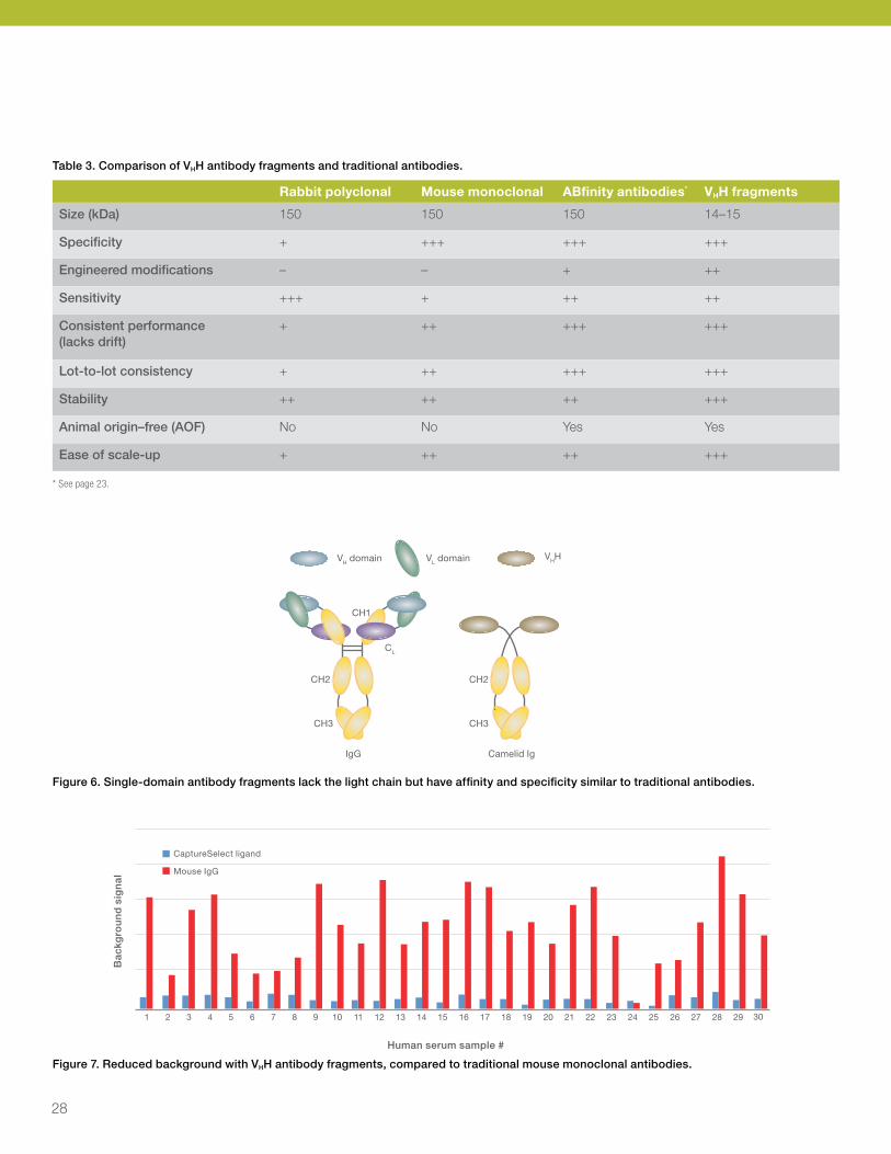
Table 3. Comparison of VHH antibody fragments and traditional antibodies.
Rabbit polyclonal Mouse monoclonal ABfinity antibodies* VHH fragments
Size (kDa) 150 150 150 14–15
Specificity + +++ +++ +++
Engineered modifications – – + ++
Sensitivity +++ + ++ ++
Consistent performance (lacks drift)
+ ++ +++ +++
Lot-to-lot consistency + ++ +++ +++
Stability ++ ++ ++ +++
Animal origin–free (AOF) No No Yes Yes
Ease of scale-up + ++ ++ +++
* See page 23.
VH domain VL domain
CH1
CL
IgG
CH2
CH3
Camelid Ig
CH2
CH3
VHH
Figure 6. Single-domain antibody fragments lack the light chain but have affinity and specificity similar to traditional antibodies.
Figure 7. Reduced background with VHH antibody fragments, compared to traditional mouse monoclonal antibodies.
1 2 3 4 5 6 7 8 9 10 11 12 13 14 15 16 17 18 19 20 21 22 23 24 25 26 27 28 29 30
CaptureSelect ligand
Mouse IgG
Human serum sample #
Ba
ckg
rou
nd
sig
na
l
28

Antibodies and detection probes
For requests or inquiries on our products for immunodiagnostic assay development, please email [email protected]
Custom antibody development and production servicesFull-service, customized production of monoclonal and polyclonal antibodies
Our custom antibody development service leverages our experience in making more than 18,500 antibodies to peptides and recombinant proteins. Our proprietary antigen design tools, including the Thermo Scientific™ Antigen Profiler software and Thermo Scientific™ Targeted Antigen Display Technology (TAD), produce robust antibodies that perform well in your targeted assays. When you initiate a custom antibody project with us, we provide you access to our online project management tool. This secure account gives you easy access to project information and allows you to provide specific instructions for your projects.
We specialize in antigen design and generation of custom peptide antibodies and monospecific peptide antibodies to highly discrete epitopes. Our detailed knowledge of antigen-determining factors allows us to produce custom antibodies with superior specificity, affinity, and assay utility.
Highlights:• Peptide design, synthesis, and conjugation—we will
help you design the best antigenic peptide using our exclusive Antigen Profiler system; then we’ll synthesize the peptide for you and conjugate it to the carrier protein of your choice.
• Fusion protein expression and purification—we offer bacterial, mammalian, and insect protein expression services as part of our antibody development platform; we’ll make your protein and purify it to prepare it for immunization and ELISA testing.
• Polyclonal or monoclonal antibody protocols—choose one of six species options (eight immunization schedules) for polyclonal antibody production or one of three package sizes (number and strains of mice) for monoclonal antibody production.
• ABfinity recombinant rabbit monoclonal antibody production services—develop recombinant rabbit monoclonal antibodies to your target. Screening can either be completed by our development scientists or in your labs.
• Biomarker antibody development—choose from several standardized packages for producing polyclonal or monoclonal antibodies to specific biomarkers of interest.
• Screening and titering analysis—we screen and characterize the antisera or hybridoma supernatants produced, and we will perform nearly any assay development and validation experiment you request.
• Purification specificities and deliverables— we purify all antibodies with an effective three-step procedure; in addition, we offer many specialized purification options for obtaining monospecific antibodies, such as to phosphorylated, acetylated, or other posttranslational modification states.
• Online project tracking and management— all antibody production services use our exclusive Thermo Scientific™ OpenProject Tool, a web portal that gives you real-time information about the status and progress of your antibody production projects, as well as complete management control over next steps.
29

Table 4. Summary of custom polyclonal and monoclonal antibody development and production service capabilities and options.
Polyclonal antibody production Monoclonal antibody production
Recombinant protein
antibody
Standard anti-peptide
antibody
Modification-specific
antibody*
Modification-specific
antibody*
Standard anti-peptide
antibody
Recombinant protein
antibody
Antigen design with Antigen Profiler ✔ ✔ ✔ ✔ ✔ ✔
Antigen design with Antigen Profiler
Peptide antigen synthesis
NA ✔ ✔ ✔ ✔ NA Peptide antigen synthesis
Depleting peptide synthesis
NA NA ✔ ✔ NA NA Control peptide synthesis
Protein expression (vs. you supply) Optional NA NA NA NA Optional
Protein expression (vs. you supply)
Animal immunization (8 protocol options) ✔ ✔ ✔ ✔ ✔ ✔
Animal immunization (3 protocol options)
Bleeds, serum prep, ELISA screening, and titration
✔ ✔ ✔ ✔ ✔ ✔
Hybridoma fusion, cloning, screening, and ELISA titration
Affinity purification and depletion (several options)
Optional Optional ✔ Optional Optional Optional
Culture, production, and purification (several options)
Specific assay validation or antibody labeling
Optional Optional Optional Optional Optional Optional Specific assay validation or antibody labeling
Complete online project tracking ✔ ✔ ✔
Coming soon
Coming soon
Coming soon
Complete online project tracking
* Modification-specific antibodies are phosphospecific antibodies or monospecific antibodies against peptides with other posttranslational modifications or specific states (phosphorylation, sumoylation, myristolation, acetylation, polymorphisms, drug binding, ubiquitination, glycosylation, isoforms, splice variants, ligand binding, prenylation, protein cleavage neoepitopes, mutations, species cross-reactivity).
ABfinity recombinant rabbit monoclonal antibody development and production services
ABfinity recombinant antibodies are rabbit monoclonal antibodies that are developed by immunizing animals, screening for functionality, and then cloning the immunogen–specific antibody genes into high-level mammalian expression vectors.
Antigendevelopment
Immunization andlibrary screening
Monoclonalantibody screening Bioproduction
3–4 months 1 month 1 month
Phase 1 Phase II* Phase III
• Customer-supplied antigen: – 5 mg protein – 20 mg peptide
• Invitrogen™ peptide or protein
• Crude serum titer check• B cell isolation and screening• Antibody cloning• Small-scale library expression
• Clone isolation• Small-scale antibody expression
• Transfection and expression of final antibody library or clone
• Customer receives 5 mg antibody
* Phase II is only performed in the monoclonal protocol. During oligoclonal development, the protocol includes phases I and III only.
30

Antibodies and detection probes
For requests or inquiries on our products for immunodiagnostic assay development, please email [email protected]
The recombinant antibodies are then produced by large-scale cell culture and purified with Protein A.
Choose the Invitrogen™ custom antibody service to obtain ABfinity antibodies with the following features:
• Greater specificity and sensitivity compared to standard antibodies
• Lot-to-lot consistency due to recombinant technology
• Animal origin–free antibodies, expressed in a mammalian cell system
Custom conjugation of antibodies or proteins
We offer different fluorescent labels for primary antibodies, secondary antibodies, anti-dye and anti-hapten antibodies, and streptavidin, including proprietary labels such as Alexa Fluor and DyLight dyes, Qdot™ nanocrystals, Invitrogen™ Texas Red™-X, and Invitrogen™ Pacific Blue™ dyes, which span the visible light spectrum from deep blue to near-infrared emission, generic fluorophores such as fluorescein and tetramethylrhodamine, RPE, APC, and peridinin chlorophyll protein complex (PerCP). We also offer conjugates of biotin, 2,4-dinitrophenyl (DNP), digoxigenin, dansyl, and other haptens.
We have more than 30 years of experience in preparing custom antibody conjugates that are optimized to meet your application-specific assay needs and match your instrument and optical specifications. The type of conjugation chemistry and linkers used can affect the functional outcome of your antibody or protein. Our expertise and our broad range of technologies enable you to get superior results. We can couple antibodies in a site-specific way and to labels like gold particles, polystyrene microspheres and nanospheres, and magnetic particles.
We are also highly experienced with conjugating antibodies and streptavidin to enzymes including AP, HRP, beta-galactosidase, and others.
Our custom conjugation service is efficient and confidential, and we stand behind the quality of our work. We can conjugate your antibody or protein to many labels, including:
• Alexa Fluor dyes
• DyLight dyes
• Qdot nanocrystals
• Pacific Blue, Pacific Green™, and Pacific Orange™ dyes
• Traditional fluorophores such as FITC, TRITC, and Texas Red dyes
• Biotin
• RPE, APC, and Alexa Fluor tandem dyes
• HRP, AP, and other enzymes
Additional testing is available upon request and includes:
• Activity validation/bioassay testing (Biacore or other applications such as IF, FACS, etc.)
• Purity testing and/or mass spectrometry (MS) analysis
• Endotoxin testing and removal
• Special purification
ANTIBODY QUALITY ICON / Final Requestor: Jane Helmer
AntibodyQuality Icon
"Performance guaranteed"
Performance guaranteed
Performanceguaranteed
Performanceguaranteed
Approved verbiage Possible application arrangments
31
ELISA productsOver 1,000 ELISA products, ranging from antibody pairs to ready-to-use kits
The enzyme-linked immunosorbent assay (ELISA) is a benchmark for the quantitation of proteins, using a solid-phase enzyme immunoassay (EIA) to detect and measure protein targets in various sample types. ELISAs are designed to provide rapid and consistent results that are relatively easy to analyze. We offer a comprehensive portfolio for research areas including immunology, inflammation, neurobiology, and cancer.
ELISA kitsOur ELISA kits help provide accurate, sensitive, and consistent quantitative results. Each target protein is tested in biologically relevant models and are calibrated to NIBSC (National Institute of Biological Standards and Controls), if available. In addition, kits are validated using common sample types, including serum, plasma, and cell culture supernatant. Cell lysates are used to validate kits that detect signaling proteins or phosphorylation.
These ELISA kits must meet rigorous quality control specifications and are manufactured in an ISO facility to help ensure excellent quality and reproducibility.
For more information on our ELISA kits, go to thermofisher.com/elisakits
Advantages of our ELISA kits:• Broad menu of over 800 targets
• Optimized for sensitive, accurate, and consistent performance
• Thorough instructions to complete protocol in 2.5 to 4 hours (varies by kit)
• Validated for typical sample types (e.g., serum, plasma, supernatant, lysates)
Protein targets:• Cytokines
• Chemokines
• Interleukins
• Inflammation targets
• Signaling proteins
• Receptors
• Neurobiology markers
• Phosphorylated proteins
• Growth factors
• Adhesion molecules
Ready-to-use ELISA kits typically include:• Antibody-coated 96-well plate
• Standards
• Primary detection antibody (typically biotinylated)
• Secondary detection reagent (usually streptavidin-HRP)
• Diluent buffers
• Wash buffers
• Substrate and stop solutions
• Plate covers
32

Antibodies and detection probes
For requests or inquiries on our products for immunodiagnostic assay development, please email [email protected]
Antibody pair kits
Antibody pair kits contain matched, pretitered, and fully optimized capture (coating) and detection antibodies. These kits enable you to build your own ELISA or any other assay platform that utilizes a matched antibody pair.
These matched pair kits are designed to accurately quantify cytokines, chemokines, growth factors, signaling pathway targets, and proteins associated with immunology, inflammation, cancer, cardiovascular, and neurodegenerative disease research.
Advantages of the antibody pair kits for ELISA:• Quality—reliable antibodies, prematched antibody pairs
and proven detection reagents
• Ease of use—simplified protocol and optimized reagents
• Flexibility—multiple detection technologies (fluorescence, absorbance, or chemiluminescence) are possible
• Cost savings—more economical than complete, ready-to-use ELISA kits contain precoated plates
Build and customize your own immunoassays:The kits are designed for use with a variety of sample types such as serum, plasma, cell culture supernatant, cell lysate, tissue homogenate, urine, and cerebrospinal fluid (CSF). For convenience, we also offer a buffer set for antibody pairs that contains premade, easy-to-use buffers and solutions that are optimized for use with the antibody pair kits.
Each antibody pair kit supplies sufficient reagents for 10 ELISA plates (five plates for intracellular targets) and includes:
• Capture antibody
• Detector antibody
• Recombinant standard
• HRP conjugates
For more information on our antibody pair kits, go to thermofisher.com/antibodypairs
33

Biotin-binding protein conjugatesEnzyme-labeled streptavidin and avidin conjugates
Pierce High Sensitivity Streptavidin-HRP
The Thermo Scientific™ Pierce™ High Sensitivity Streptavidin-HRP conjugate is an exclusive peroxidase-conjugated, biotin-binding protein that provides signal amplification and exceptional storage stability.
Highlights:• High sensitivity—detect low levels of target without
background; obtain high signal-to-noise ratios (Figure 8)
• Cost effective—use less conjugate in western blotting and ELISA applications and still obtain excellent results
• Flexible—compatible with typical chemiluminescent, fluorescent, and colorimetric peroxidase substrates
• Convenient—ready-to-use stabilized liquid format means there is no waiting for thawing and no need to aliquot
This specially manufactured variety of HRP conjugated–streptavidin protein is designed to meet the demands of today’s scientists for more sensitive detection in immunoassay applications. The conjugate is suitable for use with chemiluminescent, chemifluorescent, or colorimetric substrates. Each High Sensitivity HRP conjugate is packaged in an easy-to-use stabilized solution for convenient storage at 4°C for at least one year.
0
200
1 10 100 1,000
400
600
800
1,000
1,200
1,400
1,600
1,800
Rel
ativ
e lig
ht
un
its
(RLU
)
GST (pg)
High Sensitivity Streptavidin-HRP
MilliporeStreptavidin-HRP
GE HealthcareStreptavidin-HRP
Figure 8. Pierce High Sensitivity Streptavidin-HRP conjugate enables low-level target detection with high signal-to-noise ratios. Recombinant Thermo Scientific™ Pierce™ GST (glutathione-S-transferase) was serially diluted (0–10,000 pg/mL) with Thermo Scientific™ StartingBlock™ (PBS) Blocking Buffer. Each dilution (100 µL) was added to a 96-well Thermo Scientific™ Pierce™ glutathione-coated plate in four replicates, including a negative control. The plate was incubated for 60 minutes at room temperature and washed three times with PBS Tween™-20 detergent. Biotinylated anti-GST (100 µL at 250 ng/mL; Santa Cruz) was added to all wells. The plate was incubated for 30–60 minutes at room temperature and washed three times. Streptavidin-HRP conjugates were diluted in blocking buffer as per manufacturer’s directions. Pierce High Sensitivity Streptavidin-HRP conjugate was diluted 1:10,000 and conjugates from other suppliers were diluted to 1:1,000. The conjugate solutions (100 µL) were added to the plate and incubated for 60 minutes (light protected) at room temperature. The plate was washed five times and 150 µL/well of Thermo Scientific™ SuperSignal™ ELISA Pico Substrate was added. Signal intensity was measured using a luminometer.
34

Antibodies and detection probes
For requests or inquiries on our products for immunodiagnostic assay development, please email [email protected]
Pierce Streptavidin Poly-HRP conjugate
The Thermo Scientific™ Pierce™ Streptavidin Poly-HRP conjugate is a biotin-binding protein conjugated with polymers of HRP, enabling signal amplification and detection of biotinylated antibodies for IHC and other methods. Pierce Streptavidin Poly-HRP conjugate is designed to deliver the highest sensitivity and low background in immunoassays where sample volume is limited or when the target molecule is present at low levels. Streptavidin Poly-HRP conjugate is purified to remove unconjugated streptavidin molecules that reduce signal intensity by competing for binding sites with HRP-conjugated molecules. In addition, the conjugate is devoid of HRP monomers that can cause background signal.
Highlights:• High sensitivity—detect low-abundant targets
(low picogram to femtogram range) with high signal-to-noise ratios
• Robust—consistent manufacturing and purification to minimize low and unconjugated molecules for minimal background and high sensitivity
• Flexible—compatible with chromogenic, fluorogenic, and chemiluminescent substrates
• Versatile—compatible with ELISA, western blotting, and IHC
• Easy to use—can be directly substituted into immunoassays and other detection assays
• Convenient—ready-to-use, stabilized liquid format stored at 4°C
• Cost effective—requires less conjugate per assay than standard HRP conjugates
Pierce Streptavidin, HRP conjugate
The Thermo Scientific™ Pierce™ HRP-conjugated streptavidin includes streptavidin in a purified form, conjugated to peroxidase for substrate-based detection.
The Pierce Streptavidin, HRP conjugate enables detection of biotinylated antibodies and other probes in a variety of standard assay methods, including western blotting, ELISA, IHC, and fluorescence imaging. The conjugate is supplied as a lyophilized powder in phosphate-based buffers for immediate reconstitution with water.
Streptavidin, alkaline phosphatase conjugate
The Invitrogen™ streptavidin alkaline phosphatase conjugate can be used to detect biotin in a signal amplification scheme in conjunction with chromogenic or fluorogenic substrates.
35

For requests or inquiries on our products for immunodiagnostic assay development, please email [email protected]
Pierce High Sensitivity NeutrAvidin HRP conjugate
The Thermo Scientific™ Pierce™ High Sensitivity NeutrAvidin HRP conjugate is a specially prepared peroxidase-conjugate form of avidin-biotin–binding protein that provides signal amplification like poly-HRP conjugate and exceptional storage stability. This specially manufactured variety of HRP conjugated–avidin protein is designed to meet the demands of today’s scientists for more sensitive detection in ELISA and western blotting applications. The conjugate is suitable for use with chemiluminescent, chemifluorescent, or colorimetric substrates. Each High Sensitivity HRP conjugate is packaged in an easy-to-use, stabilized solution for convenient storage at 4°C for at least one year.
Highlights:• NeutrAvidin protein—a specially deglycosylated form
of avidin that provides highly specific, low-background binding of biotinylated antibodies in many applications
• High sensitivity—detect low levels of target without background; obtain high signal-to-noise ratios (Figure 9)
• Cost effective—use less conjugate in western blotting and ELISA applications and still obtain excellent results
• Flexible—compatible with typical chemiluminescent, fluorescent, and colorimetric peroxidase substrates
• Convenient—ready-to-use, stabilized liquid format, which means there is no waiting for thawing and no need to aliquot
NeutrAvidin-HRPdetection ofanti-GAPDH antibody
Biotinylated primary Ab Negative control
Streptavidin-HRPdetection ofanti-cytokeratin 18antibody
Figure 9. Excellent IHC staining of GAPDH and cytokeratin 18 in human colon carcinoma tissues with Pierce High Sensitivity HRP conjugates. Formalin-fixed, paraffin-embedded (FFPE) human colon carcinoma tissues were stained using a metal-enhanced DAB substrate (Thermo Scientific™ Pierce™ Immunohistochemistry Peroxidase Detection Kit). The tissues were incubated with either biotinylated anti-GAPDH or rabbit anti-cytokeratin 18 antibody followed by biotinylated anti-rabbit IgG (left panels) or blocking buffer only (right panels). The sections were subsequently incubated with Pierce High Sensitivity NeutrAvidin-HRP (top panels) or Streptavidin-HRP conjugate (bottom panels). Tissues were counterstained using the Harris-modified hematoxylin solution (blue staining in all panels). GAPDH and cytokeratin 18, stained using Pierce NeutrAvidin HRP and Streptavidin-HRP conjugates, appear brown (left panels) while the negative control panels shows no brown staining in the tissue.
Pierce NeutrAvidin HRP conjugateThermo Scientific™ Pierce™ NeutrAvidin HRP conjugate is a specially prepared form of avidin-biotin–binding protein conjugated to peroxidase that decreases background in western blotting and ELISA applications.
NeutrAvidin protein is deglycosylated native avidin from egg whites. Removal of the excess carbohydrate by an exclusive process yields a protein with a more neutral isoelectric point and less nonspecific binding properties. NeutrAvidin protein exhibits high assay specificity and sensitivity with high signal-to-noise ratios. Purified and conjugated forms of NeutrAvidin protein provide exceptional performance in western blot, ELISA, and IHC applications that require biotin-binding probes.
36

Antibodies and detection probes
NeutrAvidin Alkaline Phosphatase Conjugate
Thermo Scientific™ alkaline phosphatase conjugated NeutrAvidin protein is a specially prepared form of avidin that decreases background in biotin binding. Conjugated NeutrAvidin protein offers exceptional performance in western blot, ELISA, and IHC applications that require biotin-binding probes. NeutrAvidin protein exhibits high assay specificity and sensitivity with high signal-to-noise ratios.
Highlights: • Near-neutral isoelectric point—pI = 6.3, more neutral
than native avidin
• Nearly devoid of glycosylation—decreased possibility of lectin binding compared to native avidin
• No RYD recognition sequence—no known off-target binding domains like those contained in streptavidin
• Affordable—significantly less expensive than streptavidin
Pierce Horseradish Peroxidase Conjugated Avidin
Thermo Scientific™ Pierce™ Horseradish Peroxidase Conjugated Avidin is used in biotin-based detection methods, including western blotting or ELISA detection with appropriate enzyme substrates. Avidin from chicken egg whites is a glycoprotein that effectively binds biotin, a tag that is frequently used to tag antibodies and other probes for immunodetection methods.
Highlights:• Native avidin—glycoprotein from chicken egg whites,
67 kDa, pI = 10
• Soluble—glycosylation and isoelectric point renders avidin more soluble than streptavidin
• Affordable—significantly less expensive than recombinant streptavidin
• ABC staining—avidin is usually preferred over streptavidin for avidin-biotin complex (ABC) staining methods in IHC
• HRP conjugate—for membrane and plate- based assays
Fluorescent streptavidin and avidin conjugates
Alexa Fluor fluorescent streptavidin, avidin, and NeutrAvidin conjugates
We offer a broad range of Invitrogen™ Alexa Fluor™ fluorescent streptavidin, avidin, and NeutrAvidin conjugates (Table 5). Benefits of the Alexa Fluor Dyes and their conjugates include exceptionally bright conjugates, excellent photostability, and a broad range of emission colors (Figure 10).
Figure 10. The cytoskeleton of a fixed and permeabilized bovine pulmonary artery endothelial (BPAE) cell. Tubulin was detected using mouse monoclonal anti–alpha-tubulin antibody and visualized with Alexa Fluor 647 goat anti–mouse IgG antibody (pseudocolored magenta). Endogenous biotin in mitochondria was labeled with green-fluorescent Alexa Fluor 488 streptavidin; DNA was stained with blue-fluorescent DAPI.
37

Alexa Fluor streptavidin phycobiliprotein conjugates
Invitrogen™ Alexa Fluor™ streptavidin phycobiliprotein conjugates are fluorescent streptavidin conjugates with tandem dyes RPE (R-Phycoerythrin) and APC (Allophycocyanin) labeled with long-wavelength Alexa Fluor dyes (Table 6). These tandem conjugates are comprised of a donor phycobiliprotein–RPE conjugate or APC conjugated to longer wavelength–emitting fluorescence acceptors (Table 6). By the process of fluorescence resonance energy transfer (FRET), an energy transfer cascade is established wherein most of the light absorbed by the donor RPE (or APC) results in fluorescence of the acceptor dye. This process can be quite efficient, resulting in almost total transfer of energy to the acceptor dye.
Table 5. Alexa Fluor fluorescent streptavidin conjugates.
Streptavidin conjugates Ex (nm)* Em (nm)*
Alexa Fluor 350 streptavidin 346 442
Alexa Fluor 405 streptavidin 402 421
Alexa Fluor 430 streptavidin 434 539
Alexa Fluor 488 streptavidin 495 519
Alexa Fluor 500 streptavidin 503 525
Alexa Fluor 514 streptavidin 518 540
Alexa Fluor 532 streptavidin 530 554
Alexa Fluor 555 streptavidin 555 565
Alexa Fluor 546 streptavidin 556 573
Alexa Fluor 568 streptavidin 578 603
Alexa Fluor 594 streptavidin 590 617
Alexa Fluor 610 streptavidin 612 628
Alexa Fluor 633 streptavidin 632 647
Alexa Fluor 635 streptavidin 633 647
Alexa Fluor 647 streptavidin 650 668
Alexa Fluor 660 streptavidin 663 690
Alexa Fluor 680 streptavidin 679 702
Alexa Fluor 700 streptavidin 702 723
Alexa Fluor 750 streptavidin 749 775
NeutrAvidin and avidin conjugates
Alexa Fluor 350 NeutrAvidin 346 442
Alexa Fluor 488 avidin 495 519
* Approximate fluorescence excitation (Ex) and emission (Em) maxima for conjugates, in nm.
Table 6. Tandem Alexa Fluor streptavidin phycobiliprotein conjugates.
Tandem–RPE conjugates
Streptavidin conjugates Ex (nm)* Em (nm)*
Alexa Fluor 610–RPE Streptavidin 496, 546, 565
630
Alexa Fluor 647–RPE Streptavidin 496, 546, 565
668
Alexa Fluor 680–RPE Streptavidin 496, 546, 565
702
Alexa Fluor 750–RPE Streptavidin 496, 546, 565
771
APC and tandem APC conjugates
Streptavidin conjugates Ex (nm)* Em (nm)*
Allophycocyanin (APC) streptavidin 650 660
Alexa Fluor 680–APC streptavidin 650 702
Alexa Fluor 700–APC streptavidin 650 723
Alexa Fluor 750–APC streptavidin 650 775
* Approximate fluorescence excitation (Ex) and emission (Em) maxima for conjugates, in nm.
38
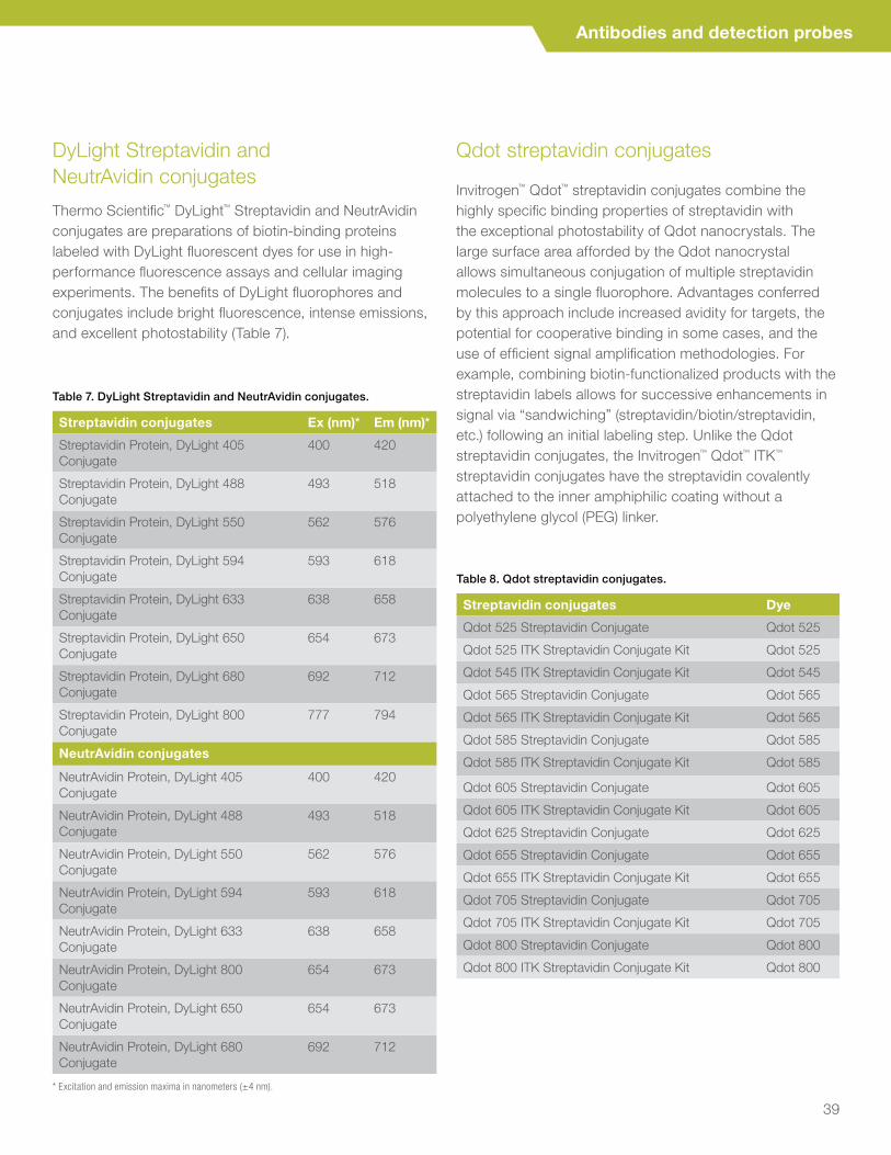
Antibodies and detection probes
DyLight Streptavidin and NeutrAvidin conjugates
Thermo Scientific™ DyLight™ Streptavidin and NeutrAvidin conjugates are preparations of biotin-binding proteins labeled with DyLight fluorescent dyes for use in high-performance fluorescence assays and cellular imaging experiments. The benefits of DyLight fluorophores and conjugates include bright fluorescence, intense emissions, and excellent photostability (Table 7).
Qdot streptavidin conjugates
Invitrogen™ Qdot™ streptavidin conjugates combine the highly specific binding properties of streptavidin with the exceptional photostability of Qdot nanocrystals. The large surface area afforded by the Qdot nanocrystal allows simultaneous conjugation of multiple streptavidin molecules to a single fluorophore. Advantages conferred by this approach include increased avidity for targets, the potential for cooperative binding in some cases, and the use of efficient signal amplification methodologies. For example, combining biotin-functionalized products with the streptavidin labels allows for successive enhancements in signal via “sandwiching” (streptavidin/biotin/streptavidin, etc.) following an initial labeling step. Unlike the Qdot streptavidin conjugates, the Invitrogen™ Qdot™ ITK™ streptavidin conjugates have the streptavidin covalently attached to the inner amphiphilic coating without a polyethylene glycol (PEG) linker.
Table 8. Qdot streptavidin conjugates.
Streptavidin conjugates Dye
Qdot 525 Streptavidin Conjugate Qdot 525
Qdot 525 ITK Streptavidin Conjugate Kit Qdot 525
Qdot 545 ITK Streptavidin Conjugate Kit Qdot 545
Qdot 565 Streptavidin Conjugate Qdot 565
Qdot 565 ITK Streptavidin Conjugate Kit Qdot 565
Qdot 585 Streptavidin Conjugate Qdot 585
Qdot 585 ITK Streptavidin Conjugate Kit Qdot 585
Qdot 605 Streptavidin Conjugate Qdot 605
Qdot 605 ITK Streptavidin Conjugate Kit Qdot 605
Qdot 625 Streptavidin Conjugate Qdot 625
Qdot 655 Streptavidin Conjugate Qdot 655
Qdot 655 ITK Streptavidin Conjugate Kit Qdot 655
Qdot 705 Streptavidin Conjugate Qdot 705
Qdot 705 ITK Streptavidin Conjugate Kit Qdot 705
Qdot 800 Streptavidin Conjugate Qdot 800
Qdot 800 ITK Streptavidin Conjugate Kit Qdot 800
Table 7. DyLight Streptavidin and NeutrAvidin conjugates.
Streptavidin conjugates Ex (nm)* Em (nm)*
Streptavidin Protein, DyLight 405 Conjugate
400 420
Streptavidin Protein, DyLight 488 Conjugate
493 518
Streptavidin Protein, DyLight 550 Conjugate
562 576
Streptavidin Protein, DyLight 594 Conjugate
593 618
Streptavidin Protein, DyLight 633 Conjugate
638 658
Streptavidin Protein, DyLight 650 Conjugate
654 673
Streptavidin Protein, DyLight 680 Conjugate
692 712
Streptavidin Protein, DyLight 800 Conjugate
777 794
NeutrAvidin conjugates
NeutrAvidin Protein, DyLight 405 Conjugate
400 420
NeutrAvidin Protein, DyLight 488 Conjugate
493 518
NeutrAvidin Protein, DyLight 550 Conjugate
562 576
NeutrAvidin Protein, DyLight 594 Conjugate
593 618
NeutrAvidin Protein, DyLight 633 Conjugate
638 658
NeutrAvidin Protein, DyLight 800 Conjugate
654 673
NeutrAvidin Protein, DyLight 650 Conjugate
654 673
NeutrAvidin Protein, DyLight 680 Conjugate
692 712
* Excitation and emission maxima in nanometers (±4 nm).
39

Crosslinking reagents are used in many techniques to assist in immobilization of a ligand to a solid phase, and also for the conjugation of a detection antibody to an enzyme or fluorescent dye. We offer crosslinkers, PEGylation reagents, biotinylation kits and reagents, and antibody and enzyme labeling kits. In addition, we provide custom conjugation of antibodies and proteins.
CrosslinkersCrosslinking is the process of chemically joining two or more molecules by a covalent bond. Crosslinking reagents, or crosslinkers, are molecules that contain two or more reactive ends capable of chemically attaching to specific functional groups (primary amines, sulfhydryls, etc.) on proteins or other molecules.
Crosslinkers are defined by their reactivities:
Homobifunctional crosslinkers have identical reactive groups at either end of a spacer arm. Generally, they must be used in one-step reaction procedures to randomly ”fix” or polymerize molecules containing like functional groups. This is useful for capturing a snapshot of all protein interactions, but it cannot provide the precision needed for other types of crosslinking applications.
Heterobifunctional crosslinkers possess different reactive groups at either end. These reagents not only allow for single-step conjugation of molecules that have the respective target functional groups, but they also allow for sequential (two-step) conjugations that minimize undesirable polymerization or self-conjugation. In sequential procedures, heterobifunctional reagents are reacted with one protein using the most labile group of the crosslinker first. After removing excess unreacted crosslinker, the modified first protein is added to a solution containing the second protein where reaction through the second reactive group of the crosslinker occurs.
Heterobifunctional crosslinkers are the preferred choice for antibody-enzyme or other protein-to-protein crosslinking applications. Unwanted self-conjugation inherent when using homobifunctional NHS-ester reagents or glutaraldehyde can be avoided by reagents such as SMCC or sulfo-SMCC. In separate reactions, one protein can be reacted to the amine-specific end of this reagent while the other protein is treated with reducing agent or sulfhydryl-addition reagent to expose or create sulfhydryl groups. Finally, after removing excess nonreacted reagents, the two proteins can be mixed to allow the sulfhydryl-reactive groups (e.g., maleimide) of the first protein to conjugate with the sulfhydryl groups of the second protein (Figure 1).
Linking mechanisms
40

Linking mechanisms
For requests or inquiries on our products for immunodiagnostic assay development, please email [email protected]
Maleimide-activated enzyme
Sulfhydryl-activated antibody
Sulfo-SMCC crosslinker
Enzyme-antibody conjugate
Enzyme
Enz
NN
S
S
OO
O
O
O
O-Na
+NH2
OO
O
NO
O
O
Enz
Ab
Ab
NH
HS
NO
O
O
Enz NH
Antibody-enzyme conjugation with heterobifunctional crosslinker Sulfo-SMCC.
NH21 H2N 2
NH
OHN
O
1
2
Primary aminePrimary amine
Sulfo-NHSSulfo-NHS
Crosslinked proteins
OHN
O
O
SO
O
Na+O-HO
N
O
O
S
O-Na+
O
O
OO
O
OBS3
N
O
O
S
O-Na+
O
ON
O
O
SO
O
Na+O-
Use of homobifunctional crosslinker BS3.
Figure 1. Example reactions for producing detection conjugates.
Pierce crosslinkers
Many types of Thermo Scientific™ Pierce™ crosslinkers are available for protein, peptide, and other macromolecular immobilization and conjugation needs. Both homobi-functional and heterobifunctional crosslinkers are offered with a variety of spacer-arm lengths, solubility, and cleaving characteristics.
Our wide selection of crosslinking reagents now includes those that contain discrete-length polyethylene glycol (PEG) spacers. These PEG groups increase reagent and conjugate solubility, minimize toxic and immunological effects compared to non-PEG spacers, and provide several options for accommodating specific crosslinking distances (Figure 2).
PEG crosslinkers offer:• Increased water solubility
• Decreased aggregation
• Better accessibility
SMCC
NHS-(PEG)4-Maleimide
N
O
O
NO
O
O
O
NO
O
O
NNH
O
O O
O
O
O
O
O
Figure 2. Chemical structures of SMCC and PEG alternative NHS-(PEG)4-maleimide.
Pierce crosslinkers are manufactured in our organic synthesis lab at scales ranging from milligram to hundreds of kilograms. Available quality tests include visual inspection, structural confirmation by infrared spectrometry (IR), purity by HPLC or quantitative NMR (qNMR), solvent analysis by GC headspace, water content by Karl Fischer titration, and elemental analysis. Custom specifications can also be set.
41

Pierce BS3 crosslinker
BS3
Bis(sulfosuccinimidyl) suberateMW 572.43
Spacer arm 11.4 Å
N OO
OO
O
N
O
O O
S O
O
S
O
O
Na+O-
O-Na+
Thermo Scientific™ Pierce™ Premium Grade BS3 (Sulfo-DSS) crosslinker is bis(sulfosuccinimidyl)suberate, an amine-to-amine crosslinker that is homobifunctional, water-soluble, non-cleavable, and membrane-impermeant.
BS3 contains an amine-reactive N-hydroxysulfosuccinimide (NHS) ester at each end of an 8-carbon spacer arm. NHS esters react with primary amines at pH 7–9 to form stable amide bonds, along with release of the N-hydroxysulfosuccinimide leaving group. Proteins, including antibodies, generally have several primary amines in the side chain of lysine (K) residues and the N-terminus of each polypeptide that are available as targets for NHS-ester crosslinking reagents.
Features of Pierce BS3 crosslinker:• Reactive groups: sulfo-NHS ester (both ends)
• Reactive with amino groups (primary amines)
• Amine-reactive sulfo-NHS ester reacts rapidly with any primary amine–containing molecule
• Water-soluble, compared with DSS
• Membrane-impermeant, allowing for cell surface labeling
• High-purity, crystalline reagent can be used to create high-purity crosslinked conjugates
Because it contains the hydrophilic sulfonyl moiety, Pierce BS3 crosslinker is soluble up to ~100 mM in water and many commonly used buffers, thus avoiding the use of organic solvents, which may perturb protein structure. DSS, the non–water-soluble analog of BS3, is also available
for applications that require a less hydrophilic crosslinker (e.g., to effect intracellular crosslinking). DSS and BS3 have essentially identical crosslinking activity toward primary amines.
Properties of Pierce BS3:• Alternative names: Sulfo-DSS
• Molecular formula: C16H18N2O14S2Na2
• Molecular weight: 572.43
• Spacer-arm length: 11.4 Å (8 atoms)
• CAS number: 82436-77-9
• Reactive groups: sulfo-NHS esters react with primary amines at pH 7–9
Applications:• Protein crosslinking to create bioconjugates via single-
step reactions
• Crosslink cell-surface proteins prior to cell lysis and immunoprecipitation
• Immobilize proteins onto amine-coated surfaces
• Identify receptor-ligand interactions by chemical crosslinking
• “Fix” protein interactions to allow identification of weak or transient protein interactions
• Identify near-neighbor protein interactions
Pierce BS3 specifications:We manufacture Pierce BS3 crosslinker to high specifications to produce specific bioconjugates, to help ensure the integrity of your data, and to offer you a high degree of consistency. Each lot of Pierce BS3 crosslinker is tested to meet the following minimum specifications:
• Purity: >93% by quantitative NMR (the highest standard for crosslinker purity)
• Solubility: >5.8 mg/mL in DI water, clear solution with no insoluble material
42

Linking mechanisms
Pierce Sulfo-SMCC crosslinker
Sulfo-SMCCSulfosuccinimidyl 4-(N-maleimidomethyl)cyclohexane-1-carboxylate
MW 436.37Spacer arm 8.3 Å
O
O
N
O
O
N
O
O
S
Na+O-
O
O
Thermo Scientific™ Pierce™ Sulfo-SMCC is a water-soluble, amine-to-sulfhydryl crosslinker that contains NHS-ester and maleimide reactive groups at opposite ends of a medium-length cyclohexane spacer arm (8.3 Å).
Features of Pierce Sulfo-SMCC crosslinker:• Reactive groups: sulfo-NHS ester and maleimide
• Reactive with amino and sulfhydryl groups
• Water-soluble (compare to SMCC), so crosslinking can be done in physiologic solutions
• High-purity, crystalline reagent can be used to create high-purity, maleimide-activated derivatives
• Cyclohexane bridge confers added stability to the maleimide group making Sulfo-SMCC the ideal crosslinking agent for maleimide activation of proteins. Maleimide groups are stable for 64 hours in 0.1 M sodium phosphate buffer, pH 7 at 4°C
Sulfosuccinimidyl-4-(N-maleimidomethyl)cyclohexane-1-carboxylate (Sulfo-SMCC) is a non-cleavable and membrane-impermeable crosslinker. It contains an amine-reactive N-hydroxysuccinimide (NHS ester) and a sulfhydryl-reactive maleimide group. NHS esters react with primary amines at pH 7–9 to form stable amide bonds. Maleimides react with sulfhydryl groups at pH 6.5–7.5 to form stable thioether bonds. The maleimide groups of Sulfo-SMCC and SMCC are unusually stable up to pH 7.5 because of the cyclohexane bridge in the spacer arm. Because it contains the hydrophilic sulfonyl moiety, Pierce
Sulfo-SMCC crosslinker is soluble up to ~10 mM in water and many commonly used buffers, thus avoiding the use of organic solvents which may perturb protein structure.
Applications:• Enzyme labeling of antibodies—both enzyme and
antibody specificity can be preserved
• Create specific bioconjugates via one- or two-step crosslinking reactions
• Create sulfhydryl-reactive, maleimide-activated carrier proteins for coupling haptens
Specifications:Pierce Sulfo-SMCC crosslinker is manufactured to high specifications to produce specific bioconjugates and maleimide-activated proteins, to help ensure the integrity of your data and to offer you a high degree of crosslinking consistency. Each lot of Pierce Sulfo-SMCC crosslinker is tested to meet the following minimum specifications.
• Identity: IR scan shows only peaks characteristic of the structure and functional groups of Sulfo-SMCC crosslinker
• Purity: ≥90% by quantitative NMR (the highest standard for crosslinker purity)
• Solubility: ≥10 mg/mL in DI water
43

Pierce Sulfo-NHS reagent
N
O
O
S
Na +O-
O
O
Sulfo-NHS AcetateM.W. 259.17
Thermo Scientific™ Pierce™ Sulfo-NHS (N-hydroxysulfosuccinimide) is a chemical modification reagent for converting carboxyl groups to amine-reactive NHS esters for bioconjugation, crosslinking, labeling, and immobilization methods.
Features of Pierce Sulfo-NHS reagent:• Efficiency of EDC-mediated coupling is increased in the
presence of NHS or Sulfo-NHS
• Amine-reactive NHS esters or sulfo-NHS esters can be made with any carboxyl-containing molecule
• Sulfo-NHS derivatives are usually readily water-soluble (can be added directly to physiologic buffers) and membrane-impermeable (can be used for cell surface labeling)
• High-purity, crystalline Sulfo-NHS can be used to create high-quality activated derivatives
Sulfo-NHS enables control and modification of carbodiimide crosslinking reactions involving activation of carboxylates (–COOH) for conjugation with primary amines (–NH2). Derivatives are easily synthesized by mixing the Sulfo-NHS with a carboxyl-containing molecule and a dehydrating agent such as the carbodiimide EDC (EDAC). The method is the basis for generating many types of protein labeling reagents, including amine-reactive fluorescent dyes, biotin affinity tags, and PEGylation compounds.
Applications:• Improve efficiency of EDC coupling reactions
• Convert carboxyls to amine-reactive sulfo-NHS esters
• Crosslink proteins to carboxyl-coated beads or surfaces more efficiently
• Activate nanoparticles with amine-reactive sulfo-NHS esters
Specifications for Pierce Sulfo-NHS reagent:We manufacture N-hydroxysulfosuccinimide to high specifications to produce specific bioconjugates, to help ensure the integrity of your data, and to offer you a high degree of consistency. Each lot of Pierce Sulfo-NHS reagent is tested to meet the following minimum specifications:
• Purity—greater than 95% by quantitative NMR (the highest standard for crosslinker purity); average lot purity is greater than 99%
• Solubility—sample dissolves at 2 mg/mL in deionized water to yield a clear, colorless solution
• Identity—IR scan shows only peaks characteristic of N-hydroxysulfosuccinimide
Bond-Breaker TCEP Solution, Neutral pH
PHO
O
OHO
OH
O
TCEPM.W. 250.15
Thermo Scientific™ Bond-Breaker TCEP Solution, Neutral pH, is a stable, 0.5 M solution of the thiol-free, phosphine-based TCEP compound, useful as a 10X stock for addition to SDS-PAGE sample loading buffers to reduce protein disulfide bonds.
44

Linking mechanisms
For requests or inquiries on our products for immunodiagnostic assay development, please email [email protected]
Features of Bond-Breaker TCEP Solution, Neutral pH:• Unlike dithiothreitol (DTT) and beta-mercaptoethanol
(beta-ME), TCEP is odor-free
• Efficient—5 to 50 mM TCEP is designed to thoroughly reduce most peptide or protein disulfide bonds within a few minutes (i.e., just as effective as DTT)
• Enables specific and complete reduction of even the most stable water-soluble alkyl disulfides
• Reduces protein disulfides at room temperature and pH 5, typically in less than five minutes
• Resistant to air oxidation; nonvolatile and nonreactive toward other protein functional groups
• Reduces peptides and proteins over a broad range of pH, salt, detergent, and temperature conditions
• Removal of the reducing agent is not necessary before most applications (e.g., histidine-tagged protein purification, maleimide conjugations), because TCEP does not contain sulfhydryl groups
Bond-Breaker TCEP Solution is a potent, odorless, thiol-free reducing agent with broad application to protein modification and other research involving reduction of disulfide bonds. It is an effective and convenient replacement for beta-ME or DTT in SDS-PAGE sample buffers. The neutral pH of this reagent provides sharp bands and avoids exposing proteins to the strong acid of TCEP-HCl, which can result in hydrolysis and carbohydrate modification.
Pierce SPDP crosslinker
S
O
S
O
NO
N
O
SPDPSuccinimidyl 3-(2-pyridyldithio)propionate
MW 312.36Spacer arm 6.8 Å
Thermo Scientific™ Pierce™ SPDP (succinimidyl 3-(2-pyridyldithio)propionate) is a short-chain crosslinker for amine-to-sulfhydryl conjugation via NHS-ester and pyridyldithiol reactive groups that form cleavable (reducible) disulfide bonds with cysteine sulfhydryls.
Features of Pierce SPDP crosslinker:• Reactive groups: NHS ester and pyridyldithiol
• Reactive with amino and sulfhydryl groups
• Releases a detectable by-product when reacted with a free sulfhydryl group; by measuring the release of pyridine-2-thione at 343 nm, the reaction can be easily followed
• Disulfide bond in the spacer arm is readily cleaved by 10–50 mM DTT or TCEP at pH 8.5
• Spacer arm is also easily cleaved using reducing SDS-PAGE sample loading buffer
• Cleavable crosslinker allows separation of crosslinked products
• Water-insoluble (dissolve first in DMF or DMSO)
• SPDP crosslinker is membrane-permeant, so crosslinking can be done inside the cells
• Compare to other SPDP-type reagents, including PEGylated forms
45

For requests or inquiries on our products for immunodiagnostic assay development, please email [email protected]
Applications:• Reversible crosslinking of proteins
• Adding sulfhydryl groups to proteins by attachment to lysine residues
• Protein crosslinking to create antibody-enzyme and other bioconjugates via a two-step reaction
• Attaching haptens to carrier proteins for antibody production
• Preparing immunotoxin conjugates
Specifications:We manufacture Pierce SPDP crosslinker to high specifications to produce specific bioconjugates to help ensure the integrity of your data and to offer you a high degree of consistency. Each lot of SPDP is tested to ensure it is >95% pure by quantitative NMR (the highest standard for crosslinker purity).
We offer three analogs of SPDP: the standard version (SPDP), a derivative with a longer spacer arm (LC-SPDP), and a sulfonated water-soluble variety (Sulfo-LC-SPDP). SPDP (N-Succinimidyl 3-(2-pyridyldithio)-propionate) and LC-SPDP (Succinimidyl 6-(3-[2-pyridyldithio]-propionamido)hexanoate) are heterobifunctional, thiol-cleavable, and membrane-permeant crosslinkers. They each contain an amine-reactive N-hydroxysuccinimide (NHS) ester that will react with lysine residues to form a stable amide bond. The other end of the spacer arm is terminated in the pyridyl disulfide group that will react with sulfhydryls to form a reversible disulfide bond.
Pierce EDC crosslinker
EDC1-Ethyl-3-(3-dimethylaminopropyl)carbodiimide • HCl
MW 191.70Spacer arm 0.0 Å
N
NC
NHCl-+
Thermo Scientific™ Pierce™ EDC (1-ethyl-3-(3-dimethylaminopropyl)carbodiimide hydrochloride) is a water-soluble carbodiimide crosslinker that activates carboxyl groups for spontaneous reaction with primary amines, enabling peptide immobilization and hapten-carrier protein conjugation.
EDC (or EDAC) is a zero-length crosslinking agent used to couple carboxyl groups to primary amines. This crosslinker has been used in diverse applications such as forming amide bonds in peptide synthesis, attaching haptens to carrier proteins to form immunogens, labeling nucleic acids through 5’ phosphate groups, and creating amine-reactive NHS-esters of biomolecules. EDC reacts with a carboxyl to form an amine-reactive O-acylisourea intermediate. If this intermediate does not encounter an amine, it will hydrolyze and regenerate the carboxyl group. In the presence of N-hydroxysulfosuccinimide (Sulfo-NHS), EDC can be used to convert carboxyl groups to amine-reactive sulfo-NHS esters. This is accomplished by mixing the EDC with a carboxyl-containing molecule and adding Sulfo-NHS.
Features of Pierce EDC crosslinker:• Reactive group: carbodiimide
• Reaction target: activates carboxyl groups to conjugate to amino groups (primary amines)
• React EDC alone with target groups or include NHS or Sulfo-NHS to increase reaction efficiency or to stabilize active intermediate for later reaction to amines
• Forms neutral amide bonds between carboxyls and amines
46

Linking mechanisms
• Water-soluble reagent can be added directly to reactions in aqueous, physiological buffers
• Soluble reaction byproducts are easily removed by washing with water or dilute acid
• High-purity, crystalline reagent can be used to create high-quality, activated derivatives
Properties of Pierce EDC crosslinker:• Molecular formula: C8H17N3·HCl
• Molecular weight: 191.7
• Spacer arm length: 0.0 Å
• CAS Number: 25952-53-8
• Reactive groups: carbodiimide
• Reactivity: forms active intermediate with carboxyl groups at pH 4.7–6.0 (optimum), then intermediate reacts with primary amines
Applications:• Conjugate carboxyl and amino groups among peptides
and proteins
• Couple haptens to immunogenic carrier proteins (e.g., attach a peptide to KLH)
• Immobilize peptide antigens to affinity-purify antibodies
• Create NHS-activated, amine-reactive labeling compounds
• Crosslink proteins to carboxyl-coated beads or surfaces
• Activate nanoparticles with amine-reactive sulfo-NHS esters
• DNA labeling through 5' phosphate groups
Also see Thermo Scientific™ Pierce™ Premium Grade EDC, page 55.
For information on all of our crosslinkers, go to thermofisher.com/crosslinkers
PEGylation reagentsWe offer activated linear and branched derivatives of PEG for PEGylation and the PEG-modification of peptides and proteins via primary amines and sulfhydryl groups. These reagents are typically used to increase solubility, prolong stability, and reduce immunogenicity. Certain experimental systems and assay platforms depend on the ability to alter the mass, solubility or other properties of proteins, immunogens, reaction vessels, and other materials.
Amine-reactive PEGylation reagents Covalent modification with PEG groups requires PEG compounds that contain a reactive or targetable functional group at one end. The simplest method for PEGylating proteins, which are rich in surface primary amines, is to use a PEG compound that contains an NHS ester group at one end.
• MS(PEG)n reagents are available in various PEG lengths (n = 4, 8, 12, and 24)
• TMS(PEG)n is a branched form of this reagent, containing three methyl-(PEG)12 arms attached to a (PEG)4-NHS ester stem
Sulfhydryl-reactive PEGylation reagents MM(PEG)n compounds are linear reagents for PEGylating sulfhydryl groups. At the end, opposite the inert methyl group(s), is a maleimide moiety, which reacts to form stable thioether bonds with sulfhydryl groups.
• MM(PEG)n is available in two PEG lengths (n = 12 and 24)
47

For requests or inquiries on our products for immunodiagnostic assay development, please email [email protected]
PEGylated amino acids and amine compounds
MA(PEG)n and CA(PEG)n are polyethylene glycol compounds of discrete length (n = 4, 8, 12, and 24) containing methyl-and-amine or carboxyl-and-amine ends. Although these functional groups are not spontaneously reactive, they are easily targeted by various crosslinking and immobilization reagents for the construction of surface chemistries.
Pierce MS(PEG)n reagentsThermo Scientific™ Pierce™ MS(PEG)n reagents are methyl-terminated, polyethylene glycol compounds (n equals 4 to 24 PEG units) activated as NHS esters for covalent PEGylation of primary amines on proteins (e.g., lysines) or assay surfaces.
Features of MS(PEG)4:• NHS-activated for efficient PEGylation of primary amines
at pH 7–9; reaction of NHS-ester group results in formation of stable, irreversible amide bonds
• Fully characterized PEGylation reagents with defined PEG chain lengths; molecules of discrete molecular weight for consistency of performance in protein-modification applications
• Provided as a series of 4, 8, 12, and 24 ethylene glycol units, enabling modification procedures to be optimized for a specific application while retaining all the benefits associated with protein PEGylation
• PEG spacer provides unique advantages, including increased stability, reduced tendency toward aggregation and reduced immunogenicity
• Easy-to-follow instructions help increase the likelihood of a successful outcome
MS(PEG)n is the abbreviation for a set of compounds having PEG spacers with methyl (–CH3) and amine-reactive NHS-ester groups at opposite ends. The unbranched, hydrophilic, discrete-length molecules have the form methyl-(PEG)n-NHS ester, where the subscript “n” denotes 4, 8, 12, or 24 ethylene glycol units. The N-hydroxysuccinimide (NHS) ester is spontaneously reactive with primary amines (–NH2), providing for efficient PEGylation of proteins, peptides, and other amine-containing molecules or surfaces.
PEGylation applications:• PEGylate amine surfaces
• Add inert mass to proteins, immunogens, drug compounds, and probes
• Improve solubility (decrease aggregation) of proteins or peptides without affecting function
• Protect proteins from proteolysis
Advantages of discrete-length mPEG-NHS ester compoundsThese reagents are specially synthesized as homogeneous compounds of discrete chain length and defined molecular weight. As such, they enable precise control and optimi- zation of surface protein modification experiments. By contrast, typical preparations of PEG compounds are heterogeneous mixtures composed of multiple chain lengths and a range of molecular weights.
48
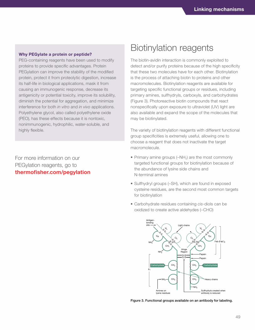
Linking mechanisms
For more information on our PEGylation reagents, go to thermofisher.com/pegylation
Biotinylation reagentsThe biotin-avidin interaction is commonly exploited to detect and/or purify proteins because of the high specificity that these two molecules have for each other. Biotinylation is the process of attaching biotin to proteins and other macromolecules. Biotinylation reagents are available for targeting specific functional groups or residues, including primary amines, sulfhydryls, carboxyls, and carbohydrates (Figure 3). Photoreactive biotin compounds that react nonspecifically upon exposure to ultraviolet (UV) light are also available and expand the scope of the molecules that may be biotinylated.
The variety of biotinylation reagents with different functional group specificities is extremely useful, allowing one to choose a reagent that does not inactivate the target macromolecule.
• Primary amine groups (–NH2) are the most commonly targeted functional groups for biotinylation because of the abundance of lysine side chains and N-terminal amines
• Sulfhydryl groups (–SH), which are found in exposed cysteine residues, are the second most common targets for biotinylation
• Carbohydrate residues containing cis-diols can be oxidized to create active aldehydes (–CHO)
Antigen-bindingsite Light chains
Carbohydrate Carbohydrate
HingeRegion
CH2
CH3
CH2
CH3
CH1 CH1
VL
CL
VL
CLVH
S-SS-S
S-SS-S
S-S
S-SS-S
Papain
Pepsin
S-S
S-SS-S
S-S
S-S
Fab (Fab')2
FC
SS
SS
SS
SS Heavy chains
VH
NH2
Amines onlysine residues
Sulfhydryls created whenantibody is reduced
NH2
NH2
NH2
Figure 3. Functional groups available on an antibody for labeling.
Why PEGylate a protein or peptide?PEG-containing reagents have been used to modify proteins to provide specific advantages. Protein PEGylation can improve the stability of the modified protein, protect it from proteolytic digestion, increase its half-life in biological applications, mask it from causing an immunogenic response, decrease its antigenicity or potential toxicity, improve its solubility, diminish the potential for aggregation, and minimize interference for both in vitro and in vivo applications. Polyethylene glycol, also called polyethylene oxide (PEO), has these effects because it is nontoxic, nonimmunogenic, hydrophilic, water-soluble, and highly flexible.
49

EZ-Link biotinylation reagents
Thermo Scientific™ EZ-Link™ biotinylation reagents and kits are used to chemically tag and label antibodies, proteins, and peptides with biotin at particular amino acid functional groups to create labeled antibody or other probes for streptavidin affinity binding, purification, and detection.
Our wide selection of reagents and kits includes traditional biotin-labeling reagents and PEG forms of biotinylation reagents. Compared to traditional reagents, PEG biotinylation reagents offer:
• Long-chain biotin alternatives
• Water-soluble, hydrophilic spacers
• PEG-biotinylated antibodies are more soluble after biotinylation
• Increased signal in ELISA applications (Figure 4)
• No aggregation issues (Figure 5)
00 500 1,000 1,500 2,000 2,500
Anti TNF-alpha LC-LC Biotin
Anti TNF-alpha LC Biotin
Anti TNF-alpha (PEG)12 Biotin
Anti TNF-alpha (PEG)4 Biotin
3,000No
rmal
ized
OD
450
−O
D55
0
TNF-alpha (pg/mL)
TNF-alpha ELISA
1.50
1.75
1.25
1.00
0.75
0.50
0.25
00 500 1,000 1,500 2,000 2,500
1:40 NHS-(PEG)4-Biotin1:40 NHS-(PEG)12-Biotin
1:40 NHS-LC-Biotin
3,000
1
OD
450
–OD
550
IL-10 (pg/mL)
IL-10 ELISA
2
3
Figure 4. Increased signal with PEG biotinylation reagents.
1:20 NHS-(PEG)4-Biotin1:20 NHS-(PEG)12-Biotin1:20 NHS-LC-Biotin1:20 NHS-LC-LC-Biotin
Relative increase in aggregation48 hours after labeling
GM-CSF0
1
2
3
4
TNF-alphaAntibody
IL-12
1:40 NHS-(PEG)4-Biotin1:40 NHS-(PEG)12-Biotin1:40 NHS-LC-Biotin1:40 NHS-LC-LC-Biotin
Relative increase in aggregation48 hours after labeling
GM-CSF0
1
2
4
3
TNF-alphaAntibody
IL-12
Figure 5. Aggregation reduction with PEG biotinylation reagents.
50

Linking mechanisms
For requests or inquiries on our products for immunodiagnostic assay development, please email [email protected]
EZ-Link NHS-PEG4-Biotin
Thermo Scientific™ EZ-Link™ NHS-PEG4-Biotin is a long (29.0 Å), PEGylated, water-soluble reagent for simple and efficient biotin labeling of antibodies, proteins, and other primary amine-containing macromolecules. The N-hydroxysuccinimide ester (NHS) group reacts specifically and efficiently with lysine and N-terminal amino groups to form stable amide bonds. The hydrophilic polyethylene glycol (PEG) spacer arm imparts water solubility that is transferred to the biotinylated molecule, thus reducing aggregation of labeled proteins stored in solution. The PEG spacer arm also gives the reagent a long and flexible connection to minimize steric hindrance for binding to avidin molecules.
Highlights:• Protein labeling—biotinylate antibodies or other proteins
for detection or purification using streptavidin probes or resins
• Amine-reactive—reacts with primary amines (–NH2), such as the side-chain of lysines (K) or the amino-termini of polypeptides
• PEGylated—spacer arm contains a hydrophilic, 4-unit, PEG group
• Enhances solubility—PEGylation imparts water solubility to the biotinylated molecule, helping to prevent aggregation of biotinylated antibodies stored in solution
• Irreversible—forms permanent amide bonds; spacer arm cannot be cleaved
• Long reach—spacer arm (total length added to target) is 29 Å; this reduces steric hindrance when binding to avidin molecules
EZ-Link Sulfo-NHS-LC-Biotin
Thermo Scientific™ EZ-Link™ Sulfo-NHS-LC-Biotin is an intermediate-length, water-soluble biotinylation reagent for labeling antibodies, proteins, and other molecules that have primary amines.
Highlights:• Protein labeling—biotinylate antibodies to facilitate
immobilization, purification or detection using streptavidin resins or probes
• Cell surface labeling—biotinylates only surface proteins of whole cells because the negatively charged reagent does not permeate cell membranes
• Amine-reactive—reacts with primary amines (–NH2), such as lysine side-chains, or the amino-termini of polypeptides
• Soluble—charged sulfo-NHS group increases reagent water solubility compared to ordinary NHS-ester compounds
• Irreversible—forms permanent amide bonds; spacer arm cannot be cleaved
• Medium length—spacer arm (total length added to target) is 22.4 Å; provides excellent balance between adequate length (minimal steric hindrance for biotin binding) and modest mass
Sulfo-NHS-LC-Biotin is one of three very similar EZ-Link reagents that are water-soluble, non-cleavable, and enable simple and efficient biotinylation of antibodies, proteins, and any other primary amine-containing macromolecules in solution. Specific labeling of cell surface proteins is another common application for these uniquely water-soluble and membrane-impermeant reagents. Differing only in their spacer arm lengths, the three Sulfo-NHS-ester reagents offer the possibility of optimizing labeling and detection experiments where steric hindrance of biotin binding is an important factor (Table 1).
51

Amine biotinylation reagents
Product categories Product Product featuresSpacer arm (Å)
Water-soluble? Cleavable?
NHS-PEG4-biotin and biotinylation kits
• EZ-Link NHS-PEG4 Biotinylation Kit
• EZ-Link Micro NHS-PEG4-Biotinylation Kit
Maximize the solubility of antibodies and other proteins
29 Yes No
Sulfo-NHS-LC-biotin and biotinylation kits
• EZ-Link Sulfo-NHS-LC- Biotinylation Kit
• EZ-Link Micro Sulfo-NHS-LC-Biotinylation Kit
Complete kit with an optimized procedure for labeling a protein and determining how much biotin has been attached
22.4 Yes No
NHS-PEG solid phase biotinylation kits
• EZ-Link NHS-PEG Solid-Phase Biotinylation Kit—1 mL Column
• EZ-Link NHS-PEG Solid-Phase Biotinylation Kit
A column-based approach to antibody biotinylation and clean-up
NA NA NA
Carbonyl biotinylation reagents
Product categories Product Product features
Spacer arm (Å)
Water-soluble? Cleavable?
Membrane permeable?
Hydrazide-biotin reagents
EZ-Link Hydrazide-Biotin
Short and mid-length reagents for labeling glycoproteins and other carbohydrate-containing compounds having oxidizable sugars or aldehydes
15.7 No No Yes
Hydrazide-biotin reagents
EZ-Link Biotin-LC-Hydrazide
Short and mid-length reagents for labeling glycoproteins and other carbohydrate-containing compounds having oxidizable sugars or aldehydes
24.7 No No Yes
Hydrazide-PEG4-biotin
EZ-Link Hydrazide-PEG4-Biotin
A carbohydrate-biotinylation reagent with a long, hydrophilic spacer arm
31.3 Yes No No
Sulfhydryl biotinylation reagents
Product categories Product Product features
Spacer arm (Å)
Water-soluble? Cleavable?
Membrane permeable?
Maleimide-PEG11-biotin
EZ-Link Maleimide PEG11-Biotin
A column-based approach to antibody biotinylation and clean-up
59.1 Yes No No
Maleimide-PEG2-biotin
EZ-Link Maleimide PEG2-Biotin
A simple, water-soluble biotinylation reagent specific for sulfhydryls
29.1 Yes No No
Maleimide solid phase biotinylation kits
EZ-Link Maleimide-PEG Solid-Phase Biotinylation Kit—Prepacked Column
A column-based approach to antibody biotinylation and clean-up
NA NA NA NA
Maleimide solid phase biotinylation kits
EZ-Link Maleimide-PEG Solid-Phase Biotinylation Kit—Mini-Spin Columns
A column-based approach to antibody biotinylation and clean-up
NA NA NA NA
Table 1. EZ-Link biotinylation reagents.
52

Linking mechanisms
For requests or inquiries on our products for immunodiagnostic assay development, please email [email protected]
Carboxyl biotinylation reagents
Product categories Product Product features
Spacer arm (Å)
Water-soluble? Cleavable?
Membrane permeable?
Amine-PEG-biotin reagents
• EZ-Link Amine-PEG2-Biotin
Short, hydrophilic reagents for biotinylation of carboxyls via EDC
20.4 Yes No No
• EZ-Link Amine-PEG3-Biotin
22.9 Yes No No
Amine-PEG11-biotin
EZ-Link Amine-PEG11-Biotin
Long, hydrophilic reagent for biotinylation of carboxyls via EDC
53.2 Yes No No
For information on all of our biotinylation reagents, go to thermofisher.com/biotinylation
Biotin quantitation kitsThese simple and accurate 4-hydroxyazobenzene-2-carboxylic acid (HABA) dye assay and fluorescence detection kits are used to measure the amount of biotin attached to proteins or other biomolecules following biotinylation, allowing you to determine the molar ratio of biotin to labeled antibody molecule in an easy, efficient manner.
Product Product featuresDetection method
Pierce Fluorescence Biotin Quantitation Kit
Fluorescent assay reagent and HABA dye for rapid and sensitive determination of biotinylation levels in labeled antibodies and other proteins with minimal sample quantities
Fluorescence
Pierce Biotin Quantitation Kit A simple, colorimetric method for determining biotinylation efficiency Colorimetric
Pierce HABA HABA dye reagent and biotinylated protein standard for convenient colorimetric determination of biotinylation levels in antibodies and other proteins
Colorimetric
Table 2. Biotin quantitation kits and reagents.
53

Pierce premium grade reagentsOur Thermo Scientific™ Pierce™ premium grade reagents are high-quality formulations of selected chemical modification reagents, specially characterized for applications where product integrity and risk minimization are paramount. The current menu of reagents offered in this premium format includes several popular crosslinkers and biotinylation reagents, and a reducing agent. The reagents are ideal for research projects that require long-term, consistent performance and an extra level of troubleshooting support afforded by robust chemical characterization and documentation. Available Pierce premium grade reagents include:
• Pierce Premium Grade Sulfo-NHS
• Pierce Premium Grade EDC
• Pierce Premium Grade Sulfo-NHS-LC-Biotin
• Pierce Premium Grade Sulfo-NHS-SS-Biotin
• Pierce Premium Grade DSP
• Pierce Premium Grade BS3
• Pierce Premium Grade Sulfo-SMCC
• Pierce Premium Grade SPDP
• Pierce Premium Grade TCEP-HCl
Highlights:• High quality—identity and purity confirmed by several
tests, including quantitative NMR
• Product integrity—enhanced level of testing and characterization compared to standard grade
• Lot retention—ample supply of past lots retained to help ensure future process testing
• Change management—Change Control Notification (CCN) service
• Consistent manufacturing—batch-specific manufacturing documentation review
Table 3. Comparison of premium grade and standard grade crosslinking reagents.
Test parameter Test method Premium grade Standard grade
Purity Quantitative NMR using an internal standard Yes Yes
Visual Color assessment Yes Yes
Solubility Sample dissolves at specified concentration in deionized water to yield a clear, colorless solution
Yes Yes
Identity Infrared spectroscopy Yes Yes
Mass identity Mass spectrometry Yes NA
Water content Karl Fischer titration Yes NA
Trace metals Inductively coupled plasma mass spectrometry (ICP-MS) Yes NA
Elemental analysis Combustion analysis (values reported for C, H, N, O, and S) Yes NA
Residual solvent analysis Headspace gas chromatography Upon request NA
54

Linking mechanisms
For requests or inquiries on our products for immunodiagnostic assay development, please email [email protected]
Pierce Premium Grade EDC crosslinker
EDC1-Ethyl-3-(3-dimethylaminopropyl)carbodiimide • HCl
MW 191.70Spacer arm 0.0 Å
N
NC
NHCl-+
Thermo Scientific™ Pierce™ Premium Grade EDC (1-Ethyl-3-(3-dimethylaminopropyl)carbodiimide • HCl) crosslinker is our highest-quality formulation of this popular carbodiimide crosslinker, specially characterized for applications where product integrity and risk minimization are paramount.
Features of Pierce Premium Grade EDC crosslinker:• Carbodiimide crosslinker for conjugation between
carboxylates and amines
• Identity and purity confirmed by several tests for exceptional quality
• High level of testing and characterization compared to standard grade for high product integrity
• Ample supply of past lots retained to help ensure future process testing
• Change control notification (CCN) service
• Batch-specific manufacturing documentation review
Compared to the standard grade product, Pierce Premium Grade EDC crosslinker provides more clearly defined quality and product support by including (a) increased analytical testing and product characterization, (b) greater batch-specific information and quality assurance review, (c) extensive lot sample retention, and (d) change control notification. EDC is a water-soluble carbodiimide crosslinker that activates carboxyl groups for spontaneous reaction with primary amines, enabling peptide immobilization and hapten-carrier protein conjugation. In the presence of N-hydroxysulfosuccinimide (sulfo-NHS), EDC can be used to convert carboxyl groups to amine-reactive sulfo-NHS esters. This is accomplished by mixing the EDC with a carboxyl containing molecule and adding sulfo-NHS.
Specifications of Pierce Premium Grade EDC crosslinker:We manufacture crosslinking reagents to offer high overall product integrity, consistency, and performance for the intended research applications.
• Visual: white crystalline powder, free of foreign matter
• Identity: IR scan shows only peaks characteristic of EDC
• Water content: Karl Fischer titration, ≤2%
• Trace metals: all metals, including lead, ≤50 ppm
• Elemental analysis: reported values for C, H, and N
Applications:• Conjugate carboxyl and amino groups among peptides
and proteins
• Couple haptens to immunogenic carrier proteins (e.g., attach a peptide to KLH)
• Immobilize peptide antigens to affinity-purify antibodies
• Create NHS-activated, amine-reactive labeling compounds
• Crosslink proteins to carboxyl-coated beads or surfaces
• Activate nanoparticles with amine-reactive, sulfo-NHS esters
For information on all of the Pierce Premium Grade Reagents, go to thermofisher.com/premiumreagents
55

For requests or inquiries on our products for immunodiagnostic assay development, please email [email protected]
Fluorescent antibody and protein labeling kitsWe offer a number of antibody and protein labeling kits for the direct attachment of a broad range of intensely fluorescent dyes and labels including Alexa Fluor dyes, DyLight dyes, Qdot labels, R-phycoerythrin, APC tandem dyes, and biotin, at scales from less than 10 μg up to 1 mg of IgG antibody. Direct labeling of antibodies allows you to use more than one same-species antibody in a single staining experiment. You can use traditional labeling chemistries optimized for your application or site-specific labeling using Invitrogen™ SiteClick™ or Zenon™ labeling technology.
Kits for fluorescent site-specific antibody labeling
SiteClick labeling technology specifically attaches the label to the heavy chains of an IgG antibody helping to ensure that the antigen binding domains remain available for binding to your antigen target. This site selectivity is achieved by targeting the carbohydrate domains present on essentially all IgG antibodies regardless of isotype and host species.
Zenon labeling technology provides a versatile, easy-to-use system for labeling human, mouse IgG1, IgG2a, and IgG2b antibodies and rabbit IgG antibodies. Zenon fragments are specifically designed to target and bind to the Fc portion of the primary antibody only, giving a rapid, noncovalent method of quickly labeling small quantities of primary antibody. Zenon labeling technology is simple and efficient and scalable; the entire labeling procedure takes only 10 minutes.
Kits for conventional antibody and protein labeling
Invitrogen™ APEX™ antibody labeling kits provide an efficient, convenient, and reliable method for covalently attaching fluorescent labels to very small amounts of IgG antibody in the presence of any contaminants, including stabilizing proteins and amine-containing buffers.
Thermo Scientific™ DyLight™ antibody labeling kits are designed for fast, efficient labeling of antibodies with DyLight dyes.
Invitrogen™ Alexa Fluor™ antibody labeling kits enable direct labeling of monoclonal and polyclonal antibodies.
Invitrogen™ microscale protein labeling kits conveniently label small amounts of purified protein or antibodies (MW between 12 and 150 kDa) with a bright, photostable Alexa Fluor dye or the hapten, biotin-XX.
Our protein labeling kits provide a nearly effortless way to stably label larger amounts of IgG antibody with a fluorescent dye or hapten in two hours.
Invitrogen™ Alexa Fluor™ SAIVI™ Rapid Antibody Labeling Kits are uniquely designed to provide the rigorous requirements to control the degree of labeling (DOL) using our near-infrared (NIR) Alexa Fluor dyes.
For more information on all of our antibody and protein labeling kits, go to thermofisher.com/antibodylabeling
56

Linking mechanisms
SiteClick antibody labeling system
The Invitrogen™ SiteClick™ antibody labeling system allows simple and gentle site-selective attachment of detection molecules to heavy chain N-linked glycans far from the antigen-binding domain, enabling excellent reproducibility from labeling to labeling and from antibody to antibody (Figures 6 and 7). A number of different detection molecules can be site-selectively attached to the heavy-chain glycans, including phycobiliproteins (e.g., RPE), Qdot probes, fluorescent dyes, metal-chelating compounds, and other small molecules such as biotin, allowing multiplex analysis with antibodies from the same species.
Highlights:• Highly efficient, site-specific, reproducible labeling
chemistry that preserves antigen binding domains
• Qdot labels for fluorescence microscopy and flow cytometry (Figure 8)
• RPE labels for flow cytometry
• Alexa Fluor labels using Invitrogen™ Click-iT™ Alexa Fluor™ DIBO Alkynes
How the SiteClick antibody labeling system works In general, IgG antibodies contain two N-linked glycans attached to specific conserved asparagine residues located in the antibody heavy-chain Fc domain. These sugar chains are structurally quite homogeneous, and the terminal sequences of the glycan branches are highly consistent. Most of the antibody glycan branches terminate with galactose-N-acetylglucosamine (Gal-GlcNAc-) or with N-acetylglucosamine (GlcNAc-). Removal of the terminal Gal residue with beta-galactosidase unmasks the majority of terminal GlcNAc labeling sites for the subsequent enzymatic beta-galactosyl transferase (GalT) reaction (Figure 7). After cleavage of terminal Gal residues by beta-galactosidase, each N-linked glycan will contain, on average, two terminal GlcNAc residues per heavy chain (four terminal GlcNAc per antibody).
The SiteClick method is compatible with antibodies from a number of different species including, but not limited to, human, rabbit, mouse, rat, goat, hamster, and chicken. Additionally, SiteClick labeling is effective with several antibody classes such as IgG, IgM, and IgY; note that chicken IgY antibodies have six heavy chain glycans instead of two and therefore can be labeled to a higher extent.
Figure 6. The SiteClick antibody labeling system. The first step in the SiteClick antibody labeling process involves removal of terminal galactose residues from the heavy chain N-linked glycans using beta-galactosidase, exposing essentially all possible modifiable GlcNAc residues. Second, the free terminal GlcNAc residues are activated with azide tags by enzymatic attachment of GalNAz to the terminal GlcNAc residues using the GalT(Y289L) enzyme. In the third step, the azide residues are reacted with the dibenzocyclooctyne (DIBO)-functionalized probe of choice (e.g., Alexa Fluor 488 DIBO Alkyne). The average degree of labeling is 3–3.5 labels per antibody.
Antibody • Polyclonal • Monoclonal • Recombinant
Beta-galactosidaseGalT(Y289L)UDP-GalNAz DIBO Alkyne label
Azide-activated antibodySite-selectively
labeled antibody
N3
N3
N3
N3
57

For requests or inquiries on our products for immunodiagnostic assay development, please email [email protected]
– Heavy chain
– Heavy chainPNGase F: – – + +
U L U L
A
B
SYPRO RubyTotal Protein Stain
SiteClickAlexa Fluor 488DIBO
– Light chain
Figure 7. Site selectivity of SiteClick antibody labeling. (A) A beta-tubulin monoclonal antibody was azide-activated using the GalT(Y289L) enzyme, labeled with DIBO-functionalized Alexa Fluor 488 Dye, and then analyzed by gel electrophoresis (left panel). Once imaged, the same gel was post-stained with Invitrogen™ SYPRO™ Ruby Total Protein Gel Stain (right panel), showing that only the heavy chain of the antibody is click-labeled with the Alexa Fluor 488 DIBO Alkyne. (B) Anti-beta-tubulin mouse monoclonal antibodies were labeled with a small-molecule DIBO-PET chelating agent (L) or left unlabeled (U) (left two lanes) and then posttreated with PNGase F, which selectively cleaves N-linked glycans from asparagine residues (right two lanes). After treatment with PNGase F, both chelator-labeled (L) and unlabeled (U) species are shifted to the same lower molecular weight, confirming the site-selective labeling of heavy chain N-linked glycans.
Figure 8. Immunocytochemistry with SiteClick-labeled antibodies. HeLa cells were fixed, permeabilized, and incubated with 10 nM Qdot 655 dye–conjugated Anti-golgin 97 antibody (magenta). This conjugate was generated using the SiteClick Qdot 655 Antibody Labeling Kit and mouse monoclonal anti-golgin-97 antibody (CDF3). After antibody incubation, the cells were counterstained with Invitrogen™ NucBlue™ Live (blue) and Invitrogen™ ActinGreen™ 488 (green) ReadyProbes™ reagents prior to imaging.
Fluorescent dyesAs one of the world’s leading developers of fluorescence-based technology, we offer many fluorescent labels for primary antibodies, secondary antibodies, anti-dye and anti-hapten antibodies, and streptavidin, including proprietary labels such as Alexa Fluor, DyLight, Texas Red-X, and Pacific Blue dyes.
Alexa Fluor dyesThe Alexa Fluor dyes produce exceptionally bright and photostable conjugates. These dyes are available in several reactive forms for labeling biopolymers and derivatizing low-molecular weight molecules. All Alexa Fluor dyes are available as an amine-reactive succinimidyl ester, and many are available in thiol, aldehyde, and carboxylic acid reactive forms (Table 4).
Highlights:• Compatible with all common excitation sources and
instruments
• Bright and unusually photostable fluorescence of their bioconjugates
• Good water solubility, which makes the reactive dyes easy to conjugate and the conjugates resistant to precipitation and aggregation
• Insensitivity of their absorption and emission spectra to pH over a broad range
• Well-differentiated spectra, providing many options for multicolor detection and fluorescence resonance energy transfer (FRET). Spans the near-UV, visible, and near-IR spectrum (Figure 9)
• High quantum yields and long fluorescence lifetimes
• Extremely high FRET efficiency, with calculated R0 values of up to 84 Å between pairs of Alexa Fluor dyes and up to 77 Å between Alexa Fluor dyes and some nonfluorescent quenchers
58

Linking mechanisms
Table 4. Properties of Alexa Fluor dyes.
Alexa Fluor dye Absorption max. (nm) Emission max. (nm) Emission color* Extinction coefficient**
Alexa Fluor 350 346 442 Blue 19,000
Alexa Fluor 405 401 421 Blue 34,000
Alexa Fluor 430 433 541 Green/yellow 16,000
Alexa Fluor 488 496 519 Green 71,000
Alexa Fluor 532 532 553 Yellow 81,000
Alexa Fluor 546 556 573 Orange 104,000
Alexa Fluor 555 555 565 Orange 150,000
Alexa Fluor 568 578 603 Orange/red 91,000
Alexa Fluor 594 590 617 Red 73,000
Alexa Fluor 610 612 628 Red 138,000
Alexa Fluor 633 632 647 Far Red 239,000
Alexa Fluor 635 633 647 Far Red 140,000
Alexa Fluor 647 650 665 Near-IR*** 239,000
Alexa Fluor 660 663 690 Near-IR*** 132,000
Alexa Fluor 680 679 702 Near-IR*** 184,000
Alexa Fluor 700 702 723 Near-IR*** 192,000
Alexa Fluor 750 749 775 Near-IR*** 240,000
Alexa Fluor 790 784 814 Near-IR*** 270,000
* Typical emission color seen through the eyepiece of a conventional fluorescence microscope with appropriate filters.
** Extinction coefficient at lmax in cm-1M-1.
*** Human vision is insensitive to light beyond ~650 nm; it is not possible to directly view near-IR fluorescent dyes.
Figure 9. Emission spectra for Alexa Fluor dyes.
Flu
ore
scen
ce e
mis
sio
n
400 450 500 550 600 650 700 750 800 850
Wavelength (nm)
1. Alexa Fluor 350 dye
2. Alexa Fluor 405 dye
3. Alexa Fluor 430 dye
4. Alexa Fluor 488 dye
5. Alexa Fluor 500 dye
6. Alexa Fluor 514 dye
7. Alexa Fluor 532 dye
8. Alexa Fluor 546 dye
9. Alexa Fluor 555 dye
10. Alexa Fluor 568 dye
11. Alexa Fluor 594 dye
12. Alexa Fluor 610 dye
13. Alexa Fluor 633 dye
14. Alexa Fluor 635 dye
15. Alexa Fluor 647 dye
16. Alexa Fluor 660 dye
17. Alexa Fluor 680 dye
18. Alexa Fluor 700 dye
19. Alexa Fluor 750 dye
20. Alexa Fluor 790 dye
19 201817161513141211109 8765 4 32 1
59

DyLight fluorescent dyes
DyLight fluorescent dyes are a complete family of high-intensity, photostable fluorescent tags for labeling antibodies and other molecular probes. DyLight dyes provide exceptional fluorescence in many applications, while maintaining excellent photostability and water solubility across a broad range of pH values.
DyLight Fluors have absorption maxima ranging from 350 nm to 777 nm, covering the entire visible light spectrum and several key near-infrared and infrared wavelengths (Table 5 and Figure 10). The absorption and emission properties of the DyLight Fluors match the output (excitation) and detection wavelengths of common fluorescence instrumentation.
Highlights:• High percentage activated dye (>80%)
• High water solubility and excellent photostability
• Available in amine- and sulfhydryl-reactive forms for fast, efficient labeling of IgG or other proteins
• Available for production of conjugates for commercialization (special conditions may apply)
• DyLight dyes are available preconjugated to various IgGs, streptavidin, and NeutrAvidin protein
Wavelength (nm)
350
350DyLight 405 488 550 594 633 650 680 755 800
400 450 500 550 600 650 700 750 800 850
Flu
ore
scen
ce e
mis
sio
n
Figure 10. Emission spectra of DyLight dyes.
Table 5. Spectral properties of DyLight fluorescent dyes.
Emission color DyLight dye Ex/Em* ε** Spectrally similar dyes
Blue 350 353/432 15,000 AMCA, Alexa Fluor 350 dye
Blue 405 400/420 30,000 Alexa Fluor 405 and Cascade Blue™ dyes
Green 488 493/518 70,000 Alexa Fluor 488, fluorescein, and FITC dyes
Yellow 550 562/576 150,000 Alexa Fluor 546, Alexa Fluor 555, Cy®3, and TRITC dyes
Red 594 593/618 80,000 Alexa Fluor 594 and Texas Red™ dyes
Red 633 638/658 170,000 Alexa Fluor 633 dye
Red 650 654/673 250,000 Alexa Fluor 647 and Cy®5 dyes
Near-IR 680 692/712 140,000 Alexa Fluor 680 and Cy®5.5 dyes
Near-IR 755 752/778 220,000 Alexa Fluor 750 and Cy®7 dyes
Near-IR 800 777/794 270,000 IRDye™ 800 dye
* Excitation and emission maxima in nanometers (±4 nm). ** Molar extinction coefficient (M-1cm-1).
60

Linking mechanisms
For requests or inquiries on our products for immunodiagnostic assay development, please email [email protected]
DyLight PEGylated dyes
The Thermo Scientific™ DyLight™ PEGylated dyes are derivatives of our high-performance DyLight dyes that can be used to fluorescently label antibodies and other proteins. Conjugates made with DyLight PEGylated dyes can be used as molecular probes for cellular imaging and other fluorescence detection methods. DyLight PEGylated dyes contain two to four nontoxic polyethylene glycol (PEG) chains. The PEG chains enhance fluorescence, reduce nonspecific binding of conjugates, improve solubility of the dyes and labeled molecules in aqueous solution, aid in cell permeability, and improve retention, especially in tumors [1]. The near-infrared (NIR) to far-red fluorescence properties of some of the PEGylated dyes such as DyLight 680, 755, and 800 dyes make them useful in biological, chemical, and pharmaceutical applications, including in vivo imaging (Figure 11).
The DyLight PEGylated Amine- and Sulfhydryl-Reactive dyes provide superior fluorescence characteristics (e.g., brightness, photostability, pH insensitivity, and water solubility). In addition, the high fluorescence intensity of DyLight PEGylated dyes provides outstanding sensitivity and requires less conjugate in most applications.
Highlights:• Soluble—PEGylated dyes and labeled molecules
improve solubility in aqueous solution, aid in cell permeability, and improve retention
• High fluorescence intensity—significantly brighter fluorescence than many other dyes
• Efficient labeling methods—well-characterized chemistry and optimized protocols enable reliable, high-quality labeling
• Reduced nonspecific binding
Table 6. Properties of the PEGylated DyLight NHS-ester fluorescent dyes.
DyLight™ Fluor Ex/Em* ε** MW (g/mol) Spectrally similar dyes
DyLight 550-2xPEG 557/571 150,000 1,102 Alexa Fluor 555, Cy3, DyLight 550, CF555
DyLight 650-4xPEG 658/681 250,000 1,425 Alexa Fluor 647, Cy5, DyLight 650, CF647
DyLight 680-4xPEG 684/706 180,000 1,729 Alexa Fluor 680, Cy5.5, DyLight 680, CF680, IR Dye 680
DyLight 755-4xPEG 754/777 220,000 1,451 Alexa Fluor 750, DyLight 755, CF750
DyLight 800-4xPEG 783/797 270,000 1,685 Alexa Fluor 790, Cy7, DyLight 800, CF790, IR Dye 800
* Excitation and emission maxima in nanometers. ** Molar extinction coefficient (M-1cm-1).
61

Figure 11. DyLight 550-2xPEG dye performance in IHC. Paraffin-embedded normal colon tissue was fluorescently probed for the EGF receptor with rabbit anti-EGFR antibody and DyLight 550-2xPEG dye–conjugated goat anti-rabbit secondary antibody. Nuclei were also counterstained with Hoechst 33342 stain.
Reference1. Knop K et al. (2010) Poly(ethylene glycol) in drug delivery: Pros and cons
as well as potential alternatives. Angew Chem Int Ed 49:6288–6308.
Table 7. Properties of the PEGylated DyLight maleimide fluors.
DyLight Fluor Ex/Em* ε** MW (g/mol) Spectrally similar dyes
DyLight 550-2xPEG 557/571 150,000 1,127 Alexa Fluor 555, Cy3, DyLight 550, CF555
DyLight 650-4xPEG 656/675 250,000 1,450 Alexa Fluor 647, Cy5, DyLight 650, CF647
DyLight 680-4xPEG 684/706 180,000 1,754 Alexa Fluor 680, Cy5.5, DyLight 680, CF680, IR Dye 680
DyLight 755-4xPEG 757/778 220,000 1,476 Alexa Fluor 750, DyLight 755, CF750
DyLight 800-4xPEG 784/798 270,000 1,710 Alexa Fluor 790, Cy7, DyLight 800, CF790, IR Dye 800
* Excitation and emission maxima in nanometers. ** Molar extinction coefficient (M-1cm-1).
62

Linking mechanisms
For requests or inquiries on our products for immunodiagnostic assay development, please email [email protected]
Enzyme labeling kitsEZ-Link HRP enzyme labeling kits and reagents
Thermo Scientific™ EZ-Link™ preactivated HRP makes it easy to convert almost any protein to a detection reagent. HRP is the enzyme most commonly used for immunoassay detection systems. This enzyme catalyzes reactions with substrates to form soluble color responses or colored precipitates, or to generate the chemical emission of light (chemiluminescence). Enzyme conjugates make stable assay reagents and can be stored for long periods at –20°C.
Pierce Maleimide Activated Horseradish Peroxidase (HRP)
Thermo Scientific™ Pierce™ Maleimide Activated Horseradish Peroxidase is for preparation of HRP conjugates with proteins, peptides, or other ligands that contain sulfhydryl groups, such as reduced cysteines.
Highlights:• Activated HRP—HRP modified with maleimide groups
for conjugation to sulfhydryl molecules
• Sulfhydryl-reactive—maleimide groups conjugate to reduced thiols (-SH), as in the side chain of cysteine residues
• High-activity HRP—enzyme activity is greater than 240 units/mg; lyophilized, activated enzyme is stable for at least 12 months at 4°C
Pierce Plus Activated Peroxidase
Thermo Scientific™ Pierce™ Plus Activated Peroxidase is an amine-reactive form of HRP that provides coupling efficiencies of greater than 95% with antibodies and other proteins.
Highlights:• Activated HRP—periodate-treated, aldehyde-activated
horseradish peroxidase, ready for conjugation to antibodies and other proteins at sites of primary amines (e.g., lysines)
• Permanent conjugation—reacts efficiently (95%) with primary amines to form covalent amide bonds upon treatment with sodium cyanoborohydride (included in kit)
• High-activity HRP—enzyme activity is 120 to 200 units/mg; lyophilized, activated enzyme is stable for at least 12 months at –20°C
• Convenient quantities—each 1 mg quantity of activated enzyme is sufficient for reaction with 1 mg of IgG to produce about 0.5 mL of conjugate
• Customizable—vary the molar ratios, reaction buffer and pH, and other parameters to achieve conjugates with different levels of HRP incorporation and activity
63

Pierce Horseradish Peroxidase
Thermo Scientific™ Pierce™ Horseradish Peroxidase is purified HRP enzyme for use in activity assays and conjugation to antibodies for ELISA, western blot, and immunohistochemistry applications.
Highlights:• Purified form—lyophilized salt-free powder;
ready to dissolve and use
• High specific activity—typically greater than 300 units/mg (lot-specific value reported)
• Compared to AP—HRP is smaller (40 kDa) than alkaline phosphatase (AP; 140 kDa) and has higher specific enzyme activity than both AP and beta-galactosidase (beta-Gal)
• Many options—numerous substrate solutions and assay techniques are available for HRP
This purified HRP is supplied lyophilized as a salt-free powder for reconstitution and use in protein research methods. The main application for HRP in molecular biology and protein research is as a reporter system for immunoassays and other probe-based assay techniques such as ELISA, western blotting, EMSA, and Southern blotting. The enzyme is usually conjugated to specific secondary antibodies or streptavidin, and its activity is detected with a color-forming (or light-generating) substrate.
One unit catalyzes the production of 1 mg of purpurogallin from pyrogallol in 20 seconds at 20°C and pH 6.0.
Pierce Alkaline Phosphatase
This purified calf intestinal alkaline phosphatase (CIP) is supplied in Tris buffer and 50% glycerol. It can be conjugated to specific primary or secondary antibodies and its activity detected with a color-forming (or light-generating) substrate.
Highlights:• Purified form—ready to dilute and conjugate
• Concentrated—approximately 20 mg/mL (lot-specific value reported)
• High specific activity—typically greater than 1,600 units/mg (lot-specific value reported)
• Tris-buffered solution—supplied in 5 mM Tris, 5 mM magnesium chloride, and 0.1 mM zinc chloride, pH ~7.0 in 50% glycerol
One unit equals the amount of protein required to hydrolyze one micromole of p-nitrophenyl phosphate (PNPP) per minute at 25°C in a glycine buffer, pH 9.6.
For requests or inquiries on our products for immunodiagnostic assay development, please email [email protected]
64

Nonspecific binding
We offer an extensive line of high-quality buffers and detergents designed for use in a variety of immunoassay procedures. Our detergents undergo extensive processing to minimize levels of oxidants and carbonyls.
Blocking buffersDesigned to reduce nonspecific binding, Thermo Scientific™ Pierce™ blocking buffers can be used in protein detection systems such as ELISA, western blotting, lateral flow devices, and protein arrays. We have a variety of blocking buffer formulations with different blocking and buffering agents (Table 1).
Nonspecific binding
Table 1. Choose the right blocking buffer for your experiment.
Product ELISA WB IHC
Nucleic acid
detection
SuperBlock Blocking Buffers
✓ ✓ ✓*
StartingBlock Blocking Buffers
✓ ✓ ✓
Blocker Casein Blocking Buffers
✓ ✓ ✓ ✓
Blocker BSA Blocking Buffers
✓ ✓ ✓ ✓
Blocker BLOTTO Blocking Buffer
✓ ✓ ✓
SEA BLOCK Blocking Buffer
✓ ✓
Pierce Protein-Free Blocking Buffers
✓ ✓
I-Block™ Protein-Based Blocking Reagent
✓ ✓
* Ideal for IHC with antibody or avidin/biotin probes (blocker is serum and biotin-free).
65

Pierce Protein-Free Blocking Buffer
Highlights of Thermo Scientific™ Pierce™ Protein-Free Blocking Buffer include:
• Protein-free blocker—minimizes or eliminates cross-reactivity associated with protein-based blocking buffers
• Application-compatible—effective in all kinds of protein detection systems, including western blots (membranes), ELISA (microplates), and arrays (coated glass slides)
• Streptavidin-friendly—absolutely free of biotin; no possible interference with avidin-biotin detection systems
• High-performance—optimized and validated in many protein methods to provide high signal-to-noise ratios (i.e., no quenching of specific binding and signal but eliminating nonspecific binding and background)
• Flexible—available in PBS and TBS formulations to suit a variety of applications
StartingBlock buffer
Highlights of Thermo Scientific™ StartingBlock™ buffer include:
• Compatible with many detection systems—western blot, ELISA, and IHC with antibody or avidin/biotin probes (blocker is serum- and biotin-free)
• Short blocking times—less than 15 minutes for nitrocellulose or PVDF membranes, even faster for polystyrene microplate wells
• Strip and reprobe without reblocking—blots stay blocked even after stripping with Thermo Scientific™ Restore™ Stripping Buffer
• High signal-to-noise ratios—biotin- and serum protein–free formulation contributes to achieving signal-to-noise ratios from 10:1 to 20:1
• Convenient formats—ready-to-use 1X formulations in PBS and TBS, with and without Tween-20 detergent
SuperBlock Blocking Buffer
Highlights of Thermo Scientific™ SuperBlock™ Blocking Buffer include:
• Fast—block membranes typically in 5 to 10 minutes and ELISA plates in 2 minutes
• Flexible—biotin-free for use with streptavidin systems
• Convenience—optimized PBS or TBS solutions available in multiple package sizes (100 mL, 1 L, 5 L, and dry-blend pouches)
• Low background—the nonserum protein solution yields a high signal-to-noise ratio
• Stable—store buffer at 4°C for one year; store blocked plates dry for up to 12 months
Blocker Casein
Highlights of Thermo Scientific™ Blocker™ Casein include:
• Purified casein—single-protein blocking buffer provides fewer chances of cross-reaction with assay components than serum or milk solutions
• Easy to use—1% solutions of Hammersten-grade casein are ready to use; can be diluted further as needed
• Flexible—available in PBS and TBS formulations to suit a variety of applications
• Safe—stable, thimerosal-free formulations
For requests or inquiries on our products for immunodiagnostic assay development, please email [email protected]
66

Nonspecific binding
Blocker BSA
Highlights of Thermo Scientific™ Blocker™ BSA include:
• Purified protein—10% solutions of high-quality bovine serum albumin; a single purified protein provides fewer chances of cross-reaction with assay components than serum or milk solutions
• Convenient—concentrated formulation helps save storage space and can be diluted easily enabling optimal blocking results for specific applications
• Easy to use—no waiting for powder to dissolve with this ready-to-dilute liquid concentrate
SEA BLOCK Blocking Buffer
Highlights of Thermo Scientific™ SEA BLOCK Blocking Buffer include:
• Nonmammalian—made from steelhead salmon serum. Since protein in the buffer is of mammalian origin, the risk of background caused by nonspecific interactions is minimized
• Convenient—filtered and stabilized in PBS for compatibility with most assay systems
• Easy to use—can be used as supplied or diluted up to 10-fold as needed
• Flexible—may be used for many different applications, including as a diluent for antibodies
Blocker BLOTTO Blocking BufferHighlights of Thermo Scientific™ Blocker™ BLOTTO Blocking Buffer include:
• Popular—nonfat milk has been used for many years in a variety of protein methods, although it is not recommended for avidin-based techniques because it contains some endogenous biotin
• Convenient—supplied as a ready-to-use 1X TBS solution; can be diluted as needed
• Easy to use—formulated with anti-foaming agent and thimerosal-free preservative
• Flexible—may be used for multiple applications, including as a diluent for antibodies
Pierce Clear Milk Blocking Buffer (10X)
Highlights of Thermo Scientific™ Pierce™ Clear Milk Blocking Buffer include:
• Excellent stability—stable for one year stored at 4°C, unlike typical homemade milk buffers
• Convenient—concentrated formulation saves storage space and can be diluted easily to obtain optimal blocking results for specific applications
• Easy to use—no waiting for powdered milk to dissolve with this ready-to-dilute solution
• Popular—nonfat milk has been used for many years in a variety of protein methods, although it is not recommended for avidin-based techniques because it contains some endogenous biotin
Pierce Fast Blocking Buffer Highlights of the Thermo Scientific™ Pierce™ Fast Blocking Buffer include:
• Fast—use to shorten the typical western blot development by over two hours
• Simple—optimized protocol makes western blot analysis easier than ever
• Low background—provides results comparable to classic western blotting buffers
67
Wash buffers
BupH dry-blend buffer packs and Pierce concentrate buffersThermo Scientific™ BupH™ packs are preblended and premeasured dry mixtures of commonly needed buffers that are easy to prepare; simply empty contents of foil envelope pack into a beaker, add ultrapure water, and stir to dissolve. The packs eliminate weighing time and tedious pH adjustments. BupH dry-blend buffer packs and Pierce concentrate buffers are offered for use in a variety of laboratory techniques (Table 2).
Highlights:• Convenient—dissolve contents of one envelope in water
and the buffer is ready to use
• Helps save time and trouble—no weighing, no pH adjustment, no need to stock individual components, and no need to make and store large volumes of stock solution in advance of daily needs
• Long shelf life—stocking and storage as dry packs minimizes concerns about long-term stability of stock solutions
• Eliminate variables—our quality control helps ensure that every pack will yield consistent buffer
Thermo Scientific™ Pierce™ concentrate buffers are ready to use without having to reconstitute with ultrapure water. The buffers are designed for use in dialysis, cross-linking, enzyme assays, ELISAs, immunohistochemistry, protein plate-coating, biotinylation, and other applications.
Highlights:• Easy to use—no packets to open and no powder to
dissolve
• Helps increase accuracy—minimizes the possibility of powder remaining in a packet
• Helps save time—20X concentration minimizes time spent waiting for powder to dissolve
• Helps save space—storage as concentrated stock minimizes bench space needed for solutions
68

Nonspecific binding
BupH packs
Description Applications Formulation after reconstitution
Phosphate buffered saline (PBS)
Crosslinking and biotinylation requiring amine-free buffer
500 mL of 0.1 M sodium phosphate, 0.15 M NaCl, pH 7.2
Modified Dulbecco’s PBS Wash buffers and antibody diluents for ELISA, western blotting, and other immunoassays
500 mL of 8 mM sodium phosphate, 2 mM potassium phosphate, 0.14 M NaCl, 10 mM potassium chloride, pH 7.4
Tris buffered saline Wash buffers and antibody diluents for ELISA, western blotting, and other immunoassays
500 mL of 25 mM Tris, 0.15 M NaCl, pH 7.2
Pierce concentrate buffers
Description Applications Formulation after reconstitution
Pierce 20X Modified Dulbecco’s PBS Buffer
Used for wash buffers and antibody diluents in applications such as ELISA, western blotting, and other immunoassays
8 mM sodium phosphate, 2 mM potassium phosphate, 0.14 M NaCl, 100 mM KCl, pH 7.4
Pierce 20X Modified Dulbecco’s PBS Tween-20 Buffer
A wash buffer for ELISA, western, and other immunoassays, as well as a blocking buffer for plate-based assays
8 mM sodium phosphate, 2 mM potassium phosphate, 0.14 M NaCl, 100 mM KCl, 0.05% Tween-20, pH 7.4
Pierce 20X Phosphate Buffered Saline
Its ionic strength makes it ideal for crosslinking and biotinylation requiring amine-free buffer
0.01 M sodium phosphate, 0.15 M NaCl, pH 7.5
Pierce 20X PBS Tween-20 Buffer
A wash buffer for ELISA, western, and other immunoassays, as well as a blocking buffer for plate-based assays
0.01 M sodium phosphate, 0.15 M NaCl, 0.05% Tween-20, pH 7.5
Pierce 20X TBS Buffer Used for wash buffers and antibody diluents in applications such as ELISA, western blotting, and other immunoassays
25 mM Tris, 0.15 M NaCl, pH 7.2
Pierce 20X TBS Tween-20 Buffer
A wash buffer for ELISA, western, and other immunoassays, as well as a blocking buffer for plate-based assays
25 mM Tris, 0.15 M NaCl, 0.05% Tween-20, pH 7.5
Table 2. BupH dry-blend buffer packs and Pierce concentrate buffers.
For requests or inquiries on our products for immunodiagnostic assay development, please email [email protected]
69

Detergents
Surfact-Amps Detergent Solutions
Thermo Scientific™ Surfact-Amps™ Detergents are highly purified, precisely diluted (10%) formulations that are ideal for applications or assays that are sensitive to contaminants present in unpurified detergents. We test every batch to help ensure that our detergents contain <1.0 μeq/mL peroxides and carbonyls and package them under nitrogen, to prevent oxidization during storage.
Highlights: • Accurate—precise 10% detergent solution in ultrapure
water
• Easy to use—solution is simple to dispense and dilute for use
• Exceptionally pure—less than 1.0 μeq/mL peroxides and carbonyls
• Stable—packaged under inert nitrogen gas in glass ampules or HDPE bottles
Unlike neat detergents, which are extremely viscous, Surfact-Amps 10% Solutions are easy to pipette and accurately dispense. The surfactant solutions are carefully prepared and packaged in non-leaching HDPE bottles under inert nitrogen gas for stability and to minimize the accumulation of peroxides and degradation products. Surfact-Amps Detergent Solutions undergo an extensive multistep purification process to remove deleterious contaminants. This process has been optimized over 20 years and uses purification reagents, phase extraction, extensive washes and other manipulations to significantly reduce the levels of peroxides and carbonyls that can hinder detergent performance and stability and create background effects.
Table 3. Surfact-Amps Detergents.
Nonionic detergents Anionic detergents
Zwitterionic detergents
Triton X-100, 10% Sodium dodecyl sulfate (lauryl SDS)
CHAPS
Triton X-114, 10% Sodium dodecyl sulfate (C12 SDS)
n-Dodecyl-beta-D-maltoside
NP-40, 10% Sodium dodecyl sulfate (lauryl SDS)
Brij™-35, 30% Sodium dodecyl sulfate (lauryl SDS), 20% solution
Brij-35, 10% Sodium cholate
Brij-58, 10% Sodium deoxycholate
Tween-20, 10%
Tween-80, 10%
Octyl-beta-Glucoside
1-S-Octyl-beta-thioglucopyranoside (OTG)
For requests or inquiries on our products for immunodiagnostic assay development, please email [email protected]
70

Nonspecific binding
Protein stabilizers
Guardian Peroxidase Conjugate Stabilizer/DiluentThermo Scientific™ Guardian™ Peroxidase Conjugate Stabilizer/Diluent preserves the functional integrity and activity of HRP-conjugated antibodies and other proteins at very dilute concentrations for long-term storage (Figure 1).
Highlights:• Preserves HRP activity—no significant loss of HRP
activity over a six-month period at room temperature (1:1,000 dilution) or 12 months at 4°C
• Convenient—store ready-to-use dilutions (1:1,000 to 1:100,000) that maintain enzyme activity in the refrigerator—no aliquoting or freezing necessary
• Assay-compatible—simply add your favorite blocking buffer to create the ideal diluent for your HRP-based ELISA system or store the HRP conjugate as a 1:1,000 stock solution for western blots and dilute in the final assay buffer
• Helps save money—less expensive than ordering new HRP conjugates
With Guardian Peroxidase Conjugate Stabilizer/Diluent, typically 1 mg/mL antibody or streptavidin peroxidase conjugates can be diluted by as much as 100,000-fold for storage at 4°C. Most ELISA and blotting applications for HRP conjugates require dilution of typical 1 mg/mL stocks by at least 1,000-fold (to 1 µg/mL). Sensitive assay systems, such as those using chemiluminescent substrates, often require dilution up to 100,000-fold (10 ng/mL). The Guardian Solution enables these 1X working concentrations of HRP conjugate to be prepared in advance and stored at 4°C for 12 months or at room temperature for six months.
100
90
80
70
60
50
40
30
20
10
1 3
Storage time at 50°C (days)
OursTheirs
HR
P a
ctiv
ity
(%)
8 150
Figure 1. Better HRP-conjugate stability with Guardian Stabilizer/Diluent. Streptavidin-HRP (1 mg/mL) was diluted 1:1,000 and stored at 50°C in Guardian Stabilizer/Diluent or another supplier’s HRP stabilizer. At each timepoint, the HRP conjugate was diluted to 1:5,000,000 with PBS/SuperBlock Blocking Buffer and incubated for one hour in a white plate that had been coated with biotinylated BSA. The plate was then washed three times with 200 µL PBS-T. The Thermo Scientific™ SuperSignal™ ELISA Femto Maximum Sensitivity Substrate (100 µL) was added to the plate and incubated for one minute and then measured in a luminometer. Samples at each timepoint were compared to control (i.e., HRP conjugate that had not been stored in diluted form). Storing enzymes at 50°C for two weeks is equivalent to 12 months at 4°C.
71

Our wide selection of high-quality substrates for use as detection reagents in immunoassays includes chemiluminescent, colorimetric, and chemifluorescent substrates. Substrate choice depends on the platform and sensitivity requirements of the assay.
Criteria for choosing immunoassay substrates Optimization of all components is very important for generating the best results with any immunoassay system. Detection substrates differ in their ease-of-use, sensitivity (i.e., lower limit of detection), dynamic range (i.e., logarithmic units of detection), and compatibility with imaging equipment. Table 1 provides some general guidelines for selecting an appropriate substrate.
Detection substrates
72

Detection substrates
Table 1. Comparison of detection substrates.
Chemiluminescent substrates Colorimetric substrates Chemifluorescent substrates
Immunoassay • Highest sensitivity
• Rapid signal generation
• Largest linear range; enhanced low-end linearity
• Medium/low sensitivity
• Slow signal generation
• Small linear range; poor low-end linearity
• Flexible (stopped, nonstopped, and kinetic assays)
• High sensitivity
• Rapid signal generation
• Large linear range; enhanced low-end linearity
• Flexible (stopped, nonstopped, and kinetic assays)
Detection equipment Luminometer Spectrophotometer Fluorometer
Chemiluminescent substrates We offer several chemiluminescent substrates for immunoassay development with either HRP, AP, or beta-Gal.
• Thermo Scientific™ SuperSignal™ ELISA Pico Chemiluminescent Substrate provides excellent performance for a large range of target protein amounts and is easily optimized to detect with greater sensitivity than entry-level colorimetric substrates. Rapid signal generation with 5- to 30-minute signal stability depending on HRP concentration
• Thermo Scientific™ SuperSignal™ ELISA Femto Maximum Sensitivity Substrate is one of the most sensitive substrates available for ELISA applications. When properly optimized, the lower detection limit is 1 to 10 orders of magnitude lower than commonly used colorimetric substrates
• Invitrogen™ DynaLight™ Substrate with RapidGlow™ Enhancer is our newest ready-to-use chemiluminescent substrate formulation that has been optimized to achieve faster results in solution-based assays. The DynaLight Substrate with RapidGlow Enhancer formulation includes 1,2-dioxetane chemiluminescent substrate and a polymeric enhancer that enables ultrasensitive immunoassay detection by alkaline phosphatase (AP) label
• Applied Biosystems™ AMPPD™, CSPD™, and CDP-Star™ Substrates are chemiluminescent AP substrates that can also be used in immunoassays. Many ready-to-use formulations of substrate with an enhancer are available, and custom formulations can be provided to better meet your specifications
• Invitrogen™ Gal-Star™ Substrate is a chemiluminescent beta-Gal substrate for use in solution-based assays. Sensitivity can be improved by using an enhancer, such as Invitrogen™ Sapphire-II™ or Invitrogen™ Emerald-II™ Luminescence Enhancers
For more information on all of our ELISA substrates, go to thermofisher.com/elisasubstrates
For requests or inquiries on our products for immunodiagnostic assay development, please email [email protected]
73

SuperSignal ELISA Pico Chemiluminescent Substrate
SuperSignal ELISA Pico Chemiluminescent Substrate is optimized to generate an intense light signal and provides exceptional performance in luminometer-based assays. SuperSignal ELISA Pico Chemiluminescent Substrate offers greater sensitivity in ELISAs or other solution-based assays for enzyme detection and quantification. The ELISAs, performed in either a test tube or a microplate, are quantified by measuring relative light units (RLU) in a luminometer.
Highlights:• Immediate light generation—intense signal is produced
immediately at room temperature or at 37°C; emits light at 425 nm
• High signal-to-noise ratios—minimal background
• High sensitivity—detects protein in ELISAs down to picogram levels
• Convenient—ambient shipping and room temperature storage conditions
• High stability—consistent performance of the working solution over an eight-hour period, with only a 10% decrease in activity at 24 hours
• Flexible—signal can be read in black or white opaque plates
SuperSignal ELISA Femto Maximum Sensitivity Substrate
SuperSignal ELISA Femto Maximum Sensitivity Substrate is formulated for superior protein detection and low-end linearity in chemiluminescent ELISA applications. The substrate provides greater sensitivity in ELISAs or any other solution-based assay for enzyme detection and quantification. The ELISAs, performed in either a test tube or a microplate, are quantified by measuring relative light units in a luminometer.
Highlights:• Immediate light generation—intense signal generated
immediately both at room temperature and 37°C; emits light at 425 nm
• Improved low-end linearity—easy detection of low quantities of proteins with high signal-to-noise ratios and low-end linearity of dose response curves
• High sensitivity—femtogram-level detection of target proteins in ELISAs (Figure 1)
• Reduction in assay time—high sensitivity allows for fewer incubation steps
• Stability—storage for six months at room temperature or a minimum of 12 months at 4°C with a six-hour working solution stability
SuperSignal ELISA Femto Maximum Sensitivity Substrate uses an improved enhancer system that meets the needs of high-throughput screening (HTS) applications and diagnostic assay development. SuperSignal ELISA Femto Maximum Sensitivity Substrate generates detectable light within one minute of addition to trace amounts of soluble HRP, saving up to 30 minutes per assay. This feature makes SuperSignal ELISA Femto Maximum Sensitivity Substrate ideal for HTS applications with the capability of running as many as 100,000 assays on robotic equipment.
For requests or inquiries on our products for immunodiagnostic assay development, please email [email protected]
74

Detection substrates
100
1,000500 1,500
120
80
60
40
20
Net
RLU
IL-2 (fg)
00
Figure 1. Femtogram detection of target protein and superior low-end linearity. The dose response curve generated from an IL-2 ELISA illustrates the exceptional low-end linearity achieved with SuperSignal ELISA Femto Maximum Sensitivity Substrate and the incredible sensitivity attainable. The SuperSignal ELISA Femto Maximum Sensitivity Substrate detected down to 168 fg of IL-2. The R2 value of the curve was calculated to be 1.00 for signal generated at less than 1,600 fg of IL-2.
DynaLight Substrate with RapidGlow Enhancer
The DynaLight Substrate with RapidGlow Enhancer is a ready-to-use chemiluminescent substrate formulation that has been optimized to achieve faster results in solution-based assays. The DynaLight Substrate with RapidGlow Enhancer formulation includes 1,2-dioxetane chemiluminescent substrate and a polymeric enhancer that enables ultrasensitive immunoassay detection by AP label. DynaLight Substrate with RapidGlow Enhancer is designed to provide superior sensitivity, wider dynamic range, and fast read times that help reduce the overall completion time for an immunoassay.
The substrate provides a robust chemiluminescent reaction catalyzed by AP that achieves maximum light emission within two minutes at 37°C and maintains a sustained signal for hours. When combined with DynaLight Trigger Solution, DynaLight Substrate with RapidGlow Enhancer delivers maximum signal within seconds. DynaLight Trigger Solution provides maximum signal within one second of addition and a longer half-life to its ground state than other chemiluminescent flash technologies, allowing ample time to collect data reliably.
Highlights:• Sensitive—attomole detection of purified
AP enzyme
• Fast—rapid kinetics and higher signal levels than colorimetric, fluorescence, or other chemiluminescence chemistries
• Optimized—for immunoassays in either microplate or magnetic bead format
• Stable—over one year of signal stability
Robust and sensitive assays using multiple formatsDynaLight Substrate with RapidGlow Enhancer can be used both in glow or flash mode, giving attomolar sensitivity and up to five logarithmic units of dynamic range of detection. As a single reagent addition, the DynaLight Substrate with RapidGlow Enhancer provides a steady chemiluminescence glow signal typically within two minutes at 37°C, or within 10 minutes at room temperature (Figure 2). The emission signal lasts several hours, offering maximum flexibility and convenience. The glow option is ideal for microplate immunoassays, where it can take several minutes to read the signal on a luminometer.
For a flash protocol, the DynaLight Trigger Solution may be added as few as five seconds after addition of DynaLight Substrate with RapidGlow Enhancer. The flash chemiluminescence signal has a longer half-life compared to direct-label chemiluminescent labels such as acridinium ester and isoluminol, allowing for further assay detection flexibility. Addition of the optional DynaLight Trigger Solution reduces the read time to seconds and increases assay signal up to 6-fold in as little as one second after its addition (Figure 3). DynaLight Substrate with RapidGlow Enhancer, using both flash and glow protocols, produced up to 1,000-fold higher sensitivity when compared to two assays from other suppliers using either an acridinium ester label or a fast AP substrate (Figure 4).
75
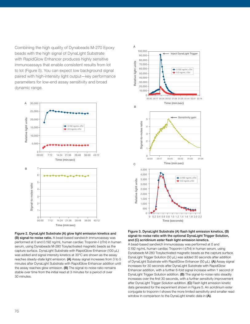
Combining the high quality of Dynabeads M-270 Epoxy beads with the high signal of DynaLight Substrate with RapidGlow Enhancer produces highly sensitive immunoassays that enable consistent results from lot to lot (Figure 5). You can expect low background signal paired with high-intensity light output—key performance parameters for low-end assay sensitivity and broad dynamic range.
30,000
25,000
20,000
15,000
10,000
5,000
00:00 7:12 14:24 21:36 28:48 36:00 43:12
Time (min:sec)
A B
0.192 ng/mL cTnl
0.0 ng/mL cTnl
Rel
ativ
e lig
ht
un
its
Sig
nal
-to
-no
ise
rati
o
000:00 7:12 14:24 21:36 28:48 36:00 43:12
Time (min:sec)
7
6
5
4
2
1
0
3
30,000
25,000
20,000
15,000
10,000
5,000
00:00 7:12 14:24 21:36 28:48 36:00 43:12
Time (min:sec)
A B
0.192 ng/mL cTnl
0.0 ng/mL cTnl
Rel
ativ
e lig
ht
un
its
Sig
nal
-to
-no
ise
rati
o
000:00 7:12 14:24 21:36 28:48 36:00 43:12
Time (min:sec)
7
6
5
4
2
1
0
3
Figure 2. DynaLight Substrate (A) glow light emission kinetics and (B) signal-to-noise ratio. A bead-based sandwich immunoassay was performed at 0 and 0.192 ng/mL human cardiac Troponin-I (cTnI) in human serum, using Dynabeads M-280 Tosylactivated magnetic beads as the capture surface. DynaLight Substrate with RapidGlow Enhancer (100 μL) was added and signal intensity kinetics at 30°C are shown as the assay reaches steady-state light emission. (A) Assay signal increases from 3 to 5 minutes after DynaLight Substrate with RapidGlow Enhancer addition until the assay reaches glow emission. (B) The signal-to-noise ratio remains stable over time from the initial read at 3 minutes for a period of over 30 minutes.
50,000
60,000
70,000
80,000
90,000
100,000
40,000
30,000
20,000
10,000
00:00 00:17 00:00 00:17 00:45 00:5200:35 00:52 01:09 01:0901:26 01:2601:44 02:01 02:18
Time (min:sec) Time (min:sec) Time (seconds)
Inject DynaLight Trigger
A B C
0.192 ng/mL cTnl
0.0 ng/mL cTnl
0.192 ng/mL cTnl
0.0 ng/mL cTnl
Rel
ativ
e lig
ht
un
its
Sig
nal
-to
-no
ise
rati
o
Rel
ativ
e lig
ht
un
its
0
2,500
3,000
3,500
4,000
4,500
2,000
1,500
1,000
500
000 0.2 0.4 0.6 0.8 1.0 1.2 1.4 1.6 1.8 2.0 2.2
1
2
3
4
5Sensitivity gain
Figure 3. DynaLight Substrate (A) flash light emission kinetics, (B) signal-to-noise ratio with the optional DynaLight Trigger Solution, and (C) acridinium ester flash light emission kinetics. A bead-based sandwich immunoassay was performed at 0 and 0.192 ng/mL human cardiac Troponin-I (cTnI) in human serum, using Dynabeads M-280 Tosylactivated magnetic beads as the capture surface. DynaLight Trigger Solution (50 μL) was added 30 seconds after addition of DynaLight Substrate with RapidGlow Enhancer (50 μL). (A) Assay signal increases for 30 seconds after DynaLight Substrate with RapidGlow Enhancer addition, with a further 6-fold signal increase within 1 second of DynaLight Trigger Solution addition. (B) The signal-to-noise ratio steadily increases over the first 30 seconds, with a further sensitivity improvement after DynaLight Trigger Solution addition. (C) Flash light emission kinetic data generated for the experiment shown in Figure 5. An acridinium ester conjugate to troponin-I shows the more limited sensitivity and smaller read window in comparison to the DynaLight kinetic data in (A).
76

Detection substrates
0.0001 0.001 0.01 0.1
1
10
100
1,000
10,000
cTnl concentration (ng/mL)
Sig
nal
-to
-no
ise
rati
o
1 10
DynaLight Substrate and
DynaLight Trigger (Flash)
DynaLight Substrate with
RapidGlow Enhancer
CDP-Star Substrate
Emerald-II Enhancer
Competitor’s FastAP substrate
1.3pg/mL
1.4pg/mL
1.6pg/mL
54.2pg/mL
Substrate Detection System MDD of cTnl concentration
Competitor's acridinium
ester assay
1,040pg/mL
0.0001 0.001 0.01 0.1
1,000
10,000
100,000
1,000,000
10,000,000
cTnl concentration (ng/mL)
Rel
ativ
e lig
ht
un
its
1 10 100
0.0001 0.001 0.01 0.1
1
10
100
1,000
10,000
cTnl concentration (ng/mL)
Sig
nal
-to
-no
ise
rati
o
1 10
DynaLight Substrate and
DynaLight Trigger (Flash)
DynaLight Substrate with
RapidGlow Enhancer
CDP-Star Substrate
Emerald-II Enhancer
Competitor’s FastAP substrate
1.3pg/mL
1.4pg/mL
1.6pg/mL
54.2pg/mL
Substrate Detection System MDD of cTnl concentration
Competitor's acridinium
ester assay
1,040pg/mL
0.0001 0.001 0.01 0.1
1,000
10,000
100,000
1,000,000
10,000,000
cTnl concentration (ng/mL)
Rel
ativ
e lig
ht
un
its
1 10 100
0.0001 0.001 0.01 0.11
10
100
1,000
10,000
cTnl concentration (ng/mL)
Sig
nal-
to-n
ois
e ra
tio
1 10
Substrate detection system
DynaLight Substrate and DynaLight Trigger (Flash)
DynaLight Substrate withRapidGlow Enhancer
CDP-Star SubstrateEmerald-II EnhancerSupplier’s FastAP substrate
Supplier’s acridinium ester assay
MDD of cTnl concentration
1.3 pg/mL
1.4 pg/mL
1.6 pg/mL
54.2 pg/mL
1,040 pg/mL
0.0001 0.001 0.01 0.11,000
10,000
100,000
1,000,000
10,000,000
cTnl concentration (ng/mL)
Rel
ativ
e lig
ht u
nits
1 10 100
Figure 4. DynaLight Substrate yields higher signals than other suppliers’ flash acridinium ester assay and fast alkaline phosphatase substrate. (A) A sandwich immunoassay was performed to detect serial dilutions of cTnI in serum. DynaLight Substrate glow and flash protocols provide >100-fold higher signal-to-noise ratios compared to another supplier’s acridinium ester conjugate. (B) Signal intensities were measured at optimal endpoint read times for DynaLight Substrate with or without DynaLight Trigger Solution, and compared to signal intensity integrated over two seconds for acridinium ester conjugate. Assay dynamic range is 5 logarithmic units for DynaLight vs. 2 logarithmic units for the acridinium ester conjugate. DynaLight glow and flash protocols provide up to 10-fold higher signal-to-noise ratios compared to a another supplier’s fast AP substrate. The optional DynaLight Trigger Solution provides ~2-fold higher signal, increasing overall assay background levels in the same proportion. MDD = minimum detectable dose.
0.01 1 100
Rel
ativ
e lig
ht
un
its
10 7
10 6
10 5
10 4
Troponin I (ng/mL)0.1 10
Figure 5. Lot-to-lot consistency and signal-to-noise ratios for DynaLight Substrate with RapidGlow Enhancer. Three different manufacturing lots of Dynabeads M-270 Epoxy magnetic beads were coupled to anti-troponin I antibody according to recommended procedures to compare lot-to-lot consistency. Calibrators were prepared from human serum spiked with native human cardiac troponin I. For each lot of Dynabeads M-270 Epoxy magnetic beads, eight replicates of each calibrator were run in a microplate-based manual magnetic bead assay using DynaLight Substrate with RapidGlow Enhancer readout.
Convert your colorimetric ELISA to help improve performance, time-to-results, and reduce reagent consumptionColorimetric, fluorimetric, or other chemiluminescent ELISAs can be converted to a chemiluminescent assay using DynaLight Substrate with RapidGlow Enhancer. Advantages of converting your ELISA include superior sensitivity and dynamic range, combined with the flexibility to help decrease reagent usage and time-to-results, allowing you to tailor your assay to meet your specific needs.
Table 2 shows an example of a colorimetric IL-10 ELISA assay optimized for performance compared to a chemiluminescence assay using DynaLight Substrate with RapidGlow Enhancer optimized for sensitivity, reagent preservation, or minimal incubation times.
For requests or inquiries on our products for immunodiagnostic assay development, please email [email protected]
77

Table 2. Example comparison of colorimetric and DynaLight chemiluminescence assay (IL-10 ELISA) conditions and outcomes. Sensitivity: conditions for optimal sensitivity were determined from using reagent titrations. Reagent preservation: the concentration of capture antibody was decreased by 2-fold to demonstrate reagent preservation. Note that the total assay time is still shorter than the colorimetric protocol, despite an increase in the sample and detector antibody incubation times relative to the other chemiluminescence assays. Minimization of incubation times: to minimize total assay time, all incubations were reduced to 10 minutes. Detector antibody concentration is still lower than in the colorimetric protocol, despite an increase relative to the optimal concentration for sensitivity.
Chemiluminescence, optimized for:
Assay conditions Colorimetric Sensitivity Reagent preservation Minimization of incubation times
Capture (µg/mL) 1 1 0.5 1
Detector (µg/mL) 0.16 0.04 0.04 0.08
Streptavidin-enzyme dilution 1/2,500 1/31,250 1/31,250 1/31,250
Total incubation (minutes) 180 70 100 30
Sample and detector Ab 120 30 60 10
Streptavidin-enzyme 30 30 30 10
Substrate 30 10 10 10
Limit of blank (pg/mL)* 2.570 0.090 0.209 0.313
Dynamic range 3 logs 4 logs 4 logs 4 logs
* Limit of blank = corresponding concentration of mean blank RLU measurements +2 standard deviations of blank RLU measurements.
Colorimetric substrates for AP and HRPWe offer the following colorimetric (also called chromogenic) substrates for immunoassay development with AP or HRP.
• PNPP (p-nitrophenyl phosphate, disodium salt) is a widely used substrate for detecting AP in ELISA applications. PNPP produces a yellow water-soluble reaction product that absorbs light at 405 nm. PNPP is available either as a crystalline powder, 5 mg tablets, or as a ready-to-use formulation
• ABTS (2,2’-Azinobis [3-ethylbenzothiazoline-6-sulfonic acid]-diammonium salt) is used to detect HRP and yields a water-soluble green end-reaction product. The green product has two major absorbance peaks, 410 nm and 650 nm. ABTS is less sensitive than the OPD and TMB substrates for HRP detection. Color development is slow (approximately 20 minutes), which may be advantageous if OPD or TMB substrates generate unacceptable background because of their higher sensitivities. ABTS is
available in either a tablet or a ready-to-use formulation
• OPD (o-phenylenediamine dihydrochloride) is used to detect HRP and yields a water-soluble yellow-orange reaction product. The reaction product has an absorbance maximum of 492 nm. OPD is available in either powder or tablet form and easily prepared by dissolving in Thermo Scientific™ Pierce™ Stable Peroxide Substrate Buffer or buffered hydrogen peroxide solution
• TMB (3,3’,5,5’-tetramethylbenzidine) soluble substrates yield a blue color when detecting HRP. The major absorbance maxima for the reaction product are 370 nm and 652 nm. The color then changes to yellow with the addition of sulfuric or phosphoric acid with maximum absorbance at 450 nm. TMB is very sensitive and may produce significant background signal if too much protein or antibody is used. TMB is more quickly oxidized than other HRP substrates, resulting in faster color development
78

Detection substrates
Pierce 1-Step Ultra TMB ELISA Substrate
Thermo Scientific™ Pierce™ 1-Step Ultra TMB ELISA Substrate detects HRP activity, yielding a blue color (Amax = 370 nm and 652 nm) that changes to yellow (Amax = 450 nm) upon addition of a sulfuric or phosphoric acid stop solution. The 1-Step Ultra TMB is the most sensitive of the chromagenic substrates.
Highlights: • Sensitivity—highest sensitivity (2 pg/well) and
signal-to-noise ratios of all TMB substrates
• Ready-to-use—single component without DMF or DMSO present in the reagent
• Stable—shelf life of three years
• Competitive pricing—available off-the-shelf in 1 L packaging
• Safe—noncarcinogenic
• Convenient—no additional reagents or filtering required
TMB is the most popular chromogenic substrate for HRP detection in ELISA and is available in several formats. The 1-Step TMB substrates are single-component substrates that require no preparation before use. Unlike other commercially available substrates, these products contain no DMF or DMSO.
1-Step Ultra TMB substrate yields the greatest sensitivity among the TMB substrates, followed by Thermo Scientific™ Pierce™ 1-Step Turbo TMB- and Thermo Scientific™ Pierce™ 1-Step Slow TMB-ELISA Substrates. The sensitivity of the 1-Step Turbo TMB substrate is similar to OPD-based substrates formulated at approximately 1 mg/mL. 1-Step Slow TMB substrate has intermediate sensitivity—more sensitive than ABTS, but less sensitive than OPD or 1-Step Turbo TMB substrate. The 1-Step Slow TMB is an ideal substrate for kinetic studies.
For requests or inquiries on our products for immunodiagnostic assay development, please email [email protected]
79

Pierce 1-Step NBT/BCIP Substrate Solution
Thermo Scientific™ Pierce™ 1-Step™ NBT/BCIP Substrate Solutions are ready-made solutions of nitro-blue tetrazolium and 5-bromo-4-chloro-3’-indolyphosphate for chromogenic blot and IHC staining with alkaline phosphatase probes. The combination of NBT (nitro-blue tetrazolium chloride) and BCIP (5-bromo-4-chloro-3’-indolyphosphate p-toluidine salt) yields an intense, insoluble black-purple precipitate when reacted with alkaline phosphatase, a popular enzyme conjugate for antibody probes. Pierce NBT and BCIP powders are available individually and in two convenient, ready-to-use solutions (with and without a levamisole suppressor of endogenous phosphatase activity).
Highlights:• AP substrate—for detection of AP activity on solid
media, including nitrocellulose and PVDF membranes and fixed tissue samples
• Chromogenic—no special equipment needed for visualization; produces purple-black precipitate that is easy to photograph
• Package options—choose individual NBT and BCIP powders, or preformulated, ready-to-use solutions with or without levamisole suppressor
Highlights of Pierce NBT/BCIP 1-Step Solution:• Best for immunoblotting applications
• Ready-to-use single component
• Sensitive with low background and minimal assay-to-assay variability
Highlights of Pierce NBT/BCIP plus Suppressor: • Best for immunohistochemistry applications
• Ready-to-use single component
• Contains 1 mM levamisole to inhibit endogenous phosphatase activity, while yielding an intense dark purple precipitate in the presence of calf intestinal phosphatase
Fluorescent substratesThe following chemifluorescent substrates are available for ELISA development with HRP.
• Thermo Scientific™ QuantaBlu™ Fluorogenic Substrate has a larger linear detection range with low-end linearity for detection of HRP. The stable fluorescent reaction product has an Emax/Amax of 420 nm/325 nm allowing stopped, nonstopped, and kinetic assays to be performed; an advantage over the more sensitive chemiluminescent substrates.
• Thermo Scientific™ QuantaRed™ Enhanced Chemifluorescent Substrate is the most sensitive fluorescent ELISA substrate available for HRP detection. The fluorescent reaction product (resorufin) is stable for four hours with an Emax/Amax of 585 nm/570 nm when the reaction is stopped. The red-shifted resorufin reaction product permits detection at a wavelength that has less interference from autofluorescence that can occur in biological samples.
• Invitrogen™ Amplex™ Red Reagent a highly sensitive and stable probe for H2O2, is one of our best fluorogenic substrates for peroxidase. Because H2O2 is produced in many different enzymatic reactions, the Amplex Red reagent allows researchers to detect the activity of many different enzymes (Amplex Red Assay Kits).
• Invitrogen™ Amplex™ UltraRed Reagent improves upon the performance of our unique Amplex Red reagent, offering brighter fluorescence and enhanced sensitivity on a per-mole basis in peroxidase or peroxidase-coupled enzyme assays.
80

World class manufacturingMany of our products are manufactured in ISO 9001– or ISO 9001/13485–certified facilities. Our resources include bioprocess, a HEPA-filtered clean room, chemical synthesis laboratories, and QC testing suites to help ensure consistent product performance. Our manufacturing suite is designed to process bulk production batches and customized formulations. For large volume and custom projects, we provide:
Reagent formulation• Formulated in a class 100,000 equivalent
clean room
• Large-scale liquid formulation (up to 5,000 L)
• Hazardous formulation labs
• Custom formulations with in-process testing
• Chromatography media preparation, and column filling
• Buffer dry-blends
Expert organic chemical synthesis• Synthetic organic chemistry (milligrams to >300 kg)
• Specialties include synthesis of:
– crosslinking and biotinylation reagents
– GC derivatization reagents
– activated supports for chromatography media
– enzyme substrates
– fluorescent dyes
• Class 1 division 1 production facility with 10–300-gallon Pfaudler reactors
• Chemical purification
• Custom synthesis
Bioprocessing• Large-scale fermentation of fine enzymes
• Cytokine standards and quantitation kits
• Protein chemical modification
• Protein immobilization onto solid supports
Custom services • Monoclonal and polyclonal antibody production
and purification
• Custom hybridoma development
• Custom antibody conjugation to enzymes, biotin, and dyes including Alexa Fluor and DyLight fluorescent dyes
• Custom peptide synthesis
• Automated high-speed 96- and 384-well microplate coating
Custom packaging and labeling• Automated high-speed liquid filling/capping/labeling lines
• Aseptic filtration
• Controlled atmosphere fills for dry materials
• Lyophilization
• Chromatography column filling
• Various packaging options from single-use packages to large-volume carboys
• Hazardous chemical and reagent handling
• Private labeling and kit production
Manufacturing capabilities
For requests or inquiries on our products for immunodiagnostic assay development, please email [email protected]
81

Quality assuranceOur controlled processes are designed for product quality and consistency, and we provide regulatory documentation and support.
Analytical testing methodsWe manufacture each lot of product to high-quality specifications using validated methods. Customizable quality specifications can be established for any large volume or custom product, with accompanying certificates of analysis. Our testing methods include:
Purity• Quantitative 1H NMR for accurate purity measurement
• LC-MS
• Infrared spectroscopy
• HPLC
• Melting point analysis
• Solvent panel by GC headspace
• Water content by Karl Fischer titration
• ICP-MS for metal content
• Benzoyl and peroxide levels for detergents
Consistency• Appearance
• Particle size analysis
• Solubility
• Concentration and pH
Activity• Immunoassay
• Enzyme activity
• Cell-based assays
• Binding capacity
Regulatory documentation and supportIn addition to standard material safety data sheets, we can provide documentation for large-volume and custom products to help meet your regulatory needs. Our logistics team is experienced in global distribution and our quality assurance team can work directly with your regulatory staff to help manage change notifications and site audits.
• Custom certificate of analysis
• Certificate of country origin
• Certificate of animal origin
• OSHA and department of transportation (DOT) hazardous material handling and shipping
• Import and export compliance expertise for chemicals and instruments
• Change control notification
• Site audits welcome
For requests or inquiries on our products for immunodiagnostic assay development, please email [email protected]
82
Naming convention on links need to be addressed before releasing to client. Double check color standards and fonts in graphs should all be Helvetica Neue not Universe.

Find out more at thermofisher.comFor Research Use Only. Not for use in diagnostic procedures. © 2016 Thermo Fisher Scientific Inc. All rights reserved. All trademarks are the property of Thermo Fisher Scientific and its subsidiaries unless otherwise specified. Brij is a trademark of ICI Americas, Inc. Tween is a trademark of Croda Americas LLC. Triton is a trademark of Union Carbide Corporation. Cy is a trademark of GE Healthcare. Santa Cruz is a trademark of Santa Cruz Biotechnology, Inc. COL02309 0816