REACTIVITY OF FROM BURKITT'S LYMPHOMA NASOPHARYNGEAL ...
12
Clin. exp. Immunol. (1970) 7, 39-50. REACTIVITY OF RADIOIODINATED SERUM ANTIBODY FROM BURKITT'S LYMPHOMA AND NASOPHARYNGEAL CARCINOMA PATIENTS AGAINST CULTURE LINES DERIVED FROM BURKITT'S LYMPHOMA M. INOUE AND G. KLEIN Institute for Tumor Biology, Karolinska Institutet, Stockholm (Received 27 January 1970) SUMMARY The IgG serum immunoglobulin fraction of two Burkitt's lymphoma (Mutua and Kiliopa) and one African nasopharyngeal carcinoma patient (Kipkoech) was conjugated to iodine-131 (1311). It is known from previous studies with fluorescein labelled conjugates that all three sera contain antibody against the Epstein-Barr virus (EBV)-associated membrane antigen complex, present on the surface of lymphoblastoid cells in EBV-carrier cultures. All three radioiodinated conjugates attached to live cells of an EBV-carrying Burkitt line (Maku), but not to EBV-free Raji cells. A Swedish control serum (Berith) did not block the binding of any of the three conjugates, whereas unconjugated sera of Mutua, Kiliopa and Kipkoech showed various degrees of blocking and cross-blocking. The blocking patterns were in good agreement with previous tests, performed with the same sera against their fluorescein conjugated derivatives. Antibody release tests, involving preincubation of live cells with one of the three conjugates, followed by incubation with unlabelled serum revealed a certain 'hier- archy' between the three sera with regard to their ability to displace radioiodinated surface-coupled immunoglobulin. This ability could be related to the competitive behaviour of the same sera in the cross blocking tests. The results are believed to reflect differences in the affinity of the three antibodies, due either to differences in fit in relation to the surface antigen(s) carried by the Maku target cell, or to differences in the duration of immunization in the three patients. INTRODUCTION Distinctive, cell membrane associated antigens have been demonstrated on Burkitt lym- phoma biopsy cells (Klein et al., 1966, 1967) and derived tissue culture lines (Klein et al., 1967, 1968, 1969) by direct and indirect membrane fluorescence. The membrane antigen complex was associated with the presence of the herpes-like Epstein-Barr virus (EBV). Correspondence: Professor George Klein, Institute for Tumor Biology, Karolinska Institutet, 104 01 Stockholm 60, Sweden. 39
Transcript of REACTIVITY OF FROM BURKITT'S LYMPHOMA NASOPHARYNGEAL ...

Clin. exp. Immunol. (1970) 7, 39-50.
REACTIVITY OF RADIOIODINATED SERUMANTIBODY FROM BURKITT'S LYMPHOMA ANDNASOPHARYNGEAL CARCINOMA PATIENTSAGAINST CULTURE LINES DERIVED FROM
BURKITT'S LYMPHOMA
M. INOUE AND G. KLEIN
Institutefor Tumor Biology, Karolinska Institutet, Stockholm
(Received 27 January 1970)
SUMMARY
The IgG serum immunoglobulin fraction of two Burkitt's lymphoma (Mutua andKiliopa) and one African nasopharyngeal carcinoma patient (Kipkoech) wasconjugated to iodine-131 (1311). It is known from previous studies with fluoresceinlabelled conjugates that all three sera contain antibody against the Epstein-Barrvirus (EBV)-associated membrane antigen complex, present on the surface oflymphoblastoid cells in EBV-carrier cultures. All three radioiodinated conjugatesattached to live cells of an EBV-carrying Burkitt line (Maku), but not to EBV-freeRaji cells. A Swedish control serum (Berith) did not block the binding of any of thethree conjugates, whereas unconjugated sera of Mutua, Kiliopa and Kipkoechshowed various degrees of blocking and cross-blocking. The blocking patternswere in good agreement with previous tests, performed with the same sera againsttheir fluorescein conjugated derivatives.Antibody release tests, involving preincubation of live cells with one of the three
conjugates, followed by incubation with unlabelled serum revealed a certain 'hier-archy' between the three sera with regard to their ability to displace radioiodinatedsurface-coupled immunoglobulin. This ability could be related to the competitivebehaviour of the same sera in the cross blocking tests. The results are believed toreflect differences in the affinity of the three antibodies, due either to differencesin fit in relation to the surface antigen(s) carried by the Maku target cell, or todifferences in the duration of immunization in the three patients.
INTRODUCTION
Distinctive, cell membrane associated antigens have been demonstrated on Burkitt lym-phoma biopsy cells (Klein et al., 1966, 1967) and derived tissue culture lines (Klein et al.,1967, 1968, 1969) by direct and indirect membrane fluorescence. The membrane antigencomplex was associated with the presence of the herpes-like Epstein-Barr virus (EBV).
Correspondence: Professor George Klein, Institute for Tumor Biology, Karolinska Institutet, 104 01Stockholm 60, Sweden.
39

40 M. Inoue and G. Klein
It could not be demonstrated on EBV-free cell lines and lines with only a low proportion(less than 1 0) of EBV-positive cells (Klein et al., 1969). The presence or absence, as wellas the level of antibodies directed against the intracellular EBV-antigen complex and themembrane associated system, respectively, was concordant in the majority (about 800')of the sera tested. The existence of a 'discordant' minority, as well as absorption experi-ments nevertheless showed that the two antigen complexes are distinct (Pearson et al.,1969).As a rule, membrane reactive antibodies were localized in the IgG fraction of positive
sera (Smith et al., 1967). In addition to membrane fluorescence, they could be demonstratedby the method of CI transfer and immune adherence as well (Nishioka et al., 1968).By fluorescein conjugation of IgG from reactive sera, the indirect membrane fluorescence
test was converted into a direct test (Goldstein et al., 1969; Klein et al., 1969). A number ofreference conjugates, derived from donors with various clinical conditions, includingBurkitt's lymphoma and nasopharyngeal carcinoma, or from healthy individuals, weretested for blocking and cross-blocking against the corresponding unconj agated sera(Svedmyr et al., 1969). The majority of the combinations showed a symmetrical behaviourin reciprocal tests, but there were some notable exceptions. In certain serum-conjugatepairs, blocking was only obtained in one direction and the reciprocal combination failed toblock or showed only partial blocking (Svedmyr et al., 1969). It was tentatively assumed thatdifferent sera contained different numbers of antibody components, directed againstdifferent receptors within the membrane antigen complex. Alternatively, asymmetricalblocking may be due to a difference in antibody affinity towards the same antigenic receptors.The present investigation is an attempt to study this question further, by using radioiodine-labelled antibodies and combining blocking with antibody release tests.
MATERIALS AND METHODS
Tissue culture linesTwo established lymphoblastoid cell lines were used, both derived from Burkitt's lym-
phoma. The cell line Maku was established from a biopsy preparation received from Nairobi(Nadkarni et al., 1969); it is strongly reactive in the EBV-associated membrane immuno-fluorescence test (Yata & Klein, 1969). The other cell line, Raji, has been derived from aNigerian case of Burkitt's lymphoma in 1963 (Pulvertaft, 1964); it is negative in membraneimmunofluorescence (Klein et al., 1967) and it carries no EB-virus, according to electronmicroscopy and intracellular immunofluorescence.
In preparation for the present tests, 8 x 10⁶ cells were added to 20 ml of Eagle's BasalMedium with 20% foetal calf serum. The cultures were kept stationary at 370C in a 500CO2 atmosphere, in loosely screw-capped bottles and were fed every 3rd day by replacingtwo-thirds of the volume with fresh medium. At each feeding, the number of cells wasadjusted to 8 x 10⁶ per bottle. The resulting cell harvest was supplied every third day for thetests; it contained 80-90% living cells, as a rule. After washing them twice with K-glucoseGVB+ +, the cells were adjusted to a concentration of I x 106/ml.
SeraFour sera were used; they have all been characterized previously with regard to their
anti-EBV and membrane reactivity (Smith et al., 1967; Klein et al., 1967, 1969; Pearson et

Radioiodinated antibody in Burkitt's lymphomaal., 1969; Svedmyr et al., 1969), including blocking and cross-blocking tests. Two, Mutua(Kenya Cancer Council No. KCC 454) and Kiliopa (KCC 834) were derived from Burkittlymphoma cases, the former in long term regression, the latter in the course of progressivetumour growth. One serum was derived from a patient with nasopharyngeal carcinoma,Kipkoech (KCC 883). These three sera were received from Mr Peter Clifford, KenyattaNational Hospital, Nairobi, in dry ice. A fourth serum, 'Berith', was taken from a healthySwedish technician, and used as an anti-EBV and membrane fluorescence negative control.All sera were inactivated at 560C for 45 min, distributed in 1-ml amounts into small tubesand stored at -20'C until use.
DEAE-cellulose column chromatographyTwo-millilitre serum aliquots were dialysed for 12 hr at 40C against several changes of
1 litre of 0 005 M phosphate buffer, pH 8 0. The material was passed through a 1 5 x 10 cmDEAE-cellulose column equilibrated with the same buffer and washed out with the samebuffer. The flow rate was 30 ml/hr. Four millilitre aliquots of the eluate was collected in atube, and the protein was checked by a Leitz spectrophotometer. The pooled eluate wasconcentrated by ultrafiltration and dialysed against 0 145 M saline or 015 M phosphatebuffer, pH 7-6. Immunodiffusion and immunoelectrophoresis of the final product revealed asingle line with goat anti-human serum and with goat antiserum to human IgG.
LabellingLabelling was carried out by the chloramin T iodination technique. Four milligrams of
IgG dissolved in I ml 015 M phosphate buffer, pH 7-6, was used for iodination. Iodine- 131,carrier free, without reducing agent added, was provided by the Radiochemical Centre,Amersham, England. As a rule, the IgG preparations had a mean iodine content of 1 atomper IgG molecule. The samples were counted in an automatic scintillation detector with awell in thallium activated Nal crystal (Tracerlab type 51). The results were printed out onpaper tape. Labelled IgG was diluted 1 :9 with non-labelled IgG of the same type before use.
Blocking testsTwo million cells were added in 0-02-ml volumes to small tubes containing 0-2 ml 1 :2
diluted serum. The mixture was incubated in a 37°C water bath for 30 min with intermittentshaking. After incubation, the cells were washed three times with K-glucose GVB+ + andincubated at 37°C for another 30 min with 0-2 ml 13"'-labelled IgG ([131I]IgG) in serialtwo-fold dilutions. After washing three times with the same buffer, the radioactivity of thesediments were counted.
Antibody release testAliquots (0-02 ml) from cell suspensions containing I x 107 cells/ml were mixed with
0-2 ml [131 I]IgG solution. The mixture was incubated at 37°C for 30 min and washedthree times with K-glucose GVB+ . After the addition of 0-2 ml serum, in serial two-folddilutions, the cells were incubated again at 37°C for 30 min. After centrifugation, 0-1 mlof the supernatant was precisely calibrated and its radioactivity was measured.
41

M. Inoue and G. Klein
RESULTS
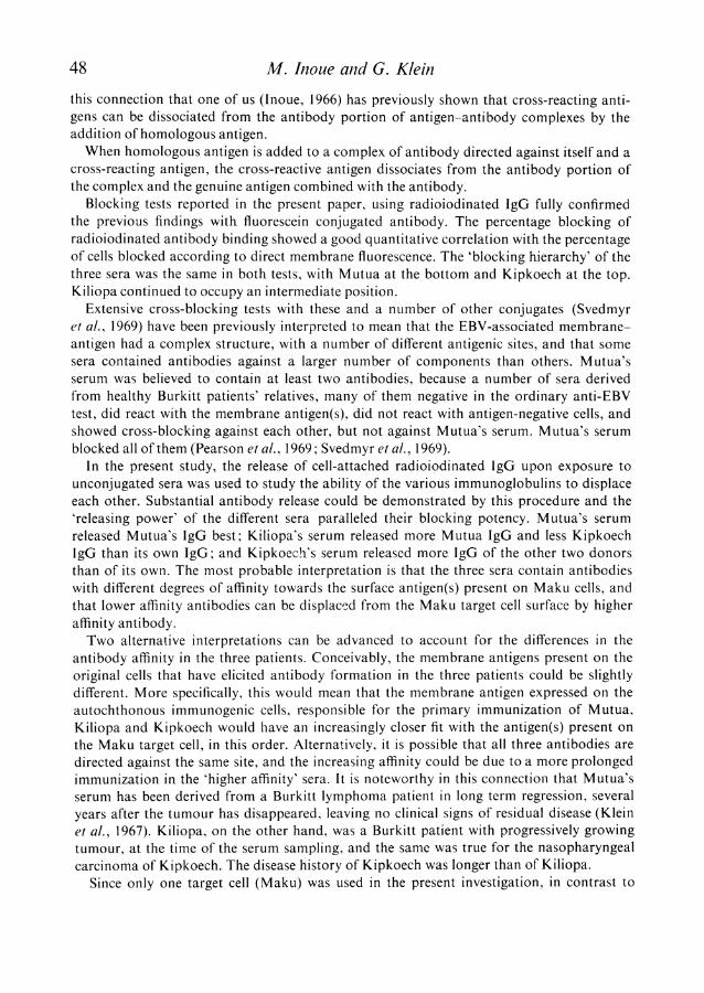
Binding ofradioiodinated immunoglobulins to the target cellsFig. 1 shows the relationship between the amounts of radioactive Mutua, Kiliopa, and
Kipkoech IgG, respectively, added to the reaction mixture and the number of counts
attached to the Maku target cells. In this experiment, 2 x 10⁶ cells, suspended in 0*02-ml
A
0c
C)
.E11
!2
10
8
6
4
2
08
A
A
ANx
0
0
0
I_
06
0o 2 3 4Reciprocal dilution in Log 2
5 6
FIG. 1. Reactivity of three radioiodinated IgG preparations against the Maku and the Rajitarget cells. Radioiodinated IgG tested against the Maku target cell: A, Kiliopa IgG; X,Kipkoech IgG; o, Mutua IgG. Radioiodinated IgG tested against Raji target cells: *,Kipkoech IgG. I/n = 9 4.
TABLE 1. Reactivity of three IgG preparations against the Maku target cell
Bound IgG (CPM) % bound IgG
Dilution Mutua Kiliopa Kipkoech Mutua Kiliopa Kipkoech
1 72750 62418 67165 2-9 2-4 2-62 50259 52240 46537 3 9 4-1 3-64 31700 29972 32095 5 0 4-7 5-08 22756 20656 16214 7 1 6 5 5 1
16 10879 12883 11905 6-8 8-1 7 5
volumes, were mixed with 0-2 ml of the serial two-fold IgG dilutions. After incubation at
370C for 30 min, followed by centrifugation, the radioactivity fixed to the cells was counted.All three immunoglobulin preparations gave a linear relationship with a slope of -94.With the same amount of IgG, the antibody activity of the three sera was approximately
42

Radioiodinated antibody in Burkitt's lymphoma 43
equal. Table 1 shows the percent IgG bound with different dilutions of the three immuno-globulin preparations. The radioactivity bound to the Raji target cell was also measured,after exposure to the Mutua, Kiliopa and Kipkoech conjugates, respectively. It was onlyabout 10-20% of the radioactivity bound to the Maku target cell, similar for all three con-jugates.
TABLE 2. Reactivity of radioiodinated Muta IgG against live and dead Maku cells
Dilution of Bound Mutua IgG CPM % bound Muta IgGMutua IgG
Live culture Dead culture Live culture Dead culture
2 50,259 58,519 3-9 4 54 31,700 38,930 5 0 6-18 22,756 21,584 7-1 6-7
TABLE 3. Blocking effect of unconjugated serum from Berith (control), Mutua(BL*), Kiliopa (BL*), and Kipkoech (NPCt) against radioiodinated IgG of
Mutua, Kiliopa and Kipkoech, respectivelyt
Radioiodinated IgG of:
Unconjugated Mutua, Kiliopa, Kipkoech,blocking reagent dilution reagent dilution reagent dilutionserum
2 4 8 16 2 4 8 16 2 4 8 16
Berith 5 0 0 0 0 0 0 0 0 0 0 0Mutua 56 62 65 89 38 54 66 76 28 37 38 39Kiliopa 52 64 73 81 48 61 76 84 26 28 41 67Kipkoech 49 58 74 87 55 63 70 76 50 63 72 78
* Burkitt's lymphoma.t Nasopharyngeal carcinoma.$ The figures designate the radioactivity bound by the cells pretreated with the
blocking serum, subtracted from the radioactivity of the cells exposed to theconjugate without blocking serum, divided by the latter figure and multipliedby 100.
The question arose whether most of the radioactivity bound to the Maku target cellreflects binding to surface antigen receptors, present on the outer membrane of viablecells. Dead cells may introduce important sources of error, due to defects in the cell mem-brane. In the fluorescence test against viable target cells, the conjugates diffuse freely acrossdead cells, resulting in a homogeneous, non-specific fluorescence. In order to assess whetherthe presence of dead cells would influence the binding of radioiodine labelled antibody to animportant extent, the binding of radioiodine-labelled Mutua IgG by living and dead Makucells was compared. The dead cells were stored in the refrigerator in GVB, until nearly theentire cell population became stainable by trypan blue. The results are shown in Table 2.
D

44 M. Inoue and G. Klein
It will be seen that slightly higher counts were obtained with the dead cell suspension, butthe difference was relatively minor. It is possible, however, that the radioactivity taken up bynon-specific penetration into the dead cells was counteracted by the degradation of surfaceantigens.
Since dead cells constituted a relatively small fraction of the cell population in all testsperformed in this study, no attempt was made to introduce any correction and the datawere calculated as if the cell suspension only contained viable cells.
Blocking testsThe results of the blocking tests, performed with radioiodinated Mutua, Kiliopa or
Kipkoech IgG, and unconjugated control serum (Berith) or Burkitt lymphoma serum(Mutua or Kiliopa) or nasopharyngeal carcinoma serum (Kipkoech) are shown in Table 3.Percentage blocking was calculated as indicated in the footnote of the table. Berith's serum
TABLE 4. Comparison of blocking against flourescein conjugated andradioiodinated Muta, Kiliopa and Kipkoech IgG, respectively, by whole
undiluted serum from the same donors
Blocking Conjugatedserum immunoglobulin: Mutua Kiliopa Kipkoech
Mutua FBI 93 70 57RBP 89 76 39
FBI/RBP ratio 1 04 0-92 1-46Kiliopa FBI 86 80 50
RBP 81 84 67FBI/RBP ratio 1-06 095 075
Kipkoech FBI 82 80 83RBP 87 76 78
FBI/RBP ratio 094 1 05 1 06
FBI, Blocking of direct membrane fluorescence expressed by the block-ing index (Svedmyr et al., 1969) x 100; RBP, percentage blocking against
the radioiodinated conjugate, dilution 1:16.
did not block any of the three immunoglobulins at any dilution tested. Mutua's serumblocked the binding of Mutua's IgG between 56 and 89%, depending on the reagentdilution. Against Kiliopa's labelled IgG, the blocking effect of Mutua's serum ranged from38 to 76%. Against the radioiodinated Kipkoech's IgG, the blocking effect ranged from28 to 39%0. The blocking effect of Kiliopa's serum against radioiodinated Mutua IgG wasnearly identical to the effect of Mutua's serum against its own conjugate when tested forblocking against Kiliopa's IgG, Kiliopa's serum gave a somewhat better effect (rangingfrom 48 to 84%) than Mutua's serum (38 to 76%). Both Kiliopa's and Mutua's serum gaveapproximately equal blocking effects against radioiodinated Kipkoech IgG, except thehighest reagent dilution (1:16) that was blocked more efficiently by Kiliopa than by Mutuaserum.The Kipkoech serum gave a high blocking effect against the Mutua conjugate (49 to
87%)0 the Kiliopa conjugate (55 to 76%) and the Kipkoech conjugate as well (50 to 78%).

Radioiodinated antibody in Burkitt's lymphoma 45
The cross-blocking tests thus show that the control (Berith) serum could not block anyof the three radioiodinated immunoglobulins. Mutua's serum showed the best blockingeffect with his own immunoglobulin and its efficiency decreased towards Kiliopa and Kip-koech IgG, in this order. Kiliopa's serum was intermediate, in showing good blockingagainst both Mutua and Kiliopa IgG, but it fell short of blocking Kipkoech IgG. The Kip-koech serum was superior to the others since it blocked all three immunoglobulin conjugateswith a high efficiency.
100_
90 MM
e80 KM
700~~
O, 60_.c~n
D 50
40 /
40 i
40 50 60 70 80 90 100% Blocking against
radioiodinoted conjugate
FIG. 2. Relationship between the blocking test against fluorescein conjugated and radioiodinatedIgG, respectively. In each circle, the first letter denotes the source of the unconjugated serumand the second the immunoglobulin conjugate: M, Mutua; K, Kiliopa; P, Kipkoech.r = 0-78; P _ 0 01.
The same three sera and their fluorescein-conjugated derivatives have been tested pre-viously by blocking and cross blocking of direct membrane fluorescence (Svedmyr et al.,1969). It was of interest to compare the present results with the corresponding valuesobtained with the same sera in the fluorescence test. The 1:16 dilution of the radioiodineconjugate which contained the same amount of IgG as used in fluorescence blocking test,was chosen for this comparison. The results are shown in Table 4 and Fig. 2. There was anexcellent correlation between the two tests.
Antibody release testsThe results are shown in Table 5. The figures are based on the radioactivity found in the
supernatant after incubating the cells, preexposed to radioiodinated IgG, without addingany serum. The counts obtained with the three IgG preparations were nearly identical. Theefficiency of the three sera in releasing the radioiodinated IgG attached to the cells wascompared by calculating a 'release index', defined as the ratio between the radioactivity

M. Inoue and G. KleinTABLE 5. Summary of antibody release tests
Serum Radioiodinated IgG
Designation Dilution Mutua Kiliopa Kipkoech
GVB++ - 100 1 00 1 00Berith 2 1-61 1-61 1-61Mutua 2 2-86 2-48 2 50
4 2 59 2-34 2-338 2-48 2-25 2-23
16 2-25 2-15 2-1632 2-19 2 05 2-06
Kiliopa 2 3 70 3-14 2-804 3 35 2-97 2 548 3 30 2-80 2 5316 3-12 2-68 2-4132 2-83 2 52 2 25
Kipkoech 2 3 85 3 70 2-844 360 353 2558 3-47 3-33 2-16
16 3-12 3-10 1-9732 3 00 2-97 1-62
The 'release index' was calculated as follows: the radio-activity of the supernatant after incubation with the test serumwas divided by the radioactivity of the supernatant followingincubation with buffer only.
4OO[
300_
2001-
:0 1 2 3 4 5
MIgG- KpMMLIaG-KDMIgG-KL
KLIgG-KL
t KpIgG-KLx\ > °MlgG-M
\--< '-°-O KLIgG-M, KpIgG-M
x KpIgG-Kp
Reciprocal dilution in log 2
FIG. 3. Antibody releasing test. The symbols on the right of each curve designate the radio-
iodinated IgG reacted with the cells, followed by the unconjugated serum. KI, Kiliopa; Kp,Kipkoech; M, Mutua.
46
aL).c
a)aL)

Radioiodinated antibody in Burkitt's lymphoma 47found in the supernatant of the serum-exposed sample, and the control sample, incubatedwith buffer alone.The addition of the control serum (Berith) at the dilution of 1:2 gave a positive release
index of 1 61. Since this serum is not known to contain any antibodies against the EBV-associated membrane-antigen complex and does not block any of the three conjugates(Klein et al., 1969; Pearson et al., 1969), it is assumed that some non-specific serum action isresponsible for this effect. Mutua's serum released considerably more radioactivity fromcells coated with all three conjugates. Its release index towards Mutua's IgG ranged from2-86 to 2 19 with increasing serum dilutions (1:2-32, cp. Table 5). Slightly lower releaseindexes were obtained against Kiliopa and Kipkoech IgG, with nearly identical values forthese two conjugates. Kiliopa's serum was more powerful against the Mutua conjugate,with a releasing index that ranged from 3 70 to 2X83. It was also stronger against Kiliopa'sIgG than Mutua's serum, although it released less radioactivity (range: 3-14-2X52) thanfrom the cells coated with Mutua IgG. Against cells coated with Kipkoech's IgG, the Kiliopaserum dilutions were only slightly more efficient than the corresponding Mutua serumdilutions (ranging from 2-80 to 2 25). Kipkoech's serum was the most powerful releaserof Mutua's IgG (3-85 to 300) and of Kiliopa's IgG as well (3 70 to 2 97) whereas its effecton cells coated with Kipkoech's own IgG was not consistently superior to the effect of thetwo other sera (2f84 to 1-62).
If the figures in Table 5 are considered in relation to the releasing effect of each serumon its own corresponding IgG conjugate, it will be seen that Mutua releases its own con-jugate best, Kiliopa releases Mutua's conjugate better and Kipkoech's conjugate less wellthan its own, whereas Kipkoech's serum releases Mutua IgG best, Kiliopa IgG nearly as well,but its own IgG much less well. There is thus a certain 'displacement hierarchy' between thethree sera, going from Mutua, through Kiliopa, to Kipkoech, also illustrated in Fig. 3.A similar hierarchy was found in the blocking test, as described above in detail.
DISCUSSION
In blocking tests against fluorescein conjugated immunoglobulin derived from the same seraas used in the present investigation, it was found (Svedmyr et al., 1969) that unconjugatedMutua serum completely blocked the membrane reactivity of EBV-carrying lymphoblastoidcell lines with Mutua's IgG conjugate, but showed only partial blocking of the Kipkoech'sconjugate. In contrast, the unconjugated Kipkoech serum blocked the Mutua and theKipkoech conjugates as well. Kiliopa's serum showed an intermediate position: it blockedthe Mutua and the Kiliopa conjugate, but showed only partial blocking against the Kip-koech conjugate; this was similar to the behaviour of the Mutua serum, but Kiliopa showedregularly a better blocking activity against the Mutua conjugate than the reciprocal test,Mutua's serum against Kiliopa conjugate.Two alternative explanations can be considered for the asymmetric blocking patterns
observed. Conceivably, the EBV-associated membrane-antigen complex may consist ofmore than one site and some sera Mutua would contain antibodies against a smallernumber of components than others, like Kipkoech. Kiliopa would be intermediate. Alterna-tively, only one site would exist, but the antibodies found in different sera would differin their affinity for the site. This would imply that Kiliopa's antibody would have higheraffinity than Mutua's and Kipkoech's antibody would be even higher. It may be relevant in
M. Inoue and G. Klein
this connection that one of us (Inoue, 1966) has previously shown that cross-reacting anti-gens can be dissociated from the antibody portion of antigen-antibody complexes by theaddition of homologous antigen.When homologous antigen is added to a complex of antibody directed against itself and a
cross-reacting antigen, the cross-reactive antigen dissociates from the antibody portion ofthe complex and the genuine antigen combined with the antibody.
Blocking tests reported in the present paper, using radioiodinated IgG fully confirmedthe previous findings with fluorescein conjugated antibody. The percentage blocking ofradioiodinated antibody binding showed a good quantitative correlation with the percentageof cells blocked according to direct membrane fluorescence. The 'blocking hierarchy' of thethree sera was the same in both tests, with Mutua at the bottom and Kipkoech at the top.Kiliopa continued to occupy an intermediate position.
Extensive cross-blocking tests with these and a number of other conjugates (Svedmyret al., 1969) have been previously interpreted to mean that the EBV-associated membrane-antigen had a complex structure, with a number of different antigenic sites, and that somesera contained antibodies against a larger number of components than others. Mutua'sserum was believed to contain at least two antibodies, because a number of sera derivedfrom healthy Burkitt patients' relatives, many of them negative in the ordinary anti-EBVtest, did react with the membrane antigen(s), did not react with antigen-negative cells, andshowed cross-blocking against each other, but not against Mutua's serum. Mutua's serumblocked all of them (Pearson et al., 1969; Svedmyr et al., 1969).
In the present study, the release of cell-attached radioiodinated lgG upon exposure tounconjugated sera was used to study the ability of the various immunoglobulins to displaceeach other. Substantial antibody release could be demonstrated by this procedure and the'releasing power' of the different sera paralleled their blocking potency. Mutua's serumreleased Mutua's IgG best; Kiliopa's serum released more Mutua IgG and less KipkoechIgG than its own IgG; and Kipkoech's serum released more IgG of the other two donorsthan of its own. The most probable interpretation is that the three sera contain antibodieswith different degrees of affinity towards the surface antigen(s) present on Maku cells, andthat lower affinity antibodies can be displaced from the Maku target cell surface by higheraffinity antibody.Two alternative interpretations can be advanced to account for the differences in the
antibody affinity in the three patients. Conceivably, the membrane antigens present on theoriginal cells that have elicited antibody formation in the three patients could be slightlydifferent. More specifically, this would mean that the membrane antigen expressed on theautochthonous immunogenic cells, responsible for the primary immunization of Mutua,Kiliopa and Kipkoech would have an increasingly closer fit with the antigen(s) present onthe Maku target cell, in this order. Alternatively, it is possible that all three antibodies aredirected against the same site, and the increasing affinity could be due to a more prolongedimmunization in the 'higher affinity' sera. It is noteworthy in this connection that Mutua'sserum has been derived from a Burkitt lymphoma patient in long term regression, severalyears after the tumour has disappeared, leaving no clinical signs of residual disease (Kleinet al., 1967). Kiliopa, on the other hand, was a Burkitt patient with progressively growingtumour, at the time of the serum sampling, and the same was true for the nasopharyngealcarcinoma of Kipkoech. The disease history of Kipkoech was longer than of Kiliopa.
Since only one target cell (Maku) was used in the present investigation, in contrast to
48

Radioiodinated antibody in Burkitt's lymphoma 49
some of the previous studies (Klein et al., 1969; Svedmyr et al., 1969), where several targetcells have been used, a clarification of this question will have to await further investigation.It is also too early to say whether the relationship between the anti-EBV negative, mem-brane-positive Burkitt relative sera (Klein et al., 1969), and particularly their asymmetricalblocking relationship to Mutua's serum, mentioned above, can be explained as anotherdifference in affinity, or will continue to show the existence of two distinct antigenic sites.In preliminary antibody release tests using fluorescein conjugated IgG (Svedmyr et al.,1969) no evidence was obtained for the hypothesis of affinity difference, but the fluorescencetechnique may not be sufficiently sensitive and quantitative to reveal this.
ACKNOWLEDGMENTS
This study was conducted under Contract No. 69-2005 within the special virus-cancerprogram of the National Cancer Institute, National Institutes of Health, Public HealthService, with grants from the Jane Coffin Childs Memorial Fund for Medical Research,the Swedish Cancer Society, Lotten Bohman's Fund, Ake Wiberg's Fund, Magnus Berg-wall's Foundation and the Cancer Society of Stockholm.We would like to express our sincere gratitude for the generous supply of the Burkitt
culture line Maku, provided by Dr Junichi Yata.Dr Inoue is in receipt of an IARC travel fellowship and is on leave of absence from the
National Cancer Centre Research Institute and Japanese Red Cross Central Hospital,Tokyo, Japan.
REFERENCES
GOLDSTEIN, G., KLEIN, G., PEARSON, G. & CLIFFORD, P. (1969) Direct membrane immunofluorescencereaction of Burkitt's lymphoma cells in culture. Cancer Res. 29, 749.
INOUE, M. (1966) Antigen exchange in cross-reacting antigen-antibody complexes. Japan J. exp. med. 36, 423.KLEIN, G., CLIFFORD, P., KLEIN, F., SMITH, R.T., MINOWADA, J., KOURILSKY, F.M. & BURCHENAL, J.H.
(1967) Membrane immunofluorescence reactions of Burkitt lymphoma cells from biopsy specimensand tissue cultures. J. nat. Cancer Inst. 39, 1027.
KLEIN, G., CLIFFORD, P., KLEIN, E. & STJERNSWXRD, J. (1966) Search for tumor specific immune reactionsin Burkitt lymphoma patients by the membrane immunofluorescence reaction. Proc. nat. Acad. Sci.(Wash.), 55, 1628.
KLEIN, G., PEARSON, G., HENLE, G., HENLE, W., GOLDSTEIN, G. & CLIFFORD, P. (1969) Relation betweenEpstein-Barr viral and cell membrane immunofluorescence in Burkitt tumor cells. III. Comparison ofblocking of direct membrane immunofluorescence and anti-EBV reactivities of different sera. J. exp.Med. 129, 697.
KLEIN, G., PEARSON, G., NADKARNI, J.S., NADKARNI, J.J., KLEIN, G., HENLE, G., HENLE, W. & CLIFFORD,P. (1968) Relation between EB viral and cell membrane immunofluorescence of Burkitt tumor cells.1. Dependence of cell membrane immunofluorescence on presence of EBV. J. exp. Med. 128, 1011.
NADKARNI, J.S., NADKARNI, J.J., CLIFFORD, P., MANOLOV, G., FENY6, E.M. & KLEIN, E. (1969) Character-istics of new cell lines derived from Burkitt lymphomas. Cancer, 23, 64.
NISHIOKA, K., TACHIBANA, T., KLEIN, G. & CLIFFORD, P. (1968) Complementological studies on tumorimmunity. Measurement of Cl bound to tumor cells and immune adherence with Burkitt lymphomacells. GANN Monograph No. 7, p. 49.
PEARSON, G., KLEIN, G., HENLE, G., HENLE, W. & CLIFFORD, P. (1969) Relation between Epstein-Barrviral and cell membrane immunofluorescence in Burkitt tumor cells. IV. Differentiation betweenantibodies responsible for membrane and viral immunofluorescence. J. exp. Med. 129, 707.
PULVERTAFT, J.V. (1964) Cytology of Burkitt's tumor (African lymphoma). Lancet, i, 238.

50 M. Inoue and G. KleinSMITH, R.T., KLEIN, G., KLEIN, E. & CLIFFORD, P. (1967) Studies of the membrane phenomenon in cultured
and biopsy cell lines from the Burkitt lymphoma. Advance in Transplantation (Ed. by DAUSSETT,HAMBURGER and MATHt) p. 779. Munksgaard, Copenhagen.
SVEDMYR, A., DEMISSIE, A., KLEIN, G. & CLIFFORD, P. (1969) Antibody patterns in different human seraagainst EBV (Epstein-Barr virus)-associated intracellular and membrane antigen complexes. J. nat.Cancer Inst. (In press).
YATA, J. & KLEIN, G. (1969) Some factors affecting membrane immunofluorescence reactivity of Burkitt'slymphoma tissue culture cell lines. Int. J. Cancer, (In press).