
Reaction to Injury - Duke University · Reaction to Injury ... compression of the medulla ... acute...
52
The Central Nervous System Reaction to Injury Anne Buckley MD PhD Neuropathology
Transcript of Reaction to Injury - Duke University · Reaction to Injury ... compression of the medulla ... acute...

The Central Nervous System
Reaction to Injury
Anne Buckley MD PhD Neuropathology
jra27
Text Box
This is the first CNS lecture, so she is going to give some background info
hulet001
Approved

Unique features of CNS that influence its response to injury -cell types unique to CNS -complex neural circuit architecture -little internal structural support -very limited room to expand -no typical lymphatic drainage
joepastry.com
sensopac.org
jra27
Callout
Brain is like Jello, no internal structure, it can't support itself. Only the skull keeps it together
jra27
Callout
A very small lesion can really cause problems when you mess up something in this complicated circuit.

R&C Chapter 28: The CNS
•Cellular Responses to Injury •Cerebral Edema, Hydrocephalus, Raised Intracranial Pressure & Herniation •Perinatal Brain Injury •Trauma
jra27
Callout
Developing brains respond differently to injury

R&C Chapter 28: The CNS
•Cellular Responses to Injury •Cerebral Edema, Hydrocephalus, Raised Intracranial Pressure & Herniation •Perinatal Brain Injury •Trauma

The cells of the CNS Neuroectodermal origin Neurons Glia (astrocytes, oligodendroglia, ependymal/choroid plexus cells) Stem/progenitor cells Mesodermal origin Resident microglia Meningial cells Blood vessels and blood cells
jra27
Text Box
Most cells in the CNS are unique to the CNS. Blood vessels are not unique.
jra27
Text Box
Glia means glue. They are the supporting cells that hold everything together, physically and functionally. They may also modulate neuronal synapses
jra27
Callout
Act as macrophages

Neurons
VandenBerg/[email protected]
H&E
Neurofilament H&E
jra27
Text Box
Up to 150 different kinds of neurons, not just the few kinds we normally learn about.
jra27
Callout
Purkinje cells have incredibly complex dendrites. Half neurons in the brain are in the cerebellum
jra27
Callout
Blood vessels
jra27
Callout
Glial cells
jra27
Callout
Neuropil - axons and dendrites
jra27
Callout
Neuron cell bodies

Neuronal reaction (acute) •“Red neurons” in hypoxia/ischemia •Selective vulnerability
VandenBerg/[email protected]
Normal cortical neurons
jra27
Text Box
Neurons are very vulnerable and are some of the first cells to be lost with injury, especially Purkinje cells. Glial cells are much more resilient.
jra27
Text Box
Red neurons are dying neurons, show up within first day of hypoxia.

Neuronal reaction (subacute/chronic) •Apoptosis (in situ and trans-synaptic)
jra27
Text Box
Neuron itself can die from insult or downstream neurons can die due to lack of signalling

Neuronal reaction (subacute/chronic) •Apoptosis (in situ and trans-synaptic) •Regeneration: axonal reaction (central chromatolysis)
Nissl stain (cresyl violet)
va
na
t.cvm
.um
n.e
du
jra27
Text Box
Neurons labeled C are bad, they are trying to recover from an insult, it is producing a lot of protein. They should normally look like the neuron labelled N

Neuronal reaction (subacute/chronic) •Apoptosis (in situ and trans-synaptic) •Regeneration: axonal reaction (central chromatolysis) •Neuronal inclusions (lipofuscin)
Nissl stain (cresyl violet)
va
na
t.cvm
.um
n.e
du
w
ww
.in
no
vita
rese
arc
h.o
rg
PAS
DAPI
EM
ncb
i.nlm
.nih
.go
v
jra27
Text Box
Lipofuscin buildup in normal in aging brains. Sign of wear and tear, accumulates as we age.

VandenBerg/[email protected]
Glial fibrillary acid protein (GFAP)
(Electron microphotograph)
Astrocytes
C. R. Green/The Biomedical Imaging Research Unit
GFAP immunostain, confocal laser scan)
capillary
astrocyte
podocyte
jra27
Text Box
One of the glial cells. Most reactive and most likely to cause problems after an insult. They put their hands on everything. They maintain synapses, nourish neurons, mop up stuff, they form the blood brain barrier on capillaries, they uptake neurotransmitters, etc.

Reactive astrocytes •Astrogliosis: hypertrophy and hyperplasia
Alzheimer type II astrocytes (H&E)
pathology.vco.edu
Gemistocytes (H&E) Reactive astrocytes (silver stain)
jra27
Text Box
When astrocytes are insulted, there are a number of changes.
jra27
Callout
Astrocytes with clear nuclei due to changes in metabolism fromP increased ammonia exposure
jra27
Callout
Pictures of astrocytes like these just tell you that something bad has been happening.
jra27
Callout
Can also become fat and plump due to metabolic changes.
jra27
Text Box
Astrocytosis is both hypertrophy and hyperplasia of astrocytes
jra27
Callout
This is not Alzhemiers disease, these are just named after the same guy
pa
tho
logy.v
co
.ed
u
Glial “scar”
Reactive astrocytes (chronic gliosis)
emedicine.medscape.com
Rosenthal fibers
Histology for Pathologists 3rd Ed
Corpora amylacea
jra27
Callout
Collections of excretions from astrocytes in areas where there are lots of astrocyte podocytes. Common as we age and in epilepsy.
jra27
Callout
Abnormal deposit in chronic gliotic states and in some tumors. Consist of heat shock proteins, GFAP. Alexander disease causes lots of GFAP buildup and kills children by age 10
jra27
Callout
Brain cant form collagenous scars like other tissues. Brain just leaves holes. Glial scars are collections of reactive astrocytes filling in the holes left by dying neurons.
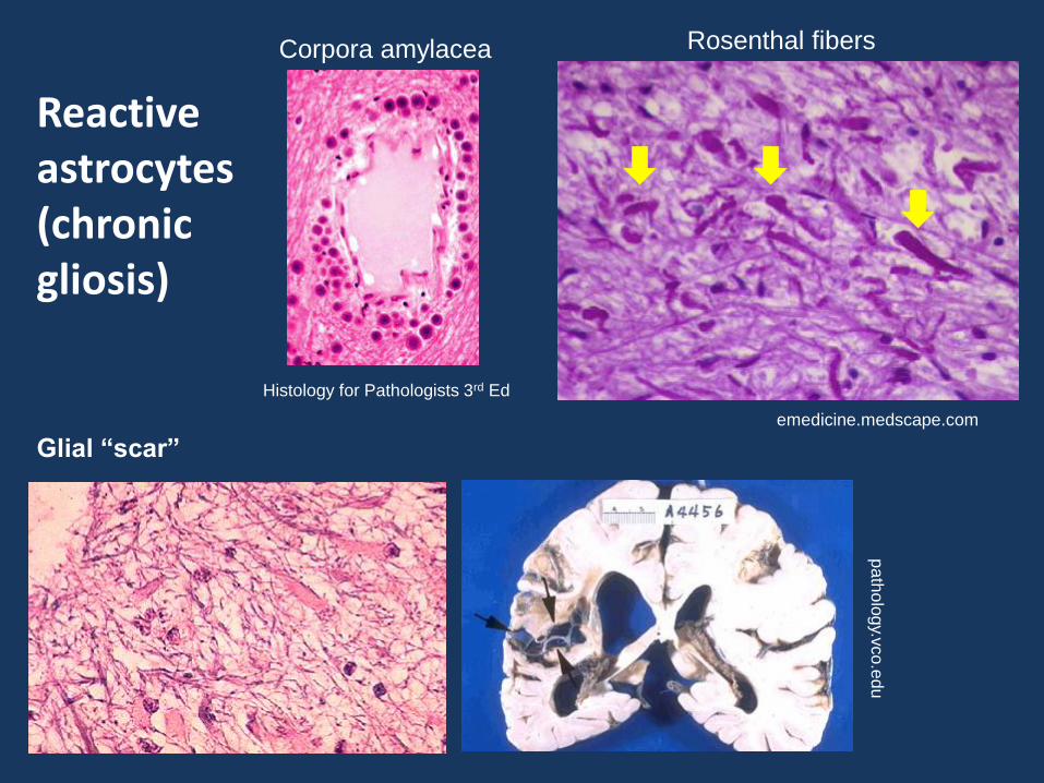
VandenBerg/[email protected]
Oligodendrocytes
jra27
Text Box
These don't really react much. They just die. When functioning, they insulate the neurons. These are lost in MS

Ependymal cells & choroid plexus
Va
nd
enB
erg
/mis
sin
glin
k@
ucsf.
edu Ependymal granulation Normal ependyma
wikimedia.org
imm
ed
iart.c
om
Normal choroid plexus (Scanning EM)
jra27
Text Box
Ependymal cells line the ventricles. They have cilia. Possible stem cells are below this layer. Ependymal granulations occur when it is damages, but it is actually just astrocytes from below this layer reacting.
jra27
Text Box
Choroid is specialized ependymal cells that produce CSF in the ventricles

VandenBerg/[email protected]
Microglia Silver stain of
resting microglia
Rod cell
Amoeboid forms
H&E stain of a microglial
nodule (viral encephalitis)
Neuronophagia
in spinal cord (H&E) Hsueh (2000) Mod Pathol13:1200
VandenBerg/[email protected]
jra27
Text Box
Microglia are the macrophages of the CNS. They are reactive cells when you have infection or insult. They try to clean up the area. Amoeboid forms (actively cleaning up) and rod cell forms.
jra27
Callout
These four arrows define a microglial nodule in a viral infection, which consists of a bunch of reactive microglia

VandenBerg/missinglink.ucsf.edu
Meninges
Arachnoid surface
Pia mater
Skull
jra27
Text Box
Mesodermal in origin. 3 layers: Dura, arachnoid and pia mater. Blood vessels usually run in the subarachnoid space.

R&C Chapter 28: The CNS
•Cellular Responses to Injury •Cerebral Edema, Hydrocephalus, Raised Intracranial Pressure & Herniation •Perinatal Brain Injury •Trauma

Cerebral (brain parenchymal) edema
•Vasogenic: blood-brain barrier is compromised (e.g. trauma, tumor)
•Cytotoxic: cellular injury (e.g. hypoxic or metabolic insult)
www.jkns.or.kr
CT scan: hemorrhage H&E frozen section: glioma
jra27
Text Box
Fluid from blood goes into brain tissue, no lymphatics to drain it
jra27
Text Box
Cellular injury can also cause edema
jra27
Callout
All these wholes are filled with fluid, lots of edema in gliomas
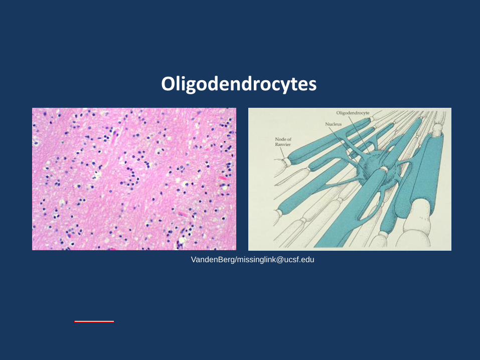
Hydrocephalus (excess cerebrospinal fluid)
choa.org
jra27
Highlight
jra27
Highlight
jra27
Highlight
jra27
Callout
Ventriculomegaly, or enlarged ventricles. This is due to excess cerebral spinal fluid (hydrocephalus)
control.tfe.umu.se
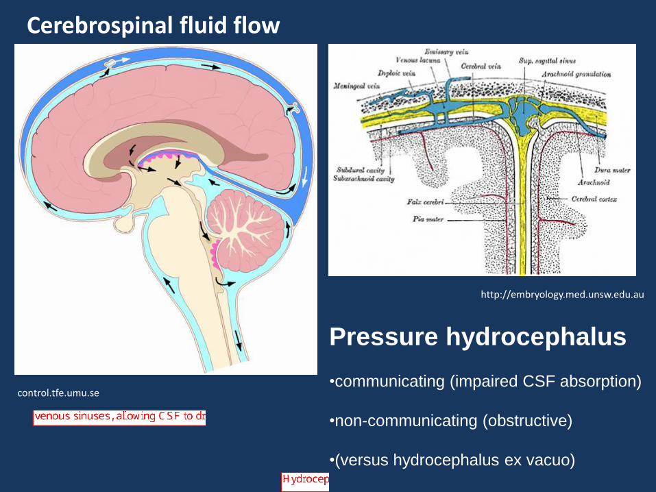
Cerebrospinal fluid flow
http://embryology.med.unsw.edu.au
Pressure hydrocephalus
•communicating (impaired CSF absorption)
•non-communicating (obstructive)
•(versus hydrocephalus ex vacuo)
jra27
Text Box
CSF normally produced in choroid plexus (bright pink) in the ventricles and out into the subarachnoid space, where it flows all over the brain. Granulations in subarachnoid space come in contact with venous sinuses, allowing CSF to drain.
jra27
Text Box
CSF can flow freely between ventricles, so all of brain is compressed
jra27
Text Box
Build up of pressure due to excess CSF, cause compression of the brain
jra27
Text Box
Obstruction between ventricles cause one region to buildup
jra27
Text Box
Hydrocephalus ex vacuo is enlarged ventricles due to brain atrophy

Initial signs and symptoms of
increased intracranial pressure:
•headache
•vomiting
•altered mental status
•papilledema (engorgement of the optic disk)
Raised intracranial pressure due to:
•Cerebral edema
•Pressure hydrocephalus
•Tumor
•Hemorrhage
•Abscess
jra27
Text Box
Increased pressure starts by shifting the brain tissues, then compression of vasculature, then flattening of sulci and gyri and serious damage.

Endpoint of severe IIP: Herniation
stroke.ahajournals.org
Falx cerebri
Tentorium
cerebelli
Foramen
magnum
jra27
Text Box
If increased pressure persists, brain squeezes out like jello. It will go wherever it can - around dural flaps, down spinal cord, etc.
jra27
Callout
Tonsillar herniation
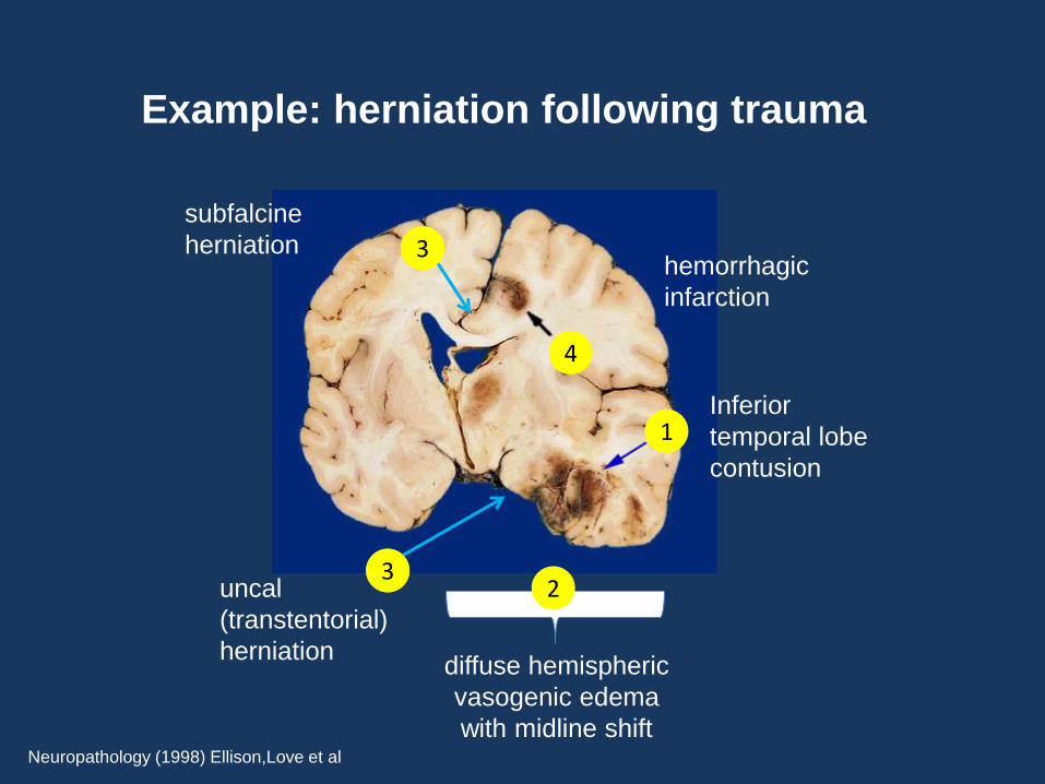
Neuropathology (1998) Ellison,Love et al
Inferior
temporal lobe
contusion
1
2
4
hemorrhagic
infarction
3
3
3
subfalcine
herniation
3 uncal
(transtentorial)
herniation
Example: herniation following trauma
diffuse hemispheric
vasogenic edema
with midline shift
jra27
Text Box
Contusion leads to edema, leads to diffuse swelling and shifting of midline to the left. This causes herniations around falx cerebri and the tentorium cerebelli. This falcine herniation gets trapped and bleeds, leading to hemorrhagic infarction.

Neu
rop
ath
olo
gy (
19
98
) E
llison
,Love
et
al Uncal (transtentorial)
herniation
Tonsillar
herniation Va
nd
enB
erg
/mis
sin
glin
k@
ucsf.
edu
jra27
Text Box
Illustrations of two of the herniations. These are real bad.

Site-specific consequences of herniation
•can be clinically silent
•ACA compression
•PCA compression (visual field defect)
•CNIII compression (blown pupil)
•Cerebral peduncle compression (paresis)
•Sylvian aqueduct occlusion (hydrocephalus)
•Duret hemorrhages
•compression of the medulla (cardiorespiratory arrest)
jra27
Text Box
Symptoms of these three herniations are different, as shown below.

Duret (secondary brainstem) hemorrhages
Neu
rop
ath
olo
gy (1
99
8) E
llison
,Love
et a
l
•Sequela of transtentorial (uncal) herniation
•Caused by stretching of the
penetrating branches of the basilar artery
as the midbrain descends
jra27
Text Box
Read this slide

R&C Chapter 28: The CNS
•Cellular Responses to Injury •Cerebral Edema, Hydrocephalus, Raised Intracranial Pressure & Herniation •Perinatal Brain Injury •Trauma

Perinatal brain injury
Sites of injury
•Germinal matrix
•Periventricular white matter
•Cortex and underlying white matter
•Deep nuclei
Sequelae
•Static motor deficits (cerebral palsy): spasticity, dystonia,
ataxia/athetosis, and paresis
•Cognitive deficits
jra27
Text Box
Childrens brains in perinatal period are still developing, have different kinds of injury.
jra27
Callout
Only in developing brain, has stem cells
jra27
Callout
non-progressive

Germinal matrix
24-week-postconception neonate (alkaline phosphatase and cresyl violet)
Ghazi-Birry et al (1997) Am J Neuroradiol 18:219
jra27
Text Box
Stem or progenitor cells in this region that are active in the developing brain. This region does not exist in the adult brain. This region is very sensitive, especially in premature babies. These neurons are not myelinated, they are easily damaged.
jra27
Text Box
Germinal matrix bleeds are especially common when pressing on the head during rescussitation, this can lead to serious brain damage or may be subclinical.

Ghazi-Birry et al (1997) Am J Neuroradiol 18:219
Germinal matrix
24-week-postconception neonate (alkaline phosphatase and cresyl violet)

Germinal matrix bleed
dartmed.dartmouth.edu
Ghazi-Birry (1997) AJNR Am J Neuroradiol 18:219
Germinal matrix
24-week-postconception neonate (alkaline phosphatase and cresyl violet)

Periventricular leukomalacia (from white matter damage)
nim.nih.gov
emedicine.medscape.com
infarct
glowm.com
ventricular wall
jra27
Text Box
Hypoxia or infection of white matter around the ventricles will lead to hydrocephalus.

Ulegyria (from cortical hypoxia/ischemia)
Thinned,
gliotic gyri
jra27
Text Box
Gray matter ribbon has different widths in different places. Due to cortical hypoxia or ischemia.
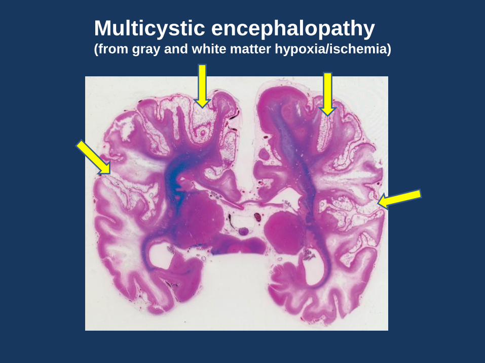
Multicystic encephalopathy (from gray and white matter hypoxia/ischemia)
jra27
Text Box
Cystic holes all over the brain due to gray and white matter hypoxia

Status marmoratus (from hypoxia/ischemia of deep nuclei)
glowm.com
Glial “scar” and aberrant myelinization in basal ganglia
Clinical sequelae include choreoathetosis and related movement disorders
jra27
Text Box
Deep nuclei hypoxia leads to a glial scar and improper myelinization. This leads to malfunctioning basal ganglia, causing inappropriate motion.

R&C Chapter 28: The CNS
•Cellular Responses to Injury •Cerebral Edema, Hydrocephalus, Raised Intracranial Pressure & Herniation •Perinatal Brain Injury •Trauma

Trauma
•Skull fractures
•Parenchymal injury
•Vascular injury
jra27
Text Box
Direct injury to the brain

Trauma: Skull fractures
wik
ipe
dia
.org
Displaced fracture
wik
ipe
dia
.org
Diastatic fractures
Emedicine.medscape.com
jra27
Text Box
Chunk of bone dives into the brain
jra27
Text Box
Fractures that span different bones
jra27
Text Box
Different signs on face depending on what bone is fractured

The CIBA collection Vol 1 Nervous System Part 2 1996
Trauma: Skull fractures
wikipedia.org
Basal skull fractures
jra27
Text Box
Raccoon sign. Blood leaks out of from anterior fossa fracture
jra27
Text Box
Battle sign from postauricular hematoma. Fracture around temporal bone
jra27
Text Box
CSF leaking out of nose and ears is also bad

Trauma: Parenchymal injury •lacerations
•contusions
•diffuse axonal injury
Contusions
remote
acute
jra27
Text Box
Coup-contre coup damage. One side hits the skull and is bruised, then the brain rebounds back and hits the opposite side. So you end up with two contusions on opposite sides.
jra27
Text Box
contusion
jra27
Text Box
yellowish plaque is an old insult that has become a glial scar

Diffuse axonal injury (traumatic rotation, deep white matter stretching)
img.m
ed
scap
e.c
om
MRI: hyperintense signal
in the corpus callosum,
septum pellucidum,
and external capsule
Beta amyloid
precursor protein
(BAPP) stain
Axonal
swelling
White matter degeneration n
eu
ropa
tholo
gy.
ne
oucom
.edu
jra27
Text Box
High torque on the brain causes stretching of axons, which can lead to damage - tearing, swelling, etc. This can easily be seen on MRI and in histology. Eventually this leads to white matter degeneration.
Trauma: Vascular injury
Intra-axial: parenchymal hemorrhage
Extra-axial: subarachnoid hemorrhage
subdural hemorrhage
epidural hemorrhage
jra27
Text Box
Inside the brain tissue
jra27
Text Box
Outside the brain

Parenchymal hemorrhage
-hypertension, vascular malformations, tumors, drugs, amyloid angiopathy
Atlas &Thulborne (1998) AJNR Am J Neuroradiol 19:1471
MRI: acute hemorrhage
with associated edema
jra27
Text Box
Due to small vessels inside the brain tissue leaking or bursting.
jra27
Callout
Hemorrhage with edema surrounding it, causing intracranial swelling.X

Subarachnoid hemorrhage
-bleeding from corticomenigeal arteries into the CSF space
-rupture of a cerebral aneurysm in most cases
-sudden onset (“thunderclap headache”)
-poor prognosis (up to 50% death rate)
VandenBerg/missinglink.ucsf.edu
em
ed
icin
e.m
edscape
.com
CT scan of SAH
jra27
Text Box
Arteries on the outside of the brain run in subarachnoid space. Rupture of vessels leads to bleeding into this space. High pressure bleed, so expands very quickly. Usually caused by ruptured aneurysm around the circle of willis. Not usually from traumatic injury like the epidural and subdural hemorrhages.
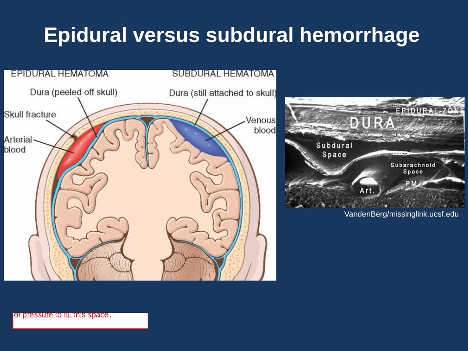
Epidural versus subdural hemorrhage
VandenBerg/missinglink.ucsf.edu
jra27
Text Box
Subdural space can be filled with venous blood with vein tears. This is a low pressure bleed, so it expands very slowly. Much less deadly.
jra27
Text Box
Epidural bleeds are arterial. Dura is very tightly connected to skull, so it takes a lot of pressure to fill this space.

Review of Meningeal Layers
http://www.profelis.org

Epidural hematoma
-ruptured dural/meningeal artery
-often associated with intoxication
-period of lucidity following trauma
-mortality rate up to 50% (varies with
level of consciousness at surgery)
jra27
Text Box
Often occurs when someone falls down and hits their head. Due to rupture of dural or meningeal artery. Develops quickly. Bleeding is on top of the dura.

Epidural hematoma
-ruptured dural/meningeal artery
-rare in infants and elderly (intracranial)
-often associated with intoxication
-period of lucidity following trauma
-mortality rate up to 50% (varies with
level of conciousness at surgery)

Subdural hemorrhage
-bleeding from bridging veins
(and accompanying arteries)
-elderly (atrophy) and
infants (thin vessel walls and larger space)
-may be subclinical; more rapid
development of symptoms if with arteries
Stein et al Forensic Science International, Volume 163, Issue 1
jra27
Text Box
This is due to breaks in the bridging vessels between the dura and the arachnoid. Veins are more fragile than the arteries. Most common in older people and children because the brains are smaller and there is more space in this bridging region. Bleeding develops slowly because it is venous. Occassionally an artery can break too, which makes it much more deadly.

R&C Chapter 28: The CNS
•Cellular Responses to Injury •Cerebral Edema, Hydrocephalus, Raised Intracranial Pressure & Herniation •Perinatal Brain Injury •Trauma