RDEB Gene Transfer Clinical Protocol (IRB #: 14563) Page 1 ... · RDEB Gene Transfer Clinical...
38
RDEB Gene Transfer Clinical Protocol (IRB #: 14563) Page 1 of 38 Stanford Dermatology, PI: Jean Tang, MD, PhD IND# 13708 ` Version Date: 10/5/2015 IND Application Section 6: Protocols 1 Protocol Title: A Phase 1 Single Center Trial of Gene Transfer for Recessive Dystrophic Epidermolysis 2 Bullosa (RDEB) using the drug LZRSE-Col7A1 Engineered Autologous Epidermal Sheets (LEAES). 3 4 Table of Contents 5 1. Abbreviations List: .............................................................................................................................. 3 6 2. Introduction:......................................................................................................................................... 4 7 2.A. Objective: ...................................................................................................................................... 4 8 2.B. Purpose: ........................................................................................................................................ 4 9 2.C. Protocol summary: ........................................................................................................................ 4 10 2.D. Study end points: .......................................................................................................................... 5 11 2.E. History of protocol and oversight: ................................................................................................ 5 12 3. Name and address and statement of qualifications of each investigator: ................................. 6 13 4. Criteria for subject selection: ............................................................................................................ 6 14 4.A. Number of subjects: ...................................................................................................................... 6 15 4.B. Characteristics Studies (Pre-Screening): ....................................................................................... 6 16 4.B.i. Plan for recruitment: ........................................................................................................... 6 17 4.B.ii. Identifying subjects eligible for gene transfer (based on Pre-Screening): ................. 7 18 4.B.iii. Procedures performed under characteristics study (Pre-screening): ......................... 7 19 4.B.iv. Inclusion/Exclusion Criteria:.............................................................................................. 9 20 4.B.v. Day -26: ............................................................................................................................. 10 21 4.B.vi. Day -7: ................................................................................................................................ 14 22 4.B.vii. Day -3: ............................................................................................................................ 15 23 4.B.viii. Day -1: ............................................................................................................................ 15 24 5. Enrollment/ Grafting: ........................................................................................................................ 15 25 5.A. Grafting (Day 0): .......................................................................................................................... 16 26 5.A.i. Final selection of graft sites ............................................................................................ 16 27 5.A.ii. Grafting of LEAES ............................................................................................................ 16 28 6. Post-grafting observation (Day +1 - Day +14) ............................................................................. 17 29 7. Post-grafting clinical follow-up: ....................................................................................................... 18 30 7.A. Blood tests .................................................................................................................................. 18 31 7.A.i. Replication competent retrovirus analysis .................................................................... 18 32 7.B. Physical examination, skin examination ..................................................................................... 18 33 Downloaded From: on 06/17/2018
Transcript of RDEB Gene Transfer Clinical Protocol (IRB #: 14563) Page 1 ... · RDEB Gene Transfer Clinical...

RDEB Gene Transfer Clinical Protocol (IRB #: 14563) Page 1 of 38 Stanford Dermatology, PI: Jean Tang, MD, PhD
IND# 13708 ` Version Date: 10/5/2015
IND Application Section 6: Protocols 1 Protocol Title: A Phase 1 Single Center Trial of Gene Transfer for Recessive Dystrophic Epidermolysis 2 Bullosa (RDEB) using the drug LZRSE-Col7A1 Engineered Autologous Epidermal Sheets (LEAES). 3 4 Table of Contents 5 1. Abbreviations List: .............................................................................................................................. 3 6
2. Introduction:......................................................................................................................................... 4 7
2.A. Objective: ...................................................................................................................................... 4 8
2.B. Purpose: ........................................................................................................................................ 4 9
2.C. Protocol summary: ........................................................................................................................ 4 10
2.D. Study end points: .......................................................................................................................... 5 11
2.E. History of protocol and oversight: ................................................................................................ 5 12
3. Name and address and statement of qualifications of each investigator: ................................. 6 13
4. Criteria for subject selection: ............................................................................................................ 6 14
4.A. Number of subjects: ...................................................................................................................... 6 15
4.B. Characteristics Studies (Pre-Screening): ....................................................................................... 6 16
4.B.i. Plan for recruitment: ........................................................................................................... 6 17
4.B.ii. Identifying subjects eligible for gene transfer (based on Pre-Screening): ................. 7 18
4.B.iii. Procedures performed under characteristics study (Pre-screening): ......................... 7 19
4.B.iv. Inclusion/Exclusion Criteria: .............................................................................................. 9 20
4.B.v. Day -26: ............................................................................................................................. 10 21
4.B.vi. Day -7: ................................................................................................................................ 14 22
4.B.vii. Day -3: ............................................................................................................................ 15 23
4.B.viii. Day -1: ............................................................................................................................ 15 24
5. Enrollment/ Grafting: ........................................................................................................................ 15 25
5.A. Grafting (Day 0): .......................................................................................................................... 16 26
5.A.i. Final selection of graft sites ............................................................................................ 16 27
5.A.ii. Grafting of LEAES ............................................................................................................ 16 28
6. Post-grafting observation (Day +1 - Day +14) ............................................................................. 17 29
7. Post-grafting clinical follow-up: ....................................................................................................... 18 30
7.A. Blood tests .................................................................................................................................. 18 31
7.A.i. Replication competent retrovirus analysis .................................................................... 18 32
7.B. Physical examination, skin examination ..................................................................................... 18 33
Downloaded From: on 06/17/2018

RDEB Gene Transfer Clinical Protocol (IRB #: 14563) Page 2 of 38 Stanford Dermatology, PI: Jean Tang, MD, PhD
IND# 13708 ` Version Date: 10/5/2015
7.C. Adverse events ............................................................................................................................ 18 34
7.D. Concomitant medications ........................................................................................................... 18 35
7.E. Photographs and graft evaluation .............................................................................................. 19 36
7.F. Skin biopsies ................................................................................................................................ 19 37
7.G. Week 8 ........................................................................................................................................ 20 38
7.H. Long term follow up protocol ..................................................................................................... 23 39
8. Discontinuation, Withdrawal, Lost to Follow-Up, or Early Termination .................................... 23 40
9. Time frame between subjects ........................................................................................................ 23 41
10. Unscheduled visits ....................................................................................................................... 24 42
11. Exceptions to protocol ................................................................................................................. 24 43
12. Method for determining dosage ................................................................................................. 24 44
13. Observations and measurements .............................................................................................. 25 45
13.A. Skin examinations and photographs ....................................................................................... 25 46
13.B. Blood tests .............................................................................................................................. 25 47
13.C. Skin biopsies ............................................................................................................................ 25 48
14. Risks ............................................................................................................................................... 25 49
14.A. Risks for investigational drug .................................................................................................. 25 50
14.A.i. Anticipated risks ............................................................................................................... 25 51
14.A.ii. EBA/Immunologic graft rejection ................................................................................ 26 52
14.A.iii. Cancer ............................................................................................................................ 26 53
14.A.iv. Advancing epithelial surfaces/migration of graft over mucous membranes: ....... 26 54
14.A.v. Systemic infection ........................................................................................................ 27 55
14.B. Risks for commercially available drugs ................................................................................... 27 56
14.B.i. Commercially available drugs used for biopsy ............................................................ 27 57
14.B.ii. Commercially available drugs used for grafting ....................................................... 28 58
14.C. Risks for procedures to be performed .................................................................................... 28 59
14.C.i. Blood draws ....................................................................................................................... 28 60
14.C.ii. Skin biopsies ................................................................................................................. 28 61
14.C.iii. Echocardiogram............................................................................................................ 29 62
14.C.iv. Electrocardiogram (ECG) ............................................................................................ 29 63
14.D. Privacy and confidentiality ...................................................................................................... 29 64
15. Protocol deviations/violations ..................................................................................................... 30 65
Downloaded From: on 06/17/2018

RDEB Gene Transfer Clinical Protocol (IRB #: 14563) Page 3 of 38 Stanford Dermatology, PI: Jean Tang, MD, PhD
IND# 13708 ` Version Date: 10/5/2015
16. Minimizing risks and monitoring for adverse events ............................................................... 30 66
16.A. Definitions ............................................................................................................................... 31 67
16.B. Triggers for temporary hold, DSMB review, and regulatory reporting: ................................. 32 68
16.C. Patient advocate ..................................................................................................................... 32 69
16.D. EB Physician ............................................................................................................................ 33 70
16.E. Data Safety Monitoring Board ................................................................................................ 33 71
16.F. Independent study monitor ........................................................................................................ 34 72
16.G. Independent medical monitor ................................................................................................ 35 73
17. Study termination: ........................................................................................................................ 35 74
18. Data management plan: .............................................................................................................. 35 75
19. Statistical plan: .............................................................................................................................. 35 76
19.A. Primary outcomes: .................................................................................................................. 36 77
19.B. Secondary outcomes: .............................................................................................................. 36 78
20. References: ................................................................................................................................... 37 79
80 Figures: 81 Figure 1: Parental Genetic Testing Algorithm……………………………………………………………………9 82 Figure 2: Flow Chart for RDEB Gene Transfer Protocol……………………………………………………….21 83 84 Tables 85 Table 1: IND Amendments for RDEB Gene Transfer Clinical Protocol………………………………………..5 86 Table 2: Schedule of Events for RDEB Gene Transfer………………………………………………………..22 87 88 89
1. Abbreviations List: 90 AEs: adverse events (defined in section 16) and 91 in Safety Monitoring Plan 92 ALT (SGPT): alanine aminotransferase, included 93 in metabolic panel 94 AST (SGOT): aspartate aminotransferase, 95 included in metabolic panel 96 BCIP/NBT: 5-bromo-4-chloro-3’indolyphosphate 97 p-toluidine salt and nitro-blue tetrazolium 98 chloride, substrate used in Stanford 99 Dermatology Research Lab 100 CBC: complete blood count 101 cDNA: complementary deoxyribonucleic acid 102 CITI: Collaborative IRB Training Initiative 103 CLIA: Clinical Laboratory Improvement 104 Amendments 105 COL7A1: Collagen 7 gene 106
CTC: Common Toxicity Criteria 107 DDEB: dominant dystrophic epidermolysis 108 bullosa 109 DEB: dystrophic epidermolysis bullosa 110 DIF: direct immunofluorescence 111 D-MEM: Dulbecco’s Modified Eagle Medium, 112 media used in Stanford Dermatology Research 113 Lab 114 DNA: deoxyribonucleic acid 115 DSMB: data safety monitoring board 116 EB: epidermolysis bullosa 117 EBA: epidermolysis bullosa acquisita 118 ECG: electrocardiogram 119 eCRF: electronic case report forms 120 EDC: electronic data capture 121 EDTA: ethylenediamine tetraacetic acid 122
Downloaded From: on 06/17/2018

RDEB Gene Transfer Clinical Protocol (IRB #: 14563) Page 4 of 38 Stanford Dermatology, PI: Jean Tang, MD, PhD
IND# 13708 ` Version Date: 10/5/2015
ELISA: enzyme-linked immunosorbent assay 123 EM: electron microscopy 124 EMLA: Eutectic mixture of local anesthetic 125 FACS: Fluorescence-activated cell sorting 126 FCS: fetal calf serum, used in Stanford 127 Dermatology Research lab 128 FDA: Food and Drug Administration 129 HIPAA: Health Insurance Portability and 130 Accountability Act 131 HIV: human immunodeficiency virus 132 IAW: in accordance with 133 ICF: informed consent form 134 IEM: immunoelectron microscopy 135 IF: immunofluorescence 136 IIF: indirect immunofluorescence 137 IND: Investigational New Drug application 138 IP: Internet Protocol 139 IRB: Institutional Review Board 140 IUVPF: Indiana University Vector Production 141 Facility 142 Keratinocyte-SFM: Keratinocyte serum free 143 media, used in Stanford Dermatology Research 144 Lab 145 KGM: keratinocyte growth media, used in 146 Stanford Dermatology Research Lab 147 LEAES: LZRSE-Col7A1 Engineered Autologous 148 Epidermal Sheets 149 LLN: Lower limit of normal 150 LPCH: Lucile Packard Children’s Hospital 151 MCH: mean corpuscular hemoglobin, included in 152 complete blood count 153
MCHC: mean corpuscular hemoglobin 154 concentration, included in complete blood count 155 MCV: mean cell volume, included in complete 156 blood count 157 MOOP: Manual of Operating Procedures 158 mRNA: messenger ribonucleic acid 159 NC1: non-collagenous region 1 of the collagen 7 160 molecule 161 NCI: National Cancer Institute 162 NIH: National Institute of Health 163 OD: optical density 164 PCP: primary care practitioner 165 PCR: polymerase chain reaction 166 PHI: protected health information 167 qPCR: quantitative polymerase chain reaction 168 qRT-PCR: quantitative real time polymerase 169 chain reaction 170 R01: NIH grant funding mechanism 171 RAC: Recombinant DNA Advisory Committee 172 RCR: replication competent retrovirus 173 RDEB: recessive dystrophic epidermolysis 174 bullosa 175 RDW: red blood cell distribution width, included 176 in complete blood count 177 REDCap: Research Electronic Data Capture 178 RNA: ribonucleic acid 179 SCC: squamous cell carcinoma 180 VPN: Virtual private network 181 WBC: white blood cell 182 WNL: within normal limits 183
184
2. Introduction: 185 186 2.A. Objective: 187 The primary objective of this protocol is to evaluate the safety of autologous skin grafts 188
transduced with a retroviral vector containing the gene encoding type VII collagen (LEAES) in 189 subjects with RDEB. 190
191 2.B. Purpose: 192 The purpose of this study is to achieve proof-of-concept for this general approach to cell-based 193
gene therapy in humans and to set the stage for further therapeutic extension in RDEB. 194 195 2.C. Protocol summary: 196 Recessive dystrophic epidermolysis bullosa (RDEB) is a severe inherited blistering skin disease 197
caused by absence of a protein known as type VII collagen. Patients with RDEB develop large, 198 severely painful blisters and open wounds from minor trauma to their skin. This trial will create a graft, 199 which we call "LEAES," of the patient's own skin that has been genetically engineered in our lab to 200
Downloaded From: on 06/17/2018
RDEB Gene Transfer Clinical Protocol (IRB #: 14563) Page 5 of 38 Stanford Dermatology, PI: Jean Tang, MD, PhD
IND# 13708 ` Version Date: 10/5/2015
express this missing protein. We will basically take a subject's own cells, correct them in culture, and 201 then transplant the corrected cells back onto them. 202
203 2.D. Study end points: 204 LEAES grafts will be evaluated at 12 weeks, 25 weeks, and 52 weeks after grafting for 205
expression of type VII collagen and presence of anchoring fibrils. Secondary endpoints include 206 evaluation at 12 weeks, 25 weeks, 52 weeks, and yearly thereafter for appearance, durability, and 207 ease of blistering. Subjects will continue to be followed for safety in a separate long-term follow-up 208 protocol under this IND. 209
210 2.E. History of protocol and oversight: 211 Stanford's Administrative Panel on Human Subjects in Medical Research, also called the 212
Institutional Review Board (IRB), and the Administrative Panel on Biosafety will review all protocols 213 and processes related to this study. 214
We obtained IRB approval and began the screening process on August 7, 2007 (IRB protocol # 215 8557, ClinicalTrials.gov, Identifier NCT00533572). We consented, biopsied and collected blood to 216 screen specifically for gene transfer on one subject under this protocol in March 2008. At the request 217 of the Food and Drug Administration (FDA), we ceased all screening procedures for gene transfer on 218 June 6, 2008 (Protocol Amendment 1). Protocol 8557 has since been closed and no additional 219 subjects have been enrolled. 220
We subsequently changed the process for subject selection for gene transfer. We will now select 221 candidates for the gene transfer trial from a pool of subjects who have completed a separate research 222 study on the characteristics of EB patients. These “characteristics” protocols were approved by the 223 Stanford IRB (protocols 17158 and 15898), and this has been communicated with the FDA (Protocol 224 Amendment 2). We will refer to the “Characteristics” protocol as “Pre-screening” throughout this 225 document to determine eligibility for gene transfer. Protocol 15898 was closed in May 2014. 226
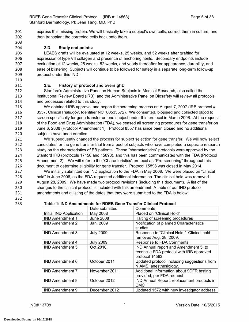
We initially submitted our IND application to the FDA in May 2008. We were placed on “clinical 227 hold” in June 2008, as the FDA requested additional information. The clinical hold was removed 228 August 28, 2009. We have made two protocol revisions (including this document). A list of the 229 changes to the clinical protocol is included with this amendment. A table of our IND protocol 230 amendments and a listing of the dates that they were submitted to the FDA is below: 231
232 Table 1: IND Amendments for RDEB Gene Transfer Clinical Protocol 233 Date submitted Comments Initial IND Application May 2008 Placed on “Clinical Hold” IND Amendment 1 June 2008 Halting of screening procedures IND Amendment 2 Jan. 2009 Notification of planned Characteristics
studies IND Amendment 3 July 2009 Response to “Clinical Hold.” Clinical hold
removed Aug. 28, 2009. IND Amendment 4 July 2009 Response to FDA Comments. IND Amendment 5 Oct 2010 IND Annual report and Amendment 5, to
reconcile FDA protocol with IRB approved protocol 14563
IND Amendment 6 October 2011 Updated protocol including suggestions from NIAMS, anesthesiology.
IND Amendment 7 November 2011 Additional information about 9CFR testing provided, per FDA request
IND Amendment 8 October 2012 IND Annual Report, replacement products in CMC
IND Amendment 9 December 2012 Updated 1572 with new investigator address
Downloaded From: on 06/17/2018

RDEB Gene Transfer Clinical Protocol (IRB #: 14563) Page 6 of 38 Stanford Dermatology, PI: Jean Tang, MD, PhD
IND# 13708 ` Version Date: 10/5/2015
IND Amendment 10 December 2012 Additional sterility testing, added possibility of hospitalization post-grafting
IND Amendment 11 January 2013 Re-submission of documents from Amendment 10, with tracked changes and final versions
IND Amendment 12 July 2013 Addition of C1 mimetic to CMC, addition of biopsy for FACS to clinical protocol
IND Amendment 13 September 2013 2013 IND Annual Report IND Amendment 14 February 2014 Revision of wound dressing protocol,
grafting protocol, CMC revision IND Amendment 15 June 2014 (this
amendment) Removal of C7 antibodies by ELISA as exclusion criteria, replaced by IDIF and DIF analysis; removal of parental mutation confirmation
IND Amendment 16 September 2014 2014 IND Annual Review IND Amendment 17 January 2015 Change of PI from Alfred Lane to Jean Tang IND Amendment 18 October 2015 2015 IND Annual Review
234 3. Name and address and statement of qualifications of each investigator: 235 This information is included in Form 1572 236 237 4. Criteria for subject selection: 238 The information below defines the inclusion and exclusion criteria for this study. Some RDEB 239
subjects may already have documented results for some of the testing listed below. At the discretion of 240 investigators and the EB physician, we may elect not to repeat previous testing that is documented and 241 meets the screening requirements listed below. This would be in order to limit the number of skin 242 biopsies and quantity of blood required from the subject. 243
244 4.A. Number of subjects: 245 We plan to graft 5 adult subjects under this protocol. 246 247 4.B. Characteristics Studies (Pre-Screening): 248 Under IRB approved pre-screening protocols 15898 (ClinicalTrials.gov Identifier NCT00533572) 249
and 17158 (ClinicalTrials.gov Identifier NCT01019148), we have completed an initial phone screen or 250 discussion in clinic for over 100 subjects interested in our clinical trials, as part of a research study in 251 which we are seeking to determine the characteristics of patients with dystrophic EB. However, this 252 information will also help us to identify a candidate for gene transfer. 253
The reason that two protocols existed for the purpose of determining the characteristics of DEB 254 patients is that these two protocols have slightly different inclusion/exclusion criteria and are funded 255 by different sources. As of May 2014, protocol 15898 is closed. 256
257 4.B.i. Plan for recruitment: 258 Subjects are currently being evaluated for their characteristics (pre-screening) through IRB 259
Approved Protocol 17158. Based on the results of the pre-screening, subjects may meet the 260 criteria to enroll in the gene transfer study. If they meet the criteria, they will be invited to 261 participate in the gene transfer protocol. There may be subjects in the pre-screening studies who 262 do not meet criteria to enroll in gene transfer, in which case they would not be invited to 263 participate in the gene transfer trial. 264
We cooperate with national and international networks of families, researchers, and 265 physicians who care for children and adults with EB and plan to use these groups to recruit 266
Downloaded From: on 06/17/2018

RDEB Gene Transfer Clinical Protocol (IRB #: 14563) Page 7 of 38 Stanford Dermatology, PI: Jean Tang, MD, PhD
IND# 13708 ` Version Date: 10/5/2015
subjects for this study. In addition we will use our email listserv 267 ([email protected]) to inform our national communities that we are recruiting for 268 this study. Our study is also listed on http://www.ClinicalTrials.gov (CT.gov Identifier 269 NCT01263379) and http://clinicaltrials.stanford.edu. We initially will limit enrollment to subjects 270 from the USA. Similar information is posted on the Department of Dermatology Epidermolysis 271 Bullosa webpage called Stanford EB Research Update at 272 http://dermatology.stanford.edu/research/research.html. Our updates for the listserv and the 273 website have been approved by the Stanford IRB. 274
We have a database of people who have contacted us regarding our epidermolysis bullosa 275 research. Each one has completed a phone screen through the Characteristics study, in which 276 they have requested to be added to a database of people contacted about future studies. A letter 277 (approved by the Stanford IRB) will be mailed to them to inform them that we are recruiting for the 278 gene transfer study and letting them know about our process for recruitment. 279
4.B.ii. Identifying subjects eligible for gene transfer (based on Pre-Screening): 280 A specific subset of individuals with RDEB will be selected for this clinical trial. RDEB subjects will 281
initially be required to express the NC1 amino-terminal fragment of collagen VII (NC1[+], 282 approximately 75% of our patients), be genotyped with confirmed recessive COL7A1 mutations, and 283 have no evidence of an immune response to type VII collagen. . 284
As described in the Introductory Statement (Section 3-4) of the original IND application and the 285 IND Annual Report, depending on the mutations involved, some RDEB patients express the amino-286 terminal fragment of type VII collagen (NC1[+]) and some do not (NC1[-], approximately 25% of our 287 patients). 1 As the NC1 domain is generally accepted to be the most antigenic region on the type VII 288 collagen molecule, we expect that NC1[+] subjects will be less likely to develop autoimmune reactions 289 to sites of grafted autologous keratinocytes that express type VII collagen since their immune system 290 should have already become tolerant to NC1 epitopes. 291
Non-CLIA data will not be shared with the subject, except as described below. Clinically relevant 292 non-CLIA results may be included in the subject’s medical record if investigators and the EB 293 physician feel that they are important for the subject’s medical care. 294
295 4.B.iii. Procedures performed under characteristics study (Pre-screening): 296 Subjects who arrive at Stanford for pre-screening will be examined to confirm the clinical 297
diagnosis of RDEB. We will examine the subject’s medical records to determine which testing has 298 been performed previously. In the pre-screening study, we will obtain a complete history, perform 299 a physical and skin examination, as well as obtaining photographs of the subject’s skin. We may 300 elect not to repeat previous testing that is documented and meets our screening requirements in 301 order to limit the number of skin biopsies and quantity of blood required from the subject. 302
303 4.B.iii.1. Skin biopsies: 304 Subjects will be asked to donate 5 skin biopsies from non-wounded skin: 305 - Two 6mm biopsies, for tissue culture, to determine NC1 status (described below) 306 - One 3 mm biopsy will be sent for Immunoelectron Microscopy (IEM): This test will 307 screen for type VII collagen by IEM using gold labeled mAb LH24 antibody which 308 recognizes the collagenous region near the NC2 domain of type VII collagen (Gift of Dr. I. 309 Leigh).2 310 - One 4 mm biopsy for indirect and direct immunofluorescence (IIF and DIF, 311 respectively): The biopsy tissue will be screened for multiple epidermal and BMZ 312 antigens (collagen XVII [BP180], collagen IV, collagen VII, laminin-332 gamma 2 chain) 313 which should be positive and also for LH 7.2 mAb which recognizes the NC1 portion of 314
Downloaded From: on 06/17/2018

RDEB Gene Transfer Clinical Protocol (IRB #: 14563) Page 8 of 38 Stanford Dermatology, PI: Jean Tang, MD, PhD
IND# 13708 ` Version Date: 10/5/2015
type VII collagen.3,4 This biopsy will also be analyzed by direct IF for presence of IgG, 315 IgM, IgA, and complement at the basement membrane. 316 - One 3 mm biopsy for electron microscopy (EM): This test will evaluate anchoring fibrils, 317 to confirm the diagnosis of dystrophic EB. The EM biopsy should show absent or 318 significantly defective anchoring fibrils. 319 320 The Stanford University Dermatopathology Laboratory is CLIA certified 321 (http://dermatopathology.stanford.edu/services/epiderm.html) to perform the IIF, DIF and 322 EM diagnostic tests. Physicians in the Departments of Dermatology and 323 Dermatopathology at Stanford University have extensive experience in immunomapping 324 and EM diagnosis for EB. 325
326 4.B.iii.1.a. Assessment of NC1 status: 327 As the NC1 domain is generally accepted to be the most antigenic region on the type 328
VII collagen molecule, we expect that NC1[+] subjects will be less likely to develop 329 autoimmune reactions to sites of grafted autologous keratinocytes that express type VII 330 collagen since their immune system should have already become tolerant to NC1 331 epitopes. We are concerned that NC1[-] RDEB subjects may have a higher risk of 332 developing autoimmune reactions at sites of grafted autologous keratinocytes that 333 express type VII collagen because NC1 may represent a previously immunologically 334 “unseen” neoantigen. In order to decrease this potential risk, the initial RDEB subjects will 335 be NC1[+]. 336
For those subjects with confirmed RDEB, IF microscopic analysis is not sensitive 337 enough to document or exclude the expression of NC1 protein. Determining NC1 338 expression will be accomplished by culturing the keratinocytes and extracting the NC1 339 protein.1 The skin biopsies obtained for culture will be placed in keratinocyte media 340 containing KGM (Keratinocyte-SFM, Invitrogen Corporation, Carlsbad, CA). Skin biopsies 341 will be washed 3 times in PBS with antibiotics/antimycotics and cut into pieces not bigger 342 than 1cm2. Epidermis will be then separated by incubation in 50 caseolytic units/ml 343 dispase (Invitrogen) for 2 hours at 37°C. After incubation in 0.25 mg/ml trypsin/EDTA 344 (Invitrogen Corporation) for 30 minutes at 37°C, a single cell suspension of keratinocytes 345 will be released by gentle pipetting. After neutralization with Dulbecco’s Modified Eagle 346 Medium (D-MEM, Invitrogen) with 10% Fetal Calf Serum (FCS, Omega Scientific, 347 Tarzana, CA) cells will be cultured in KGM in cell culture plate at 37°C in a humidified 348 atmosphere. Keratinocyte extracts will be prepared and subjected to denaturing gel 349 electrophoresis on a 6% polyacrylamide gel. After electrophoresis, protein will be 350 transferred to nitrocellulose membrane and incubated with rabbit anti-FNC1 antibodies to 351 human type VII collagen1, and the mAbs NP 32 and NP185 (gift of Lynn Y.Sakai) to 352 detect NC1 presence2. 353
Subjects who have retained expression of NC1 on immunoblots will be considered 354 NC1[+] and subjects who do not have retained expression of NC1 on immunoblots will be 355 considered NC1[-]. Immunoblot of subject cell extracts will be compared side by side with 356 the cell lysates of our previously published NC1[+] patients cells1. To be considered for 357 the study, the subject's cells must show an intensity of NC1 staining by densitometry 358 equal to or greater than 25% of the mean of our published cells. 359
360 4.B.iii.2. Blood tests: 361 At the time of the diagnostic skin biopsies, blood will be drawn for the following CLIA 362
tests: 363
Downloaded From: on 06/17/2018

RDEB Gene Transfer Clinical Protocol (IRB #: 14563) Page 9 of 38 Stanford Dermatology, PI: Jean Tang, MD, PhD
IND# 13708 ` Version Date: 10/5/2015
- Complete Blood Count (CBC) 364 - Complete Metabolic Panel 365 - Direct and Indirect Bilirubin 366 - HIV test 367 - Hepatitis B surface antigen screening 368 - Hepatitis C antibodies 369 - IIF on monkey esophagus to rule out circulating antibodies to the basement 370
membrane. 371 - Genetic testing for COL7A1 mutations (GeneDx, Gaithersburg, MD). 372 373 If genetic testing doesn’t demonstrate two mutations that have recessive inheritance 374 patterns, we will follow the algorithm depicted in Figure 1. 375 376
Figure 1: Parental Genetic Testing Algorithm 377
378 379
4.B.iv. Inclusion/Exclusion Criteria: 380 381 Inclusion Criteria for Gene Transfer and Autologous Grafting with LEAES: 382 1. Clinical diagnosis of RDEB 383 2. Age 18 years or older, willing and able to give consent 384 3. Confirmation of RDEB diagnosis by IIF and EM 385 4. NC1[+] 386 5. mAb LH24 antibody staining negative, or significantly decreased 387 6. Two confirmed RDEB type VII collagen mutations with recessive inheritance patterns (or 388 confirmation that parents don’t have any evidence of dominant disease) 389 7. At least 100 to 200cm2 areas of open erosions on the trunk and/or extremities suitable for skin 390 grafting 391 8. Able to undergo adequate anesthesia to allow grafting procedures to take place 392 393
Downloaded From: on 06/17/2018

RDEB Gene Transfer Clinical Protocol (IRB #: 14563) Page 10 of 38 Stanford Dermatology, PI: Jean Tang, MD, PhD
IND# 13708 ` Version Date: 10/5/2015
Exclusion criteria for Gene Transfer and Autologous Grafting with LEAES: 394 1. Medical instability limiting ability to travel to Stanford University Medical Center 395 2. The presence of medical illness expected to complicate participation and/or compromise the 396 safety of this technique, such as active infection with HIV, hepatitis B or hepatitis C 397 3. Evidence of immune response to type VII collagen 398 4. Active infection in the area that will undergo grafting 399 5. Evidence of systemic infection 400 6. Current evidence or a history of squamous cell carcinoma in the area that will undergo grafting 401 7. Active drug or alcohol addiction 402 8. Hypersensitivity to vancomycin or amikacin 403 9. Receipt of chemical or biological study product for the specific treatment of RDEB in the past 404 six months 405 10. Positive pregnancy test or breast-feeding 406 11. Clinically significant medical or laboratory abnormalities as determined by investigators and 407 the EB physician 408 409
4.B.v. Day -26: 410 If initial screening criteria have been met, we will ask the subject to return to Stanford to 411
continue the screening procedure and to begin the process of culturing their cells for gene 412 transfer. Subjects will be allowed to bring a companion in order to assist with travel procedures 413 as well as dressing changes. If the subject does not have a companion, or the companion is not 414 able to stay the entire time the subject is at Stanford, we will attempt to provide nursing services. 415
The time of this visit will be 20-35 days prior to grafting. For simplicity, we will refer to this 416 timepoint throughout the protocol and the consent as “Day -26.” The time variability is related to 417 the speed at which the cultured keratinocytes can grow. It is possible that a subject may not 418 meet criteria (e.g. abnormal labs, unable to undergo anesthesia, cells do not grow, etc.) and 419 would be considered a “screen fail.” Please note that the subject will be considered “enrolled” in 420 the gene transfer study on Day 0, when they receive the LEAES graft. 421
When the subject comes to Stanford, we will review the gene transfer consent with them. 422 They will have received a copy in the mail before the appointment, so they will have ample time to 423 review it, discuss it with their primary care physician, etc. 424
425 4.B.v.1. Skin biopsies for LEAES manufacture 426 Two 8 mm punch biopsies will be obtained from non-blistered skin for keratinocyte 427
culture, in order to manufacture the LEAES graft. The manufacturing aim is to produce and 428 deliver four to six of the 40 cm2 to 50 cm2 sheets for grafting (LEAES). Approximately two to 429 six of the 40-50 cm2 epithelial sheets will be used in a single grafting session. The maximum 430 total grafting surface area for all the graft sites will be 300 cm2. Skin biopsies for LEAES 431 manufacture will be labeled with the subject’s name, date of birth, medical record number, 432 and study number. 433
We anticipate a maximum of 3 grafting sessions over the course of six months. Subjects 434 who do not have initial graft attachment because of wound infections or mechanical causes 435 will have the option (upon approval by the EB physician) of receiving additional grafts in the 436 future prepared from frozen keratinocytes or a new biopsy if necessary. 437
We will graft two types of wounds. One site will be an acute wound (induced 438 approximately 24 hours prior to grafting, at Day -1, see section 4.B.viii) not to exceed 40-50 439 cm2, produced by inducing a blister on intact skin and removing the blister roof just prior to 440 grafting. The other sites will be areas of chronic wounds of approximately 25-50 cm2 or 441
Downloaded From: on 06/17/2018

RDEB Gene Transfer Clinical Protocol (IRB #: 14563) Page 11 of 38 Stanford Dermatology, PI: Jean Tang, MD, PhD
IND# 13708 ` Version Date: 10/5/2015
greater that have been prepared for grafting. We plan to graft multiple areas of chronic 442 wounds or possibly one large wound with several 40 cm2 to 50 cm2 sheets (see Day 0, 443 section 5.A). LEAES grafts will be labeled with the subject’s name and study number. 444
445 446 4.B.v.2. Research biopsies 447 In addition the following biopsies may be obtained from non-blistered, non-traumatized 448
skin near where one of the wounds may be grafted. The biopsies obtained at screening will 449 be used as controls for later biopsies. 450
We will obtain the minimum number of biopsies necessary; some may be excluded at the 451 discretion of investigators and the EB physician. At this timepoint, we may obtain: 452
- one 4 mm biopsy for IIF for BMZ antigens to be examined in our research laboratory 453 - one 4mm biopsy for DIF for immunoreactants in the skin; 454 - one 4 mm punch biopsy for FACS; 455 - one 6mm punch biopsy for molecular analysis 456 - one 3 mm biopsy for EM 457 - one 3mm biopsy for IEM 458 459
4.B.v.2.a. Immunoelectron microscopy (IEM) and electron microscopy (EM): 460 Both 3 mm biopsies will be shipped over night to one of our collaborators at Shriner’s 461
Hospital in Portland, OR who will do the research immunoelectron microscopy (IEM) and 462 electron microscopy (EM).5 IEM will be performed on one of the 3 mm biopsies using 463 gold-labeled LH24 antibodies to detect and localize delivered type VII collagen protein. 464 Evidence for blistering on an ultrastuctural level will be examined. EM will be performed 465 on the other 3 mm biopsy to determine presence of anchoring fibrils. 466
467 4.B.v.2.b. Immunofluorescence (IF), FACS and molecular analysis: 468 IF analysis for expression of type VII collagen will be performed on one 4 mm biopsy. 469 Another 4mm biopsy may be used for FACS analysis. FACS analysis will consist of 470
separating T regulatory cells from the skin samples, followed by PCR analysis of their 471 cytokine profile. The investigator may decide to obtain an additional biopsy for the FACS 472 analysis. This sample will be sent to collaborators at UCSF for analysis. 473
We may do one 6 mm punch biopsy for molecular analysis which will be placed in 474 keratinocyte media containing KGM (Keratinocyte-SFM, Invitrogen Corporation, 475 Carlsbad, CA). This biopsy may be flash frozen in liquid N2 for future analysis. Analysis 476 will include qPCR to quantify the amount of vector DNA present and qRT-PCR to detect 477 mRNA expression of type VII collagen. 478
These biopsies will be analyzed in the Stanford Dermatology research lab. The 479 molecular analysis biopsy is not required by the FDA but it was strongly suggested by the 480 NIH review and it may be omitted at times that the investigators think it would add less 481 information (subject to approval of the EB physician). 482
483 4.B.v.3. Blood and urine tests 484 At this time we will also obtain blood for a CBC, Complete Metabolic Panel, and Hepatic 485
Function Testing. If follow-up is required clinically, additional blood tests may be obtained as 486 needed, at the discretion of the EB physician. We will obtain a sample to use as a baseline 487 test to assay cytotoxic T cells (see below). We will also send a blood sample as a baseline 488 test for replication competent retrovirus (RCR) to Indiana University Vector Production Facility 489
Downloaded From: on 06/17/2018

RDEB Gene Transfer Clinical Protocol (IRB #: 14563) Page 12 of 38 Stanford Dermatology, PI: Jean Tang, MD, PhD
IND# 13708 ` Version Date: 10/5/2015
(IUVPF). A blood sample may be obtained for IIF on monkey esophagus analysis to rule out 490 an immune response to type VII collagen, if the IIF test has not been completed recently. 491
On female subjects of childbearing potential, we will perform a urine pregnancy test to 492 confirm that they are not pregnant. 493
494 4.B.v.3.a. Cytotoxic T cell assay 495 15 mL of whole blood will be collected for the cytotoxic T cell assay. Studies are 496
currently being conducted to determine if a smaller amount of blood can be used for the 497 assay. Testing will be conducted in the Stanford Dermatology research labs. Samples 498 will be labeled with subject number and initials and the date the sample was obtained. 499
Isolation of T lymphocytes and monocytes from peripheral blood (15mL): Peripheral 500 blood mononuclear cells are isolated from buffy coats or whole blood using Ficoll-Paque 501 (GE Healthcare) density-gradient centrifugation. Adherent monocytes are then recovered 502 after a 2-hour incubation in Petri dishes. CD4+ and CD8+ T lymphocytes are purified 503 together using MACS magnetic cell sorting kits (Miltenyi Biotech), by incubating the 504 nonadherent cells with anti-CD4 and anti-CD8 antibodies conjugated to paramagnetic 505 microbeads. 506
Th1 (IFN-γ) and Th2 (IL-4) ELISPOT: Ninety-six-well PVDF-filter plates (Millipore) 507 are coated with monoclonal antibody against IFN-γ (BD Pharmingen) or IL-4 (BD 508 Pharmingen), blocked using RPMI medium with 5% human AB serum, and washed with 509 serum-free RPMI. CD4+ and CD8+ T lymphocytes (2×10⁵ cells/well), and γ-irradiated 510 monocytes (2.5×10⁴ cells/well) are co-incubated on the plate in the presence of 20 UI/ml 511 IL-2 for 40 hours at 37°C, in a humidified, 5% CO2 in air incubator. The medium contains 512 either 10 µg/ml of recombinant type VII collagen or 3 μg/ml of concanavalin A (Sigma) to 513 stimulate the lymphocytes. The plates are washed and the IFN-γ or IL-4 secreted by 514 individual cells are detected in situ by successively reacting each well with (1) biotinylated 515 anti-IFN-γ or anti-IL-4 monoclonal antibody (BD Pharmingen) at 1 µg/ml, (2) a 1:1000 516 dilution of streptavidin-conjugated alkaline phosphatase (Roche), and (3) a solution of 517 BCIP/NBT chromogenic substrate (Promega), with suitable intermediate washes. The 518 reaction is halted by washing with water, and spots are counted using a CTL ELISPOT 519 reader. Negative controls are run in parallel using T cells without antigen, and the 520 corresponding scores are subtracted from those of the unknowns. 521
522 4.B.v.4. Selection of target chronic wounds 523 At the first screening visit (Day -26) we will select multiple potential wounds for grafting. 524
At the discretion of investigators, target wound selection may occur at a later visit. We will 525 follow these wounds clinically until the day of grafting/enrollment (Day 0). The decision on 526 which sites to be grafted will not be finalized until Day 0. The determination that target 527 wounds meet all grafting criteria will be made at that time. 528
The grafted wound areas will be selected by several criteria. The wounds should appear 529 clean with adequate granulation tissue, adequate vascularization, and not appear infected. 530 The surface area should have a smooth texture that can accept a graft. 531
The duration that the subject thinks that they have each wound will be recorded, but often 532 the subject is unsure of the duration of wounds at specific sites. We believe that the 533 appearance and characteristics of the wound site are more important than the duration as 534 many of the ulcerated areas have been wounded for many years. 535
The wounds will also need to meet mechanical requirements that decrease trauma to the 536 grafted areas. Joints that are mobile may stretch the graft with movement and force the graft 537
Downloaded From: on 06/17/2018

RDEB Gene Transfer Clinical Protocol (IRB #: 14563) Page 13 of 38 Stanford Dermatology, PI: Jean Tang, MD, PhD
IND# 13708 ` Version Date: 10/5/2015
to tear off before it has adequately attached. Areas that are exposed to constant sheering 538 trauma may limit the ability of the graft to adhere before it has firmly attached. In addition, the 539 areas to be grafted must also be easily covered with effective wound dressings. Sites that 540 cannot be adequately protected by effective wound dressing will be excluded. 541
Appropriate sites will be on the anterior and/or lateral trunk and/or upper and/or lower 542 extremities in areas protected from frequent trauma or injury. Excluded areas will usually 543 include the face, areas close to mucous membranes (genito-urinary, oral or anal mucosa). 544 Areas over joints and on the back are frequently areas of chronic non-healing wounds. We 545 will consider grafting these areas if we are capable of immobilizing the grafts onto the areas 546 long enough for securing effective graft attachment. The grafting surgeon, investigators, and 547 the EB physician will jointly decide if areas over the joints, the back, and buttocks may be 548 appropriate for grafting on a case-by-case basis. The distance from objective body landmarks 549 will be identified and measured. 550
551 4.B.v.5. Assessment of chronic target wounds 552 The health of target chronic wounds will be determined by investigators and the EB 553
physician. Each wound will be examined for signs of inflammation and infection, and the 554 quality of granulation tissue will be assessed. Wounds appropriate for grafting have a clean, 555 vascularized, nonexudative wound bed, appearing clinically as granulation tissue without 556 drainage. We will avoid any areas that may already contain potential SCC even if that area 557 has been previously biopsied and the biopsy did not document SCC. 558
Bacterial cultures will be obtained from several wounds for culture and antibiotic 559 sensitivities. Wound cultures may be repeated as needed based upon standard of care and 560 medical judgment of the investigators and EB physician. Chronic wounds in patients with 561 RDEB are oftentimes colonized with bacteria even in the absence of clinical infection. Thus, 562 the diagnosis of an infected wound or cellulitis is made clinically while taking into account the 563 results of the wound culture. Systemic antibiotics will be prescribed for wounds with cellulitis 564 or excessive exudates, with antibiotic choice determined by culture results and judgment of 565 the investigators and EB physician. 566
The subject may remain in housing near Stanford between Day -26 and Day -7 if specific 567 medical treatment or wound care is needed to prepare for the grafts. They may need to have 568 this treatment at an outside hospital. If additional treatment is not needed, the subject may 569 return home and then come back to Stanford in time for Day -7. This decision will be made by 570 the EB physician. 571
572 4.B.v.6. Photographs and wound measurements 573 Digital photographs will be taken of each site, including a centimeter (cm) scale, to clearly 574
document the location and size of each estimated treated area. A color chart may be included 575 in photographs in order to standardize colors in the photographs. We will also measure the 576 area of each wound using the ARANZ SilhouetteStar digital device, which uses a series of 577 lasers to accurately determine wound measurements, or with the Canfield Vectra system 578 which is able to produce 3D images.. Data captured by the camera systems is stored to track 579 wound progress over time, with easy visualization of the wounds, and calculation of wound 580 progression. We will also measure the distance from the center of the target wound to at 581 least 2 body landmarks (i.e. bony prominence, freckle, etc.). 582
583 4.B.v.7. Anesthesia and Plastic Surgery consults, ECG, Echocardiogram 584 The subject and possibly investigators will meet with an anesthesiologist and plastic 585
surgeon prior to grafting. The plastic surgeon will be the physician who will place the skin 586
Downloaded From: on 06/17/2018

RDEB Gene Transfer Clinical Protocol (IRB #: 14563) Page 14 of 38 Stanford Dermatology, PI: Jean Tang, MD, PhD
IND# 13708 ` Version Date: 10/5/2015
grafts. We will perform an echocardiogram and/or an ECG to determine the cardiac function 587 in preparation for grafting and anesthesia. Special precautions for EB patients will be taken 588 when undergoing the ECG (i.e. use of Mepitel instead of the usual leads), as is commonplace 589 at Stanford/Lucile Packard Children’s Hospital. We may also request a cardiology 590 consultation if the ECG or echocardiogram is abnormal. 591
Additionally, the anesthesiologists will request and review the subject’s previous 592 anesthesia records, and their intubation history. 593
If, in the opinion of the anesthesiologists, cardiologists, grafting surgeon, and/or EB 594 physician, the subject will not be able to undergo adequate local anesthesia, general 595 anesthesia or conscious sedation, the candidate would be considered a screen fail. 596
In order to limit unnecessary biopsies, we may choose to have subjects undergo these 597 non-invasive tests prior to performing the skin biopsies described in 4.B.v.1 and 4.B.v.2. 598
599 4.B.v.8. Adverse event reporting and assessment 600 We will provide the subject with a paper diary, in which to record all adverse events 601
(AEs). This diary will be reviewed with subjects at every study visit. The diary will be 602 approved by the Stanford IRB. We will also send a letter (approved by the Stanford IRB) to 603 the subject’s primary care provider requesting that they inform us immediately of adverse 604 events or any other problems that the subject experiences. We will ask subjects to begin 605 recording adverse events after their first screening visit to Stanford. 606
At the time that investigators are informed of adverse events, they will be assessed for 607 severity, etiology/causality, and expectedness. Adverse events will be graded according to 608 the National Cancer Institute’s Common Toxicity Criteria. At the first screening visit, subjects 609 will be assessed for baseline medical conditions according to these criteria. Any increase in 610 the grade of the condition will be considered an adverse event. Subjects with Grade 2 611 abnormalities identified prior to Day 0 (other than those specified as exceptions in the 612 Inclusion/Exclusion criteria or by the EB physician) will not receive LEAES grafts and will be 613 considered a “screen fail”. 614
Abnormal laboratory values will be assessed for clinical significance, based on the 615 investigator’s judgment, subject to review by the EB physician. Clinically significant, 616 abnormal lab values identified after the subject’s initial visit will be captured as AEs, and 617 graded according to the NCI toxicity criteria. EB patients with open wounds have abnormal 618 laboratory values that have not been quantified in enough detail to create normal value 619 limits. Markers of inflammation such as white blood count and sedimentation rate are usually 620 elevated. Anemia is common and is associated with chronic blood loss through wounds as 621 well as anemia associated with chronic disease. Subjects will be followed comparing their 622 own laboratory results over time in order to identify noteworthy deviations from normal for 623 them (as determined by the investigators or EB physician). 624
625 4.B.vi. Day -7: 626 Seven to 14 days before grafting we should have an estimate of the grafting date based upon 627
the status of the growth of the keratinocyte grafts (LEAES). For simplicity, we will refer to this as 628 “Day -7” in the protocol. At Day -7, the target wound areas will again be assessed, possibly 629 photographed and possibly recultured depending on the clinical appearance at the discretion of 630 the investigator and EB physician. Additional blood tests may also be obtained at the discretion of 631 the investigator and EB physician. If necessary, the EB physician may approve additional 632 treatments necessary for the subject to meet grafting inclusion criteria, including the ability to 633 undergo anesthesia. A skin and physical examination will occur, and the subject will be asked 634 about concomitant medications and adverse events. 635
Downloaded From: on 06/17/2018

RDEB Gene Transfer Clinical Protocol (IRB #: 14563) Page 15 of 38 Stanford Dermatology, PI: Jean Tang, MD, PhD
IND# 13708 ` Version Date: 10/5/2015
636 4.B.vi.1. Wound preparation for grafting 637 The subject should continue their normal dressing regimen. Site selection for the 638
grafting will be finalized on Day 0 (the date of grafting). Oral antibiotics may be used 639 depending on wound appearance and culture results, at the discretion of the investigator and 640 EB physician. 641
642 4.B.vii. Day -3: 643 Subjects may have their target wounds re-examined at Day -3. The target wound areas will 644
again be assessed, possibly photographed and possibly recultured depending on the clinical 645 appearance. Additional blood tests may also be obtained at the discretion of the investigator and 646 EB physician. If necessary, the EB physician may approve additional treatments necessary for 647 the subject to meet grafting inclusion criteria, including the ability to undergo anesthesia. A skin 648 and physical examination will occur (at the discretion of the EB physician and investigator) and 649 the subject will be asked about concomitant medications and adverse events. 650
At the discretion of the investigator and EB physician, this visit may be omitted. 651 652 4.B.viii. Day -1: 653 Subjects will have their target wounds re-examined at Day -1. The target wound areas will 654
again be assessed, possibly photographed and possibly recultured depending on the clinical 655 appearance. Additional blood tests may also be obtained at the discretion of the investigator and 656 EB physician. If necessary, the EB physician may approve additional treatments necessary for 657 the subject to meet grafting inclusion criteria, including the ability to undergo anesthesia. A skin 658 and physical examination will occur (at the discretion of the EB physician and investigator), and 659 the subject will be asked about concomitant medications and adverse events. 660
Patients with RDEB understand how much trauma is necessary to develop a blister and how 661 rapidly blisters can form on their skin. The timing necessary to develop a blister that can be 662 developed into an acute wound will be based upon the suggestions of the RDEB subject. Usually 663 less than 24 hours are necessary. Twenty-four hours or less before grafting we plan to create an 664 acute wound (Study Day -1). 665
666 4.B.viii.1. Creation of acute blister 667 For this process, we will select an area of intact skin on the anterior or lateral torso or 668
upper or lower extremity not to exceed 40-50 cm2 in size. The area will be marked with a 669 sterile surgical marking pen and photographed with a digital camera and/or Canfield system 670 prior to induction of a blister or applying trauma to the area. . 671
Either the investigator or the subject will firmly rub the skin within the marked area until 672 they think they have generated enough trauma to develop a blister. The blister may be 673 subclinical at this time but will fill with fluid over the next several hours. This area should not 674 be disturbed until the time of grafting, unless the subject feels the blister is enlarging and 675 extending beyond the marked areas. In this case, a sterile needle may be used to drain the 676 blister, but the roof should not be removed until the time of grafting procedure. At the time of 677 the grafting the roof will be removed and the area grafted as described below. We will 678 provide the subject with written instructions (approved by the Stanford IRB) on how to care 679 for the induced wound. 680
681 5. Enrollment/ Grafting: 682 Subjects will be considered “enrolled” in the gene transfer study when the determination is made that 683
they will undergo grafting with LEAES at Day 0. 684
Downloaded From: on 06/17/2018
RDEB Gene Transfer Clinical Protocol (IRB #: 14563) Page 16 of 38 Stanford Dermatology, PI: Jean Tang, MD, PhD
IND# 13708 ` Version Date: 10/5/2015
685 5.A. Grafting (Day 0): 686
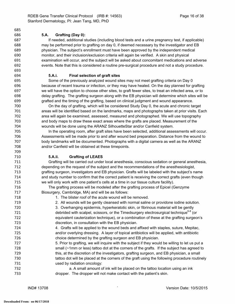
If needed, additional studies (including blood tests and a urine pregnancy test, if applicable) 687 may be performed prior to grafting on day 0, if deemed necessary by the investigator and EB 688 physician. The subject’s enrollment must have been approved by the independent medical 689 monitor, and their inclusion/exclusion criteria will again be verified. A skin and physical 690 examination will occur, and the subject will be asked about concomitant medications and adverse 691 events. Note that this is considered a routine pre-surgical procedure and not a study procedure. 692 693
5.A.i. Final selection of graft sites 694 Some of the previously analyzed wound sites may not meet grafting criteria on Day 0 695
because of recent trauma or infection, or they may have healed. On the day planned for grafting 696 we will have the option to choose other sites, to graft fewer sites, to treat an infected area, or to 697 delay grafting. The grafting surgeon along with the EB physician will determine which sites will be 698 grafted and the timing of the grafting, based on clinical judgment and wound appearance. 699
On the day of grafting, which will be considered Study Day 0, the acute and chronic target 700 areas will be identified based on the landmarks, maps and photographs taken at prior visits. Each 701 area will again be examined, assessed, measured and photographed. We will use topography 702 and body maps to draw these exact areas where the grafts are placed. Measurement of the 703 wounds will be done using the ARANZ SilhouetteStar and/or Canfield system. 704
In the operating room, after graft sites have been selected, additional assessments will occur. 705 Assessments will be made prior to and after wound bed preparation. Distance from the wound to 706 body landmarks will be documented. Photographs with a digital camera as well as the ARANZ 707 and/or Canfield will be obtained at these timepoints. 708
709 5.A.ii. Grafting of LEAES 710 Grafting will be carried out under local anesthesia, conscious sedation or general anesthesia, 711
depending on the request of the subject and the recommendations of the anesthesiologist, 712 grafting surgeon, investigators and EB physician. Grafts will be labeled with the subject’s name 713 and study number to confirm that the correct patient is receiving the correct grafts (even though 714 we will only work with one patient’s cells at a time in our tissue culture facility). 715
The grafting process will be modeled after the grafting process of Epicel (Genzyme 716 Biosurgery, Cambridge, MA) and will be as follows: 717
1. The blister roof of the acute wound will be removed. 718 2. All wounds will be gently cleansed with normal saline or providone iodine solution. 719 3. Overhanging epidermis, hyperkeratotic skin, or fibrinous material will be gently 720 debrided with scalpel, scissors, or the Timedsurgery electrosurgical technique6-8 (or 721 equivalent cauterization technique), or a combination of these at the grafting surgeon’s 722 discretion, in consultation with the EB physician. 723 4. Grafts will be applied to the wound beds and affixed with staples, suture, Mepitac, 724 and/or overlying dressing. A layer of topical antibiotics will be applied, with antibiotic 725 choice determined by the grafting surgeon and EB physician. 726 5. Prior to grafting, we will inquire with the subject if they would be willing to let us put a 727 small (~1mm or less) tattoo dot at the corners of the grafts. If the subject has agreed to 728 this, at the discretion of the investigators, grafting surgeon, and EB physician, a small 729 tattoo dot will be placed at the corners of the graft using the following procedure routinely 730 used by radiation oncology: 731 a. A small amount of ink will be placed on the tattoo location using an ink 732 dropper. The dropper will not make contact with the patient’s skin. 733
Downloaded From: on 06/17/2018

RDEB Gene Transfer Clinical Protocol (IRB #: 14563) Page 17 of 38 Stanford Dermatology, PI: Jean Tang, MD, PhD
IND# 13708 ` Version Date: 10/5/2015
b. Using a sterile hypodermic needle, a physician will pierce the skin only enough 734 to deliver the ink into the skin. 735 c. Any ink remaining on the skin will be wiped away to ensure tattoo accuracy. 736 6. Photographs will be obtained with a digital camera, ARANZ, and/or Canfield system 737 following graft placement. 738 7. Dressings will consist of a single layer of Interface (silk gauze), Mepitel, Adaptic, 739 Restore, or other equivalent contact layer followed by an absorbent foam dressing 740 (Mepilex, Restore Foam, or Allevyn), which will be held in place with rolled gauze and 741 surgical netting. Dressings will be decided upon by the grafting surgeon, in consultation 742 with the EB physician. The subject will be given written instructions on how to care for 743 their grafts (see section 6) and how to contact study staff. 744 8. The dressings will stay in place until changed at day +5 to +10. We will plan to see 745 the subject every 3 days (+/- 1 day) following grafting to monitor wound healing if the 746 subject is not hospitalized. If the subject is experiencing problems, we will see them 747 immediately in clinic. 748 9. At the discretion of the grafting surgeon, investigators, and EB physician, splints may 749 be used to immobilize the grafted areas to prevent any trauma to the grafts. 750
751 6. Post-grafting observation (Day +1 - Day +14) 752 The subject will remain in the local area for observation and frequent examinations for at least 14 753
days following grafting. Based on the discretion of the grafting surgeon and EB physician, the subject may 754 be admitted to the hospital for observation for several days following grafting. This is in order to minimize 755 movement and immobilize the grafted areas and also to facilitate monitoring of the patient. In the post-756 grafting period, it is crucial that the graft is not disturbed for at least 5-10 days (until Study Days +5 to 757 +10). We plan to see the subject every 3 days (+/- 1 day) for follow up. This may be as a study visit, or 758 during regular rounds if the subject is hospitalized. If deemed necessary by the investigator or EB 759 physician, we may see subjects more frequently. At these visits, subjects will be assessed for adverse 760 events and changes to concomitant medications. The investigator may perform a physical examination at 761 their discretion and that of the EB physician. If the subject is unable to come to Stanford, investigators 762 may be able to come to the subject’s home or hotel to check on their progress. We will also try to provide 763 nursing support for bandage changes. 764
At the discretion of the grafting surgeon, investigators, and EB physician, the subject may be given 765 prophylactic antibiotics, as is routine following surgery. 766
If the dressings become moistened with exudates, or if the subject has unexplained fevers over 767 38.5°C on Study Day +3 or later, the absorbent layers may be changed, but the underlying contact layer 768 should not be removed, irrigated or cleansed. If drainage is excessive or purulent, antibiotics may be 769 used at the discretion of the investigator and the EB physician. The subject will be provided with written 770 instructions (approved by the Stanford IRB) on how to take care of their wounds, and how to contact 771 study staff if they have any problems. 772
Five to 10 days after placement of the graft, the investigator will perform a physical exam and a skin 773 examination. The investigator and EB physician will determine if any adverse events have occurred. 774
The outer dressing layers will be removed. At this point, the Vaseline backing of the LEAES grafts 775 will still be attached to the wound. As the sutures dissolve, the Vaseline backing should come off on its 776 own, as the LEAES cells incorporate into the wound beds. The subject and their caretaker will be 777 instructed to let this happen naturally, not to try to remove the gauze. 778
If results from post-release criteria (i.e. mycoplasma, sterility) are unacceptable, the investigator will 779 be notified immediately and failure investigations will be conducted in accordance with our internal SOPs 780
Downloaded From: on 06/17/2018

RDEB Gene Transfer Clinical Protocol (IRB #: 14563) Page 18 of 38 Stanford Dermatology, PI: Jean Tang, MD, PhD
IND# 13708 ` Version Date: 10/5/2015
(available upon request) and Safety Monitoring Plan. The investigator will discuss treatment options with 781 the EB physician, who will have the authority to determine the appropriate course of action. 782
At the discretion of investigators and the EB physician, the subject may return on Day +12 (+/- 1 day) 783 for another follow-up visit. At this visit, adverse events will be assessed and concomitant medications will 784 be recorded. Additionally, the investigator and/or EB physician will perform a physical examination and a 785 skin examination. Most likely the LEAES grafts will still be covered by the Vaseline gauze backing. If 786 grafts are visible at this time, they will be assessed and photographed as described below in section 7.E. 787
788 7. Post-grafting clinical follow-up: 789 Please see Schedule of Events (Table 3) for additional clarification on follow up procedures. Routine 790
skin care for RDEB patients will continue throughout the study. Additional skin biopsies, wound cultures, 791 or laboratory tests will be performed as necessary to evaluate variations from the expected protocol, 792 inflammation, infection, or possible SCC growths within the grafted sites at the discretion of the 793 investigator and the EB physician. 794
795 7.A. Blood tests 796
At Study Day +14, 4 weeks, 12 weeks, 25 weeks and 52 weeks after grafting, the subject will 797 return to Stanford University Medical Center where blood will be obtained for CBC and platelets, 798 Complete Metabolic Panel and hepatic function testing, cytotoxic T cell assay, and analysis for an 799 immune response against type VII collagen. Whenever possible, we will draw the minimum 800 (pediatric) amount of blood. Additional blood tests may be added at the discretion of the 801 investigator and the EB physician. If it is not possible to obtain enough blood for all of these 802 studies, tests may be omitted, at the discretion of the investigator and the EB physician. 803
804 7.A.i. Replication competent retrovirus analysis 805 Replication competent retroviral analysis (RCR) will be performed on the blood collected at 806
12 weeks, 25 weeks and 52 weeks after grafting then yearly thereafter (possibly in a follow-up 807 protocol, as described in section 7.H) for at least 5 years. 808
809 7.B. Physical examination, skin examination 810
At each study visit, the subject will undergo a physical examination and skin examination in 811 addition to the examination of the grafted area. 812
813 7.C. Adverse events 814
The subject will also be asked if they have experienced any adverse events since the 815 previous visit and the study diary will be reviewed. This information will be recorded in the 816 subject’s medical record. All adverse events will be recorded, regardless of their attribution to 817 LEAES and will be discussed with the DSMB on a routine basis (section 16.E). Unexpected, 818 harmful, and related adverse events will be reported immediately to the FDA, the Stanford IRB, 819 Stanford Biosafety, and the DSMB, as described in the Safety Monitoring Plan (section 7.E). 820
821 7.D. Concomitant medications 822
At each study visit, the subject will be asked if they have used any concomitant medications 823 or undergone any concomitant therapies. This information will be recorded in the subject’s 824 medical record. There are no excluded concomitant therapies or medications in this protocol. 825
826
Downloaded From: on 06/17/2018

RDEB Gene Transfer Clinical Protocol (IRB #: 14563) Page 19 of 38 Stanford Dermatology, PI: Jean Tang, MD, PhD
IND# 13708 ` Version Date: 10/5/2015
7.E. Photographs and graft evaluation 827 Photographs will be taken of the grafted sites using the ARANZ SilhouetteStar and/or 828
Canfield Vectra system to generate an accurate image of the graft area and calculate the cm2 of 829 the wound/graft area. 830
We will observe the grafts closely in order to separate technical grafting problems from 831 immunological rejection or loss of genetic correction. The graft site will be clinically evaluated 832 with a global score of: 1) 100% to 75% healed, 2) 74% to 50% healed, 3) 49% to 25% healed, 4) 833 25% to 1% healed, 5) complete graft loss, or 99) unable to determine. It may be several weeks 834 before we are able to adequately evaluate the condition of the grafted areas, as the grafts may 835 still be covered by Vaseline gauze. 836
The early time points of graft evaluation may not be as accurately recorded because the area 837 of graft acceptance may not clearly be apparent within the wound. We will be able to quantitate 838 the precise surface area of blistering, erosions and graft absence from the digital photographs of 839 the areas. 840
Blinded observers will confirm the accuracy of the evaluation without knowledge of the 841 duration of the graft or when the digital image was obtained. 842
We will record the subject’s impression of the durability of the grafted sites and the ease of 843 blistering with trauma. We will not physically damage the grafts in order to develop minimum 844 requirements for blistering. Future studies can include methods used to estimate the force 845 necessary to cause blistering in RDEB and RDEB skin grafted with LEAES. 846
Attention of the clinical examination will also be focused on the sites of the previous biopsies 847 to evaluate healing and scar appearance. 848
849 7.F. Skin biopsies 850
At Study Day +14, 4 weeks, 12 weeks, 25 weeks and 52 weeks after grafting, the graft sites 851 will be identified. Biopsies may be obtained from multiple sites. The following biopsies may be 852 obtained at the discretion of investigators and the EB physician: 853
- One 4mm punch biopsy for IF to be performed in our research laboratory 854 - One 4mm punch biopsy for FACS to be performed at UCSF 855 - One 4mm punch biopsy for DIF to determine if immune complexes are present at the 856
basement membrane. This is a CLIA test which will be performed by the Stanford 857 Dermatopathology Laboratory. 858
- One 3mm punch biopsy for EM to be performed by Doug Keene, an outside collaborator 859 - One 3mm punch biopsy for IEM to be performed by Doug Keene, an outside collaborator 860 - One 6mm punch biopsy for molecular analysis to be performed in our research 861
laboratories 862 The priority for obtaining skin biopsies will focus on obtaining the IF biopsies for type VII 863
collagen antigens and DIF for immunological response at 12 weeks, 25 weeks and 52 weeks 864 after grafting and EM for presence of anchoring fibrils. Additional biopsies may need to be 865 obtained at the discretion of the investigator and the EB physician. The biopsies may be needed 866 to document presence or absence of grafted tissue in a specific area. Biopsies may also be 867 omitted at the discretion of the investigator and the EB physician. 868
If the tissue appears abnormal the biopsy will be sent to pathology for histologic evaluation. 869 Additional skin biopsies or laboratory tests will be done as necessary (in consultation with the EB 870 physician) to evaluate variations from the expected protocol, inflammation, or possible SCC 871 growths within the grafted sites. 872
Based on the success of the graft attachment and/or a sense by the subject that the non-873 grafted adjacent skin may be more resistant to trauma or may demonstrate healing of wounds 874 that previously would not heal, additional biopsies may be obtained outside of the grafted area (at 875
Downloaded From: on 06/17/2018

RDEB Gene Transfer Clinical Protocol (IRB #: 14563) Page 20 of 38 Stanford Dermatology, PI: Jean Tang, MD, PhD
IND# 13708 ` Version Date: 10/5/2015
the discretion of the EB physician) in order to evaluate potential type VII collagen spreading 876 outside of the grafted area. Biopsies on the periphery of the grafted area may also be obtained if 877 it appears that the graft is spreading outside of the initial graft boundaries. 878
All biopsies will be obtained after local anesthesia and all sites will be marked and 879 photographed to confirm the location of the biopsies. Location of biopsies will be recorded in the 880 source documentation. When multiple grafts are placed on different body sites, additional biopsy 881 sites will be decided based on the graft appearance, previously obtained information, and 882 consultation with the EB physician. Based upon subject preferences and in order to limit the 883 number of procedures, the biopsies may be obtained by one or more surgical elliptical biopsies 884 representing the surface area of the punch biopsies described above. 885
Research biopsies will be labeled with the subject’s study number and possibly initials, 886 anatomical location, and the date the biopsy was obtained. The evaluator of the biopsies will be 887 blinded as to the source of the biopsies, which could be from wounded areas, grafted areas, or 888 non-grafted, non-wounded skin. 889
890 7.G. Week 8 891
At 8 weeks after grafting, blood will be drawn at a laboratory close to the subject’s home and 892 shipped to Stanford for CBC and platelets, Complete Metabolic Panel, cytotoxic T cell assay, and 893 for an immune response to type VII collagen. If it is not possible to obtain enough blood for all of 894 these studies, specific tests may be omitted at the discretion of investigators and the EB 895 physician. It will not be necessary for the subject to come to Stanford for the Week 8 visit. 896 However, they may come to Stanford if they prefer. If they come to Stanford for this visit, 897 additional graft assessment and physical examination may be performed. 898
899 Figure 2: Flow Chart for RDEB Gene Transfer Protocol: 900
901
Downloaded From: on 06/17/2018
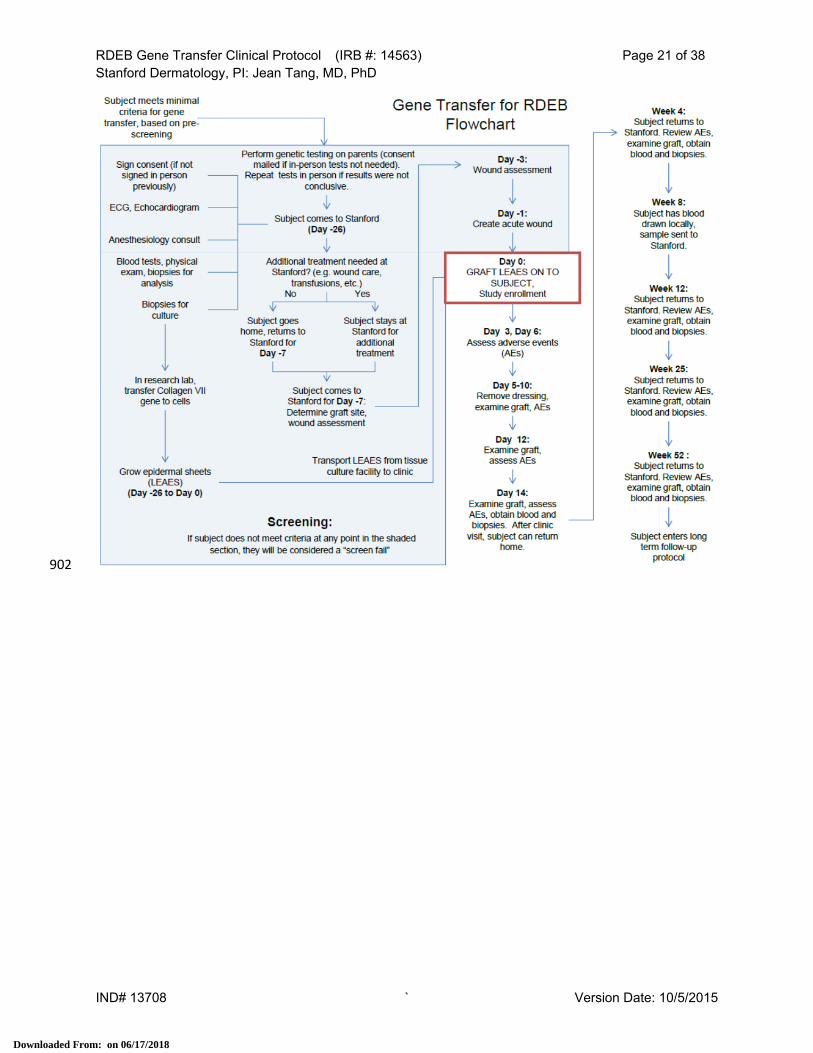
RDEB Gene Transfer Clinical Protocol (IRB #: 14563) Page 21 of 38 Stanford Dermatology, PI: Jean Tang, MD, PhD
IND# 13708 ` Version Date: 10/5/2015
902
Downloaded From: on 06/17/2018

RDEB Gene Transfer Clinical Protocol (IRB #: 14563) Page 22 of 38 Stanford Dermatology, PI: Jean Tang, MD, PhD
IND# 13708 ` Version Date: 10/5/2015
Table 2: Schedule of Events for RDEB Gene Transfer: 903
904 905
906
Downloaded From: on 06/17/2018

RDEB Gene Transfer Clinical Protocol (IRB #: 14563) Page 23 of 38 Stanford Dermatology, PI: Jean Tang, MD, PhD
IND# 13708 ` Version Date: 10/5/2015
7.H. Long term follow up protocol 907 After 52 weeks (1 year) subjects will be enrolled in a long term follow-up protocol under this IND, 908
in which we will follow them for life. 909 Regarding the NIH sponsored DSMB, the end of the DSMB follow-up will be 1 year after the last 910
subject is enrolled. 911 912
8. Discontinuation, Withdrawal, Lost to Follow-Up, or Early Termination 913 Patients who discontinue due to adverse events or protocol deviations/violations (section 15 and 914
section 6 of the Safety Monitoring Plan) shall not be replaced. Any and all patients who discontinue or 915 withdraw from the study after receipt of LEAES will be included in the long-term follow up. 916
A subject may withdraw at any time, but we would plan to follow them for at least 15 years and up to 917 their lifetime (section 7.H). They will have the option to not follow our suggestions, but we will still attempt 918 to contact them. If a subject does not follow directions or refuses to return for follow up we will still 919 attempt to evaluate them or have their local physician evaluate them. 920
Collected skin tissue may be used until the subject asks that it not be used. 921 If subjects withdraw from the study, we will record the date of withdrawal, as well as the reason for 922
withdrawal in our study records. This information will be communicated to the DSMB at each meeting, 923 when we give an enrollment update (see Safety Monitoring Plan, section 8). 924
If a subject is no longer able to travel to Stanford due to illness, either related to the study treatment, 925 or due to their underlying disease, we will attempt to send a study investigator to the subject, to see them 926 when they visit their PCP. 927
If we must remove the LEAES graft from a subject, we will record the date of graft removal, and the 928 reason for graft removal in source documents and will present it to the DSMB at the next scheduled 929 meeting, unless LEAES removal is due to a serious adverse event that warrants an ad hoc meeting of the 930 DSMB. If we decide to re-graft the subject (subject to approval of the EB physician), they will restart the 931 study protocol at Day -26 (if they must be re-biopsied) or at Day -7 (if another biopsy is not necessary) in 932 order to prepare the wound beds to receive gene transfer. If we decide not to re-graft, we will contact the 933 subject for safety monitoring every 3 months up to one year following the graft removal, after which we 934 will contact the subject yearly for safety monitoring. Safety monitoring in this case will include 935 communication with the subject as well as the subject’s PCP to determine if any AEs or SAEs have 936 occurred. 937
If the subject withdraws for other reasons, we will continue to attempt to contact them at least once 938 per year to perform safety monitoring as described above. We may elect to contact subjects more 939 frequently if it is deemed necessary by investigators and/or EB physician. 940
If a subject is lost to follow up, we will document our attempts to contact them, in an effort to show 941 due diligence. 942
If a subject is terminated early from the study, we will follow the same process as for “withdrawal” of a 943 subject as described above. 944
945 9. Time frame between subjects 946 After grafting the second subject, we will plan to have at least one week between grafting of each 947
subsequent subject and starting cell culture of the next subject. However, we will not work with more than 948 one subject’s cells at a time in our tissue culture room in order to reduce the risks of contamination or 949 mistaken identity. If complications develop we will make additional grafting decisions based upon the 950 suggestions of the EB physician and Data Safety Monitoring Board (DSMB), as well as the requirements 951 of the FDA and other regulatory bodies. 952
Based on the success or failure of the grafting process, we will work with the FDA to determine an 953 appropriate time to begin evaluation and grafting of children less than 18 years of age. A meeting of the 954 DSMB will also take place at that time. If we are unable to identify five adult subjects that meet the 955
Downloaded From: on 06/17/2018

RDEB Gene Transfer Clinical Protocol (IRB #: 14563) Page 24 of 38 Stanford Dermatology, PI: Jean Tang, MD, PhD
IND# 13708 ` Version Date: 10/5/2015
requirements for entry into this protocol we may petition the FDA to allow us to enroll children between 956 the ages of 12 and 18 years of age. 957
958 10. Unscheduled visits 959 Unscheduled visits will be handled at the discretion of the investigator and the EB physician. Study 960
procedures performed at unscheduled visits will be determined by the investigator and the EB physician 961 and may include, but are not limited to physical exam, photographs, graft assessment, skin biopsies, 962 wound cultures, and/or blood draws for lab procedures. 963
964 11. Exceptions to protocol 965 Exceptions to inclusion/exclusion criteria, target chronic wound criteria, or grafting criteria may be 966
made at the discretion of the EB physician and the investigator(s). Exceptions that have been made in 967 advance of a protocol deviation will be considered “planned protocol deviations” and will be reported as 968 described in the Safety Monitoring Plan (section 6). 969
970 12. Method for determining dosage 971 The RDEB keratinocyte preparation, transduction and epidermal sheet production are described in 972
detail in the Chemistry, Manufacturing, and Control section of FDA Investigational New Drug Application 973 13708 and our Basic Biosafety Application. The investigational drug will be developed in a clean room at 974 the Lokey Stem Cell Building. Only designated, trained personnel will have access to the clean room. We 975 will maintain batch records for each graft that we manufacture. The LEAES grafts will go directly onto the 976 subject once it removed from the Lokey Stem Cell Building tissue culture facility and transported to the 977 facility where grafting is taking place. 978
The manufacturing aim is to produce and deliver four to six of the 40 cm2 to 50 cm2 sheets for grafting 979 (LEAES). Approximately two to six of the 40-50 cm2 epithelial sheets will be used in a single grafting 980 session. We anticipate a maximum of 3 grafting sessions over the course of six months. Subjects who 981 do not have initial graft attachment because of wound infections or mechanical causes will have the 982 option (upon approval by the investigators and EB physician) of receiving additional grafts in the future 983 prepared from frozen keratinocytes or a new biopsy if necessary. 984
The maximum total grafting surface area for all the graft sites will be 300 cm2. We will graft two types 985 of wounds. One site will be an acute wound (induced approximately 24 hours prior to grafting) not to 986 exceed 40-50 cm2, produced by inducing a blister on intact skin and removing the blister roof just prior to 987 grafting. The other sites will be areas of chronic wounds of approximately 25-50 cm2 or greater that have 988 been prepared for grafting. We plan to graft multiple areas of chronic wounds or possibly one large wound 989 with several 40 cm2 to 50 cm2 sheets. 990
The amount of area grafted will depend on the appropriate wound surface area available for grafting 991 and the quantity of grafts available. We expect the grafts to remain in place for the life of the subject 992 unless they are lost because of infection, rejection, or they need to be removed because of development 993 of SCC or they are migrating over mucous membranes. If they occur, these adverse events will be 994 discussed immediately with the DSMB (see Safety Monitoring Plan). 995
The success rate of attaching cultured keratinocytes in epithelial sheets in burn patients ranges from 996 0 to 100%.9,10 We will be grafting wound areas that may have existed for months to years. We are 997 unsure of the ability of the old, chronic wounds to accept grafts without additional specialized techniques 998 of graft bed preparation that may need to be developed in the future. We plan to graft between 100 cm2 999 to 300 cm2 areas in order to minimize the risks of graft failure based on grafting techniques. Grafting a 1000 surface area of up to 300 cm2 will require little additional discomfort for the research subjects. We will 1001 also graft different locations on the body and wounds with different appearances in order to define the 1002 best wound appearance for successful grafting. If an adverse reaction occurs, the grafts can be easily 1003
Downloaded From: on 06/17/2018

RDEB Gene Transfer Clinical Protocol (IRB #: 14563) Page 25 of 38 Stanford Dermatology, PI: Jean Tang, MD, PhD
IND# 13708 ` Version Date: 10/5/2015
removed as they are easily accessible and we will have clearly documented their location by photographs 1004 and measurements. 1005
1006 13. Observations and measurements 1007 Table 3 describes the time line and expected type of observations and measurements that are 1008
planned. Skin biopsies, blood tests, or medical interventions may occur at the discretion of the EB 1009 physician because of clinical changes that require evaluation or therapy. Wound cultures and other 1010 routine EB care will continue as necessary. 1011
1012 13.A. Skin examinations and photographs 1013 Throughout the study the grafted and non-grafted skin will be examined in addition to examining 1014
the non-blistered skin. The specific observations that will be done may include measuring the 1015 changes in the wounds using the ARANZ SilhouetteStar, the Canfield system, and/or digital 1016 photographs. The digital photographs can be examined in the future by individuals blinded to grafted 1017 or not grafted lesions and the timing of the grafting. 1018
1019 13.B. Blood tests 1020 Blood tests will be obtained at the specified intervals and more frequently if necessary. These 1021
include but are not limited to CBC, Complete Metabolic Panel, Hepatic Function Tests, Replication 1022 Competent Retrovirus (RCR), cytotoxic T cell assay, and IIF evaluating the subject’s immune 1023 response to type VII collagen as well as additional tests necessary for patient care and evaluation of 1024 potential immunological reactions. RCR testing will be performed yearly (at a minimum) for at least 5 1025 years. Subjects will be enrolled in an additional protocol to facilitate long term follow-up (section 7.H). 1026
1027 13.C. Skin biopsies 1028 Skin biopsies will be obtained to observe physical development of the anchoring fibrils using 1029
electron microscopy (EM). In addition skin biopsies will be obtained to evaluate expression of type VII 1030 collagen, both NC1 and NC2 epitopes, using immunoelectron microscopy (IEM) and 1031 immunofluorescent light microscopy (IF). Skin biopsies will also be evaluated for genomic retention of 1032 type VII collagen proviral vector sequences (DNA retention) as well as mRNA expression in the 1033 LEAES grafts. We may also evaluate skin biopsies for cytokines using FACS. Immune responses to 1034 the grafted skin will be evaluated by DIF analysis for immune reactants. 1035
1036 14. Risks 1037
1038 14.A. Risks for investigational drug 1039 Since our toxicological studies in animals were done on an immunological suppressed animal 1040
model, we are concerned about human immunological responses in this study. Furthermore, 1041 additional risks have been identified in other published gene transfer trials and in the technique of 1042 skin grafting. These risks are examined below and were communicated to the FDA in our IND 1043 application. We plan to be in frequent communications with the subjects in this study. We expect 1044 them to contact us with any concern that may develop. 1045
1046 14.A.i. Anticipated risks 1047 There are several risks anticipated in this trial. Subjects could develop physical difficulties 1048
which could destroy individual grafts. These events include wound infections or physical trauma 1049 which removes the graft before it has attached. 1050
1051
Downloaded From: on 06/17/2018

RDEB Gene Transfer Clinical Protocol (IRB #: 14563) Page 26 of 38 Stanford Dermatology, PI: Jean Tang, MD, PhD
IND# 13708 ` Version Date: 10/5/2015
14.A.ii. EBA/Immunologic graft rejection 1052 A subject could develop autoantibodies to type VII collagen (EBA). If that happens we expect 1053
the individual to form blisters under the grafts and lose the graft. We expect immunological graft 1054 rejection to initially appear as redness and possibly spontaneous blisters within the grafts. We 1055 will evaluate inflammation within the grafted sites with a routine biopsy for Hematoxylin and Eosin 1056 (H&E) and Direct Immunofluorescence (DIF) as we would any inflammatory lesion in the skin in a 1057 patient. Immunological rejection will be immediately reported to the DSMB, FDA and IRB and 1058 additional subjects will not be enrolled until approved by the DSMB. Immunologic rejection will 1059 initially be treated with potent topical steroids. As clinical experienced is gained, combinations of 1060 systemic and topical therapy will be considered. If we see no signs of immunological response in 1061 the NC1[+] subjects we will consider NC1[-] subjects in future. The decision to enroll NC1[-] 1062 subjects or to use systemic anti-rejection medications would first need to be approved by our EB 1063 physician. Subsequently we would need to have review and approval by the FDA, RAC and IRB 1064 before proceeding with enrollment of NC1[-] subjects. 1065
If a subject does develop EBA, we will consider use of immune suppression to protect the 1066 grafts. Immune suppression may increase the subject’s overall risk of infection. The decision to 1067 use immune suppression or another alternative treatment will require approval from our EB 1068 physician. This adverse event would be reported expeditiously to the DSMB, the IRB, and the 1069 FDA (see Safety Monitoring Plan sections 5.B and 7.E). 1070
1071 14.A.iii. Cancer 1072 We know from other retroviral studies that a risk of inducing cancer is possible. Gene transfer 1073
of keratinocytes may increase the risks of SCC dependent upon the site of retrovirus vector 1074 integration as was seen in autologous bone marrow cells developing leukemia.11,12 1075
Since the subject with RDEB is already at increased risk for SCC which can be metastatic 1076 and lethal, we are concerned of an increased risk with gene transfer. Grafted sites that develop 1077 thickening or an abnormal appearance will be biopsied. Identified SCC will be removed and the 1078 subject will be evaluated for metastasis. Throughout this trial we will be observing the subjects for 1079 development of squamous cell carcinoma (SCC) in the grafted sites as well as other sites on their 1080 body. We will consult with the EB physician and biopsy any suspicious lesions in order to identify 1081 any lesion that could potentially be a SCC. 1082
If an SCC is identified, it will be removed surgically. In order to assess if the SCC contains 1083 proviral genome, approximately a 100 mg SCC biopsy sample will be taken and used for genomic 1084 DNA and total RNA purification. The sample will be stored in RNA Later solution upon 1085 processing to prevent RNA degradation. The genomic DNA and total RNA will be purified. qPCR 1086 and qRT-PCR will be performed using multiple primer sets specific for the proviral genome 1087 extended packaging sequence (5’ primers) and sequence within exon 2 of Col7A1 cDNA (3’ 1088 primers). 1089
We are aware that SCC is a common complication of RDEB subjects over the age of 15 1090 years old. It will be important for us to confirm if the SCC is related to the grafted cells or related 1091 to the underlying disease process. Presence of SCC containing proviral genome will require that 1092 we graft no additional subjects and the DSMB will be informed expeditiously as well as the FDA 1093 and IRB (see Safety Monitoring Plan sections 5.B and 7.E). 1094
1095 14.A.iv. Advancing epithelial surfaces/migration of graft over mucous membranes: 1096 Advancing epithelial surfaces from a grafted area over an ungrafted area will be of concern, 1097
and treatment options (including the decision to remove the graft) will be made by investigators 1098 and the EB physician. This will be reported expeditiously to the DSMB, FDA and IRB (see Safety 1099 Monitoring Plan sections 5.B and 7.E). There is also a remote risk that the grafts may expand 1100
Downloaded From: on 06/17/2018
RDEB Gene Transfer Clinical Protocol (IRB #: 14563) Page 27 of 38 Stanford Dermatology, PI: Jean Tang, MD, PhD
IND# 13708 ` Version Date: 10/5/2015
beyond the wound area and possibly migrate to cover mucous membrane areas. In order to avoid 1101 this possibility we will attempt to graft areas away from the mucous membranes. If migration does 1102 become a problem, we may be required to surgically remove portions of the graft. If this occurs, 1103 we will consult with the EB physician and notify the FDA and other regulatory bodies. We will not 1104 enroll any additional subjects until details of the affected subject are understood and the extent of 1105 the spread is recognized. 1106
A successful grafting outcome may be present if the graft advancement is only onto the 1107 adjacent chronically wounded surface and not onto contiguous unwounded skin. The DSMB will 1108 approve enrolling additional subjects after appropriate consultation with the FDA and IRB. 1109
1110 14.A.v. Systemic infection 1111 Because they have many open wounds, RDEB patients have frequent cutaneous infections 1112
and occasional systemic infections. Any subject who has a severe infection will be treated for the 1113 infections as is standard care in consultation with the EB physician. Systemic infections thought 1114 to be associated with the grafting will be reported immediately to the DSMB, FDA and IRB. The 1115 DSMB will have the authority to terminate or delay enrollment of additional subjects if concerns 1116 exist that the grafts may have had an association with the source of the infection. 1117
1118 14.B. Risks for commercially available drugs 1119 Local anesthesia will be done prior to any biopsy procedures. We are currently planning to do the 1120
grafting procedures using local anesthesia, general anesthesia, or conscious sedation. A pediatric 1121 anesthesiologist experienced with EB patients will assist with this procedure. As described below 1122 (section 14.B.ii), the anesthesiologist will go over the risks of the drugs used for conscious sedation or 1123 general anesthesia. The anesthesiologist will review the subject’s intubation history and other tests to 1124 verify that they meet the requirements to undergo anesthesia (section 4.B.v.7). 1125
1126 14.B.i. Commercially available drugs used for biopsy 1127 EMLA Cream (lidocaine 2.5% and prilocaine 2.5%) [One gram contains lidocaine 25 mg, 1128
prilocaine 25 mg] or LMX 4 Cream (Lidocaine 4%) [One gram contains lidocaine 40 mg] topical 1129 anesthetic creams, or similar formulations will be used for local anesthesia prior to biopsies. 1130 EMLA Cream is a prescription drug and LMX 4 is over-the-counter. They are usually applied 30 1131 minutes to an hour before the procedure. 1132
Usually only 4 to 5 grams of cream are needed prior to the multiple biopsy procedures. If we 1133 assume 100% absorption then the total dose would be 200 mg of lidocaine for LMX 4 and 125 mg 1134 of lidocaine for EMLA. The absorption through the intact skin that will be biopsied will be much 1135 lower than 100%. 1136
The suggested maximum dose of infiltrated lidocaine over a 2 hour period, assuming 100% 1137 absorption of the injection, is 300 mg alone and 500 mg if injected with epinephrine containing 1138 solution. Topically to intact skin larger quantities can be applied. In addition to the topical 1139 lidocaine, 1% lidocaine with epinephrine (10 mg/ml with epinephrine) will be injected to the sites 1140 where the biopsy will be done. After the topical lidocaine this usually only requires about 3 ml (30 1141 mg) injected. These doses are under the minimum toxicity doses. 1142
It has been described that a solution of 1% lidocaine hydrochloride with epinephrine buffered 1143 with 8.4% sodium bicarbonate decreases pain associated with the injection of local anesthetic.13 1144 We may elect to add sodium bicarbonate to the lidocaine in a 10:1 dilution when performing 1145 biopsies. This will be at the discretion of the investigator, subject to approval of the EB physician. 1146
1147
Downloaded From: on 06/17/2018

RDEB Gene Transfer Clinical Protocol (IRB #: 14563) Page 28 of 38 Stanford Dermatology, PI: Jean Tang, MD, PhD
IND# 13708 ` Version Date: 10/5/2015
14.B.ii. Commercially available drugs used for grafting 1148 For the grafting procedure we will need to anesthetize a larger surface area. For this 1149
procedure we will assume greater absorption of the topical anesthesia so a much smaller quantity 1150 will be used. In addition we expect that the grafting will be done over a period longer than one 1151 hour which will allow some metabolism of the lidocaine. 1152
The main local anesthetic will be injected 1% lidocaine with epinephrine. This will be injected 1153 about 10 minutes before the grafting to the specific site. By using a 30 G needle and a 1 cc 1154 syringe we are able to anesthetize large areas with a small amount of injected anesthesia. We 1155 assume that each 50 cm2 graft surface area will require 3 ml to 4 ml of injected 1% lidocaine with 1156 epinephrine (10 mg/ml with epinephrine) for a total injection of 30 mg to 40 mg of lidocaine. If we 1157 do graft six grafts we should only inject between 180 to 240 mg of lidocaine with epinephrine (24 1158 ml of the 1% solution), well below the 500 mg maximum. Ten to 30 minutes prior to injecting, 1159 approximately 1/5 of a gram of EMLA Cream or LMX 4 Cream can be applied to one corner of the 1160 grafted area. This allows the initial site of the needle insertion to be anesthetized prior to the first 1161 injection. By injecting slowly with the 30 G needle and 1 cc syringe the lidocaine anesthesia can 1162 be extended through the 50 cm2 surface area with little or no discomfort. 1163
If general anesthesia or conscious sedation are recommended by the EB physician or 1164 grafting surgeon, or at the request of the subject, the risks will be explained to the subject by the 1165 anesthesiologist who will be performing the procedure. LPCH has a team of pediatric 1166 anesthesiologists who are experienced with anesthesia for children and adults with all severe 1167 forms of EB. We will rely on the anesthesiologist to review with the subject the medications that 1168 will be used during the procedure. 1169
1170 14.C. Risks for procedures to be performed 1171 Risks of the gene transfer procedure are as described above, under the section "Investigational 1172
Drugs." 1173 1174
14.C.i. Blood draws 1175 Blood drawing is usually accomplished with the risks of pain and occasionally bruising at the 1176
site of the blood drawing. The subjects with RDEB commonly have great difficulty finding veins for 1177 blood drawing because of the scars and open wounds. We may try to use topical anesthesia prior 1178 to the blood drawing, if possible, in order to limit the pain of blood drawing. 1179
1180 14.C.ii. Skin biopsies 1181 Skin biopsy is a common procedure performed in our dermatology clinics. Infection of a skin 1182
biopsy is a rare event occurring less than 1% of the time. Patients with RDEB frequently have 1183 wound infections secondary to their large surface areas of open wounds. The subjects in this trial 1184 will have an increased risk of infection at the biopsy site because of their potentially infected 1185 wounds at sites distant to the biopsy site. 1186
It is easier than expected for an experienced surgeon to biopsy a patient with RDEB. The 1187 members of our team have completed many diagnostic and research biopsies on RDEB patients 1188 and RDEB research subjects without major complications or chronic wound formation. Although 1189 the epidermal-dermal junction is extremely fragile, the dermal strength is normal. Wounded or 1190 non-wounded skin can be sutured easily during most biopsy procedures. These wounds heal and 1191 remain closed after the sutures are removed. For punch biopsies of 3 mm or less we have found 1192 that sutures are not needed and the wounds will contract normally. For biopsies of 4 mm or larger 1193 sutures are frequently beneficial. Occasionally we may elect to do an elliptical biopsy rather than 1194 punch biopsies for keratinocyte culture or molecular analysis. This decision will depend on the 1195 mobility of the research subject's skin and the location of the biopsy. The most important 1196
Downloaded From: on 06/17/2018

RDEB Gene Transfer Clinical Protocol (IRB #: 14563) Page 29 of 38 Stanford Dermatology, PI: Jean Tang, MD, PhD
IND# 13708 ` Version Date: 10/5/2015
technique during the biopsy procedure is to minimize lateral trauma during the biopsy and suture 1197 removal process. 1198
The success of genetically corrected keratinocyte grafts in this application will depend on 1199 many factors. The design of this trial, including the frequency of skin biopsies, was calculated for 1200 maximum patient protection as well as maximum scientific value to improve and refine future 1201 therapeutic efforts. The 6 mm biopsies for tissue culture are needed to collect a large enough 1202 surface to isolate adequate keratinocytes for culture. The 4 mm biopsies for IF and 3 mm biopsies 1203 for EM are specifically done in order to maximize the diagnostic IF or EM tissue preparation 1204 techniques and minimize the trauma to the biopsied tissue during the procedure. These are the 1205 smallest biopsy techniques that will consistently give adequate tissue for evaluation. Based upon 1206 the condition of the grafts and the medical judgment of the investigator and the EB physician, we 1207 may obtain these biopsies from the same graft or from several grafted areas. 1208
The specific biopsies of the grafted skin were also chosen to minimize the required trauma 1209 while obtaining adequate information. Again, the 4 mm biopsy for IF, the 6 mm biopsy for 1210 molecular analysis, and the two 3 mm biopsies for EM and IEM are the smallest tissues that will 1211 give adequate information with minimal trauma to the biopsied tissues. Our protocol and consents 1212 are designed so that we have flexibility and will try to biopsy as infrequently as possible and to 1213 obtain as small amount of tissue as possible in order to document subject safety and success or 1214 failure of the grafting process. We will try to use photographs and observations as effectively as 1215 possible in order to limit biopsies. On the other hand, biopsies can give precise detail and 1216 information that clinical observation cannot provide. 1217
14.C.iii. Echocardiogram 1218 An echocardiogram is a safe, non-invasive procedure. It requires the patient to lie down, and 1219
a cool gel will be applied to their skin. This gel should not cause any pain, and will prevent EB 1220 skin from forming blisters due to the echo transducer, allowing cardiologists to visualize the heart 1221 and its function. There are no additional risks anticipated with this procedure. 1222
14.C.iv. Electrocardiogram (ECG) 1223 An ECG is a safe, non-invasive procedure used to measure electrical activity of the heart. 1224
However, EB patients have some special requirements for ECGs. Instead of the usual sticky 1225 leads, ECG technicians will use a piece of Mepitel or defibrillator pads, in order to prevent the 1226 skin from tearing. They are familiar with this procedure for EB patients. There are no other 1227 anticipated risks with this procedure. 1228
1229 14.D. Privacy and confidentiality 1230 In order to protect the privacy interests of participants, study appointments will take place in an 1231
isolated, private examination room. Only authorized personnel will be allowed to conduct study visits. 1232 Phone interviews will only take place if study personnel are in a private location. If the subject does 1233 not have enough privacy during the phone call, the call will be postponed until the subject is 1234 comfortable with their privacy level. 1235
Hard copies of documentation containing PHI will be stored in a secure, locked cabinet. Only 1236 authorized personnel will have access to this cabinet. Any electronic information will be kept on a 1237 secure, password protected computer. Only authorized study personnel will have access to this 1238 electronic data. Data and coded specimens will be coded with the subject's initials and a unique study 1239 code number. Only authorized personnel will have access to this code. It will be kept on a secure, 1240 password protected computer accessible only to authorized study personnel. Whenever possible, 1241 subjects will be referred to by their study code only. 1242
Downloaded From: on 06/17/2018

RDEB Gene Transfer Clinical Protocol (IRB #: 14563) Page 30 of 38 Stanford Dermatology, PI: Jean Tang, MD, PhD
IND# 13708 ` Version Date: 10/5/2015
For non-CLIA testing, specimens will only be identified with subject initials and a unique study 1243 number. Only authorized personnel will have access to the code. For CLIA approved testing, 1244 specimens will be identified with the subject's name, medical record number, and date of birth. This 1245 information will be made available to the subject and may be included in the subject's medical record. 1246 Only research staff (listed on this protocol) will have access to data or specimens. All study staff have 1247 completed the appropriate training, including HIPAA and CITI training, and blood borne pathogens 1248 training as necessary. 1249
Grafts will be labeled with the subject’s name, and study number. 1250 Data related to this study may be transferred via email between study personnel, or between 1251
study personnel, however any email correspondence that contains PHI will be sent via Stanford’s 1252 secure email service. PHI may be sent to medical records, but it will be sent via secure email, 1253 accessible only to authorized personnel. 1254
The investigator will permit direct access to source data and documents by the FDA, and other 1255 applicable regulatory authorities. The access may consist of study-related monitoring, audits, IRB 1256 reviews, and FDA/regulatory authority inspections. 1257
1258 15. Protocol deviations/violations 1259 Any unplanned excursion from the protocol can be referred to as a protocol deviation or violation. For 1260
our purposes, the terms “deviation” and “violation” are synonymous. A protocol deviation/violation may 1261 be intended or not intended. 1262
The EB physician will grant “protocol exceptions” if necessary. Investigators may request these 1263 exceptions from the EB physician. These exceptions will be considered intended protocol 1264 deviations/violations. (See section 11, and section 6 of the Safety Monitoring Plan for additional 1265 information including timelines for reporting exceptions). 1266
The EB physician, investigators, study staff, and the independent study monitor will work together to 1267 determine if any protocol deviations/violations have occurred, and to report them as necessary (see 1268 Safety Monitoring Plan, section 6). All protocol deviations/violations (whether intended or unintended) will 1269 be reported to the Stanford IRB at Continuing Review (see Safety Monitoring Plan, section 6) and to the 1270 DSMB during their regularly scheduled meetings. 1271
Please note that the Stanford IRB only requires immediate notification of protocol 1272 deviations/violations if intended to eliminate apparent immediate hazard to a research participant, or if 1273 harmful (caused harm to participants or others, or placed them at increased risk of harm – including 1274 physical, psychological, economic, or social harm), or if a possible serious or continued noncompliance. 1275
1276 16. Minimizing risks and monitoring for adverse events 1277 The schedule of events (Table 2) describes the time line and expected type of observations, 1278
laboratory tests, and measurements that are planned. Additional testing and patient care will occur as 1279 necessary, in consultation with the EB physician. Long term follow up is also planned as described above 1280 (section 7.H). 1281
All treatments will be at the discretion of the investigator, subject to approval of the EB physician. If 1282 the graft is removed because of infection or trauma, we will consider re-grafting. If lesions occur that are 1283 indurated or unusual we will perform diagnostic skin biopsies and treat the lesions as appropriate. If the 1284 grafted area expands over mucous membranes we will consider surgical removal of that portion of the 1285 graft. If the subject develops EBA or cell mediated inflammation we will evaluate the reaction and treat 1286 topically or systemically under the recommendations of the investigator and the EB physician. 1287
In order to verify that subjects are able to undergo anesthesia for grafting, they will meet with the 1288 anesthesiologists prior to grafting. The anesthesiologists will review the subject’s previous intubation 1289 records. Additionally, a cardiology consult may be required, in which the subject may undergo an 1290
Downloaded From: on 06/17/2018
RDEB Gene Transfer Clinical Protocol (IRB #: 14563) Page 31 of 38 Stanford Dermatology, PI: Jean Tang, MD, PhD
IND# 13708 ` Version Date: 10/5/2015
echocardiogram as well as an ECG. They may also need additional lab studies to verify that they will be 1291 able to tolerate anesthesia. 1292
1293 16.A. Definitions 1294 Please note that the information listed here is identical to that listed in the Safety Monitoring Plan 1295
(section 7.B). These definitions are in accordance with the FDA final rule for 21 CFR 312 and 320, 1296 effective March 28, 2011. 1297
1298 Adverse event (AE): any untoward medical occurrence associated with the use of a drug in 1299 humans, whether or not considered drug related. AEs include any new events not present during 1300 the pre-intervention period or events that were present during the pre-intervention period, which 1301 have increased in severity. 1302
Life threatening AE: Any AE or adverse gene transfer product experience that places the subject, 1303 in the view of the investigator, its occurrence places the subject at immediate risk of death. It 1304 does not include an adverse event or suspected adverse reaction that, had it occurred in a more 1305 severe form, might have caused death. 1306
Serious Adverse Event (SAE): Any adverse event that, in the view of the investigator, results in 1307 any of the following outcomes: 1308
o Death 1309 o a life-threatening adverse event 1310 o requiring inpatient hospitalization or prolongation of existing hospitalization. Note: 1311
hospitalization per protocol after grafting (if it is in the best interest of the patient as 1312 judged by the treating physician) will not be considered an SAE. However if there is an 1313 event triggering the decision to hospitalize the patient, this will be considered an SAE. 1314
o a persistent or significant incapacity or substantial disruption of the ability to conduct 1315 normal life functions 1316
o or a congenital anomaly/birth defect. 1317 Important medical events that may not result in death, be life-threatening or require 1318
hospitalization may be considered serious when, based upon appropriate medical judgment, they 1319 may jeopardize the subject and may require medical or surgical intervention to prevent one of the 1320 outcomes listed in this definition. Examples of such medical events include allergic 1321 bronchospasm requiring intensive treatment in an emergency room or at home, blood dyscrasias 1322 or convulsions that do not result in inpatient hospitalization, or the development of drug 1323 dependency or drug abuse. 1324
Additionally, any finding from tests in laboratory animals that suggests a significant risk for 1325 human research participants, including reports of mutagenicity, teratogenicity, or carcinogenicity 1326 will be reported as a serious adverse event. 1327
Associated with the use of LEAES: There is a reasonable possibility, according to the investigator 1328 or EB physician, that the experience may have been caused by the gene transfer product 1329 (LEAES). Including: 1330
o A single occurrence of an event that is uncommon and known to be strongly associated 1331 with drug exposure (e.g., angioedema, hepatic injury, Stevens-Johnson Syndrome). 1332
o One or more occurrences of an event that is not commonly associated with drug 1333 exposure, but is otherwise uncommon in the population exposed to the drug (e.g., tendon 1334 rupture). 1335
Downloaded From: on 06/17/2018

RDEB Gene Transfer Clinical Protocol (IRB #: 14563) Page 32 of 38 Stanford Dermatology, PI: Jean Tang, MD, PhD
IND# 13708 ` Version Date: 10/5/2015
o An aggregate analysis of specific events observed in a clinical trial (such as known 1336 consequences of the underlying disease or condition under investigation or other events 1337 that commonly occur in the study population independent of drug therapy) that indicates 1338 those events occur more frequently in the drug treatment group than in a concurrent or 1339 historical control group. 1340
1341 Unexpected AE: Any adverse event, the specificity or severity of which is not consistent with the 1342 risk information described in the Clinical Protocol, or what is expected based on the medical 1343 judgment of the investigator and/or EB physician. 1344
Severity: Severity of the AE will be determined by the investigator and the EB physician. Severity 1345 may be mild, moderate, or severe. 1346 1347 Nonconformance: any departure from approved manufacturing standard operating procedures or 1348 specifications whether or not LEAES product quality is affected. It is possible that a 1349 nonconformance can result in an AE or SAE, in which case the AE will be reported in accordance 1350 with the processes described in section 7.E of the Safety Monitoring Plan. 1351
Protocol deviation/violation: Any unplanned excursion from the protocol can be referred to as a 1352 protocol deviation or violation. For our purposes, the terms “deviation” and “violation” are 1353 synonymous. A protocol deviation/violation may be intended or not intended. A protocol 1354 exception is an example of an intended protocol deviation. Please see section 6 of the Safety 1355 Monitoring Plan for more information on protocol exceptions. 1356 1357 16.B. Triggers for temporary hold, DSMB review, and regulatory reporting: 1358 The following will require reporting to the DSMB for an ad hoc meeting and will initiate a 1359 temporary hold on the study: 1360 • Occurrence of an SAE related or possibly related to the study intervention 1361 • Milestone reached (i.e., recommendation needed for grafting of second participant or moving 1362
from grafting adults to pediatric population) 1363 • Outside information is discovered that may affect the study and its participants 1364 • Consideration of graft removal 1365
1366 Corrective actions/treatment will occur at the discretion of the investigator and the EB 1367
physician. They are subject to urgent review by the DSMB, IRB, FDA, and other regulatory bodies 1368 (see Safety Monitoring Plan, section 7.E). Please note that the following are examples of SAEs 1369 that would be related or possibly related to the study intervention, listed as a trigger for DSMB 1370 review above. 1371
o Evidence of allergic reaction at graft site 1372 o Evidence of graft rejection 1373 o Evidence of malignancy at graft site 1374 o Evidence of systemic infection 1375 o Evidence of advancing epithelial surfaces 1376 o Death of a study subject 1377
1378 1379 16.C. Patient advocate 1380 Because of the severity of this disease and the complex nature of this study we will offer 1381
additional help for the subjects in deciding whether or not to enter or to continue with this research 1382
Downloaded From: on 06/17/2018

RDEB Gene Transfer Clinical Protocol (IRB #: 14563) Page 33 of 38 Stanford Dermatology, PI: Jean Tang, MD, PhD
IND# 13708 ` Version Date: 10/5/2015
study. We will ask that each subject communicate with a patient advocate. The advocate will not have 1383 any investment in the outcome of our research and will be knowledgeable about all the risks and 1384 benefits of participation in this research trial. 1385
1386 16.D. EB Physician 1387 A dermatologist with expertise caring for patients with RDEB has been designated as the EB 1388
physician for this study. The EB physician will be involved with the routine care and problems that a 1389 patient with RDEB faces in their daily life. The EB physician will have the following responsibilities: 1390 • Exceptions: 1391
o Review/approve major and minor exceptions to clinical protocol (e.g., lab results not 1392 within stated limits, additional clinical abnormalities not specified in protocol, exceptions 1393 to target wound criteria. The EB physician will also write a brief justification for the 1394 exception, for documentation purposes. 1395
• Treatments: 1396 o Determine treatments for complications, either due to underlying disease (e.g., low 1397
hemoglobin or hematocrit, wound infection, other medical conditions) or grafting with 1398 LEAES (adverse events, wound infection, post-release criteria out of specification). 1399
o Determine if subject needs to remain at Stanford between Day -26 and Day -7 for 1400 additional wound or systemic treatments. 1401
o Determine if additional (unscheduled) visits are needed. Determine which procedures 1402 will occur at unscheduled visits. 1403
o Determine corrective actions for adverse events. 1404 o Determine whether to re-graft, or perform additional grafting sessions. 1405
• Assessments: 1406 o Review investigators’ assessment of adverse events (e.g., severity, causality). 1407 o Assess laboratory results for clinically significant abnormalities. 1408 o Assess vital signs and physical exam for clinically significant abnormalities. 1409
• Study visits: 1410 o Assist with/conduct study visits caring for the common problems and complications 1411
associated with routine care of a patient with RDEB. 1412 o Assist with grafting procedure, consult with grafting surgeon on graft technique, which 1413
sites to be grafted, timing of grafting, and which dressings to use on the graft. 1414 o Determine which laboratory studies are needed at study visits and in order to prepare for 1415
grafting (e.g., additional blood tests, fewer blood tests, urine pregnancy test, omit 1416 cytotoxic T cell assay, additional skin biopsies, fewer skin biopsies, determine which graft 1417 to obtain biopsies from). 1418
o Determine if subject meets all requirements to undergo grafting (in consultation with 1419 grafting surgeon, investigators, anesthesiologists, cardiologists, etc.). 1420
• Other: 1421 o Act as a resource for investigators. Provide recommendations in the best interest of the 1422
subject. Provide medical oversight for trial. 1423 1424 16.E. Data Safety Monitoring Board 1425 For detailed information on the Data Safety Monitoring Board, please see the Safety Monitoring 1426
Plan, which describes the triggers for ad hoc DSMB review (section 4.A.i.1.a), as well as the process 1427 for reporting adverse events to each of the regulatory bodies (section 7.E), including the DSMB. 1428
Downloaded From: on 06/17/2018

RDEB Gene Transfer Clinical Protocol (IRB #: 14563) Page 34 of 38 Stanford Dermatology, PI: Jean Tang, MD, PhD
IND# 13708 ` Version Date: 10/5/2015
Briefly, the DSMB will meet in person prior to the enrollment of the first subject in the Phase 1 trial 1429 in order to review all the safety plans and information. Meeting frequency will occur as described in 1430 the DSMB charter. 1431
As described in the Safety Monitoring Plan (section 4), the DSMB will be informed of any 1432 complications and variations in the trial as the trial progresses. The DSMB will monitor the gene 1433 transfer clinical trial and have the ability to stop or delay the trial. 1434
The DSMB will be involved in the decision whether or not to enroll a second subject in the trial. 1435 This decision will be made either during a routine DSMB meeting or a meeting specific to this issue. 1436
Periodically the DSMB will need to have an ad hoc meeting and will be required to consult with 1437 the investigators to determine whether or not to proceed with the study. The situations in which an ad 1438 hoc meeting would occur are listed in the Safety Monitoring Plan, and include notification of serious 1439 adverse events related or possibly related to the study intervention. 1440
A complete listing of the reports that will be reviewed by the DSMB at each meeting are included 1441 in the Safety Monitoring Plan (section 8). 1442
1443 16.F. Independent study monitor 1444 This trial will have a paid Consultant for Regulatory Compliance who will monitor for data integrity 1445
and good clinical practice. Monitoring will take place at regularly scheduled intervals. 1446 A pre-study site evaluation and initiation visit will be conducted prior to study start to document 1447
the appropriateness of the site, to ensure that all documentation required for study start is in place, to 1448 review study protocol with staff involved in the trial, and to discuss Good Clinical Practices (GCPs) 1449 and study visit documentation. 1450
A monitoring visit will occur after the first visit by the first patient. Monitoring visits will occur at a 1451 minimum of every eight weeks unless the volume of the study dictates more frequent monitoring. 1452
The monitor will have access to all study documents. They will review all signed consent forms, 1453 entry criteria, source documentation, adverse events, efficacy parameters, and product accountability 1454 logs. The monitor will identify any protocol deviations not previously reported (e.g. enrollment of 1455 ineligible participant, failure to obtain informed consent, entering of participant into another study, 1456 failure to keep IRB approval up to date, etc.). The monitor will also review documentation of reported 1457 protocol deviations, including exceptions (see Safety Monitoring Plan, section 6). 1458
All protocol deviations will be recorded in the protocol deviations log (see Manual of Operating 1459 Procedures [MOOP], section 29.c) and reported to the DSMB at their regularly scheduled meetings 1460 (see Safety Monitoring Plan, section 8). Investigators will ultimately determine if the deviations require 1461 reporting to regulatory agencies. See section 7.E.iii of the Safety Monitoring Plan for information on 1462 reporting protocol deviations to the Stanford IRB and section 6 of the Safety Monitoring Plan for 1463 information on intended deviations (exceptions). 1464
The monitor will also be responsible for ensuring rights/safety of participants, confirming GCP 1465 guidelines are followed, ensuring maintenance of required documents, verifying adherence to 1466 protocol, monitoring quality of data collected, ensuring accurate reporting and documentation of AEs, 1467 ensuring ICF obtained and documented IAW IRB/FDA regulations, information on forms is 1468 complete/accurate, there are no omissions in forms, missing examinations are indicated on forms. 1469 The monitor will also ensure that the regulatory binder is complete and up to date (see MOOP, 1470 section 28). 1471
At the monitoring visit, the monitor will complete the monitoring log (see MOOP, section 29.g). 1472 The monitor will review their findings with the PI and will send a letter following the visit documenting 1473 any action items, or other items outstanding. 1474
1475
Downloaded From: on 06/17/2018

RDEB Gene Transfer Clinical Protocol (IRB #: 14563) Page 35 of 38 Stanford Dermatology, PI: Jean Tang, MD, PhD
IND# 13708 ` Version Date: 10/5/2015
16.G. Independent medical monitor 1476 An independent medical monitor will be responsible for verifying subject eligibility prior to their 1477
enrollment in the study. The medical monitor will have access to all study documents, including 1478 results of the Characteristics study, preliminary data from Day -26 visit and any other pre-grafting 1479 visits, and non-study medical records. 1480
17. Study termination: 1481 The study protocol does not have a specific end date. Individual subject participation will end one 1482
year after grafting, at which time they will be enrolled in a separate follow up protocol, as stated 1483 previously in the clinical protocol (section 7.H). As previously stated, we plan to enroll 5 subjects over 5 1484 years, based on the amount of funding received from the NIH. The responsibilities of the NIH supported 1485 DSMB will end 1 year after grafting of the last subject supported by the NIH grant. 1486
If the gene transfer trial terminates at the request of the DSMB, FDA, IRB, or other regulatory body 1487 because of severe complications, all the subjects will be informed and we will attempt to treat the 1488 associated complications as best as possible with surgical or medical interventions at the discretion of 1489 investigators and the EB physician. We will follow any additional instructions from the EB physician, 1490 DSMB, FDA, or other regulatory bodies. 1491
1492 18. Data management plan: 1493 Paper source documents will be prepared prior to study start. These source documents will be used 1494
in clinic to collect data for the study. 1495 REDCap, an electronic data capture (EDC) system, will be utilized in this study as an electronic case 1496
report form (eCRF). REDCap is a HIPAA-secure database system available through Stanford at 1497 http://redcap.stanford.edu.This system can only be accessed by authorized users, and only users with 1498 permission to view identifiers will be able to view PHI. Users must sign into REDCap using their Stanford 1499 University ID and password. Additionally, users may only access the database from a Stanford IP 1500 address, or via a Virtual Private Network (VPN). Prior to study start, a database will be designed for this 1501 study that incorporates the data points collected in the source documentation. 1502
After each study visit, data will be transcribed from paper source documents into the electronic data 1503 capture (EDC) REDCap system. Hard copies of source documentation will also be retained and stored in 1504 the subject binder. Photographs, laboratory reports, and dictations will be uploaded to REDCap. 1505
Study staff will be responsible for “cleaning” the source document data and EDC prior to monitoring. 1506 This includes but is not limited to: making sure that dates are logical, reasons for concomitant medication 1507 have a corresponding adverse event, making sure that GCP is followed, etc. 1508
Every 8 weeks (unless the volume of study dictates more frequent monitoring), the study monitor will 1509 verify that the data entered into REDCap matches the data recorded in the source documentation. 1510 REDCap data will be printed and the monitor will verify that this information is the same as in the source 1511 documentation. 1512
Data from REDCap will be exported to excel, SPSS, or SAS for analysis. When exporting data, PHI 1513 will not be included in the dataset. 1514
1515 19. Statistical plan: 1516 Sample size and power calculations: Due to the pilot nature of this first-in-human study, it is not 1517
possible to calculate power or sample size. The effect of the treatment is not known at this time. 1518 Sample size (n=5) was chosen based upon funding constraints as well as the orphan nature of 1519
RDEB, and the intensive nature of this protocol. Initially we are selecting only adult subjects (n=5), 1520 limiting our potential subject population, as many patients with RDEB do not live to the age of 18. It is 1521 currently not known how many RDEB patients are over the age of 18. We are developing additional 1522 epidemiological studies to determine this. Further, we are selecting subjects who are NC1[+], which is 1523
Downloaded From: on 06/17/2018

RDEB Gene Transfer Clinical Protocol (IRB #: 14563) Page 36 of 38 Stanford Dermatology, PI: Jean Tang, MD, PhD
IND# 13708 ` Version Date: 10/5/2015
approximately 75% of those with RDEB. Additionally, subjects must have both parents alive who are 1524 willing to undergo genetic testing in order to confirm the genetic mutations. 1525
Variables to consider: 1526 We will determine time to response, with “response” defined as each of the primary outcomes. 1527 1528
19.A. Primary outcomes: 1529 Determined at Week 12, Week 25, Week 52: 1530
• Production of collagen VII for each graft: (determined by IF). Rater will be blinded as to the time 1531 point of the biopsy, whether or not it was obtained from grafted skin or non-grafted, non-wounded skin. 1532
• Production of anchoring fibrils for each graft: (determined by EM). Rater will be blinded as to the 1533 time point of the biopsy, whether or not it was obtained from grafted skin or non-grafted, non-wounded 1534 skin. 1535
• Incidence of adverse events associated with graft (infection, cancer, autoimmune reaction) 1536 • Investigator’s assessment of graft for each graft: 1537
o Global score: 100-75% healed, 74-50% healed, 49-25% healed, 25-1% healed, Complete 1538 graft loss, Unable to determine 1539
1540 19.B. Secondary outcomes: 1541
Collected at Day +14, Week 4, Week 12, Week 25, Week 52: 1542
• Subject’s impression of graft, for each graft: 1543 o Overall impression of graft: rated as totally healed, mostly healed, stable, or worsened 1544 o Durability (compared to non-grafted skin): rated as more durable, no change, less 1545
durable 1546 o Ease of blistering (compared to non-grafted skin): more difficult to blister, no change, 1547
blisters easier 1548 • Collagen VII production and presence of anchoring fibrils at Day +14 and Week 4. Note that 1549
these measures are primary outcomes at week 12, week 25 and week 52. 1550 • Investigator’s assessment of previous biopsy sites (for each graft): healed, healing, scarred, 1551
blistered, N/A (no previous biopsy sites), other 1552 • Dimensions of wound at grafting site (for each graft): Dimensions, including length, width, and 1553
area (in cm2), will be obtained using the ARANZ SilhouetteStar and/or the Canfield system. Changes in 1554 dimensions between visits as well as changes in dimensions from baseline will be recorded. 1555
o Dimensions of intact skin (not wounded) at all graft sites 1556 o Dimensions of erosions/graft absence at graft sites 1557
• Clinically significant changes in laboratory values: Clinical significance will be determined based 1558 upon investigator’s judgment. 1559
o Immune response against collagen VII 1560 o Cytotoxic T cell assay 1561 o Replication Competent Retrovirus (RCR) 1562 o Complete Blood Count with Differential: 1563
• WBC 1564 • Hemoglobin 1565 • Hematocrit 1566 • Platelet Count 1567 • MCV 1568 • RDW 1569 • RBC 1570
• MCH 1571 • MCHC 1572 • Neutrophils, % and abs 1573 • Lymphocytes, % and 1574
abs 1575 • Monocytes, % and abs 1576
Downloaded From: on 06/17/2018

RDEB Gene Transfer Clinical Protocol (IRB #: 14563) Page 37 of 38 Stanford Dermatology, PI: Jean Tang, MD, PhD
IND# 13708 ` Version Date: 10/5/2015
• Eosinophils, % and 1577 abs 1578
• Basophils, % and abs 1579
o Metabolic Panel: 1580 • Albumin 1581 • Alkaline 1582
Phosphatase Total 1583 • Anion Gap 1584 • ALT (SGPT) 1585 • AST (SGOT) 1586 • Urea Nitrogen 1587 • Calcium 1588 • Chloride 1589
• CO2 1590 • Creatinine 1591 • Glucose 1592 • Globulin 1593 • Potassium 1594 • Sodium 1595 • Total Bilirubin 1596 • Total Protein1597
o Bilirubin: 1598 • Direct 1599 • Indirect 1600
• Clinically significant changes in vital signs: blood pressure, heart rate, respiratory rate, 1601 temperature 1602
• Clinically significant changes seen in physical exam: i.e. changes in total % surface area 1603 wounded 1604
• Blinded investigator’s assessment of healing (photographs): 1605 o Digital photographs of wounded and non-wounded skin obtained at entry into study, 1606
target wounds prior to grafting, target wounds immediately after grafting, grafts at Day 14, 1607 Week 4, Week 12, and Week 25. If photographs are assessed at Week 52 (or later as 1608 part of a follow up protocol), those photographs will be included in the analysis. 1609
o Photographs will be standardized for size and color. 1610 o Blinded observers will confirm the accuracy of the investigator’s graft assessments 1611
without knowledge of the duration of the graft or when the digital image was obtained. 1612 o In order to confirm the agreement of the observers’ ratings, a Cohen’s kappa test will be 1613
applied. 1614 For the lab values and other outcomes, we will report the mean or median as appropriate at different 1615
time points. We will also look at change in the values using an appropriate statistical test (Wilcoxon 1616 signed rank test or McNemar’s test for paired measures). 1617
1618 20. References: 1619
1 Ortiz-Urda S, Garcia J, Green CL et al. Type VII collagen is required for Ras-driven human 1620 epidermal tumorigenesis. Science 2005; 307: 1773-6. 1621
2 Sakai LY, Keene DR, Morris NP et al. Type VII collagen is a major structural component of 1622 anchoring fibrils. Journal of Cell Biology 1986; 103: 1577-86. 1623
3 Yiasemides E, Walton J, Marr P et al. A comparative study between transmission electron 1624 microscopy and immunofluorescence mapping in the diagnosis of epidermolysis bullosa. Am J 1625 Dermatopathol 2006; 28: 387-94. 1626
4 Heagerty AH, Kennedy AR, Gunner DB et al. Rapid prenatal diagnosis and exclusion of 1627 epidermolysis bullosa using novel antibody probes. J Invest Dermatol 1986; 86: 603-5. 1628
5 Keene DR, Sakai LY, Lunstrum GP et al. Type VII collagen forms an extended network of 1629 anchoring fibrils. The Journal of Cell Biology 1987; 104: 611-21. 1630
6 Guerra L, Capurro S, Melchi F et al. Treatment of "Stable" Vitiligo by Timedsurgery and 1631 Transplantation of Cultured Epidermal Autografts. Arch Dermatol 2000; 136: 1380-9. 1632
7 Mavilio F, Pellegrini G, Ferrari S et al. Correction of junctional epidermolysis bullosa by 1633 transplantation of genetically modified epidermal stem cells. Nat Med 2006; 12: 1397-402. 1634
Downloaded From: on 06/17/2018

RDEB Gene Transfer Clinical Protocol (IRB #: 14563) Page 38 of 38 Stanford Dermatology, PI: Jean Tang, MD, PhD
IND# 13708 ` Version Date: 1/20/2015
8 Capurro S, Fiallo P. Timed Surgery for Treatment of Angiofibromas in Tuberous Sclerosis. 1635 Dermatologic Surgery 2001; 27: 486-8. 1636
9 Wood FM, Kolybaba ML, Allen P. The use of cultured epithelial autograft in the treatment of major 1637 burn wounds: Eleven years of clinical experience. Burns 2006; 32: 538-44. 1638
10 Atiyeh BS, Costagliola M. Cultured epithelial autograft (CEA) in burn treatment: Three decades 1639 later. Burns 2007; 33: 405-13. 1640
11 Hacein-Bey-Abina S, Von Kalle C, Schmidt M et al. LMO2-associated clonal T cell proliferation in 1641 two patients after gene therapy for SCID-X1. Science 2003; 302: 415-9. 1642
12 Woods N-B, Bottero V, Schmidt M et al. Gene therapy: Therapeutic gene causing lymphoma. 1643 Nature 2006; 440: 1123-. 1644
13 Burns CA, Ferris G, Feng C et al. Decreasing the pain of local anesthesia: A prospective, double-1645 blind comparison of buffered, premixed 1% lidocaine with epinephrine versus 1% lidocaine freshly 1646 mixed with epinephrine. Journal of the American Academy of Dermatology 2006; 54: 128-31. 1647
1648
Downloaded From: on 06/17/2018