RC1339/APRc from Rickettsia conoriiIs a Novel Aspartic ... · RC1339/APRc from Rickettsia conoriiIs...
18
RC1339/APRc from Rickettsia conorii Is a Novel Aspartic Protease with Properties of Retropepsin-Like Enzymes Rui Cruz 1,2 , Pitter Huesgen 3 , Sean P. Riley 4 , Alexander Wlodawer 5 , Carlos Faro 1,2 , Christopher M. Overall 3,6 , Juan J. Martinez 4 *, Isaura Simo ˜ es 1,2 * 1 The Center for Neuroscience and Cell Biology (CNC), Coimbra, Portugal, 2 Biocant, Biotechnology Innovation Center, Cantanhede, Portugal, 3 Centre for Blood Research and Department of Biological and Medical Sciences, Faculty of Dentistry, University of British Columbia, Vancouver, British Columbia, Canada, 4 Vector-Borne Diseases Laboratories, Department of Pathobiological Sciences, School of Veterinary Medicine, Louisiana State University, Baton Rouge, Louisiana, United States of America, 5 Protein Structure Section, Macromolecular Crystallography Laboratory, National Cancer Institute at Frederick, Frederick, Maryland, United States of America, 6 Department of Biochemistry and Molecular Biology, University of British Columbia, Vancouver, British Columbia, Canada Abstract Members of the species Rickettsia are obligate intracellular, gram-negative, arthropod-borne pathogens of humans and other mammals. The life-threatening character of diseases caused by many Rickettsia species and the lack of reliable protective vaccine against rickettsioses strengthens the importance of identifying new protein factors for the potential development of innovative therapeutic tools. Herein, we report the identification and characterization of a novel membrane-embedded retropepsin-like homologue, highly conserved in 55 Rickettsia genomes. Using R. conorii gene homologue RC1339 as our working model, we demonstrate that, despite the low overall sequence similarity to retropepsins, the gene product of rc1339 APRc (for Aspartic Protease from Rickettsia conorii) is an active enzyme with features highly reminiscent of this family of aspartic proteases, such as autolytic activity impaired by mutation of the catalytic aspartate, accumulation in the dimeric form, optimal activity at pH 6, and inhibition by specific HIV-1 protease inhibitors. Moreover, specificity preferences determined by a high-throughput profiling approach confirmed common preferences between this novel rickettsial enzyme and other aspartic proteases, both retropepsins and pepsin-like. This is the first report on a retropepsin-like protease in gram-negative intracellular bacteria such as Rickettsia, contributing to the analysis of the evolutionary relationships between the two types of aspartic proteases. Additionally, we have also shown that APRc is transcribed and translated in R. conorii and R. rickettsii and is integrated into the outer membrane of both species. Finally, we demonstrated that APRc is sufficient to catalyze the in vitro processing of two conserved high molecular weight autotransporter adhesin/invasion proteins, Sca5/OmpB and Sca0/OmpA, thereby suggesting the participation of this enzyme in a relevant proteolytic pathway in rickettsial life-cycle. As a novel bona fide member of the retropepsin family of aspartic proteases, APRc emerges as an intriguing target for therapeutic intervention against fatal rickettsioses. Citation: Cruz R, Huesgen P, Riley SP, Wlodawer A, Faro C, et al. (2014) RC1339/APRc from Rickettsia conorii Is a Novel Aspartic Protease with Properties of Retropepsin-Like Enzymes. PLoS Pathog 10(8): e1004324. doi:10.1371/journal.ppat.1004324 Editor: Craig R. Roy, Yale University School of Medicine, United States of America Received March 9, 2014; Accepted July 9, 2014; Published August 21, 2014 This is an open-access article, free of all copyright, and may be freely reproduced, distributed, transmitted, modified, built upon, or otherwise used by anyone for any lawful purpose. The work is made available under the Creative Commons CC0 public domain dedication. Data Availability: The authors confirm that all data underlying the findings are fully available without restriction. All data are included within the manuscript and the Supporting Information files. Funding: This work was funded by Fundo Europeu de Desenvolvimento Regional (FEDER) Funds through the Operational Competitiveness Programme (COMPETE), by National Funds through the Fundac ¸a ˜ o para a Cie ˆ ncia e a Tecnologia (FCT) under the project PTDC/SAU-MII/107942/2008 (to IS) and in part by NIH/ NIAID award AI072606 (to JJM). RC was the recipient of a FCT PhD grant SFRH/BD/47638/2008. Authors would like to acknowledge grant PEst-C/SAU/LA0001/ 2013-2014. This project was also supported in part by the Intramural Research Program of the NIH, National Cancer Institute, Center for Cancer Research. The funders had no role in study design, data collection and analysis, decision to publish, or preparation of the manuscript. Competing Interests: The authors have declared that no competing interests exist. * Email: [email protected] (JJM); [email protected] (IS) Introduction The genus Rickettsia represents a group of gram-negative obligate intracellular bacteria that exist as pathogens and symbionts of eukaryotic cells. These bacteria are transmitted to mammals by arthropod vectors such as ticks, lice, and fleas. With the advent of new molecular biology tools and whole genome sequence analysis Rickettsia species have been classified into several distinct genetic groups including the ancestral group (AG), spotted fever group (SFG), typhus group (TG), and transitional group (TRG) [1–4]. Many rickettsial species belonging to the TG and SFG are pathogenic to humans causing serious illnesses, including epidemic typhus (Rickettsia prowazekii), Rocky Mountain spotted fever (RMSF) (Rickettsia rickettsii), and Med- iterranean spotted fever (MSF) (Rickettsia conorii) [5–7]. The life- threatening character of many rickettsial species is the conse- quence of their highly virulent properties and unique biological characteristics including aerosol transmission, persistence in infected hosts, and low infectious dose. There is growing concern about rickettsial diseases and their impact on global health, with members of the genus Rickettsia being identified, together with other bacteria, as emerging/re-emerging pathogens, responsible for the majority of emerging infectious diseases events between 1940 and 2004 [8]. Although in the U.S. the case fatality rate for RMSF has declined over the years (to less than 0.5% in 2010), the Brazilian spotted fever (also caused by R. rickettsii) has case fatality PLOS Pathogens | www.plospathogens.org 1 August 2014 | Volume 10 | Issue 8 | e1004324
Transcript of RC1339/APRc from Rickettsia conoriiIs a Novel Aspartic ... · RC1339/APRc from Rickettsia conoriiIs...
RC1339/APRc from Rickettsia conorii Is a Novel AsparticProtease with Properties of Retropepsin-Like EnzymesRui Cruz1,2, Pitter Huesgen3, Sean P. Riley4, Alexander Wlodawer5, Carlos Faro1,2,
Christopher M. Overall3,6, Juan J. Martinez4*, Isaura Simoes1,2*
1 The Center for Neuroscience and Cell Biology (CNC), Coimbra, Portugal, 2 Biocant, Biotechnology Innovation Center, Cantanhede, Portugal, 3 Centre for Blood Research
and Department of Biological and Medical Sciences, Faculty of Dentistry, University of British Columbia, Vancouver, British Columbia, Canada, 4 Vector-Borne Diseases
Laboratories, Department of Pathobiological Sciences, School of Veterinary Medicine, Louisiana State University, Baton Rouge, Louisiana, United States of America,
5 Protein Structure Section, Macromolecular Crystallography Laboratory, National Cancer Institute at Frederick, Frederick, Maryland, United States of America,
6 Department of Biochemistry and Molecular Biology, University of British Columbia, Vancouver, British Columbia, Canada
Abstract
Members of the species Rickettsia are obligate intracellular, gram-negative, arthropod-borne pathogens of humans andother mammals. The life-threatening character of diseases caused by many Rickettsia species and the lack of reliableprotective vaccine against rickettsioses strengthens the importance of identifying new protein factors for the potentialdevelopment of innovative therapeutic tools. Herein, we report the identification and characterization of a novelmembrane-embedded retropepsin-like homologue, highly conserved in 55 Rickettsia genomes. Using R. conorii genehomologue RC1339 as our working model, we demonstrate that, despite the low overall sequence similarity to retropepsins,the gene product of rc1339 APRc (for Aspartic Protease from Rickettsia conorii) is an active enzyme with features highlyreminiscent of this family of aspartic proteases, such as autolytic activity impaired by mutation of the catalytic aspartate,accumulation in the dimeric form, optimal activity at pH 6, and inhibition by specific HIV-1 protease inhibitors. Moreover,specificity preferences determined by a high-throughput profiling approach confirmed common preferences between thisnovel rickettsial enzyme and other aspartic proteases, both retropepsins and pepsin-like. This is the first report on aretropepsin-like protease in gram-negative intracellular bacteria such as Rickettsia, contributing to the analysis of theevolutionary relationships between the two types of aspartic proteases. Additionally, we have also shown that APRc istranscribed and translated in R. conorii and R. rickettsii and is integrated into the outer membrane of both species. Finally,we demonstrated that APRc is sufficient to catalyze the in vitro processing of two conserved high molecular weightautotransporter adhesin/invasion proteins, Sca5/OmpB and Sca0/OmpA, thereby suggesting the participation of thisenzyme in a relevant proteolytic pathway in rickettsial life-cycle. As a novel bona fide member of the retropepsin family ofaspartic proteases, APRc emerges as an intriguing target for therapeutic intervention against fatal rickettsioses.
Citation: Cruz R, Huesgen P, Riley SP, Wlodawer A, Faro C, et al. (2014) RC1339/APRc from Rickettsia conorii Is a Novel Aspartic Protease with Properties ofRetropepsin-Like Enzymes. PLoS Pathog 10(8): e1004324. doi:10.1371/journal.ppat.1004324
Editor: Craig R. Roy, Yale University School of Medicine, United States of America
Received March 9, 2014; Accepted July 9, 2014; Published August 21, 2014
This is an open-access article, free of all copyright, and may be freely reproduced, distributed, transmitted, modified, built upon, or otherwise used by anyone forany lawful purpose. The work is made available under the Creative Commons CC0 public domain dedication.
Data Availability: The authors confirm that all data underlying the findings are fully available without restriction. All data are included within the manuscriptand the Supporting Information files.
Funding: This work was funded by Fundo Europeu de Desenvolvimento Regional (FEDER) Funds through the Operational Competitiveness Programme(COMPETE), by National Funds through the Fundacao para a Ciencia e a Tecnologia (FCT) under the project PTDC/SAU-MII/107942/2008 (to IS) and in part by NIH/NIAID award AI072606 (to JJM). RC was the recipient of a FCT PhD grant SFRH/BD/47638/2008. Authors would like to acknowledge grant PEst-C/SAU/LA0001/2013-2014. This project was also supported in part by the Intramural Research Program of the NIH, National Cancer Institute, Center for Cancer Research. Thefunders had no role in study design, data collection and analysis, decision to publish, or preparation of the manuscript.
Competing Interests: The authors have declared that no competing interests exist.
* Email: [email protected] (JJM); [email protected] (IS)
Introduction
The genus Rickettsia represents a group of gram-negative
obligate intracellular bacteria that exist as pathogens and
symbionts of eukaryotic cells. These bacteria are transmitted to
mammals by arthropod vectors such as ticks, lice, and fleas. With
the advent of new molecular biology tools and whole genome
sequence analysis Rickettsia species have been classified into
several distinct genetic groups including the ancestral group (AG),
spotted fever group (SFG), typhus group (TG), and transitional
group (TRG) [1–4]. Many rickettsial species belonging to the TG
and SFG are pathogenic to humans causing serious illnesses,
including epidemic typhus (Rickettsia prowazekii), Rocky
Mountain spotted fever (RMSF) (Rickettsia rickettsii), and Med-
iterranean spotted fever (MSF) (Rickettsia conorii) [5–7]. The life-
threatening character of many rickettsial species is the conse-
quence of their highly virulent properties and unique biological
characteristics including aerosol transmission, persistence in
infected hosts, and low infectious dose. There is growing concern
about rickettsial diseases and their impact on global health, with
members of the genus Rickettsia being identified, together with
other bacteria, as emerging/re-emerging pathogens, responsible
for the majority of emerging infectious diseases events between
1940 and 2004 [8]. Although in the U.S. the case fatality rate for
RMSF has declined over the years (to less than 0.5% in 2010), the
Brazilian spotted fever (also caused by R. rickettsii) has case fatality
PLOS Pathogens | www.plospathogens.org 1 August 2014 | Volume 10 | Issue 8 | e1004324
rates ranging from 30% to 80% [9]. MSF is also associated with
high morbidity and mortality, with case fatality rates varying from
21% to 33% in Portugal [9]. In fact, from 1989 to 2000 the
incidence rate of MSF in Portugal was one of the highest in the
Mediterranean area (9.8 cases per million) [10]. From the typhus
group, R. prowazekii infection is still recognized as one of the most
severe rickettsioses with case fatality rates as high as 12%. The
emergent and severe character of rickettsioses with their associated
high morbidity and mortality rates, together with the lack of
protective vaccines, strengthen the importance of identifying new
protein factors that may work as potential targets for the
development of more efficacious therapies against these diseases
[6,11].
In line with what has been described for other obligate
intracellular bacteria, rickettsial species have highly conserved
and reduced genome sizes, which derive from reduction of
originally larger genomes accompanying the adaptation to strict
intracellular lifestyles [12–15]. Significant progress has been made
with regards to the analysis of genetic composition of a number of
rickettsial species (now 55 sequenced genomes); however, the
genetic intractability of these bacteria has severely limited
molecular dissection of virulence factors associated with their
intracellular parasitism and pathogenic mechanisms. Comparative
genomics has resulted in identification of several genes encoding
secreted proteins that are potential virulence factors involved in
pathogenesis [16–18]. However, many more putative rickettsial
virulence factors and hypothetical proteins remain to be function-
ally defined.
Bacterial pathogenicity generally results from a combination of
factors and there are different bacterial components and strategies
contributing to virulence [19]. Among these components emerges
a diverse array of proteolytic enzymes (mainly localized to the
bacterial surface or secreted), which have been recognized as
virulence factors in several pathogenic bacteria. Such enzymes
play critical functions related to colonization and evasion of host
immune defenses, acquisition of nutrients for growth and
proliferation, and facilitation of dissemination or tissue damage
during infection [19–21]. The relevance of proteolytic events for
bacterial pathogenicity and the progressive increase in antibiotic
resistance among pathogenic bacteria contribute to positioning
proteases as potential candidate targets for the development of
alternative antibacterial strategies [20]. However, thus far only a
few proteases have been identified and characterized in Rickettsia[18,22,23].
In this work, we have identified a gene coding for a putative
membrane-embedded aspartic protease (AP) of the retropepsin
type, conserved in all 55 sequenced Rickettsia genomes. The
retropepsins (also known as family A2 of aspartic proteases) were
first identified with the discovery of the Human Immunodeficiency
Virus 1 (HIV-1) protease (PR) in the late 1980’s [24] and the
recognition of its essential role in the maturation of HIV-1. These
proteases require homodimerization of two monomeric units in
order to form a functional enzyme, structurally related to the
pepsin family (A1) of bi-lobal APs [25–27]. Interestingly, the
existence of retropepsin-like enzymes in prokaryotes has always
been a matter of debate [28,29] and never unequivocally
demonstrated. Herein, we describe the characterization of the
retropepsin-like homologue from Rickettsia conorii (RC1339/
APRc – for Aspartic Protease from Rickettsia conorii) and
demonstrate that this protease shares several enzymatic properties
(e.g. autolytic activity, optimum pH, sensitivity to specific HIV-1
PR inhibitors) with other APs. Moreover, we demonstrate that this
novel protease is expressed in vivo in two pathogenic species of
Rickettsia and provide experimental evidence for its potential role
as a modulator of rickettsial surface cell antigen (Sca) proteins
OmpB (Sca5/rOmpB) and OmpA (Sca0/rOmpA).
This work provides the first evidence for a retropepsin-like
protease in gram-negative intracellular bacteria such as Rickettsia,
and the contribution of these results to change the paradigm on
the evolutionary relationships between pepsin-like and retroviral
APs is also discussed.
Results
The gene encoding a putative retropepsin-like enzyme ishighly conserved in Rickettsia species
In silico analysis of the genome sequence of R. conorii str.
Malish 7, the etiologic agent of MSF, revealed a gene (RC1339)
with 696 bp, encoding a putative retropepsin-like aspartic
protease. This gene is highly conserved among 55 sequenced
Rickettsia genomes, with deduced amino acid sequences sharing
more than 84% sequence identity among each other. This striking
pattern of conservation is illustrated in Figure 1A, which shows the
alignment of the deduced amino acid sequence of R. conoriiRC1339/APRc with eight other homologues from representative
species of each rickettsial group (SFG, TG, TRG and AG). Protein
sequences are highly conserved, with amino acid identities ranging
from 84.0% (R. bellii str. OSU 85–389 APRc homologue) to
99.6% (R. parkeri str. Portsmouth APRc homologue) (Table 1).
Strikingly, this novel type of rickettsial AP showed no detectable
sequence homology when compared with APs from other
organisms, except for the presence of the hallmark sequence
motifs of family A2 members. Although the overall sequence
identity with other retropepsins was found to be lower than 14%
(with only 6% for the HIV-1 PR which is considered the
archetypal member of this family of APs), it was possible to identify
the active site consensus motif Asp-Thr-Gly (contained in the
sequence Xaa-Xaa-Asp-Xbb-Gly-Xcc, where a Xaa is hydropho-
bic, Xbb is Thr or Ser, and Xcc is Ser, Thr or Ala) corresponding
to the sequence Met-Val-Asp-Thr-Gly-Ala (amino acids 138–143),
Author Summary
Several rickettsiae are pathogenic to humans by causingsevere infections, including epidemic typhus (Rickettsiaprowazekii), Rocky Mountain spotted fever (Rickettsiarickettsii), and Mediterranean spotted fever (Rickettsiaconorii). Progress in correlating rickettsial genes and genefunctions has been greatly hampered by the intrinsicdifficulty in working with these obligate intracellularbacteria, despite the increasing insights into the mecha-nisms of pathogenesis of and the immune response torickettsioses. Therefore, comparison of the multiple avail-able genomes of Rickettsia is proving to be the mostpractical method to identify new factors that may play arole in pathogenicity. Here, we identified and characterizeda novel retropepsin-like enzyme, APRc, that is expressed byat least two pathogenic rickettsial species, R. conorii and R.rickettsii. We have also established that APRc acts toprocess two major surface antigen/virulence determinants(OmpB/Sca5, OmpA/Sca0) in vitro and we suggest that thisprocessing event is important for protein function. Wedemonstrate that APRc is specifically inhibited by drugsclinically used to treat HIV infections, providing theexciting possibility of targeting this enzyme for therapeuticintervention. With this work, we demonstrate that retro-pepsin-type aspartic proteases are indeed present inprokaryotes, suggesting that these enzymes may representan ancestral form of these proteases.
RC1339/APRc Is a Novel Retropepsin-Like Enzyme
PLOS Pathogens | www.plospathogens.org 2 August 2014 | Volume 10 | Issue 8 | e1004324

followed downstream by a hydrophobic-hydrophobic-Gly se-
quence (Leu-Leu-Gly, amino acids 208–210). These features are
characteristic of retropepsin-like APs which are obligate homodi-
mers, with each monomer contributing one catalytic residue and
one hydrophobic-hydrophobic-Gly motif to form the structural
feature known as psi loop [25,26]. A distinguishing feature of
APRcs compared to retroviral enzymes is their predicted
membrane-embedded nature, with different algorithms predicting
three putative transmembrane (TM) a-helix segments in the N-
terminal domain of APRc (Figure 1A). The presence of cysteine
residues in these predicted transmembrane regions, which may be
linking the three a-helical chains together through interchain
disulfide bonds, may likely contribute to structural stability.
Additionally, an inside orientation for the N terminus and an
outside orientation for the C-terminal soluble protease domain of
APRc (Arg87-Tyr231) relative to the membrane was predicted by
the HMMTOP2 [30]. Interestingly, a similar domain organization
- putatively membrane embedded with a soluble catalytic domain -
with variations in the number of predicted TM helices has been
also described for eukaryotic retropepsin-like proteases such as
human and mouse SASPase [31,32], as well as for one putative
retroviral-type AP (SpoIIGA) [33] identified in Bacillus subtilis, for
which no enzymatic characterization is yet available.
Despite the high divergence at the sequence level, a structure-
based alignment of the putative soluble catalytic domain of
RC1339/APRc with HIV, Equine Infectious Anemia Virus
(EIAV), and Xenotropic Murine Leukemia Virus-related Virus
(XMRV) retropepsins, as well as with Ddi1 putative protease
domain (Figure 1B), further suggested an overall retention of
structural similarity through conservation of secondary structure,
thereby anticipating an evolutionary relationship between APRc
and retropepsins.
RC1339/APRc displays autoprocessing activity strictlydependent on the catalytic aspartate residue
Using R. conorii RC1339 as our working model, we started
assessing, by producing the soluble catalytic domain fused to GST
in E. coli, whether this gene would indeed encode an active
aspartic protease. Assuming the predicted boundary between the
transmembrane and soluble catalytic domains at Phe86-Arg87, the
synthetic codon optimized sequence coding for the whole soluble
domain was cloned into pGEX-4T2 (pGST-APRc87–231) and the
fusion construct was expressed in E. coli (BL21 Star (DE3) strain).
The soluble fraction of the cell lysates was subjected to a GSTrap
HP affinity chromatography and the eluted fractions were
pooled and further purified by size-exclusion chromatography
on a Superdex 200 HiLoad 26/60. Purified fractions analyzed
by SDS-PAGE confirmed the presence of the fusion protein
with the molecular mass of approximately 42 kDa, as well as
free GST (25 kDa), which likely results from proteolytic
degradation by the host. One of the features shared by the
retropepsins is their autoprocessing activity which promotes
their own release from a larger polyprotein precursor [25]. As
shown in Figure 2A, our results demonstrate that recombinant
rGST-APRc87–231 also undergoes a multi-step processing invitro upon incubation at pH 6.0, resulting in the generation of
different cleavage products over activation time. Edman
sequencing of these APRc fragments allowed the identification
of the three autolytic cleavage sites: Tyr92-Ala93, Met98-Ser99,
and Ser104-Tyr105 (Figure 2B).
As a first approach to assess enzyme activity, we used oxidized
insulin B chain as a substrate as this polypeptide is usually cleaved
by aspartic proteases, and tested its cleavage over activation time
for purified rGST-APRc87–231. As illustrated in Figure 2C,
samples from each time point (0, 12, 24, 36 and 48 h) were
tested and the reaction products separated by RP-HPLC.
Interestingly, the presence of several insulin cleavage products
was concomitant with the appearance of the activation product
APRc105–231, suggesting that autoprocessing may be an essential
step for the activation of recombinant APRc.
In order to evaluate the role of the putative catalytic aspartate
for this autoprocessing activity, an active site mutant of rGST-
APRc87–231, where the putative catalytic aspartate residue was
mutated to an alanine [rGST-APRc(D140A)87–231] was produced,
purified, and activated under the same conditions as for the WT
fusion protein. As predicted, the mutation significantly affected the
activation process (Figure 2A, right panel), suggesting that APRc is
dependent on the conserved catalytic aspartate residue for
triggering autolytic activity. To confirm that the previously
observed insulin degradation resulted from APRc activity, parallel
tests were performed with the active site mutant rGST-
APRc(D140A)87–231 incubated under similar conditions (Figure
S1). The autoprocessing ability of APRc and the importance of the
catalytic aspartate were further confirmed by expressing the
constructs harboring the soluble domain (rGST-APRc87–231) and
its active site mutant (rGST-APRc(D140A)87–231) in E. coli and by
analyzing total soluble fractions for the presence of APRc-
activated forms with a specific APRc polyclonal antibody (raised
Table 1. Protein sequence identity of each APRc homologue in relation to NP_360976 as well as the taxonomic group of analyzedrickettsial spp.
APRc HOMOLOGUES SPECIES IDENTITY TAXONOMY
NP_360976 R. conorii str. Malish 7 100% SFG
YP_005393543 R. parkeri str. Portsmouth 99.6% SFG
YP_001495413 R. rickettsii str. Sheila Smith 99.1% SFG
YP_005364747 R. amblyomii str. GAT-30V 97.4% SFG
YP_005391701 R. montanensis str. OSU 85–930 96.5% SFG
NP_221215 R. prowazekii str. Madrid E 90.0% TG
YP_067793 R. typhi str. Wilmington 88.3% TG
YP_247382 R. felis str. URRWXCal2 95.2% TRG
YP_001495500 R. bellii str. OSU 85–389 84.0% AG
(SFG: spotted fever group; TG: typhus group; TRG: transitional group; AG: ancestral group).doi:10.1371/journal.ppat.1004324.t001
RC1339/APRc Is a Novel Retropepsin-Like Enzyme
PLOS Pathogens | www.plospathogens.org 3 August 2014 | Volume 10 | Issue 8 | e1004324

towards amino acids 165–178). As shown in Figure 2D, and
consistent with the results obtained in our in vitro assays, the
activation products were only detected when the WT sequence
was expressed, further corroborating the role of the catalytic
aspartate in autoprocessing. This intrinsic autoprocessing observed
during expression in E. coli is in line with what has been
documented for other retropepsins (e.g. HIV-1 PR and XMRV
PR [34,35]).
Since the expression of the soluble domain of APRc fused to
GST resulted in a high degree of contamination with free GST, an
alternative strategy was undertaken to streamline the production of
APRc activation product with higher yield and purity. For this we
designed two new constructs where the sequences encoding the
intermediate of activation APRc99–231, as well as the final product
APRc105–231, were cloned into pET23a expression vector (Invitro-
gen) in frame with a C-terminal 66His-tag. Both constructs were
Figure 1. RC1339/APRc gene homologues from Rickettsia spp. display a striking pattern of sequence conservation among each otherand retain structural similarity with other members of the retropepsin family. (A) Multi-alignment of deduced amino acid sequences of theputative retropepsin-like protease from representative species from all rickettsial taxonomic groups (spotted fever group, typhus group, transitionalgroup and ancestral group). Sequences were aligned against RC1339/APRc sequence from R. conorii (NP_360976) using the ClustalW software [69].Accession numbers and corresponding species are described in Table 1. The predicted a-helical transmembrane domains are represented bycylinders and the box indicates the active site motif (DTG). (B) Structure-based alignment of the soluble catalytic domain of RC1339/APRc with HIV-1(PDB 3hvp), EIAV (PDB 2fmb) and XMRV (PDB 3nr6) retropepsins and with DdI1 putative protease domain (PDB 2i1a), performed with PROMALS3D[71]. The first line shows conservation indices for positions with a conservation index above 4. Consensus_ss represent consensus predictedsecondary structures (alpha-helix: h; beta-strand: e). Sequences are colored according to predicted secondary structures (red: alpha-helix, blue: beta-strand). Red nines highlight the most conserved positions. Active site consensus motif Asp-Thr-Gly and hydrophobic-hydrophobic-Gly sequence areboxed.doi:10.1371/journal.ppat.1004324.g001
RC1339/APRc Is a Novel Retropepsin-Like Enzyme
PLOS Pathogens | www.plospathogens.org 4 August 2014 | Volume 10 | Issue 8 | e1004324

Figure 2. The recombinant soluble catalytic domain of APRc displays autoprocessing activity dependent on the catalytic aspartateresidue. (A) The soluble catalytic domain (amino acids 87–231) was fused to GST and produced in E. coli. Upon purification, the auto-activation ofrGST-APRc87–231 was performed in vitro in 0.1 M sodium acetate buffer pH 6 at 37uC for 48 h and monitored by SDS-PAGE stained with Coomassieblue. rGST-APRc87–231 undergoes multi-step auto-activation processing, resulting in the formation of the activated form APRc105–231 with ,14.2 kDa(left panel). Mutation of the active site aspartic acid by alanine in this fusion construct [rGST-APRc(D140A)87–231] clearly impaired the auto-catalyticactivity of the protease (right panel). (B) Schematic representation of full-length APRc domain organization. APRc is predicted to comprise threetransmembrane domains (TM 1–3) at the N terminus and the soluble catalytic domain at the C terminus. The three auto-cleavage sites (shown in A)identified by Edman degradation are highlighted by order of cleavage (1–3). (C) Activity of wt rGST-APRc87–231 towards oxidized insulin B chain wastested over activation time. Samples from each time point evaluated in panel A were incubated with oxidized insulin B chain. Reaction products werethen evaluated by RP-HPLC showing that substrate cleavage (appearance of four major peaks) was concomitant with appearance of the finalactivation product. T0, T12, T24, T36 and T48, correspond to the different time points of activation (hours) tested, as shown in A. (D) rGST-APRc87–231
RC1339/APRc Is a Novel Retropepsin-Like Enzyme
PLOS Pathogens | www.plospathogens.org 5 August 2014 | Volume 10 | Issue 8 | e1004324

readily expressed in the soluble form in E. coli. A purification
protocol consisting of a Ni-IMAC step, followed by dialysis of
APRc-enriched polled fractions, and further purification through a
cation exchange chromatography with a Mono S column was
optimized. As shown in Figure 3A, the His-tagged intermediate
rAPRc99–231 was also able to undergo auto-activation into the
mature form at pH 6.0. To further characterize the enzymatic
activity of APRc we designed a specific fluorogenic substrate,
which mimics the identified auto-cleavage site between Ser104-
Tyr105 residues (Rick14 peptide: MCA-Lys-Ala-Leu-Ile-Pro-Ser-
Tyr-Lys-Trp-Ser-Lys-DNP), and tested this substrate during
rAPRc99–231 activation. As previously observed with the GST-
fusion precursor, activity towards this substrate was shown to be
also dependent on the conversion step and the highest activity was
observed upon accumulation of the conversion product (Fig-
ure 3A–B), further strengthening the importance of enzyme
activation. Interestingly, when the final product APRc105–231 was
directly produced in E. coli, no proteolytic activity was observed
towards the same substrate (data not shown). This result suggests
that protease autoprocessing may indeed be accompanied by some
conformational change that is not observed when the activation
product is directly expressed in E. coli. Wan and co-workers have
reported a similar result for HIV-1 PR by showing that a
recombinant protein corresponding to the mature form of the
protease (99 amino acids) with two additional amino-acids at the
N-terminus (Met and Gly) displayed no proteolytic activity [36].
Based on this result, we have focused on the construct of the
precursor form APRc99–231 for further analysis.
Given the observed impact of mutating the catalytic Asp residue
on the autoprocessing ability of APRc we decided to evaluate the
effect of pepstatin (a classical inhibitor of aspartic proteases) and
indinavir (an HIV-1 PR inhibitor) in rAPRc99–231 autoprocessing.
Our results (Figure S2A) show that in the presence of pepstatin the
auto-activation step was slightly slowed, whereas indinavir had no
apparent inhibitory effect on this auto-processing activity.
Surprisingly, EDTA inhibited rAPRc99–231 auto-activation, sug-
gesting that a metal ion may be involved in proper folding and/or
enzyme activity.
Given the homodimeric nature of retropepsins, crosslinking
studies using DSS as the crosslinking agent were also conducted
with purified rAPRc99–231, as well as with the derived activation
product, in order to provide an evidence for APRc dimer
formation. Reaction products and control samples were analyzed
by immunoblotting (Figure 3D) and, as expected, the results
revealed a significant amount of APRc associated as dimer,
although monomeric and larger aggregate species were also
visible. Similar results were observed when glutaraldehyde, which
differs from DSS in the length of connecting backbone (11.4 A for
DSS and 7 A for glutaraldehyde) was used (Figure S2B). These
results were consistent with the analysis of both forms by analytical
size-exclusion chromatography under native conditions (Figure
S2C) where the presence of oligomeric species was observed,
although the large majority of the protein was shown to
accumulate as a monomer.
The soluble domain of RC1339/APRc is an active enzymewith properties resembling those of retropepsins
Based on the observed enzymatic activity upon conversion of
the precursor form rAPRc99–231, all characterization studies were
focused exclusively on this derived activation product (for
simplification denoted APRc). The effect of pH was determined
using the same fluorogenic substrate - MCA-Lys-Ala-Leu-Ile-Pro-
Ser-Tyr-Lys-Trp-Ser-Lys-DNP - in a range of pH values from 4 to
9. From this analysis an optimal activity at pH 6.0 was observed
(Figure 4A), with no appreciable hydrolytic activity below pH 5.0.
auto-processing ability during expression was evaluated in total lysates of E. coli cells expressing wt rGST-APRc87–231 or the correspondent active sitemutant rGST-APRc(D140A)87–231 over a time-course of 3 h and subsequently subjected to Western blot analysis with anti-APRc antibody. A band withapproximately 15 kDa was only detected for the wt construct. Incubation and expression time course in hours (h) are indicated above gels and themolecular weight markers in kilodaltons (kDa) are shown on the left.doi:10.1371/journal.ppat.1004324.g002
Figure 3. Auto-processing activity of the last intermediate of activation rAPRc99–231. (A) The intermediate of activation APRc99–231 wasfused to C-terminal His-tag and produced in E. coli. Upon purification, the auto-activation assays were performed in vitro in 0.1 M sodium acetatebuffer pH 6 at 37uC for 48 h and monitored by SDS-PAGE stained with Coomassie blue. rAPRc99–231 undergoes auto-processing, resulting in theformation of the activated form. (B) Activity of rAPRc99–231 towards the fluorogenic substrate MCA-Lys-Ala-Leu-Ile-Pro-Ser-Tyr-Lys-Trp-Ser-Lys-DNPwas tested over activation time. Substrate cleavage increased with accumulation of the final activation product. The error bars represent standarddeviation of the mean. (C) The quaternary configuration of rAPRc99–231 precursor and activated forms was assessed by incubating the protease withthe cross-linker DSS. Both DSS treated and untreated protein samples were subjected to Western blot analysis with anti-APRc antibody. In thepresence of the cross-linking agent, a significant proportion of the protein migrated as a dimer, although the monomeric forms and larger aggregateswere also observed. The molecular weight markers in kilodaltons (kDa) are shown on the left.doi:10.1371/journal.ppat.1004324.g003
RC1339/APRc Is a Novel Retropepsin-Like Enzyme
PLOS Pathogens | www.plospathogens.org 6 August 2014 | Volume 10 | Issue 8 | e1004324
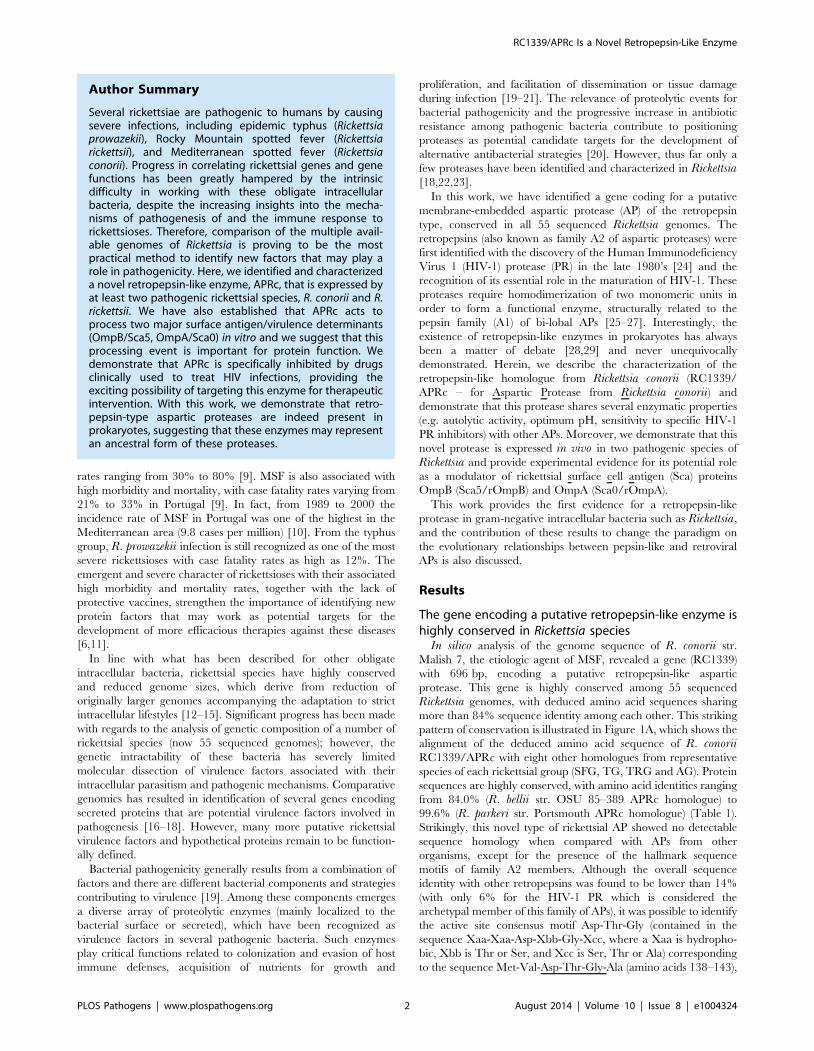
This higher optimal pH value is consistent with optimum pH
values reported for other retropepsins [37,38].
When investigating the susceptibility of APRc to classical
protease inhibitors (Figure 4B), this protease was shown to be
insensitive to pepstatin A, even though a slightly inhibitory effect
was observed during autolytic processing. In contrast, APRc
activity was strongly inhibited by EDTA, retaining only 25%
activity, and a small inhibitory effect was also observed with
Pefabloc. No significant effect was observed after incubation with
E-64, whereas incubation with Zn2+ (Figure 4B) slightly affected
enzyme activity.
In order to provide additional evidence that APRc is indeed a
retropepsin-like enzyme we analyzed the effects of different clinical
inhibitors of HIV-1 PR. Strikingly, incubation with indinavir
resulted in a near complete inhibition of APRc, even when tested
at a final concentration of 0.25 mM in the assay. Additionally,
nelfinavir, saquinavir, amprenavir and atazanavir also had a
significant inhibitory effect, ranging between approximately 30–
50% of inhibition (Figure 4C). This inhibitory effect of specific
HIV-1 protease inhibitors against a prokaryotic retropepsin-like
enzyme has not been previously described.
APRc displays a unique specificity pattern with aminoacid preferences resembling those of retropepsin- andpepsin-like PRs
To determine the APRc sequence specificity we employed
Proteomic Identification of Protease Cleavage Sites (PICS)
[39,40], a high-throughput profiling approach based on the use
of database-searchable proteome-derived oligopeptide libraries
representing the natural biological sequence diversity as test
substrates [39]. A unique advantage of PICS is the simultaneous
determination of sequence specificity on both sides of the scissile
bond, the prime-side (P9) and non-prime side (P) [41]. PICS
substrate peptide libraries are prepared by digestion of a complex
proteome from a sequenced model organism with highly specific
endoproteases such as trypsin (cleavage after Arg and Lys) or GluC
(cleavage after Glu and Asp), followed by blocking of primary
amines at peptide N termini and in Lys side chains. Cleavage of
library peptides by the test protease of interest generates C-
terminal cleavage products with free a- amines that are exploited
for selective enrichment and identification by LC-MS/MS. The
identified cleavage products constitute the prime side sequences
(P9) of the cleaved library peptides. Due to the non-random nature
of the PICS libraries the non-prime side sequences can be inferred
by database matching to allow reconstruction of the complete
cleavage sites.
In this work, active APRc was incubated with PICS libraries
generated by digestion of total human THP1 (human monocytic
leukemia cell line) cell proteins by either trypsin or GluC. These
PICS experiments resulted in the identification of 830 and 327 C-
terminal cleavage products from tryptic and GluC libraries,
respectively (Tables S1 and S2). The corresponding N-terminal
cleavage products and complete cleavage sites were obtained and
summarized using the WebPICS tool [40]. The PICS-based APRc
specificity profiles are shown in Figure 5 and a good agreement
was observed between the two complementary peptide libraries.
APRc displays only moderate specificity and accepts multiple
amino acids at each position. At P1, directly preceding the scissile
bond, APRc showed a preference for large hydrophobic residues
such as phenylalanine, tyrosine, methionine, leucine, and carbox-
yamidomethylated cysteine (modified during library preparation).
In addition, APRc also preferred the neutral amino acids
threonine and asparagine at this site. A similar preference was
observed for P19, although this further included small amino acids
alanine, serine, and glycine, as well as aspartate. Notably, cleavage
sites were almost devoid of Pro at P1 and P19. Furthermore, PICS
revealed distinct preferences for selected amino acids at other
positions, likely reflecting structural constraints imposed by the
substrate recognition and binding to the pocket site. In P2, APRc
preferences include valine, isoleucine, proline, and threonine,
whereas a predominant preference for small and branched
Figure 4. rAPRc activation product displays optimal activity atpH 6 and is strongly inhibited by specific HIV-1 PR inhibitors.The effect of pH, class-specific and HIV-1 PR specific inhibitors on theproteolytic activity of rAPRc activation product was evaluated using thesynthetic fluorogenic substrate (MCA)Lys-Ala-Leu-Ile-Pro-Ser-Tyr-Lys-Trp-Ser-Lys(DNP). (A) Activity at different pH values. Activated rAPRcwas incubated with the substrate at 37uC in buffers ranging betweenpH 4 and pH 9 containing 100 mM NaCl (50 mM sodium acetatepH 4.0, 5.0, 5.5 and 6.0 and 50 mM Tris-HCl pH 7.0, 8.0 and 9.0). (B) and(C) To test the effect different compounds, the protease was pre-incubated in the presence of each inhibitor for 10 minutes at roomtemperature in 50 mM sodium acetate pH 6.0 containing 100 mM NaClbefore adding the substrate. The rate of substrate hydrolysis (RFU/sec)was monitored for 3 hours and the relative activity normalized bysetting the maximum activity at 100%. The error bars representstandard deviation of the mean.doi:10.1371/journal.ppat.1004324.g004
RC1339/APRc Is a Novel Retropepsin-Like Enzyme
PLOS Pathogens | www.plospathogens.org 7 August 2014 | Volume 10 | Issue 8 | e1004324

aliphatic amino acids alanine, valine, and isoleucine was observed
at P29. More distant from the cleavage site, small preferences for
valine and isoleucine at P3 and for alanine and glycine in P39 and
a strong preference for leucine or isoleucine at P49 were observed.
Interestingly, basic and acidic residues were significantly under-
represented throughout. The large number of APRc cleavage sites
identified from the tryptic PICS library further allowed investiga-
tion of subsite cooperativity. When comparing two of the strongest
cleavage site determinants, phenylalanine at P1 and proline at P2,
we observed apparent mutual exclusion. Of the 103 unique
cleavage sites that contained proline in P2, only 4 had
phenylalanine in P1 (3.7% compared to 10.5% occurrence for
all identified cleavage sites), which was compensated by more
frequent occurrence of P1 methionine (10.3% compared to 5.8%
total occurrence) and P1 asparagine (14% compared to 8.3% total
occurrence). Correspondingly, peptides with phenylalanine in P1
yielded 4.6% P2 proline (compared with 12.9% total occurrence),
whereas peptides with methionine or asparagine in P1 yielded
22.9% or 21.7% P2 proline, respectively. A similar trend was
observed in identified cleavage sites from GluC libraries,
indicating subsite cooperativity between P2 and P1.
These results clearly show that, although displaying a unique
profile, APRc shares some specificity requirements with
retropepsins as well as with pepsin-like enzymes (particularly
BACE), further supporting APRc has being a member of the
aspartic protease family.
RC1339/APRc accumulates in the outer membrane in E.coli
As previously mentioned, full-length RC1339/APRc was
predicted to be membrane-embedded with an extracytoplasmic
orientation of the C-terminal domain. In order to provide
experimental validation of these theoretical observations we used
E. coli as our working model. An untagged construct in pET28a
comprising RC1339/APRc full-length coding sequence was
generated and protein expression carried out as described under
Experimental Procedures. Protease insertion into the membrane
was first assessed by subcellular fractionation studies followed by
Western blot analysis with a specific APRc antibody. A band of
approximately 21 kDa, whose nature was confirmed by peptide
competition assays, was detected in the total membrane fraction
and shown to accumulate in the outer membrane (Figure 6A). The
purity of the outer membrane (OM) fraction was confirmed by
Western blotting against E. coli Lep and OmpA proteins, as inner
and outer membrane markers [42], respectively, and compared to
the total membrane fraction (Figure 6B). As expected, OmpA was
Figure 5. rAPRc specificity profiling reveals similar amino acid preferences to both retropepsin and pepsin-like proteases. Graphicrepresentation of APRc specificity profile by Heatmaps and IceLogos. Results are from Tryptic and GluC peptide libraries derived from a Homo sapiensproteome (THP1 cells) incubated with activated rAPRc at a ratio of 1:50 (enzyme/library). The analytical strategy applied was similar to that describedin [40]. PICS libraries were analyzed by multiple sequence alignments and applying correction for natural amino acid abundance. For each class ofPICS library, the average amino acid occurrences in P4–P49 were calculated from three experiments and are either shown in the form of (A) a two-dimensional heatmap of log(2) transformed values of fold-enrichment over natural abundance of amino acids and (B) % difference IceLogos. Bothtryptic and GluC display consistency between them. In IceLogos representation, horizontal axis represents the amino acid position and vertical axisdenotes the over- and under-representation of amino acid occurrence compared with the Swiss-Prot Homo sapiens protein database. Cysteines arecarboxyamidomethylated and lysines are dimethylated.doi:10.1371/journal.ppat.1004324.g005
RC1339/APRc Is a Novel Retropepsin-Like Enzyme
PLOS Pathogens | www.plospathogens.org 8 August 2014 | Volume 10 | Issue 8 | e1004324

detected in the outer membrane fraction and the absence of cross-
contamination with inner membrane proteins was confirmed
through loss of signal for Lep, when compared with total
membrane fraction. Interestingly, APRc displayed a molecular
weight lower than expected (,21 kDa instead of the predicted
26.4 kDa), and parallel experiments with a C-terminal His-tagged
construct confirmed the presence of the tag in the membrane
fractions (data not shown), clearly suggesting that the protease may
be processed at the N terminus during translocation to the
membrane.
In an attempt to expand our knowledge about the membrane
topology of APRc, further studies were performed in order to
determine the overall in/out orientation of this protein relative to
the outer membrane of E. coli. To investigate this, PFA-fixed E.coli cells expressing untagged full-length APRc were subjected to
flow cytometry with both anti-APRc and anti-a-subunit of RNA
polymerase (mAb 4RA2) antibodies. The staining of E. coli cells
with the 4RA2 mAb was primarily used to restrict the analysis to
the non-permeable cells. As shown in Figure 6C, after gating out
all the cells that stained positive for RNAPaprotein (permeable
cells), bacterial surface staining with anti-APRc was observed,
confirming the integration of RC1339/APRc into the outer
membrane of E. coli and the orientation of the soluble catalytic
domain to the extracellular milieu.
APRc is expressed in Rickettsia conorii and Rickettsiarickettsii and is localized in the outer membrane
To determine whether RC1339/APRc and the R. rickettsiihomologue are expressed in the context of the intact bacterium,
we isolated total RNA from R. conorii and R. rickettsii grown in
Vero cells and performed reverse transcriptase PCR (RT-PCR).
As shown in Figure 7A, both R. conorii and R. rickettsii produce
transcripts for rc1339 and A1G_07330, respectively, when grown
in culture. To confirm expression of these transcripts, protein
lysates from each rickettsial species were separated by SDS-PAGE
and immunoblotting analyses were carried out with the specific
APRc antibody. As depicted in Figure 7B, a major reactive species
with an apparent molecular mass of 21 kDa was detected in R.rickettsii whole cell lysate and in the insoluble fraction of the R.conorii extract. These results clearly confirmed that rc1339 gene
and its R. rickettsii homologue are indeed translated in both
rickettsial species. Interestingly, and as previously observed in E.coli, a molecular weight of around 21 kDa was also detected for
APRc in rickettsial extracts. Although we cannot exclude
abnormal migration of the protease in the gel, the observed lower
molecular weight may also be correlated with APRc processing at
the N terminus, as anticipated by our results in E. coli.To provide additional insights on the localization of APRc in
these rickettsial species, fractionation studies were performed on
purified bacteria. Whole cell lysates as well as isolated inner and
outer membrane fractions were separated by SDS-PAGE and
analyzed by Western blots. For both species tested, our results
were consistent with localization of the protease at the outer
membrane, as confirmed by the immunodetection of rickettsial
OmpB, which was used as an internal marker for the outer
membrane in these assays (Figure 7C). We further confirmed the
presence of APRc on the surface of intact R. conorii by flow
cytometry analysis and also showed that the enzyme’s catalytic
domain is presented to the extracellular milieu (Figure 7D).
Together, these results further illustrate that a novel retropepsin-
like enzyme is expressed in two pathogenic rickettsial species and
that the APRc catalytic domain is oriented towards the
extracellular environment when present at the outer membrane
of these bacteria.
Recombinant R. conorii APRc is sufficient to mediatecleavage of OmpB
The evidence that a proportion of APRc is associated with the
outer membrane led us to hypothesize that rickettsial surface
proteins might be potential substrates for this newly characterized
Figure 6. Recombinant full-length APRc accumulates in the outer membrane in E. coli and the soluble catalytic domain is exposed tothe cell surface. (A) Full-length APRc was expressed in E. coli and total (TM) as well as outer membrane (OM) fractions were isolated and analyzed byWestern blot with anti-APRc antibody (left panel). As a control for non-specific staining, peptide competition assays were performed by blocking theanti-APRc antibody with the immunizing peptide (right panel). One specific band with approximately 21 kDa was detected in the OM fraction. (B) Thepurity of OM fractions was confirmed by using OmpA and Lep proteins as internal markers for the outer and inner membranes of E. coli, respectively.Both proteins were present in TM faction, while only OmpA is detected in OM faction. (C) Flow cytometric analysis was carried out for recombinantAPRc recognition at the surface of E. coli cells. PFA-fixed E. coli cells were incubated with anti-APRc and anti-RNA polymerase a (RNAPa mAb 4RA2,)followed by secondary detection using goat anti-rabbit IgG Alexa Fluor 488- and goat anti-mouse IgG R-PE-Cy5.5 conjugated secondary antibodies,respectively. Porous, permeabilized cells staining positive for RNAPa were excluded from the analysis by selective gating of this population.Fluorescence was detected on the remaining E. coli cells incubated with anti-APRc, thereby confirming the expression of recombinant APRc at theouter membrane and its exposure to extracellular milieu.doi:10.1371/journal.ppat.1004324.g006
RC1339/APRc Is a Novel Retropepsin-Like Enzyme
PLOS Pathogens | www.plospathogens.org 9 August 2014 | Volume 10 | Issue 8 | e1004324

enzyme. As has been shown for other autotransporter proteins,
rickettsial OmpB, OmpA, Sca1, and Sca2 are involved in
mediating important interactions with mammalian cells and
undergo processing events at the outer membrane [43–49]. As
an example, R. conorii OmpB (rOmpB) is expressed as a
preprotein of 168 kDa and is subsequently cleaved to release the
passenger domain (120 kDa) from the b-barrel translocation
domain (32 kDa) [46]. Interestingly, R. conorii and R. japonicaOmpB do not undergo proteolytic cleavage when expressed at the
outer membrane of E. coli, suggesting that the processing event is
not autocatalytic [43]. However, the identity of the enzyme
responsible for Sca protein maturation still remains elusive.
Therefore, and based on the observed APRc outer membrane
localization, we sought to determine whether APRc might
participate in the processing of rOmpB (Figure 8A). In order to
do this, we performed transactivation assays using E. coli outer
membrane fractions enriched in recombinant rOmpB (C-termi-
nally His-tagged) and purified active APRc (soluble catalytic
domain). Reaction products were then separated by SDS-PAGE
and analyzed by Western blot. As shown in Figure 8B, the
detection of an anti-His immune reactive product with ,35 kDa
in the presence of APRc was correlated with the disappearance of
rOmpB preprotein, suggesting that this enzyme may be indeed
capable of promoting cleavage of recombinant rOmpB. Moreover,
the generated reactive protein product has approximately the
same molecular weight as that expected for rOmpB b-barrel
(32 kDa), further suggesting that this proteolytic cleavage may
likely be occurring somewhere between the passenger and the b-
barrel domains, in agreement with what has been described for
native rOmpB [46]. To further validate these results, parallel
assays were performed in the presence of APRc active site
mutant and the integrity of rOmpB proprotein evaluated by
Figure 7. RC1339/APRc is expressed in Rickettsia conorii and Rickettsia rickettsii and accumulates at the outer membrane in bothspecies. (A) RT-PCR analysis of RC1339/APRc expression on rickettsial spp.. The housekeeping gene hrtA (17 kDa surface antigen) was used as acontrol. The negative control for the cDNA synthesis lacking reverse transcriptase is identified by (RTase -). Rickettsial species are identified on the topand the gene names are shown on the left side of the agarose gel. (B) A whole cell lysate from R. rickettsii (1) and insoluble (2) and soluble (3) fractionsfrom R. conorii extracts were isolated and then subjected to Western Blot analysis with anti-APRc antibody. A specific band with approximately21 kDa was detected. (C) Whole cell lysates (WCL), inner (IM) and outer membrane (OM) fractions from sarkosyl treatment of R. rickettsii and R. conoriiextracts were isolated and then subjected to Western Blot analysis with anti-APRc and anti-rOmpB antibody. APRc shares the same localization ofrOmpB, an internal marker for outer membrane of Rickettsia spp. Molecular mass markers in kilodaltons (kDa) are shown on the left. (D) For flowcytometric analysis of APRc expression in R. conorii, fixed bacteria were queried for deposition of anti-APRc (orange trace), negative control lackingprimay antibody (blue trace), or the positive control anti-OmpB (red trace), a known rickettsial surface protein. After incubation with fluorescentsecondary antibody, both anti-APRc and anti-OmpB detected on the bacterial surface (increased fluorescence), indicating accessibility of these targetproteins to exogenously applied antibody.doi:10.1371/journal.ppat.1004324.g007
RC1339/APRc Is a Novel Retropepsin-Like Enzyme
PLOS Pathogens | www.plospathogens.org 10 August 2014 | Volume 10 | Issue 8 | e1004324

immunoblotting with a specific antibody to this outer membrane
protein. As expected, the disappearance of rOmpB proprotein was
observed in the presence of active APRc but not when the cell
extract was incubated with the active site mutant protein
(APRc(D140A)99–231). Interestingly, we observed a similar phe-
nomenon using as a substrate another conserved rickettsial
antigen, Sca0/OmpA, demonstrating that a protein other than
OmpB can be processed by APRc in vitro (Figure 9). Altogether,
these results suggest that APRc is sufficient to mediate rOmpB
maturation and rOmpA maturation in vitro, thereby raising an
exciting hypothesis regarding possible functional significance of
APRc as being able to process these and possibly other
autotransporter proteins in the context of intact R. conorii cells.
Discussion
The intrinsic difficulty in working with obligate intracellular
parasites such as rickettsiae greatly hampers the correlation of
rickettsial gene products with their function. Therefore, valuable
information on the nature of conserved genes as well as on the
identification of new bacterial factors that may play a role in
rickettsiae pathogenesis is mostly being provided by comparative
genomics. Using this approach, we identified a gene encoding a
putative membrane embedded aspartic protease with a retroviral-
type signature, highly conserved in 55 Rickettsia genomes. Using
the R. conorii gene homologue RC1339 as our working model we
demonstrate that the gene product (APRc) displays a high degree
of identity among Rickettsia spp., although no significant
homology is observed when compared to other aspartic proteases,
except for the conservation of the motif around the catalytic
aspartate as well as the hydrophobic-hydrophobic-glycine motif
required for the formation of the psi loop. These features resemble
the retroviral APs comprising family A2, which are characterized
by being active only as symmetric dimers with a single active site,
where each monomer contributes one aspartate [25,27]. Despite
the observed low overall sequence similarity with retropepsins, our
results on the enzymatic characterization of the soluble catalytic
domain of RC1339/APRc further revealed that this novel
rickettsial enzyme indeed shares several properties with this family
of APs. The common properties include autolytic activity impaired
by mutation of the catalytic aspartate, accumulation in the dimeric
form, optimal activity at pH 6, inhibition by specific HIV-1 PR
inhibitors, and specificity preferences resembling those of both AP
families. The presence of retroviral-type APs in bacteria has been
previously demonstrated (SpoIIGA in Bacillus subtilis [33] and
PerP in Caulobacter crescentus [50]). However, no enzymatic
characterization is yet available for these enzymes and their
inclusion as retropepsin-type protease members has not been
universally accepted [29]. Therefore, the results described here
Figure 8. APRc can process rOmpB in vitro. (A) rOmpB is proteolytically processed between the passenger and b-barrel domains through a yetunknown mechanism (?) and APRc was tested as the candidate enzyme to perform rOmpB preprotein processing in vitro. (B) Total membranefractions of E. coli enriched in rOmpB were incubated with activated APRc soluble domain and the reaction products analyzed by Western blot withan anti-Histidine antibody. The integrity of rOmpB proprotein was confirmed in the absence of APRc whereas in the presence of the protease aproduct with approximately 35 kDa was observed, correlated with the disappearance of the full-length unprocessed form. (C) The integrity ofrecombinant rOmpB was further evaluated upon incubation with both activated APRc and the active site mutant form (D140A) for 16 h. The reactionproducts were then subjected to immunoblot analysis with anti-rOmpB MAb, confirming the disappearance of rOmpB in the presence of the activeform of the enzyme. Molecular weight markers in kilodaltons (kDa) are shown on the left. Protein loading controls: Coomassie blue staining.doi:10.1371/journal.ppat.1004324.g008
Figure 9. APRc can process rOmpA in vitro. Total membranefractions of E. coli enriched in rOmpA were incubated with bothactivated APRc and the active site mutant form (D140A) for 16 h. Thereaction products were then subjected to immunoblot analysis withanti-rOmpA Ab, confirming the disappearance of rOmpA in thepresence of the active form of the enzyme. Molecular weight markersin kilodaltons (kDa) are shown on the left. Protein loading controls:Coomassie blue staining.doi:10.1371/journal.ppat.1004324.g009
RC1339/APRc Is a Novel Retropepsin-Like Enzyme
PLOS Pathogens | www.plospathogens.org 11 August 2014 | Volume 10 | Issue 8 | e1004324

provide experimental substantiation that RC1339/APRc is a
novel retropepsin-like enzyme expressed in bacteria.
Most viral retropepsins are strictly required for the processing of
Gag and Gag-Pol polyproteins into mature structural and
functional proteins (including themselves) and are, therefore,
indispensable for viral maturation [51]. Because of this, retro-
pepsin-type APs are generally characterized by their inherent
autolytic function. Interestingly, our results with APRc soluble
catalytic domain fused to GST also demonstrated the ability of this
protein to undergo a multi-step autocatalytic conversion in vitrointo APRc105–231 mature form, and this autolytic activity was
again confirmed when the last intermediate of activation was
produced in E. coli. As expected for a retropepsin-like enzyme,
mutation of the catalytic aspartate impaired this process. The
enzymatic activity assays performed during these autoactivation
studies (either using oxidized insulin B chain or the fluorogenic
peptide mimicking the final cleavage site between the Ser104-
Tyr105 residues) clearly indicated that APRc activity appears to be
dependent on the presence of the final activation product. These
results suggest that the processing at the N terminus must be the
determining step for the regulation of enzymatic activity,
presumably through a conformational change occurring upon
conversion from rAPRc99–231 to APRc105–231 form. This is in line
with what has been already described for recombinant HIV-1 PR,
where the increase in catalytic activity upon protease autolytic
conversion has been correlated with a conformational rearrange-
ment between the precursor/inactive vs. mature/active forms of
the enzyme [51,52]. However, further studies are required to
better understand the maturation of APRc precursor forms in vitroand how this is accomplished and controlled in vivo. In fact, we
have shown that APRc accumulates in the outer membrane in R.conorii and R. rickettsii and, therefore, we cannot rule out that the
presence of the transmembrane domain may play an important
role in this maturation process in vivo.
Another interesting observation was that APRc autolytic
activity, as well as cleavage of the fluorogenic substrate, occurred
at a pH optimum of 6.0. This is again in good agreement with the
optimal pH of other retropepsin-like [37,38] enzymes as well as of
the pepsin-like renin [53,54] and, actually, it is consistent with the
presence in APRc of an alanine residue downstream from the
catalytic motif (Asp-Thr-Gly-Ala), instead of the common threo-
nine residue found in most pepsin-like APs [27]. Together with the
observed inhibitory effect of specific HIV-1 PR inhibitors, these
results strengthen the striking resemblance between the enzymatic
properties of APRc and those of viral retropepsins. Unexpectedly,
we observed a drastic inhibitory effect of EDTA on both APRc
maturation and hydrolysis of the fluorogenic substrate, suggesting
that this protease may depend on a metal ion for folding and/or
activity. A similar effect has not been reported for other
retropepsins and no homology to a metalloprotease consensus
motif was identified in APRc that could justify this inhibition.
Therefore, further structural studies will be required to help in
understanding this effect.
To provide additional evidence on the nature of APRc as a
retropepsin-like AP we determined both the prime and nonprime
side specificity using PICS [39]. Although HIV-1 PR is the only
AP for which a PICS analysis has been reported [40], there are
several studies for many different APs on specificity towards
individual substrates (compiled, at least partially, by MEROPS
[55]) providing a collection of cleavage patterns for these enzymes.
A comparison of the substrate specificity of APs with our PICS
results confirmed common preferences between APRc and both
retropepsin and pepsin-type APs. The amino acid preference of
APRc for P1 position is in good agreement with the canonical
specificity of APs for large hydrophobic amino acids, such as
phenylalanine, methionine, carboxyamidomethylated cysteine
(which results from the modification during generation of peptide
libraries), or leucine. Despite the observed lower selectivity, a
similar trend for accommodating hydrophobic amino acids is also
observed in P19. As observed in both tryptic and GluC libraries,
APRc appears to display broader specificity for P1 and P19, while
a more constrained amino acid preference is observed for P3, P2,
and P29 positions. This observation may account for an important
role on substrate recognition and binding to the active pocket site
and may ultimately influence hydrolytic efficiency. Strikingly, a
high degree of similarity is found with more specialized pepsin-like
proteases such as BACE for P3 (with a preference for valine and
isoleucine) and P29 (alanine and valine) positions, as well as with
cathepsin D (also for P29). Interestingly, APRc also displays unique
amino acid preferences such as proline at P2 (although the
preference observed for valine and threonine in this position has
also been described for feline immunodeficiency virus retropepsin
[55,56]), and leucine and isoleucine in P49 position. When
compared with the two major types of cleavage sites proposed
for HIV-1 PR and other retropepsins, APRc specificity profile
suggests a preference for type 2-like substrates with hydrophobic
amino acids in P1 and P19, whereas type 1-like substrates with the
typical combination of tyrosine(phenylalanine)-proline at P1-P19
appear disfavored [51,57]. Moreover, our results suggest a
cooperative effect between P2 and P1 positions by revealing that
a P2 proline co-occurs more frequently with P1 methionine or
asparagine residues and that proline is not favored at this position
when P1 is occupied by phenylalanine. Curiously, APRc autolytic
cleavage sites do not perfectly match the observed specificity
preferences of the activated form used in PICS, suggesting either a
different conformational arrangement of the protease or a
dependence on the sequence context and/or conformation of
the substrate. This is not totally unexpected, as for HIV-1 PR it
has also been reported that specificity towards nonviral protein
substrates significantly differed from viral polyprotein cleavage
sites [51].
Aspartic proteases were assumed for a long time to be restricted
to viruses and eukaryotes. However, more recently proteins
bearing the characteristic hallmark features of the pepsin family
have been identified in seven bacterial genomes [29] and the
detailed biochemical characterization of the pepsin-like homo-
logue from the Shewanella amazonensis, shewasin A [58], has
clearly demonstrated that this bacterial AP is strongly reminiscent
of its eukaryotic counterparts. These observations have raised a
discussion on the evolutionary relationships between bacterial and
eukaryotic pepsin-like APs, by suggesting that bi-lobal pepsin-like
proteases may have evolved from primordial homodimeric
aspartic proteases before divergence between eukaryotes and
prokaryotes (through the proposed gene duplication and fusion
event [59]). Our current results on RC1339/APRc further support
this hypothesis by providing the first experimental evidence that a
gene for a single-lobed AP is indeed present in prokaryotes, coding
for an active enzyme with properties resembling those of
retropepsins. Moreover, these results offer additional clues on
the relationships between retropepsin-like and pepsin-like APs.
The presence of single-lobed AP genes in prokaryotes suggests that
enzymes such as APRc may actually represent the most ancestral
forms of these proteases, whereas retroviral retropepsins may
instead correspond to a derived state.
Besides demonstrating that RC1339 encodes an active enzyme,
we have also shown that this rickettsial protease is expressed
in both R. conorii and R. rickettsii. Unlike the large majority
of a-helical type of integral membrane proteins, sub-cellular
RC1339/APRc Is a Novel Retropepsin-Like Enzyme
PLOS Pathogens | www.plospathogens.org 12 August 2014 | Volume 10 | Issue 8 | e1004324

localization studies revealed an outer membrane accumulation for
APRc which was also confirmed by expression of the full-length
protease in E. coli and in the context of intact R. conorii. So far,
only three transmembrane proteins with a-helical architecture
have been reported to be embedded in the outer membrane of
gram-negative bacteria [60–62]. Therefore, our results provide
additional evidence that the bacterial surface is not restricted to
proteins with b-barrel structures known to play essential roles in
energetics, metabolism, signal transduction, and transport [63,64],
further suggesting that the repertoire of proteins with a-helices
localized to the OM may be higher than anticipated. Nevertheless,
transport and insertion of APRc into the OM definitely requires
further studies in order to clarify whether the detected 21 kDa
band is an intermediate processed form or the result of different
gel mobility.
In line with our evidence for the native expression of APRc in
R. conorii and R. rickettsii, a multiomics study performed in
Rickettsia prowazekii to identify potential virulence factors has also
confirmed transcription of RC1339 gene homologue (RP867)
[17]. Importantly, these studies also showed differential regulation
of RP867 expression in different strains of R. prowazekii with a
fold change of 1.77 between the virulent strain Rp22 and the
avirulent strain Erus. This evidence for an up-regulation of APRc’s
gene expression in R. prowazekii Rp22, combined with our results
confirming protease expression and accumulation into the OM in
R. conorii and R. rickettsii, strongly support a relevant role of this
highly conserved protease in rickettsial life cycle. Serine-, cysteine-,
and metalloproteases are widely spread in many pathogenic
bacteria, where they play critical functions related to pathogenesis
and virulence [19,20]. However, much less is known about the role
of aspartic proteases since the presence of this class of enzymes in
pathogenic bacteria has not been previously reported. Taking
under consideration the unique biochemical and enzymatic
features of APRc presented in this work: i) the apparent non-
stringent sequence requirement; ii) outer membrane localization
and extracellular orientation of recombinant APRc catalytic
domain and iii) autolytic activity suggesting that the soluble
biological unit may be released from the surface of rickettsial cells
by an ectodomain shedding-like process, we anticipate a potential
multi-functional role for this rickettsial protease. One of the
proposed functions concerns APRc contribution for the degrada-
tion of host tissues for supplying bacteria with nutrients, similar to
that described for other extracellular proteases secreted by many
pathogens [19]. Second, this protease may also support the spread
of the infection and dissemination of bacteria into deeper tissue
through the shedding of cell surface adhesion molecules or the
inactivation of the components of the host immune system such as
proteins from the complement system [21,65,66]. And third,
APRc may participate in the degradation and/or maturation of
other rickettsial proteins, in particular those located at the OM,
such as Sca proteins [45,67], exemplified by Sca5/rOmpB and
Sca0/OmpA. In contrast to other autotransporter proteins from
gram-negative bacteria with auto-proteolytic activity such as
SPATEs (Serine Protease Autotransporters) [68], rOmpB process-
ing is thought to implicate a protease as previous expression studies
in E. coli have failed to demonstrate autocatalytic activity [43,47].
In this work, we have started addressing this last hypothesis and we
showed that APRc is indeed sufficient to catalyze the processing of
Sca0/OmpA and Sca5/rOmpB in vitro and that, for the latter,
the generated product is consistent with the cleavage between the
passenger and the b-peptide regions. The N-terminal sequence of
the b-peptide has been experimentally determined for R. typhi and
R. prowazekii [46] rOmpB and the region spanning the cleavage
site (/) corresponds to the sequence Ala-Ala-Val-Ala-Ala/
Gly-Asp-Glu-Ala-Val. Although we cannot exclude that in R.conorii the cleavage of rOmpB may occur slightly upstream from
this region, if considering a similar cleavage site the amino acids
present in P4, P3, P1, P19 and P49 are in good agreement with the
observed specificity preferences for APRc, while the differences
observed for the remaining positions may reflect again the
importance of sequence context/substrate conformation for APRc
cleavage. Nevertheless, additional experiments are required to
determine the cleavage site and its relevance in the context of
intact rickettsiae as well as APRc role in the degradation of other
rickettsial and/or host proteins.
In summary, the findings described herein show that this newly
characterized aspartic protease from Rickettsia is an active enzyme
with features highly reminiscent of retropepsin-type proteases and
we anticipate its participation in a relevant proteolytic pathway in
rickettsial life-cycle, likely as a modulator of activity of other
rickettsial membrane-localized proteins. Determination of APRc
three-dimensional structure and dissection of its contribution to
rickettsial pathogenesis will be critical to start unveiling the
significance of this novel protease as a potential target for
therapeutic intervention.
With this work we expect to contribute to start changing the
currently accepted evolutionary paradigm of aspartic proteases, by
positioning what we denominate as ‘‘prokaryopepsins’’ as the new
archetypes of modern APs.
Materials and Methods
MaterialsOligonucleotide primers were purchased from Integrated DNA
Technologies, Leuven, Belgium. Synthetic genes encoding the full-
length RC1339 and the predicted soluble catalytic domain, the
fluorogenic peptide PepRick14 (MCA-Lys-Ala-Leu-Ile-Pro-Ser-
Tyr-Lys-Trp-Ser-Lys-DNP) and the rabbit polyclonal antibody
raised towards the sequence Cys-Tyr-Thr-Arg-Thr-Tyr-Leu-Thr-
Ala-Asn-Gly-Glu-Asn-Lys-Ala (anti-APRc) were produced by
GenScript (Piscataway, NJ, USA). N-terminal amino acid
sequence analyses were performed in the Analytical Services Unit
- Protein Sequencing Service, ITQB (Oeiras, Portugal). Rabbit
polyclonal antibody against the purified APRc99–231His construct
(see below) was generated by standard immunization schemes
approved by the LSU School of Veterinary Medicine Institutional
Animal Care and Use Committee (IACUC).
Bioinformatics analysisGene and protein sequences for R. conorii str. Malish 7
RC1339 were obtained from the genome sequence at NCBI
(NC_003103) (AAL03877). Amino acid sequence alignment and
the degree of identity between RC1339/APRc homologues from
Rickettsia (genus) (TaxID 780) were obtained with ClustalW [69],
by comparing the sequences deposited in NCBI database with the
following accession numbers: NP_360976 (R. conorii str. Malish
7), YP_005393543 (R. parkeri str. Portsmouth), YP_001495413
(R. rickettsii str. Sheila Smith), YP_005364747 (R. amblyomii str.
GAT-30V), YP_005391701 (R. montanensis str. OSU 85–930),
NP_221215 (R. prowazekii str. Madrid E), YP_067793 (R. typhistr. Wilmington), YP_247382 (R. felis URRWXCal2) and
YP_001495500 (R. bellii OSU 85–389). The protein family,
domain, and functional sites were searched using the InterProScan
program [70]. Topology structure was predicted with
HMMTOP2 algorithm [30]. A structure-based alignment of
RC1339/APRc soluble catalytic domain with HIV-1 (PDB 3hvp),
EIAV (PDB 2fmb), and XMRV (PDB 3nr6) retropepsins and with
RC1339/APRc Is a Novel Retropepsin-Like Enzyme
PLOS Pathogens | www.plospathogens.org 13 August 2014 | Volume 10 | Issue 8 | e1004324

DdI1 putative protease domain (PDB 2i1a) was performed with
PROMALS3D [71].
DNA constructsThe sequences encoding the full-length and the predicted
soluble domain of RC1339/APRc were chemically synthetized
with OptimumGene codon optimization technology for E. colicodon usage and cloned into pUC57 vector. The gene encoding
the full-length APRc (construct coding amino acids 1–231) was
then amplified to include restriction sites for NcoI and NotI at 59-
and 39-ends, respectively, using the forward primer 59-CCATGG-
GAATGAACAAAAAACTGATCAAACTG-39 and the reverse
primer 59-CTCGAGATAATTCAGAATCAGCAGATCTTT-
39; the resulting PCR product was cloned into pGEM-T
Easy plasmid (Promega). After digestion with NcoI and NotI,
APRc1–231 insert was subcloned into pET28a expression vector
(Invitrogen) in frame with a C-terminal His-tag (pET-APRc1–
231His). In order to generate the untagged construct, an insertion
mutagenesis was performed to include the TGA stop codon at the
end of the full-length sequence using the Quick Change site-
directed mutagenesis kit (Stratagene) and the primers 59-
ATTCTGAATTATTGACTCGAGCACCAC-39 (forward) and
59-GTGG-
TGCTCGAGTCAATAATTCAGAAT-39 (reverse) (pET-AP-
Rc1–231).
The optimized sequence encoding the predicted soluble
catalytic domain of APRc (construct coding amino acids 87–231)
flanked by restriction sites for BamHI (59)/EcoRI (39) was inserted
in frame to the C terminus of GST in pGEX-4T2 expression
vector (Amersham) using the same pair of restriction enzymes
(pGST-APRc87–231).
For generating the expression construct bearing the sequence
encoding the intermediate activation form APRc99–231 (construct
coding amino acids 99–231), the sequence was firstly amplified
using the construct pETAPRc1–231 as the template and the
forward primer containing a NdeI restriction site (59-CATATG-
TATAAATGGAGTACCGAAGTT-39) and the same reverse
primer used for amplification of APRc1–231 (59-CTCGAGA-
TAATTCAGAATCAGCAGATCTTT-39), and cloned into
pGEM-T Easy (Promega). The insert was then digested with
NdeI/NotI and subcloned into pET23a expression vector
(Invitrogen) in frame with a C-terminal His-tag (pETAPRc99–231
His).
The active site mutant of APRc (both in pGEX4T2 and pET23a
constructs) was generated by replacing the putative active site
aspartic acid residue by alanine (D140A) using the Quick Change
site-directed mutagenesis kit (Stratagene) and the primers 59-
AAAATCAAATTCATGGTGAATACCGGCGCCTCTGATA-
TTGCA-39 (forward) and 59-TGCAATATCAGAGGCGCCGG-
TATTCACCATGAATTTGATTTT-39 (reverse) (mutation un-
derlined). All positive clones were selected by restriction analysis and
confirmed by DNA sequencing.
The construct for expression of rOmpB in E. coli was generated
as described elsewhere [43].
Expression and purification of the soluble forms of APRcrGST-APRc87–231 and the corresponding active site mutant
protein were expressed by standard procedures. Briefly, E. coliBL21 Star (DE3) cells transformed with each plasmid construct,
pGST-APRc87–231 and pGST-APRc87–231(D140A), were grown at
37uC until an OD600 nm of 0.7. Protein expression was then
induced with 0.1 mM IPTG for 3 hours, after which cells were
harvested by centrifugation at 9000 g for 20 minutes at 4uC,
and resuspended in PBS buffer. Lysozyme (100 mg/ml) was added
and the harvested cells were frozen at 220uC. After freezing and
thawing, bacterial cell lysates were incubated with DNase (1 mg/
ml) and MgCl2 (5 mM) for 1 hour at 4uC. The total cell lysate was
then centrifuged at 27216 g for 20 minutes at 4uC and the
resulting supernatant filtered (0.2 mm) before loading onto a
GSTrap HP 5 ml column (GE Healthcare Life Sciences)
previously equilibrated in PBS buffer. After extensive washing,
the protein of interest was eluted in 50 mM Tris-HCl pH 8 with
10 mM glutathione and immediately loaded onto a Superdex 200
HiLoad 26/60 (GE Healthcare Life Sciences) equilibrated in PBS
buffer for further purification and glutathione removal.
Expression of E. coli BL21 Star (DE3) cells transformed with
pETAPRc99–231His plasmid as well as isolation of total soluble
protein were performed under the same conditions as described
for rGST-APRc87–231, except that in this case the cell pellet was
resupended in 20 mM phosphate buffer pH 7.5, 500 mM NaCl
and 10 mM imidazole. The resultant supernatant was then loaded
onto a Histrap HP 5 ml column (GE Healthcare Life Sciences)
pre-equilibrated in the same buffer. Protein elution was performed
by a three-step gradient of imidazole (50 mM, 100 mM and
500 mM) and fractions containing the protein of interest (100 mM
imidazole gradient step) were pooled and buffer exchanged into
20 mM phosphate buffer pH 7.5 by an overnight dialysis step.
Dialyzed protein was further purified by cation-exchange chro-
matography with a MonoS column (GE Healthcare Life Sciences)
equilibrated in the same buffer and elution was carried out by a
linear gradient of NaCl (0–1 M).
Auto-processing studiesTime-course studies of APRc activation were undertaken with
two recombinant forms of the soluble catalytic domain of APRc
(rGST-APRc87–231 and rAPRc99–231). Purified samples of rAPRc
were first diluted to 0.1 mg/mL with PBS and then diluted 1:1
with 0.1 M sodium acetate buffer pH 6. Diluted samples were
incubated up to 48 h at 37uC and aliquots were taken every 12 h
for SDS-PAGE analysis and proteolytic activity assays. To
evaluate the effect of inhibitors on rAPRc auto-activation
processing, a time-course analysis was carried out in the presence
of 20 mM pepstatin, 1 mM indinavir or 5 mM EDTA and protein
samples were analyzed by SDS-PAGE.
Dimerization assaysCrosslinking reactions with disuccinimidyl suberate (DSS)
(Pierce) were performed in 20 mM phosphate buffer pH 7.5
containing 150 mM NaCl. A solution of 0.2 mg/ml of purified
rAPRc (rAPRc99–231 and activated product rAPRc105–231) was
treated with a 50-fold molar excess of DSS in a total volume of
50 ml and allowed to react for 30 min at room temperature. For
glutaraldehyde treatment, a solution of 0.5 mg/ml of purified
rAPRc99–231 was treated with 5 ml of 1.15% freshly prepared
solution of glutaraldehyde for 4 minutes at 37uC, in a total volume
of 50 ml, under similar buffer conditions. To terminate the
reactions, 5 ml of the quenching buffer 1 M Tris-HCl pH 8.0
were added. Crosslinked proteins were separated by SDS-PAGE
and analyzed by Western blot with anti-APRc antibody.
Analytical size-exclusion chromatographyPrecursor rAPRc99–231 and activated rAPRc110–231 forms were
analyzed under nondenaturing conditions by analytical size-
exclusion chromatography on a Superdex 200 5/150 GL (GE
Healthcare Life Sciences) column connected to a Prominence
HPLC system (Shimadzu Corporation, Tokyo, Japan). The
column was equilibrated in 20 mM phosphate buffer pH 7.5
containing 150 mM NaCl, and calibrated with Gel Filtration
RC1339/APRc Is a Novel Retropepsin-Like Enzyme
PLOS Pathogens | www.plospathogens.org 14 August 2014 | Volume 10 | Issue 8 | e1004324

LMW and HMW calibration kits (GE Healthcare Life Sciences),
according to the manufacturer’s instructions. The molecular mass
markers used for calibration were conalbumin (75 kDa), ovalbu-
min (43 kDa), carbonic anhydrase (29 kDa), and ribonuclease A
(13.7 kDa).
Enzyme activity assaysThe effect of pH on activity and inhibitory profile of purified
recombinant rAPRc (rAPRc105-231) was determined by fluores-
cence assays in 96-well plates in in a Gemini EM Fluorescence
Microplate Reader, using the fluorogenic substrate PepRick14
(MCA-Lys-Ala-Leu-Ile-Pro-Ser-Tyr-Lys-Trp-Ser-Lys-DNP) (final
concentration of 2.5 mM). For determination of the pH profile,
rAPRc105-231 was assayed for activity at 37uC in buffers ranging
between pH 4 and 9 (50 mM sodium acetate pH 4.0, 5.0, 5.5 and
6.0; 50 mM Tris-HCl pH 7.0, 8.0 and 9.0) containing 100 mM
NaCl. To test the effect of classical inhibitors, the protease was
pre-incubated in the presence of each inhibitor, 20 mM pepstatin,
5 mM EDTA, 1 mM ZnCl2, 1 mM Pefabloc, or 10 mM E-64, for
10 minutes at room temperature in 50 mM sodium acetate
pH 6.0 containing 100 mM NaCl before determination of
proteolytic activity. The effect of the HIV inhibitors on rAPRc
proteolytic activity was also evaluated. The following reagents
were obtained through the NIH AIDS Research and Reference
Reagent Program, Division of AIDS, NIAID, NIH: indinavir
sulfate, nelfinavir, ritonavir, saquinavir, amprenavir, atazanavir
sulfate, sarunavir, and lopinavir. Each inhibitor was again
incubated with rAPRc for 10 minutes at room temperature in
50 mM sodium acetate pH 6.0 containing 100 mM NaCl and 5%
DMSO, except for indinavir and darunavir that were assayed
without DMSO. Indinavir, nelfinavir, ritonavir, saquinavir,
amprenavir, atazanavir and lopinavir were tested in the range of
0.25 mM–1 mM and the inhibitor darunavir in the range of
2.5 mM–10 mM (final concentration). The rate of substrate
hydrolysis was monitored for 3 hours by the increase in
fluorescence intensity with excitation/emission wavelengths of
328/393 nm and the relative activity normalized by setting rAPRc
activity as 100%.
Evaluation of proteolytic activity during auto-processing time-
course analysis was performed towards oxidized insulin b chain.
Substrate (1 mg/ml) was incubated with purified recombinant
APRc enzyme: substrate mass ratio of 1:15) in 0.1 mM sodium
acetate buffer pH 6.0. After an overnight incubation at 37uC the
reaction mixture was centrifuged at 20000 g during 6 min and the
digestion fragments were separated by RP-HPLC on a C18
column (KROMASIL 100 C18 250, 4.6 mm), using a Prominence
system (Shimadzu Corporation, Tokyo, Japan). Elution was
carried out with a linear gradient (0–80%) of acetonitrile in
0.1% v/v trifluoroacetic acid for 30 min at a flow rate of 1 ml/
min. Absorbance was monitored at 220 nm.
PICSrAPRc (APRc105-231) specificity profiling was determined
according to the Proteomic Identification of Protease Cleavage
Sites (PICS) methodology as described elsewhere [40], with minor
changes. Tryptic and GluC peptide libraries were generated from
THP1 cells. rAPRc-to-library ratios were 1:50 (wt/wt) and
incubation was performed at 37uC for 16 h in 50 mM sodium
acetate buffer pH 6.0, 150 mM NaCl. The separation of carboxy-
peptide cleavage products was carried out on a column packed
with PepMap C18 resin (Dionex) connected to a LC-MS/MS
QSTAR Pulsar (AB SCIEX) operated by the UBC Michael Smith
Laboratory/Laboratory for Molecular Biophysics Proteomics
Core Facility, and on HALO C18 column (Eksigent) connected
to a LC-MS/MS TripleTOF 5600, AB SCIEX (Center for
Neuroscience and Cell Biology Proteomics Unit). Peptide
sequences were identified with both Mascot [72] and X!Tandem
[73], in conjunction with PeptideProphet [74] at a confidence level
.95%. Mass tolerance was 10 ppm for parent ions and 0.6 Da for
fragment ions. Search parameters were set to identify static
modifications as carboxyamidomethylation of cysteine residues
(+57.02 Da), dimethylation of lysines (+28.03 Da) and thioacyla-
tion of peptide amino termini (+88.00 Da). Semi-style cleavage
searches were applied with no constraints for the orientation of the
specific terminus. The Web-based PICS service [40] was used to
derive nonprime sequences. Sequence logos were generated with
IceLogo with a p-value of 5% [75].
RT-PCR analysisFor cDNA synthesis, total RNA was isolated from an aliquot of
frozen R. conorii Malish 7 and R. rickettsii ‘‘Sheila Smith’’-
infected Vero cells using the SurePrep TrueTotal RNA Purifica-
tion Kit (Fisher Scientific), according to the manufacturer’s
instructions. After extraction, RNA samples were treated with
DNase I, RNase-free set (Thermo Scientific) for 30 minutes at
37uC. The reaction was inactivated by adding 50 mM EDTA and
heating the mixture at 65uC for 10 minutes. Next, approximately
1 ug of total RNA was used as the template for reverse
transcription using the iScript cDNA Synthesis Kit (BioRad),
according to the manufacturer’s instructions. For all extracted
samples, negative RT-PCR controls were processed in the absence
of reverse transcriptase. APRc gene expression was assessed by
PCR reaction with the specific primers RC1339_RT-Fwd (59-
AAAGCCGCCCCTATAACCTT-39) and RC1339_RT-Rev (59-
TCCTGAAACCTTTGAAACGCTC-39) which were designed
for the amplification of a segment with 136 bp. The PCRs were
performed in a 50 ml volume, with 1 ml of cDNA as the DNA
template, 0.1 mM of each primer, 16PCR buffer (100 mM Tris-
HCl (pH 9.0), 15 mM MgCl2, 500 mM KCl), 200 mM of dNTP
mix, and 1 U of Taq DNA polymerase (GE Healthcare). The PCR
mixtures were incubated at 95uC for 3 min, followed by 35 cycles
of 95uC (30 sec), 55uC (30 sec), and 72uC (30 sec). The gene hrtA(17 kDa surface antigen) was used as the positive control using the
primers Rc_htrA_RT-Fwd (59-GGACAGCTTGTTGGAG-
TAGG-39) and Rc_htrA_RT-Rev (59-TCCGGATTACGC-
CATTCTAC-39). An aliquot of 20 ml of each PCR product was
electrophoresed on a 1.7% agarose gel and stained with ethidium
bromide. The size of the PCR product was determined by
comparison with GeneRuler 1 kb Plus DNA Ladder (Thermo
Scientific).
R. conorii and E. coli fractionationCell fractionation studies with Rickettsia spp. were performed as
previously described [76]. Briefly, approximately 56106 plaque
forming units (pfu) of purified R. conorii Malish 7 or R. rickettsii‘‘Sheila Smith’’ was fixed in 4% paraformaldehyde (PFA) in PBS,
washed in PBS and then removed from BSL3 containment after
verification that viable rickettsiae were no longer present
according to standard operating procedures (SOPs). For whole-
cell lysates, cells were resuspended in SDS-PAGE loading buffer
and boiled. Total outer membrane proteins were extracted
essentially as described in [77]. The sample was resuspended in
1.5 ml of 20 mM Tris pH 8.0 containing 16 protease inhibitor
cocktail and then subjected to three rounds of French press
treatment for cell lysis. The resulting lysate was centrifuged at
10,000 g for 3 minutes to remove unbroken cells and then
incubated in 0.5% sarkosyl at room temperature for 5 minutes.
The sarkosyl-treated lysate was centrifuged at .16,000 g for
RC1339/APRc Is a Novel Retropepsin-Like Enzyme
PLOS Pathogens | www.plospathogens.org 15 August 2014 | Volume 10 | Issue 8 | e1004324
30 minutes at 40uC. The sarkosyl soluble protein fraction was
removed and the remaining insoluble pellet representing the outer-
membrane protein fraction was washed in 20 mM Tris pH 8.0
and then boiled in 0.5 ml of 20 mM Tris pH 8.0 containing
0.5 ml of 26 SDS sample buffer. Protein samples were aliquoted
and frozen at 220uC until use.
For isolation of total and outer membrane fractions of E. coliBL21 Star (DE3) cells expressing full-length rAPRc1–231 the
protocol used was essentially as described in [77]. BL21 (DE3) cells
were transformed with pETAPRc1–231 construct and grown at
37uC until an OD600 nm of 0.6–0.7. Expression of rAPRc1–231 was
induced with 0.1 mM IPTG for 3 hours, after which cells were
pelleted by centrifugation at 9000 g for 20 minutes at 4uC and
resuspended in PBS buffer. Cells were then mechanically
disrupted on a FrenchPress following the manufacturer’s instruc-
tions (36, 1500 psi), and cleared by centrifugation at 20000 g for
20 minutes. Total membrane were directly pelleted by ultracen-
trifugation at 144028 g for 1 hour at 4uC and resuspended in PBS
buffer. For enrichment and purification of OM proteins, inner
membrane proteins were extracted by incubating the supernatant
of lysate clearance with sarkosyl (final concentration of 0.5%) at
room temperature for 5 minutes. Outer membranes were pelleted
by ultracentrifugation at 144028 g for 1 hour at 4uC and
resuspended in PBS buffer. Total membrane, soluble/sarkosyl-
solubilized and outer membrane proteins were resolved by SDS-
PAGE and analyzed by immunoblotting using antibodies against
APRc, Lep and OmpA, the last two used as internal markers for
inner and outer membranes of E. coli, respectively.
Flow cytometryE. coli BL21 (DE3) were transformed with pETAPRc1–231
construct and grown at 37uC until an OD600 nm of 0.6–0.7.
Protein expression was induced with 0.1 mM IPTG for 3 hours.
Cells were then fixed for 20 minutes in 4% PFA and subsequently
washed in cold PBS. Fixed cells were incubated with anti-APRc
rabbit polyclonal (2 mg/ml) and anti-RNAPa mouse monoclonal
antibodies (mAb 4RA2, 50 ng/ml), and then labeled with both
goat anti-rabbit IgG Alexa Fluor 488 (Life Technologies) and goat
anti-mouse IgG R-PE-Cy5.5 conjugated secondary antibodies
(SouthernBiotech) at the specified concentration (4 mg/ml).
Bacteria were analyzed by flow cytometry using a BD FACSCa-
libur (BD Biosciences) instrument and FlowJo software. For
analysis of non-permeable E. coli cells, positive anti-RNAPastaining cells were gated out, and intact cells analyzed for surface
expression of APRc1–231 with anti-APRc antibody. Detection of
APRc and OmpB on the surface of PFA-fixed, intact R. conoriicells was determined using rabbit polyclonal anti-APRc99–231
antibody, anti-OmpB mAb 5C7.27 [44] and the appropriate
AlexaFluor 488 conjugated secondary antibody as described above
for the E. coli samples.
In vitro cleavage assaysThe expression of rOmpB and rOmpA independently in E. coli
BL21 (DE3) cells was performed as previously described [43,78].
To assess for in vitro proteolytic cleavage of this outer membrane
protein by rAPRc, the total membrane fraction of E. coli cells
overexpressing rOmpB and rOmpA was isolated as described
under the section R. conorii and E. coli fractionation and then
incubated for 16 hours at 37uC in 50 mM sodium acetate pH 4.0,
100 mM NaCl with 25 mg of purified active rAPRc105–231. A
parallel incubation was performed under similar conditions with
the active site mutant rAPRc(D140A)99–231 as a negative control.
The reaction products were separated by SDS-PAGE and
analyzed by Western blot with anti-APRc, anti-rOmpB35–1334
and anti-rOmpA [78] rabbit polyclonal antibodies.
SDS-PAGE and western blottingSDS-PAGE analysis was performed in a Bio-Rad Mini Protean
III electrophoresis apparatus using 4–20% or 12.5% polyacryl-
amide gels. Samples were treated with loading buffer (0.35 M
Tris-HCl, 0.28% SDS buffer pH 6.8, 30% glycerol, 10% SDS,
0.6 M DTT and 0.012% Bromophenol Blue) and boiled for
5 minutes before loading. Gels were stained with Coomassie
Brilliant Blue R-250 (Sigma). For Western blot analysis, protein
samples were resolved by SDS-PAGE and electrotransferred onto
PVDF or nitrocellulose membranes by standard wet (using the
buffer 25 mM Tris, 192 mM glycine and 20% methanol) or semi-
dry (in buffer 25 mM Tris, 192 mM glycine, 20% methanol and
0.025% SDS) transfer apparatus. Membranes were then blocked
for one hour in standard TBS containing 1% (v/v) Tween-20
supplemented with 5% (w/v) skim milk or 2% (w/v) BSA and then
incubated with the antibodies, anti-APRc rabbit polyclonal
(GenScript, 2 ml/ml), anti-rOmpB35–1334 and anti-rOmpA [78]
rabbit polyclonal, anti-OmpA (E. coli) serum (1:20000), anti-Lep
(E. coli) serum (1:1000). Anti-OmpA and anti-Lep antibodies were
kindly provided by Professor Gunnar von Heijne (Stockholm
University, Sweden). Membranes were washed in TBS, containing
0.1% (v/v) Tween-20 and incubated with secondary anti-mouse or
anti-rabbit alkaline phosphatase-conjugated (GE Healthcare) and
IRDye-conjugated (LI-COR Biotechnology) antibodies and re-
vealed using ECF chemiluminescence detection kit (GE Health-
care) in a Molecular Imager FX (Bio-Rad) or by infrared detection
using an Odyssey infrared dual-laser scanning unit (LI-COR
Biotechnology), respectively.
To confirm the specific band reactivity of anti-APRc antibody, a
peptide competition assay was performed. The primary antibody
was pre-incubated with 100-fold (mass) excess of immunizing
peptide (CYTRTYLTANGENKA) for 20 minutes at room
temperature prior immunoblotting analysis and parallel experi-
ments were performed with pre-incubated and non-incubated
antibody.
Supporting Information
Figure S1 Mutation of the active site aspartic acidimpairs activity towards oxidized insulin B chain.Activity of wt rGST-APRc87–231 and rGST-APRc(D140A)87–231
towards oxidized insulin B chain was tested upon activation assays
in vitro in 0.1 M sodium acetate buffer pH 6 at 37uC for 48 h.
T48_WT and T48_Mut correspond to the analysis of reaction
products by RP-HPLC for the wt and active site mutant,
respectively. Ctrl Insulin corresponds to the RP-HPLC profile of
oxidized insulin B chain in the absence of protease. The presence
of several peaks upon incubation with wt protease is consistent
with substrate cleavage.
(TIF)
Figure S2 Auto-processing activity of the last interme-diate of activation rAPRc99–231 and oligomeric status.(A) The intermediate of activation APRc99–231 fused to C-
terminal His-tag was subjected to auto-activation assays in vitro in
0.1 M sodium acetate buffer pH 6 at 37uC for 48 h, in the
presence of pepstatin, indinavir and EDTA. Assays were
monitored by SDS-PAGE stained with Coomassie blue. In the
presence of pepstatin A this conversion was slower and no
significant effect was detected under the presence of indinavir. The
presence of EDTA completely inhibited protease conversion. (B)
The quaternary configuration of rAPRc99–231-His precursor was
RC1339/APRc Is a Novel Retropepsin-Like Enzyme
PLOS Pathogens | www.plospathogens.org 16 August 2014 | Volume 10 | Issue 8 | e1004324

assessed by incubating the protease with the cross-linker
glutaraldehyde. Both glutaraldehyde treated and untreated protein
samples were subjected to Western blot analysis with anti-APRc
antibody. In the presence of the cross-linking agent, a significant
proportion of the protein migrated as a dimer, although the
monomeric forms and larger aggregates were also observed. (C)
Analysis of precursor rAPRc99–231 and activated rAPRc110–231
forms by analytical size exclusion chromatography. The Superdex
200 5/150 GL was equilibrated in 20 mM phosphate buffer
pH 7.5 containing 150 mM NaCl. The black dots refer to elution
volumes of molecular mass markers used for calibration. From left
to right: conalbumin (75 kDa), ovalbumin (43 kDa), carbonic
anhydrase (29 kDa) and ribonuclease A (13.7 kDa).
(TIF)
Table S1 APRc cleavage sites identified from a trypticpeptide library using Mascot and X!Tandem. Peptides
identified by LC-MS/MS spectrum-to-sequence assignment with
Mascot and X!Tandem are listed with PeptideProphet probability
score, calculated neutral mass and one exemplary accession
number of a matching UniProt protein entry is listed. This data
was further processed and rendered non-redundant for generation
of cleavage specificity profiles.
(DOCX)
Table S2 APRc cleavage sites identified from a GluCpeptide library using Mascot and X!Tandem. Peptides
identified by LC-MS/MS spectrum-to-sequence assignment with
Mascot and X!Tandem are listed with PeptideProphet probability
score, calculated neutral mass and one exemplary accession
number of a matching UniProt protein entry is listed. This data
was further processed and rendered non-redundant for generation
of cleavage specificity profiles.
(DOCX)
Acknowledgments
Authors would like to acknowledge Professor Gunnar Von Heijne and Dr.
IngMarie Nilsson from Center for Biomembrane Research, Department of
Biochemistry and Biophysics, Arrhenius Laboratories of Natural Sciences,
Stockholm University, for kindly providing Lep and OmpA antibodies.
Edman sequencing data were obtained by the Analytical Laboratory,
Analytical Services Unit, Instituto de Tecnologia Quımica e Biologica,
Universidade Nova de Lisboa. The following reagents were obtained
through the AIDS Research and Reference Reagent Program, Division of
AIDS, NIAID, NIH: indinavir sulfate, nelfinavir, ritonavir, saquinavir,
amprenavir, atazanavir sulfate, lopinavir and darunavir (Cat# 11447) from
Tibotec, Inc. Part of MS analysis was performed in the Proteomic Facility
of the Center for Neuroscience and Cell biology (CNC).
Author Contributions
Conceived and designed the experiments: RC CF JJM IS. Performed the
experiments: RC PH SPR. Analyzed the data: RC PH SPR CMO JJM IS.
Contributed reagents/materials/analysis tools: CMO JJM AW CF IS.
Wrote the paper: RC PH CMO AW JJM IS.
References
1. Weinert LA, Werren JH, Aebi A, Stone GN, Jiggins FM (2009) Evolution and
diversity of Rickettsia bacteria. BMC Biol 7: 6.
2. Fournier PE, Raoult D (2009) Current knowledge on phylogeny and taxonomy
of Rickettsia spp. Ann N Y Acad Sci 1166: 1–11.
3. Gillespie JJ, Williams K, Shukla M, Snyder EE, Nordberg EK, et al. (2008)
Rickettsia phylogenomics: unwinding the intricacies of obligate intracellular life.
PLoS One 3: e2018.
4. Goddard J (2009) Historical and recent evidence for close relationships among
Rickettsia parkeri, R. conorii, R. africae, and R. sibirica: implications for
rickettsial taxonomy. J Vector Ecol 34: 238–242.
5. Parola P, Paddock CD, Raoult D (2005) Tick-borne rickettsioses around the
world: emerging diseases challenging old concepts. Clin Microbiol Rev 18: 719–
756.
6. Walker DH (2007) Rickettsiae and rickettsial infections: the current state of
knowledge. Clin Infect Dis 45 Suppl 1: S39–S44.
7. Walker DH, Ismail N (2008) Emerging and re-emerging rickettsioses: endothelial
cell infection and early disease events. Nat Rev Microbiol 6: 375–386.
8. Jones KE, Patel NG, Levy MA, Storeygard A, Balk D, et al. (2008) Global trends
in emerging infectious diseases. Nature 451: 990–993.
9. Stephen Dumler (2012) Clinical Disease: Current Treatment and New
Challenges. In: Guy Hughes Palmer AFA, editor. Intracellular Pathogens II:Rickettsiales. Washington, D.C.: ASM Press. pp. 1–39.
10. Galvao MA, Silva LJ, Nascimento EM, Calic SB, Sousa R, et al. (2005)
Rickettsial diseases in Brazil and Portugal: occurrence, distribution and
diagnosis. Rev Saude Publica 39: 850–856.
11. Azad AF (2007) Pathogenic rickettsiae as bioterrorism agents. Clin Infect Dis 45
Suppl 1: S52–S55.
12. Andersson SG, Kurland CG (1998) Reductive evolution of resident genomes.
Trends Microbiol 6: 263–268.
13. Blanc G, Ogata H, Robert C, Audic S, Suhre K, et al. (2007) Reductive genome
evolution from the mother of Rickettsia. PLoS Genet 3: e14.
14. Darby AC, Cho NH, Fuxelius HH, Westberg J, Andersson SG (2007)
Intracellular pathogens go extreme: genome evolution in the Rickettsiales.
Trends Genet 23: 511–520.
15. Wixon J (2001) Featured organism: reductive evolution in bacteria: Buchnera
sp., Rickettsia prowazekii and Mycobacterium leprae. Comp Funct Genomics 2:
44–48.
16. Andersson SG, Zomorodipour A, Andersson JO, Sicheritz-Ponten T, Alsmark
UC, et al. (1998) The genome sequence of Rickettsia prowazekii and the origin
of mitochondria. Nature 396: 133–140.
17. Bechah Y, El KK, Mediannikov O, Leroy Q, Pelletier N, et al. (2010) Genomic,
proteomic, and transcriptomic analysis of virulent and avirulent Rickettsia
prowazekii reveals its adaptive mutation capabilities. Genome Res 20: 655–663.
18. Felsheim RF, Kurtti TJ, Munderloh UG (2009) Genome sequence of the
endosymbiont Rickettsia peacockii and comparison with virulent Rickettsia
rickettsii: identification of virulence factors. PLoS One 4: e8361.
19. Lebrun I, Marques-Porto R, Pereira AS, Pereira A, Perpetuo EA (2009)
Bacterial toxins: an overview on bacterial proteases and their action as virulence
factors. Mini Rev Med Chem 9: 820–828.
20. Potempa J, Travis J (2000) Proteinases as Virulence Factors in Bacterial Diseases
and as Potential Targets for Therapeutic Intervention with Proteinase Inhibitors.
In: Helm K, Korant B, Cheronis J, editors. Proteases as Targets for Therapy.
140th edition. New York: Springer Berlin Heidelberg. pp. 159–188.
21. Wladyka B, Pustelny K (2008) Regulation of bacterial protease activity. Cell Mol
Biol Lett 13: 212–229.
22. Ammerman NC, Gillespie JJ, Neuwald AF, Sobral BW, Azad AF (2009) A
typhus group-specific protease defies reductive evolution in rickettsiae.
J Bacteriol 191: 7609–7613.
23. Temenak JJ, Anderson BE, McDonald GA (2001) Molecular cloning, sequence
and characterization of cjsT, a putative protease from Rickettsia rickettsii.
Microb Pathog 30: 221–228.
24. Ratner L, Haseltine W, Patarca R, Livak KJ, Starcich B, et al. (1985) Complete
nucleotide sequence of the AIDS virus, HTLV-III. Nature 313: 277–284.
25. Dunn BM, Goodenow MM, Gustchina A, Wlodawer A (2002) Retroviral
proteases. Genome Biol 3: REVIEWS3006.
26. Dunn BM (2002) Structure and mechanism of the pepsin-like family of aspartic
peptidases. Chem Rev 102: 4431–4458.
27. Wlodawer A, Gustchina A (2000) Structural and biochemical studies of
retroviral proteases. Biochim Biophys Acta 1477: 16–34.
28. Cascella M, Micheletti C, Rothlisberger U, Carloni P (2005) Evolutionarily
conserved functional mechanics across pepsin-like and retroviral aspartic
proteases. J Am Chem Soc 127: 3734–3742.
29. Rawlings ND, Bateman A (2009) Pepsin homologues in bacteria. BMC
Genomics 10: 437.
30. Tusnady GE, Simon I (2001) The HMMTOP transmembrane topology
prediction server. Bioinformatics 17: 849–850.
31. Bernard D, Mehul B, Thomas-Collignon A, Delattre C, Donovan M, et al.
(2005) Identification and characterization of a novel retroviral-like aspartic
protease specifically expressed in human epidermis. J Invest Dermatol 125: 278–
287.
32. Matsui T, Kinoshita-Ida Y, Hayashi-Kisumi F, Hata M, Matsubara K, et al.
(2006) Mouse homologue of skin-specific retroviral-like aspartic protease
involved in wrinkle formation. J Biol Chem 281: 27512–27525.
33. Imamura D, Zhou R, Feig M, Kroos L (2008) Evidence that the Bacillus subtilis
SpoIIGA protein is a novel type of signal-transducing aspartic protease. J Biol
Chem 283: 15287–15299.
RC1339/APRc Is a Novel Retropepsin-Like Enzyme
PLOS Pathogens | www.plospathogens.org 17 August 2014 | Volume 10 | Issue 8 | e1004324

34. Louis JM, Aniana A, Weber IT, Sayer JM (2011) Inhibition of autoprocessing of
natural variants and multidrug resistant mutant precursors of HIV-1 protease byclinical inhibitors. Proc Natl Acad Sci U S A 108: 9072–9077.
35. Li M, Dimaio F, Zhou D, Gustchina A, Lubkowski J, et al. (2011) Crystal
structure of XMRV protease differs from the structures of other retropepsins.Nat Struct Mol Biol 18: 227–229.
36. Wan M, Takagi M, Loh BN, Xu XZ, Imanaka T (1996) Autoprocessing: anessential step for the activation of HIV-1 protease. Biochem J 316 (Pt 2): 569–
573.
37. Ido E, Han HP, Kezdy FJ, Tang J (1991) Kinetic studies of humanimmunodeficiency virus type 1 protease and its active-site hydrogen bond
mutant A28S. J Biol Chem 266: 24359–24366.
38. Fodor SK, Vogt VM (2002) Characterization of the protease of a fish retrovirus,
walleye dermal sarcoma virus. J Virol 76: 4341–4349.
39. Schilling O, Overall CM (2008) Proteome-derived, database-searchable peptidelibraries for identifying protease cleavage sites. Nat Biotechnol 26: 685–694.
40. Schilling O, Huesgen PF, Barre O, auf dem KU, Overall CM (2011)Characterization of the prime and non-prime active site specificities of proteases
by proteome-derived peptide libraries and tandem mass spectrometry. NatProtoc 6: 111–120.
41. Schechter I, Berger A (1967) On the size of the active site in proteases. I. Papain.
Biochem Biophys Res Commun 27: 157–162.
42. Marani P, Wagner S, Baars L, Genevaux P, de Gier JW, et al. (2006) New
Escherichia coli outer membrane proteins identified through prediction andexperimental verification. Protein Sci 15: 884–889.
43. Chan YG, Cardwell MM, Hermanas TM, Uchiyama T, Martinez JJ (2009)
Rickettsial outer-membrane protein B (rOmpB) mediates bacterial invasionthrough Ku70 in an actin, c-Cbl, clathrin and caveolin 2-dependent manner.
Cell Microbiol 11: 629–644.
44. Chan YG, Riley SP, Chen E, Martinez JJ (2011) Molecular basis of immunity to
rickettsial infection conferred through outer membrane protein B. Infect
Immun79: 2303–2313.
45. Chan YG, Riley SP, Martinez JJ (2010) Adherence to and invasion of host cells
by spotted Fever group rickettsia species. Front Microbiol 1: 139.
46. Hackstadt T, Messer R, Cieplak W, Peacock MG (1992) Evidence for proteolytic
cleavage of the 120-kilodalton outer membrane protein of rickettsiae:
identification of an avirulent mutant deficient in processing. Infect Immun 60:159–165.
47. Uchiyama T, Kawano H, Kusuhara Y (2006) The major outer membraneprotein rOmpB of spotted fever group rickettsiae functions in the rickettsial
adherence to and invasion of Vero cells. Microbes Infect 8: 801–809.
48. Riley SP, Goh KC, Hermanas TM, Cardwell MM, Chan YG, et al. (2010) TheRickettsia conorii autotransporter protein Sca1 promotes adherence to
nonphagocytic mammalian cells. Infect Immun 78: 1895–1904.
49. Cardwell MM, Martinez JJ (2009) The Sca2 autotransporter protein from
Rickettsia conorii is sufficient to mediate adherence to and invasion of culturedmammalian cells. Infect Immun 77: 5272–5280.
50. Chen JC, Hottes AK, McAdams HH, McGrath PT, Viollier PH, et al. (2006)
Cytokinesis signals truncation of the PodJ polarity factor by a cell cycle-regulatedprotease. EMBO J 25: 377–386.
51. Louis JM, Weber IT, Tozser J, Clore GM, Gronenborn AM (2000) HIV-1protease: maturation, enzyme specificity, and drug resistance. Adv Pharmacol
49: 111–146.
52. Louis JM, Wondrak EM, Kimmel AR, Wingfield PT, Nashed NT (1999)Proteolytic processing of HIV-1 protease precursor, kinetics and mechanism.
J Biol Chem 274: 23437–23442.
53. Sibanda BL, Blundell T, Hobart PM, Fogliano M, Bindra JS, et al. (1984)
Computer graphics modelling of human renin. Specificity, catalytic activity and
intron-exon junctions. FEBS Lett 174: 102–111.
54. Yamauchi T, Nagahama M, Hori H, Murakami K (1988) Functional
characterization of Asp-317 mutant of human renin expressed in COS cells.FEBS Lett 230: 205–208.
55. Rawlings ND, Waller M, Barrett AJ, Bateman A (2013) MEROPS: the database
of proteolytic enzymes, their substrates and inhibitors. Nucleic Acids Res 42:D503–509.
56. Dunn BM (2013) Chapter 54 - Feline Immunodeficiency Virus Retropepsin. In:
Rawlings ND, Salvesen G, editors. Handbook of Proteolytic Enzymes. Oxford:Academic Press. pp. 230–234.
57. Goldfarb NE, Dunn BM (2013) Chapter 44 - Human Immunodeficiency Virus 1Retropepsin. In: Rawlings ND, Salvesen G, editors. Handbook of Proteolytic
Enzymes. Oxford: Academic Press. pp. 190–199.
58. Simoes I, Faro R, Bur D, Kay J, Faro C (2011) Shewasin A, an active pepsinhomolog from the bacterium Shewanella amazonensis. FEBS J 278: 3177–3186.
59. Tang J, James MN, Hsu IN, Jenkins JA, Blundell TL (1978) Structural evidencefor gene duplication in the evolution of the acid proteases. Nature 271: 618–621.
60. Chandran V, Fronzes R, Duquerroy S, Cronin N, Navaza J, et al. (2009)Structure of the outer membrane complex of a type IV secretion system. Nature
462: 1011–1015.
61. Dong C, Beis K, Nesper J, Brunkan-Lamontagne AL, Clarke BR, et al. (2006)Wza the translocon for E. coli capsular polysaccharides defines a new class of
membrane protein. Nature 444: 226–229.62. Ziegler K, Benz R, Schulz GE (2008) A putative alpha-helical porin from
Corynebacterium glutamicum. J Mol Biol 379: 482–491.
63. Koebnik R, Locher KP, Van GP (2000) Structure and function of bacterial outermembrane proteins: barrels in a nutshell. Mol Microbiol 37: 239–253.
64. Schulz GE (2002) The structure of bacterial outer membrane proteins. BiochimBiophys Acta 1565: 308–317.
65. Mansueto P, Vitale G, Cascio A, Seidita A, Pepe I, et al. (2012) New insight intoimmunity and immunopathology of Rickettsial diseases. Clin Dev Immunol
2012: 967852.
66. Blom AM, Hallstrom T, Riesbeck K (2009) Complement evasion strategies ofpathogens-acquisition of inhibitors and beyond. Mol Immunol 46: 2808–2817.
67. Blanc G, Ngwamidiba M, Ogata H, Fournier PE, Claverie JM, et al. (2005)Molecular evolution of rickettsia surface antigens: evidence of positive selection.
Mol Biol Evol 22: 2073–2083.
68. Dautin N (2010) Serine protease autotransporters of enterobacteriaceae(SPATEs): biogenesis and function. Toxins (Basel) 2: 1179–1206.
69. Thompson JD, Higgins DG, Gibson TJ (1994) CLUSTAL W: improving thesensitivity of progressive multiple sequence alignment through sequence
weighting, position-specific gap penalties and weight matrix choice. NucleicAcids Res 22: 4673–4680.
70. Mulder NJ, Apweiler R, Attwood TK, Bairoch A, Barrell D, et al. (2003) The
InterPro Database, 2003 brings increased coverage and new features. NucleicAcids Res 31: 315–318.
71. Pei J, Kim BH, Grishin NV (2008) PROMALS3D: a tool for multiple proteinsequence and structure alignments. Nucleic Acids Res 36: 2295–2300.
72. Perkins DN, Pappin DJ, Creasy DM, Cottrell JS (1999) Probability-based
protein identification by searching sequence databases using mass spectrometrydata. Electrophoresis 20: 3551–3567.
73. Craig R, Beavis RC (2004) TANDEM: matching proteins with tandem massspectra. Bioinformatics 20: 1466–1467.
74. Keller A, Nesvizhskii AI, Kolker E, Aebersold R (2002) Empirical statisticalmodel to estimate the accuracy of peptide identifications made by MS/MS and
database search. Anal Chem 74: 5383–5392.
75. Colaert N, Helsens K, Martens L, Vandekerckhove J, Gevaert K (2009)Improved visualization of protein consensus sequences by iceLogo. Nat Methods
6: 786–787.76. Riley SP, Patterson JL, Nava S, Martinez JJ (2013) Pathogenic Rickettsia species
acquire vitronectin from human serum to promote resistance to complement-
mediated killing. Cell Microbiol 16: 849–861.77. Nikaido H (1994) Isolation of outer membranes. Methods Enzymol 235: 225–
234.78. Hillman RD Jr, Baktash YM, Martinez JJ (2013) OmpA-mediated rickettsial
adherence to and invasion of human endothelial cells is dependent upon
interaction with alpha2beta1 integrin. Cell Microbiol 15: 727–741.
RC1339/APRc Is a Novel Retropepsin-Like Enzyme
PLOS Pathogens | www.plospathogens.org 18 August 2014 | Volume 10 | Issue 8 | e1004324